
Cationic Ordering and Its Influence on the Magnetic Properties of Co-Rich Cobalt Ferrite Thin Films Prepared by Reactive Solid Phase Epitaxy on Nb-Doped SrTiO3(001)

 , , , , , , and
Abstract
:1. Introduction
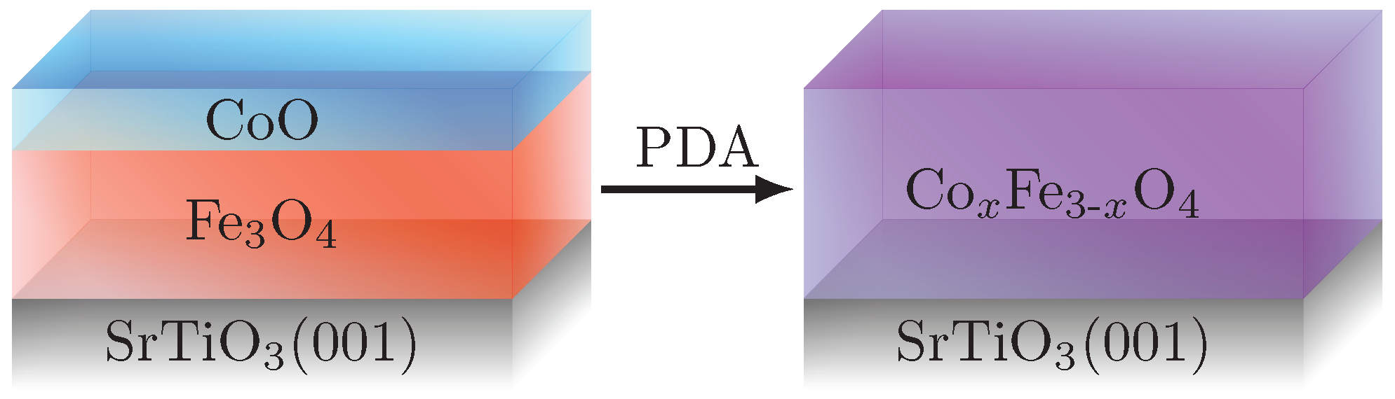
2. Materials and Methods
3. Results and Discussion
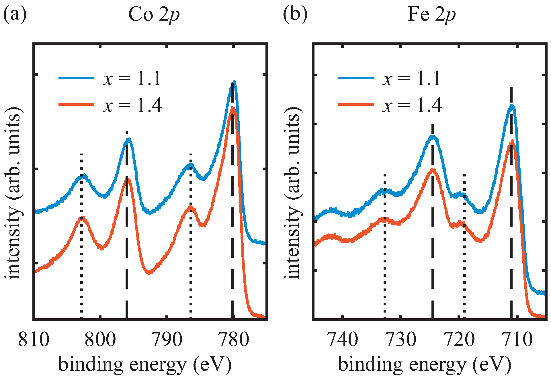
3.1. AR-HAXPES
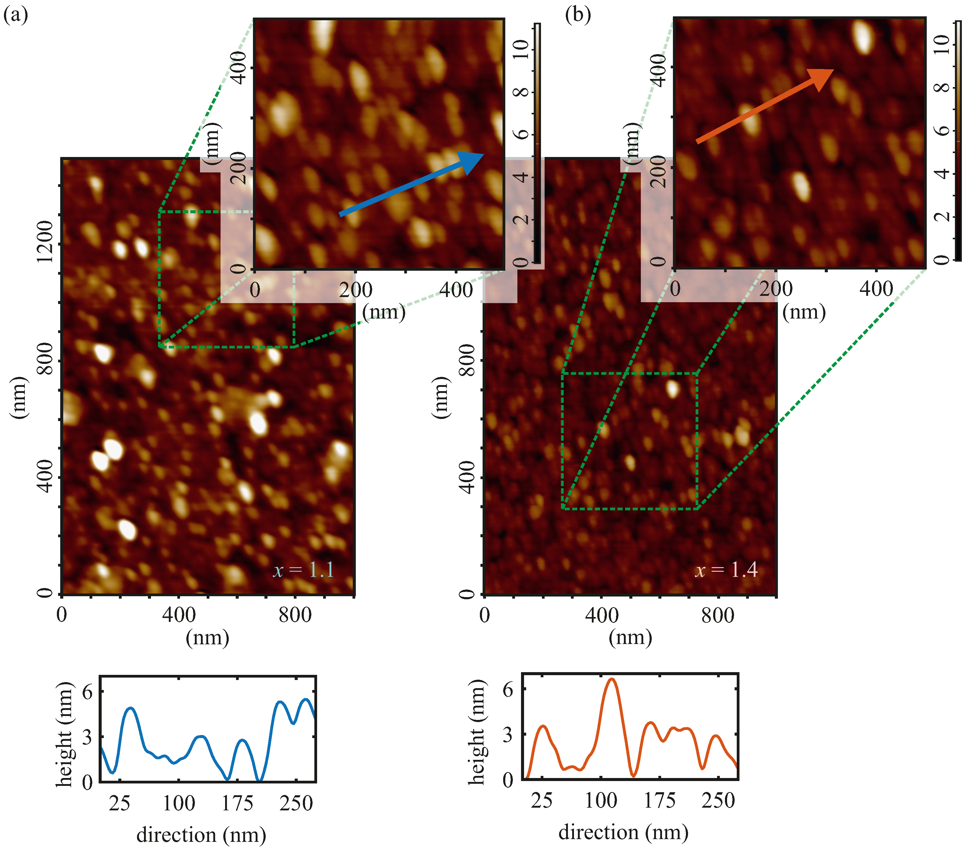
3.2. AFM
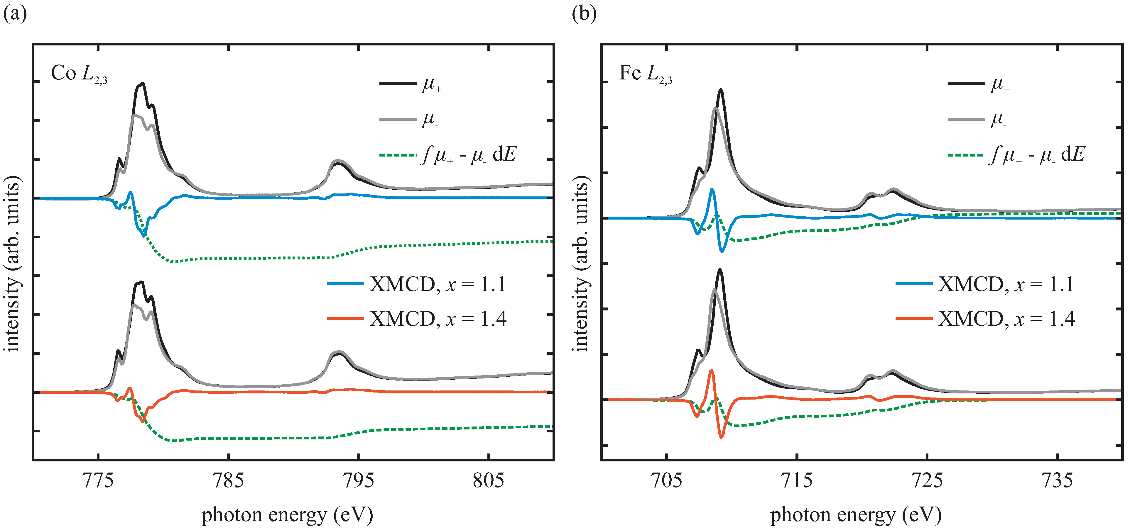
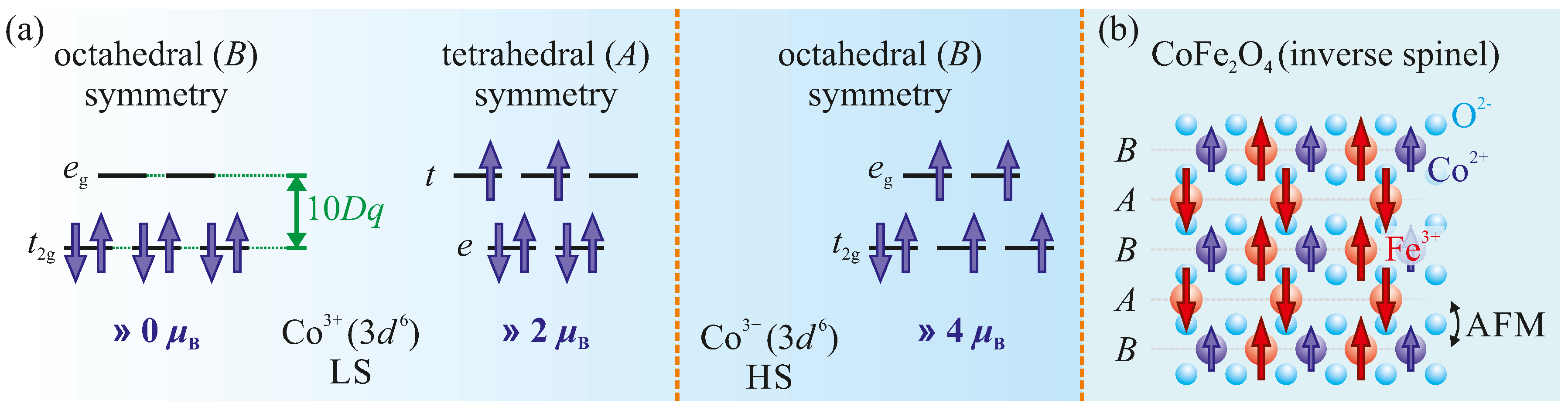
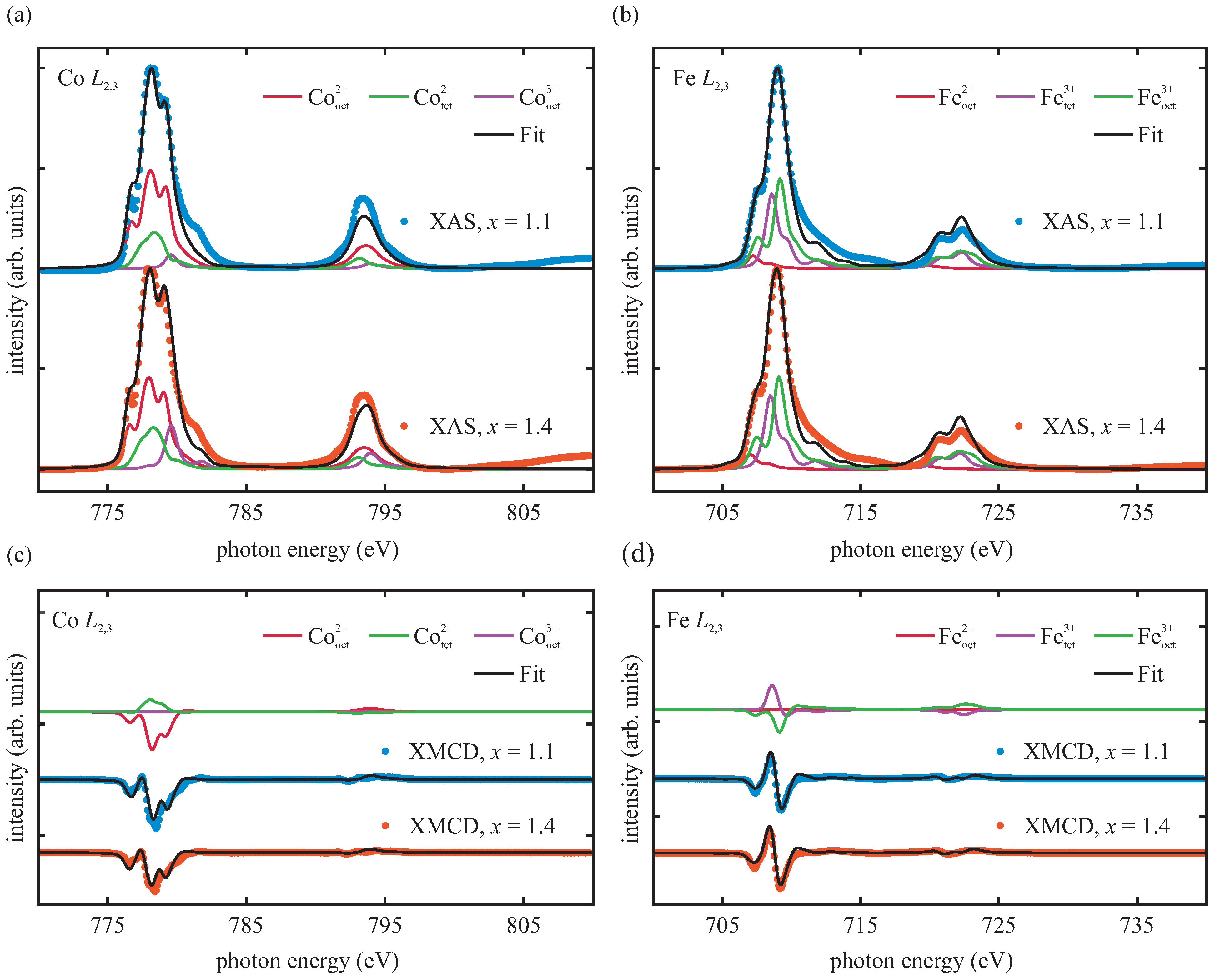
3.3. XAS/XMCD
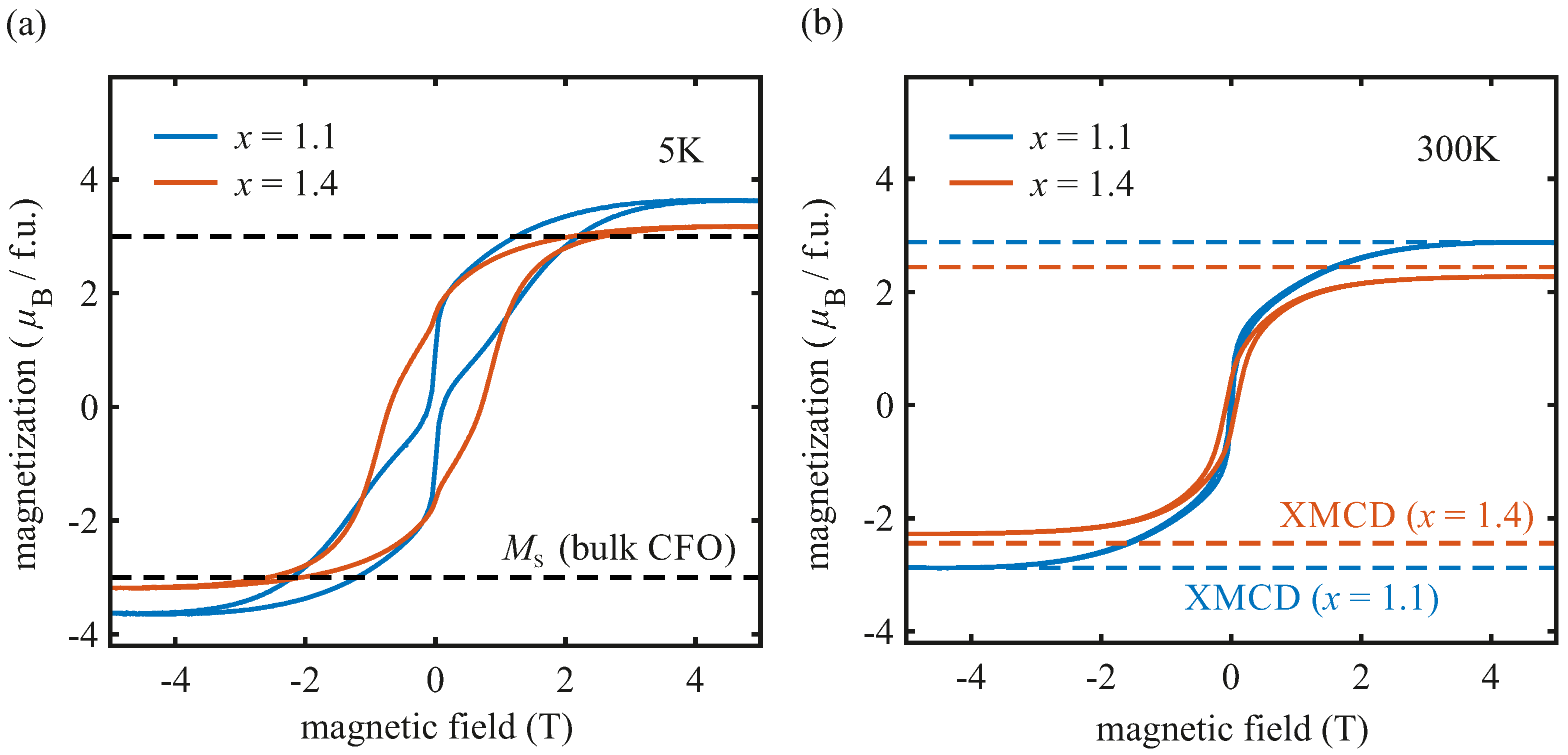
3.4. SQUID
4. Conclusions
Author Contributions
Funding
Institutional Review Board Statement
Informed Consent Statement
Data Availability Statement
Acknowledgments
Conflicts of Interest
References
- Moussy, J.B. From epitaxial growth of ferrite thin films to spin-polarized tunnelling. J. Phys. D Appl. Phys. 2013, 46, 143001. [Google Scholar] [CrossRef]
- Ramos, A.V.; Guittet, M.J.; Moussy, J.B.; Mattana, R.; Deranlot, C.; Petroff, F.; Gatel, C. Room temperature spin filtering in epitaxial cobalt-ferrite tunnel barriers. Appl. Phys. Lett. 2007, 91, 122107. [Google Scholar] [CrossRef] [Green Version]
- Matzen, S.; Moussy, J.B.; Mattana, R.; Bouzehouane, K.; Deranlot, C.; Petroff, F. Nanomagnetism of cobalt ferrite-based spin filters probed by spin-polarized tunneling. Appl. Phys. Lett. 2012, 101, 042409. [Google Scholar] [CrossRef]
- Caffrey, N.M.; Fritsch, D.; Archer, T.; Sanvito, S.; Ederer, C. Spin-filtering efficiency of ferrimagnetic spinels CoFe2O4 and NiFe2O4. Phys. Rev. B 2013, 87, 024419. [Google Scholar] [CrossRef] [Green Version]
- Foerster, M.; Rigato, F.; Bouzehouane, K.; Fontcuberta, J. Tunnel transport through CoFe2O4 barriers investigated by conducting atomic force microscopy. J. Phys. D Appl. Phys. 2010, 43, 295001. [Google Scholar] [CrossRef] [Green Version]
- Takahashi, Y.K.; Kasai, S.; Furubayashi, T.; Mitani, S.; Inomata, K.; Hono, K. High spin-filter efficiency in a Co ferrite fabricated by a thermal oxidation. Appl. Phys. Lett. 2010, 96, 072512. [Google Scholar] [CrossRef]
- Guo, E.J.; Herklotz, A.; Kehlberger, A.; Cramer, J.; Jakob, G.; Kläui, M. Thermal generation of spin current in epitaxial CoFe2O4 thin films. Appl. Phys. Lett. 2016, 108, 022403. [Google Scholar] [CrossRef] [Green Version]
- Niizeki, T.; Kikkawa, T.; Uchida, K.I.; Oka, M.; Suzuki, K.Z.; Yanagihara, H.; Kita, E.; Saitoh, E. Observation of longitudinal spin-Seebeck effect in cobalt-ferrite epitaxial thin films. AIP Adv. 2015, 5, 053603. [Google Scholar] [CrossRef] [Green Version]
- Uchida, K.I.; Adachi, H.; Kikkawa, T.; Kirihara, A.; Ishida, M.; Yorozu, S.; Maekawa, S.; Saitoh, E. Thermoelectric Generation Based on Spin Seebeck Effects. Proc. IEEE 2016, 104, 1946–1973. [Google Scholar] [CrossRef] [Green Version]
- Valvidares, M.; Dix, N.; Isasa, M.; Ollefs, K.; Wilhelm, F.; Rogalev, A.; Sánchez, F.; Pellegrin, E.; Bedoya-Pinto, A.; Gargiani, P.; et al. Absence of magnetic proximity effects in magnetoresistive Pt/CoFe2O4 hybrid interfaces. Phys. Rev. B 2016, 93, 214415. [Google Scholar] [CrossRef] [Green Version]
- Huang, S.Y.; Wang, W.G.; Lee, S.F.; Kwo, J.; Chien, C.L. Intrinsic Spin-Dependent Thermal Transport. Phys. Rev. Lett. 2011, 107, 216604. [Google Scholar] [CrossRef] [Green Version]
- Zhu, S.; Li, J.; Deng, X.; He, C.; Liu, E.; He, F.; Shi, C.; Zhao, N. Ultrathin-Nanosheet-Induced Synthesis of 3D Transition Metal Oxides Networks for Lithium Ion Battery Anodes. Adv. Funct. Mater. 2017, 27, 1605017. [Google Scholar] [CrossRef]
- Lu, X.F.; Gu, L.F.; Wang, J.W.; Wu, J.X.; Liao, P.Q.; Li, G.R. Bimetal-Organic Framework Derived CoFe2O4/C Porous Hybrid Nanorod Arrays as High-Performance Electrocatalysts for Oxygen Evolution Reaction. Adv. Mater. 2017, 29, 1604437. [Google Scholar] [CrossRef]
- Coll, M.; Moreno, J.M.M.; Gazquez, J.; Nielsch, K.; Obradors, X.; Puig, T. Low Temperature Stabilization of Nanoscale Epitaxial Spinel Ferrite Thin Films by Atomic Layer Deposition. Adv. Funct. Mater. 2014, 24, 5368–5374. [Google Scholar] [CrossRef]
- Nlebedim, I.C.; Moses, A.J.; Jiles, D.C. Non-stoichiometric cobalt ferrite, CoxFe33xO4 (x = 1.0 to 2.0): Structural, magnetic and magnetoelastic properties. J. Magn. Magn. Mater. 2013, 343, 49–54. [Google Scholar] [CrossRef]
- Trong, H.L.; Presmanes, L.; Grave, E.D.; Barnabé, A.; Bonningue, C.; Tailhades, P. Mössbauer characterisations and magnetic properties of iron cobaltites CoxFe3−xO4 (1 ≤ x ≤ 2.46) before and after spinodal decomposition. J. Magn. Magn. Mater. 2013, 334, 66–73. [Google Scholar] [CrossRef] [Green Version]
- Sinha, A.K.; Singh, M.N.; Archary, S.N.; Sagdeo, A.; Shukla, D.K.; Phase, D.M. Crystal field splitting and spin states of Co ions in cobalt ferrite with composition Co1.5Fe1.5O4 using magnetization and X-ray absorption spectroscopy measurements. J. Magn. Magn. Mater. 2017, 435, 87–95. [Google Scholar] [CrossRef]
- Thien, J.; Bahlmann, J.; Alexander, A.; Hoppe, M.; Pohlmann, T.; Ruwisch, K.; Meyer, C.; Bertram, F.; Küpper, K.; Wollschläger, J. Effects of Post-deposition Annealing on Epitaxial CoO/Fe3O4 Bilayers on SrTiO3(001) and Formation of Thin High-Quality Cobalt Ferrite-like Films. J. Phys. Chem. C 2020, 124, 23895–23904. [Google Scholar] [CrossRef]
- Chambers, S.A.; Farrow, R.F.C.; Maat, S.; Toney, M.F.; Folks, L.; Catalano, J.G.; Trainor, T.P.; Brown, G.E. Molecular beam epitaxial growth and properties of CoFe2O4 on MgO(001). J. Magn. Magn. Mater. 2002, 246, 124–139. [Google Scholar] [CrossRef]
- Eskandari, F.; Porter, S.B.; Venkatesan, M.; Kameli, P.; Rode, K.; Coey, J.M.D. Magnetization and anisotropy of cobalt ferrite thin films. Phys. Rev. Mater. 2017, 1, 074413. [Google Scholar] [CrossRef] [Green Version]
- Rodewald, J.; Thien, J.; Pohlmann, T.; Hoppe, M.; Timmer, F.; Bertram, F.; Kuepper, K.; Wollschläger, J. Formation of ultrathin cobalt ferrite films by interdiffusion of Fe3O4/CoO bilayers. Phys. Rev. B 2019, 100, 155418. [Google Scholar] [CrossRef] [Green Version]
- Fritsch, D.; Ederer, C. Epitaxial strain effects in the spinel ferrites CoFe2O4 and NiFe2O4 from first principles. Phys. Rev. B 2010, 82, 104117. [Google Scholar] [CrossRef] [Green Version]
- Fritsch, D.; Ederer, C. Effect of epitaxial strain on the cationdistribution in spinel ferrites CoFe2O4 and NiFe2O4: A density functional theory study. Appl. Phys. Lett. 2011, 99, 081916. [Google Scholar] [CrossRef] [Green Version]
- Chen, Y.Z.; Sun, J.R.; Han, Y.N.; Xie, X.Y.; Shen, J.; Rong, C.B.; He, S.L.; Shen, B.G. Microstructure and magnetic properties of strained Fe3O4 films. J. Appl. Phys. 2008, 103, 07D703. [Google Scholar] [CrossRef]
- Monti, M.; Sanz, M.; Oujja, M.; Rebollar, E.; Castillejo, M.; Pedrosa, F.J.; Bollero, A.; Camarero, J.; Cunado, J.L.F.; Nemes, N.M.; et al. Room temperature in-plane 〈100〉 magnetic easy axis for Fe3O4/SrTiO3(001):Nb grown by infrared pulsed laser deposition. J. Appl. Phys. 2013, 114, 223902. [Google Scholar] [CrossRef] [Green Version]
- Sawatzky, G.A.; van der Woude, F.; Morrish, A.H. Cation Distributions in Octahedral and Tetrahedral Sites of the Ferrimagnetic Spinel CoFe2O4. J. Appl. Phys. 1968, 39, 1204–1206. [Google Scholar] [CrossRef]
- Hu, G.; Choi, J.H.; Eom, C.B.; Harris, V.G.; Suzuki, Y. Structural tuning of the magnetic behavior in spinel-structure ferrite thin films. Phys. Rev. B 2000, 62, R779–R782. [Google Scholar] [CrossRef]
- de Groot, F. Multiplet effects in X-ray spectroscopy. Coord. Chem. Rev. 2005, 249, 31–63. [Google Scholar] [CrossRef]
- Stavitski, E.; de Groot, F.M.F. The CTM4XAS program for EELS and XAS spectral shape analysis of transition metal L edges. Micron 2010, 41, 687–694. [Google Scholar] [CrossRef] [PubMed] [Green Version]
- Cowan, R.D. The Theory of Atomic Structure and Spectra, 1st ed.; University of California Press: Berkely, CA, USA, 1981. [Google Scholar] [CrossRef]
- McIntyre, N.S.; Cook, M.G. X-ray photoelectron studies on some oxides and hydroxides of cobalt, nickel, and copper. Anal. Chem. 1975, 47, 2208–2213. [Google Scholar] [CrossRef]
- Yamashita, T.; Hayes, P. Analysis of XPS spectra of Fe2+ and Fe2+ ions in oxide materials. Appl. Surf. Sci. 2008, 254, 2441–2449. [Google Scholar] [CrossRef]
- Fujii, T.; de Groot, F.M.F.; Sawatzky, G.A.; Voogt, F.C.; Hibma, T.; Okada, K. In situ XPS analysis of various iron oxide films grown by NO2-assisted molecular-beam epitaxy. Phys. Rev. B 1999, 59, 3195–3202. [Google Scholar] [CrossRef] [Green Version]
- Graat, P.C.J.; Somers, M.A.J. Simultaneous determination of composition and thickness of thin iron-oxide films from XPS Fe 2p spectra. Appl. Surf. Sci. 1996, 100–101, 36–40. [Google Scholar] [CrossRef]
- Rigato, F.; Geshev, J.; Skumryev, V.; Fontcuberta, J. The magnetization of epitaxial nanometric CoFe2O4(001) layers. J. Appl. Phys. 2009, 106, 113924. [Google Scholar] [CrossRef] [Green Version]
- van der Laan, G.; Arenholz, E.; Chopdekar, R.V.; Suzuki, Y. Influence of crystal field on anisotropic x-ray magnetic linear dichroism at the Co2+L2,3 edges. Phys. Rev. B 2008, 77, 064407. [Google Scholar] [CrossRef]
- Moyer, J.A.; Vaz, C.A.F.; Kumah, D.P.; Arena, D.A.; Henrich, V.E. Enhanced magnetic moment in ultrathin Fe-doped CoFe2O4 films. Phys. Rev. B 2012, 86, 174404. [Google Scholar] [CrossRef]
- Moyer, J.A.; Vaz, C.A.F.; Negusse, E.; Arena, D.A.; Henrich, V.E. Controlling the electronic structure of Co1−xFe2+xO4 thin films through iron doping. Phys. Rev. B 2011, 83, 035121. [Google Scholar] [CrossRef] [Green Version]
- Crocombette, J.P.; Pollak, M.; Jollet, F.; Thromat, N.; Gautier-Soyer, M. X-ray-absorption spectroscopy at the Fe L2,3 threshold in iron oxides. Phys. Rev. B 1995, 52, 3143–3150. [Google Scholar] [CrossRef] [PubMed]
- Kuiper, P.; Searle, B.G.; Rudolf, P.; Tjeng, L.H.; Chen, C.T. X-ray magnetic dichroism of antiferromagnet Fe2O3: The orientation of magnetic moments observed by Fe 2p x-ray absorption spectroscopy. Phys. Rev. Lett. 1993, 70, 1549–1552. [Google Scholar] [CrossRef] [Green Version]
- Thole, B.T.; Carra, P.; Sette, F.; van der Laan, G. X-ray circular dichroism as a probe of orbital magnetization. Phys. Rev. Lett. 1992, 68, 1943–1946. [Google Scholar] [CrossRef]
- Carra, P.; Thole, B.T.; Altarelli, M.; Wang, X. X-ray circular dichroism and local magnetic fields. Phys. Rev. Lett. 1993, 70, 694–697. [Google Scholar] [CrossRef] [PubMed]
- Chen, C.T.; Idzerda, Y.U.; Lin, H.J.; Smith, N.V.; Meigs, G.; Chaban, E.; Ho, G.H.; Pellegrin, E.; Sette, F. Experimental Confirmation of the X-Ray Magnetic Circular Dichroism Sum Rules for Iron and Cobalt. Phys. Rev. Lett. 1995, 75, 152–155. [Google Scholar] [CrossRef] [PubMed]
- Piamonteze, C.; Miedema, P.; de Groot, F.M.F. Accuracy of the spin sum rule in XMCD for the transition-metal L edges from manganese to copper. Phys. Rev. B 2009, 80, 184410. [Google Scholar] [CrossRef] [Green Version]
- Teramura, Y.; Tanaka, A.; Jo, T. Effect of Coulomb Interaction on the X-Ray Magnetic Circular Dichroism Spin Sum Rule in 3d Transition Elements. J. Phys. Soc. Jpn. 1996, 65, 1053–1055. [Google Scholar] [CrossRef]
- Trong, H.L.; Barnabé, A.; Presmanes, L.; Tailhades, P. Phase decomposition study in CoxFe3−xO4 iron cobaltites: Synthesisand structural characterization of the spinodal transformation. Solid State Sci. 2008, 10, 550–556. [Google Scholar] [CrossRef] [Green Version]
- Murray, P.J.; Linnett, J.W. Cation distribution in the spinels CoxFe3−xO4 . J. Phys. Chem. Solids 1976, 37, 1041–1042. [Google Scholar] [CrossRef]
- Lee, D.H.; Kim, H.S.; Lee, J.Y.; Yo, C.H.; Kim, K.H. Characterization of the magnetic properties and transport mechanisms of CoxFe3−xO4 spinel. Solid State Commun. 1995, 96, 445–449. [Google Scholar] [CrossRef]
- Kita, N.; Shibuichi, N.; Sasaki, S. X-ray magnetic circular dichroism in cobalt-iron spinels and electronic states of Co ions. J. Synchrotron Rad. 2001, 8, 446–448. [Google Scholar] [CrossRef] [Green Version]
- Takahashi, M.; Fine, M.E. Magnetic behavior of quenched and aged CoFe2O4-Co3O4 alloys. J. Appl. Phys. 1972, 43, 4205–4216. [Google Scholar] [CrossRef]
- Smith, P.A.; Spencer, C.D.; Stillwell, R.P. Co57 and Fe57 mössbauer studies of the spinels FeCo2O4 and Fe0.5Co2.5O4. J. Phys. Chem. Solids 1978, 39, 107–111. [Google Scholar] [CrossRef]
- Jonker, G.H. Analysis of the semiconducting properties of cobalt ferrite. J. Phys. Chem. Solids 1959, 9, 165–175. [Google Scholar] [CrossRef]
- Lewis, F.B.; Saunders, N.H. The thermal conductivity of NiO and CoO at the Neel temperature. J. Phys. C Solid State Phys. 1973, 6, 2525–2532. [Google Scholar] [CrossRef]
- Goodenough, J.B. Metallic oxides. Prog. Solid State Chem. 1971, 5, 145–399. [Google Scholar] [CrossRef]
- Beltrán, J.J.; Osorio, J.A.; Barrero, C.A.; Hanna, C.B.; Punnoose, A. Magnetic properties of Fe doped, Co doped, and Fe+Co co-doped ZnO. J. Appl. Phys. 2013, 113, 17C308. [Google Scholar] [CrossRef] [Green Version]
- Kim, J.H.; Kim, H.; Kim, D.; Ihm, Y.E.; Choo, W.K. The origin of room temperature ferromagnetism in cobalt-doped zinc oxide thin films fabricated by PLD. J. Eur. Ceram. Soc. 2004, 24, 1847–1851. [Google Scholar] [CrossRef]
- Basith, N.M.; Vijaya, J.J.; Kennedy, L.J.; Bououdina, M. Structural, morphological, optical, and magnetic properties of Ni-doped CuO nanostructures prepared by a rapid microwave combustion method. Mater. Sci. Semicond. Process. 2014, 17, 110–118. [Google Scholar] [CrossRef]
- Li, Y.; Xu, M.; Pan, L.; Zhang, Y.; Guo, Z.; Bi, C. Structural and room-temperature ferromagnetic properties of Fe-doped CuO nanocrystals. J. Appl. Phys. 2010, 107, 113908. [Google Scholar] [CrossRef]
- Negi, D.S.; Loukya, B.; Dileep, K.; Sahu, R.; Nagaraja, K.K.; Kumar, N.; Datta, R. Robust room temperature ferromagnetism in epitaxial CoO thin film. Appl. Phys. Lett. 2013, 103, 242407. [Google Scholar] [CrossRef]
- Gheisari, M.; Mozafari, M.; Niyaifar, M.; Amighian, J.; Soleimani, R. Observation of Small Exchange Bias in Defect Wüstite (Fe0.93O) Nanoparticles. J. Supercond. Nov. Magn. 2013, 26, 237–242. [Google Scholar] [CrossRef]
- Qin, H.; Zhang, Z.; Liu, X.; Zhang, Y.; Hu, J. Room-temperature ferromagnetism in CuO sol-gel powders and films. J. Magn. Magn. Mater. 2010, 322, 1994–1998. [Google Scholar] [CrossRef]
- Xu, Q.; Schmidt, H.; Zhou, S.; Potzger, K.; Helm, M.; Hochmuth, H.; Lorenz, M.; Setzer, A.; Esquinazi, P.; Meinecke, C.; et al. Room temperature ferromagnetism in ZnO films due to defects. Appl. Phys. Lett. 2008, 92, 082508. [Google Scholar] [CrossRef]
- Jedrecy, N.; Aghavnian, T.; Moussy, J.B.; Magnan, H.; Stanescu, D.; Portier, X.; Arrio, M.A.; Mocuta, C.; Vlad, A.; Belkhou, R.; et al. Cross-Correlation between Strain, Ferroelectricity, and Ferromagnetism in Epitaxial Multiferroic CoFe2O4/BaTiO3 Heterostructures. ACS Appl. Mater. Interfaces 2018, 10, 28003–28014. [Google Scholar] [CrossRef]
- Kuschel, O.; Buß, R.; Spiess, W.; Schemme, T.; Wöllermann, J.; Balinski, K.; N’Diaye, A.T.; Kuschel, T.; Wollschläger, J.; Kuepper, K. From Fe3O4/NiO bilayers to NiFe2O4-like thin films through Ni interdiffusion. Phys. Rev. B 2016, 94, 094423. [Google Scholar] [CrossRef] [Green Version]
- Arenholz, E.; van der Laan, G.; Chopdekar, R.V.; Suzuki, Y. Anisotropic X-ray magnetic linear dichroism at the Fe L2,3 edges in Fe3O4. Phys. Rev. B 2006, 74, 094407. [Google Scholar] [CrossRef] [Green Version]
- Kuepper, K.; Kuschel, O.; Pathé, N.; Schemme, T.; Schmalhorst, J.; Thomas, A.; Arenholz, E.; Gorgoi, M.; Ovsyannikov, R.; Bartkowski, S.; et al. Electronic and magnetic structure of epitaxial Fe3O4(001)/NiO heterostructures grown on MgO(001) and Nb-doped SrTiO3(001). Phys. Rev. B 2016, 94, 024401. [Google Scholar] [CrossRef] [Green Version]
- Tainosho, T.; ichiro Inoue, J.; Sharmin, S.; Takeguchi, M.; Kita, E.; Yanagihara, H. Large negative uniaxial magnetic anisotropyin highly distorted Co-ferrite thin films. Appl. Phys. Lett. 2019, 114, 092408. [Google Scholar] [CrossRef] [Green Version]
- Lüders, U.; Bibes, M.; Bobo, J.F.; Cantoni, M.; Bertacco, R.; Fontcuberta, J. Enhanced magnetic moment and conductive behavior in NiFe2O4 spinel ultrathin films. Phys. Rev. B 2005, 71, 134419. [Google Scholar] [CrossRef] [Green Version]
- Rigato, F.; Estradé, S.; Arbiol, J.; Peiró, F.; Lüders, U.; Martí, X.; Sánchez, F.; Fontcuberta, J. Strain-induced stabilization of new magnetic spinel structures in epitaxial oxide heterostructures. Mater. Sci. Eng. B 2007, 144, 43–48. [Google Scholar] [CrossRef] [Green Version]
- Yanagihara, H.; Uwabo, K.; Minagawa, M.; Kita, E.; Hirota, N. Perpendicular magnetic anisotropy in CoFe2O4(001) films epitaxially grown on MgO(001). J. Appl. Phys. 2011, 109, 07C122. [Google Scholar] [CrossRef] [Green Version]
- Horng, L.; Chern, G.; Chen, M.C.; Kang, P.C.; Lee, D.S. Magnetic anisotropic properties in Fe3O4 and CoFe2O4 ferrite epitaxy thin films. J. Magn. Magn. Mater. 2004, 270, 389–396. [Google Scholar] [CrossRef]
- Zhang, Y.; Deng, C.; Ma, J.; Lin, Y.; Nan, C.W. Enhancement in magnetoelectric response in CoFe2O4-BaTiO3 heterostructure. Appl. Phys. Lett. 2008, 92, 062911. [Google Scholar] [CrossRef]
- Zeng, X.; Zhang, J.; Zhu, S.; Deng, X.; Ma, H.; Zhang, J.; Zhang, Q.; Li, P.; Xue, D.; Mellors, N.J.; et al. Direct observation of cation distributions of ideal inverse spinel CoFe2O4 nanofibres and correlated magnetic properties. Nanoscale 2017, 9, 7493–7500. [Google Scholar] [CrossRef]
- Franco, A.; Machado, F.L.A.; Zapf, V.S. Magnetic properties of nanoparticles of cobalt ferrite at high magnetic field. J. Appl. Phys. 2011, 110, 053913. [Google Scholar] [CrossRef]
- Verma, K.C.; Singh, V.P.; Ram, M.; Shah, J.; Kotnala, R.K. Structural, microstructural and magnetic properties of NiFe2O4, CoFe2O4 and MnFe2O4nanoferrite thin films. J. Magn. Magn. Mater. 2011, 323, 3271–3275. [Google Scholar] [CrossRef]
- Guillot, M.; Ostorero, J.; Marchand, A. High magnetic field magnetization study in cadmium-cobalt ferrite single crystals. Z. Phys. B 1988, 71, 193–197. [Google Scholar] [CrossRef]
- Bercoff, P.G.; Bertorello, H.R. Exchange constants and transfer integrals of spinel ferrites. J. Magn. Magn. Mater. 1997, 169, 314–322. [Google Scholar] [CrossRef]
- Srivastava, C.M.; Srinivasan, G.; Nanadikar, N.G. Exchange constants in spinel ferrites. Phys. Rev. B 1979, 19, 499–508. [Google Scholar] [CrossRef] [Green Version]


| Co Content | Co Moment (/Co ion) | Fe Moment (/Fe ion) | ||||
|---|---|---|---|---|---|---|
| total | total | |||||
| 1.1 | ||||||
| 1.4 | ||||||
| total | total | |||||
| 1.1 | ||||||
| 1.4 | ||||||
Publisher’s Note: MDPI stays neutral with regard to jurisdictional claims in published maps and institutional affiliations. |
© 2021 by the authors. Licensee MDPI, Basel, Switzerland. This article is an open access article distributed under the terms and conditions of the Creative Commons Attribution (CC BY) license (https://creativecommons.org/licenses/by/4.0/).
Share and Cite
Thien, J.; Bahlmann, J.; Alexander, A.; Ruwisch, K.; Rodewald, J.; Pohlmann, T.; Hoppe, M.; Alarslan, F.; Steinhart, M.; Altuncevahir, B.; et al. Cationic Ordering and Its Influence on the Magnetic Properties of Co-Rich Cobalt Ferrite Thin Films Prepared by Reactive Solid Phase Epitaxy on Nb-Doped SrTiO3(001). Materials 2022, 15, 46. https://doi.org/10.3390/ma15010046
Thien J, Bahlmann J, Alexander A, Ruwisch K, Rodewald J, Pohlmann T, Hoppe M, Alarslan F, Steinhart M, Altuncevahir B, et al. Cationic Ordering and Its Influence on the Magnetic Properties of Co-Rich Cobalt Ferrite Thin Films Prepared by Reactive Solid Phase Epitaxy on Nb-Doped SrTiO3(001). Materials. 2022; 15(1):46. https://doi.org/10.3390/ma15010046
Chicago/Turabian StyleThien, Jannis, Jascha Bahlmann, Andreas Alexander, Kevin Ruwisch, Jari Rodewald, Tobias Pohlmann, Martin Hoppe, Fatih Alarslan, Martin Steinhart, Baki Altuncevahir, and et al. 2022. "Cationic Ordering and Its Influence on the Magnetic Properties of Co-Rich Cobalt Ferrite Thin Films Prepared by Reactive Solid Phase Epitaxy on Nb-Doped SrTiO3(001)" Materials 15, no. 1: 46. https://doi.org/10.3390/ma15010046