Article

Green Synthesis of Silver-Decorated Magnetic Particles for Efficient and Reusable Antimicrobial Activity

1
Department of Chemical Engineering, Konkuk University, Seoul 05029, Korea
2
Institute of SK-KU Biomaterials, Konkuk University, Seoul 05029, Korea
*
Author to whom correspondence should be addressed.
Materials 2021, 14(24), 7893; https://doi.org/10.3390/ma14247893
Submission received: 2 November 2021 / Revised: 11 December 2021 / Accepted: 16 December 2021 / Published: 20 December 2021
(This article belongs to the Special Issue Functional Materials for Therapeutic and Industrial Applications)

Abstract

:
Metal and metal hybrid nanostructures have shown tremendous application in the biomedical and catalytic fields because of their plasmonic and catalytic properties. Here, a green and clean method was employed for the synthesis of silver nanoparticle (Ag NP)-SiO2-Fe2O3 hybrid microstructures, and biomolecules from green tea extracts were used for constructing the hybrid structures. The SiO2-Fe2O3 structures were synthesized using an ethanolic green tea leaf extract to form Bio-SiO2-Fe2O3 (BSiO2-Fe2O3) structures. Biochemical studies demonstrated the presence of green tea biomolecules in the BSiO2 layer. Reduction of the silver ions was performed by a BSiO2 layer to form Ag NPs of 5–10 nm in diameter in and on the BSiO2-Fe2O3 microstructure. The reduction process was observed within 600 s, which is faster than that reported elsewhere. The antimicrobial activity of the Ag-BSiO2-Fe2O3 hybrid structure was demonstrated against Staphylococcus aureus and Escherichia coli, and the nanostructures were further visualized using confocal laser scanning microscopy (CLSM). The magnetic properties of the Ag-BSiO2-Fe2O3 hybrid structure were used for studying reusable antimicrobial activity. Thus, in this study, we provide a novel green route for the construction of a biomolecule-entrapped SiO2-Fe2O3 structure and their use for the ultra-fast formation of Ag NPs to form antimicrobial active multifunctional hybrid structures.

1. Introduction

For the last seven decades since the discovery of antibiotics, their uncontrolled use has led to a generation of antibiotic-resistant strains that are unresponsive to currently available conventional antibiotics, creating a major concern for the health sector [1,2]. More new-generation antibiotics have been introduced in the field of medicine; however, the toxicity and adverse effects of these antibiotics are worrying factors for implementation [3,4]. Therefore, alternate therapeutic strategies have been studied for the last two decades, and several types of nanomaterials have been tested for their antimicrobial activity that is nontoxic to human and nonhuman hosts [5,6,7]. Silver nanoparticles (Ag NPs) have been extensively studied because of their effective antimicrobial activity known since ancient times [8]. A detailed study of the antimicrobial activity of Ag NPs against a wide range of pathogenic microorganisms was performed; it was demonstrated that the Ag NPs act by releasing Ag+ ions, which act on bacteria through reactive oxygen species [8]. The antimicrobial activity of the Ag NPs decreases with aggregate formation, where Ag NPs form a bulky material that makes it difficult for them to act on small bacteria and viruses [9,10,11]. In addition, Ag NPs are not only toxic to the bacteria but also to mammalian cells, which is a major concern for the therapeutic use of Ag NPs [12]. Therefore, several functionalizations or immobilization procedures have been used to enhance their antimicrobial efficiency and avoid the aggregation and toxicity of Ag NPs before release into the environment [13]. Currently, extensive research is ongoing for the construction of an ideal support to maintain the activity, stability, and reusability of Ag NPs [14].
Magnetic nanoparticles have been used as carriers for several nanomaterials, as they facilitate the separation of the nanoparticles by an external magnetic field and stability of the catalyst, which can reduce the adverse effects of the Ag NPs after use and reuse of Ag NPs for repeated applications [15]. Several researchers have demonstrated various methods for the synthesis of metal nanoparticle-supported SiO2 NPs and magnetic nanoparticles; these methods are time-consuming and require hazardous materials and expensive instruments with harsh reaction procedures [16,17]. Ag@nanosilica NPs have been synthesized using a protein extract of Rhizopus oryzae after 72 h of incubation [18]. Kaloti et al. used glucose to form Ag–γ-Fe2O3 nanocomposites by applying a very high temperature [19]. Tzounis et al. used polyethyleneimine for the functionalization of SiO2 nanospheres, wherein Ag NPs were decorated using sodium borohydride (NaBH4) as the reducing agent [20]. Muthukumar et al. prepared silver ferrite nanoparticles from A. blitum leaf extract at 50 °C and pH 9 after 4 h of reaction time [21]. To the best of our knowledge, this is the first report where reducing molecules, i.e., plant leaf extract immobilized in the silica layer of silica-coated magnetic nanoparticles, were used for the reduction of Ag+ ions to form metal-magnetic hybrid microstructures with ultrafine, spherical Ag NPs on the surface, within a short reaction duration and under mild reaction conditions. A major advantage of the hybrid structure synthesis method is the minimization of steps involving very complex, expensive, and cumbersome functionalization and reduction reaction conditions.
In this report, we present an economical and clean method for the construction of Ag-SiO2-Fe2O3 hybrid structures by entrapping biomolecules from green tea leaf extract of Camellia sinensis, thus forming BioSiO2 (BSiO2)-Fe2O3 particles. The entrapped biomolecules reduced Ag+ ions and formed Ag NPs on the BSiO2-Fe2O3 particles. Different techniques were used for analyzing the crystal structure and morphology of the prepared hybrid structures, such as X-ray diffraction (XRD), high-resolution transmission electron microscopy (HR-TEM), and magnetic property measurement using a superconducting quantum interference device (SQUID). The prepared hybrid structures showed reusable active antimicrobial activity.

2. Materials and Methods

2.1. Materials

Analytical reagent-grade (AR-grade) chemicals were used for all experiments without purification. Silver nitrate (AgNO3), iron (III) nitrate nonahydrate (Fe(NO3)3·9H2O), ethanol (C2H5OH; 99.9%), tetraethyl orthosilicate (TEOS), sodium carbonate, gallic acid monohydrate, 2,2-diphenyl-1-picrylhydrazyl (DPPH), 2,2′-azino-bis(3-ethylbenzothiazoline-6-sulphonic acid) (ABTS), potassium persulfate (K2Cr2O7), and ammonium hydroxide (NH4OH) were purchased from Sigma-Aldrich Co. Ltd. (St. Louis, MO, USA). Green tea bags (Jeju company, Korea) were purchased from the local market.

2.2. Preparation of the Ethanolic Extract of Green Tea Leaves

Green tea leaves (1.2 g) were ground and refluxed for 4 h in anhydrous ethyl alcohol (99.9%) under magnetic stirring. The suspension was cooled at room temperature and centrifuged at 4000 rpm for 10 min to remove large particulates of green tea leaves. Next, the green tea leaf extract was filtered through Whatman filter paper to remove small leaf particulates. Before further experimental procedures, the purified ethanolic green tea extract was kept in the dark at 4 °C.

2.3. Synthesis of Fe2O3 NPs

Fe2O3 NPs were synthesized using a previously reported method [22]. Briefly, 1 M of iron nitrate Fe(NO3)3 was prepared in 50 mL of distilled water (DW) under N2 gas bubbling for 30 min. The pH of this solution was maintained at more than 10 under vigorous stirring by using aqueous ammonium hydroxide (30%). Then, the solution was autoclaved at 150 °C for 10 h in a Teflon-lined stainless-steel reactor. The formed precipitate was centrifuged and washed several times with ethanol and water until a neutral pH was obtained. The particles were vacuum-dried at 50 °C for 12 h.

2.4. Synthesis of BSiO2-Fe2O3 Particles

BSiO2-Fe2O3 particles were synthesized using a previously reported method with modifications [23]. In the purified ethanolic green tea extract (40 mL), 50 mg of Fe2O3 NPs were dispersed under sonication for 20 min. Two milliliters of TEOS was added dropwise to the Fe2O3 NP suspension and further mixed with mechanical stirring for 1 h. The pH of the solution was adjusted to 11 by adding aqueous ammonium hydroxide dropwise. After 24 h of incubation, the formed particles were collected by centrifugation and repeatedly washed with ethanol and water. The BSiO2-Fe2O3 particles were vacuum-dried at 50 °C for 12 h.

2.5. Synthesis of the Ag-BSiO2-Fe2O3 Hybrid Microstructure

For the construction of the Ag-BSiO2-Fe2O3 hybrid structure, the BSiO2-Fe2O3 particles were dispersed in 10 mL of DW and sonicated for 20 min to disturb the agglomerates. Ten milliliters of aqueous AgNO3 was poured into the solution to form a 10 mM AgNO3 solution. The mixture was further incubated and stirred for 30 min and observed for color change. The formed hybrid structure was washed with water and ethanol several times, obtained with centrifugation, and vacuum-dried at 50 °C overnight.

2.6. Antimicrobial Activity

The antimicrobial activity of the Ag-BSiO2-Fe2O3 hybrid microstructure was tested against Staphylococcus aureus and Escherichia coli. The growth media used for S. aureus and E. coli were trypticase soy broth and Luria–Bertani broth, respectively. The bacterial cultures were grown in the broths for 8 h to obtain the desired optical density (OD). Initially, the effect of the hybrid structure, streptomycin, AgNPs, and Fe2O3 NPs was tested on the growth curve of the bacterial cells for 22 h. The growth of both microorganisms was monitored using UV-visible spectroscopy, that is, optical density at 610 nm. Various concentrations of the hybrid structure, from 50 to 90 µg mL−1, were added to the broths (106 to 107 bacterial cells mL−1) and further incubated for 24 h at 37 °C. The antimicrobial activity of streptomycin, AgNPs, and Fe2O3 NPs was also studied with varied concentrations. After incubation, 0.1 mL from the incubated broth was spread over the respective agar and incubated for 24 h and observed for growth, and the colony-forming units (CFUs) mL−1 were measured. The higher concentrations of the hybrid nanostructure inhibited the growth of the bacteria completely, and they were used for the antimicrobial reusability experiment, in which 200 µg mL−1 of the hybrid structure was inoculated in 106 to 107 bacterial cells mL−1. To visualize the antimicrobial activity of the prepared particles, a live/dead staining assay was performed after the exposure of 50 and 200 µg mL−1 of the nanohybrid nanostructure to S. aureus and E. coli cultures, as specified in earlier reports. The cells were observed under CLSM (Carl Zeiss LSM 710, Jena, Germany) [24].

2.7. Biochemical Analysis

2.7.1. Estimation of Total Phenolic Content

The total phenolic content of the green tea extract prepared using water and ethanol was determined using the McDonald method [25]. Briefly, a standard solution of gallic acid monohydrate (100 μg mL−1) was prepared in methanol. Different concentrations of 0.1 to 1.0 mL of green tea leaf extract were mixed with a 1:1 mixture of Folin–Ciocalteu reagent and DW in 10 different tubes. To each tube, 7.5% sodium carbonate (2 mL) was added, and the mixture was allowed to react further for 30 min. The absorbance was measured at 765 nm, and the standard calibration curve was plotted. Then, 100 μL of the test samples was used to estimate the total phenolic content with a standard graph in terms of gallic acid equivalent (mg GAE g−1 extract).

2.7.2. DPPH Radical Scavenging Activity

The antioxidant properties of the ethanolic green tea extract and BSiO2 NPs were measured based on the DPPH free radical scavenging activity [26]. In 100 mL of methanol, DPPH (6 mg) solution was prepared. The antioxidant activity was measured based on the discoloration of stable DPPH radicals in methanol, and at 517 nm, the absorbance was obtained. Different aliquots of the test samples were tested for the discoloration of DPPH solution. Using the given formula, the radical scavenging activity was calculated as follows: % Radical scavenging activity = absorbance (control) − absorbance (sample)/absorbance (control) × 100.

2.7.3. ABTS Radical Scavenging Assay

The ABTS radical scavenging assay was followed to demonstrate the antioxidant capability of the ethanolic green tea extract before and after TEOS hydrolysis by using a previously published method with some modifications [26]. Briefly, green tea extract and solution obtained from TEOS hydrolysis were diluted to different concentrations. They were added to 7 mM of ABTS standard solution prepared with 2.45 mM of potassium persulfate (K2S2O8), and the mixture was further allowed to react for 6 min at 30 °C. After incubation, absorbance was measured at 734 nm. The radical scavenging activities of the extracts were calculated using the following equation and were compared with that of BHT: ABTS radical scavenging activity = absorbance (control) − absorbance (sample)/absorbance (control) × 100.

2.8. Analytical Instrumentation

UV-Visible spectroscopy was performed using a Schimadzu UV 1600 spectrophotometer with a 1 cm well-stoppered quartz cuvette. The morphology of the microstructures was evaluated by HR-TEM analysis using a JEM-3010 (JEOL) working at 300 kV and by energy-dispersive spectroscopy (EDS). Crystallography analysis of the nanostructures was performed using an X-ray diffractometer (Rigaku, Tokyo, Japan) with a Cu target. The magnetic properties of Fe2O3 and Fe2O3 hybrid structures were measured with a vibrating sample magnetometer (VSM; Lakeshore Cryotronics, Inc., Westerville, OH, USA). The antimicrobial activity was observed under CLSM. Dynamic light scattering (DLS) spectroscopy and zeta potential measurement of the particles in colloidal solution were performed using a NICOMPTM 380 ZIS (Santa Barbara, CA, USA) for the determination of the hydrodynamic diameter (HDD) and charge on the particles.

3. Results and Discussion

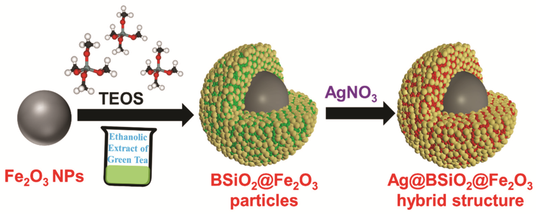
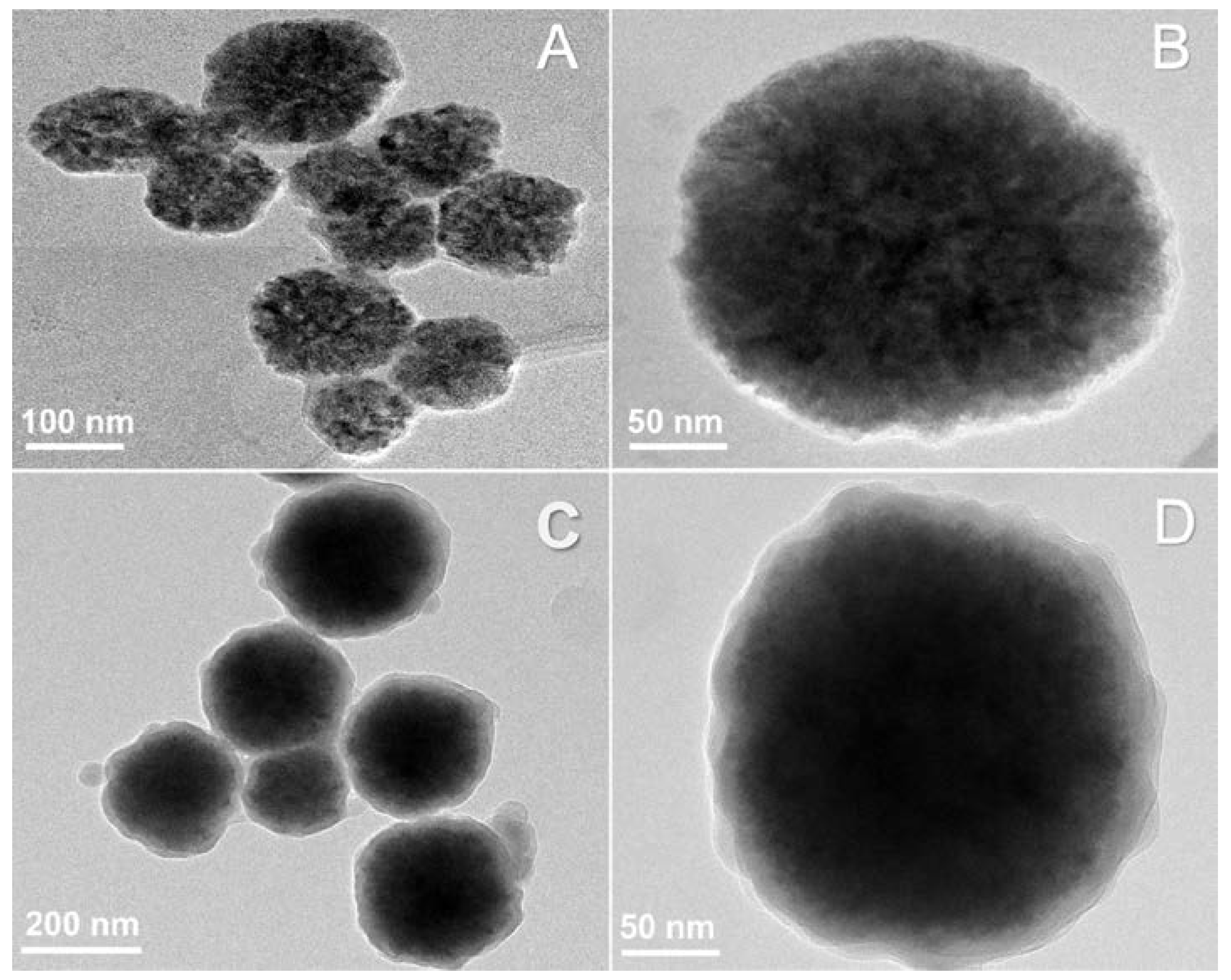
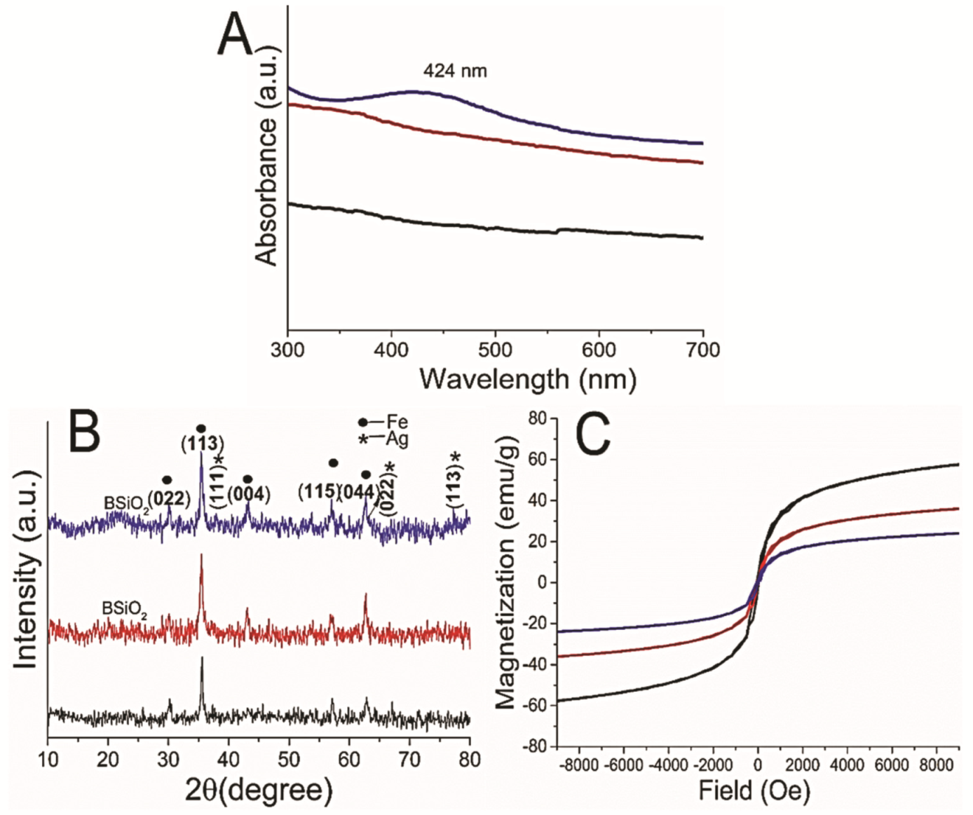
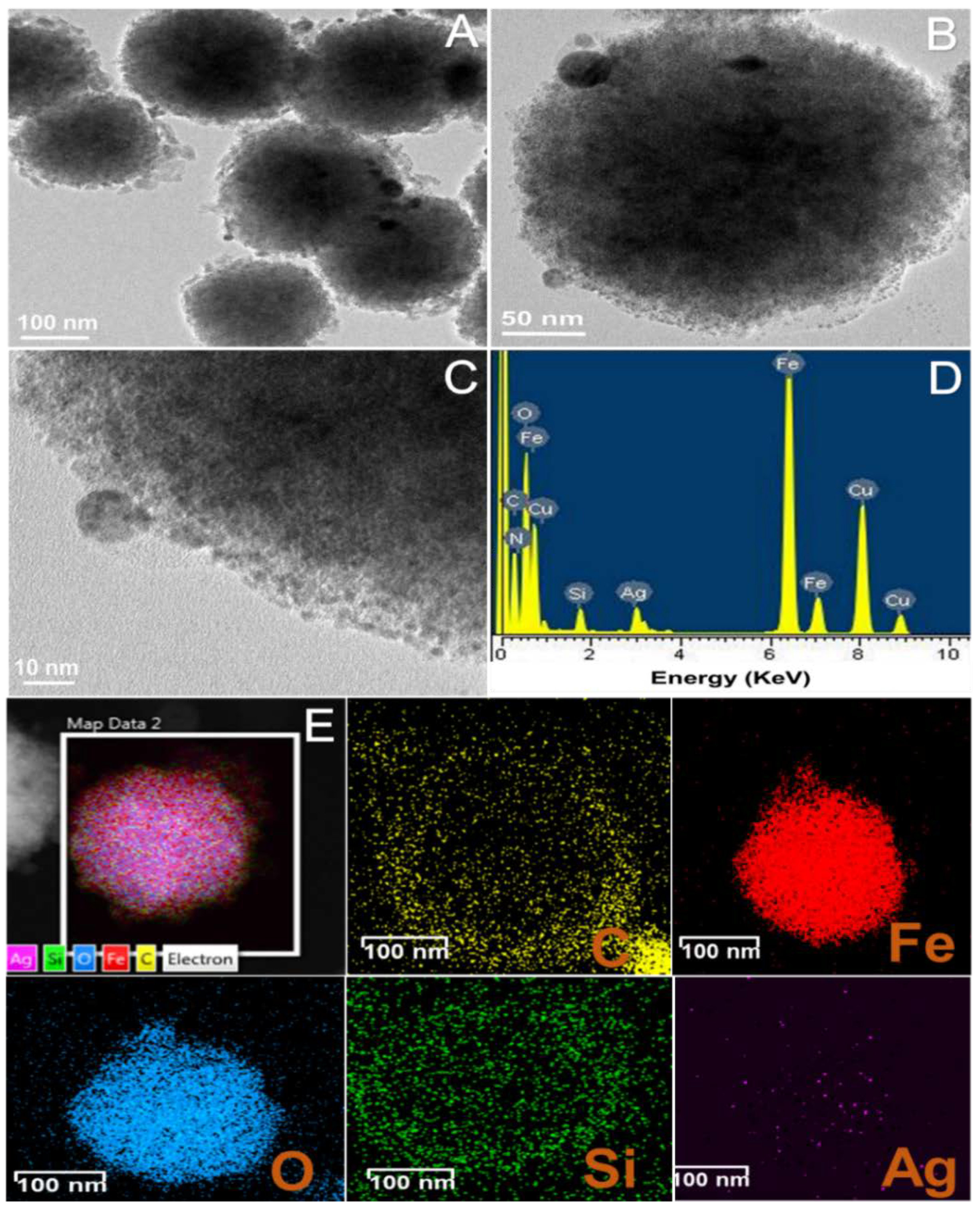
3.1. Synthesis of the Hybrid Microstructure

The plant extract-mediated synthesis of nanomaterials has been one of the green chemistry approaches followed for the preparation of various materials. Extracts from several plant parts have been studied for the synthesis of metal nanoparticles that contain various polysaccharides, proteins, vitamins, or alkaloids, functionalizing and capping the nanomaterials with nontoxic and biodegradable biomaterials [27]. The presence of biomolecules on the surface of the nanoparticles leads to an increase in the colloidal stability of the nanoparticles forming a biomolecular corona on the surface, which contributes to enhanced bioactivity of the nanoparticles [28,29]. Here, in the presented work, the magnetic, catalytic active hybrid structure was synthesized with plant leaf extract biomolecules entrapped in SiO2 on the surface of the magnetic nanostructure and further dispersed in aqueous Ag+ ions for the reduction process to form Ag NPs on the BSiO2-Fe2O3 particle surface. Table S1 provides the total phenolic content of green tea in water and ethanol. Though the phenolic content in ethanol compared to water was low, ethanol is the most favorable for the extraction of total phenols because it prevents the oxidation of phenolic compounds. In addition, the antioxidant property of the ethanolic and water extract, obtained from ABTS and DPPH scavenging assays, revealed effective antioxidant activity in both solvents. In alkaline ethanolic green tea extract, TEOS was added for the formation of the SiO2 layer, where the biomolecules present in the green tea extract become trapped, resulting in biomolecule-entrapped SiO2 (BSiO2) (Figure 1).
The Fe2O3 NPs were synthesized using a previously reported hydrothermal method. Figure 2A,B show Fe2O3 NPs of an average diameter of 100 nm. To these magnetite NPs, the alkaline mixture of ethanolic green tea extract and TEOS was added and allowed to form the BSiO2 layer on the surface of the Fe2O3 NPs for 12 h at room temperature. In Figure 2C,D, the smooth layer of BSiO2 on the surface of the magnetite NPs can be observed to form a core–shell structure. The size of the magnetite NPs increased by approximately 30 nm, and a layer of 10–20 nm of BSiO2 was formed. The formed core–shell structure was exposed to aqueous AgNO3, and the active biomolecules of the BSiO2 layer reduced AgNO3 to form Ag NPs.
The synthesis of Ag NPs was confirmed using UV-visible spectroscopy (Figure 3A). No absorbance for Fe2O3 and BSiO2-Fe2O3 NPs was observed in the range of 400–500 nm, whereas Ag-BSiO2-Fe2O3 NPs showed characteristic absorbance at 424 nm, confirming the formation of Ag NPs on the BSiO2-Fe2O3 hybrid nanostructure. There are several reports on the synthesis of Ag NPs using biomaterials originating from plants and bacteria such as carbohydrates, amino acids, enzymes, proteins, and polyphenols [30]. Moulton et al. proposed a mechanism for the synthesis of Ag NPs using epigallocatechin-3-gallate obtained from C. sinensis leaf extract [31]. In addition, tea polyphenols, flavonoids, and epicatechin present in C. sinensis leaf extract were found to function as reducing and capping agents in the Ag NP synthesis process [31]. DFT calculations showed that the catechol moiety of flavonoids has a low energy of dissociation for the O–H bond of -OH groups, which is less than that of the -OH groups present in other parts of flavonoids [32]. The -OH groups of the catechol moiety of flavonoids have been demonstrated to function as metal-ion-reducing molecules. Various solvents have been utilized for the extraction of polyphenols and epicatechins from various plant extracts to maintain their high antioxidant activity [33]. Green tea extracts prepared in ethanol have high polyphenol content and retain high antioxidant properties [33]. Here, microhybrid structures were fabricated using the concentrated catechin derivatives from ethanolic green tea extract with active reducing capacity.
XRD spectra of the γ-Fe2O3 NPs (black), BSiO2-Fe2O3 particles (red), and Ag-BSiO2-Fe2O3 hybrid (blue) are shown in Figure 3B, demonstrating the crystallinity of the hybrid materials. The γ-Fe2O3 NPs showed a typical XRD pattern with the face-centered cubic structure of magnetite consistent with JCPDS card no. 96-101-3050 (Figure 3B, black). The characteristic peaks obtained at 2θ were 30.06°, 35.4°, 43.0°, 56.9°, and 62.5° and corresponded to (022), (113), (004), (115), and (044) indices, respectively [34]. The XRD data for BSiO2-Fe2O3 particles (Figure 3B, red) were similar to those for Fe2O3 NPs, with a broad peak of 21° at 2θ corresponding to the amorphous BSiO2 layer [20]. The XRD pattern of the Ag-BSiO2-Fe2O3 hybrid (Figure 3B, blue) showed characteristic peaks for Fe2O3 along with characteristic peaks for Ag of 37.8°, 63.9°, and 76.7° at 2θ corresponding to phase indices (111), (022), and (113), respectively (JCPDS card no. 969013048) [35]. The XRD pattern demonstrated the face-centered-cubic structure of metallic silver. A broad peak of 21–23° at 2θ for SiO2 was observed in the XRD of the Ag-BSiO2-Fe2O3 hybrid structure. The VSM measurement with a peak field of 20 KOe at room temperature was used to calculate the magnetic hysteresis loops of γ-Fe2O3 NPs (black), BSiO2-Fe2O3 NPs (red), and Ag BSiO2-Fe2O3 hybrid and to analyze changes in the magnetic behavior of the NPs with modifications (Figure 3C, blue). The saturation magnetization (Ms) value of the γ-Fe2O3 NPs was 61 emu g−1 at 300 K. After modification with BSiO2 on the surface of the γ-Fe2O3 NPs, the saturation magnetization value reduced to 39 emu g−1, and it further declined to 23 emu/g because of the presence of Ag NPs in the BSiO2 layer. The decrease in the saturation magnetization may be due to the mass replacement and shielding of the magnetic NPs with SiO2 and Ag NPs. The remanent magnetization (Mr) and coercivity (Hc) of the bare Fe2O3 were also affected by the modifications with SiO2 and Ag NPs. The Mr decreased from 16.3 emu g−1 for γ-Fe2O3 NPs to 11.5 emu g−1 for BSiO2-Fe2O3 NPs and then to 6.4 emu g−1 for the Ag-BSiO2-Fe2O3 hybrid structure. The Hc value of Fe2O3 NP increased from 114 Oe to 121 Oe for the Ag-BSiO2-Fe2O3 hybrid structure. The increase in Hc may be attributed to changes in the domain structure or surface coating anisotropy. Although the Ag-BSiO2-Fe2O3 structure showed decreased saturation magnetization due to the presence of Ag NPs and SiO2, it displayed extreme magnetic behavior, which suggests its magnetic separation capability. Thus, the Ag-BSiO2-Fe2O3 nanohybrid can be recovered and reused by applying an external magnetic field. Therefore, the proposed method for modifying the SiO2 nanostructures can be used for the construction of various hybrid nanostructures.
Figure 4A,B show HR-TEM images of the hybrid structure demonstrating the presence of small Ag NPs on the surface of the BSiO2-Fe2O3 NPs, thus forming the Ag-BsiO2-Fe2O3 hybrid structure. The Ag NPs were 5–10 nm in size and were distributed evenly in and on the BsiO2 layer (Figure 4C). EDS analysis confirmed the presence of elemental silver in the hybrid nanostructure (Figure 4D). Figure 4E shows the elemental distribution of the Ag-BSiO2-Fe2O3 structure displaying the elemental maps of C, Fe, O, Si, and Ag individually. The elemental mapping clearly shows that biomolecules from the green tea extracts were well distributed in the SiO2 layer around the Fe2O3 nanoparticles, which further led to the synthesis of Ag NPs to form the hybrid nanostructure. The FT-IR spectrum of the extract of green tea and Ag-BSiO2-Fe2O3 NPs with the spectral range from 400 to 4000 cm–1 showed the presence of bands at ~3327 cm−1, ~2974 cm−1, and 2880 cm−1, corresponding to aliphatic OH groups, νCH2, and C-H stretching, confirming the presence of phenolic compounds in green tea and Ag-BSiO2-Fe2O3 NPs (Figure S1) [36]. The band at 1252 cm−1 corresponds to the vibrations of the C–O groups, which suggest the existence of polyols such as hydroxy flavonoids. The bands at 1044 cm−1 and 877 cm−1 correspond to CO stretching vibrations and C–C stretching vibrations, respectively, corresponding to the extract’s flavonoids. The spectra of Ag-BSiO2-Fe2O3 NPs at ~1074 cm−1 showed Si–O–Si stretching vibrations, confirming the presence of a SiO2 layer on the Fe2O3 NPs [37].

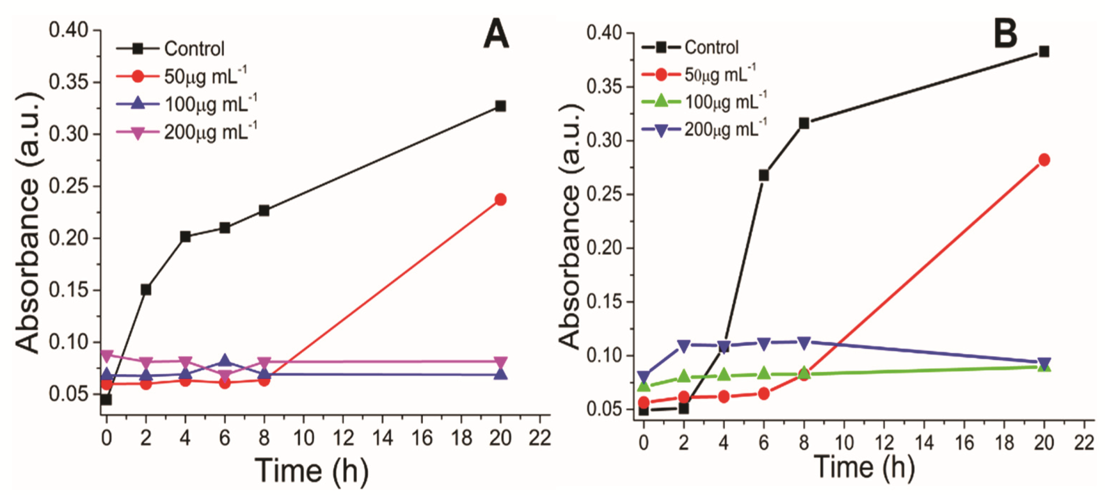
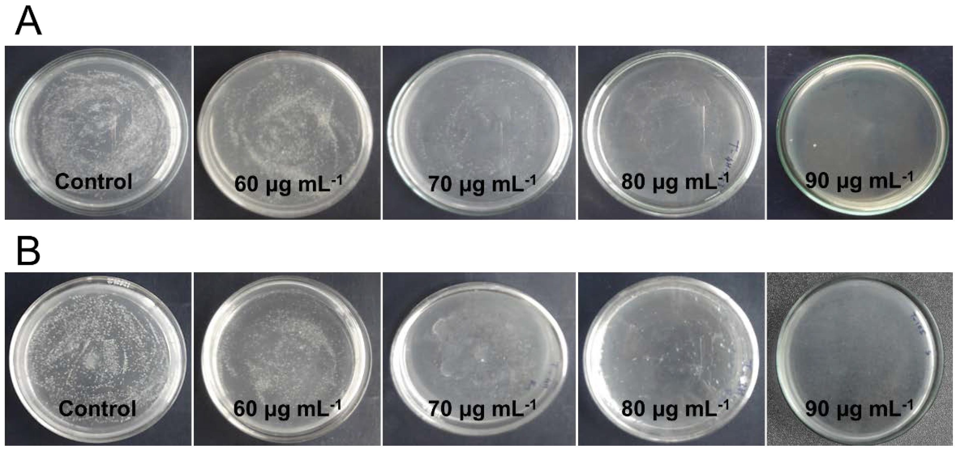
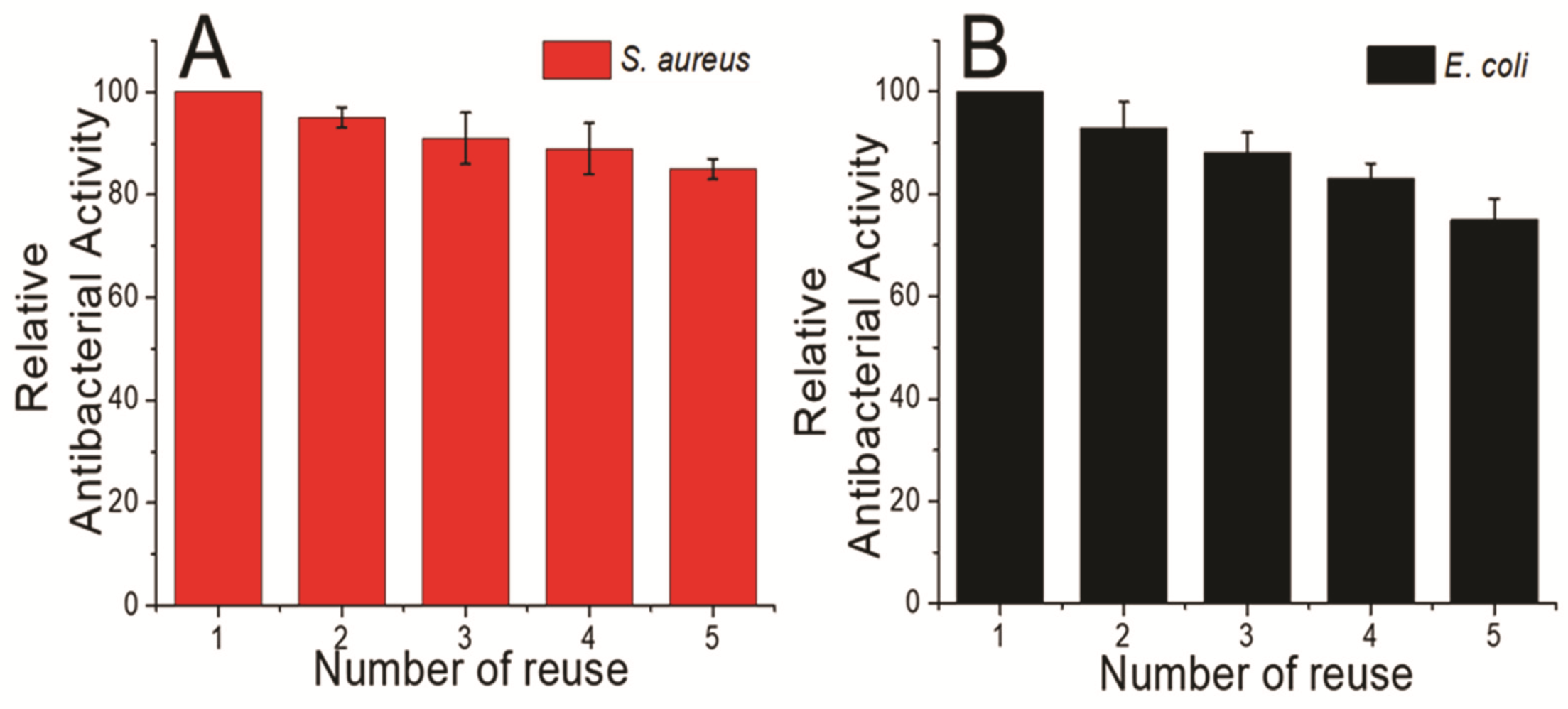
3.2. Antimicrobial Activity

The effective antimicrobial activity of the NPs is dependent on the NPs aggregation and interaction of the NPs with microbial cells. Thus, the hydrodynamic diameter (HDD) and zeta potential studies of the NPs provide information regarding the size and charge on the NPs in the colloidal solution, demonstrating the stability of NPs. The average HDD of the AgNPs, Fe2O3 NPs, and Ag-BSiO2-Fe2O3 hybrid structure in the saline solution was 30 (Figure S2A), 200 (Figure S2B), and 500 nm (Figure S2C), respectively. The zeta potential of the AgNPs, Fe2O3 NPs, and Ag-BSiO2-Fe2O3 hybrid structure was −28.6 (Figure S3A), −5.36 (Figure S3B), and −17.8 mV (Figure S3C), respectively. The antimicrobial activity of the streptomycin, AgNPs, Fe2O3 NPs, and Ag-BSiO2-Fe2O3 hybrid structure was analyzed using S. aureus (Gram-positive) and E. coli (Gram-negative). The effective concentration of the various structures was determined by analyzing the inhibitory effect on the growth curve of both microorganisms; the time-dependent growth of the microorganisms was monitored at 610 nm. Figure 5 shows that 50 µg mL−1 of the microhybrid affected the growth rates of S. aureus (Figure 5A) and E. coli (Figure 5B), but it did not completely inhibit both microorganisms. At 100 and 200 µg mL−1, the growth of both microorganisms was hampered, and no changes in the absorbance were observed after 20 h of incubation with the Ag-BSiO2-Fe2O3 structure. In similar experiments, a 10 µg mL−1 concentration of streptomycin and AgNPs showed an inhibitory effect on both microorganisms. The complete inhibition of S. aureus (Figure S4A) and E. coli (Figure S4B) was observed at 30 µg mL−1 of streptomycin, whereas 50 µg mL−1 of AgNPs showed complete inhibition of S. aureus (Figure S5A) and E. coli (Figure S5B). The Fe2O3 and BSiO2-Fe2O3 NPs showed no effect on the bacterial growth curve in 50–200 µg mL−1 for both microorganisms (Figures S6 and S7). It is clear that 50 µg mL−1 of the hybrid structure was most effective for S. aureus, whereas E. coli showed some inhibition at this concentration. However, both microorganisms were highly sensitive to 200 µg mL−1 of the Ag-BSiO2-Fe2O3 hybrid structure. To determine the minimum bactericidal concentration, both bacterial strains were exposed to various concentrations of the Ag-BSiO2-Fe2O3 structure. Although 50 µg mL−1 of the hybrid structure reduced the growth of the microorganisms, it did not inhibit growth completely. However, as the concentration of the microhybrid increased from 50 to 90 µg mL−1, the growth of both microorganisms constantly decreased. At 90 µg mL−1 of the hybrid structure, complete inhibition was observed for S. aureus (Figure 6A) and E. coli (Figure 6B); therefore, it can be considered the minimum bactericidal concentration (MBC) for both bacterial strains. For streptomycin, 20 µg mL−1 and 30 µg mL−1 were inhibitory for S. aureus and E. coli, respectively, as no growth was observed on growth plates (Figure S8). AgNPs inhibited both microorganisms at 30 µg mL−1 (Figure S9). However, the Fe2O3 and BSiO2-Fe2O3 particles showed no effect on the microbial growth in 50 to 90 µg mL−1 (Figures S10 and S11). The AgNPs showed high stability and low HDD in the aqueous solution, which allowed the maximum interaction with microorganisms, leading to its effective antimicrobial activity, whereas the presence of plant extract and AgNPs on the surface Fe2O3 NPs increased the stability of the hybrid nanostructure compared to Fe2O3 NPs, allowing it to perform more effectively in antimicrobial activity. The CLSM study was performed to visualize the antimicrobial activity of the hybrid structure where a 200 μg mL−1 concentration of the hybrid structure is completely inhibitory to both species showing the red dead bacteria cells (Figures S12 and S13).
Because 200 µg mL−1 of the microhybrid caused complete inhibition of both pathogenic strains, the concentration of the hybrid structure was checked for repeated use of antimicrobial activity. The Ag-BSiO2-Fe2O3 hybrid was added to each bacterial suspension, and the mixture was incubated for 24 h. After incubation, the hybrid material was isolated with an external magnet, and the bacterial suspension was analyzed spectroscopically. The hybrid structure was washed with a buffer before each repeated use, and the new bacterial suspension was added to this washed buffer. This was repeated for the next 5 cycles. The Ag-BSiO2-Fe2O3 hybrid structure maintained 80% of its relative antimicrobial activity against S. aureus and 75% against E. coli (Figure 7). The loss of antimicrobial activity of the Ag-BSiO2-Fe2O3 hybrid structure may be due to the loss of the hybrid material during isolation from the bacterial suspension.

4. Conclusions

Here, we have developed a prominent, clean, robust method for the synthesis of an antimicrobial hybrid nanostructure using green tea active biomolecules. C. sinensis green tea ethanolic extract comprises polyphenols and other active ingredients and has a high antioxidant and reducing activity. The extract was allowed to become trapped in the SiO2 layer by hydrolysis. This process of entrapment was employed to synthesize the separable SiO2-coated Fe2O3 NPs magnetically. The reducing capacity of biomolecule-entrapped SiO2-Fe2O3 NPs was used to synthesize Ag NPs on the surface of the magnetic structure, thus forming the Ag-BSiO2-Fe2O3 hybrid nanostructure. As the entrapped biomolecules have a high reducing capacity, the reduction of Ag+ ions occurred quickly, which led to the formation of spherical Ag NPs with diameters in the range of 5–10 nm on the magnetic nanostructure. Elemental dot mapping revealed the uniform distribution of biomolecules and Ag NPs in the SiO2 layer coated on Fe2O3 NPs. The Ag-BSiO2-Fe2O3 hybrid microstructure showed excellent antimicrobial activity against S. aureus and E. coli. As the hybrid nanostructure was magnetic in nature, it showed excellent reusable antimicrobial activity. Toxicology studies need to be performed to use the antioxidant hybrid structure for medicinal and environmental applications. The outcome of the presented work creates opportunities for the construction of various metal microhybrid structures using eco-friendly biomaterials.

Supplementary Materials

The following are available online at https://www.mdpi.com/article/10.3390/ma14247893/s1, Figure S1: FT-IR spectroscopy analysis of green tea extract and Ag-BSiO2 NPs-Fe2O3 NPs; Figure, S2: The Dynamic light scattering (DLS) analysis of colloids suspension of (A) Ag NPs, (B) Fe2O3 NPs, and (C) Ag-BSiO2- Fe2O3 NPs; Figure S3: The zeta potential analysis of colloids suspension of (A) Ag NPs, (B) Fe2O3 NPs, and (C) Ag-BSiO2- Fe2O3 NPs; Figure S4: Growth curves of (A) S. aureus and (B) E. coli with various concentrations of the streptomycin antibiotics; Figure S5: Growth curves of (A) S. aureus and (B) E. coli with various concentrations of the AgNPs; Figure S6: Growth curves of (A) S. aureus and (B) E. coli with various concentrations of the Fe2O3 NPs; Figure S7: Growth curves of (A) S. aureus and (B) E. coli with various concentrations of the BSiO2-Fe2O3 NPs; Figure S8: Growth plate photographs of (A) S. aureus and (B) E. coli with various concentrations of streptomycin antibiotics; Figure S9: Growth plate photographs of (A) S. aureus and (B) E. coli with various concentrations of Ag NPs; Figure S10: Growth plate photographs of (A) S. aureus and (B) E. coli with various concentrations of Fe2O3 NPs; Figure S11: Growth plate photographs of (A) S. aureus and (B) E. coli with various concentrations of BSiO2-Fe2O3 NPs; Figure S12: CLSM microscopy images of the antimicrobial activity of Ag-BSiO2-Fe3O4 nanoparticles against E. coli.; Figure S13: CLSM microscopy images of the antimicrobial activity of Ag-BSiO2-Fe3O4 nanoparticles against S. aureus. Table S1: Total phenolic contents and antioxidant activity of the green tea extract in different solvents.

Author Contributions

Conceptualization, S.V.O. and J.-K.L.; methodology, S.V.O. and A.B.; software, S.V.O.; validation, V.C.K., I.-W.K. and J.-K.L.; formal analysis, V.C.K.; investigation, S.V.O.; resources, S.V.O., A.B. and J.-K.L.; data curation, V.C.K.; writing—original draft preparation, S.V.O.; writing—review and editing, S.V.O., A.B. and J.-K.L.; visualization, V.C.K. and J.-K.L.; supervision, J.-K.L.; project administration, I.-W.K. and J.-K.L.; funding acquisition, J.-K.L. All authors have read and agreed to the published version of the manuscript.

Funding

This research received no external funding.

Institutional Review Board Statement

Not applicable.

Informed Consent Statement

Not applicable.

Data Availability Statement

Not applicable.

Acknowledgments

This work was supported by Konkuk University in 2019.

Conflicts of Interest

The authors declare no conflict of interest.

References

  1. Tenover, F.C. Mechanisms of antimicrobial resistance in bacteria. Am. J. Infect. Med. 2006, 119, S3–S10. [Google Scholar] [CrossRef] [PubMed]
  2. Holmes, A.H.; Moore, L.S.P.; Sundsfjord, A.; Steinbakk, M.; Regmi, S.; Karkey, A.; Guerin, P.J.; Piddock, L.J.V. Understanding the mechanisms and drivers of antimicrobial resistance. Lancet 2016, 387, 176–187. [Google Scholar] [CrossRef]
  3. Kim, H.U.; Blin, K.; Lee, S.Y.; Weber, T. Recent development of computational resources for new antibiotics discovery. Curr. Opin. Microbiol. 2017, 39, 113–120. [Google Scholar] [CrossRef] [PubMed]
  4. Gupta, A.; Mumtaz, S.; Li, C.H.; Hussain, I.; Rotello, V.M. Combatting antibiotic-resistant bacteria using nanomaterials. Chem. Soc. Rev. 2019, 48, 415–427. [Google Scholar] [CrossRef] [PubMed]
  5. Hemeg, H.A. Nanomaterials for alternative antibacterial therapy. Int. J. Nanomed. 2017, 12, 8211–8225. [Google Scholar] [CrossRef] [PubMed] [Green Version]
  6. Bandow, J.E.; Metzler-Nolte, N. New ways of killing the beast: Prospects for inorganic-organic hybrid nanomaterials as antibacterial agents. ChemBioChem 2009, 10, 2847–2850. [Google Scholar] [CrossRef] [PubMed]
  7. Yacoby, I.; Benhar, I. Antibacterial nanomedicine. Antibact. Nanomed. 2008, 3, 329–341. [Google Scholar] [CrossRef]
  8. Rai, M.; Yadav, A.; Gade, A. Silver nanoparticles as a new generation of antimicrobials. Biotechnol. Adv. 2009, 27, 76–83. [Google Scholar] [CrossRef]
  9. Otari, S.V.; Patil, R.M.; Nadaf, N.H.; Ghosh, S.J.; Pawar, S.H. Green biosynthesis of silver nanoparticles from an actinobacteria Rhodococcus sp. Mater. Lett. 2012, 72, 92–94. [Google Scholar] [CrossRef]
  10. Lok, C.N.; Ho, C.M.; Chen, R.; He, Q.Y.; Yu, W.Y.; Sun, H.; Tam, P.K.H.; Chiu, J.F.; Che, C.M. Silver nanoparticles: Partial oxidation and antibacterial activities. JBIC J. Biol. Inorg. Chem. 2007, 12, 527–534. [Google Scholar] [CrossRef] [PubMed]
  11. Choi, O.; Deng, K.K.; Kim, N.J.; Ross, L.; Surampalli, R.Y.; Hu, Z. The inhibitory effects of silver nanoparticles, silver ions, and silver chloride colloids on microbial growth. Water Res. 2008, 42, 3066–3074. [Google Scholar] [CrossRef] [PubMed]
  12. Ahamed, M.; AlSalhi, M.S.; Siddiqui, M.K.J. Silver nanoparticle applications and human health. Clin. Chim. Acta 2010, 411, 1841–1848. [Google Scholar] [CrossRef]
  13. Wu, F.; Harper, B.J.; Harper, S.L. Differential dissolution and toxicity of surface functionalized silver nanoparticles in small-scale microcosms: Impacts of community complexity. Environ. Sci. Nano 2017, 4, 359–372. [Google Scholar] [CrossRef]
  14. Zhao, X.S.; Bao, X.Y.; Guo, W.; Lee, F.Y. Immobilizing catalysts on porous materials. Mater. Today 2006, 9, 32–39. [Google Scholar] [CrossRef]
  15. Dalpozzo, R. Magnetic nanoparticle supports for asymmetric catalysts. Green Chem. 2015, 17, 3671–3686. [Google Scholar] [CrossRef]
  16. Tzounis, L.; Logothetidis, S. Fe3O4@SiO2 core shell particles as platforms for the decoration of Ag nanoparticles. Mater. Today Proc. 2017, 4, 7076–7082. [Google Scholar] [CrossRef]
  17. Sui, N.; Monnier, V.; Salvia, M.V.; Chevolot, Y.; Souteyrand, E. Magnetic and optical properties of Ag@SiO2-FITC-Fe3O4 hybrid nanoparticles. Mater. Sci. Eng. B 2014, 182, 92–95. [Google Scholar] [CrossRef]
  18. Das, S.K.; Khan, M.M.R.; Parandhaman, T.; Laffir, F.; Guha, A.K.; Sekaran, G.; Mandal, A.B. Nano-silica fabricated with silver nanoparticles: Antifouling adsorbent for efficient dye removal, effective water disinfection and biofouling control. Nanoscale 2013, 5, 5549–5560. [Google Scholar] [CrossRef]
  19. Kaloti, M.; Kumar, A.; Navani, N.K. Synthesis of glucose-mediated Ag-γ-Fe2O3 multifunctional nanocomposites in aqueous medium—A kinetic analysis of their catalytic activity for 4-nitrophenol reduction. Green Chem. 2015, 17, 4786–4799. [Google Scholar] [CrossRef]
  20. Tzounis, L.; Contreras-Caceres, R.; Schellkopf, L.; Jehnichen, D.; Fischer, D.; Cai, C.; Uhlmann, P.; Stamm, M. Controlled growth of Ag nanoparticles decorated onto the surface of SiO2 spheres: A nanohybrid system with combined SERS and catalytic properties. RSC Adv. 2014, 4, 17846–17855. [Google Scholar] [CrossRef]
  21. Muthukumar, H.; Palanirajan, S.K.; Shanmugam, M.K.; Gummadi, S.N. Plant extract mediated synthesis enhanced the functional properties of silver ferrite nanoparticles over chemical mediated synthesis. Biotechnol. Rep. 2020, 26, e00469. [Google Scholar] [CrossRef]
  22. Darezereshki, E. Synthesis of maghemite (γ-Fe2O3) nanoparticles by wet chemical method at room temperature. Mater. Lett. 2010, 64, 1471–1472. [Google Scholar] [CrossRef]
  23. Deng, Y.H.; Wang, C.C.; Hu, J.H.; Yang, W.L.; Fu, S.K. Investigation of formation of silica-coated magnetite nanoparticles via sol-gel approach. Colloids Surf. A Physicochem. Eng. Asp. 2005, 262, 87–93. [Google Scholar] [CrossRef]
  24. Chaurasia, A.K.; Thorat, N.D.; Tandon, A.; Kim, J.H.; Park, S.H.; Kim, K.K. Coupling of radiofrequency with magnetic nanoparticles treatment as an alternative physical antibacterial strategy against multiple drug resistant bacteria. Sci. Rep. 2016, 6, 33662. [Google Scholar] [CrossRef] [Green Version]
  25. de Oliveira, A.C.; Valentim, I.B.; Silva, C.A.; Bechara, E.J.H.; de Barros, M.P.; Mano, C.M.; Goulart, M.O.F. Total phenolic content and free radical scavenging activities of methanolic extract powders of tropical fruit residues. Food Chem. 2009, 115, 469–475. [Google Scholar] [CrossRef]
  26. Ahmed, S.; Ahmad, M.; Swami, B.L.; Ikram, S. A review on plants extract mediated synthesis of silver nanoparticles for antimicrobial applications: A green expertise. J. Adv. Res. 2016, 7, 17–28. [Google Scholar] [CrossRef] [Green Version]
  27. Rónavári, A.; Kovács, D.; Igaz, N.; Vágvölgyi, C.; Boros, I.M.; Kónya, Z.; Pfeiffer, I.; Kiricsi, M. Biological activity of green-synthesized silver nanoparticles depends on the applied natural extracts: A comprehensive study. Int. J. Nanomed. 2017, 12, 871–883. [Google Scholar] [CrossRef] [Green Version]
  28. Rónavári, A.; Bélteky, P.; Boka, E.; Zakupszky, D.; Igaz, N.; Szerencsés, B.; Pfeiffer, I.; Kónya, Z.; Kiricsi, M. Polyvinyl-pyrrolidone-coated silver nanoparticles—The colloidal, chemical and biological consequences of steric stabilization under biorelevant conditions. Int. J. Mol. Sci. 2021, 22, 8673. [Google Scholar] [CrossRef] [PubMed]
  29. Bélteky, P.; Rónavári, A.; Igaz, N.; Szerencsés, B.; Tóth, I.Y.; Pfeiffer, I.; Kiricsi, M.; Kónya, Z. Silver nanoparticles: Aggregation behavior in biorelevant conditions and its impact on biological activity. Int. J. Nanomed. 2019, 14, 667–687. [Google Scholar] [CrossRef] [Green Version]
  30. Hussain, S.; Khan, Z. Epigallocatechin-3-gallate-capped Ag nanoparticles: Preparation and characterization. Bioprocess Biosyst. Eng. 2014, 37, 1221–1231. [Google Scholar] [CrossRef] [PubMed]
  31. Moulton, M.C.; Braydich-Stolle, L.K.; Nadagouda, M.N.; Kunzelman, S.; Hussain, S.M.; Varma, R.S. Synthesis, characterization and biocompatibility of “green” synthesized silver nanoparticles using tea polyphenols. Nanoscale 2010, 2, 763–770. [Google Scholar] [CrossRef] [PubMed]
  32. Trouillas, P.; Marsal, P.; Siri, D.; Lazzaroni, R.; Duroux, J.L. A DFT study of the reactivity of OH groups in quercetin and taxifolin antioxidants: The specificity of the 3-OH site. Food Chem. 2006, 97, 679–688. [Google Scholar] [CrossRef]
  33. Navale, S.T.; Bandgar, D.K.; Nalage, S.R.; Khuspe, G.D.; Chougule, M.A.; Kolekar, Y.D.; Sen, S.; Patil, V.B. Synthesis of Fe2O3 nanoparticles for nitrogen dioxide gas sensing applications. Ceram. Int. 2013, 39, 6453–6460. [Google Scholar] [CrossRef]
  34. Otari, S.V.; Patil, R.M.; Waghmare, S.R.; Ghosh, S.J.; Pawar, S.H. A novel microbial synthesis of catalytically active Ag-alginate biohydrogel and its antimicrobial activity. Dalton Trans. 2013, 42, 9966–9975. [Google Scholar] [CrossRef] [PubMed]
  35. Shin, K.S.; Cho, Y.K.; Choi, J.-Y.; Kim, K. Facile synthesis of silver-deposited silanized magnetite nanoparticles and their application for catalytic reduction of nitrophenols. Appl. Catal. A Gen. 2012, 413–414, 170–175. [Google Scholar] [CrossRef]
  36. Yi, T.; Zhu, L.; Peng, W.L.; He, X.C.; Chen, H.L.; Li, J.; Yu, T.; Liang, Z.T.; Zhao, Z.Z.; Chen, H.B. Comparison of ten major constituents in seven types of processed tea using HPLC-DAD-MS followed by principal component and hierarchical cluster analysis. LWT—Food Sci. Technol. 2015, 62, 194–201. [Google Scholar] [CrossRef] [Green Version]
  37. Li, Z.; Zhu, Y. Surface-modification of SiO2 nanoparticles with oleic acid. Appl. Surf. Sci. 2003, 211, 315–320. [Google Scholar] [CrossRef]
Figure 1. Schematics for the synthesis of silver-decorated biomolecule-entrapped SiO2-coated Fe2O3 (Ag-BSiO2-Fe2O3) hybrid structure.
Materials 14 07893 g001
Figure 2. HR-TEM images of the (A,B) Fe2O3 nanosphere and (C,D) BSiO2-Fe2O3 particles.
Materials 14 07893 g002
Figure 3. (A) UV-visible spectroscopic analysis of Fe2O3 NPs, BSiO2-Fe2O3 particles, and Ag-BSiO2-Fe2O3 structure. (B) XRD analysis. (C) Magnetization curves of γ-Fe2O3 NPs (black), BSiO2-Fe2O3 particles (red), and Ag- BSiO2-Fe2O3 structure (blue).
Materials 14 07893 g003
Figure 4. (AC) HR-TEM images, (D) EDS spectrum analysis, and (E) elemental dot mapping of Ag-BSiO2-Fe2O3 hybrid structure.
Materials 14 07893 g004
Figure 5. Growth curves of (A) S. aureus and (B) E. coli with various concentrations of the Ag-BSiO2-Fe2O3 hybrid structure.
Materials 14 07893 g005
Figure 6. Growth plate photographs of (A) S. aureus and (B) E. coli with various concentrations of Ag-BSiO2-Fe2O3 hybrid structure.
Materials 14 07893 g006
Figure 7. Reusable antibacterial effects of Ag-BSiO2-Fe2O3 hybrid structure (200 μg mL−1) on (A) S. aureus and (B) E. coli.
Materials 14 07893 g007
Publisher’s Note: MDPI stays neutral with regard to jurisdictional claims in published maps and institutional affiliations.

