Hierarchical Composite Meshes of Electrospun PS Microfibers with PA6 Nanofibers for Regenerative Medicine


Abstract
:
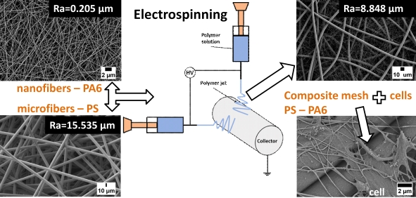
1. Introduction
2. Materials and Methods
2.1. Solutions Preparation
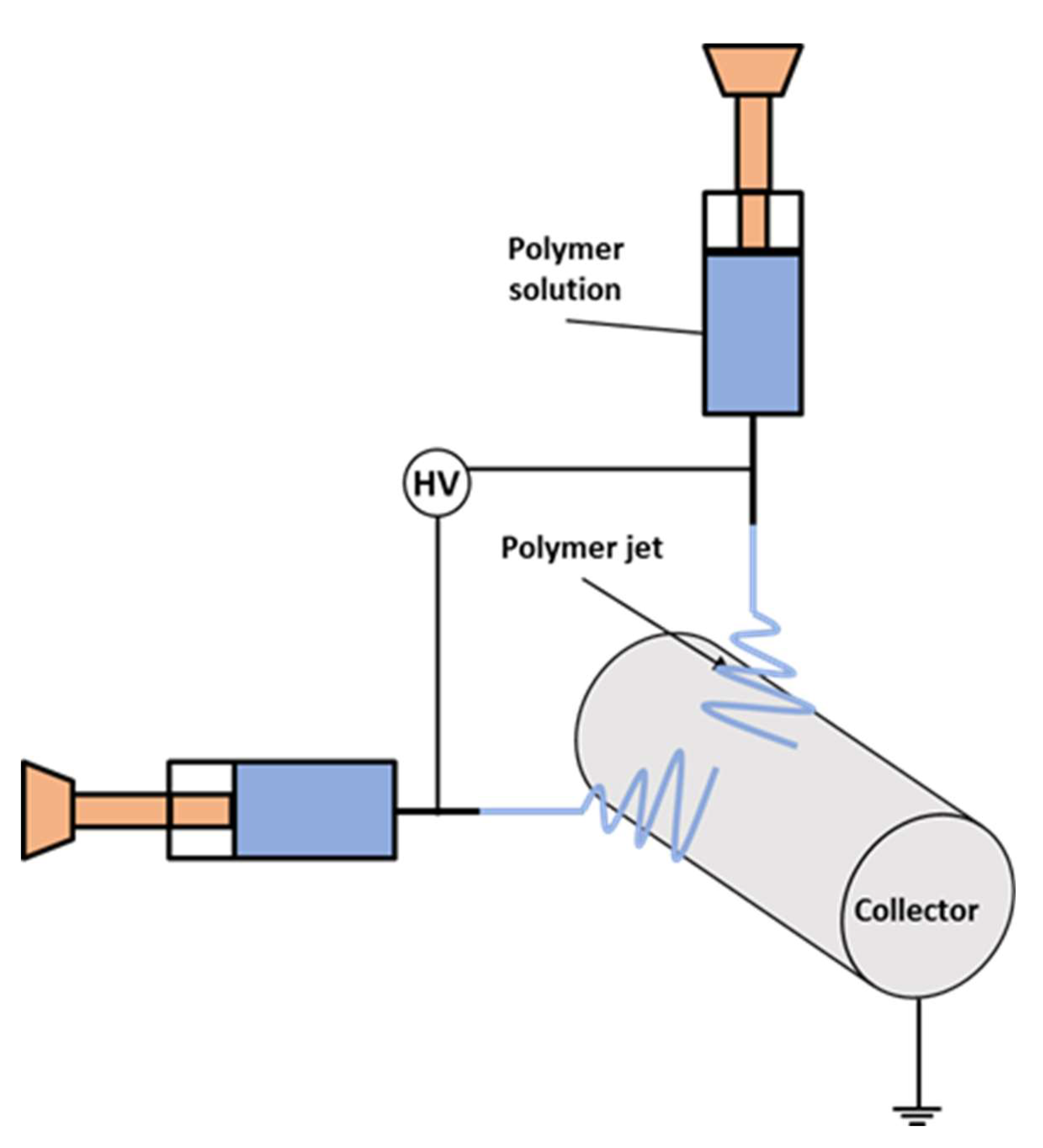
2.2. Electrospinning and Meshes Characterization
2.3. Cell Culture Studies
2.3.1. Adhesion Test
2.3.2. MTS Proliferation Assay
2.3.3. Statistical Analyses
3. Results and Discussion
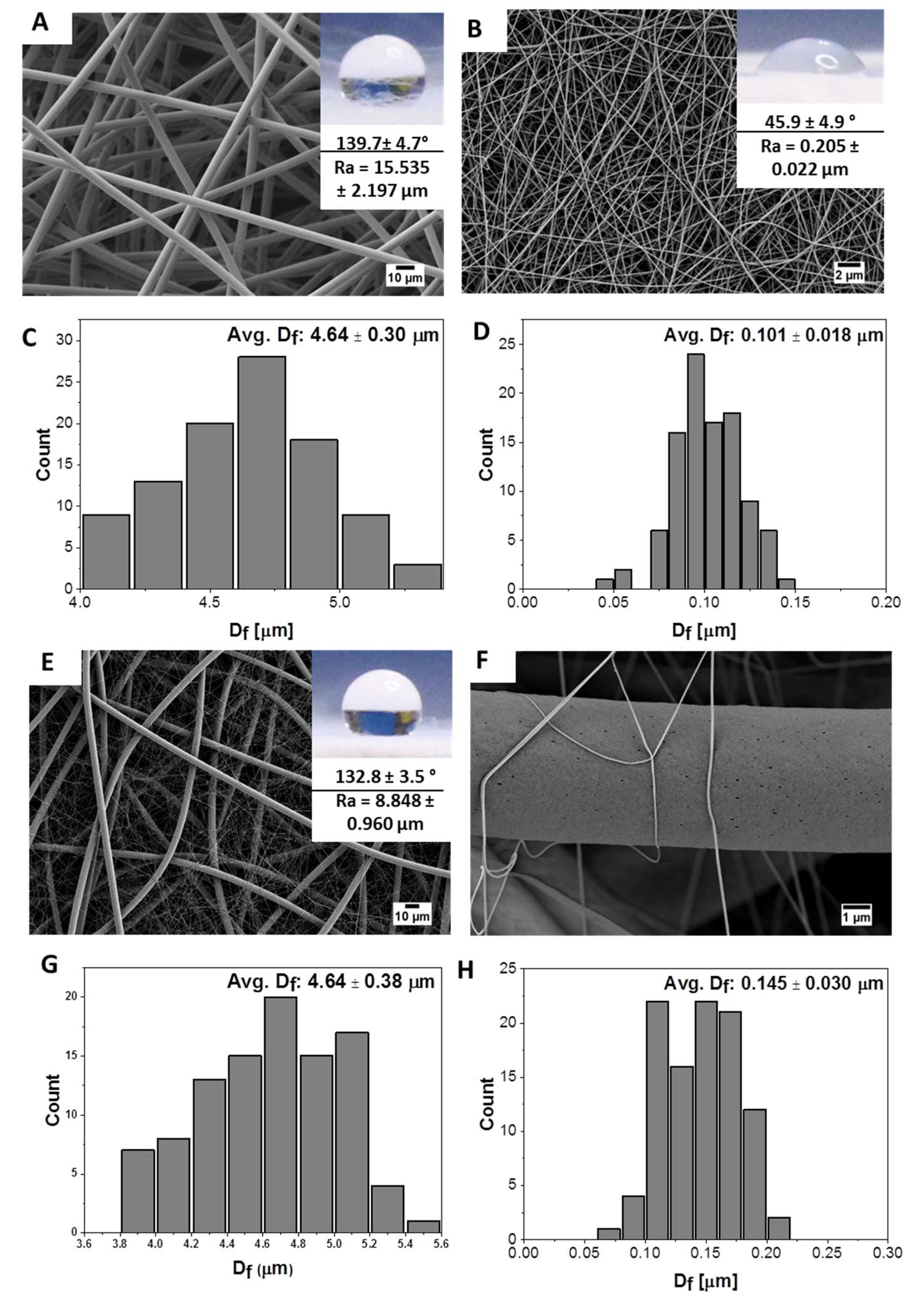
3.1. Fibers Characterization
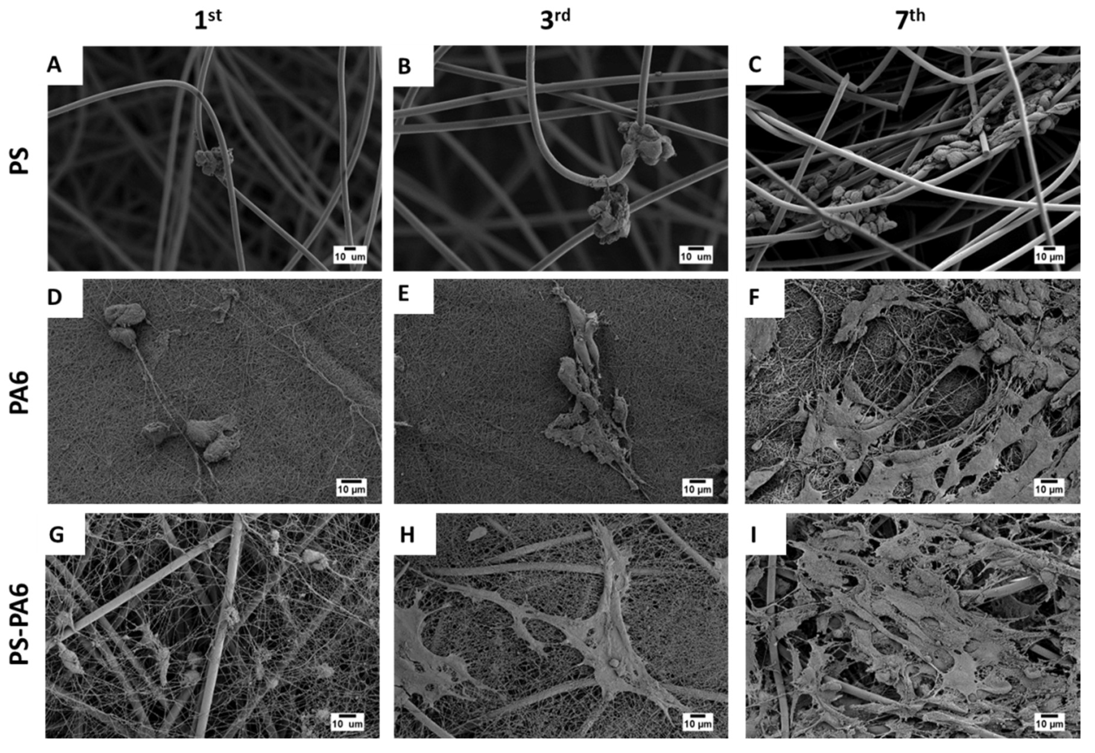
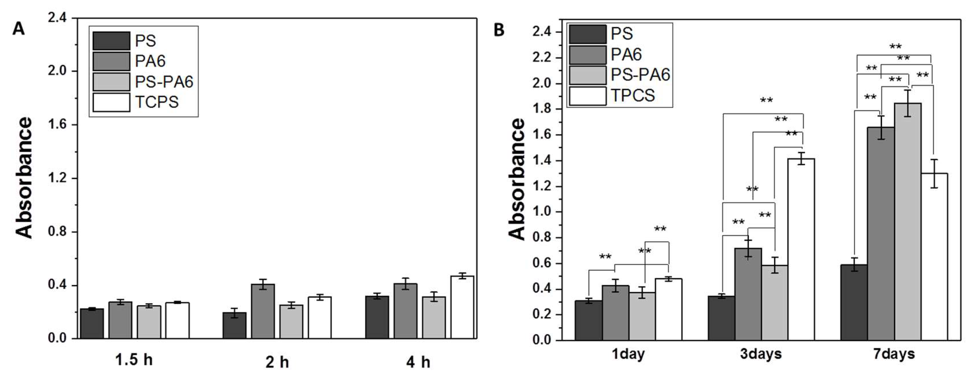
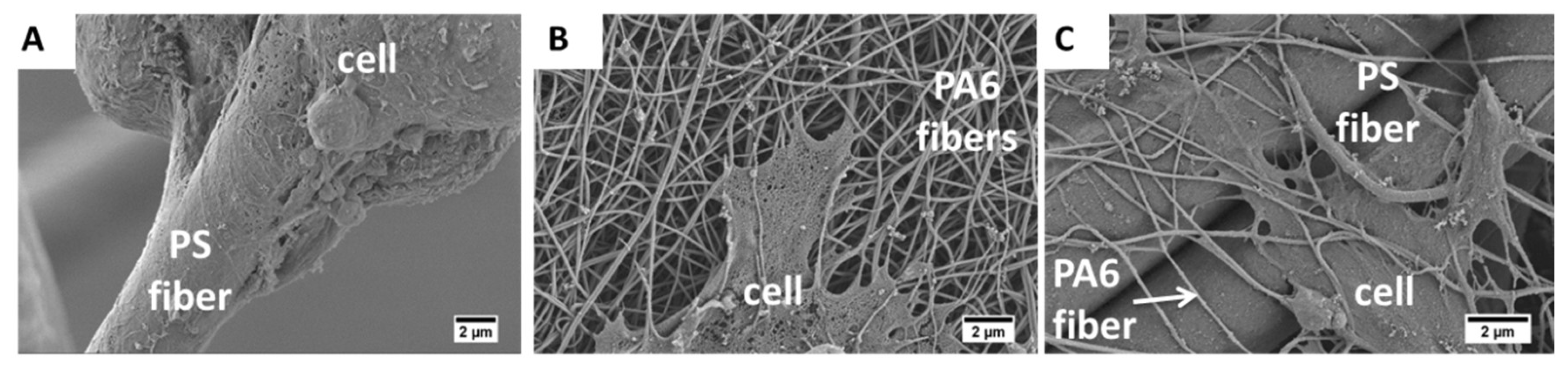
3.2. Cell Culture Study
4. Conclusions
Author Contributions
Funding
Conflicts of Interest
References
- Kooten, T.G.; Spijker, H.T.; Busscher, H.J. Plasma-treated polystyrene surfaces: Model surfaces for studying cell–biomaterial interactions. Biomaterials 2004, 25, 1735–1747. [Google Scholar] [CrossRef] [PubMed]
- Szewczyk, P.; Metwally, S.; Karbowniczek, J.E.; Marzec, M.M.; Stodolak-Zych, E.; Gruszczynski, A.; Bernasik, A.; Stachewicz, U.; Stodolak, E. Surface-Potential-Controlled Cell Proliferation and Collagen Mineralization on Electrospun Polyvinylidene Fluoride (PVDF) Fiber Scaffolds for Bone Regeneration. ACS Biomater. Sci. Eng. 2018, 5, 582–593. [Google Scholar] [CrossRef]
- Metwally, S.; Karbowniczek, J.; Szewczyk, P.; Marzec, M.M.; Gruszczyński, A.; Bernasik, A.; Stachewicz, U. Single-Step Approach to Tailor Surface Chemistry and Potential on Electrospun PCL Fibers for Tissue Engineering Application. Adv. Mater. Interfaces 2018, 6, 1–12. [Google Scholar] [CrossRef]
- Curtis, A.S.; Forrester, J.V.; McInnes, C.; Lawrie, F. Adhesion of cells to polystyrene surfaces. J. Cell Boil. 1983, 97, 1500–1506. [Google Scholar] [CrossRef] [Green Version]
- Steele, J.G.; Dalton, B.A.; Johnson, G.; Underwood, P.A. Polystyrene chemistry affects vitronectin activity: An explanation for cell attachment to tissue culture polystyrene but not to unmodified polystyrene. J. Biomed. Mater. Res. 1993, 27, 927–940. [Google Scholar] [CrossRef]
- Baker, S.; Atkin, N.; Gunning, P.A.; Granville, N.; Wilson, K.; Wilson, D.; Southgate, J. Characterisation of electrospun polystyrene scaffolds for three-dimensional in vitro biological studies. Biomaterials 2006, 27, 3136–3146. [Google Scholar] [CrossRef]
- Simonet, M.; Schneider, O.D.; Neuenschwander, P.; Stark, W.J. Ultraporous 3D polymer meshes by low-temperature electrospinning: Use of ice crystals as a removable void template. Polym. Eng. Sci. 2007, 47, 2020–2026. [Google Scholar] [CrossRef]
- Ma, P.X. Biomimetic materials for tissue engineering. Adv. Drug Deliv. Rev. 2007, 60, 184–198. [Google Scholar] [CrossRef] [Green Version]
- Stachewicz, U.; Szewczyk, P.; Kruk, A.; Barber, A.H.; Czyrska-Filemonowicz, A. Pore shape and size dependence on cell growth into electrospun fiber scaffolds for tissue engineering: 2D and 3D analyses using SEM and FIB-SEM tomography. Mater. Sci. Eng. C 2019, 95, 397–408. [Google Scholar] [CrossRef]
- Pham, Q.P.; Sharma, U.; Mikos, A.G. Electrospinning of Polymeric Nanofibers for Tissue Engineering Applications: A Review. Tissue Eng. 2006, 12, 1197–1211. [Google Scholar] [CrossRef] [Green Version]
- Li, X.; Wang, X.; Yao, D.; Jiang, J.; Guo, X.; Gao, Y.; Li, Q.; Shen, C. Effects of aligned and random fibers with different diameter on cell behaviors. Colloids Surfaces B Biointerfaces 2018, 171, 461–467. [Google Scholar] [CrossRef] [PubMed]
- Stachewicz, U.; Qiao, T.; Rawlinson, S.; Almeida, F.; Li, W.; Cattell, M.J.; Barber, A. 3D imaging of cell interactions with electrospun PLGA nanofiber membranes for bone regeneration. Acta Biomater. 2015, 27, 88–100. [Google Scholar] [CrossRef] [PubMed]
- Kaniuk, Ł.; Krysiak, Z.J.; Metwally, S.; Stachewicz, U. Osteoblasts and fibroblasts attachment to poly(3-hydroxybutyric acid-co-3-hydrovaleric acid) (PHBV) film and electrospun scaffolds. Mater. Sci. Eng. C 2020, 110, 110668. [Google Scholar] [CrossRef]
- Sensini, A.; Gualandi, C.; Focarete, M.L.; Belcari, J.; Zucchelli, A.; Boyle, L.; Reilly, G.C.; Kao, A.; Tozzi, G.; Cristofolini, L. Multiscale hierarchical bioresorbable scaffolds for the regeneration of tendons and ligaments. Biofabrication 2019, 11, 035026. [Google Scholar] [CrossRef]
- Tuzlakoglu, K.; Bölgen, N.; Salgado, A.J.; Gomes, M.E.; Pişkin, E.; Reis, R.L. Nano- and micro-fiber combined scaffolds: A new architecture for bone tissue engineering. J. Mater. Sci. Mater. Electron. 2005, 16, 1099–1104. [Google Scholar] [CrossRef] [Green Version]
- Theron, S.; Yarin, A.; Zussman, E.; Kroll, E. Multiple jets in electrospinning: Experiment and modeling. Polymer 2005, 46, 2889–2899. [Google Scholar] [CrossRef]
- Bolbasov, E.N.; Buznik, V.M.; Stankevich, K.S.; Goreninskii, S.I. Composite Materials Obtained via Two-Nozzle Electrospinning from Polycarbonate and Vinylidene Fluoride/Tetrafluoroethylene Copolymer. Inorg. Mater. Appl. Res. 2018, 9, 184–191. [Google Scholar] [CrossRef]
- Zhang, C.; Li, Y.P.; Wang, W.; Zhan, N.; Xiao, N.; Wang, S.; Li, Y.; Yang, Q. A novel two-nozzle electrospinning process for preparing microfiber reinforced pH-sensitive nano-membrane with enhanced mechanical property. Eur. Polym. J. 2011, 47, 2228–2233. [Google Scholar] [CrossRef]
- Karaca, E.; Hockenberger, A.S. Analysis of the fracture morphology of polyamide, polyester, polypropylene, and silk sutures before and after implantationin vivo. J. Biomed. Mater. Res. Part B Appl. Biomater. 2008, 87, 580–589. [Google Scholar] [CrossRef]
- Jaganathan, S.K.; Supriyanto, E.; Murugesan, S.; Balaji, A.; Asokan, M.K. Biomaterials in Cardiovascular Research: Applications and Clinical Implications. BioMed Res. Int. 2014, 2014, 1–11. [Google Scholar] [CrossRef] [Green Version]
- Shen, J.; Li, Y.; Zuo, Y.; Zou, Q.; Cheng, L.; Zhang, L.; Gong, M.; Gao, S. Characterization and cytocompatibility of biphasic calcium phosphate/polyamide 6 scaffolds for bone regeneration. J. Biomed. Mater. Res. Part B Appl. Biomater. 2010, 95, 330–338. [Google Scholar] [CrossRef] [PubMed]
- Wisniewski, N.; Rajamand, N.; Adamsson, U.; Lins, P.E.; Reichert, W.M.; Klitzman, B.; Ungerstedt, U. Analyte flux through chronically implanted subcutaneous polyamide membranes differs in humans and rats. Am. J. Physiol. Metab. 2002, 282, E1316–E1323. [Google Scholar] [CrossRef] [Green Version]
- Panthi, G.; Barakat, N.A.M.; Risal, P.; Yousef, A.; Pant, B.; Unnithan, A.R.; Kim, H.Y. Preparation and Characterization of Nylon-6/Gelatin Composite Nanofibers Via Electrospinning for Biomedical Applications. Fibers Polym. 2013, 14, 718–723. [Google Scholar] [CrossRef]
- Pant, H.R.; Risal, P.; Park, C.H.; Tijing, L.D.; Jeong, Y.J.; Kim, C.S. Core–shell structured electrospun biomimetic composite nanofibers of calcium lactate/nylon-6 for tissue engineering. Chem. Eng. J. 2013, 221, 90–98. [Google Scholar] [CrossRef]
- Pant, H.R.; Kimc, H.J.; Bhatt, L.R.; Joshi, M.K.; Kima, E.K.; Kima, J.I.; Abdal-haya, A.; Hui, K.S.; Kim, C.S. Applied Surface Science Chitin butyrate coated electrospun nylon-6 fibers for biomedical applications. Appl. Surf. Sci. 2013, 285, 538–544. [Google Scholar] [CrossRef]
- Abdal-hay, A.; Khali, K.A.; Al-Jassir, F.F.; Gamal-Eldeen, A.M. Biocompatibility properties of polyamide 6/PCL blends composite textile scaffold using EA. hy926 human endothelial cells. Biomed. Mater. 2017, 12, 035002. [Google Scholar] [CrossRef] [PubMed]
- Winnacker, M. Polyamides and their functionalization: Recent concepts for their applications as biomaterials. Biomater. Sci. 2017, 5, 1230–1235. [Google Scholar] [CrossRef]
- Stachewicz, U.; Stone, C.A.; Willis, C.R.; Barber, A. Charge assisted tailoring of chemical functionality at electrospun nanofiber surfaces. J. Mater. Chem. 2012, 22, 22935. [Google Scholar] [CrossRef]
- Stachewicz, U.; Barber, A. Enhanced Wetting Behavior at Electrospun Polyamide Nanofiber Surfaces. Langmuir 2011, 27, 3024–3029. [Google Scholar] [CrossRef]
- Oliveira, S.; Alves, N.; Mano, J.F. Cell interactions with superhydrophilic and superhydrophobic surfaces. J. Adhes. Sci. Technol. 2012, 28, 843–863. [Google Scholar] [CrossRef]
- Anselme, K.; Ploux, L.; Ponche, A. Cell/Material Interfaces: Influence of Surface Chemistry and Surface Topography on Cell Adhesion. J. Adhes. Sci. Technol. 2010, 24, 831–852. [Google Scholar] [CrossRef]
- Chauvel-Lebret, D.; Pellen-Mussi, P.P.O.; Auroy, P.; Bonnaure-Mallet, M. Evaluation of the in vitro biocompatibility of various elastomers. Biomaterials 1999, 20, 291–299. [Google Scholar] [CrossRef]
- Tsuji, H.; Satoh, H.; Ikeda, S.; Ikemoto, N.; Gotoh, Y.; Ishikawa, J. Surface modification by silver-negative-ion implantation for controlling cell-adhesion properties of polystyrene. Surf. Coatings Technol. 1998, 103, 124–128. [Google Scholar] [CrossRef]
- Shen, M.; Horbett, T.A. The effects of surface chemistry and adsorbed proteins on monocyte / macrophage adhesion to chemically modified polystyrene surfaces. J. Biomed. Res. 2001, 57, 336–345. [Google Scholar] [CrossRef]
- Dowling, D.P.; Miller, I.; Ardhaoui, M.; Gallagher, W.M. Effect of Surface Wettability and Topography on the Adhesion of Osteosarcoma Cells on Plasma-modified Polystyrene. J. Biomater. Appl. 2010, 26, 327–347. [Google Scholar] [CrossRef] [PubMed]
- Metwally, S.; Stachewicz, U. Surface potential and charges impact on cell responses on biomaterials interfaces for medical applications. Mater. Sci. Eng. C 2019, 104, 109883. [Google Scholar] [CrossRef]
- Yoon, J.W.; Park, Y.; Kim, J.; Park, C.H. Multi-jet electrospinning of polystyrene/polyamide 6 blend: Thermal and mechanical properties. Fash. Text. 2017, 4, 1438. [Google Scholar] [CrossRef] [Green Version]
- Knapczyk-Korczak, J.; Ura, D.P.; Gajek, M.; Marzec, M.M.; Berent, K.; Bernasik, A.; Chiverton, J.P.; Stachewicz, U. Fiber-Based Composite Meshes with Controlled Mechanical and Wetting Properties for Water Harvesting. ACS Appl. Mater. Interfaces 2019, 12, 1665–1676. [Google Scholar] [CrossRef]
- Lourenço, B.N.; Marchioli, G.; Song, W.; Reis, R.L.; Van Blitterswijk, C.; Karperien, M.; Van Apeldoorn, A.; Mano, J.F. Wettability Influences Cell Behavior on Superhydrophobic Surfaces with Different Topographies. Biointerphases 2012, 7, 46–11. [Google Scholar] [CrossRef] [Green Version]
- Berger, D.; Bientzle, M. Polyvinylidene fluoride: A suitable mesh material for laparoscopic incisional and parastomal hernia repair! Hernia 2008, 13, 167–172. [Google Scholar] [CrossRef]
- Schumpelick, V.; Klinge, U.; Junge, K.; Stumpf, M. Incisional abdominal hernia: The open mesh repair. Langenbeck’s Arch. Surgery 2004, 389, 1–5. [Google Scholar] [CrossRef] [PubMed]
- Khan, A.; Shah, M.H.; Nauman, M.; Hakim, I.; Shahid, G.; Niaz, P.; Sethi, H.; Aziz, S.; Arabdin, M. In vitro evaluation of electrospun nanofiber scaffolds for vascular graft application. J. Pak. Med. Assoc. 2017, 67, 1180–1185. [Google Scholar] [PubMed]
- Kunzler, T.P.; Drobek, T.; Schuler, M.; Spencer, N.D. Systematic study of osteoblast and fibroblast response to roughness by means of surface-morphology gradients. Biomaterials 2007, 28, 2175–2182. [Google Scholar] [CrossRef] [PubMed]
- Szewczyk, P.; Ura, D.; Metwally, S.; Knapczyk-Korczak, J.; Gajek, M.; Marzec, M.M.; Bernasik, A.; Stachewicz, U. Roughness and Fiber Fraction Dominated Wetting of Electrospun Fiber-Based Porous Meshes. Polymers 2018, 11, 34. [Google Scholar] [CrossRef] [Green Version]
- Stachewicz, U.; Modaresifar, F.; Bailey, R.J.; Peijs, T.; Barber, A.H. Manufacture of Void-Free Electrospun Polymer Nanofiber Composites with Optimized Mechanical Properties. ACS Appl. Mater. Interfaces 2012, 4, 2577–2582. [Google Scholar] [CrossRef]
- Knight, E.; Przyborski, S.A. Advances in 3D cell culture technologies enabling tissue-like structures to be created in vitro. J. Anat. 2014, 227, 746–756. [Google Scholar] [CrossRef] [Green Version]
- Dalby, M.J.; Riehle, M.; Johnstone, H.; Affrossman, S.; Curtis, A. Polymer-Demixed Nanotopography: Control of Fibroblast Spreading and Proliferation. Tissue Eng. 2002, 8, 1099–1108. [Google Scholar] [CrossRef]
- Oliveira, S.M.; Song, W.; Alves, N.M.; Mano, J.F. Chemical modification of bioinspired superhydrophobic polystyrene surfaces to control cell attachment/proliferation. Soft Matter 2011, 7, 8932–8941. [Google Scholar] [CrossRef]
- Wei, J.; Igarashi, T.; Okumori, N.; Igarashi, T.; Maetani, T.; Liu, B.; Yoshinari, M. Influence of surface wettability on competitive protein adsorption and initial attachment of osteoblasts. Biomed. Mater. 2009, 4, 45002. [Google Scholar] [CrossRef]
- Liu, W.; Zhan, J.; Su, Y.; Wu, T.; Wu, C.; Ramakrishna, S.; Mo, X.; Al-Deyab, S.S.; El-Newehy, M. Biointerfaces Effects of plasma treatment to nanofibers on initial cell adhesion and cell morphology. Colloids Surf. B Biointerfaces 2014, 113, 101–106. [Google Scholar] [CrossRef]
- Chen, M.; Patra, P.K.; Warner, S.B.; Bhowmick, S. Role of Fiber Diameter in Adhesion and Proliferation of NIH 3T3 Fibroblast on Electrospun Polycaprolactone Scaffolds. Tissue Eng. 2007, 13, 579–587. [Google Scholar] [CrossRef] [PubMed]
- Kim, C.H.; Khil, M.S.; Kim, H.Y.; Lee, H.U.; Jahng, K.Y. An improved hydrophilicity via electrospinning for enhanced cell attachment and proliferation. J. Biomed. Mater. Res. Part B Appl. Biomater. 2006, 78, 283–290. [Google Scholar] [CrossRef] [PubMed]
- Ura, D.; Karbowniczek, J.; Szewczyk, P.; Metwally, S.; Kopyściański, M.; Stachewicz, U. Cell Integration with Electrospun PMMA Nanofibers, Microfibers, Ribbons, and Films: A Microscopy Study. Bioengineering 2019, 6, 41. [Google Scholar] [CrossRef] [PubMed] [Green Version]


| Polymer | Voltage Applied to the Needle (kV) | Voltage Applied to the Collector (kV) | Distance Between Needle and Collector (cm) | Flow Rate (mL·h−1) |
|---|---|---|---|---|
| PS | 13 | 0 | 15 | 1.5 |
| PA6 | 18 | −2 | 15 | 0.2 |
| PS-PA6 | 20 | 0 | 22/17 | 1.8/0.1 |
© 2020 by the authors. Licensee MDPI, Basel, Switzerland. This article is an open access article distributed under the terms and conditions of the Creative Commons Attribution (CC BY) license (http://creativecommons.org/licenses/by/4.0/).
Share and Cite
Krysiak, Z.J.; Gawlik, M.Z.; Knapczyk-Korczak, J.; Kaniuk, Ł.; Stachewicz, U. Hierarchical Composite Meshes of Electrospun PS Microfibers with PA6 Nanofibers for Regenerative Medicine. Materials 2020, 13, 1974. https://doi.org/10.3390/ma13081974
Krysiak ZJ, Gawlik MZ, Knapczyk-Korczak J, Kaniuk Ł, Stachewicz U. Hierarchical Composite Meshes of Electrospun PS Microfibers with PA6 Nanofibers for Regenerative Medicine. Materials. 2020; 13(8):1974. https://doi.org/10.3390/ma13081974
Chicago/Turabian StyleKrysiak, Zuzanna J., Małgorzata Z. Gawlik, Joanna Knapczyk-Korczak, Łukasz Kaniuk, and Urszula Stachewicz. 2020. "Hierarchical Composite Meshes of Electrospun PS Microfibers with PA6 Nanofibers for Regenerative Medicine" Materials 13, no. 8: 1974. https://doi.org/10.3390/ma13081974