A Review of Bioactive Glass/Natural Polymer Composites: State of the Art


Abstract
:1. Introduction
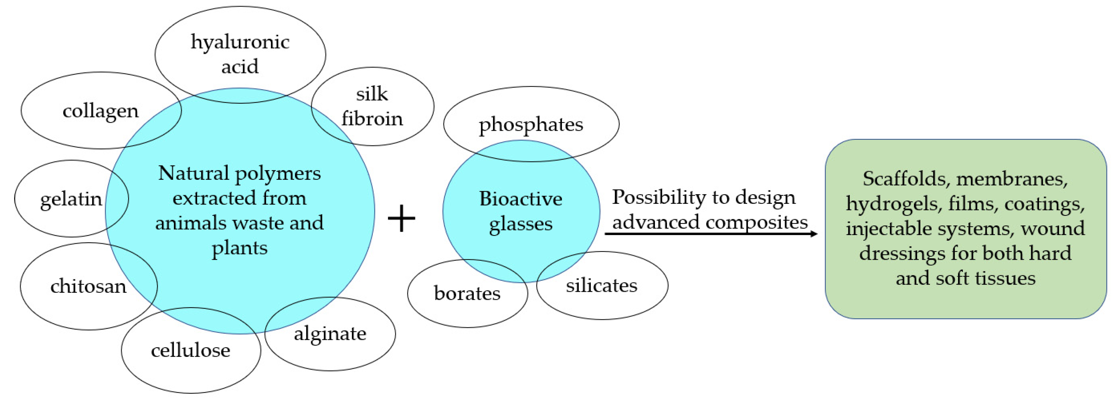
2. Natural Polymers: Proteins
2.1. Collagen/Bioactive Glass Composites
2.2. Gelatin/Bioactive Glass Composites
2.3. Silk Fibroin/Bioactive Glass Composites
3. Natural Polymers: Polysaccharides
3.1. Hyaluronic Acid/Bioactive Glass Composites
3.2. Chitosan/Bioactive Glass Composites
3.3. Alginate/Bioactive Glass Composites
3.4. Cellulose/Bioactive Glass Composites
4. Additional Remarks
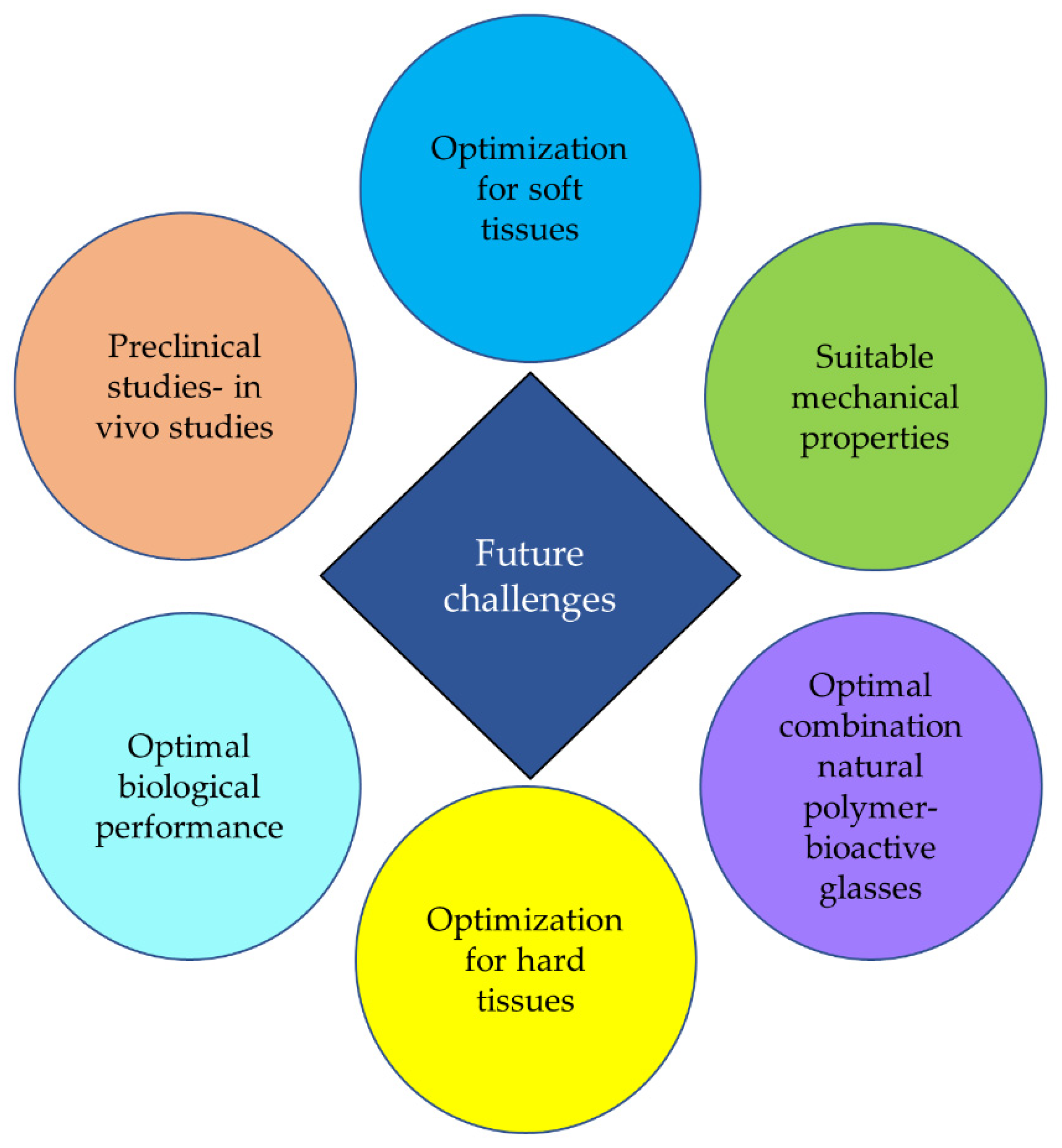
5. Conclusions and Future Challenges
Author Contributions
Funding
Conflicts of Interest
References
- Linden, M.; Ray, D. Life expectancy effects of public and private health expenditures in OECD countries 1970–2012: Panel time series approach. Econ. Anal. Policy 2017, 56, 101–113. [Google Scholar] [CrossRef] [Green Version]
- Vaupel, J.W. Biodemography of human ageing. Nature 2010, 464, 536–542. [Google Scholar] [CrossRef] [PubMed] [Green Version]
- Daware, M.A. Review Article Osteoporosis in Elderly. Vidarbha. J. Intern. Med. 2014, 17, 19–27. [Google Scholar]
- Lorentzon, M.; Cummings, S.R. Osteoporosis: The evolution of a diagnosis. J. Intern. Med. 2015, 277, 650–661. [Google Scholar] [CrossRef]
- Hench, L.L. Biomaterials: A forecast for the future. Biomaterials 1998, 19, 1419–1423. [Google Scholar] [CrossRef]
- Blackwood, D.J. Biomaterials: Past Successes and Future Problems. Corros. Rev. 2003, 21, 97–124. [Google Scholar] [CrossRef]
- Pirhonen, E. Fibres and Composites for Potential Biomaterials Applications. Ph.D. Thesis, Tampere University of Technology, Tampere, Finland, 2006; pp. 1–84. [Google Scholar]
- Hench, L.L.; Polak, J.M. Third-generation biomedical materials. Science 2002, 295, 1014–1017. [Google Scholar] [CrossRef] [Green Version]
- Griffith, L.G. Polymeric biomaterials. Acta Mater. 2000, 48, 263–277. [Google Scholar] [CrossRef]
- Boccaccini, A.; Gough, J. Tissue Engineering Using Ceramics and Polymers; Woodhead Publishing Limited: Cambridge, UK, 2007; pp. 1–587. [Google Scholar]
- Bhatia, S. Natural Polymer Drug Delivery Systems; Springer: Berlin/Heidelberg, Germany, 2016; pp. 1–225. [Google Scholar]
- Kumbar, S.; Laurencin, C.; Deng, M. Natural and Synthetic Biomedical Polymers; Elsevier: Berlin/Heidelberg, Germany, 2014; pp. 1–402. [Google Scholar]
- Sabu, T.; Neethu, N.; Sneha, M.; Elizabeth, F. Natural Polymers, Biopolymers, Biomaterials and Their Composites, Blends and IPNS; Apple Academic Press: Toronto, ON, Canada, 2012; pp. 1–407. [Google Scholar]
- Kulkarni, V.; Butte, K.; Rathod, S. Natural Polymers—A Comprehensive Review. Int. J. Res. Pharm. Biomed. Sci. 2012, 3, 1597–1613. [Google Scholar]
- Ratner, B.; Hoffman, A.; Schoen, F.; Lemons, J. Biomaterials Science an Introduction to Materials in Medicine, 2nd ed.; Elsevier: Amsterdam, The Netherlands, 2004; pp. 1–851. [Google Scholar]
- Odian, G. Principles of Polymerization, 4th ed.; Odian, G., Ed.; Wiley Interscience a John Wiley & Sons: Hoboken, NJ, USA, 2004; pp. 1–812. [Google Scholar]
- Maitz, M.F. Applications of synthetic polymers in clinical medicine. Biosurface Biotribology 2015, 1, 161–176. [Google Scholar] [CrossRef] [Green Version]
- Leite, A.; Mano, J. Biomedical applications of natural-based polymers combined with bioactive glass nanoparticles. J. Mater. Chem. B 2017, 5, 4555–4568. [Google Scholar] [CrossRef] [PubMed] [Green Version]
- Marelli, B.; Ghezzi, C.E.; Barralet, J.E.; Boccaccini, A.R.; Nazhat, S.N. Three-dimensional mineralization of dense nanofibrillar collagen-bioglass hybrid scaffolds. Biomacromolecules 2010, 11, 1470–1479. [Google Scholar] [CrossRef] [PubMed]
- Dziadek, M.; Stodolak-Zych, E.; Cholewa-Kowalska, K. Biodegradable ceramic-polymer composites for biomedical applications: A review. Mater. Sci. Eng. C 2017, 71, 1175–1191. [Google Scholar] [CrossRef]
- Mano, J.F.; Silva, G.A.; Azevedo, H.S.; Sousa, P.B.; Sousa, R.A.; Silva, S.S.; Boesel, L.F.; Oliveira, J.M.; Santos, T.C.; Marques, A.P.; et al. Natural origin biodegradable systems in tissue engineering and regenerative medicine: Present status and some moving trends. J. R. Soc. Interface 2007, 4, 999–1030. [Google Scholar] [CrossRef] [PubMed] [Green Version]
- Badylak, S.F. The extracellular matrix as a biologic scaffold material. Biomaterials 2007, 28, 3587–3593. [Google Scholar] [CrossRef]
- D’Ayala, G.G.; Malinconico, M.; Laurienzo, P. Marine derived polysaccharides for biomedical applications: Chemical modification approaches. Molecules 2008, 13, 2069–2106. [Google Scholar] [CrossRef] [Green Version]
- Klouda, L.; Mikos, A.G. Thermoresponsive hydrogels in biomedical applications—A review. Eur. J. Pharm. Biopharm. 2011, 68, 34–45. [Google Scholar] [CrossRef] [Green Version]
- Araújo, M.; Viveiros, R.; Philippart, A.; Miola, M.; Doumett, S.; Baldi, G.; Perez, J.; Boccaccini, A.R.; Aguiar-Ricardo, A.; Verné, E. Bioactivity, mechanical properties and drug delivery ability of bioactive glass-ceramic scaffolds coated with a natural-derived polymer. Mater. Sci. Eng. C 2017, 768, 342–351. [Google Scholar] [CrossRef]
- Harini, B.; Shadamarshan, R.P.K.; Rao, S.H.; Selvamurugan, N.; Balagangadharan, K. Natural and synthetic polymers/bioceramics/bioactive compounds-mediated cell signalling in bone tissue engineering. Int. J. Biol. Macromol. 2017, 110, 88–96. [Google Scholar]
- Stanić, V. Variation in Properties of Bioactive Glasses after Surface Modification; Springer International Publishing AG: Berlin/Heidelberg, Germany, 2017; pp. 1–467. [Google Scholar]
- Carvalho, S.M.; Moreira, C.D.F.; Oliveira, A.C.X.; Oliveira, A.A.R.; Lemos, E.M.F.; Pereira, M.M. Bioactive Glass Nanoparticles for Periodontal Regeneration and Applications in Dentistry Nanobiomaterials in Clinical Dentistry; Elsevier Inc.: Amsterdam, The Netherlands, 2019; pp. 351–383. [Google Scholar]
- Ali, S.; Farooq, I.; Iqbal, K. A review of the effect of various ions on the properties and the clinical applications of novel bioactive glasses in medicine and dentistry. Saudi Dent. J. 2014, 26, 1–5. [Google Scholar] [CrossRef] [Green Version]
- Profeta, A.C.; Prucher, G.M. Bioactive-glass in periodontal surgery and implant dentistry. Dent. Mater. J. 2015, 34, 559–571. [Google Scholar] [CrossRef] [PubMed] [Green Version]
- Montazerian, M.; Zanotto, E.D. A guided walk through Larry Hench’s monumental discoveries. J. Mater. Sci. 2017, 52, 8695–8732. [Google Scholar] [CrossRef]
- Kokubo, T. Apatite formation on surfaces of ceramics, metals and polymers in body environment. Acta Mater. 1998, 46, 2519–2527. [Google Scholar] [CrossRef]
- Bellows, C.G.; Aubin, J.E.; Heersche, J.N.M. Initiation and progression of mineralization of bone nodules formed in vitro: The role of alkaline phosphatase and organic phosphate. Bone Miner. 1991, 14, 27–40. [Google Scholar] [CrossRef]
- Hench, L.L. The story of Bioglass®. J. Mater. Sci. Mater. Med. 2006, 17, 967–978. [Google Scholar] [CrossRef]
- O’Donnell, M.D.; Hill, R.G.; Donnell, M.D.O. Influence of strontium and the importance of glass chemistry and structure when designing bioactive glasses for bone regeneration. Acta Biomater. 2010, 6, 2382–2385. [Google Scholar] [CrossRef]
- Bellucci, D.; Sola, A.; Salvatori, R.; Anesi, A.; Chiarini, L.; Cannillo, V. Role of magnesium oxide and strontium oxide as modifiers in silicate-based bioactive glasses: Effects on thermal behaviour, mechanical properties and in-vitro bioactivity. Mater. Sci. Eng. C 2017, 72, 566–575. [Google Scholar] [CrossRef]
- Aydin, H. Magnesium supplementation and bone. Magnes. Hum. Health Dis. 2013, 57, 149–157. [Google Scholar]
- Bellucci, D.; Cannillo, V. A novel bioactive glass containing strontium and magnesium with ultra-high crystallization temperature. Mater. Lett. 2018, 213, 67–70. [Google Scholar] [CrossRef]
- Bellucci, D.; Salvatori, R.; Anesi, A.; Chiarini, L.; Cannillo, V. SBF assays, direct and indirect cell culture tests to evaluate the biological performance of bioglasses and bioglass-based composites: Three paradigmatic cases. Mater. Sci. Eng. C 2019, 96, 757–764. [Google Scholar] [CrossRef]
- Bellucci, D.; Salvatori, R.; Giannatiempo, J.; Anesi, A.; Bortolini, S.; Cannillo, V. A New Bioactive Glass/Collagen Hybrid Composite for Applications in Dentistry. Materials 2019, 12, 2079. [Google Scholar] [CrossRef] [PubMed] [Green Version]
- Bellucci, D.; Veronesi, E.; Strusi, V.; Petrachi, T.; Murgia, A.; Mastrolia, I.; Dominici, M.; Cannillo, V. Human Mesenchymal Stem Cell Combined with a New Strontium-Enriched Bioactive Glass: An ex-vivo Model for Bone Regeneration. Materials 2019, 12, 3633. [Google Scholar] [CrossRef] [PubMed] [Green Version]
- Elsayed, H.; Romero, A.R.; Bellucci, D.; Cannillo, V.; Bernardo, E. Advanced open-celled structures from low-temperature sintering of a crystallization-resistant bioactive glass. Materials 2019, 12, 3653. [Google Scholar] [CrossRef] [PubMed] [Green Version]
- Bellucci, D.; Veronesi, E.; Dominici, M.; Cannillo, V. On the In vitro biocompatibility testing of bioactive glasses. Materials 2020, 13, 1816. [Google Scholar] [CrossRef] [PubMed] [Green Version]
- Sergi, R.; Cannillo, V.; Boccaccini, A.R.; Liverani, L. Incorporation of bioactive glasses containing Mg, Sr and Zn in electrospun PCL fibers by using benign solvents. Appl. Sci. 2020, 10, 5530. [Google Scholar] [CrossRef]
- Di Tinco, R.; Sergi, R.; Bertani, G.; Pisciotta, A.; Bellucci, D.; Carnevale, G.; Cannillo, V.; Bertoni, L. Effects of a novel bioactive glass composition on biological properties of human dental pulp stem cells. Materials 2020, 13, 4049. [Google Scholar] [CrossRef]
- Bellucci, D.; Veronesi, E.; Dominici, M.; Cannillo, V. A new bioactive glass with extremely high crystallization temperature and outstanding biological performance. Mater. Sci. Eng. C 2020, 110, 110699. [Google Scholar] [CrossRef]
- Sergi, R.; Bellucci, D.; Salvatori, R.; Anesi, A.; Cannillo, V. A novel bioactive glass containing therapeutic ions with enhanced biocompatibility. Materials 2020, 13, 4600. [Google Scholar] [CrossRef]
- Cacciotti, I. Bivalent cationic ions doped bioactive glasses: The influence of magnesium, zinc, strontium and copper on the physical and biological properties. J. Mater. Sci. 2017, 52, 8812–8831. [Google Scholar] [CrossRef]
- Hoppe, A.; Güldal, N.S.; Boccaccini, A.R. A review of the biological response to ionic dissolution products from bioactive glasses and glass-ceramics. Biomaterials 2011, 32, 2757–2774. [Google Scholar] [CrossRef]
- Balasubramanian, P.; Strobel, L.A.; Kneser, U.; Boccaccini, A.R. Zinc-containing bioactive glasses for bone regeneration, dental and orthopedic applications. Biomed. Glas. 2015, 1, 51–69. [Google Scholar] [CrossRef]
- Huang, M.; Hill, R.G.; Rawlinson, S.C.F. Zinc bioglasses regulate mineralization in human dental pulp stem cells. Dent. Mater. 2017, 33, 543–552. [Google Scholar] [CrossRef] [PubMed]
- Kim, Y. Antibacterial and remineralization effects of orthodontic bonding agents containing bioactive glass. Korean J. Orthod. 2018, 48, 163–171. [Google Scholar] [CrossRef] [PubMed]
- Bari, A.; Bloise, N.; Fiorilli, S.; Novajra, G.; Vallet-Regí, M.; Bruni, G.; Torres-Pardo, A.; González-Calbet, J.M.; Visai, L.; Vitale-Brovarone, C. Copper-containing mesoporous bioactive glass nanoparticles as multifunctional agent for bone regeneration. Acta Biomater. 2017, 55, 493–504. [Google Scholar] [CrossRef]
- Sergi, R.; Bellucci, D.; Salvatori, R.; Maisetta, G.; Batoni, G.; Cannillo, V. Zinc containing bioactive glasses with ultra-high crystallization temperature, good biological performance and antibacterial effects. Mater. Sci. Eng. C 2019, 104, 109910. [Google Scholar] [CrossRef]
- Nour, S.; Baheiraei, N.; Imani, R.; Rabiee, N.; Khodaei, M. Bioactive Materials: A Comprehensive Review on Interactions with Biological Microenvironment Based on the Immune Response. J. Bionic. Eng. 2019, 16, 563–581. [Google Scholar] [CrossRef]
- Sengupta, D.; Heilshorn, S.C. Protein-engineered biomaterials: Highly tunable tissue engineering scaffolds. Tissue Eng. Part B Rev. 2010, 16, 285–293. [Google Scholar] [CrossRef] [Green Version]
- Weiner, S.; Wagner, H.D. The material bone: Structure-mechanical function relations. Ann. Rev. Mater. Sci. 1998, 28, 271–298. [Google Scholar] [CrossRef]
- Zhang, D.; Wu, X.; Chen, J.; Lin, K. The development of collagen based composite scaffolds for bone regeneration. Bioact. Mater. 2018, 3, 129–138. [Google Scholar] [CrossRef]
- Ignatius, A.; Blessing, H.; Liedert, A.; Schmidt, C.; Neidlinger-Wilke, C.; Kaspar, D.; Friemert, B.; Claes, L. Tissue engineering of bone: Effects of mechanical strain on osteoblastic cells in type I collagen matrices. Biomaterials 2005, 26, 311–318. [Google Scholar] [CrossRef]
- Geiger, M.; Li, R.H.; Friess, W. Collagen sponges for bone regeneration with rhBMP-2. Adv. Drug Deliv. Rev. 2003, 55, 1613–1629. [Google Scholar] [CrossRef] [PubMed]
- Lutolf, M.P.; Weber, F.E.; Schmoekel, H.G.; Schense, J.C.; Kohler, T.; Müller, R.; Hubbell, J.A. Repair of bone defects using synthetic mimetics of collagenous extracellular matrices. Nat. Biotechnol. 2003, 21, 513–518. [Google Scholar] [CrossRef] [PubMed]
- Guilak, F.; Cohen, D.; Estes, B.; Gimble, J.; Liedtke, W.; Chen, C. Control of stem cell fate by physical interactions with the extracellular matrix. Bone 2012, 23, 17–26. [Google Scholar] [CrossRef] [PubMed] [Green Version]
- Prodanov, L.; te Riet, J.; Lamers, E.; Domanski, M.; Luttge, R.; van Loon, J.J.W.A.; Jansen, J.A.; Walboomers, X.F. The interaction between nanoscale surface features and mechanical loading and its effect on osteoblast-like cells behavior. Biomaterials 2010, 31, 7758–7765. [Google Scholar] [CrossRef]
- Sundararaghavan, H.G.; Monteiro, G.A.; Firestein, B.L.; Shreiber, D.I. Neurite growth in 3D collagen gels with gradients of mechanical properties. Biotechnol. Bioeng. 2009, 102, 632–643. [Google Scholar] [CrossRef]
- Ma, W.; Fitzgerald, W.; Liu, Q.Y.; O’Shaughnessy, T.J.; Maric, D.; Lin, H.J.; Alkon, D.L.; Barker, J.L. CNS stem and progenitor cell differentiation into functional neuronal circuits in three-dimensional collagen gels. Exp. Neurol. 2004, 190, 276–288. [Google Scholar] [CrossRef]
- Law, J.X.; Liau, L.L.; Saim, A.; Yang, Y.; Idrus, R. Electrospun Collagen Nanofibers and Their Applications in Skin Tissue Engineering. Tissue Eng. Regen. Med. 2017, 14, 699–718. [Google Scholar] [CrossRef]
- Sheehy, E.J.; Cunniffe, G.M.; O’Brien, F.J. Collagen-based biomaterials for tissue regeneration and repair. In Peptides and Proteins Biomaterials of Tissue Regeneration Repair; Elsevier: Duxford, UK, 2018; pp. 127–150. [Google Scholar]
- Matsuno, T.; Nakamura, T.; Kuremoto, K.; Notazawa, S. Development of β-tricalcium Phosphate/Collagen Sponge Composite for Bone Regeneration. Dent. Mater. J. 2006, 25, 38–144. [Google Scholar] [CrossRef] [Green Version]
- Zhang, Z.; Ma, Z.; Zhang, Y.; Chen, F.; Zhou, Y.; An, Q. Dehydrothermally crosslinked collagen/hydroxyapatite composite for enhanced in vivo bone repair. Colloids Surfaces B Biointerfaces 2018, 163, 394–401. [Google Scholar] [CrossRef]
- Powell, H.M.; Supp, D.M.; Boyce, S.T. Influence of electrospun collagen on wound contraction of engineered skin substitutes. Biomaterials 2008, 29, 834–843. [Google Scholar] [CrossRef]
- Chen, R.; Wang, G.; Chen, C.; Ho, H.; Sheu, M.-T. Development of N,O-(Carboxymethyl) chitosan/Collagen Matrixes as a Wound Dressing. Biomacromolecules 2006, 7, 1058–1064. [Google Scholar] [CrossRef] [PubMed]
- Chen, J.P.; Chang, G.Y.; Chen, J.K. Electrospun collagen/chitosan nanofibrous membrane as wound dressing. Colloids Surfaces A Physicochem. Eng. Asp. 2008, 313, 183–188. [Google Scholar] [CrossRef]
- Sarker, B.; Hum, J.; Nazhat, S.N.; Boccaccini, A.R. Combining collagen and bioactive glasses for bone tissue engineering: A review. Adv. Healthc. Mater. 2015, 4, 176–194. [Google Scholar] [CrossRef] [PubMed]
- Cancedda, R.; Giannoni, P.; Mastrogiacomo, M. A tissue engineering approach to bone repair in large animal models and in clinical practice. Biomaterials 2007, 28, 4240–4250. [Google Scholar] [CrossRef] [PubMed]
- Karageorgiou, V.; Kaplan, D. Porosity of 3D biomaterial scaffolds and osteogenesis. Biomaterials 2005, 26, 5474–5491. [Google Scholar] [CrossRef]
- Shekaran, A.; Garcia, A. Extracellular matrix-mimetic adhesive biomaterials for bone repair. J. Biomed. Mater. Res. A 2012, 96, 261–272. [Google Scholar] [CrossRef] [Green Version]
- Lanza, R.; Langer, R.; Vacanti, J. Principles of Tissue Engineering, 3rd ed.; Elsevier: Amsterdam, The Netherlands, 2007; pp. 1–1291. [Google Scholar]
- Long, T.; Yang, J.; Shi, S.S.; Guo, Y.P.; Ke, Q.F.; Zhu, Z.A. Fabrication of three-dimensional porous scaffold based on collagen fiber and bioglass for bone tissue engineering. J. Biomed. Mater. Res. Part B Appl. Biomater. 2015, 103, 1455–1464. [Google Scholar] [CrossRef]
- Hong, S.J.; Yu, H.S.; Noh, K.T.; Oh, S.A.; Kim, H.W. Novel scaffolds of collagen with bioactive nanofiller for the osteogenic stimulation of bone marrow stromal cells. J. Biomater. Appl. 2010, 24, 733–750. [Google Scholar] [CrossRef]
- Marelli, B.; Ghezzi, C.E.; Mohn, D.; Stark, W.J.; Barralet, J.E.; Boccaccini, A.R.; Nazhat, S.N. Accelerated mineralization of dense collagen-nano bioactive glass hybrid gels increases scaffold stiffness and regulates osteoblastic function. Biomaterials 2011, 32, 8915–8926. [Google Scholar] [CrossRef]
- Rezwan, K.; Chen, Q.Z.; Blaker, J.J.; Boccaccini, A.R. Biodegradable and bioactive porous polymer/inorganic composite scaffolds for bone tissue engineering. Biomaterials 2006, 27, 3413–3431. [Google Scholar] [CrossRef]
- Kim, H.-W.; Song, J.-H.; Kim, H.-E. Bioactive glass nanofiber–collagen nanocomposite as a novel bone regeneration matrix. J. Biomed. Mater. Res. 2006, 79, 698–705. [Google Scholar] [CrossRef] [PubMed]
- Hattar, S.; Loty, S.; Gaisser, D.; Berdal, A.; Sautier, J.M. Effects of 58s sol-gel glasses on the temporal expression of bone markers during mouse osteoblastic differentiation. J. Biomed. Mater. Res. Part A 2006, 76, 811–819. [Google Scholar] [CrossRef] [PubMed]
- Xynos, I.D.; Edgar, A.J.; Buttery, L.D.K.; Hench, L.L.; Polak, J.M. Ionic products of bioactive glass dissolution increase proliferation of human osteoblasts and induce insulin-like growth factor II mRNA expression and protein synthesis. Biochem. Biophys. Res. Commun. 2000, 276, 461–465. [Google Scholar] [CrossRef] [PubMed]
- O’Neill, E.; Awale, G.; Daneshmandi, L.; Umerah, O.; Lo, K.W.H. The roles of ions on bone regeneration. Drug Discov. Today 2018, 23, 879–890. [Google Scholar] [CrossRef] [PubMed]
- Dashnyam, K.; El-Fiqi, A.; Buitrago, J.O.; Perez, R.A.; Knowles, J.C.; Kim, H.W. A mini review focused on the proangiogenic role of silicate ions released from silicon-containing biomaterials. J. Tissue Eng. 2017, 8, 1–13. [Google Scholar] [CrossRef]
- Maeno, S.; Niki, Y.; Matsumoto, H.; Morioka, H.; Yatabe, T.; Funayama, A.; Toyama, Y.; Taguchi, T.; Tanaka, J. The effect of calcium ion concentration on osteoblast viability, proliferation and differentiation in monolayer and 3D culture. Biomaterials 2005, 26, 4847–4855. [Google Scholar] [CrossRef]
- Gao, X.; Wang, S.; Dong, Y.; Chen, X.; Gong, W. A novel nano-sized bioactive glass stimulates osteogenesis via the MAPK pathway. RSC Adv. 2017, 7, 13760–13767. [Google Scholar]
- Humeau, J.; Bravo-San Pedro, J.M.; Vitale, I.; Nuñez, L.; Villalobos, C.; Kroemer, G.; Senovilla, L. Calcium signaling and cell cycle: Progression or death. Cell Calcium 2018, 70, 3–15. [Google Scholar] [CrossRef]
- Allen-Durrance, A.E. A Quick Reference on Phosphorus. Vet. Clin. N. Am. Small Anim. Pract. 2017, 47, 257–262. [Google Scholar] [CrossRef]
- Julien, M.; Khoshniat, S.; Lacreusette, A.; Gatius, M.; Bozec, A.; Wagner, E.F.; Wittrant, Y.; Masson, M.; Weiss, P.; Beck, L.; et al. Phosphate-dependent regulation of MGP in osteoblasts: Role of ERK1/2 and Fra-1. J. Bone Miner. Res. 2009, 24, 1856–1868. [Google Scholar] [CrossRef]
- Wang, Y.; Xu, C.; Meng, Y.; Xiang, A.P.; Su, P.; Yu, W.; Xiang, A.P.; Wang, Y. Biocompatibility and osteogenesis of biomimetic Bioglass-Collagen-Phosphatidylserine composite scaffolds for bone tissue engineering. Biomaterials 2010, 32, 1051–1058. [Google Scholar]
- Freyman, T.M.; Yannas, I.V.; Gibson, L.J. Cellular materials as porous scaffolds for tissue engineering. Prog. Mater. Sci. 2001, 46, 273–282. [Google Scholar] [CrossRef]
- Naahidi, S.; Jafari, M.; Logan, M.; Wang, Y.; Yuan, Y.; Bae, H.; Dixon, B.; Chen, P. Biocompatibility of hydrogel-based scaffolds for tissue engineering applications. Biotechnol. Adv. 2017, 35, 530–544. [Google Scholar] [CrossRef] [PubMed]
- Jafari, M.; Paknejad, Z.; Rad, M.R.; Motamedian, S.R.; Eghbal, M.J.; Nadjmi, N.; Khojasteh, A. Polymeric scaffolds in tissue engineering: A literature review. J. Biomed. Mater. Res. Part B Appl. Biomater. 2017, 105, 431–459. [Google Scholar] [CrossRef]
- Eglin, D.; Maalheem, S.; Livage, J.; Coradin, T. In vitro apatite forming ability of type I collagen hydrogels containing bioactive glass and silica sol-gel particles. J. Mater. Sci. Mater. Med. 2006, 17, 161–167. [Google Scholar] [CrossRef]
- Vargas, G.; Durand, L.; Cadena, V.; Romero, M.; Mesones, R.; Mackovic, M.; Spallek, S.; Spiecker, E.; Boccaccini, A.R.; Gorustovich, A.A. Effect of nano-sized bioactive glass particles on the angiogenic properties of collagen based composites. J. Mater. Sci. Mater. Med. 2013, 24, 1261–1269. [Google Scholar] [CrossRef]
- El-fiqi, A.; Ho, J.; Lee, E.; Kim, H. Collagen hydrogels incorporated with surface-aminated mesoporous nanobioactive glass: Improvement of physicochemical stability and mechanical properties is effective for hard tissue engineering. Acta Biomater. 2013, 9, 9508–9521. [Google Scholar] [CrossRef]
- Heinemann, S.; Heinemann, C.; Jäger, M.; Neunzehn, J.; Wiesmann, H.P.; Hanke, T. Effect of silica and hydroxyapatite mineralization on the mechanical properties and the biocompatibility of nanocomposite collagen scaffolds. ACS Appl. Mater. Interfaces 2011, 3, 4323–4331. [Google Scholar] [CrossRef]
- Desimone, M.F.; Hélary, C.; Rietveld, I.B.; Bataille, I.; Mosser, G.; Giraud-Guille, M.M.; Livage, J.; Coradin, T. Silica-collagen bionanocomposites as three-dimensional scaffolds for fibroblast immobilization. Acta Biomater. 2010, 6, 3998–4004. [Google Scholar] [CrossRef]
- Zhou, T.; Sui, B.; Mo, X.; Sun, J. Multifunctional and biomimetic fish collagen/bioactive glass nanofibers: Fabrication, antibacterial activity and inducing skin regeneration In vitro and in vivo. Int. J. Nanomed. 2017, 12, 3495–3507. [Google Scholar] [CrossRef] [Green Version]
- Ayuk, S.M.; Houreld, N.N.; Abrahamse, H. Collagen Production in Diabetic Wounded Fibroblasts in Response to Low-Intensity Laser Irradiation at 660 nm. Diabetes Technol. Ther. 2012, 14, 1110–1117. [Google Scholar] [CrossRef] [PubMed]
- Constantin, V.D.; Carap, A.; Bobic, S.; Budu, V.; Albu Kaya, M.; Marin, Ş.; Marin, M.M.; Socea, B. Tissue Engineering—Collagen Sponge Dressing for Chronic Wounds. In Proceedings of the ICAMS 2018—7th International Conference on Advanced Materials and Systems, Bucharest, Romania, 18–20 October 2018; pp. 63–68. [Google Scholar]
- Gorustovich, A.A.; Roether, J.A.; Boccaccini, A.R. Effect of Bioactive Glasses on Angiogenesis: A Review of In vitro and In vivo Evidences. Tissue Eng. Part B Rev. 2010, 16, 199–207. [Google Scholar] [CrossRef] [PubMed]
- Li, H.; Chang, J. Bioactive silicate materials stimulate angiogenesis in fibroblast and endothelial cell co-culture system through paracrine effect. Acta Biomater. 2013, 9, 6981–6991. [Google Scholar] [CrossRef] [PubMed]
- Oates, M.; Chen, R.; Duncan, M.; Hunt, J.A. The angiogenic potential of three-dimensional open porous synthetic matrix materials. Biomaterials 2007, 28, 3679–3686. [Google Scholar] [CrossRef] [PubMed]
- Bose, S.; Tarafder, S.; Bandyopadhyay, A. Effect of Chemistry on Osteogenesis and Angiogenesis towards Bone Tissue Engineering Using 3D Printed Scaffolds. Ann. Biomed. Eng. 2017, 45, 261–272. [Google Scholar] [CrossRef] [Green Version]
- Engler, A.J.; Sen, S.; Sweeney, H.L.; Discher, D.E. Matrix Elasticity Directs Stem Cell Lineage Specification. Cell 2006, 126, 677–689. [Google Scholar] [CrossRef] [Green Version]
- Zandstra, P.W.; Discher, D.E.; Mooney, D.J. Growth Factors, Matrices, and Forces Combine and Control Stem Cells. Science 2009, 324, 1673–1677. [Google Scholar]
- Discher, D.E.; Janmey, P.; Wang, Y.L. Tissue cells feel and respond to the stiffness of their environment. Science 2005, 10, 1139–1143. [Google Scholar] [CrossRef] [Green Version]
- Zheng, J.; Zhao, F.; Zhang, W.; Mo, Y.; Zeng, L.; Li, X. Sequentially-crosslinked biomimetic bioactive glass/gelatin methacryloyl composites hydrogels for bone regeneration. Mater. Sci. Eng. C 2018, 89, 119–127. [Google Scholar] [CrossRef]
- Kazemi, M.; Azami, M.; Johari, B. Bone Regeneration in rat using a gelatin/bioactive glass nanocomposite scaffold along with endothelial cells (HUVECs). Int. J. Appl. Ceram. Technol. 2018, 15, 1427–1438. [Google Scholar] [CrossRef]
- Kargozar, S.; Jafar, S.; Soleimani, M.; Brouki, P. Acceleration of bone regeneration in bioactive glass/gelatin composite scaffolds seeded with bone marrow-derived mesenchymal stem cells over-expressing bone morphogenetic protein-7. Mater. Sci. Eng. C 2017, 75, 688–698. [Google Scholar] [CrossRef] [PubMed]
- Mozafari, M.; Moztarzadeh, F.; Rabiee, M.; Azami, M.; Nezafati, N.; Moztarzadeh, Z.; Tahriri, M. Development of 3D bioactive nanocomposite scaffolds made from gelatin and nano bioactive glass for biomedical applications. Adv. Compos. Lett. 2010, 19, 91–96. [Google Scholar] [CrossRef] [Green Version]
- Mozafari, M.; Moztarzadeh, F.; Rabiee, M.; Azami, M. Development of macroporous nanocomposite scaffolds of gelatin/bioactive glass prepared through layer solvent casting combined with lamination technique for bone tissue engineering. Ceram. Int. 2010, 36, 2431–2439. [Google Scholar] [CrossRef]
- Mozafari, M.; Rabiee, M.; Azami, M.; Maleknia, S. Biomimetic formation of apatite on the surface of porous gelatin/bioactive glass nanocomposite scaffolds. Appl. Surf. Sci. 2010, 257, 1740–1749. [Google Scholar] [CrossRef]
- Gupta, N.; Santhiya, D. In situ mineralization of bioactive glass in gelatin matrix. Mater. Lett. 2017, 188, 127–129. [Google Scholar] [CrossRef]
- Gao, C.; Gao, Q.; Li, Y.; Rahaman, M.N.; Teramoto, A.; Abe, K. In vitro Evaluation of Electrospun Gelatin-Bioactive Glass Hybrid Scaffolds for Bone Regeneration. J. Appl. Polym. Sci. 2013, 127, 2588–2599. [Google Scholar] [CrossRef]
- Lao, J.; Dieudonné, X.; Fayon, F.; Montouillout, V.; Jallot, E. Bioactive glass-gelatin hybrids: Building scaffolds with enhanced calcium incorporation and controlled porosity for bone regeneration. J. Mater. Chem. B 2016, 4, 2486–2497. [Google Scholar] [CrossRef]
- Nadeem, D.; Kiamehr, M.; Yang, X.; Su, B. Fabrication and In vitro evaluation of a sponge-like bioactive-glass/gelatin composite scaffold for bone tissue engineering. Mater. Sci. Eng. C 2013, 33, 2669–2678. [Google Scholar] [CrossRef]
- Gao, C.; Rahaman, M.N.; Gao, Q.; Teramoto, A.; Abe, K. Robotic deposition and In vitro characterization of 3D gelatin-bioactive glass hybrid scaffolds for biomedical applications. J. Biomed. Mater. Res. Part A 2013, 101, 2027–2037. [Google Scholar] [CrossRef]
- Lacroix, J.; Jallot, E.; Lao, J. Gelatin-bioactive glass composites scaffolds with controlled macroporosity. Chem. Eng. J. 2014, 256, 9–13. [Google Scholar] [CrossRef]
- Jalise, Z.S.; Bagheiraei, N.; Bagheri, F. The effects of strontium incorporation on a novel gelatin/bioactive glass bone graft: In vitro and in vivo characterization. Ceram. Int. 2018, 44, 14217–14227. [Google Scholar] [CrossRef]
- Raz, M.; Moztarzadeh, F.; Kordestani, S.S. Sol-gel Based Fabrication and Properties of Mg-Zn Doped Bioactive Glass/Gelatin Composite Scaffold for Bone Tissue Engineering. Silicon 2018, 10, 667–674. [Google Scholar] [CrossRef]
- Guo, W.; Zhao, F.; Wang, Y.; Tang, J.; Chen, X. Characterization of the mechanical behaviors and bioactivity of tetrapod ZnO whiskers reinforced bioactive glass/gelatin composite scaffolds. J. Mech. Behav. Biomed. Mater. 2017, 68, 8–15. [Google Scholar] [CrossRef] [PubMed]
- Yazdimamaghani, M.; Vashaee, D.; Assefa, S.; Walker, K.J.; Madihally, S.V.; Köhler, G.A.; Tayebi, L. Hybrid Macroporous Gelatin/Bioactive-Glass/Nanosilver Scaffolds with Controlled Degradation Behavior and Antimicrobial Activity for Bone Tissue Engineering. J. Biomed. Nanotechnol. 2014, 10, 911–931. [Google Scholar] [CrossRef] [PubMed]
- Zheng, K.; Wu, J.; Li, W.; Dippold, D.; Wan, Y.; Boccaccini, A.R. Incorporation of Cu-Containing Bioactive Glass Nanoparticles in Gelatin-Coated Sca ff olds Enhances Bioactivity and Osteogenic Activity. ACS Biomater. Sci. Eng. 2018, 4, 1546–1557. [Google Scholar]
- Kundu, B.; Rajkhowa, R.; Kundu, S.C.; Wang, X. Silk fibroin biomaterials for tissue regenerations. Adv. Drug Deliv. Rev. 2013, 65, 457–470. [Google Scholar] [CrossRef]
- Koh, L.-D.; Yu, H.-D.; Han, M.-Y.; Teng, C.-P.; Zhang, Y.-W.; Khin, Y.-W.; Loh, X.-J.; Tee, S.-Y.; Low, M.; Ye, E.; et al. Structures, mechanical properties and applications of silk fibroin materials. Prog. Polym. Sci. 2015, 46, 86–110. [Google Scholar] [CrossRef]
- Thurber, A.E.; Omenetto, F.G.; Kaplan, D.L. In vivo bioresponses to silk proteins. Biomaterials 2015, 71, 145–157. [Google Scholar] [CrossRef] [Green Version]
- Melke, J.; Midha, S.; Ghosh, S.; Ito, K.; Hofmann, S. Silk fibroin as biomaterial for bone tissue engineering. Acta Biomater. 2016, 31, 1–16. [Google Scholar] [CrossRef] [Green Version]
- Ma, D.; Wang, Y.; Dai, W. Silk fibroin-based biomaterials for musculoskeletal tissue engineering. Mater. Sci. Eng. C 2018, 89, 456–469. [Google Scholar] [CrossRef]
- Farokhi, M.; Mottaghitalab, F.; Fatahi, Y.; Khademhosseini, A.; Kaplan, D.L. Overview of Silk Fibroin Use in Wound Dressings. Trends Biotechnol. 2018, 36, 907–922. [Google Scholar] [CrossRef] [PubMed]
- Du, X.; Wei, D.; Huang, L.; Zhu, M.; Zhang, Y.; Zhu, Y. 3D printing of mesoporous bioactive glass/silk fibroin composite scaffolds for bone tissue engineering. Mater. Sci. Eng. C 2019, 103, 109731. [Google Scholar] [CrossRef] [PubMed]
- Wu, C.; Zhang, Y.; Zhou, Y.; Fan, W.; Xiao, Y. A comparative study of mesoporous glass/silk and non-mesoporous glass/silk scaffolds: Physiochemistry and in vivo osteogenesis. Acta Biomater. 2011, 7, 2229–2236. [Google Scholar] [CrossRef] [PubMed] [Green Version]
- Shen, X.; Yu, P.; Chen, H.; Wang, J.; Lu, B.; Cai, X. Icariin controlled release on a silk fibroin/mesoporous bioactive glass nanoparticles scaffold for promoting stem cell osteogenic differentiation. RSC Adv. 2020, 10, 12105–12112. [Google Scholar] [CrossRef]
- Moses, J.C.; Nandi, S.K.; Mandal, B.B. Multifunctional Cell Instructive Silk-Bioactive Glass Composite Reinforced Scaffolds Toward Osteoinductive, Proangiogenic, and Resorbable Bone Grafts. Adv. Healthc. Mater. 2018, 7, 1701418. [Google Scholar] [CrossRef]
- Joseph, M.C.; Reardon, P.J.T.; Konwarh, R.; Knowles, J.C.; Mandal, B.B. Mimicking Hierarchical Complexity of the Osteochondral Interface Using Electrospun Silk—Bioactive Glass Composites. ACS Appl. Mater. Interfaces 2017, 9, 8000–8013. [Google Scholar]
- Bidgoli, M.; Tamjid, E.; Khafaji, M.; Alemzadeh, I.; Vossoughi, M. Fabrication of hierarchically porous silk fibroin-bioactive glass composite scaffold via indirect 3D printing: Effect of particle size on physico- mechanical properties and in vitro cellular behavior. Mater. Sci. Eng. C 2019, 103, 109688. [Google Scholar] [CrossRef]
- Zhu, H.; Liu, N.; Feng, X.; Chen, J. Fabrication and characterization of silk fibroin/bioactive glass composite films. Mater. Sci. Eng. C 2012, 32, 822–829. [Google Scholar] [CrossRef]
- Rubert, M.; Alonso-Sande, M.; Monjo, M.; Ramis, J.M. Evaluation of alginate and hyaluronic acid for their use in bone tissue engineering. Biointerphases 2012, 7, 44. [Google Scholar] [CrossRef] [Green Version]
- Fallacara, A.; Baldini, E.; Manfredini, S.; Vertuani, S. Hyaluronic acid in the third millennium. Polymers 2018, 10, 701. [Google Scholar] [CrossRef] [Green Version]
- Boeriu, C.G.; Springer, J.; Kooy, F.K.; van den Broek, L.A.M.; Eggink, G. Production Methods for Hyaluronan. Int. J. Carbohydr. Chem. 2013, 624967. [Google Scholar] [CrossRef] [Green Version]
- Liu, H.; Li, H.; Cheng, W.; Yang, Y.; Zhu, M.; Zhou, C. Novel injectable calcium phosphate/chitosan composites for bone substitute materials. Acta Biomater. 2006, 2, 557–565. [Google Scholar] [CrossRef] [PubMed]
- Wang, X.; Chen, L.; Xiang, H.; Ye, J. Influence of Anti-Washout Agents on the Rheological Properties and Injectability of a Calcium Phosphate Cement. J. Biomed. Mater. Res. Part B Appl. Biomater. 2007, 81B, 410–418. [Google Scholar] [CrossRef] [PubMed]
- Wu, L.; Zhu, F.; Tao, G. In-Vitro Biocompatibility Evaluation of Collagen-Hyaluronic Acid/Bioactive Glass Nanocomposite Scaffold. J. Macromol. Sci. Part A 2013, 50, 1121–1125. [Google Scholar] [CrossRef]
- Zhou, Z.; He, S.; Ou, B.; Huang, T.; Zeng, W.; Liu, L.; Liu, Q.; Chen, J.; Zhao, Y.; Yang, Z.; et al. Influence of Nano-Bioactive Glass (NBG) content on properties of gelatin-hyaluronic acid/NBG composite scaffolds. J. Macromol. Sci. Part B Phys. 2014, 53, 1145–1155. [Google Scholar] [CrossRef]
- Lou, J.; Stowers, R.; Nam, S.; Xia, Y.; Chaudhuri, O. Stress relaxing hyaluronic acid-collagen hydrogels promote cell spreading, fiber remodeling, and focal adhesion formation in 3D cell culture. Biomaterials 2018, 154, 213–222. [Google Scholar] [CrossRef] [PubMed]
- Mohan, N.; Mohanan, P.V.; Sabareeswaran, A.; Nair, P. Chitosan-hyaluronic acid hydrogel for cartilage repair. Int. J. Biol. Macromol. 2017, 104, 1936–1945. [Google Scholar] [CrossRef]
- Almond, A.; Deangelis, P.L.; Blundell, C.D. Hyaluronan: The Local Solution Conformation Determined by NMR and Computer Modeling is Close to a Contracted Left-handed 4-Fold Helix. J. Mol. Biol. 2006, 358, 1256–1269. [Google Scholar] [CrossRef]
- Sohrabi, M.; Hesaraki, S.; Kazemzadeh, A.; Alizadeh, M. Development of injectable biocomposites from hyaluronic acid and bioactive glass nano-particles obtained from different sol-gel routes. Mater. Sci. Eng. C 2013, 33, 3730–3744. [Google Scholar] [CrossRef]
- Croisier, F.; Jérôme, C. Chitosan-based biomaterials for tissue engineering. Eur. Polym. J. 2013, 49, 780–792. [Google Scholar] [CrossRef] [Green Version]
- Bano, I.; Arshad, M.; Yasin, T.; Ghauri, M.A.; Younus, M. Chitosan: A potential biopolymer for wound management. Int. J. Biol. Macromol. 2017, 102, 380–383. [Google Scholar] [CrossRef] [PubMed]
- Levengood, S.K.L.; Zhang, M. Chitosan-based scaffolds for bone tissue engineering. J. Mater. Chem. B 2014, 2, 3161. [Google Scholar] [CrossRef] [PubMed]
- Nandi, S.K.; Kundu, B.; Basu, D. Protein growth factors loaded highly porous chitosan scaffold: A comparison of bone healing properties. Mater. Sci. Eng. C 2013, 33, 1267–1275. [Google Scholar] [CrossRef] [PubMed]
- Chesnutt, B.; Yuan, Y.; Buddington, K.; Haggard, W.; Bumgardner, J. Composite Chitosan = Nano-Hydroxyapatite Scaffolds Induce Osteocalcin Production by Osteoblasts In vitro and Support Bone Formation In vivo. Tissue Eng. A 2009, 15, 2571. [Google Scholar] [CrossRef] [PubMed]
- DiMartino, A.; Sittinger, M.; Risbud, M.V. Chitosan: A versatile biopolymer for orthopaedic tissue-engineering. Biomaterials 2005, 26, 5983–5990. [Google Scholar] [CrossRef]
- Chen, Y.; Chung, Y.; Wang, L.W.; Chen, K.; Chung, Y.; Wang, L.W.; Chen, K.-T.; Li, S.-Y. Antibacterial properties of chitosan in waterbone pathogen. J. Environ. Sci. Heal. Part A Toxic/Hazard. Subst. Environ. Eng. 2007, 4529, 37–41. [Google Scholar]
- Hu, S.G.; Jou, C.H.; Yang, M.C. Protein adsorption, fibroblast activity and antibacterial properties of poly(3-hydroxybutyric acid-co-3-hydroxyvaleric acid) grafted with chitosan and chitooligosaccharide after immobilized with hyaluronic acid. Biomaterials 2003, 24, 2685–2693. [Google Scholar] [CrossRef]
- Vaz, J.M.; Taketa, T.B.; Hernandez-Montelongo, J.; Chevallier, P.; Cotta, M.A.; Mantovani, D.; Beppu, M.M. Antibacterial properties of chitosan-based coatings are affected by spacer-length and molecular weight. Appl. Surf. Sci. 2018, 445, 478–487. [Google Scholar] [CrossRef]
- Dotto, G.L.; Campana-Filho, S.P.; Pinto, L.A.A. Chitosan Based Materials and its Applications; Bentham Science Publishers: Sharjah, UAE, 2017; pp. 1–322. [Google Scholar]
- Albanna, M.Z.; Bou-akl, T.H.; Blowytsky, O.; Walters, H.L.; Matthew, H.W.T. Chitosan fibers with improved biological and mechanical properties for tissue engineering applications. J. Mech. Behav. Biomed. Mater. 2013, 20, 217–226. [Google Scholar] [CrossRef]
- Lee, K.; Jin, G.; Jang, C.H.; Jung, W.; Kim, G. Preparation and characterization of multi-layered poly(3-caprolactone)/chitosan scaffolds fabricated with a combination of melt-plotting/in situ plasma treatment and a coating method for hard tissue regeneration. J. Mater. Chem. B 2013, 1, 5831. [Google Scholar] [CrossRef]
- Caridade, S.G.; Merino, E.G.; Alves, N.M.; de Bermudez, V.Z.; Boccaccini, A.R.; Mano, J.F. Chitosan membranes containing micro or nano-size bioactive glass particles: Evolution of biomineralization followed by in situ dynamic mechanical analysis. J. Mech. Behav. Biomed. Mater. 2013, 20, 173–183. [Google Scholar] [CrossRef] [PubMed] [Green Version]
- Oliveira, M.B.; Luz, G.M.; Mano, J.F. A combinatorial study of nanocomposite hydrogels: On-chip mechanical/viscoelastic and pre-osteoblast interaction characterization. J. Mater. Chem. B 2014, 2, 5627–5638. [Google Scholar] [CrossRef] [PubMed]
- Couto, D.S.; Hong, Z.; Mano, J.F. Development of bioactive and biodegradable chitosan-based injectable systems containing bioactive glass nanoparticles. Acta Biomater. 2009, 5, 115–123. [Google Scholar] [CrossRef] [PubMed]
- Yang, J.; Long, T.; He, N.-F.; Guo, Y.-P.; Zhu, Z.-A.; Ke, Q.-F. Fabrication of a chitosan/bioglass three-dimensional porous scaffold for bone tissue engineering applications. J. Mater. Chem. B 2014, 2, 6611–6618. [Google Scholar] [CrossRef] [PubMed]
- Cui, X.; Huang, W.; Zhou, J.; Wang, H.; Zhou, N.; Wang, D.; Pan, H.; Rahaman, M.N. Evaluation of an injectable bioactive borate glass cement to heal bone defects in a rabbit femoral condyle model. Mater. Sci. Eng. C 2017, 73, 585–595. [Google Scholar] [CrossRef]
- Correia, C.O.; Leite, Á.J.; Mano, J.F. Chitosan/bioactive glass nanoparticles scaffolds with shape memory properties. Carbohydr. Polym. 2015, 123, 39–45. [Google Scholar] [CrossRef] [PubMed]
- Mota, J.; Yu, N.; Caridade, S.G.; Luz, G.M.; Gomes, M.E.; Reis, R.L.; Jansen, J.A.; Walboomers, X.F.; Mano, J.F. Chitosan/bioactive glass nanoparticle composite membranes for periodontal regeneration. Acta Biomater. 2012, 8, 4173–4180. [Google Scholar] [CrossRef] [PubMed] [Green Version]
- Xianmiao, C.; Yubao, L.; Yi, Z.; Li, Z.; Jidong, L.; Huanan, W. Properties and In vitro biological evaluation of nano-hydroxyapatite/chitosan membranes for bone guided regeneration. Mater. Sci. Eng. C 2009, 29, 29–35. [Google Scholar] [CrossRef]
- Bottino, M.C.; Thomas, V.; Janowski, G.M. A novel spatially designed and functionally graded electrospun membrane for periodontal regeneration. Acta Biomater. 2011, 7, 216–224. [Google Scholar] [CrossRef]
- Dashnyam, K.; Perez, R.A.; Singh, R.K.; Lee, E.J.; Kim, H.W. Hybrid magnetic scaffolds of gelatin-siloxane incorporated with magnetite nanoparticles effective for bone tissue engineering. RSC Adv. 2014, 4, 40841–40851. [Google Scholar] [CrossRef]
- Nardecchia, S.; Carriazo, D.; Ferrer, M.L.; Gutiérrez, M.C.; Del Monte, F. Three dimensional macroporous architectures and aerogels built of carbon nanotubes and/or graphene: Synthesis and applications. Chem. Soc. Rev. 2013, 42, 794–830. [Google Scholar] [CrossRef] [PubMed]
- Turnbull, G.; Clarke, J.; Picard, F.; Riches, P.; Jia, L.; Han, F.; Li, B.; Shu, W. 3D bioactive composite scaffolds for bone tissue engineering. Bioact. Mater. 2018, 3, 278–314. [Google Scholar] [CrossRef] [PubMed] [Green Version]
- Murphy, C.M.; Haugh, M.G.; O’Brien, F.J. The effect of mean pore size on cell attachment, proliferation and migration in collagen-glycosaminoglycan scaffolds for bone tissue engineering. Biomaterials 2010, 31, 461–466. [Google Scholar] [CrossRef] [PubMed]
- Gupte, M.J.; Swanson, W.B.; Hu, J.; Jin, X.; Ma, H.; Zhang, Z.; Liu, Z.; Feng, K.; Feng, G.; Xiao, G.; et al. Pore size directs bone marrow stromal cell fate and tissue regeneration in nanofibrous macroporous scaffolds by mediating vascularization. Acta Biomater. 2018, 82, 1–11. [Google Scholar] [CrossRef] [PubMed]
- Valerio, P.; Pereira, M.M.; Goes, A.M.; Leite, M.F. The effect of ionic products from bioactive glass dissolution on osteoblast proliferation and collagen production. Biomaterials 2004, 25, 2941–2948. [Google Scholar] [CrossRef] [PubMed]
- Reffitt, D.M.; Ogston, N.; Jugdaohsingh, R.; Cheung, H.F.J.; Evans, B.A.J.; Thompson, R.P.H.; Powell, J.J.; Hampson, G.N. Orthosilicic acid stimulates collagen type 1 synthesis and osteoblastic differentiation in human osteoblast-like cells in vitro. Bone 2003, 32, 127–135. [Google Scholar] [CrossRef]
- Orrenius, S.; Gogvadze, V.; Zhivotovsky, B. Calcium and mitochondria in the regulation of cell death. Biochem. Biophys. Res. Commun. 2015, 460, 72–81. [Google Scholar] [CrossRef]
- Kohn, A.D.; Moon, R.T. Wnt and calcium signaling: β-Catenin-independent pathways. Cell Calcium 2005, 38, 439–446. [Google Scholar] [CrossRef]
- Clapham, D. Calcium signaling. Cell 2007, 131, 1047–1058. [Google Scholar] [CrossRef] [Green Version]
- Kühl, M. The Wnt/Calcium Pathway. Front. Biosci. 2004, 9, 967–974. [Google Scholar] [CrossRef] [Green Version]
- Khoshakhlagh, P.; Rabiee, S.M.; Kiaee, G.; Heidari, P.; Miri, A.K.; Moradi, R.; Moztarzadeh, F.; Ravarian, R. Development and characterization of a bioglass/chitosan composite as an injectable bone substitute. Carbohydr. Polym. 2017, 157, 1261–1271. [Google Scholar] [CrossRef] [PubMed]
- Silver, I.A.; Deas, J.; Erecińska, M. Interactions of bioactive glasses with osteoblasts in vitro: Effects of 45S5 Bioglass(®), and 58S and 77S bioactive glasses on metabolism, intracellular ion concentrations and cell viability. Biomaterials 2001, 22, 175–185. [Google Scholar] [CrossRef]
- Bonnelye, E.; Chabadel, A.; Saltel, F.; Jurdic, P. Dual effect of strontium ranelate: Stimulation of osteoblast differentiation and inhibition of osteoclast formation and resorption in vitro. Bone 2008, 42, 129–138. [Google Scholar] [CrossRef]
- Chattopadhyay, N.; Quinn, S.J.; Kifor, O.; Ye, C.; Brown, E.M. The calcium-sensing receptor (CaR) is involved in strontium ranelate-induced osteoblast proliferation. Biochem. Pharmacol. 2007, 74, 438–447. [Google Scholar] [CrossRef] [PubMed]
- Hurtel-lemaire, A.S.; Mentaverri, R.; Caudrillier, A.; Cournarie, F.; Wattel, A.; Kamel, S.; Terwilliger, E.F.; Brown, E.M.; Brazier, M. The Calcium-sensing Receptor Is Involved in Strontium Ranelate-induced Osteoclast Apoptosis. J. Biol. Chem. 2009, 284, 575–584. [Google Scholar] [CrossRef] [Green Version]
- Isaac, J.; Nohra, J.; Lao, J.; Jallot, E.; Nedelec, J.M.; Berdal, A.; Sautier, J.-M. Effects of strontium-doped bioactive glass on the differentiation of cultured osteogenic cells. Eur. Cells Mater. 2011, 21, 130–143. [Google Scholar] [CrossRef]
- Zhang, W.; Shen, Y.; Pan, H.; Lin, K.; Liu, X.; Darvell, B.W.; Lu, W.W.; Chang, J.; Deng, L.; Wang, D.; et al. Effects of strontium in modified biomaterials. Acta Biomater. 2011, 7, 800–808. [Google Scholar] [CrossRef]
- Naruphontjirakul, P.; Porter, A.E.; Jones, J.R. In vitro osteogenesis by intracellular uptake of strontium containing bioactive glass nanoparticles. Acta Biomater. 2018, 66, 67–80. [Google Scholar] [CrossRef]
- Yang, F.; Yang, D.; Tu, J.; Zheng, Q.; Cai, L.; Wang, L. Strontium enhances osteogenic differentiation of mesenchymal stem cells and in vivo bone formation by activating Wnt/catenin signaling. Stem Cells 2011, 29, 981–991. [Google Scholar] [CrossRef]
- Zhang, Y.; Cui, X.; Zhao, S.; Wang, H.; Rahaman, M.N.; Liu, Z.; Huang, W.; Zhang, C. Evaluation of injectable strontium-containing borate bioactive glass cement with enhanced osteogenic capacity in a critical-sized rabbit femoral condyle defect model. ACS Appl. Mater. Interfaces 2015, 7, 2393–2403. [Google Scholar] [CrossRef]
- Lin, K.; Xia, L.; Li, H.; Jiang, X.; Pan, H.; Xu, Y.; Lu, W.W.; Zhang, Z.; Chang, J. Enhanced osteoporotic bone regeneration by strontium-substituted calcium silicate bioactive ceramics. Biomaterials 2013, 34, 10028–10042. [Google Scholar] [CrossRef] [PubMed]
- Luz, G.M.; Mano, J.F. Chitosan/bioactive glass nanoparticles composites for biomedical applications. Biomed. Mater. 2012, 7, 054104. [Google Scholar] [CrossRef] [PubMed]
- Diba, M.; Goudouri, O.M.; Tapia, F.; Boccaccini, A.R. Magnesium-containing bioactive polycrystalline silicate-based ceramics and glass-ceramics for biomedical applications. Curr. Opin. Solid State Mater. Sci. 2014, 18, 147–167. [Google Scholar] [CrossRef]
- Zreiqat, H.; Howlett, C.R.; Zannettino, A.; Evans, P.; Schulze-Tanzil, G.; Knabe, C.; Shakibaei, M. Mechanisms of magnesium-stimulated adhesion of osteoblastic cells to commonly used orthopaedic implants. J. Biomed. Mater. Res. 2002, 62, 175–184. [Google Scholar] [CrossRef]
- Sergi, R.; Bellucci, D.; Salvatori, R.; Cannillo, V. Chitosan—Based Bioactive Glass Gauze: Microstructural Properties, In vitro Bioactivity, and Biological Tests. Materials 2020, 13, 2819. [Google Scholar] [CrossRef]
- Luz, G.M.; Boesel, L.; Campo, A.; Del Mano, J.F. Micropatterning of bioactive glass nanoparticles on chitosan membranes for spatial controlled biomineralization. Langmuir 2012, 28, 6970–6977. [Google Scholar] [CrossRef] [Green Version]
- Peter, M.; Binulal, N.S.; Soumya, S.; Nair, S.V.; Furuike, T.; Tamura, H.; Jayakumar, R. Nanocomposite scaffolds of bioactive glass ceramic nanoparticles disseminated chitosan matrix for tissue engineering applications. Carbohydr. Polym. 2010, 79, 284–289. [Google Scholar] [CrossRef]
- Martins, T.; Oliveira, A.A.R.; Oliveira, A.C.; Boaventura, T.P.; Barrioni, B.R.; Costa-Júnior, E.S.; Pereira, M.M. Novel 3D composites with highly flexible behavior based on chitosan and bioactive glass for biomedical applications. Mater. Chem. Phys. 2017, 189, 1–11. [Google Scholar] [CrossRef]
- Rinaudo, M. Biomaterials based on a natural polysaccharide: Alginate. © TIP Rev. Espec en Ciencias Químico-Biológicas 2014, 17, 92–96. [Google Scholar]
- Venkatesan, J.; Bhatnagar, I.; Manivasagan, P.; Kang, K.H.; Kim, S.K. Alginate composites for bone tissue engineering: A review. Int. J. Biol. Macromol. 2015, 72, 269–281. [Google Scholar] [CrossRef]
- Rehm, B.H.A.; Moradali, M.F. Alginates and Their Biomedical Applications; Springer: Berlin/Heidelberg, Germany, 2018; Volume 11, pp. 1–268. [Google Scholar]
- Pawar, S.N.; Edgar, K.J. Alginate derivatization: A review of chemistry, properties and applications. Biomaterials 2012, 33, 3279–3305. [Google Scholar] [CrossRef] [PubMed]
- Lee, K.Y.; Mooney, D.J. Alginate: Properties and biomedical applications. Prog. Polym. Sci. 2012, 37, 106–126. [Google Scholar] [CrossRef] [PubMed] [Green Version]
- Shapiro, L.; Cohen, S. Novel alginate sponges for cell culture and transplantation. Biomaterials 1997, 18, 583–590. [Google Scholar] [CrossRef]
- Turco, G.; Marsich, E.; Bellomo, F.; Semeraro, S.; Donati, I.; Brun, F.; Grandolfo, M.; Accardo, A.; Paoletti, S. Alginate/Hydroxyapatite Biocomposite for Bone Ingrowth: A Trabecular Structure with High and Isotropic Connectivity Gianluca. Biomacromolecules 2009, 10, 1575–1583. [Google Scholar] [CrossRef] [PubMed]
- Gabbai-Armelin, P.R.; Cardoso, D.A.; Zanotto, E.D.; Peitl, O.; Leeuwenburgh, S.C.G.; Jansen, J.A.; Renno, A.C.M.; van den Beucken, J.J.J.P. Injectable composites based on biosilicate® and alginate: Handling and In vitro characterization. RSC Adv. 2014, 4, 45778–45785. [Google Scholar] [CrossRef]
- Park, D.; Choi, B.; Zhu, S.; Huh, J.; Kim, B.; Lee, S. Injectable bone using chitosan-alginate gel/mesenchymal stem cells/BMP-2 composites. J. Cranio Maxillofac. Surg. 2005, 33, 50–54. [Google Scholar] [CrossRef]
- Iwasaki, N. Development of cartilage tissue engineering techniques based on biomedical research. Jpn. Orthop. Assoc. 2014, 19, 699–706. [Google Scholar] [CrossRef] [Green Version]
- Igarashi, T.; Iwasaki, N.; Kasahara, Y.; Minami, A. A cellular implantation system using an injectable ultra-purified alginate gel for repair of osteochondral defects in a rabbit model. J. Biomed. Mater. Res. Part A 2010, 94, 844–855. [Google Scholar] [CrossRef]
- De Long, W.G.; Einhorn, T.A.; Koval, K.; Mckee, M.; Smith, W.; Sanders, R.; Watson, T. Bone Grafts and Bone Graft Substitutes in Orthopaedic Trauma Surgery A Critical Analysis. J. Bone Jt. Surg. 2007, 89, 649–658. [Google Scholar] [CrossRef]
- Zeng, Q.; Han, Y.; Li, H.; Chang, J. Bioglass/alginate composite hydrogel beads as cell carriers for bone regeneration. J. Biomed. Mater. Res. Part B Appl. Biomater. 2014, 102, 42–51. [Google Scholar] [CrossRef]
- Jell, G.; Notingher, I.; Tsigkou, O.; Notingher, P.; Polak, J.M.; Hench, L.L.; Stevens, M.M. Bioactive glass-induced osteoblast differentiation: A noninvasive spectroscopic study. J. Biomed. Mater. Res. 2008, 86A, 31–40. [Google Scholar] [CrossRef] [PubMed]
- Zhao, F.; Zhang, W.; Fu, X.; Xie, W.; Chen, X. Fabrication and characterization of bioactive glass/alginate composite scaffolds by a self-crosslinking processing for bone regeneration. RSC Adv. 2016, 6, 91201–91208. [Google Scholar] [CrossRef]
- Srinivasan, S.; Jayasree, R.; Chennazhi, K.P.; Nair, S.V.; Jayakumar, R. Biocompatible alginate/nano bioactive glass ceramic composite scaffolds for periodontal tissue regeneration. Carbohydr. Polym. 2012, 87, 274–283. [Google Scholar] [CrossRef]
- Luo, Y.; Wu, C.; Lode, A.; Gelinsky, M. Hierarchical mesoporous bioactive glass/alginate composite scaffolds fabricated by three-dimensional plotting for bone tissue engineering. Biofabrication 2013, 5, 015005. [Google Scholar] [CrossRef] [PubMed]
- Luo, G.; Ma, Y.; Cui, X.; Jiang, L.; Wu, M.; Hu, Y.; Luo, Y.; Pan, H.; Ruan, C. 13–93 bioactive glass/alginate composite scaffolds 3D printed under mild conditions for bone regeneration. RSC Adv. 2017, 7, 11880–11889. [Google Scholar] [CrossRef] [Green Version]
- Wu, C.; Ramaswamy, Y.; Zhu, Y.; Zheng, R.; Appleyard, R.; Howard, A.; Zreiqat, H. The effect of mesoporous bioactive glass on the physiochemical, biological and drug-release properties of poly(dl-lactide-co-glycolide) films. Biomaterials 2009, 30, 2199–2208. [Google Scholar] [CrossRef]
- Liu, W.; Wang, T.; Yang, C.; Darvell, B.W.; Wu, J.; Lin, K.; Chang, J.; Pan, H.; Lu, W.W. Alkaline biodegradable implants for osteoporotic bone defects—Importance of microenvironment pH. Osteoporos. Int. 2015, 27, 93–104. [Google Scholar] [CrossRef]
- Dasgupta, S.; Banerjee, S.S.; Bandyopadhyay, A.; Bose, S. Zn- and Mg-Doped Hydroxyapatite Nanoparticles for Controlled Release of Protein. Langmuir 2010, 26, 4958–4964. [Google Scholar] [CrossRef] [Green Version]
- Badr-Mohammadi, M.-R.; Hesaraki, S.; Zamanian, A. Mechanical properties and In vitro cellular behavior of zinc- containing nano-bioactive glass doped biphasic calcium phosphate bone substitutes. J. Mater. Sci. Mater. Med. 2014, 25, 185–197. [Google Scholar] [CrossRef]
- Zamani, D.; Moztarzadeh, F.; Bizari, D. Alginate-bioactive glass containing Zn and Mg composite scaffolds for bone tissue engineering. Int. J. Biol. Macromol. 2019, 137, 1256–1267. [Google Scholar] [CrossRef]
- Loh, Q.L.; Choong, C. Three-dimensional scaffolds for tissue engineering applications: Role of porosity and pore size. Tissue Eng. Part B Rev. 2013, 19, 485–502. [Google Scholar] [CrossRef] [PubMed] [Green Version]
- Jones, J.R. Reprint of: Review of bioactive glass: From Hench to hybrids. Acta Biomater. 2015, 23, S53–S82. [Google Scholar] [CrossRef] [PubMed]
- Liu, J.; Rawlinson, S.C.F.; Hill, R.G.; Fortune, F. Strontium-substituted bioactive glasses in vitro osteogenic and antibacterial effects. Dent. Mater. 2016, 32, 412–422. [Google Scholar] [CrossRef] [PubMed]
- Reginster, J.; Badurski, J.; Bellamy, N.; Bensen, W.; Chapurlat, R.; Chevalier, X.; Christiansen Genant, H.; Navarro, F.; Nasonov, E.; Sambrook, P.N.; et al. Efficacy and safety of strontium ranelate in the treatment of knee osteoarthritis: Results of a double-blind, randomised placebo-controlled trial. Ann. Rheum. Dis. 2013, 72, 179–186. [Google Scholar] [CrossRef]
- Tao, Z.; Zhou, W.; He, X.; Liu, W.; Bai, B.; Zhou, Q.; Huang, Z.-L.; Tu, K.-K.; Li, H.; Sun, T.; et al. comparative study of zinc, magnesium, strontium-incorporated hydroxyapatite-coated titanium implants for osseointegration of osteopenic rats. Mater. Sci. Eng. C 2016, 62, 226–232. [Google Scholar] [CrossRef]
- Haider, A.; Waseem, A.; Karpukhina, N.; Mohsin, S. Strontium-and zinc-containing bioactive glass and alginates scaffolds. Bioengineering 2020, 7, 10. [Google Scholar] [CrossRef] [Green Version]
- Hatton, J.; Davis, G.R.; Mourad, A.H.I.; Cherupurakal, N.; Hill, R.G.; Mohsin, S. Fabrication of porous bone scaffolds using alginate and bioactive glass. J. Funct. Biomater. 2019, 10, 15. [Google Scholar] [CrossRef] [Green Version]
- Wu, C.; Chang, J. Degradation, Bioactivity, and Cytocompatibility of Diopside, Akermanite, and Bredigite Ceramics. J. Biomed. Mater. Res. Part B Appl. Biomater. 2007, 81B, 66–75. [Google Scholar] [CrossRef]
- Li, M.; Mondrinos, M.J.; Chen, X.; Gandhi, M.R.; Ko, F.K.; Lelkes, P.I. Elastin Blends for Tissue Engineering Scaffolds. J. Biomed. Mater. Res. Part A 2006, 79, 963–973. [Google Scholar] [CrossRef]
- Chakkalakal, D.A.; Mashoof, A.A.; Novak, J.; Strates, B.S.; Mcguire, M.H. Mineralization and pH relationships in healing skeletal defects grafted with demineralized bone matrix. J. Biomed. Mater. Res. 1994, 28, 1439–1443. [Google Scholar] [CrossRef]
- Kumar, S. Biocompatible β-chitin Hydrogel/Nanobioactive Glass Ceramic Nanocomposite Scaffolds for Periodontal Bone Regeneration. Trends Biomater. Artif. Organs 2011, 25, 1–11. [Google Scholar]
- Thamaria, N. Synthesis and characterization of alginate scaffolds containing bioactive glass for bone tissue engineering applications. In Proceedings of the 2017 24th National and 2nd International Iranian Conference on Biomedical Engineering, Tehran, Iran, 30 November–1 December 2017; p. 278. [Google Scholar]
- Liu, M.; Dai, L.; Shi, H.; Xiong, S.; Zhou, C. In vitro evaluation of alginate/halloysite nanotube composite scaffolds for tissue engineering. Mater. Sci. Eng. C 2015, 49, 700–712. [Google Scholar] [CrossRef] [PubMed]
- Yan, J.; Miao, Y.; Tan, H.; Zhou, T.; Ling, Z.; Chen, Y.; Xing, X.; Hu, X. Injectable alginate/hydroxyapatite gel scaffold combined with gelatin microspheres for drug delivery and bone tissue engineering. Mater. Sci. Eng. C 2016, 63, 274–284. [Google Scholar] [CrossRef] [PubMed]
- Zmora, S.; Glicklis, R.; Cohen, S. Tailoring the pore architecture in 3-D alginate scaffolds by controlling the freezing regime during fabrication. Biomaterials 2002, 23, 4087–4094. [Google Scholar] [CrossRef]
- Mouriño, V.; Newby, P.; Boccaccini, A.R. Preparation and characterization of gallium releasing 3-d alginate coated 45s5 bioglass® based scaffolds for bone tissue engineering. Adv. Eng. Mater. 2010, 12, 283–291. [Google Scholar] [CrossRef]
- Clarkin, O.M.; Wu, B.; Cahill, P.A.; Brougham, D.F.; Banerjee, D.; Brady, S.A.; Fox, E.K.; Lally, C. Novel injectable gallium-based self-setting glass-alginate hydrogel composite for cardiovascular tissue engineering. Carbohydr. Polym. 2019, 217, 152–159. [Google Scholar] [CrossRef]
- Brady, S.A.; Fox, E.K.; Lally, C.; Clarkin, O.M. Optimisation of a novel glass-alginate hydrogel for the treatment of intracranial aneurysms. Carbohydr. Polym. 2017, 176, 227–235. [Google Scholar] [CrossRef] [Green Version]
- Bériault, R.; Hamel, R.; Chenier, D.; Mailloux, R.J.; Joly, H.; Appanna, V.D. The overexpression of NADPH-producing enzymes counters the oxidative stress evoked by gallium, an iron mimetic. BioMetals 2007, 20, 165–176. [Google Scholar] [CrossRef]
- Mourino, V.; Newby, F.; Cattalini, J.; Lucangioli, S.; Boccaccini, A. Physicochemical, biological and drug-release properties of gallium crosslinked alginate/nanoparticulate bioactive glass composite films. Soft Matter 2011, 7, 6705. [Google Scholar] [CrossRef]
- Cattalini, J.P.; García, J.; Boccaccini, A.R.; Lucangioli, S.; Mouriño, V. A new calcium releasing nano-composite biomaterial for bone tissue engineering scaffolds. Procedia Eng. 2013, 59, 78–84. [Google Scholar] [CrossRef] [Green Version]
- Cattalini, J.P.; Hoppe, A.; Pishbin, F.; Roether, J.; Boccaccini, A.R.; Lucangioli, S.; Mourin˜o, V. Novel nanocomposite biomaterials with controlled copper/calcium release capability for bone tissue engineering multifunctional scaffolds. J. R. Soc. Interface 2015, 12, 20150509. [Google Scholar] [CrossRef] [PubMed]
- Finney, L.; Vogt, S.; Fukai, T.; Glesne, D. Copper and angiogenesis: Unravelling a relationship key to cancer progression. Clin. Exp. Pharmacol. Physiol. 2009, 36, 88–94. [Google Scholar] [CrossRef] [PubMed] [Green Version]
- Barralet, J.; Gbureck, U.; Habibovic, P.; Vorndran, E.; Gerard, C.; Doillon, C.J. Angiogenesis in calcium phosphate scaffolds by inorganic copper ion release. Tissue Eng. Part A 2009, 15, 1601–1609. [Google Scholar] [CrossRef] [PubMed]
- Gérard, C.; Bordeleau, L.J.; Barralet, J.; Doillon, C.J. The stimulation of angiogenesis and collagen deposition by copper. Biomaterials 2010, 31, 824–831. [Google Scholar] [CrossRef] [PubMed]
- Kong, N.; Lin, K.; Li, H.; Chang, J. Synergy effects of copper and silicon ions on stimulation of vascularization by copper-doped calcium silicate. J. Mater. Chem. B 2014, 2, 1100–1110. [Google Scholar] [CrossRef] [PubMed]
- Li, S.; Xie, H.; Li, S.; Kang, Y.J. Copper stimulates growth of human umbilical vein endothelial cells in a vascular endothelial growth factor-independent pathway. Exp. Biol. Med. 2012, 237, 77–82. [Google Scholar] [CrossRef] [PubMed]
- Mouriño, V.; Cattalini, J.P.; Boccaccini, A.R. Metallic ions as therapeutic agents in tissue engineering scaffolds: An overview of their biological applications and strategies for new developments. J. R. Soc. Interface 2012, 9, 401–419. [Google Scholar] [CrossRef] [Green Version]
- Esteban-Tejeda, L.; Malpartida, F.; Esteban-Cubillo, A.; Pecharromn, C.; Moya, J.S. Antibacterial and antifungal activity of a soda-lime glass containing copper nanoparticles. Nanotechnology 2009, 20, 505701. [Google Scholar] [CrossRef]
- Moon, R.J.; Martini, A.; Nairn, J.; Youngblood, J.; Martini, A.; Nairn, J. Cellulose nanomaterials review: Structure, properties and nanocomposites. Chem. Soc. Rev. 2011, 40, 3941–3994. [Google Scholar] [CrossRef]
- Wang, X.; Cheng, F.; Liu, J.; Smått, J.H.; Gepperth, D.; Lastusaari, M.; Xu, C.; Hupa, L. Biocomposites of copper-containing mesoporous bioactive glass and nanofibrillated cellulose: Biocompatibility and angiogenic promotion in chronic wound healing application. Acta Biomater. 2016, 46, 286–298. [Google Scholar] [CrossRef]
- Ferreira, F.; Souza, L.; Martins, T.; Lopes, J.; Mattos, B.; Mariano, M.; Pinheiro, I.F.; Valverde, T.M.; Livi, S.; Camilli, J.A.; et al. Nanocellulose/bioactive glass cryogels as scaffolds for bone regeneration. Nanoscale 2019, 11, 19842. [Google Scholar] [CrossRef] [PubMed] [Green Version]
- Cattini, A.; Latka, L.; Bellucci, D.; Bolelli, G.; Sola, A.; Lusvarghi, L.; Pawlowski, L.; Cannillo, V. Suspension plasma sprayed bioactive glass coatings: Effects of processing on microstructure, mechanical properties and in-vitro behavior. Surface Coat. Technol. 2013, 220, 52–59. [Google Scholar] [CrossRef]
- Sergi, R.; Bellucci, D.; Cannillo, V. A comprehensive review of bioactive glass coatings: State of the art, challenges and future perspectives. Coatings 2020, 10, 757. [Google Scholar] [CrossRef]
- Chen, Q.; Pe, R.; Munoz, J.; Pe, U.; Larraya, D.; Garmendia, N.; Yao, Q.; Boccaccini, A.R. Cellulose Nanocrystals-Bioactive Glass Hybrid Coating as Bone Substitutes by Electrophoretic Co-deposition: In situ Control of Mineralization of Bioactive Glass and Enhancement of Osteoblastic Performance. ACS Appl. Mater. Interfaces 2015, 7, 24715–24725. [Google Scholar] [CrossRef] [PubMed]
- Chen, Q.; Yang, Y.; Pérez, U.; Larraya, D.; Garmendia, N.; Virtanen, S.; Boccaccini, A.R. Electrophoretic co-deposition of cellulose nanocrystals-45S5 bioactive glass nanocomposite coatings on stainless steel. Appl. Surf. Sci. 2016, 362, 323–328. [Google Scholar] [CrossRef]
- Parenteau-Bareil, R.; Gauvin, R.; Berthod, F. Collagen-based biomaterials for tissue engineering applications. Materials 2010, 3, 1863–1887. [Google Scholar] [CrossRef] [Green Version]
- Rinaudo, M. Chitin and chitosan: Properties and applications. Prog. Polym. Sci. 2006, 31, 603–632. [Google Scholar] [CrossRef]
- Gentleman, E.; Fredholm, Y.C.; Jell, G.; Lotfibakhshaiesh, N.; O’Donnell, M.D.; Hill, R.G.; Stevens, M.M. The effects of strontium-substituted bioactive glasses on osteoblasts and osteoclasts in vitro. Biomaterials 2010, 31, 3949–3956. [Google Scholar] [CrossRef] [Green Version]


| Bioactive Glass | Technique | Composites’ Characteristics | Applications | Ref. |
|---|---|---|---|---|
| BGMS10 (2.3Na2O-2.3K2O-25.6CaO-10.0MgO-10.0 SrO-2.6 P2O5-47.2 SiO2 mol%) | Manually mixing | Non-cytotoxic; putties containing bioactive glass enhanced cell proliferation | Dentistry and reconstructive surgery | [40] |
| 65SiO2-30CaO-5P2O5 wt% | Slurry-dipping technique | Porosity of 81 ± 4.6% and pore size of 40–200 μm; water adsorption 52.26 ± 11.37%; compression strength 5.80 ± 1.60 MPa; elastic modulus 0.35 ± 0.10 GPa; flexure strength 1.90 ± 0.15 MPa; flexure modulus 27.90 ± 1.32 MPa; tensile strength 3.56 ± 0.22 MPa; tensile modulus 82.00 ± 17.40 MPa. The mechanical properties well-matched with the mechanical properties of trabecular bones; good biocompatibility | Scaffolds for hard tissue | [78] |
| 58S, 70S | Freeze-drying and lyophilization technique | Porosity 87–88%; density 0.0015–0.0016 g/cm3. Induced bone mineral-like phase, signs of in vitro bone bioactivity | Scaffolds for hard tissue | [79] |
| 45S5 (nanoparticles) | Plastic compression technique | Good cell viability, high ALP activity, crystalline carbonated hydroxyl apatite growth | Scaffolds for potential bone tissue engineering | [80] |
| 58SiO2-38CaO-4P2O5 | Freeze-drying technique | Average thickness 181 μm. Active induction of apatite minerals; significantly high ALP levels | Membranes and scaffolds for bone regeneration | [82] |
| CaO-P2O5-SiO2 or silica particles | Mixing solution | In vitro osteoconductivity properties | Hydrogels for bone repair and tissue engineering | [96] |
| 45S5 | Compression molding | Early angiogenic response | Films for tissue engineering and regenerative medicine | [97] |
| Mesoporous bioactive glass nanoparticles mBGn (85SiO2–15CaO mol%) | Solvent casting | Improved chemical stability, reduced hydrolytic and enzymatic degradation, and increased resistance to loading and stiffness. Storage modulus (E’) of collagen (Col) = 75 kPa, E’ of collagen/bioactive glass (Col-mBGn) = 130–160 kPa and E’ Col–mBGn (Aminated) = 150–280 kPa | Hydrogel for hard tissue engineering | [98] |
| Si/Ca/P = 80:15:5 mol% | Electrospinning | Improved tensile strength, promotion the secretion of type I collagen (COL-I) and vascular endothelial growth factor (VEGF) in human dermal fibroblast (HDFs), in vivo: skin regeneration in the wound area | Skin wound dressing | [101] |
| Bioactive Glass | Technique | Composites’ Characteristics | Applications | Ref. |
|---|---|---|---|---|
| 40SiO2-45CaO-15P2O5 | Sequential physical and chemical crosslinking (gelation + UV) approach | Average pore size = 90–200 μm. Compressive modulus: “regular” composites hydrogels (0, 2.5, 5 and 10% w/v BG) = 54.1, 51.2, 50.2, 46.0 kPa; “enhanced” composite hydrogels were approximately 4.4-, 3.8-, 3.5-and 3.3-folds greater than those of “regular” composites hydrogels at 0 to 10% w/v BG. High apatite forming ability. Highly biocompatible; enhanced ALP activity | Hydrogels for bone regeneration | [111] |
| 64S | Layer solvent casting combined with freeze-drying and lamination techniques | Porosity = 85%; average pore size = 200–500 μm. No cytotoxic effect on the cell survival and proliferation in vitro; bone regeneration in vivo | Scaffolds for tissue-engineered bone defects | [112] |
| 64SiO2-5P2O5-31CaO mol% | Freeze-drying technique | Total porosity = 80%; micro-sized porous surface structure = 200–500 μm; elastic modulus = 64 ± 1.3 MPa; compressive yield strength = 4.3 ± 0.23 MPa. No cytotoxic effects; good viability of cells in vitro. Woven bone tissue formation in vivo | Scaffolds for bone defects | [113] |
| nBG (64SiO2 -5P2O5 - 31CaO mol%) | Layer solvent casting combined with freeze-drying and lamination techniques | Pore size = 200–500 μm; elastic modulus = 50–80 MPa. Good cellular migration and osteoconductivity | Scaffolds for tissue engineering | [114] |
| BaG (64SiO2 -5P2O5 - 31CaO mol%) | Layer solvent casting combined with freeze-drying and lamination techniques | Parallel aligned and inter-connected pores = 200–500 μm; porosity = 70–86%; Young’s modulus = 50–80 MPa. Improved attachment and penetration of cells into the pores | Scaffolds for bone tissue engineering | [115] |
| BaG (64SiO2 -5P2O5 - 31CaO mol%) | Freeze-drying technique | Pore size = 200–500 μm; porosity = 70–86%. Young’s modulus: 10% BaG = 51 ± 1.8 MPa, 20% BaG = 58 ± 2.1 MPa, 30% BaG = 64 ± 1.3 MPa, 40% BaG = 72 ± 1.7 MPa, 50% BaG = 78 ± 1.2 MPa. Compressive yield strength: 10% BaG = 2.8 ± 0.26 MPa, 20% BaG = 3.7 ± 0.19 MPa, 30% BaG = 4.3 ± 0.23 MPa, 40% BaG = 4.9 ± 0.30 MPa, 50% BaG = 5.6 ± 0.61 MPa. Improved cell culture response | Scaffolds for bone tissue engineering implant | [116] |
| 45S5 | Casting technique | Tensile stress = 0.75–2.1 MPa | Films for soft tissue engineering | [117] |
| 70SiO2–25CaO–5P2O5 mol% | Electrospinning technique | Free from bead-like defects; average fiber diameter = 192 ± 8 nm; tensile strength = 4.3 ± 1.2 MPa; elongation to failure = 168 ± 14%. 14 days after seeding, surfaces covered by multicellular layers; improved ALP activity | Hybrid scaffolds for bone regeneration | [118] |
| BG (75SiO2–25CaO wt%) | Sol–gel method | Porosity = 90%; after immersion in physiological fluids: compressive strength of class I (without covalent linkages between organic–inorganic networks) BG-gelatin = 1 kPa at 10% deformation and of class II (with covalent linkages between organic– inorganic networks) BG-gelatin = 108 kPa at 10% deformation; Young’s modulus class II BG-gelatin = 5-166 MPa; yield strength class II BG-gelatin = 0.2–4 MPa | Hybrid scaffolds for bone regenerative medicine and tissue engineering | [119] |
| 70S30C | Modified direct foaming technique | Modal pore size = 160–170 μm; effective stiffness = 5.87 ± 2.22 MPa; maximum compressive strength = 0.32 ± 0.03 MPa. Samples crosslinked: approximate stiffness = 7.19 ± 2.78 MPa, compressive strength = 0.24 ± 0.04 MPa. Rapid formation of apatite in SBF; excellent cell attachment 3 days after seeding | Scaffolds for bone augmentation and clinical applications | [120] |
| 70SiO2-25CaO-5P2O5 mol% | Solid free form fabrication (SFF) method | Large shrinkage after deposition and drying = 33% linear shrinkage in the plane of deposition and 40–50% in thickness; compressive strength = 3.7 ± 0.2 MPa; compressive strength crosslinked = 5.1 ± 0.6 MPa. Enhanced proliferation, ALP activity, and mineralization of osteogenic MC3T3-E1 (line of mouse pre-osteoblastic cells) in vitro | Hybrid scaffolds for bone regeneration | [121] |
| 75SiO2–25CaO wt % | Mixing solution and drying | Total porosity = 91 ± 1%; average pore size = 187 ± 6 μm with pores ranging from 105 to 295 μm; average interconnection diameter = 74 ± 4 μm with diameters ranging from 25 to 115 μm | Scaffolds for tissue engineering | [122] |
| 45S5, BG/Sr (SiO2-CaO-SrO-P2O5) Gel = gelatin | Freeze-drying technique | Pore size: Gel-45S5 15% = 215 ± 12 μm; Gel-BG/Sr 5% = 201 ± 15 μm; Gel-BG/Sr 10% = 164 ± 10 μm; Gel-BG/Sr 15% = 154 ± 16 μm. Porosity: Gel-45S5 15% = 95 ± 2.0%; Gel-BG/Sr 5% = 89 ± 3.25 %; Gel-BG/Sr 10% = 85 ± 1.5 %; Gel-BG/Sr 15% = 80 ± 2.8 %. Young Modulus: Gel-45S5 15% = 14.36 ± 1.3 MPa; Gel-BG/Sr 5% = 10.21 ± 2.0 MPa; Gel-BG/Sr 10% = 19.30 ± 0.68 MPa; Gel-BG/Sr 15% = 70.62 ± 1.91 MPa. ALP secretion significantly enhanced in cells on Gel-BG/Sr15%; cell infiltration and migration enhanced for Gel-BG/Sr15% in vivo | Scaffolds for bone tissue engineering | [123] |
| SiO2-CaO-P2O5-MgO-ZnO | Freeze-drying technique | Pore diameter in the range of 100–500 μm; average Young’s modulus = 28 ± 2 MPa; average yield strength = 4 ± 0.4 MPa. Insignificant reduction in cells proliferation and no severe toxicity | Scaffolds for bone tissue | [124] |
| BG (60SiO2-36CaO-4P2O5 mol%) mixed 0 wt%, 1 wt%, 2 wt%, 3 wt%, 4 wt% tetrapod-like ZnO whiskers (indicated as BGZ0, BGZ1, BGZ2, BGZ3, BGZ4) | Freeze-drying technique | Pore size = 100–800 μm; porosity = 80–90%; compression strength (MPa): BGZ0 = 2.62 ± 0.23, BGZ1 = 3.11 ± 0.28, BGZ2 = 3.69 ± 0.30, BGZ3 = 3.24 ± 0.12, BGZ4 = 2.97 ± 0.13. Elastic modulus (MPa): BGZ0 = 112.3 ± 15.9, BGZ1 = 154.6 ± 26.7, BGZ2 = 208.6 ± 31.4, BGZ3 = 147.3 ± 12.6, BGZ4 = 119.6 ± 10.8. Flexure strength (MPa): BGZ0 = 7.28 ± 0.82, BGZ1 = 8.54 ± 1.23, BGZ2 = 9.41 ± 0.72, BGZ3 = 8.48 ± 0.83, BGZ4 = 7.51 ± 1.09. Flexural modulus (MPa): BGZ0 = 612.6 ± 89.5, BGZ1 = 711.5 ± 121.5, BGZ2 = 883.4 ± 45.5, BGZ3 = 756.2 ± 80.9, BGZ4 = 666.0 ± 70.7. Increased proliferation of rat mesenchymal stem cells (rMSCs) | Scaffolds for bone repair | [125] |
| Silver nanoparticles, 63S | Freeze-drying and crosslinking technique | Pore size of gelatin/nanosilver/bioactive glass scaffolds = 350–635 μm. Gelatin/nanosilver scaffolds high water uptake; gel fraction = 70–85%. Improved human mesenchymal stem cells (hMSC) viability. Antibacterial effects against E. coli and S. aureus | Antibacterial scaffolds for bone tissue engineering | [126] |
| Cu-BGN (95SiO2-2.5CaO-2.5CuO mol%) | Dip coating technique | Porosity = 91%; The obtained scaffolds were designated as 5Cu-BGS and 20Cu-BGS according to the ratios (in wt%) of Cu-BGN/gelatin used (5 and 20 wt%). Compressive strength: 0Cu-BGS = 1.3 ± 0.2 MPa, 5Cu-BGS = 1.2 ± 0.2 MPa, 20Cu-BGS = 0.7 ± 0.3 MPa. Good HCA formation; improved mouse preosteoblastic cell lines (MC3T3-E1) proliferation and improved osteogenic activity | Coatings on BG scaffolds for bone regeneration/repair | [127] |
| Bioactive Glass | Technique | Composites’ Characteristics | Applications | Ref. |
|---|---|---|---|---|
| MBG (mesoporous bioactive glass) | 3D printing | Compressive strength = 19.9 ± 0.6 MPa. Improved ALP activity. Expression of bone morphogenic protein (BMP-2) = 0.22 ± 0.05 | Scaffolds for bone tissue engineering | [134] |
| Non-mesoporous glass BG, mesoporous bioactive glass MBG (Si/Ca/P = 80/15/5 mol%) | Freeze-drying technique | Porosity: MBG/silk = 76 ± 4%; BG/silk = 76 ± 2%. Compressive strength: MBG/silk = 420 kPa, BG/silk = 300 kPa. Compressive modulus: MBG/silk = 0.7 MPa, BG/silk = 0.5 MPa. MBG/silk and BG/silk good bone repair ability | Scaffolds for bone tissue repair | [135] |
| Mesoporous bioactive glass nanoparticles (MBGNs) | Freeze-drying technique | Pore diameter = 36.2 ± 9.3 μm. Good adhesion and spreading and proliferation of bone marrow stromal cells (BMSCs) | Scaffolds for orthopedic applications | [136] |
| 70S (70SiO2-22CaO-5P2O5-3CuO mol%) | Freeze-drying and lyophilization technique | Average pore size: -silk fibroin scaffolds without reinforcement 175–145 μm; -silk fibroin matrices reinforced with unmodified silk microfibers 112–143 μm; -composite silk fibroin matrices reinforced with functionalized silk microfibers 108–119 μm. Compressive strength: Silk fibroin matrices reinforced with functionalized silk microfibers 91–123 kPa. Favored homing of endothelial cells through C-X-C chemokine receptor type 4 (CXCR4/SDF-1) signaling in vitro. Facilitated the neo-osseous tissue formation in vivo | Scaffolds as resorbable bone grafts | [137] |
| 70S (70SiO2-25CaO-5P2O5 mol%) | Electrospinning technique | Contact angle bioactive glass/silk fibroin (B. mori silk) biphasic: Silk fibroin side = 73.51 ± 1.8°, bioactive glass side = 24.2 ± 2.2°; contact angle bioactive glass/silk fibroin (A. assama) biphasic: Silk fibroin side = 47.14 ± 2.9°, bioactive glass side = 51.23 ± 2.2°. Elastic modulus: bioactive glass/silk fibroin (B. mori silk) = 29.36 ± 1.38 MPa; bioactive glass/silk fibroin (A. assama) = 27.48 ± 3.96 MPa. Elongation at break: bioactive glass/silk fibroin (B. mori silk) = 2.28 ± 0.59%; bioactive glass/silk fibroin (A. assama) = 8.52 ± 1.43%. Enhanced ALP activity | Biphasic scaffolds for osteochondral tissue repair | [138] |
| 45S5 | Combination of indirect 3D inkjet printing and freeze-drying methods | Size of macropores = 500–600 μm; pore size = 20–30 μm. Water uptake: silk fibroin (SF) and 45S5 nanoparticles (nBG) while 45S5 microparticles (μBG) SF-nBG = 85.19 ± 1.6%, SF-μBG = 87.14 ± 0.86%; swelling ratios: SF-nBG = 5.79 ± 0.76%, SF-μBG = 6.79 ± 0.5%. Contact angle: SF-nBG = 63.65 ± 0.74°, SF-μBG = 73.9 ± 0.72°. Compressive strength: SF-nBG = 942 ± 81 kPa, SF-μBG = 1210 ± 80 kPa; compressive modulus: SF-nBG = 8660 ± 660 kPa, SF-μBG = 10353 ± 620 kPa. Viability of cell significantly high on SF-nBG; high ALP activity on SF-nBG and SF-μBG | Scaffolds for bone tissue regeneration in high load-bearing applications | [139] |
| 58sNBG (58SiO2-23CaO-9P2O5 wt%) | Solvent casting method | Thickness = 50–200 μm. Water contact angle = 60–67°. Improved hydrophilicity and in vitro bioactivity. Enhanced proliferation of osteoblasts | Films for bone tissue engineering | [140] |
| Bioactive Glass | Technique | Composites’ Characteristics | Applications | Ref. |
|---|---|---|---|---|
| 64SiO2-31CaO-5P2O5 | Manually mixing | Better apatite formation ability of bioactive glass/hyaluronic acid (BG2–HAc) compared to bioactive glass/hyaluronic acid (BG1–HAc), where BG1 is the bioactive glass developed by a one-step acid-catalyzed process, while two-step acid–base sol–gel processing was used to produce BG2 bioactive glass. BG2/HAc paste had higher viscosity than BG1/HAc paste. | Injectable pastes for the treatment of hard and even soft tissues | [151] |
| Bioactive Glass | Technique | Composites’ Characteristics | Applications | Ref. |
|---|---|---|---|---|
| BG (46.08SiO2-22.96Na2O-27.18CaO-3.77P2O5 wt%) | Solvent casting | Water contact angle chitosan/bioactive glass microparticles (CS/μBG) control = 7.45 ± 6.5°, CS/μBG 7d in PBS = 86.4 ± 6.5°; chitosan/bioactive glass nanoparticles (CS/nBG) control = 65.0 ± 6.3°, CS/nBG 7d in PBS = 81.2 ± 14.6°; Young’s Modulus CS/μBG = 17 MPa, Young’s Modulus CS/nBG = 20 MPa. Excellent hydroxyapatite forming ability | Membranes for bone regeneration | [164] |
| 55SiO2-40CaO-5P2O5 mol% | Dispensing the precursor solutions in wettable spots previously patterned onto superhydrophobic surfaces | Storage modulus = 0.03-5 MPa. Enhanced proliferation and spreading of pre-osteoblast cell line with a fibroblast-like phenotype (MC3T3-E1) | Hydrogels for bone tissue engineering | [165] |
| BG (55SiO2-40CaO-5P2O5 mol%) | Mixing solution | BG nanoparticles: 0% (control), 10%, 20%, 30%, 40% and 50% (wBG/wchitosan) Storage modulus: BG10 = 24.1 Pa, BG20 = 76.2 Pa, BG30 = 65.0 Pa, BG40 = 105.2 Pa, BG50 = 107.2 Pa. Loss modulus: BG10 = 17.0 Pa, BG20 = 19.8 Pa, BG30 = 16.7 Pa, BG40 = 18.2 Pa, BG50 = 17.7 Pa. Gelation point BG10 = 38.3 °C, BG20 = 29.3 °C, BG30 = 37.0 °C, BG40 = 36.9 °C; BG50 = 36.8 °C | Injectable systems for small bone defects | [166] |
| 60SiO2-36CaO-4P2O5 mol% | Needle punching process | Porosity (%) = 77.52 ± 1.67; water absorption (%) = 58.89 ± 7.3; compression strength = 7.68 ± 0.38 MPa; elastic modulus = 0.46 ± 0.02 GPa; flexure strength = 6.0 ± 0.4 MPa, flexure modulus = 102.0 ± 10 MPa; tensile strength = 3.11 ± 0.24 MPa; tensile modulus = 196.0 ± 17.0 MPa; fracture toughness = 0.24 ± 0.02 MPa m1/2. Good biocompatibility | Scaffolds for bone tissue engineering | [167] |
| 6Na2O-8K2O-8MgO-22CaO-54B2O3-2P2O5 mol% | Mixing chitosan solution and glass particles | Injectability = 84–97%; compressive strength = 8–32 MPa; density = 2–3 gcm−3; disintegration resistance = 90–95%. Enhanced proliferation and ALP activity; in vivo stimulation of new bone formation | Injectable systems for healing bone defects | [168] |
| BG-NPs (55SiO2-40CaO-5P2O5 mol%), where NPs means nanoparticles | Freeze-drying technique | Maximum swelling reached for 100 vol% of water = 358%; Young’s Modulus = 55 kPa; strain recovery under deformation strain 30% = 10–80 %; shape fixity ratio = 98.2 ± 0.7 and shape recovery ratio = 89.9 ± 2.7 under deformation strain = 30%. Enhanced apatite formation in vitro | Scaffolds for bone tissue engineering | [169] |
| 55SiO2-40CaO-5P2O5 mol% | Solvent casting | Young’s Modulus = 2639 ± 212 MPa (dry), 4.7 ± 0.3 MPa (wet); ultimate tensile strength = 49.6 ± 9.2 MPa (dry), 3.3 ± 0.6 MPa (wet); elongation at break = 2.5 ± 0.6% (dry), 71.6 ± 11.6 % (wet); water uptake = 130 ± 9 %. Enhanced metabolic activity of human periodontal ligament cells (hPDL) and human bone marrow stromal cells (hBMSC) | Membranes for guided tissue regeneration (GTR) and guided bone regeneration (GBR). | [170] |
| 64SiO2 -31CaO-5P2O5 mol% | Mixing solutions | In vitro hydroxyapatite formation and enhanced cell proliferation. In vivo high rate of new bone regeneration | Injectable system for bone substitute | [184] |
| Sr-BBG (6Na2O-8K2O-8MgO-9SrO, 22CaO-54B2O3-2P2O5 mol%); BBG (6Na2O-8K2O-8MgO-22CaO-54B2O3-2P2O5 mol%) | Manually mixing | Injectability: Sr-BBG = 98 ± 1%, BBG = 95 ± 1%. Compressive strength: Sr-BBG = 19 ± 1 MPa, BBG = 20 ± 1 MPa. Enhanced proliferation and osteogenic differentiation of human bone marrow stromal cells (hBMSCs) in vitro; good capacity to regenerate bone at the implant bone interface in vivo | Injectable system to treat irregularly shaped bone defects | [193] |
| 55SiO2-40CaO-5P2O5 mol%; 64SiO2-26CaO-5P2O5-5MgO mol% | Solvent casting | High hydrophilicity, good osteoblastic response toward cellular differentiation and mineralization | Membranes for guided tissue regeneration: orthopedic field | [195] |
| 45S5, BGMS10 (47.2SiO2-2.3Na2O-2.3K2O-25.6CaO-2.6P2O5-10MgO-10SrO mol%); BGMS_2Zn (47.2SiO2-2.3Na2O-2.3K2O-25.6CaO-2.6P2O5-8MgO-10SrO-2ZnO mol%) | Manually mixing | Enhanced bioactivity, cellular viability, and cells migration rate of chitosan/bioactive glass wound dressings | Wound dressings suitable for healing devices | [198] |
| BG_NPs (55SiO2-40CaO-5P2O5 mol%), where NPs means nanoparticles | Solvent casting | Nucleation and growth of apatite. Density of cells on the patterned substrate increased with increasing culture time | Membranes pattern with BG to promote guided tissue regeneration in the bone side | [199] |
| Bioactive glass ceramic nanoparticles (nBGCs) | Lyophilization technique | Interconnected pores 150–300 μm, controlled swelling behavior, good attachment and spread of cells | Scaffolds for tissue applications | [200] |
| 60SiO2-36CaO-4P2O5 mol% | Foaming method | Young’s modulus = 750 ± 0.08 KPa; compressive strength = 120 ± 0.09 KPa; toughness = 1936 ± 0.07 KPa. Total porosity = 65.0 ± 3.6%; average pore size = 118.2 ± 8.2 mm; highly interconnected pore system = 99.8 ± 0.1%. Increased swelling at low pH; adequate cells viability | Scaffolds for tissue regeneration and stimulation of healing | [201] |
| Bioactive Glass | Technique | Composites’ Characteristics | Applications | Ref. |
|---|---|---|---|---|
| Biosilicate® | Manual mixing | Injectability time = 198–119 s; storage modulus = 10–77 kPa. Cell viability = 800RFU | Injectable systems for bone regenerative | [209] |
| 80SiO2-15CaO-5P2O5 mol% | Self-crosslinking process | Porosity = 60–75%; compressive strength = 0.3–2.5 MPa; compressive modulus = 5–65 MPa. Relatively excellent apatite formation ability in vitro. Promotion of early cell adhesion | Scaffolds for bone regeneration | [216] |
| CaO–SiO2–P2O5 nano bioactive glass ceramic particles (nBGC) | Lyophilization technique | Pore size 100–300 μm; swelling ability; limited degradation; enhanced biomineralization. Good protein adsorption; good cell attachment; good cell proliferation | Scaffolds for periodontal tissue regeneration | [217] |
| Mesoporous bioactive glass (MBG) | 3D plotting | Pore size = 190–415 μm; porosity = 50–69%; compressive strength = 0.4–1.6 MPa; compressive modulus = 1.5–6 MPa. Increased apatite mineralization and cytocompatibility | Scaffolds for bone tissue engineering | [218] |
| 13-93 BG | 3D printing | Pore size = 250–500 μm; porosity = 65–87%; compressive strength = 9–16 MPa; modulus = 40–80 MPa. Increased apatite mineralization; rat bone mesenchymal stem cells (rBMSC) spread, adhere and improved osteogenical differentiation | - | [219] |
| 60SiO2-26CaO-4P2O5-5ZnO-5MgO mol% | Freeze-drying technique | Pore size = 75–275 μm; compressive strength = 0.2–1.7 MPa; elastic modulus = 3.5–18 MPa; toughness = 0.25–0.75 MJ/m3. Improved degradation rate; improved HA formation; good MG-63 cell line (CRL-1427, 14 years, Caucasian, Passage 4) response (viability, attachment and proliferation); restrict growth of both S. aureus and E. coli | Scaffolds for bone tissue engineering | [224] |
| A0 (36.4SiO2-6.0P2O5-26.5CaO-2.2CaF2-26.5SrO-2.2SrF2-0Na2O-0K2O-0ZnO mol%); A1 (44.0SiO2-5.0P2O5-15.0CaO-0CaF2 -15.0SrO-0SrF2-10.0Na2O-10.0K2O-1.0ZnO mol%) | Freeze-drying technique | Pore size = 100–133 μm; Young’s Modulus = 1.6–2.8 MPa | Scaffolds for bone applications | [230] |
| ICIE16M (49.46SiO2-6.60Na2O-27.27CaO-3.00SrO-6.60K2O-3.00MgO-3.00ZnO-1.07P2O5 mol %); ICIE16 (49.46SiO2-6.60Na2O-36.27CaO-0SrO-6.60K2O-0MgO-0ZnO-1.07P2O5 mol%) | Freeze-drying technique | Average pore size of 110 µm and maximum pore size of 309 µm; average collapse stress/yield stress = 0.175 ± 0.04 MPa; average Young’s Modulus = 1.83 ± 0.66 MPa | Scaffolds for bone applications | [231] |
| AL100 (0.33SiO2-0.18Al2O3-0.00Ga2O3-0.23CaO-0.11P2O5-0.15CaCl2 mol%); AL067 (0.33SiO2-0.12Al2O3-0.06Ga2O3-0.23CaO-0.11P2O5-0.15CaCl2 mol%); GA067 (0.33SiO2-0.06Al2O3-0.12Ga2O3-0.23CaO-0.11P2O5-0.15CaCl2 mol%); GA100 (0.33SiO2-0.00Al2O3-0.18Ga2O3-0.23CaO-0.11P2O5-0.15CaCl2 mol%) | Mixing solutions | Ultimate compressive strength = 10–70 kPa; elastic modulus = 70–340 kPa. High biocompatibility | Hydrogels for applications in tissue enginnering | [241] |
| 46SiO2-23Na2O-27CaO-4P2O5 wt% | Solvent casting, drying and crosslinking technique | Young’s modulus = 0.3–1.45 GPa. Improved acellular bioactivity; enhanced osteoblast-like cell proliferation | Films with prophylaxis effect against infections and potential use in bone tissue engineering | [244] |
| 45S5 | Solvent casting | Tensile strength = 7 MPa. Improved biomineralization in vitro | Scaffolds for bone tissue engineering | [245] |
| Nbg: nominal composition close to Bioglass 45S5 (46SiO2-27CaO-23Na2O-4P2O5 wt%) | Solvent casting and drying | Ultimate strength = 6.5–7 MPa. Enhanced bioactivity; stimulation of rat bone marrow-derived mesenchymal stem cells (rBMSCs) differentiation; enhanced human umbilical vein endothelial cells (HUVEC) proliferation and vascular endothelial growth factor (VEGF) secretion | Scaffolds for bone tissue engineering | [246] |
| Bioactive Glass | Technique | Composites’ Characteristics | Applications | Ref. |
|---|---|---|---|---|
| MBGSi80 (molar ratio Si/Ca/P = 80/15/5); MBGSi78Cu2 (molar ratio Si/Cu/Ca/P = 78/2/15/5); MBGSi75Cu5 (molar ratio Si/Cu/Ca/P = 75/5/15/5 | Freeze-drying and lyophilization technique | Angiogenic effect in the angiogenesis assay; enhanced the gene expression | Membranes for wound healing dressings | [255] |
| BG | Freeze-casting technique | Pore size = 135 ± 33 μm. Improved hydroxyapatite formation; enhanced cellular proliferation | Scaffolds for bone tissue engineering | [256] |
| 45S5 | One step electrophoretic co-deposition process | Thickness = 28 μm. Enhanced attachment, proliferation, and differentiation of cells; accelerated mineralization capability | Hybrid coatings for orthopedic implants | [259] |
| 45S5 | One-step electrophoretic deposition process | As-deposited thickness = 14.0 ± 0.9 μm; after 14d in SBF thickness = 7.1 ± 0.6 μm | Coatings for bio-functionalization of metallic orthopedic implants | [260] |
| Polymer | Advantages | Disadvantages | Refs. |
|---|---|---|---|
| Collagen | Biodegradable, biocompatible, hemostatic, easily modifiable and versatile. Compatible with synthetic polymers | Poor mechanical strength and stiffness. Variability of isolated collagen, hydrophilicity | [12,73,261] |
| Gelatin | Biodegradable, biocompatible, very low antigenicity, good cell recognition | Fast degradation rate in physiological fluids, brittle | [12] |
| Silk fibroin | Biodegradable, biocompatible, good structural stability, strength, biodegradability, elasticity, low tissue reactivity | Lacks the potential to induce osteogenesis, sericin needs to be removed | [12,128] |
| Hyaluronic acid | Biodegradable, biocompatible | High risk of biological contamination, high purification costs | [142] |
| Chitosan | Biodegradable, biocompatible, hemostatic, hydrophilicity, antibacterial activity, its structure allows specific modifications without too many difficulties | Long period for bone formation, crosslinkers needed to maintain the integrity of structure | [262] |
| Alginate | Biodegradable, non-immunogenicity, hydrophilicity, formation of gels capable of encapsulating cells, drugs and other biological entities, relative low cost | Biocompatibility under discussion for possible impurities due to the alginate extract, partial oxidation required to make alginate degradable in physiological environment | [205,206] |
| Cellulose | Biodegradable, biocompatible, most abundant | Low dimensional stability, and it showed poor hydrophilicity, bioactivity, and antibacterial properties | [12] |
Publisher’s Note: MDPI stays neutral with regard to jurisdictional claims in published maps and institutional affiliations. |
© 2020 by the authors. Licensee MDPI, Basel, Switzerland. This article is an open access article distributed under the terms and conditions of the Creative Commons Attribution (CC BY) license (http://creativecommons.org/licenses/by/4.0/).
Share and Cite
Sergi, R.; Bellucci, D.; Cannillo, V. A Review of Bioactive Glass/Natural Polymer Composites: State of the Art. Materials 2020, 13, 5560. https://doi.org/10.3390/ma13235560
Sergi R, Bellucci D, Cannillo V. A Review of Bioactive Glass/Natural Polymer Composites: State of the Art. Materials. 2020; 13(23):5560. https://doi.org/10.3390/ma13235560
Chicago/Turabian StyleSergi, Rachele, Devis Bellucci, and Valeria Cannillo. 2020. "A Review of Bioactive Glass/Natural Polymer Composites: State of the Art" Materials 13, no. 23: 5560. https://doi.org/10.3390/ma13235560