Smart Supra- and Macro-Molecular Tools for Biomedical Applications


Abstract
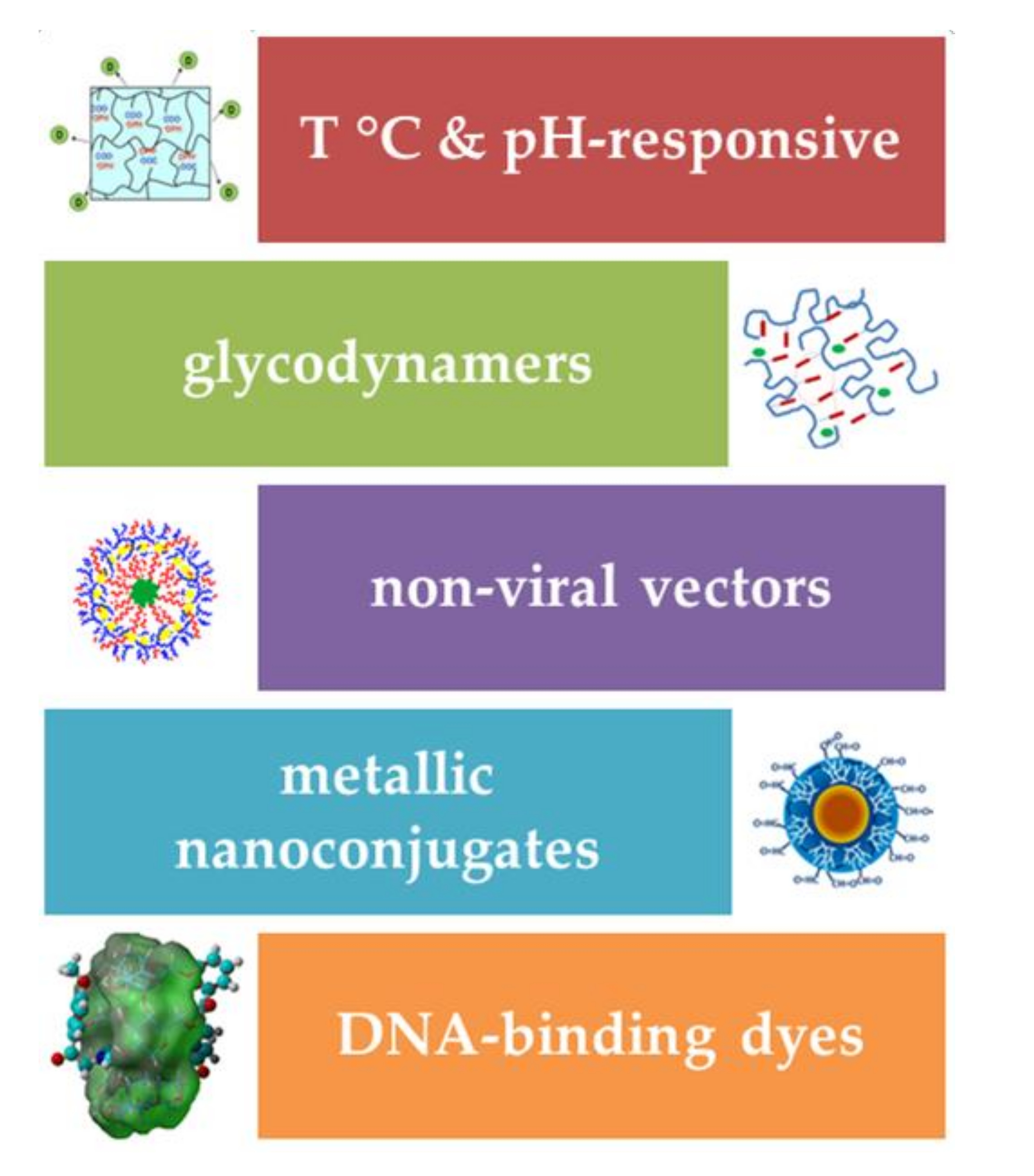
:
1. Introduction
2. Temperature- and pH-Responsive Systems for Controlled Drug Delivery
3. Glycodynameric Gels for Drug Delivery
4. Polymeric Non-Viral Vectors for Gene Delivery
5. Metallic Nanoconjugates for Biomedical Applications
6. Smart Organic Tools for Biomedical Imaging
7. Conclusions and Future Directions
Author Contributions
Funding
Conflicts of Interest
References
- Gillies, E.R. Reflections on the evolution of smart polymers. Isr. J. Chem. 2019, 59, 1–12. [Google Scholar] [CrossRef] [Green Version]
- Cohen Stuart, M.A.; Huck, W.T.A.; Genzer, J.; Müller, M.; Ober, C.; Stamm, M.; Sukhorukov, G.B.; Szleifer, I.; Tsukruk, V.V.; Urban, M.; et al. Emerging applications of stimuli-responsive polymer materials. Nat. Mater. 2010, 9, 101–113. [Google Scholar] [CrossRef] [PubMed]
- Doberenz, F.; Zeng, K.; Willems, C.; Zhang, K.; Groth, T. Thermoresponsive polymers and their biomedical application in tissue engineering—A review. J. Mater. Chem. B 2020, 8, 607–628. [Google Scholar] [CrossRef] [PubMed]
- Alejo, T.; Uson, L.; Arruebo, M. Reversible stimuli-responsive nanomaterials with on-off switching ability for biomedical applications. J. Control. Release 2019, 314, 162–176. [Google Scholar] [CrossRef] [PubMed]
- Zhang, A.; Jung, K.; Li, A.; Liu, J.; Boyer, C. Recent advances in stimuli-responsive polymer systems for remotely controlled drug release. Prog. Polym. Sci. 2019, 99, 101164. [Google Scholar] [CrossRef]
- Mrinalini, M.; Prasanthkumar, S. Recent advances on stimuli-responsive smart materials and their applications. ChemPlusChem 2019, 84, 1103–1121. [Google Scholar] [CrossRef] [Green Version]
- Wells, C.M.; Harris, M.; Choi, L.; Murali, V.P.; Delbuque Guerra, F.; Jennings, J.A. Stimuli-Responsive drug release from smart polymers. J. Funct. Biomater. 2019, 10, 34. [Google Scholar] [CrossRef] [Green Version]
- De las Heras Alarcon, C.; Pennadam, S.; Alexander, C. Stimuli responsive polymers for biomedical applications. Chem. Soc. Rev. 2005, 34, 276–285. [Google Scholar] [CrossRef]
- Rosca, I.; Petrovici, A.R.; Peptanariu, D.; Nicolescu, A.; Dodi, G.; Avadanei, M.; Ivanov, I.C.; Bostanaru, A.C.; Mares, M.; Ciolacu, D. Biosynthesis of dextran by Weissella confusa and its in vitro functional characteristics. Int. J. Biol. Macromol. 2018, 107, 1765–1772. [Google Scholar] [CrossRef]
- Petrovici, A.R.; Rosca, I.; Stoica, I.; Silion, M.; Nicolescu, A.; Dodi, G.; Simionescu, N.; Varganici, C.D.; Ivanov, I.C.; Pinteala, M. Biosynthesis of exopolysaccharides by Weissella confusa in a new culture medium. Rom. Biotechnol. Lett. 2018, 23, 13637–13646. [Google Scholar]
- Petrovici, A.R.; Nicolescu, A.; Silion, M.; Rosca, I.; Ciolacu, D. Biopolymer biosynthesis by lactic acid bacteria strain in four different culture media. Rev. Roum. Chim. 2018, 63, 637–642. [Google Scholar]
- Balan, G.G.; Rosca, I.; Ursu, E.L.; Fifere, A.; Varganici, C.D.; Doroftei, F.; Turin-Moleavin, I.A.; Sandru, V.; Constantinescu, G.; Timofte, D.; et al. Duodenoscope-Associated infections beyond the elevator channel: Alternative causes for difficult reprocessing. Molecules 2019, 24, 2343. [Google Scholar] [CrossRef] [Green Version]
- Bostanaru, A.C.; Rosca, I.; Minea, B.; Nastasa, V.; Foia, L.; Marincu, I.; Mares, M.; Mederle, O.A. Genotype comparison of Candida albicans isolates from different clinical samples. Rev. Rom. Med. Lab. 2019, 27, 327–332. [Google Scholar] [CrossRef] [Green Version]
- Rosca, I.; Bostanaru, A.C.; Minea, B.; Nastasa, V.; Gherghel, I.; Panzaru, C.V.; Mares, M.; Moroti-Constantinescu, V.R. Phenotypic and genotypic variations in Candida albicans isolates from Romanian patients. Rev. Rom. Med. Lab. 2018, 26, 405–413. [Google Scholar] [CrossRef] [Green Version]
- Balan, G.G.; Rosca, I.; Ursu, E.L.; Doroftei, F.; Bostanaru, A.C.; Hnatiuc, E.; Nastasa, V.; Sandru, V.; Stefanescu, G.; Trifan, A.; et al. Plasma-Activated water: A new and effective alternative for duodenoscope reprocessing. Infect. Drug Resist. 2018, 11, 727–733. [Google Scholar] [CrossRef] [PubMed] [Green Version]
- Mares, M.; Minea, B.; Nastasa, V.; Rosca, I.; Bostanaru, A.C.; Marincu, I.; Toma, V.; Cristea, V.C.; Murariu, C.; Pinteala, M. In vitro activity of echinocandins against 562 clinical yeast isolates from a Romanian multicentre study. Med. Mycol. 2018, 56, 442–451. [Google Scholar] [CrossRef] [PubMed] [Green Version]
- Rotaru, A.; Pricope, G.; Plank, T.N.; Clima, L.; Ursu, E.L.; Pinteala, M.; Davis, J.T.; Barboiu, M. G-Quartet hydrogels for effective cell growth applications. Chem. Commun. 2017, 53, 12668. [Google Scholar] [CrossRef] [PubMed]
- Ursu, E.L.; Gavril, G.; Morariu, S.; Pinteala, M.; Barboiu, M.; Rotaru, A. Single-Walled carbon nanotubes–G-quadruple hydrogel nanocomposite matrixes for cell support applications. Mater. Sci. Eng. C Mater. Biol. Appl. 2020, 111, 110800. [Google Scholar] [CrossRef]
- Barboiu, M. Artificial water channels-incipient innovative developments. Chem. Commun. 2016, 52, 5657–5665. [Google Scholar] [CrossRef]
- Sun, Z.; Barboiu, M.; Legrand, Y.M.; Petit, E.; Rotaru, A. Highly selective artificial cholesteryl crown ether K(+)-channels. Angew. Chem. Int. Ed. 2015, 54, 14473–14477. [Google Scholar] [CrossRef]
- Murail, S.; Vasiliu, T.; Neamtu, A.; Barboiu, M.; Sterpone, F.; Baaden, M. Water permeation across artificial I-quartet membrane channels: From structure to disorder. Faraday Discuss. 2018, 209, 125–148. [Google Scholar] [CrossRef] [PubMed]
- Nastasa, V.V.; Stavarache, C.; Hanganu, A.; Coroaba, A.; Nicolescu, A.; Deleanu, C.; Sadet, A.; Vasos, P. Hyperpolarised NMR to follow water proton transport through membrane channels via exchange with biomolecules. Faraday Discuss. 2018, 209, 67–82. [Google Scholar] [CrossRef]
- Pinteala, T.; Chiriac, A.E.; Rosca, I.; Larese Filon, F.; Pinteala, M.; Chiriac, A.; Podoleanu, C.; Stolnicu, S.; Coros, M.F.; Coroaba, A. Nail damage (severe onychodystrophy) induced by acrylate glue: Scanning electron microscopy and energy dispersive X-Ray investigations. Skin Appendage Disord. 2017, 2, 137–142. [Google Scholar] [CrossRef] [Green Version]
- Coroaba, A.; Chiriac, A.E.; Sacarescu, L.; Pinteala, T.; Minea, B.; Ibanescu, S.A.; Pertea, M.; Moraru, A.; Esanu, I.; Maier, S.S.; et al. New insights into human hair: SAXS, SEM, TEM and EDX for Alopecia areata investigations. PeerJ 2020, 8, e8376. [Google Scholar] [CrossRef] [PubMed] [Green Version]
- Gritsch, L.; Motta, F.L.; Negrini, N.C.; Yahia, L.; Fare, S. Crosslinked gelatin hydrogels as carriers for controlled heparin release. Mater. Lett. 2018, 228, 375–378. [Google Scholar] [CrossRef]
- Chen, Y.; Chen, B.Z.; Wang, Q.L.; Jin, X.; Guo, X.D. Fabrication of coated polymer microneedles for transdermal drug delivery. J. Control. Release 2017, 265, 14–21. [Google Scholar] [CrossRef] [PubMed]
- Hyun, H.; Park, J.; Willis, K.; Park, J.E.; Lyle, L.T.; Lee, W.; Yeo, Y. Surface modification of polymer nanoparticles with native albumin for enhancing drug delivery to solid tumors. Biomaterials 2018, 180, 206–224. [Google Scholar] [CrossRef]
- Gaudio, C.; Crognale, V.; Serino, G.; Galloni, P.; Audenino, A.; Ribatti, D.; Morbiducci, U. Natural polymeric microspheres for modulated drug delivery. Mater. Sci. Eng. C Mater. Biol. Appl. 2017, 75, 408–417. [Google Scholar] [CrossRef]
- Fundueanu, G.; Constantin, M.; Bucatariu, S.; Ascenzi, P. Poly (N-isopropylacrylamide-co-Nisopropylmethacrylamide) thermo-responsive microgels as self-regulated drug delivery system. Macromol. Chem. Phys. 2016, 217, 2525–2533. [Google Scholar] [CrossRef]
- Fundueanu, G.; Constantin, M.; Oanea, I.; Harabagiu, V.; Ascenzi, P.; Simionescu, B.C. Entrapment and release of drugs by a strict ‘on-off’ mechanism in pullulan microspheres with pendant thermosensitive groups. Biomaterials 2010, 31, 9544–9553. [Google Scholar] [CrossRef]
- Fundueanu, G.; Constantin, M.; Stanciu, C.; Theodoridis, G.; Ascenzi, P. pH- and temperature-sensitive polymeric microspheres for drug delivery: The dissolution of copolymers modulates drug release. J. Mater. Sci. Mater. Med. 2009, 20, 2465–2475. [Google Scholar] [CrossRef]
- Heskins, M.; Guillet, J.E. Solution properties of poly(N-isopropylacrylamide). J. Macromol. Sci. Chem. 1968, 2, 1441–1455. [Google Scholar] [CrossRef]
- Enas, M.A. Hydrogel: Preparation, characterization, and applications: A review. J. Adv. Res. 2015, 6, 105–121. [Google Scholar]
- Sherbiny, I.M.E.; Magdi, H.Y. Hydrogel scaffolds for tissue engineering: Progress and challenges. Glob. Cardiol. Sci. Pract. 2013, 3, 316–342. [Google Scholar] [CrossRef] [PubMed] [Green Version]
- Yang, J.A.; Yeom, J.; Hwang, B.W.; Hoffman, A.S.; Hahn, S.K. In situ-forming injectable hydrogels for regenerative medicine. Prog. Polym. Sci. 2014, 39, 1973–1986. [Google Scholar] [CrossRef]
- Buengera, D.; Topuza, F.; Groll, J. Hydrogels in sensing applications. Prog. Polym. Sci. 2014, 37, 1678–1719. [Google Scholar] [CrossRef]
- Jianyu, L.; Mooney, D.J. Designing hydrogels for controlled drug delivery. Nat. Rev. Mater. 2016, 1, 16071. [Google Scholar]
- D’Emanuele, A.; Dinarvand, R. Preparation, characterisation, and drug release from thermoresponsive microspheres. Int. J. Pharm. 1995, 118, 237–242. [Google Scholar] [CrossRef]
- Sato-Matsuo, E.; Tanaka, T. Kinetics of discontinuous volume–phase transition of gels. J. Chem. Phys. 1988, 89, 1695–1703. [Google Scholar] [CrossRef]
- Tanaka, T.; Fillmore, D.J. Kinetics of swelling of gels. J. Chem. Phys. 1979, 70, 1214–1218. [Google Scholar] [CrossRef]
- Fundueanu, G.; Constantin, M.; Ascenzi, P. Poly (N-isopropylacrylamide-co-acrylamide) cross-linked thermoresponsive microspheres obtained from preformed polymers: Influence of the physico-chemical characteristics of drugs on their release profiles. Acta Biomater. 2009, 5, 363–373. [Google Scholar] [CrossRef]
- Zhang, X.Z.; Yang, Y.Y.; Chung, S.T.; Ma, K.X. Preparation and characterization of fast response macroporous poly (N-isopropylacrylamide) hydrogels. Langmuir 2001, 17, 6094–6099. [Google Scholar] [CrossRef]
- Wang, W.; Xu, X.D.; Wang, Z.C.; Cheng, S.X.; Zhang, X.Z.; Zhuo, R.X. Synthesis and properties of pH and temperature sensitive P (NIPAAm-co-DMAEMA) hydrogels. Colloid Surf. B 2008, 64, 34–41. [Google Scholar] [CrossRef] [PubMed]
- Djokpé, E.; Vogt, W. N-Isopropylacrylamide and N-Isopropylmethacryl-amide: Cloud points of mixtures and copolymers. Macromol. Chem. Phys. 2001, 202, 750–757. [Google Scholar] [CrossRef]
- Constantin, M.; Cristea, M.; Ascenzi, P.; Fundueanu, G. Lower critical solution temperature versus volume phase transition temperature in thermoresponsive drug delivery systems. Express Polym. Lett. 2011, 5, 839–848. [Google Scholar] [CrossRef]
- Fundueanu, G.; Constantin, M.; Bucatariu, S.; Ascenzi, P. Poly (N-isopropylacrylamide-co-N-vinylpyrrolidone) thermoresponsive microspheres: The low drug loading ensures the pulsatile release mechanism. Express Polym. Lett. 2020, 14, 63–76. [Google Scholar] [CrossRef]
- Kwon, S.S.; Kong, B.J.; Park, S.N. Physicochemical properties of pH-sensitive hydrogels based on hydroxyethyl cellulose–hyaluronic acid and for applications as transdermal delivery systems for skin lesions. Eur. J. Pharm. Biopharm. 2015, 92, 146–154. [Google Scholar] [CrossRef]
- Fundueanu, G.; Constantin, M.; Asmarandei, I.; Harabagiu, V.; Ascenzi, P.; Simionescu, B.C. The thermosensitivity of pH/thermoresponsive microspheres activated by the electrostatic interaction of pH-sensitive units with a bioactive compound. J. Biomed. Mater. Res. A 2013, 101, 1661–1669. [Google Scholar] [CrossRef]
- Yoo, M.K.; Sung, Y.K.; Lee, Y.M.; Cho, C.S. Effect of polyelectrolyte on the lower critical solution temperature of poly (Nisopropylacrylamide) in the poly (NIPAAm-co-acrylic acid) hydrogel. Polymer 2000, 41, 5713–5719. [Google Scholar] [CrossRef]
- Salgado-Rodríguez, R.; Licea-Claveríe, A.; Arndt, K.F. Random copolymers of N-isopropylacrylamide and methacrylic acid monomers with hydrophobic spacers: pH-tunable temperature sensitive materials. Eur. Polym. J. 2004, 40, 1931–1946. [Google Scholar] [CrossRef]
- Fundueanu, G.; Constantin, M.; Bucatariu, S.; Ascenzi, P. pH/thermo-responsive poly (N-isopropylacrylamide-co-maleic acid) drug delivery system with a sensor and an actuator. Polymer 2017, 110, 177–186. [Google Scholar] [CrossRef]
- Miyata, K.; Christie, R.J.; Kataoka, K. Polymeric micelles for nano-scale drug delivery. React. Funct. Polym. 2011, 71, 227–234. [Google Scholar] [CrossRef]
- Kim, T.I.; Kim, S.W. Bioreducible polymers for gene delivery. React. Funct. Polym. 2011, 71, 344–349. [Google Scholar] [CrossRef] [PubMed] [Green Version]
- Colfen, H. Double-Hydrophilic block copolymers: Synthesis and application as novel surfactants and crystal growth modifiers. Macromol. Rapid Commun. 2001, 22, 219–252. [Google Scholar] [CrossRef]
- Nakashima, K.; Bahadur, P. Aggregation of water-soluble block copolymers in aqueous solutions: Recent trends. Adv. Colloid Interface Sci. 2006, 123, 75–96. [Google Scholar] [CrossRef] [PubMed]
- Nakayama, M.; Okano, T. Multi-Targeting cancer chemotherapy using temperature-responsive drug carrier systems. React. Funct. Polym. 2011, 71, 235–244. [Google Scholar] [CrossRef]
- Isoda, K.; Kanayama, N.; Miyamoto, D.; Takarada, T.; Maeda, M. RAFT-Generated poly (N-isopropylacrylamide)-DNA block copolymers for temperature-responsive formation of polymer micelles. React. Funct. Polym. 2011, 71, 367–371. [Google Scholar] [CrossRef]
- Zhang, J.X.; Qiu, L.Y.; Zhu, K.J.; Jin, Y. Thermosensitive micelles self-assembled by novel N-isopropylacrylamide oligomer grafted polyphosphazene. Macromol. Rapid Commun. 2004, 25, 1563–1567. [Google Scholar] [CrossRef]
- Zhang, J.X.; Qiu, L.Y.; Jin, Y.; Zhu, J.Y. Physicochemical characterization of polymeric micelles constructed from novel amphiphilic polyphosphazenewith poly (N-isopropylacrylamide) and ethyl 4-aminobenzoate as side groups. Colloids Surf. B Biointerfaces 2005, 43, 123–130. [Google Scholar] [CrossRef]
- Sen, G.; Sharon, A.; Pal, S. Grafted polysaccharides: Smart materials of the future, their synthesis and applications. In Biopolymers: Biomedical and Environmental Applications; Kalia, S., Averious, L., Eds.; John Wiley & Sons: Austin, TX, USA, 2011; pp. 99–128. [Google Scholar]
- Constantin, M.; Bucatariu, S.; Stoica, I.; Fundueanu, G. Smart nanoparticles based onpullulan-g-poly(N-isopropylacrylamide) for controlled delivery of indomethacin. Int. J. Biol. Macromol. 2017, 94, 698–708. [Google Scholar] [CrossRef]
- Constantin, M.; Bucatariu, S.M.; Doroftei, F.; Fundueanu, G. Smart composite materials based on chitosan microspheres embedded in thermosensitive hydrogel for controlled delivery of drugs. Carbohydr. Polym. 2017, 157, 493–502. [Google Scholar] [CrossRef] [PubMed]
- Ahmadi, F.; Oveisi, Z.; Mohammadi Samani, S.; Amoozgar, Z. Chitosan based hydrogels: Characteristics and pharmaceutical applications. Res. Pharm. Sci. 2015, 10, 1–16. [Google Scholar] [PubMed]
- del Valle, L.J.; Dıaz, A.; Puiggali, J. Hydrogels for biomedical applications: Cellulose, chitosan, and protein/peptide derivatives. Gels 2017, 3, 27. [Google Scholar] [CrossRef] [Green Version]
- Barcan, G.A.; Zhang, X.; Waymouth, R.M. Structurally dynamic hydrogels derived from 1,2-dithiolanes. J. Am. Chem. Soc. 2015, 137, 5650–5653. [Google Scholar] [CrossRef]
- Deng, G.; Tang, C.; Li, F.; Jiang, H.; Chen, Y. Covalent cross-linked polymer gels with reversible sol-gel transition and self-healing properties. Macromolecules 2010, 43, 1191–1194. [Google Scholar] [CrossRef]
- Brooks, W.L.A.; Sumerlin, B.S. Synthesis and applications of boronic acid-containing polymers: From materials to medicine. Chem. Rev. 2016, 116, 1375–1397. [Google Scholar] [CrossRef] [PubMed]
- Zhang, Y.; Tao, L.; Li, S.; Wei, Y. Synthesis of multiresponsive and dynamic chitosan-based hydrogels for controlled release of bioactive molecules. Biomacromolecules 2011, 12, 2894–2901. [Google Scholar] [CrossRef]
- Casuso, P.; Odriozol, I.; Pérez-San Vicente, A.; Loinaz, I.; Cabañero, G.; Grande, H.J.; Dupin, D. Injectable and self-healing dynamic hydrogels based on metal(I)-thiolate/disulfide exchange as biomaterials with tunable mechanical properties. Biomacromolecules 2015, 16, 3552–3561. [Google Scholar] [CrossRef]
- Roy, N.; Bruchmannb, B.; Lehn, J.M. Dynamers: Dynamic polymers as self-healing materials. Chem. Soc. Rev. 2015, 44, 3786–3807. [Google Scholar] [CrossRef] [Green Version]
- Marin, L.; Bejan, A.; Ailincai, D.; Belei, D. Poly(azomethine-phenothiazine)s with efficient emission in solid state. Eur. Polym. J. 2017, 95, 127–137. [Google Scholar] [CrossRef]
- Fisher, H.F.; Viswanathan, T.S. Carbonyl oxygen exchange evidence ofimine formation in the glutamate dehydrogenase reaction and identification of the occult role of NADPH. Proc. Natl. Acad. Sci. USA 1984, 81, 2747–2751. [Google Scholar] [CrossRef] [PubMed] [Green Version]
- Perez, D.M.; Karnik, S.S. Multiple signalling states of G-protein-coupled receptors. Pharmacol. Rev. 2005, 57, 147–161. [Google Scholar] [CrossRef] [PubMed]
- Qin, W.; Long, S.; Panunzio, M.; Biondi, S. Schiff bases: A short survey on an evergreen chemistry tool. Molecules 2013, 18, 12264–12289. [Google Scholar] [CrossRef] [PubMed]
- Mi, F.L.; Kuan, C.Y.; Shyu, S.S.; Lee, S.T.; Chang, S.F. Study of gelation kinetics and chain-relaxation properties of glutaraldehyde-cross-linked chitosan gel and their effects on microspheres preparation and drug release. Carbohydr. Polym. 2000, 41, 389–396. [Google Scholar] [CrossRef]
- Beauchamp, R.O.; St Clair, M.B.; Fennell, T.R.; Clarke, D.O.; Morgan, K.T. A critical review of the toxicology of glutaraldehyde. Crit. Rev. Toxicol. 1992, 22, 143–174. [Google Scholar] [CrossRef]
- Berger, J.; Reist, M.; Mayer, J.M.; Felt, O.; Peppas, N.A.; Gurny, R. Structure and interactions in covalently and ionically crosslinked chitosan hydrogels for biomedical applications. Eur. J. Pharm. Biopharm. 2004, 57, 19–34. [Google Scholar] [CrossRef]
- Ailincai, D.; Marin, L.; Morariu, S.; Mares, M.; Bostanaru, A.C.; Pinteala, M.; Simionescu, B.C.; Barboiu, M. Dual crosslinked iminoboronate-chitosan hydrogels with strong antifungal activity against Candida planktonic yeasts and biofilms. Carbohydr. Polym. 2016, 152, 306–316. [Google Scholar] [CrossRef]
- Iftime, M.M.; Morariu, S.; Marin, L. Salicyl-Imine-Chitosan hydrogels: Supramolecular architecturing as a crosslinking method toward multifunctional hydrogels. Carbohydr. Polym. 2017, 165, 39–50. [Google Scholar] [CrossRef]
- Marin, L.; Ailincai, D.; Morariu, S.; Tartau-Mititelu, L. Development of biocompatible glycodynameric hydrogels joining two natural motifs by dynamic constitutional chemistry. Carbohydr. Polym. 2017, 170, 60–71. [Google Scholar] [CrossRef]
- Ruff, Y.; Buhler, E.; Candau, S.J.; Kesselman, E.; Talmon, Y.; Lehn, J.M. Glycodynamers: Dynamic polymers bearing oligosaccharides residues-Generation, structure, physicochemical, component exchange, and lectin binding properties. J. Am. Chem. Soc. 2010, 132, 2573–2584. [Google Scholar] [CrossRef]
- Pasa, S.; Aydın, S.; Kalayci, S.; Boga, M.; Atlan, M.; Bingul, M.; Sahin, F.; Temel, H. The synthesis of boronic-imine structured compounds and identification of their anticancer, antimicrobial and antioxidant activities. J. Pharm. Anal. 2016, 6, 39–48. [Google Scholar] [CrossRef] [PubMed] [Green Version]
- Bandyopadhyay, A.; McCarthy, K.A.; Kelly, M.A.; Gao, J. Targeting bacteria via iminoboronate chemistry of amine-presenting lipids. Nat. Commun. 2015, 6, 6561. [Google Scholar] [CrossRef] [PubMed] [Green Version]
- Ailincai, D.; Pamfil, D.; Marin, L. Multiple bio-responsive polymer dispersed liquid crystal composites for sensing applications. J. Mol. Liq. 2018, 272, 572–582. [Google Scholar] [CrossRef]
- Ailincai, D.; Farcau, C.; Paslaru, E.; Marin, L. PDLC composites based on polyvinyl boric acid matrix–a promising pathway towards biomedical engineering. Liq. Cryst. 2016, 43, 1973–1985. [Google Scholar] [CrossRef]
- Kim, J.H.; Campbell, B.C.; Mahoney, N.; Chan, K.L.; Molyneux, R.J. Chemosensitization of aflatoxigenic fungi to antimycin a and strobilurin using salicylaldehyde, a volatile natural compound targeting cellular antioxidation system. Mycopathologia 2011, 171, 291–298. [Google Scholar] [CrossRef] [PubMed]
- Adams, T.B.; Cohen, S.M.; Doull, J.; Feron, V.; Goodman, J.I.; Marnett, L.J.; Munro, I.C.; Portoghese, P.S.; Smith, R.L.; Waddell, W.J.; et al. The FEMA GRAS assessment of hydroxy- and alkoxy-substituted benzylderivatives used as flavor ingredients. Food Chem. Toxicol. 2005, 43, 1241–1271. [Google Scholar] [CrossRef] [PubMed]
- Salicylaldehyde. Food Cosmet. Toxicol. 1979, 17, 903–905. [CrossRef]
- Hong, S.C.; Yoo, S.Y.; Kim, H.; Lee, J. Chitosan-Based multifunctional platforms for local delivery of therapeutics. Mar. Drugs 2017, 15, 60. [Google Scholar] [CrossRef] [Green Version]
- Sedghi, R.; Shaabani, A.; Mohammadi, Z.; Samadi, Z.Y.; Isae, F. Biocompatible electrospinning chitosan nanofibers: A novel delivery system with superior local cancer therapy. Carbohydr. Polym. 2017, 159, 1–10. [Google Scholar] [CrossRef] [PubMed]
- Olaru, A.M.; Marin, L.; Morariu, S.; Pricope, G.; Pinteala, M.; Tartau-Mititelu, L. Biocompatible chitosan based hydrogels for potential application in local tumour therapy. Carbohydr. Polym. 2018, 179, 59–70. [Google Scholar] [CrossRef]
- Montembault, A.; Viton, C.; Domard, A. Rheometric study of the gelation of chitosan in aqueous solution without cross-linking agent. Biomacromolecules 2005, 6, 653–662. [Google Scholar] [CrossRef] [PubMed]
- Zhang, X.; Lin, Y.; Gillies, R.J. Tumor pH and its measurement. J. Nucl. Med. 2010, 51, 1167–1170. [Google Scholar] [CrossRef] [PubMed] [Green Version]
- Luo, H.; Sui, Y.; Lin, W.H.; Wu, H.Q. Study on the antiproliferative activity of four Schiff bases derived from natural biomass dehydroabietylamine. Indian J. Chem. 2016, 55B, 248–251. [Google Scholar]
- Zahedifard, M.; Faraj, F.L.; Paydar, M.; Looi, C.Y.; Hajrezaei, M.; Hasanpourghadi, M.; Kamalidehghan, B.; Majid, N.A.; Ali, H.M.; Ameen, M. Synthesis, characterization and apoptotic activity of quinazolinone Schiff base derivatives toward MCF-7 cells via intrinsic and extrinsic apoptosis pathways. Sci. Rep. 2015, 5, 11544. [Google Scholar] [CrossRef]
- Craciun, A.M.; Mititelu-Tartau, L.; Pinteala, M.; Marin, L. Nitrosalicyl-imine-chitosan hydrogels based drug delivery systems for long term sustained release in local therapy. J. Colloid Interface Sci. 2019, 536, 196–207. [Google Scholar] [CrossRef]
- Williams, B.S.; Buvanendran, A. Nonopioid analgesics: NSAIDs, COX-2 inhibitors, and acetaminophen. In Essentials of Pain Medicine, 3rd ed.; Benzon, H., Fishman, S., Raja, S., Cohen, S., Liu, S., Eds.; Saunders: Philadelphia, PA, USA, 2011; pp. 130–139. [Google Scholar]
- Craciun, A.M.; Barhalescu, M.L.; Agop, M.; Ochiuz, L. Theoretical modeling of long-time drug release from nitrosalicyl-imine-chitosan hydrogels through multifractal logistic type laws. Comput. Math. Methods Med. 2019, 2019, 4091464. [Google Scholar] [CrossRef] [Green Version]
- Craciun, A.M. Cinnamyl-imine-chitosan hydrogels. Morphology control. Acta Chem. Iasi 2018, 26, 221–232. [Google Scholar] [CrossRef] [Green Version]
- Ailincai, D.; Tartau-Mititelu, L.; Marin, L. Drug delivery systems based on biocompatible imino-chitosan hydrogels for local anticancer therapy. Drug Deliv. 2018, 25, 1080–1090. [Google Scholar] [CrossRef] [Green Version]
- Craciun, A.M.; Serban, G.; Crumpei, I.; Agop, M.; Cioca, G. Operational procedures in the theory of the drug release from chitosan hydrogels. Mater. Plast. 2018, 55, 590–594. [Google Scholar] [CrossRef]
- Yin, H.; Kanasty, R.L.; Eltoukhy, A.A.; Vegas, A.J.; Dorkin, J.R.; Anderson, D.G. Nonviral vectors for gene-based therapy. Nat. Rev. Genet. 2014, 15, 541–555. [Google Scholar] [CrossRef]
- O’Connor, D.M.; Boulis, N.M. Gene therapy for neurodegenerative diseases. Trends Mol. Med. 2015, 21, 504–512. [Google Scholar] [CrossRef] [PubMed]
- Somia, N.; Verm, I.M. Gene therapy: Trials and tribulations. Nat. Rev. Genet. 2000, 1, 91–99. [Google Scholar] [CrossRef] [PubMed]
- Rusu, R.D.; Clima, L.; Pinteala, M. Dendritic architectures as non-viral gene delivery vectors: Challenges and perspectives. Rev. Roum. Chim. 2018, 63, 581–592. [Google Scholar]
- Yang, J.; Zhang, Q.; Chang, H.; Cheng, Y. Surface-engineered dendrimers in gene delivery. Chem. Rev. 2015, 115, 5274–5300. [Google Scholar] [CrossRef] [PubMed]
- Tschiche, A.; Malhotra, S.; Haag, R. Nonviral gene delivery with dendritic self-assembling architectures. Nanomedicine 2014, 9, 667–693. [Google Scholar] [CrossRef]
- Ailincai, D.; Peptanariu, D.; Pinteala, M.; Marin, L. Dynamic constitutional chemistry towards efficient nonviral vectors. Mater. Sci. Eng. C Mater. Biol. Appl. 2019, 94, 635–646. [Google Scholar] [CrossRef]
- Ardeleanu, R.; Dascalu, A.; Neamtu, A.; Peptanariu, D.; Uritu, C.M.; Maier, S.; Nicolescu, A.; Simionescu, B.C.; Barboiu, M.; Pinteala, M. Multivalent polyrotaxane vectors as adaptive cargo complexes for gene therapy. Polym. Chem. 2017, 9, 845–859. [Google Scholar] [CrossRef]
- Vasiliu, T.; Cojocaru, C.; Rotaru, A.; Pricope, G.; Pinteala, M.; Clima, L. Optimization of polyplex formation between DNA oligonucleotide and poly(L-lysine): Experimental study and modeling approach. Int. J. Mol. Sci. 2017, 18, 1291. [Google Scholar] [CrossRef]
- Yanan, Y. How Free Cationic Polymer Chains Promote Gene Transfection; Springer International Publishing: Cham, Switzerland, 2013; pp. 15–89. [Google Scholar]
- Simionescu, B.C.; Drobota, M.; Timpu, D.; Vasiliu, T.; Constantinescu, C.A.; Rebleanu, D.; Calin, M.; David, G. Biopolymers/poly(ε-caprolactone)/polyethylenimine functionalized nano-hydroxyapatite hybrid cryogel: Synthesis, characterization and application in gene delivery. Mater. Sci. Eng. C Mater. Biol. Appl. 2017, 81, 167–176. [Google Scholar] [CrossRef]
- Mintzer, M.A.; Simanek, E.E. Nonviral vectors for gene delivery. Chem. Rev. 2009, 109, 259–302. [Google Scholar] [CrossRef]
- Uritu, C.M.; Varganici, D.C.; Ursu, L.; Coroaba, A.; Nicolescu, A.; Dascalu, A.I.; Peptanariu, D.; Stan, D.; Constantinescu, C.A.; Simion, V.; et al. Hybrid fullerene conjugates as vectors for DNA cell-delivery. J. Mater. Chem. B 2015, 3, 2433–2446. [Google Scholar] [CrossRef] [PubMed]
- Godbey, W.T.; Wu, K.K.; Mikos, A.G. Tracking the intracellular path of poly(ethylenimine)/DNA complexes for gene delivery. Proc. Natl. Acad. Sci. USA 1999, 96, 5177–5181. [Google Scholar] [CrossRef] [PubMed] [Green Version]
- Ketola, T.M.; Hanzlíková, M.; Utti, A.; Lemmetyinen, H.; Yliperttula, M.; Vuorimaa-Laukkanen, E. Role of polyplex intermediate species on gene transfer efficiency: Polyethylenimine-DNA complexes and time-resolved fluorescence spectroscopy. J. Phys. Chem. B 2013, 117, 10405–10413. [Google Scholar] [CrossRef] [PubMed]
- Arima, H.; Motoyama, K.; Higashi, T. Sugar-appended polyamidoamine dendrimer conjugates with cyclodextrins as cell-specific non-viral vectors. Adv. Drug Deliv. Rev. 2013, 65, 1204–1214. [Google Scholar] [CrossRef]
- Voicu, G.; Rebleanu, D.; Constantinescu, C.A.; Fuior, E.A.; Ciortan, L.; Droc, I.; Uritu, C.M.; Pinteala, M.; Manduteanu, I.; Simionescu, M.; et al. Nano-polyplexes mediated transfection of Runx2-shRNA mitigates the osteodifferentiation of human valvular interstitial cells. Pharmaceutics 2020, 12, 507. [Google Scholar] [CrossRef]
- David, G.; Clima, L.; Calin, M.; Constantinescu, C.A.; Balan-Porcarasu, M.; Uritu, C.M.; Simionescu, B.C. Squalene/polyethylenimine based non-viral vectors: Synthesis and use in systems for sustained gene release. Polym. Chem. 2018, 9, 1072–1081. [Google Scholar] [CrossRef]
- Craciun, B.F.; Gavril, G.; Peptanariu, D.; Ursu, E.L.; Clima, L.; Pinteala, M. Synergistic effect of low molecular weight polyethylenimine and polyethylene glycol components in dynamic nonviral vector structure, toxicity, and transfection efficiency. Molecules 2019, 24, 1460. [Google Scholar] [CrossRef] [Green Version]
- Pricope, G.; Pinteala, M.; Clima, L. Dynamic self-organizing systems for DNA delivery. Rev. Roum. Chim. 2018, 63, 613–619. [Google Scholar]
- Clima, L.; Craciun, B.F.; Gavril, G.; Pinteala, M. Tunable composition of dynamic non-viral vectors over the DNA polyplex formation and nucleic acid transfection. Polymers 2019, 11, 1313. [Google Scholar] [CrossRef] [Green Version]
- Uritu, C.M.; Calin, M.; Maier, S.S.; Cojocaru, C.; Nicolescu, A.; Peptanariu, D.; Constantinescu, C.A.; Stan, D.; Barboiu, M.; Pinteala, M. Flexible cyclic siloxane core enhances the transfection efficiency of polyethylenimine-based non-viral gene vectors. J. Mater. Chem. B 2015, 3, 8250–8267. [Google Scholar] [CrossRef]
- Fischer, D.; Bieber, T.; Li, Y.; Elsasser, H.P.; Kissel, T. A novel non-viral vector for DNA delivery based on low molecular weight, branched polyethylenimine: Effect of molecular weight on transfection efficiency and cytotoxicity. Pharm. Res. 1999, 16, 1273–1279. [Google Scholar] [CrossRef]
- Neuberg, P.; Kichler, A. Recent developments in nucleic acid delivery with polyethylenimines. In Advances in Genetics; Huang, L., Liu, D., Wagner, E., Eds.; Academic Press: San Diego, CA, USA, 2014; Volume 88, pp. 263–288. [Google Scholar]
- Remant Bahadur, K.C.; Uludag, H. 2-PEI and its derivatives for gene therapy. In Polymers and Nanomaterials for Gene Therapy; Narain, R., Ed.; Woodhead Publishing: Sawston, UK, 2016; pp. 29–54. [Google Scholar]
- Albuquerque, L.J.C.; de Castro, C.E.; Riske, K.A.; Carlan da Silva, M.C.; Muraro, P.I.R.; Schmidt, V.; Giacomelli, C.; Giacomelli, F.C. Gene transfection mediated by catiomers requires free highly charged polymer chains to overcome intracellular barriers. Biomacromolecules 2017, 18, 1918–1927. [Google Scholar] [CrossRef] [PubMed]
- Dascalu, A.I.; Ardeleanu, R.; Neamtu, A.; Maier, S.S.; Uritu, C.M.; Nicolescu, A.; Silion, M.; Peptanariu, D.; Calin, M.; Pinteala, M. Transfection-capable PEGylated-cyclodextrin-containing polycationic nanovectors: A new synthesis pathway. J. Mater. Chem. B 2017, 5, 7164–7174. [Google Scholar] [CrossRef] [PubMed] [Green Version]
- Fierascu, I.; Fierascu, I.C.; Brazdis, R.I.; Baroi, A.M.; Fistos, T.; Fierascu, R.C. Phytosynthesized metallic nanoparticles-between nanomedicine and toxicology. A brief review of 2019′s findings. Materials 2020, 13, 574. [Google Scholar] [CrossRef] [PubMed] [Green Version]
- Uritu, C.M.; Tamba, B.I.; Dodi, G.; Pintela, M.; Ghizdovat, V.; Gutu, M.M.; Stefanescu, C. Design and synthesis of polymer coated gold nanoparticles, AuPEI-Glc-99mTc, for targeted in vivo imaging. Eur. J. Nucl. Med. Mol. Imaging 2018, 45, S682–S683. [Google Scholar]
- Corciova, A.; Mircea, C.; Burlec, A.F.; Cioanca, O.; Tuchilus, C.; Fifere, A.; Lungoci, A.L.; Marangoci, N.; Hancianu, M. Antioxidant, antimicrobial and photocatalytic activities of silver nanoparticles obtained by bee propolis extract assisted biosynthesis. Farmacia 2019, 67, 482–489. [Google Scholar] [CrossRef]
- Dascalu, A.; Ignat, L.; Ignat, M.E.; Doroftei, F.; Belhacene, K.; Froidevaux, R.; Pinteala, M. Reusable biocatalyst by pepsin immobilization on functionalized magnetite nanoparticles. Rev. Roum. Chim. 2018, 63, 685–695. [Google Scholar]
- Alexandrica, M.C.; Silion, M.; Timpu, D.; Popa, M.I. A study on the intercalation process of tramadol hydrochloride in ZnAl-nitrate layered double hydroxides. Rev. Roum. Chim. 2016, 61, 683–688. [Google Scholar]
- Kim, K.S.; Lee, D.; Song, C.G.; Kang, P.M. Reactive oxygen species-activated nanomaterials as theranostic agents. Nanomedicine 2015, 10, 2709–2723. [Google Scholar] [CrossRef]
- Fu, S.; Wang, S.; Zhang, X.; Qi, A.; Liu, Z.; Yu, X.; Chen, C.; Li, L. Structural effect of Fe3O4 nanoparticles on peroxidase-like activity for cancer therapy. Colloids Surf. B Biointerfaces 2017, 154, 239–245. [Google Scholar] [CrossRef]
- Turin-Moleavin, I.A.; Fifere, A.; Lungoci, A.L.; Rosca, I.; Coroaba, A.; Peptanariu, D.; Nastasa, V.; Pasca, S.A.; Bostanaru, A.C.; Mares, M.; et al. In Vitro and in vivo antioxidant activity of the new magnetic-cerium oxide nanoconjugates. Nanomaterials 2019, 9, 1565. [Google Scholar] [CrossRef] [PubMed] [Green Version]
- Charbgoo, F.; Ahmad, M.; Darroudi, M. Cerium oxide nanoparticles: Green synthesis and biological applications. Int. J. Nanomed. 2017, 12, 1401–1413. [Google Scholar] [CrossRef] [PubMed] [Green Version]
- Nguyen, A.; Abdelrasoul, G.; Lin, D.; Maadi, H.; Tong, J.; Chen, G.; Wang, R.; Anwar, A.; Shoute, L.; Fang, Q.; et al. Polyethylenimine-coated iron oxide magnetic nanoparticles for high efficient gene delivery. Appl. Nanosci. 2018, 8, 811–821. [Google Scholar] [CrossRef]
- Lungoci, A.L.; Turin-Moleavin, I.A.; Corciova, A.; Mircea, C.; Arvinte, A.; Fifere, A.; Marangoci, N.L.; Pinteala, M. Multifunctional magnetic cargo-complexes with radical scavenging properties. Mater. Sci. Eng. C Mater. Biol. Appl. 2019, 94, 608–618. [Google Scholar] [CrossRef] [PubMed]
- Xia, T.; Li, N.; Fang, X. Single-molecule fluorescence imaging in living cells. Annu. Rev. Phys. Chem. 2013, 64, 459–480. [Google Scholar] [CrossRef] [PubMed]
- Stennett, E.M.; Ciuba, M.A.; Levitus, M. Photophysical processes in single molecule organic fluorescent probes. Chem. Soc. Rev. 2014, 43, 1057–1075. [Google Scholar] [CrossRef]
- Vegesna, G.K.; Sripathi, S.R.; Zhang, J.; Zhu, S.; He, W.; Luo, F.T.; Jahng, W.J.; Frost, M.; Liu, H. Highly water-soluble BODIPY-based fluorescent probe for sensitive and selective detection of nitric oxide in living cells. ACS Appl. Mater. Interfaces 2013, 5, 4107–4112. [Google Scholar] [CrossRef]
- Qin, W.; Ding, D.; Liu, J.; Yuan, W.Z.; Hu, Y.; Liu, B.; Tang, B.Z. Biocompatible nanoparticles with aggregation-induced emission characteristics as far-red/near-infrared fluorescent bioprobes for in vitro and in vivo imaging applications. Adv. Funct. Mater. 2012, 22, 771–779. [Google Scholar] [CrossRef]
- Ursu, E.L.; Doroftei, F.; Peptanariu, D.; Pinteala, M.; Rotaru, A. DNA-assisted decoration of single-walled carbon nanotubes with gold nanoparticles for applications in surface enhanced Raman scattering imaging of cells. J. Nanopart. Res. 2017, 19, 181. [Google Scholar] [CrossRef]
- Rotaru, A.; Druta, I.; Oeser, T.; Müller, T.J.J. A novel coupling 1,3-dipolar cycloaddition sequence as a three-component approach to highly fluorescent indolizines. Helv. Chim. Acta 2005, 88, 1798–1812. [Google Scholar] [CrossRef]
- Danac, R.; Rusu, R.; Rotaru, A.; Pui, A.; Shova, S. New conjugates of calix [4]arenes bearing dipyridine and indolizine heterocycles. Supramol. Chem. 2012, 24, 424–435. [Google Scholar] [CrossRef]
- Marangoci, N.L.; Popovici, L.; Ursu, E.L.; Danac, R.; Clima, L.; Cojocaru, C.; Coroaba, A.; Neamtu, A.; Mangalagiu, I.; Pinteala, M.; et al. Pyridyl-indolizine derivatives as DNA binders and pH-sensible fluorescent dyes. Tetrahedron 2016, 72, 8215–8222. [Google Scholar] [CrossRef]
- Mizobe, Y.; Tohnai, N.; Miyata, M.; Hasegawa, Y. A tunable solid-state fluorescence system consisting of organic salts of anthracene-2,6-disulfonic acid with primary amines. Chem. Commun. 2005, 1839–1841. [Google Scholar] [CrossRef]
- Yoshizawa, M.; Klosterman, J.K. Molecular architectures of multi-anthracene assemblies. Chem. Soc. Rev. 2014, 43, 1885–1898. [Google Scholar] [CrossRef] [PubMed]
- Kumar, C.V.; Punzalan, E.H.A.; Tan, W.B. Adenine-thymine base pair recognition by an anthryl probe from the DNA minor groove. Tetrahedron 2000, 56, 7027–7040. [Google Scholar] [CrossRef]
- Ghosh, S.; Chattoraj, S.; Chattopadhyay, N. Interaction of β-cyclodextrin with nile red in a single live CHO cell: An initiative towards developing a prospective strategy for excretion of adsorbed drugs from the cell membrane. Analyst 2014, 139, 5664–5668. [Google Scholar] [CrossRef]
- Di Donato, C.; Lavorgna, M.; Fattorusso, R.; Isernia, C.; Isidori, M.; Malgieri, G.; Piscitelli, C.; Russo, C.; Russo, L.; Iacovino, R. Alpha- and beta-cyclodextrin inclusion complexes with 5-fluorouracil: Characterization and cytotoxic activity evaluation. Molecules 2016, 21, 1644. [Google Scholar] [CrossRef] [PubMed]
- Edetsberger, M.; Knapp, M.; Gaubitzer, E.; Miksch, C.; Gvichiya, K.E.; Kohler, G. Effective staining of tumor cells by coumarin-6 depends on the stoichiometry of cyclodextrin complex formation. J. Incl. Phenom. Macrocycl. Chem. 2011, 70, 327–331. [Google Scholar] [CrossRef]
- Pricope, G.; Ursu, E.L.; Sardaru, M.; Cojocaru, C.; Clima, L.; Marangoci, N.; Danac, R.; Mangalagiu, I.; Simionescu, B.C.; Pinteala, M.; et al. Novel cyclodextrin-based pH-sensitive supramolecular host-guest assembly for staining acidic cellular organelles. Polym. Chem. 2018, 9, 968–975. [Google Scholar] [CrossRef]


© 2020 by the authors. Licensee MDPI, Basel, Switzerland. This article is an open access article distributed under the terms and conditions of the Creative Commons Attribution (CC BY) license (http://creativecommons.org/licenses/by/4.0/).
Share and Cite
Pinteala, M.; Abadie, M.J.M.; Rusu, R.D. Smart Supra- and Macro-Molecular Tools for Biomedical Applications. Materials 2020, 13, 3343. https://doi.org/10.3390/ma13153343
Pinteala M, Abadie MJM, Rusu RD. Smart Supra- and Macro-Molecular Tools for Biomedical Applications. Materials. 2020; 13(15):3343. https://doi.org/10.3390/ma13153343
Chicago/Turabian StylePinteala, Mariana, Marc J. M. Abadie, and Radu D. Rusu. 2020. "Smart Supra- and Macro-Molecular Tools for Biomedical Applications" Materials 13, no. 15: 3343. https://doi.org/10.3390/ma13153343