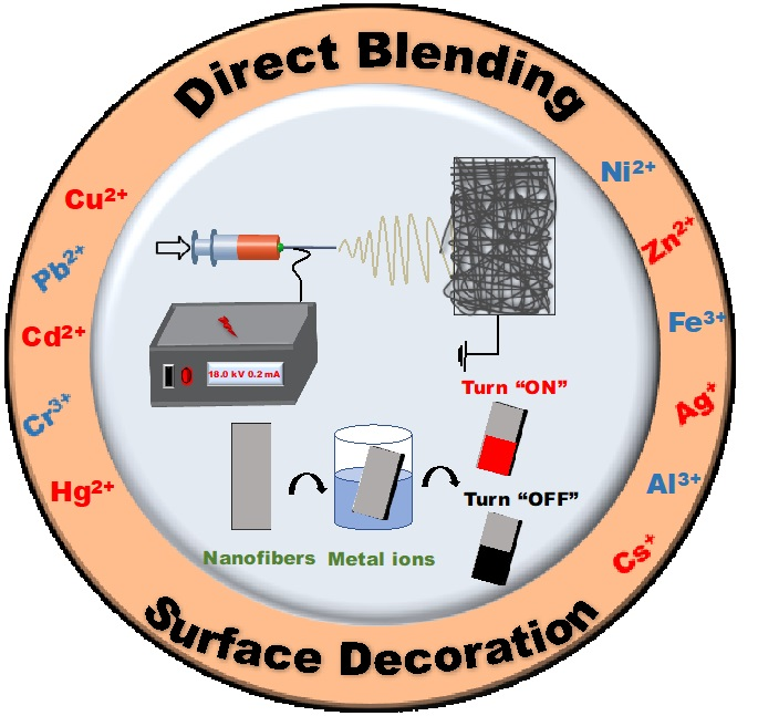
Functionalized Electrospun Nanofibers as a Versatile Platform for Colorimetric Detection of Heavy Metal Ions in Water: A Review


Abstract
:
1. Introduction
2. Designing of Functionalized Electrospun Nanofibers for the Colorimetric Detection of Metal Ions
2.1. Detection of Copper Ions
2.2. Detection of Lead Ions
2.3. Detection of Cadmium Ions
2.4. Detection of Chromium Ions
2.5. Detection of Mercury Ions
2.6. Detection of Nickel Ions
2.7. Detection of Zinc Ions
2.8. Detection of Iron Ions
2.9. Detection of Other (Silver, Aluminum, and Cesium) Ions
3. Discussion, Conclusions and Future Outlook
Author Contributions
Funding
Conflicts of Interest
References
- National Systems to Support Drinking-Water, Sanitation and Hygiene: Global Status Report 2019. UN-Water Global Analysis and Assessment of Sanitation and Drinking-Water (GLAAS) 2019 Report; World Health Organization: Geneva, Switzerland, 2019.
- Johnson, A.C.; Jin, X.; Nakada, N.; Sumpter, J.P. Learning from the past and considering the future of chemicals in the environment. Science 2020, 367, 384–387. [Google Scholar] [CrossRef] [PubMed] [Green Version]
- Escher, B.I.; Stapleton, H.M.; Schymanski, E.L. Tracking complex mixtures of chemicals in our changing environment. Science 2020, 367, 388–392. [Google Scholar] [CrossRef]
- Kortenkamp, A.; Faust, M. Regulate to reduce chemical mixture risk. Science 2018, 361, 224–226. [Google Scholar] [CrossRef] [PubMed] [Green Version]
- Kortenkamp, A. Low dose mixture effects of endocrine disrupters and their implications for regulatory thresholds in chemical risk assessment. Curr. Opin. Pharm. 2014, 19, 105–111. [Google Scholar] [CrossRef] [PubMed]
- Mohammed, A.S.; Kapri, A.; Goel, R. Heavy metal pollution: Source, impact, and remedies. In Biomanagement of Metal-Contaminated Soils; Khan, M., Zaidi, A., Goel, R., Musarrat, J., Eds.; Springer: Dordrecht, The Netherlands, 2011; Volume 20, pp. 1–28. [Google Scholar]
- Tchounwou, P.B.; Yedjou, C.G.; Patlolla, A.K.; Sutton, D.J. Heavy metal toxicity and the environment. In Molecular, Clinical and Environmental Toxicology: Experientia Supplementum; Luch, A., Ed.; Springer: Basel, Switzerland, 2012; Volume 101, pp. 133–164. [Google Scholar]
- Häder, D.P.; Banaszak, A.T.; Villafañe, V.E.; Narvarte, M.A.; González, R.A.; Helbling, E.W. Anthropogenic pollution of aquatic ecosystems: Emerging problems with global implications. Sci. Total Environ. 2020, 713, 136586. [Google Scholar] [CrossRef] [PubMed]
- Dutta, S.; Sharma, R.K. Sustainable magnetically retrievable nanoadsorbents for selective removal of heavy metal ions from different charged wastewaters. In Evaluating Water Quality to Prevent Future Disasters; Ahuja, S., Ed.; Elsevier: London, UK, 2019; Volume 11, pp. 371–416. [Google Scholar]
- Inoue, K.I. Heavy metal toxicity. J Clin. Toxicol. 2013, 3, 2161–2495. [Google Scholar]
- Das, S.; Raj, R.; Mangwani, N.; Dash, H.R.; Chakraborty, J. Heavy metals and hydrocarbons: Adverse effects and mechanism of toxicity. In Microbial Biodegradation and Bioremediation, 1st ed.; Das, S., Ed.; Elsevier: London, UK, 2014; pp. 23–54. [Google Scholar]
- Rehman, K.; Fatima, F.; Waheed, I.; Akash, M.S.H. Prevalence of exposure of heavy metals and their impact on health consequences. J. Cell Biochem. 2018, 119, 157–184. [Google Scholar] [CrossRef]
- Barbier, O.; Jacquillet, G.; Tauc, M.; Cougnon, M.; Poujeol, P. Effect of heavy metals on, and handling by, the kidney. Nephron Physiol. 2005, 99, 105–110. [Google Scholar] [CrossRef]
- Fu, Z.; Xi, Z. The effects of heavy metals on human metabolism. Toxicol. Mech. Method 2020, 30, 167–176. [Google Scholar] [CrossRef]
- Valko, M.; Morris, H.; Cronin, M.T. Metals, toxicity and oxidative stress. Curr. Med. Chem. 2005, 12, 1161–1208. [Google Scholar] [CrossRef] [Green Version]
- Jomova, K.; Valko, M. Advances in metal-induced oxidative stress and human disease. Toxicology 2011, 283, 65–87. [Google Scholar] [CrossRef] [PubMed]
- Bánfalvi, G. Cellular Effects of Heavy Metals; Springer: New York, NY, USA, 2011. [Google Scholar]
- Valko, M.; Jomova, K.; Rhodes, C.J.; Kuča, K.; Musílek, K. Redox-and non-redox-metal-induced formation of free radicals and their role in human disease. Arch. Toxicol. 2016, 90, 1–37. [Google Scholar] [CrossRef] [PubMed]
- Jomova, K.; Vondrakova, D.; Lawson, M.; Valko, M. Metals, oxidative stress and neurodegenerative disorders. Mol. Cell Biochem. 2010, 345, 91–104. [Google Scholar] [CrossRef] [PubMed]
- United States Environmental Protection Agency. Ground Water and Drinking Water; National Primary Drinking Water Regulations. Available online: https://www.epa.gov/ground-water-and-drinking-water/national-primary-drinking-water-regulations (accessed on 5 March 2020).
- World Health Organization. Guidelines for Drinking Water Quality. Available online: https://www.who.int/water_sanitation_health/publications/2011/9789241548151_ch12.pdf (accessed on 5 March 2020).
- The European Parliament and the Council of the European Union. Directive on the Restriction of the Use of Certain Hazardous Substances in Electrical and Electronic Equipment, Directive 2002/95/EC. Available online: https://eur-lex.europa.eu/legal-content/EN/TXT/?uri=CELEX:32002L0095 (accessed on 6 March 2020).
- Malik, L.A.; Bashir, A.; Qureashi, A.; Pandith, A.H. Detection and removal of heavy metal ions: A review. Environ. Chem. Lett. 2019, 17, 1495–1521. [Google Scholar] [CrossRef]
- Eddaif, L.; Shaban, A.; Telegdi, J. Sensitive detection of heavy metals ions based on the calixarene derivatives-modified piezoelectric resonators: A review. Int. J. Environ. Anal. Chem. 2019, 99, 824–853. [Google Scholar] [CrossRef] [Green Version]
- Das, R.; Vecitis, C.D.; Schulze, A.; Cao, B.; Ismail, A.F.; Lu, X.; Chen, J.; Ramakrishna, S. Recent advances in nanomaterials for water protection and monitoring. Chem. Soc. Rev. 2017, 46, 6946–7020. [Google Scholar] [CrossRef]
- Kumar, P.; Kim, K.H.; Bansal, V.; Lazarides, T.; Kumar, N. Progress in the sensing techniques for heavy metal ions using nanomaterials. J. Ind. Eng. Chem. 2017, 54, 30–43. [Google Scholar] [CrossRef]
- Li, M.; Gou, H.; Al-Ogaidi, I.; Wu, N. Nanostructured sensors for detection of heavy metals: A review. ACS Sustain. Chem. Eng. 2013, 1, 713–723. [Google Scholar] [CrossRef]
- Schroeder, V.; Savagatrup, S.; He, M.; Lin, S.; Swager, T.M. Carbon nanotube chemical sensors. Chem. Rev. 2019, 119, 599–663. [Google Scholar] [CrossRef]
- Liu, X.; Yao, Y.; Ying, Y.; Ping, J. Recent advances in nanomaterial-enabled screen-printed electrochemical sensors for heavy metal detection. Trac. Trend Anal. Chem. 2019, 115, 187–202. [Google Scholar] [CrossRef]
- Kukkar, D.; Vellingiri, K.; Kumar, V.; Deep, A.; Kim, K.H. A critical review on the metal sensing capabilities of optically active nanomaterials: Limiting factors, mechanism, and performance evaluation. Trac. Trend Anal. Chem. 2018, 109, 227–246. [Google Scholar] [CrossRef]
- Gan, X.; Zhao, H.; Schirhagl, R.; Quan, X. Two-dimensional nanomaterial based sensors for heavy metal ions. Microchim. Acta 2018, 185, 478. [Google Scholar] [CrossRef] [PubMed]
- Zhang, J.; Cheng, F.; Li, J.; Zhu, J.J.; Lu, Y. Fluorescent nanoprobes for sensing and imaging of metal ions: Recent advances and future perspectives. Nano Today 2016, 11, 309–329. [Google Scholar] [CrossRef] [PubMed] [Green Version]
- Willner, M.R.; Vikesland, P.J. Nanomaterial enabled sensors for environmental contaminants. J. Nanobiotechnol. 2018, 16, 95. [Google Scholar] [CrossRef] [PubMed]
- Lin, T.; Fang, J. Fundamentals of Electrospinning & Electrospun Nanofibers; DEStech Publications Inc.: Lancaster, PA, USA, 2017. [Google Scholar]
- Li, Z.; Wang, C. One-Dimensional Nanostructures: Electrospinning Technique and Unique Nanofibers; Springer-Verlag: Berlin/Heidelberg, Germany, 2013. [Google Scholar]
- Mitchell, G.R. Electrospinning: Principles, Practice and Possibilities; The Royal Society of Chemistry: Cambridge, UK, 2015. [Google Scholar]
- Topuz, F.; Uyar, T. Electrospinning of cyclodextrin functional nanofibers for drug delivery applications. Pharmaceutics 2019, 11, 6. [Google Scholar] [CrossRef] [Green Version]
- Ding, B.; Yu, J. Electrospun Nanofibers for Energy and Environmental Applications, 1st ed.; Springer: Berlin/Heidelberg, Germany, 2014. [Google Scholar]
- Ismail, A.F.; Hilal, N.; Jaafar, J.; Wright, C. Nanofiber Membranes for Medical, Environmental, and Energy Applications; CRC Press: Boca Raton, FL, USA, 2019. [Google Scholar]
- Wei, Q. Functional Nanofibers and their Applications.; Woodhead Publishing in Textiles, Elsevier: Cambridge, UK, 2012. [Google Scholar]
- Sun, G.; Sun, L.; Xie, H.; Liu, J. Electrospinning of nanofibers for energy applications. Nanomaterials 2016, 6, 129. [Google Scholar] [CrossRef] [Green Version]
- Wang, L.; Yang, G.; Peng, S.; Wang, J.; Yan, W.; Ramakrishna, S. One-dimensional nanomaterials toward electrochemical sodium-ion storage applications via electrospinning. Energy Storage Mater. 2020, 25, 443–476. [Google Scholar] [CrossRef]
- Balusamy, B.; Senthamizhan, A.; Uyar, T. Surface functionalized electrospun nanofibers for removal of toxic pollutants in water. In Nanofiber Membranes for Medical, Environmental, and Energy Applications; Ismail, A.F., Hilal, N., Jaafar, J., Wright, C., Eds.; CRC Press: Boca Raton, FL, USA, 2019; p. 189. [Google Scholar]
- Senthamizhan, A.; Uyar, T. Electrospun fluorescent nanofibers for explosive detection. In Electrospinning for High Performance Sensors; Macagnano, A., Zampetti, E., Kny, E., Eds.; Springer: Cham, Switzerland, 2015; pp. 179–204. [Google Scholar]
- Senthamizhan, A.; Balusamy, B.; Uyar, T. Electrospun filters for organic pollutants removal. In Filtering Media by Electrospinning; Focarete, M., Gualandi, C., Ramakrishna, S., Eds.; Springer: Cham, Switzerland, 2018; pp. 115–150. [Google Scholar]
- Balusamy, B.; Sarioglu, O.F.; Senthamizhan, A.; Uyar, T. Rational design and development of electrospun nanofibrous biohybrid composites. ACS Appl. Biol. Mater. 2019, 2, 3128–3143. [Google Scholar] [CrossRef]
- Senthamizhan, A.; Balusamy, B.; Aytac, Z.; Uyar, T. Grain boundary engineering in electrospun ZnO nanostructures as promising photocatalysts. Cryst. Eng. Comm. 2016, 18, 6341–6351. [Google Scholar] [CrossRef] [Green Version]
- Anitha, S.; Brabu, B.; Rajesh, K.P.; Natarajan, T.S. Fabrication of UV sensor based on electrospun composite fibers. Mater. Lett. 2013, 92, 417–420. [Google Scholar] [CrossRef]
- Balusamy, B.; Senthamizhan, A.; Uyar, T. Functionalized electrospun nanofibers as colorimetric sensory probe for mercury detection: A review. Sensors 2019, 19, 4763. [Google Scholar] [CrossRef] [PubMed] [Green Version]
- Senthamizhan, A.; Balusamy, B.; Celebioglu, A.; Uyar, T. “Nanotraps” in porous electrospun fibers for effective removal of lead (II) in water. J. Mater. Chem. A 2016, 4, 2484–2493. [Google Scholar] [CrossRef] [Green Version]
- Wang, X.; Yu, J.; Sun, G.; Ding, B. Electrospun nanofibrous materials: A versatile medium for effective oil/water separation. Mater Today 2016, 19, 403–414. [Google Scholar] [CrossRef]
- Veeramuthu, L.; Venkatesan, M.; Liang, F.C.; Benas, J.S.; Cho, C.J.; Chen, C.W.; Zhou, Y.; Lee, R.H.; Kuo, C.C. Conjugated copolymers through electrospinning synthetic strategies and their versatile applications in sensing environmental toxicants, pH, temperature, and humidity. Polymers 2020, 12, 587. [Google Scholar] [CrossRef] [PubMed] [Green Version]
- Ye, D.; Ding, Y.; Duan, Y.; Su, J.; Yin, Z.; Huang, Y.A. Large-scale direct-writing of aligned nanofibers for flexible electronics. Small 2018, 14, 1703521. [Google Scholar] [CrossRef] [PubMed]
- Nan, N.; He, J.; You, X.; Sun, X.; Zhou, Y.; Qi, K.; Shao, W.; Liu, F.; Chu, Y.; Ding, B. A stretchable, highly sensitive, and multimodal mechanical fabric sensor based on electrospun conductive nanofiber yarn for wearable electronics. Adv. Mater. Technol. 2019, 4, 1800338. [Google Scholar] [CrossRef]
- Mukhlish, M.Z.B.; Horie, Y.; Higashi, K.; Ichigi, A.; Guo, S.; Nomiyama, T. Self-standing conductive ITO-silica nanofiber mats for use in flexible electronics and their application in dye-sensitized solar cells. Ceram. Int. 2017, 43, 8146–8152. [Google Scholar] [CrossRef]
- Uyar, T.; Kny, E. Electrospun Materials for Tissue Engineering and Biomedical Applications: Research, Design and Commercialization, 1st ed.; Elsevier: Cambridge, UK, 2017. [Google Scholar]
- Senthamizhan, A.; Balusamy, B.; Uyar, T. Recent progress on designing electrospun nanofibers for colorimetric biosensing applications. Curr. Opin. Biomed. Eng. 2020, 13, 1–8. [Google Scholar] [CrossRef]
- Balusamy, B.; Senthamizhan, A.; Uyar, T. Electrospun nanofibrous materials for wound healing applications. In Electrospun Materials for Tissue Engineering and Biomedical Applications: Research, Design and Commercialization; Uyar, T., Kny, E., Eds.; Elsevier: Cambridge, UK, 2017; pp. 147–177. [Google Scholar]
- Senthamizhan, A.; Balusamy, B.; Uyar, T. Electrospinning-a versatile processing technology for producing biomedical materials and tissue engineering. In Electrospun Materials for Tissue Engineering and Biomedical Applications: Research, Design and Commercialization; Uyar, T., Kny, E., Eds.; Elsevier: Cambridge, UK, 2017; pp. 3–41. [Google Scholar]
- Balusamy, B.; Senthamizhan, A.; Uyar, T. In vivo safety evaluations of electrospun nanofibers for biomedical applications. In Electrospun Materials for Tissue Engineering and Biomedical Applications: Research, Design and Commercialization; Uyar, T., Kny, E., Eds.; Elsevier: Cambridge, UK, 2017; pp. 101–113. [Google Scholar]
- Balusamy, B.; Senthamizhan, A.; Uyar, T. Design and development of electrospun nanofibers in regenerative medicine. In Nanomaterials for Regenerative Medicine. Stem Cell Biology and Regenerative Medicine; Springer: Cham, Switzerland, 2019; pp. 47–79. [Google Scholar]
- Senthamizhan, A.; Balusamy, B.; Uyar, T. Glucose sensors based on electrospun nanofibers: A review. Anal. Bioanal. Chem. 2016, 408, 1285–1306. [Google Scholar] [CrossRef]
- Senthamizhan, A.; Balusamy, B.; Aytac, Z.; Uyar, T. Ultrasensitive electrospun fluorescent nanofibrous membrane for rapid visual colorimetric detection of H2O2. Anal. Bioanal. Chem. 2016, 408, 1347–1355. [Google Scholar] [CrossRef] [Green Version]
- Anitha, S.; Brabu, B.; Thiruvadigal, D.J.; Gopalakrishnan, C.; Natarajan, T.S. Preparation of free-standing electrospun composite ZnO membrane for antibacterial applications. Adv. Sci. Lett. 2012, 5, 468–474. [Google Scholar] [CrossRef]
- Anitha, S.; Brabu, B.; Thiruvadigal, D.J.; Gopalakrishnan, C.; Natarajan, T.S. Optical, bactericidal and water repellent properties of electrospun nano-composite membranes of cellulose acetate and ZnO. Carbohydr. Polym. 2012, 87, 1065–1072. [Google Scholar] [CrossRef]
- Sabra, S.; Ragab, D.M.; Agwa, M.M.; Rohani, S. Recent advances in electrospun nanofibers for some biomedical applications. Eur. J. Pharm. Sci. 2020, 144, 105224. [Google Scholar] [CrossRef] [PubMed]
- Schoolaert, E.; Hoogenboom, R.; Clerck, K.D. Colorimetric nanofibers as optical sensors. Adv. Funct. Mater. 2017, 27, 1702646. [Google Scholar] [CrossRef] [Green Version]
- Terra, I.A.A.; Mercante, L.A.; Andre, R.S.; Correa, D.S. Fluorescent and colorimetric electrospun nanofibers for heavy-metal sensing. Biosensors 2017, 7, 61. [Google Scholar] [CrossRef] [PubMed] [Green Version]
- Zhang, N.; Qiao, R.; Su, J.; Yan, J.; Xie, Z.; Qiao, Y.; Wang, X.; Zhong, J. Recent advances of electrospun nanofibrous membranes in the development of chemosensors for heavy metal detection. Small 2017, 13, 1604293. [Google Scholar] [CrossRef]
- Yang, T.; Zhan, L.; Huang, C.Z. Recent insights into functionalized electrospun nanofibrous films for chemo-/bio-sensors. Trac. Trend Anal. Chem. 2020, 121, 115813. [Google Scholar] [CrossRef]
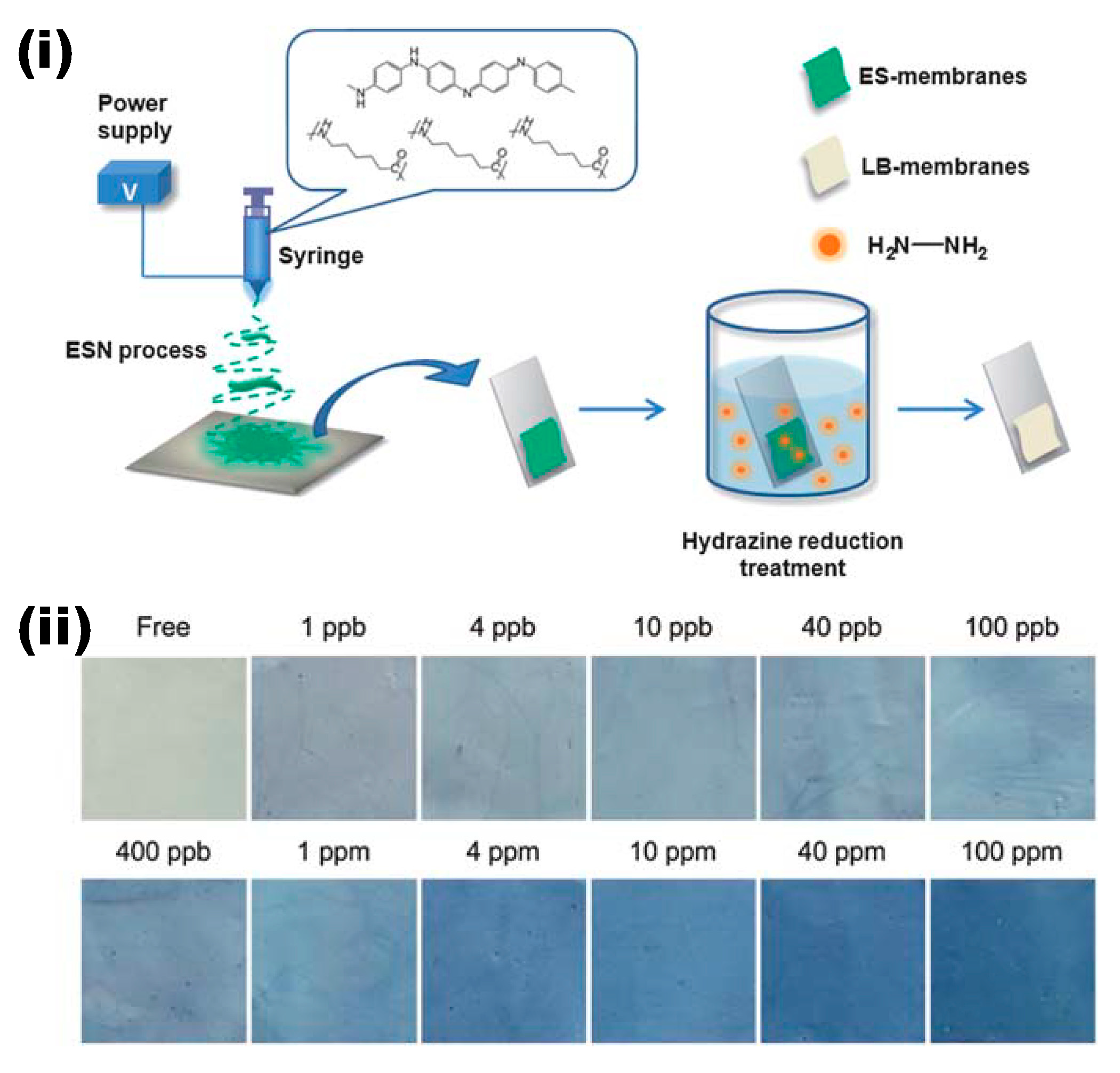
- Ding, B.; Si, Y.; Wang, X.; Yu, J.; Feng, L.; Sun, G. Label-free ultrasensitive colorimetric detection of copper (II) ions utilizing polyaniline/polyamide-6 nano-fiber/net sensor strips. J. Mater. Chem. 2011, 21, 13345–13353. [Google Scholar] [CrossRef]
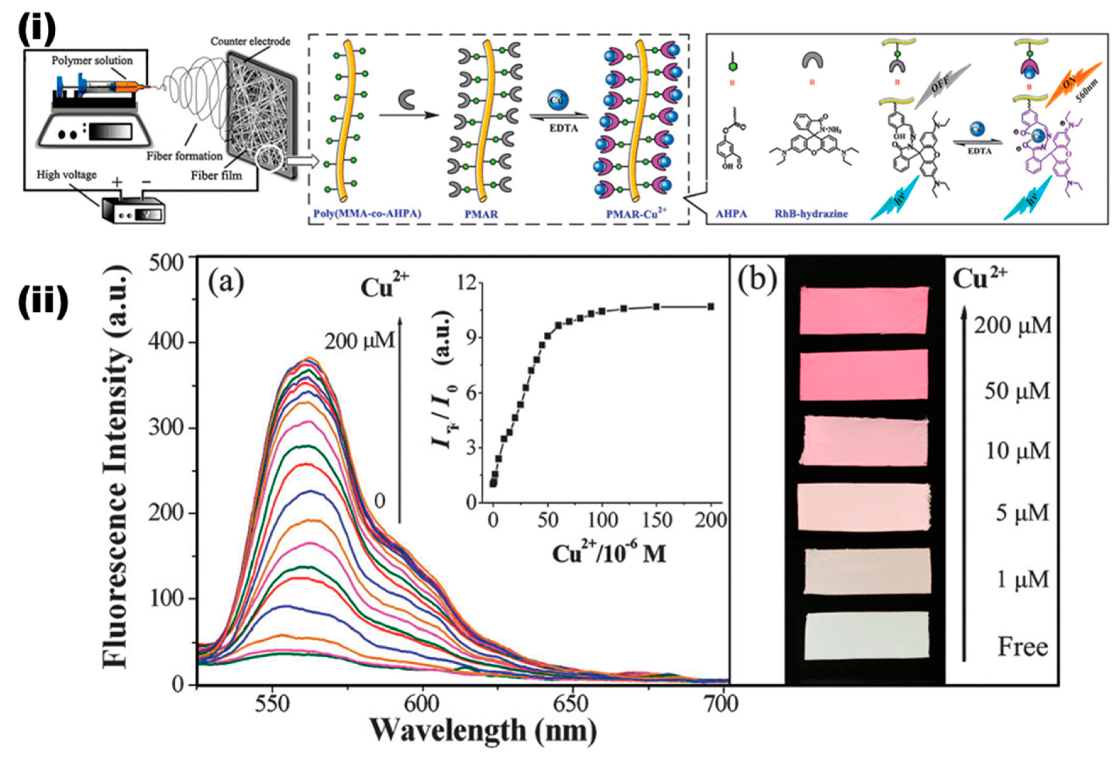
- Wang, W.; Yang, Q.; Sun, L.; Wang, H.; Zhang, C.; Fei, X.; Sun, M.; Li, Y. Preparation of fluorescent nanofibrous film as a sensing material and adsorbent for Cu2+ in aqueous solution via copolymerization and electrospinning. J. Hazard Mater. 2011, 194, 185–192. [Google Scholar] [CrossRef]
- Wu, W.C.; Lai, H.J. Preparation of thermo-responsive electrospun nanofibers containing rhodamine-based fluorescent sensor for Cu2+ detection. J. Polym. Res. 2016, 23, 223. [Google Scholar] [CrossRef]
- Hung, C.C.; Kuo, C.C.; Weng, N.K.; Wu, W.C.; Chen, B.Y.; Cho, C.J.; Hsu, I.J.; Chiu, Y.C.; Chen, W.C. Novel highly sensitive and reversible electrospun nanofibrous chemosensor-filters composed of poly (HEMA-co-MNA) and bpy-F-bpy with metal-ion-modulated multicolor fluorescence emission. Polym. J. 2016, 48, 439–449. [Google Scholar] [CrossRef]
- Lin, H.J.; Chen, C.Y. Thermo-responsive electrospun nanofibers doped with 1,10-phenanthroline-based fluorescent sensor for metal ion detection. J. Mater. Sci. 2016, 51, 1620–1631. [Google Scholar] [CrossRef]
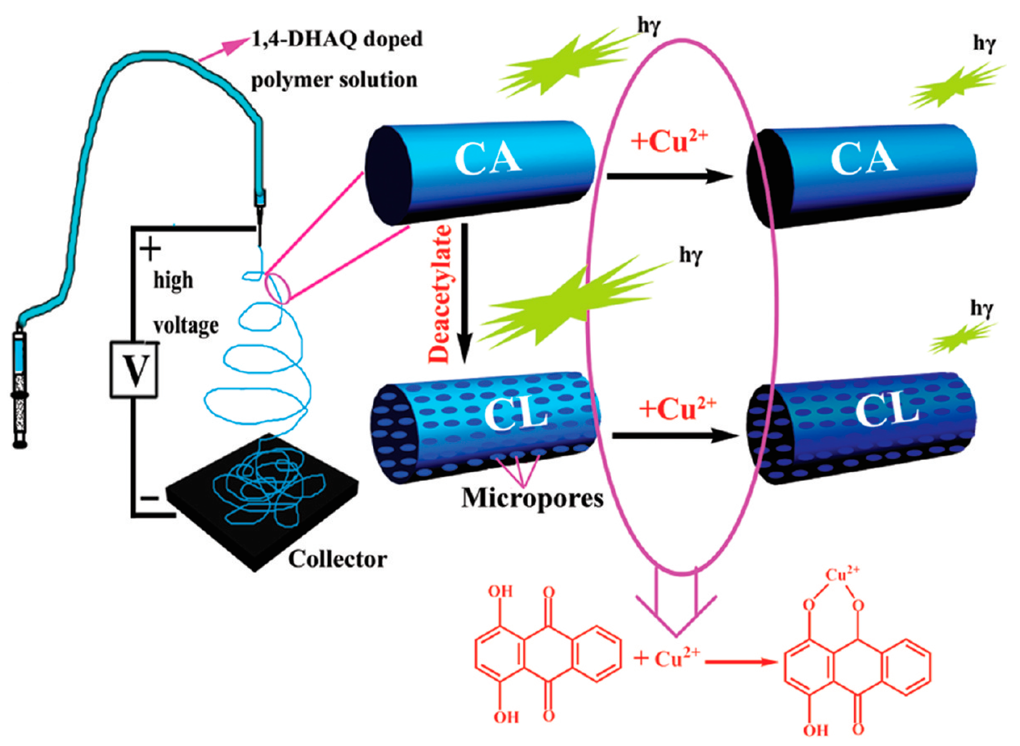
- Wang, M.; Meng, G.; Huang, Q.; Qian, Y. Electrospun 1,4-DHAQ-doped cellulose nanofiber films for reusable fluorescence detection of trace Cu2+ and further for Cr3+. Environ. Sci. Technol. 2012, 46, 367–373. [Google Scholar] [CrossRef]
- Min, M.; Wang, X.; Chen, Y.; Wang, L.; Huang, H.; Shi, J. Highly sensitive and selective Cu2+ sensor based on electrospun rhodamine dye doped poly (ether sulfones) nanofibers. Sens. Actuators B Chem. 2013, 188, 360–366. [Google Scholar] [CrossRef]
- Tungsombatvisit, N.; Inprasit, T.; Rohmawati, D.; Pisitsak, P. Rhodamine derivative-based cellulose acetate electrospun colorimetric sensor for Cu2+ sensing in water: Effects of alkaline treatment. Fibers Polym. 2019, 20, 481–489. [Google Scholar] [CrossRef]
- Ongun, M.Z.; Ertekin, K.; Gocmenturk, M.; Ergun, Y.; Suslu, A. Copper ion sensing with fluorescent electrospun nanofibers. Spectrochim. Acta A 2012, 90, 177–185. [Google Scholar] [CrossRef] [PubMed]
- Gao, W.; Haratipour, P.; Kahkha, M.R.R.; Tahvili, A. Ultrasound-electrospinning-assisted fabrication and sensing evaluation of a novel membrane as ultrasensitive sensor for copper (II) ions detection in aqueous environment. Ultrason. Sonochem. 2018, 44, 152–161. [Google Scholar] [CrossRef] [PubMed]
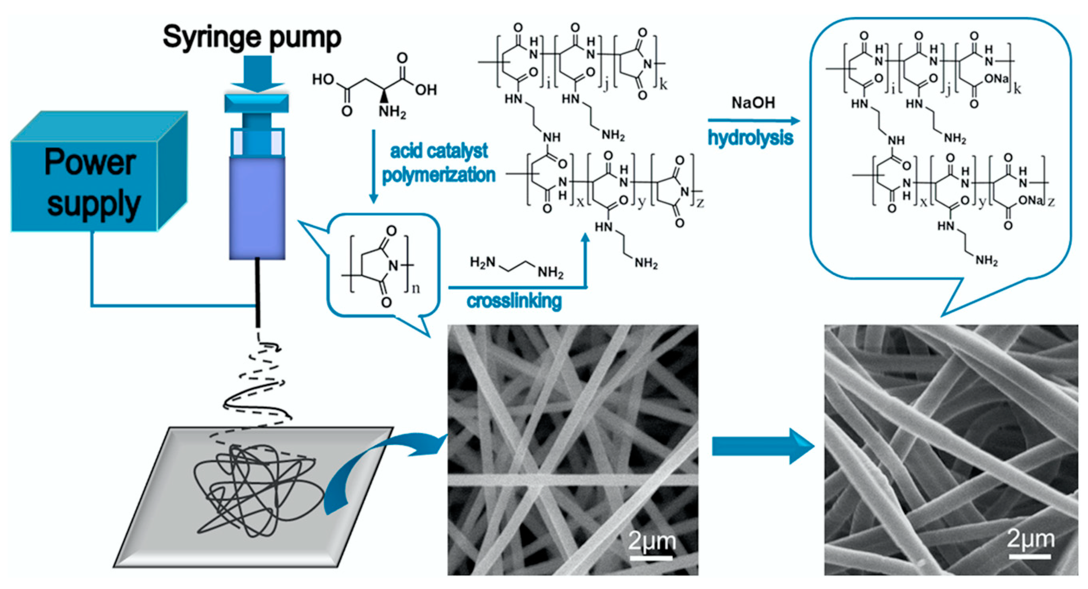
- Zhang, C.; Wan, L.Y.; Wu, S.; Wu, D.; Qin, X.; Ko, F. A reversible colorimetric chemosensor for naked-eye detection of copper ions using poly (aspartic acid) nanofibrous hydrogel. Dyes Pigm. 2015, 123, 380–385. [Google Scholar] [CrossRef]
- Zhang, C.; Li, H.; Yu, Q.; Jia, L.; Wan, L.Y. Poly (aspartic acid) electrospun nanofiber hydrogel membrane-based reusable colorimetric sensor for Cu (II) and Fe (III) detection. ACS Omega 2019, 4, 14633–14639. [Google Scholar] [CrossRef] [Green Version]
- Wang, Y.; Zhu, Y.; Huang, J.; Cai, J.; Zhu, J.; Yang, X.; Shen, J.; Li, C. Perovskite quantum dots encapsulated in electrospun fiber membranes as multifunctional supersensitive sensors for biomolecules, metal ions and pH. Nanoscale Horiz. 2017, 2, 225–232. [Google Scholar] [CrossRef]
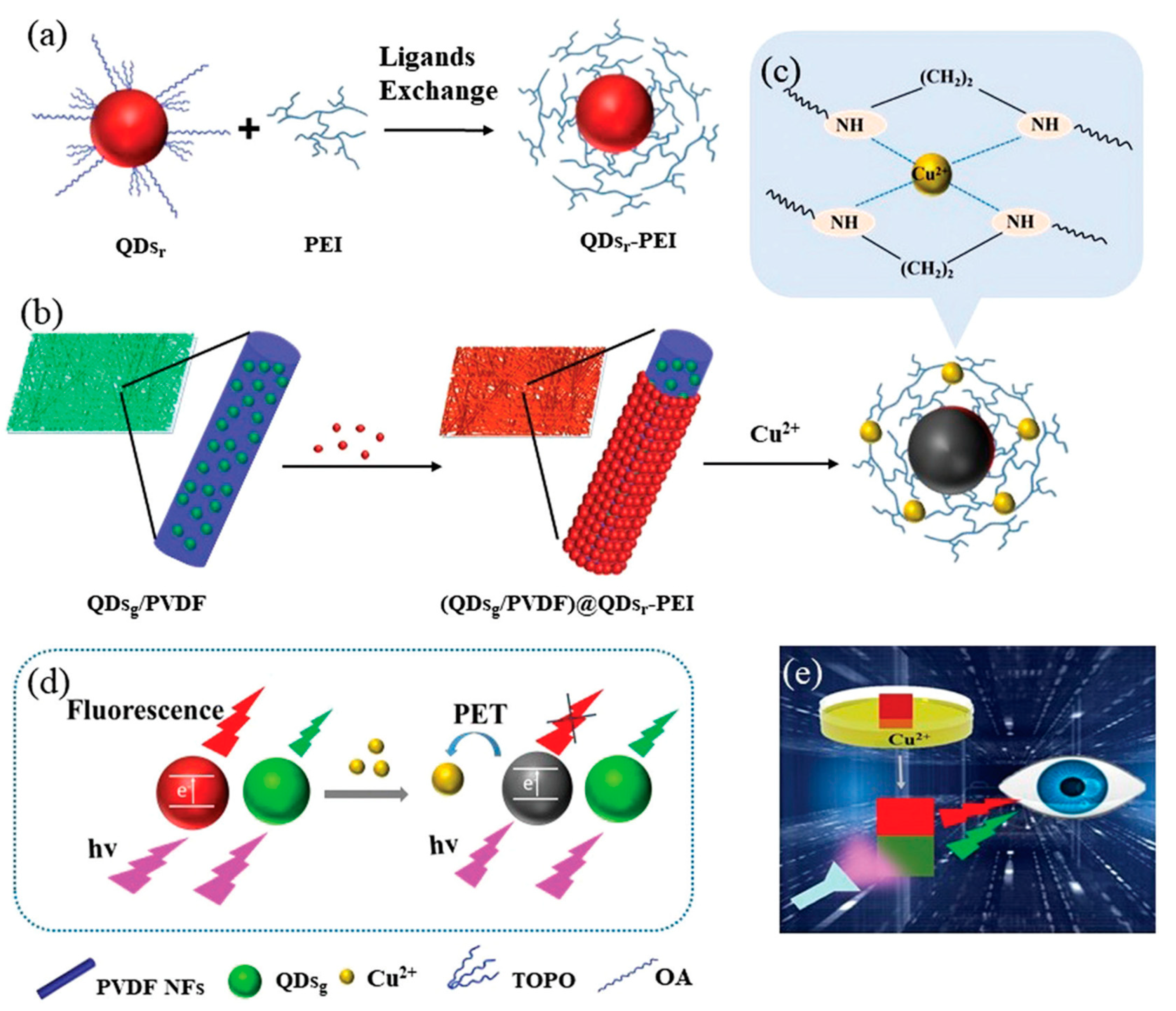
- Han, T.; Yuan, Y.; Kang, H.; Zhang, Y.; Dong, L. Ultrafast, sensitive and visual sensing of copper ions by a dual-fluorescent film based on quantum dots. J. Mater. Chem. C 2019, 7, 14904–14912. [Google Scholar] [CrossRef]
- Wang, W.; Wang, X.; Yang, Q.; Fei, X.; Sun, M.; Song, Y. A reusable nanofibrous film chemosensor for highly selective and sensitive optical signaling of Cu2+ in aqueous media. Chem. Commun. 2013, 49, 4833–4835. [Google Scholar] [CrossRef] [PubMed]
- Cho, C.J.; Lu, S.T.; Kuo, C.C.; Liang, F.C.; Chen, B.Y.; Chu, C.C. Pyrene or rhodamine derivative-modified surfaces of electrospun nanofibrous chemosensors for colorimetric and fluorescent determination of Cu2+, Hg2+, and pH. React. Funct. Polym. 2016, 108, 137–147. [Google Scholar] [CrossRef]
- Cui, X.; Li, T.; Li, J.; An, Y.; An, L.; Zhang, X.; Zhang, Z. A highly selective and reversible turn-off fluorescent chemosensor for Cu2+ based on electrospun nanofibrous membrane modified with pyrenecarboxaldehyde. Spectrochim. Acta A 2019, 207, 173–182. [Google Scholar] [CrossRef] [PubMed]
- Gu, Y.; Wang, M.; Li, G. CNT-anchored cellulose fluorescent nanofiber membranes as a fluorescence sensor for Cu2+ and Cr3+. Anal Methods 2017, 9, 6044–6048. [Google Scholar] [CrossRef]
- Abedalwafa, M.A.; Li, Y.; Li, D.; Sanbhal, N.; Yang, J.; Wang, L. Aminated polyacrylonitrile nanofibers with immobilized gold-silver core-shell nanoparticles for use in a colorimetric test strip for copper (II). Microchim. Acta 2018, 185, 402. [Google Scholar] [CrossRef]
- Senthamizhan, A.; Celebioglu, A.; Balusamy, B.; Uyar, T. Immobilization of gold nanoclusters inside porous electrospun fibers for selective detection of Cu (II): A strategic approach to shielding pristine performance. Sci. Rep. 2015, 5, 15608. [Google Scholar] [CrossRef] [Green Version]
- Li, Y.; Liu, Z.; Bai, L.; Liu, Y. Nitrogen-doped carbon dots derived from electrospun carbon nanofibers for Cu (II) ion sensing. New J. Chem. 2019, 43, 1812–1817. [Google Scholar] [CrossRef]
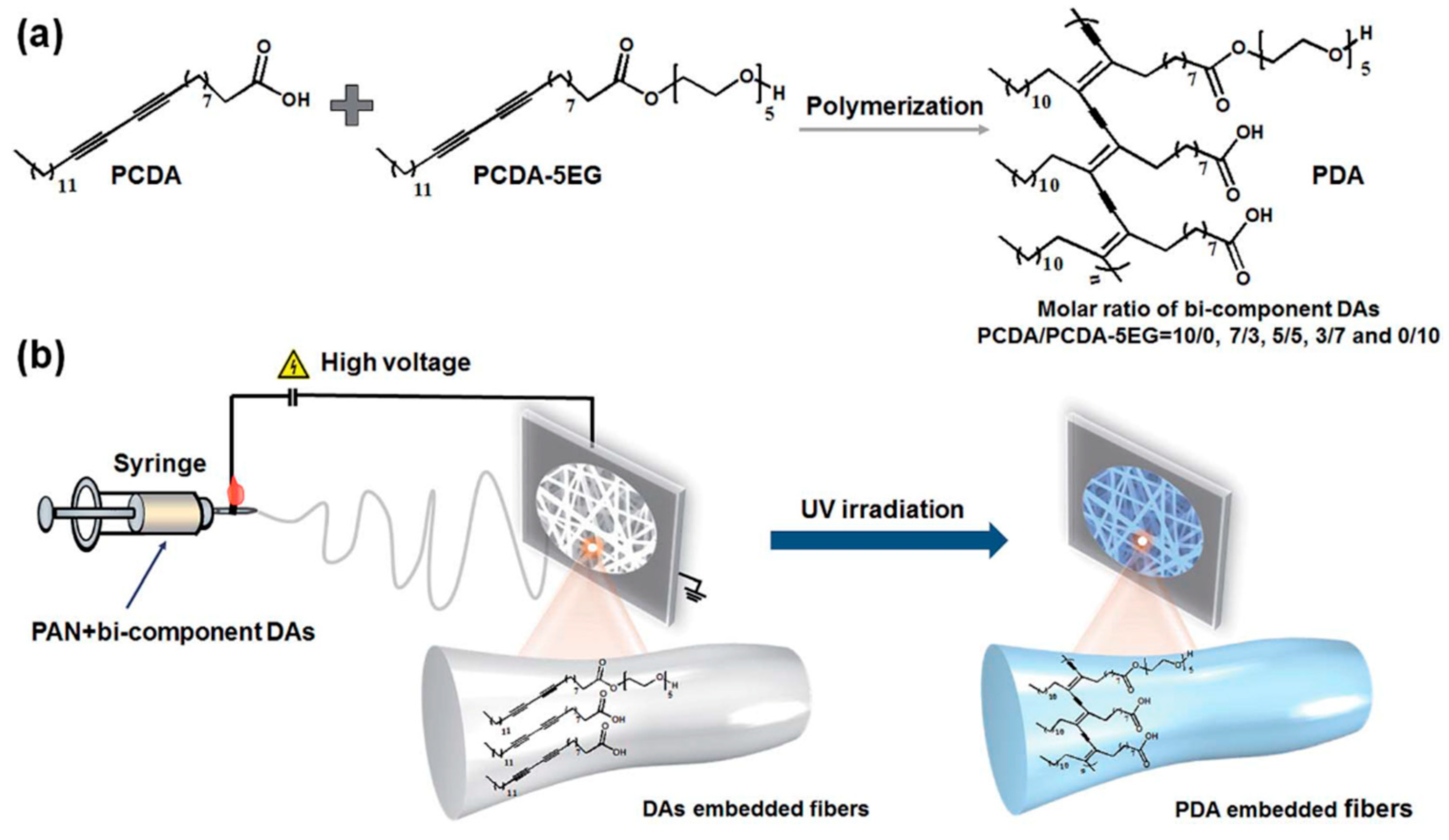
- Li, Y.; Wang, L.; Yin, X.; Ding, B.; Sun, G.; Ke, T.; Chen, J.; Yu, J. Colorimetric strips for visual lead ion recognition utilizing polydiacetylene embedded nanofibers. J. Mater. Chem. A 2014, 2, 18304–18312. [Google Scholar] [CrossRef]
- Li, Y.; Wang, L.; Wen, Y.; Ding, B.; Sun, G.; Ke, T.; Chen, J.; Yu, J. Constitution of a visual detection system for lead (II) on polydiacetylene-glycine embedded nanofibrous membranes. J. Mater. Chem. A 2015, 3, 9722–9730. [Google Scholar] [CrossRef]
- Raj, S.; Shankaran, D.R. Curcumin based biocompatible nanofibers for lead ion detection. Sens. Actuators B Chem. 2016, 226, 318–325. [Google Scholar] [CrossRef]
- Raj, S.; Shankaran, D.R. Synthesis and characterization of curcumin nanoparticles loaded nanofibers for lead ion detection. Sens. Lett. 2016, 14, 889–895. [Google Scholar]
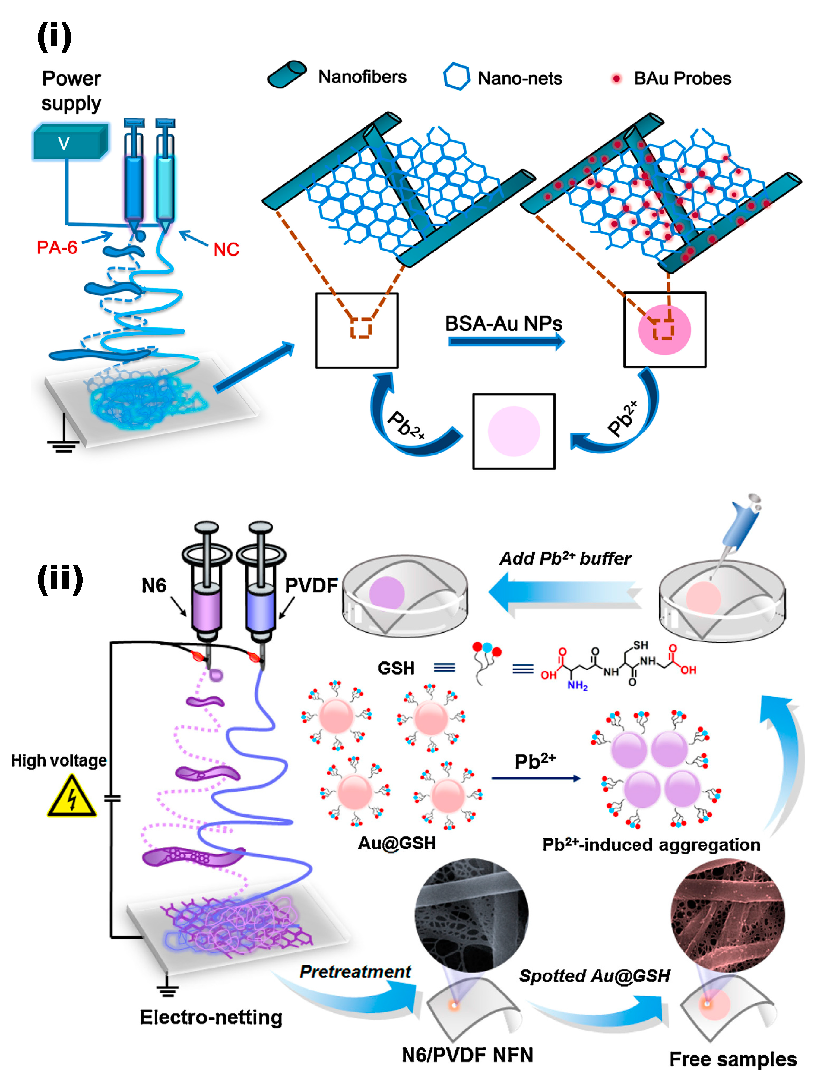
- Li, Y.; Si, Y.; Wang, X.; Ding, B.; Sun, G.; Zheng, G.; Luo, W.; Yu, J. Colorimetric sensor strips for lead (II) assay utilizing nanogold probes immobilized polyamide-6/nitrocellulose nano-fibers/nets. Biosens. Bioelectron. 2013, 48, 244–250. [Google Scholar] [CrossRef] [PubMed]
- Li, Y.; Ding, B.; Sun, G.; Ke, T.; Chen, J.; Al-Deyab, S.S.; Yu, J. Solid-phase pink-to-purple chromatic strips utilizing gold probes andnanofibrous membranes combined system for lead (II) assaying. Sens. Actuators B Chem. 2014, 204, 673–681. [Google Scholar] [CrossRef]
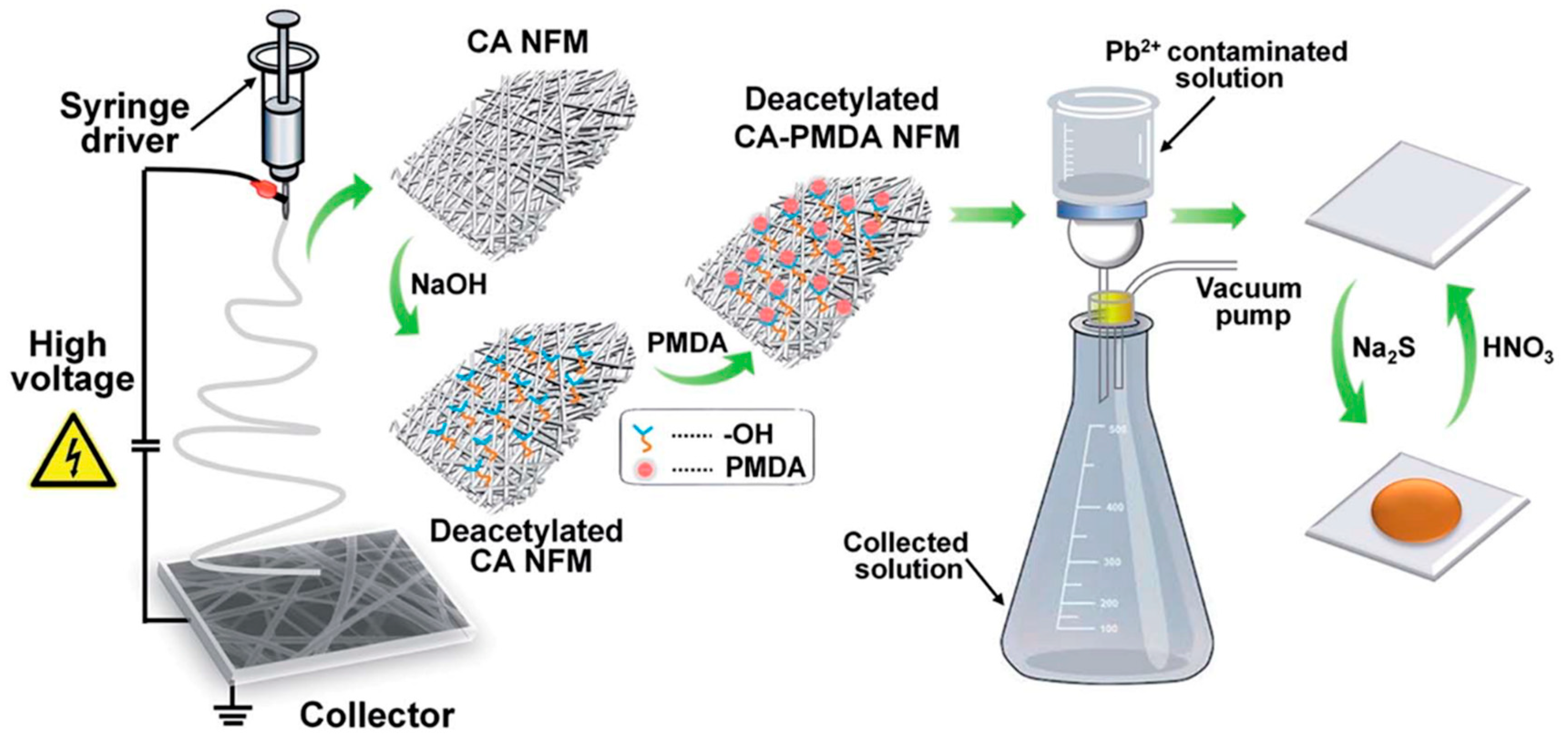
- Li, Y.; Wen, Y.; Wang, L.; He, J.; Al-Deyab, S.S.; El-Newehy, M.; Yu, J.; Ding, B. Simultaneous visual detection and removal of lead (II) ions with pyromellitic dianhydride-grafted cellulose nanofibrous membranes. J. Mater. Chem. A 2015, 3, 18180–18189. [Google Scholar] [CrossRef]
- Zhang, H.; Cao, M.; Wu, W.; Xu, H.; Cheng, S.; Fan, L.J. Polyacrylonitrile/noble metal/SiO2 nanofibers as substrates for the amplified detection of picomolar amounts of metal ions through plasmon-enhanced fluorescence. Nanoscale 2015, 7, 1374–1382. [Google Scholar] [CrossRef]
- Godt, J.; Scheidig, F.; Grosse-Siestrup, C.; Esche, V.; Brandenburg, P.; Reich, V.; Groneberg, D.A. The toxicity of cadmium and resulting hazards for human health. J. Occup. Med. Toxicol. 2006, 1, 22. [Google Scholar] [CrossRef] [Green Version]
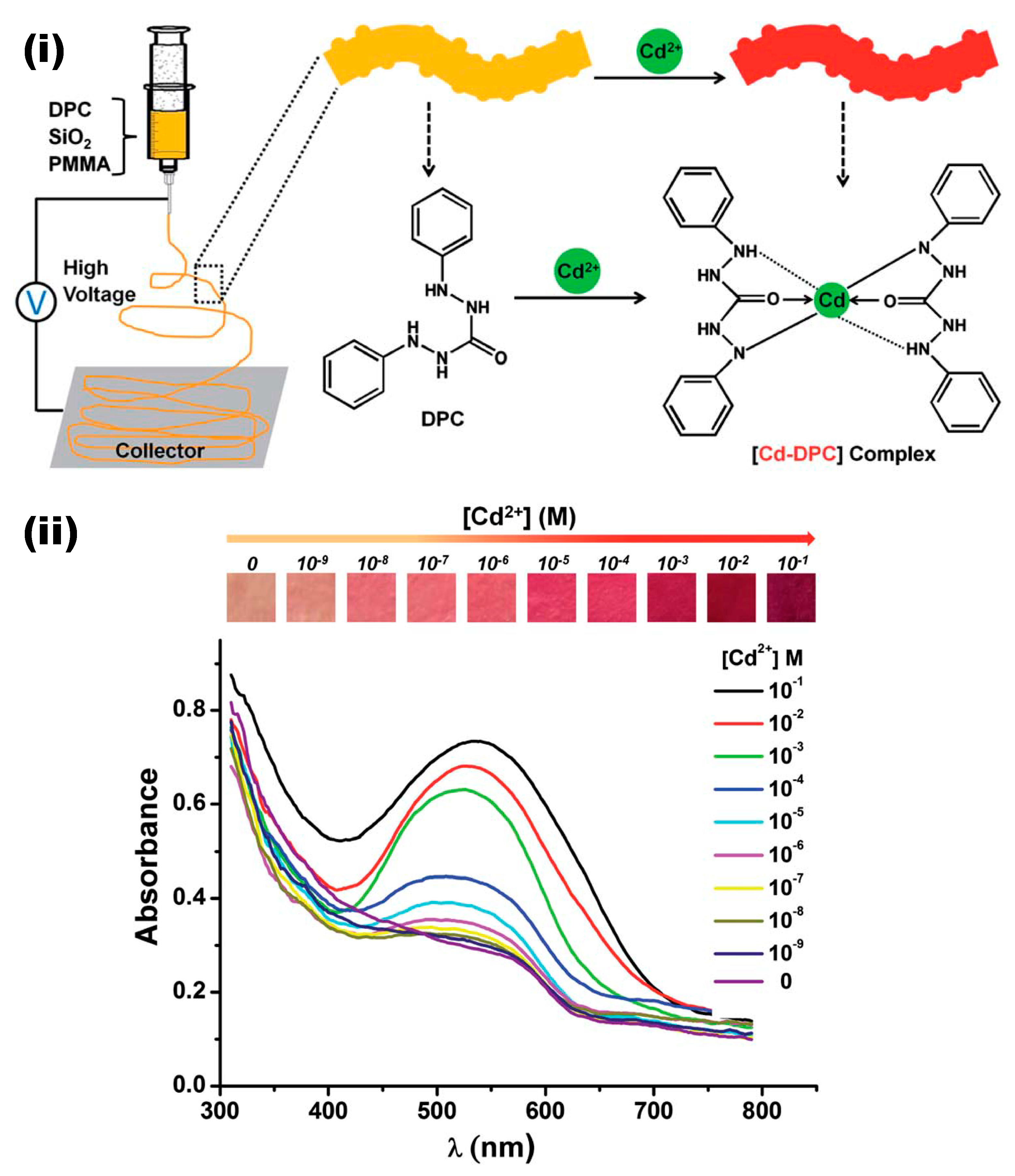
- Yao, T.; Tu, Q.; Han, X.; Zhang, L.; Wang, D.E.; Li, M.; Chen, S.; Wang, J. SiO2 nanoparticles and diphenylcarbazide doped polymethylmethacrylate electrospun fibrous film for Cd2+ colorimetric detection. Anal. Methods 2014, 6, 4102–4106. [Google Scholar] [CrossRef]
- Baruthio, F. Toxic effects of chromium and its compounds. Biol. Trace. Elem. Res. 1992, 32, 145–153. [Google Scholar] [CrossRef]
- Katz, S.A.; Salem, H. The toxicology of chromium with respect to its chemical speciation: A review. J. Appl. Toxicol. 1993, 13, 217–224. [Google Scholar] [CrossRef]
- Krabbenhoft, D.P.; Sunderland, E.M. Global change and mercury. Science 2013, 341, 1457–1458. [Google Scholar] [CrossRef] [PubMed]
- Streets, D.G.; Horowitz, H.M.; Jacob, D.J.; Lu, Z.; Levin, L.; ter Schure, A.F.H.; Sunderland, E.M. Total mercury released to the environment by human activities. Environ. Sci. Technol. 2017, 51, 5969–5977. [Google Scholar] [CrossRef] [PubMed]
- Liu, M.; Zhang, Q.; Cheng, M.; He, Y.; Chen, L.; Zhang, H.; Cao, H.; Shen, H.; Zhang, W.; Tao, S.; et al. Rice life cycle-based global mercury biotransport and human methylmercury exposure. Nat. Commun. 2019, 10, 5164. [Google Scholar] [CrossRef] [PubMed]
- Senthamizhan, A.; Celebioglu, A.; Uyar, T. Flexible and highly stable electrospun nanofibrous membrane incorporating gold nanoclusters as an efficient probe for visual colorimetric detection of Hg (II). J. Mater. Chem. A 2014, 2, 12717–12723. [Google Scholar] [CrossRef] [Green Version]
- Wang, X.; Drew, C.; Lee, S.H.; Senecal, K.J.; Kumar, J.; Samuelson, L.A. Electrospun nanofibrous membranes for highly sensitive optical sensors. Nano Lett. 2002, 2, 1273–1275. [Google Scholar] [CrossRef]
- Ma, L.; Liu, K.; Yin, M.; Chang, J.; Geng, Y.; Pan, K. Fluorescent nanofibrous membrane (FNFM) for the detection of mercuric ion (II) with high sensitivity and selectivity. Sens. Actuators B Chem. 2017, 238, 120–127. [Google Scholar] [CrossRef]
- Tahvili, A.; Poush, M.K.; Ahmed, M.; Parsaee, Z. New efficient inorganic-organic nanofibers electrospun membrane for fluorescence detection and removal of mercury (II) ions. J. Mol. Struct. 2019, 1179, 242–251. [Google Scholar] [CrossRef]
- Chen, B.Y.; Kuo, C.C.; Cho, C.J.; Liang, F.C.; Jeng, R.J. Novel fluorescent chemosensory filter membranes composed of electrospun nanofibers with ultra-selective and reversible pH and Hg2+ sensing characteristics. Dye. Pigment 2017, 143, 129–142. [Google Scholar] [CrossRef]
- Liang, F.C.; Luo, Y.L.; Kuo, C.C.; Chen, B.Y.; Cho, C.J.; Lin, F.J.; Yu, Y.Y.; Borsali, R. Novel magnet and thermoresponsive chemosensory electrospinning fluorescent nanofibers and their sensing capability for metal ions. Polymers 2017, 9, 136. [Google Scholar] [CrossRef] [PubMed]
- Parsaee, Z. Electrospun nanofibers decorated with bio-sonochemically synthesized gold nanoparticles as an ultrasensitive probe in amalgam-based mercury (II) detection system. Ultrason. Sonochem. 2018, 44, 24–35. [Google Scholar] [CrossRef]
- Liang, F.C.; Kuo, C.C.; Chen, B.Y.; Cho, C.J.; Hung, C.C.; Chen, W.C.; Borsali, R. RGB-switchable porous electrospun nanofiber chemoprobe-filter prepared from multifunctional copolymers for versatile sensing of pH and heavy metals. ACS Appl. Mater. Interfaces 2017, 9, 16381–16396. [Google Scholar] [CrossRef] [PubMed]
- Heng, L.; Wang, B.; Zhang, Y.; Zhang, L.; Tang, B.Z.; Jiang, L. Sensing mechanism of nanofibrous membranes for fluorescent detection of metal ion. J. Nanosci. Nanotechnol. 2012, 12, 8443–8447. [Google Scholar] [CrossRef] [PubMed]
- Kacmaz, S.; Ertekin, K.; Suslu, A.; Ergun, Y.; Celik, E.; Cocen, U. Sub-nanomolar sensing of ionic mercury with polymeric electrospun nanofibers. Mater. Chem. Phys. 2012, 133, 547–552. [Google Scholar] [CrossRef]
- Senthamizhan, A.; Celebioglu, A.; Uyar, T. Real-time selective visual monitoring of Hg2+ detection at ppt level: An approach to lighting electrospun nanofibers using gold nanoclusters. Sci. Rep. 2015, 5, 10403. [Google Scholar] [CrossRef] [PubMed]
- Orriach-Fernández, F.J.; Medina-Castillo, A.L.; Díaz-Gómez, J.E.; Muñoz de la Peña, A.; Fernández-Sánchez, J.F.; Fernández-Gutiuérrez, A. A sensing microfibre mat produced by electrospinning for the turn-on luminescence determination of Hg2+ in water samples. Sens. Actuators B Chem. 2014, 195, 8–14. [Google Scholar] [CrossRef]
- Ongun, M.Z.; Ertekin, K.; Hizliates, C.G.; Oter, O.; Ergun, Y.; Celik, E. Determination of Hg (II) at sub-nanomolar levels: A comparative study with nanofibrous materials and continuous thin films. Sens. Actuators B Chem. 2013, 181, 244–250. [Google Scholar] [CrossRef]
- Si, Y.; Wang, X.; Li, Y.; Chen, K.; Wang, J.; Yu, J.; Wang, H.; Ding, B. Optimized colorimetric sensor strip for mercury (II) assay using hierarchical nanostructured conjugated polymers. J. Mater. Chem. A 2014, 2, 645–652. [Google Scholar] [CrossRef]
- Wang, W.; Li, Y.; Sun, M.; Zhou, C.; Zhang, Y.; Li, Y.; Yang, Q. Colorimetric and fluorescent nanofibrous film as a chemosensor for Hg2+ in aqueous solution prepared by electrospinning and host-guest interaction. Chem. Commun. 2012, 48, 6040–6042. [Google Scholar] [CrossRef]
- Das, K.K.; Reddy, R.C.; Bagoji, I.B.; Das, S.; Bagali, S.; Mullur, L.; Khodnapur, J.P.; Biradar, M.S. Primary concept of nickel toxicity-an overview. J. Basic Clin. Physiol. Pharm. 2018, 30, 141–152. [Google Scholar] [CrossRef] [Green Version]
- Lu, H.; Shi, X.; Costa, M.; Huang, C. Carcinogenic effect of nickel compounds. Mol. Cell. Biochem. 2005, 279, 45–67. [Google Scholar] [CrossRef]
- Zambelli, B.; Uversky, V.N.; Ciurli, S. Nickel impact on human health: An intrinsic disorder perspective. Biochim. Biophys. Acta 2016, 1864, 1714–1731. [Google Scholar] [CrossRef] [PubMed]
- Poltue, T.; Rangkupan, R.; Dubas, S.T.; Dubas, L. Nickel (II) ions sensing properties of dimethylglyoxime/poly(caprolactone) electrospun fibers. Mater. Lett. 2011, 65, 2231–2234. [Google Scholar] [CrossRef]
- Najarzadekan, H.; Sereshti, H. Development of a colorimetric sensor for nickel ion based on transparent electrospun composite nanofibers of polycaprolactam-dimethylglyoxime/polyvinyl alcohol. J. Mater. Sci. 2016, 51, 8645–8654. [Google Scholar] [CrossRef]
- Adewuyi, S.; Ondigo, D.A.; Zugle, R.; Tshentu, Z.; Nyokong, T.; Torto, N. A highly selective and sensitive pyridylazo-2-naphthol-poly (acrylic acid) functionalized electrospun nanofiber fluorescence ‘‘turn-off’’ chemosensory system for Ni2+. Anal. Methods 2012, 4, 1729–1735. [Google Scholar] [CrossRef]
- Koh, J.Y.; Suh, S.W.; Gwag, B.J.; He, Y.Y.; Hsu, C.Y.; Choi, D.W. The role of zinc in selective neuronal death after transient global cerebral ischemia. Science 1996, 272, 1013–1016. [Google Scholar] [CrossRef]
- Frederickson, C.J.; Koh, J.Y.; Bush, A.I. The neurobiology of zinc in health and disease. Nat. Rev. Neurosci. 2005, 6, 449–462. [Google Scholar] [CrossRef]
- Syu, J.H.; Cheng, Y.K.; Hong, W.Y.; Wang, H.P.; Lin, Y.C.; Meng, H.F.; Zan, H.W.; Horng, S.F.; Chang, G.F.; Hung, C.H.; et al. Electrospun fibers as a solid-state real-time zinc ion sensor with high sensitivity and cell medium compatibility. Adv. Funct. Mater. 2013, 23, 1566–1574. [Google Scholar] [CrossRef]
- Chen, L.N.; Kuo, C.C.; Chiu, Y.C.; Chen, W.C. Ultra metal ions and pH sensing characteristics of thermoresponsive luminescent electrospun nanofibers prepared from poly (HPBO-co-NIPAAm-co-SA). RSC Adv. 2014, 4, 45345–45353. [Google Scholar] [CrossRef]
- Chen, L.N.; Weng, N.K.; Wu, W.C.; Chen, W.C. Electrospun polymer nanofibers of P(NIPAAm-co-SA-co-FBPY): Preparation, structural control, metal ion sensing and thermoresponsive characteristics. Mater. Chem. Phys. 2015, 163, 63–72. [Google Scholar] [CrossRef]
- Abbaspour, N.; Hurrell, R.; Kelishadi, R. Review on iron and its importance for human health. J. Res. Med. Sci. 2014, 19, 164–174. [Google Scholar]
- Staniek, H.; Wójciak, R.W. The combined effects of iron excess in the diet and chromium (III) supplementation on the iron and chromium status in female rats. Biol. Trace. Elem. Res. 2018, 184, 398–408. [Google Scholar] [CrossRef] [Green Version]
- Saithongdee, A.; Praphairaksit, N.; Imyim, A. Electrospun curcumin-loaded zein membrane for iron (III) ions sensing. Sens. Actuators B Chem. 2014, 202, 935–940. [Google Scholar] [CrossRef]
- Qiao, Y.; Shi, C.; Wang, X.; Wang, P.; Zhang, Y.; Wang, D.; Qiao, R.; Wang, X.; Zhong, J. Electrospun nanobelt-shaped polymer membranes for fast and high-sensitivity detection of metal ions. ACS Appl. Mater. Interfaces 2019, 11, 5401–5413. [Google Scholar] [CrossRef] [PubMed]
- Kacmaz, S.; Ertekin, K.; Gocmenturk, M.; Suslu, A.; Ergun, Y.; Celik, E. Selective sensing of Fe3+ at pico-molar level with ethyl cellulose based electrospun nanofibers. React. Funct. Polym. 2013, 73, 674–682. [Google Scholar] [CrossRef]
- Najarzadekan, H.; Sereshti, H. Transparent polycaprolactam electrospun nanofibers doped with 1,10-phenanthroline optical sensor for colorimetric determination of iron (II) and vitamin C. Fibers Polym. 2018, 19, 2149–2156. [Google Scholar] [CrossRef]
- Rijin, K.K.; Sagitha, P.; Amitha, G.S.; Vasudevan, S.; Sujith, A. 4,4′-Fluoresceinoxy bisphthalonitrile (FPN)-incorporated polycaprolactone electrospun membranes: A portable sensor strip for detection of Fe3+ ions. J. Mater. Sci. 2019, 54, 13433–13444. [Google Scholar] [CrossRef]
- Martwiset, S.; Nijpanich, S.; Banturngsaksiri, A.; Sriring, M.; Pandhumas, T.; Youngme, S. Pyrene-doped electrospun PMMA-PVC fibers for ferric ion detection. J. Appl. Polym. Sci. 2013, 130, 3205–3211. [Google Scholar] [CrossRef]
- Chen, B.Y.; Kuo, C.C.; Huang, Y.S.; Lu, S.T.; Liang, F.C.; Jiang, D.H. Novel highly selective and reversible chemosensors based on dual-ratiometric fluorescent electrospun nanofibers with pH- and Fe3+-modulated multicolor fluorescence emission. ACS Appl. Mater. Interfaces 2015, 7, 2797–2808. [Google Scholar] [CrossRef]
- Wang, J.T.; Chiu, Y.C.; Sun, H.S.; Yoshida, K.; Chen, Y.; Satoh, T.; Kakuchi, T.; Chen, W.C. Synthesis of multifunctional poly (1-pyrenemethyl methacrylate)-b-poly (N-isopropylacrylamide)-b-poly (N-methylolacrylamide)s and their electrospun nanofibers for metal ion sensory applications. Polym. Chem. 2015, 6, 2327–2336. [Google Scholar] [CrossRef]
- Li, Z.; Li, H.; Shi, C.; Zhang, W.; Zhou, W.; Wei, L.; Yu, M. Naked-eye-based highly selective sensing of Fe3+ and further for PPi with nano copolymer film. Sens. Actuators B Chem. 2016, 226, 127–134. [Google Scholar] [CrossRef]
- Ondigo, D.A.; Tshentu, Z.R.; Torto, N. Electrospun nanofiber based colorimetric probe for rapid detection of Fe2+ in water. Anal. Chim. Acta 2013, 804, 228–234. [Google Scholar] [CrossRef]
- Li, S.; Zhou, S.; Xu, H.; Xiao, L.; Wang, Y.; Shen, H.; Wang, H.; Yuan, Q. Luminescent properties and sensing performance of a carbon quantum dot encapsulated mesoporous silica/polyacrylonitrile electrospun nanofibrous membrane. J. Mater. Sci. 2016, 51, 6801–6811. [Google Scholar] [CrossRef]
- Kacmaz, S.; Ertekin, K.; Suslu, A.; Ozdemir, M.; Ergun, Y.; Celik, E.; Cocen, U. Emission based sub-nanomolar silver sensing with electrospun nanofibers. Sens. Actuators B Chem. 2011, 153, 205–213. [Google Scholar] [CrossRef]
- Berthon, G. Aluminium speciation in relation to aluminium bioavailability, metabolism and toxicity. Coord. Chem. Rev. 2002, 228, 319–341. [Google Scholar] [CrossRef]
- Kim, C.; Hwang, J.Y.; Ku, K.S.; Angupillai, S.; Son, Y.A. A renovation of non-aqueous Al3+ sensor to aqueous media sensor by simple recyclable immobilize electrospun nano-fibers and its uses for live sample analysis. Sens. Actuators B Chem. 2016, 228, 259–269. [Google Scholar] [CrossRef]
- Melnikov, P.; Zanoni, L.Z. Clinical effects of cesium intake. Biol. Trace Elem. Res. 2010, 135, 1–9. [Google Scholar] [CrossRef] [PubMed]
- Jung, S.H.; Park, J.S.; Choi, Y.; Kim, S.K.; Jung, J.H. Calix [4] arene-based fluorescent probe and the adsorption capacity of its electrospun nanofibrous film for the cesium cation as an adsorbent. Supramol. Chem. 2017, 29, 139–145. [Google Scholar] [CrossRef]
- Ahn, J.; Lim, N.Y.; Choi, Y.; Choi, M.Y.; Jung, J.H. Highly selective chromogenic probe for cesium ions prepared from an electrospun film of self-assembled benzenetricarboxyamide nanofibers. Sens. Actuators B Chem. 2018, 255, 325–331. [Google Scholar] [CrossRef]


| Fibrous Matrix | Functional Molecule | Encapsulation Approach | Limit of Detection (LOD) | Response Time | Ref |
|---|---|---|---|---|---|
| Polyamide-6/PANI | PANI leucoemeraldine base | Direct blending | 1 ppb | 30 min | [71] |
| Poly(methyl methacrylate-co-1,8-naphthalimide) | 1,8-naphthalimide | Direct blending | 20 × 10−6 M | - | [72] |
| Poly[(N-isopropylacrylamide)-co-(N-hydroxymethyl acrylamide)-co-(4-rhodamine hydrazonomethyl-3-hydroxy-phenyl methacrylate)] | 4-rhodamine hydrazonomethyl-3-hydroxy-phenyl methacrylate | Direct blending | - | - | [73] |
| Poly(2-hydroxyethyl methacrylate-co-N-methylolacrylamide) | 9,9-dihexylfluorene-2,7-bipyridine | Direct blending | - | - | [74] |
| Poly(N-isopropylacrylamide-co-N-hydroxymethyl-acrylamide) | 1,10-phenanthroline | Direct blending | - | - | [75] |
| Cellulose | 1,4-Dihydroxyanthraquinone | Direct blending | 3 nM | - | [76] |
| Poly(ether sulfones) | Rhodamine dye (spirolactam moiety) | Direct blending | 1.1 × 10⁻⁹ M | 10 min | [77] |
| Cellulose Acetate | Rhodamine B derivative | Direct blending | 18 ppm | <100 s | [78] |
| Poly(methyl methacrylate) (PMMA) Ethyl cellulose (EC) | N’-3-(4-(dimethylamino phenly)allylidene) isonicotinohydrazide | Direct blending | 3.8 × 10⁻¹⁴ M for EC 1.4 × 10⁻¹³ M for PMAA | - | [79] |
| Polyvinyl alcohol (PVA)/tetraethyl orthosilicate (TEOS) | Schiff base | Direct blending | 1.27 × 10⁻⁸ mol L−1 | 90 s | [80] |
| Polysuccinimide | Poly (aspartic acid) | Direct blending | 0.01 mg/L | 5 min | [81] |
| Polysuccinimide | Poly (aspartic acid) | Direct blending | 0.3 mg/L | - | [82] |
| Polymethyl methacrylate | CsPbBr3 perovskite quantum dots and cyclam | Direct blending and surface functionalization | 10−15 M | - | [83] |
| Poly(vinylidene fluoride) | Quantum dots modified with polyethylenimine | Direct blending and surface functionalization | 2 µM | 30 s | [84] |
| Poly (MMA-co-AHPA) | Rhodamine B-hydrazine | Surface functionalization | 1.5 × 10⁻⁶ mol L−1 | <10 s | [85] |
| Poly(HEMA-co-NMA | 2-((pyren-1-yl)methyleneamino)-3-amino maleonitrile) (PyDAN2) | Surface functionalization | 10−7–10−6 M | - | [86] |
| Ethylene-vinyl alcohol copolymer (EVOH) | 4-aminobenzoic acid (PABA) and 1-pyrenecarboxaldehyde (Py-CHO) | Surface functionalization | 1 × 10⁻⁹ M | - | [87] |
| Cellulose | 1,4-Dihydroxyanthraquinone and carbon nanotubes | Direct blending and Surface functionalization | 2.17 × 10⁻⁹ M | 6 min | [88] |
| Polyacrylonitrile | Gold/Silver core/shell nanoparticles | Surface functionalization | 50 nM | 3 min | [89] |
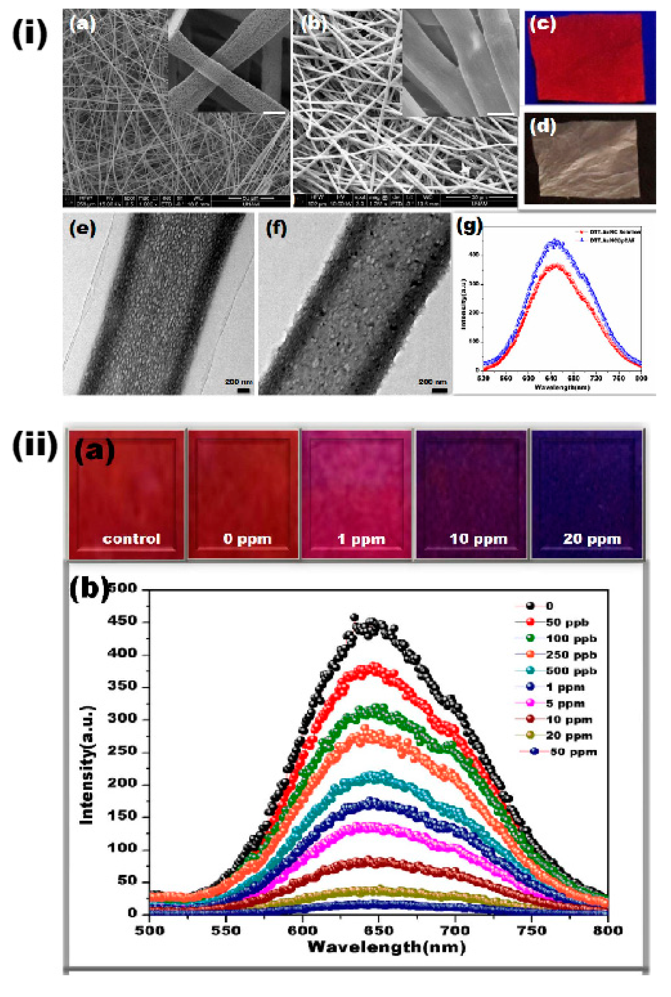
| Cellulose acetate | Dithiothreitol capped gold nanocluster | Surface decoration | 50 ppb | 10 min | [90] |
| Polyacrylonitrile | Nitrogen-doped carbon dots | Carbonization | 5 nM | - | [91] |
| Fibrous Matrix | Functional Molecule | Encapsulation Approach | Limit of Detection (LOD) | Response Time | Ref |
|---|---|---|---|---|---|
| Polyacrylonitrile | Polydiacetylene | Direct blending | 0.48 µM | 30 min | [92] |
| Polyacrylonitrile | Polydiacetylene | Direct blending | 0.24 mM | 10 min | [93] |
| Cellulose acetate | Curcumin | Direct blending | 0.12 ± 0.01 µM | - | [94] |
| Cellulose acetate | Curcumin nanoparticles | Direct blending | 0.14 ± 0.01 mM | - | [95] |
| Polyamide-6 /Nitrocellulose | Bovine serum albumin decorated gold nanoparticles | Surface functionalization | 0.2 µM | 60 min | [96] |
| Nylon-6/Polyvinylidene fluoride | Glutathione conjugated gold nanoparticles | Surface functionalization | 10 µg/dL | 10 min | [97] |
| Deacetylated cellulose acetate | Pyromellitic dianhydride | Surface functionalization | 0.048 µM | - | [98] |
| Polyacrylonitrile | Silver and silica | Direct blending/ Surface functionalization | - | - | [99] |
| Fibrous Matrix | Functional Molecule | Encapsulation Approach | Limit of Detection (LOD) | Response Time | Ref |
|---|---|---|---|---|---|
| Polyvinyl alcohol | Bovine serum albumin (BSA)-capped fluorescent gold nanoclusters (AuNC) | Direct blending | 1 ppb | 2 min | [107] |
| Poly(acrylic acid)−poly(pyrene methanol) (PAA−PM) | Poly(pyrene methanol) | Direct blending | - | - | [108] |
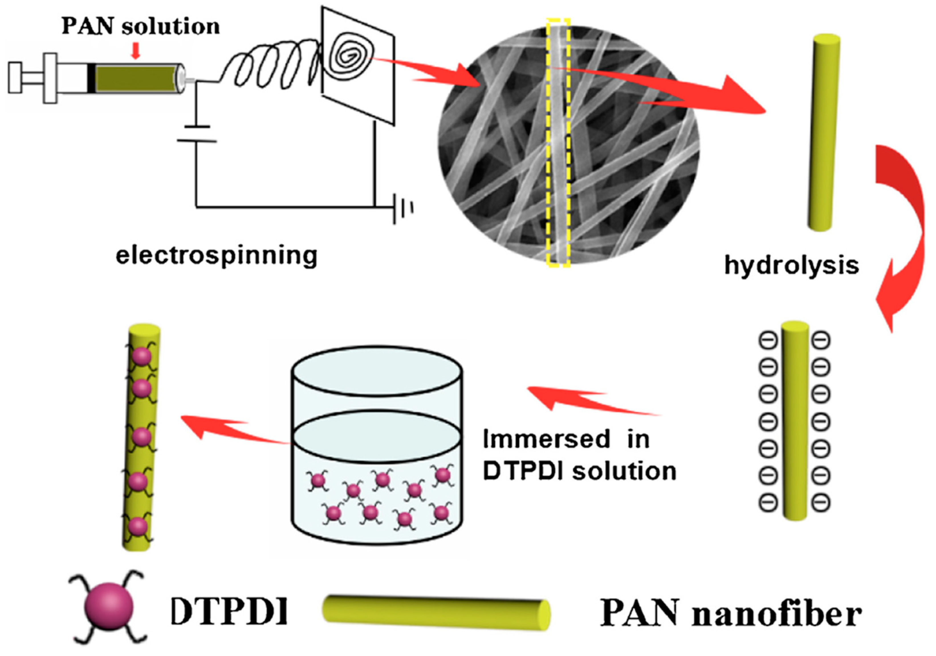
| Polyacrylonitrile | Dithioacetal-modified perylenediimide (DTPDI) | Surface functionalization | 1 ppb | 2 h | [109] |
| Polyvinyl alcohol - tetraethyl orthosilicate | Carbazol-based Schiff base (S) | Direct blending | 0.018 ppb | 60 s | [110] |
| Poly(2-hydroxyethyl methacrylate-co-N-methylolacrylamide-co-rhodamine derivative) | Rhodamine derivative | Direct blending | 10−7 M | 2 min | [111] |
| Poly(N-isopropylacrylamide)-co-(N-methylolacrylamide)-co-(Acrylic acid) | 1-benzoyl-3-[2-(2-allyl-1,3-dioxo-2,3-dihydro- 1Hbenzo[de] isoquinolin-6-ylamino)-ethyl]-thiourea | Direct blending | 10−3 M | - | [112] |
| Gold nanoparticles, rhodamine B, and TEOS | Gold nanoparticles and rhodamine B (RhB) | Direct blending | 1.10 nM | - | [113] |
| Poly(HEMA-co-NMA) | Pyrene derivative (PyDAN2) or rhodamine B derivative (RhBN2) | Surface functionalization | 10−2–10−1 M | - | [86] |
| (poly(methyl methacrylatete-co-1,8-naphthalimide derivatives-co-rhodamine derivative) | Rhodamine derivative | Direct blending | 2 × 10⁻⁸ M | - | [114] |
| Polymethyl methacrylate | Hexaphenylsilole | Direct blending | - | - | [115] |
| Ethyl cellulose | 4-(dimethylamino)benzaldehyde 2- [[4-cyanophenyl] methylene]hydrazone dye (DC-AZM) | Direct blending | 0.07 nM | - | [116] |
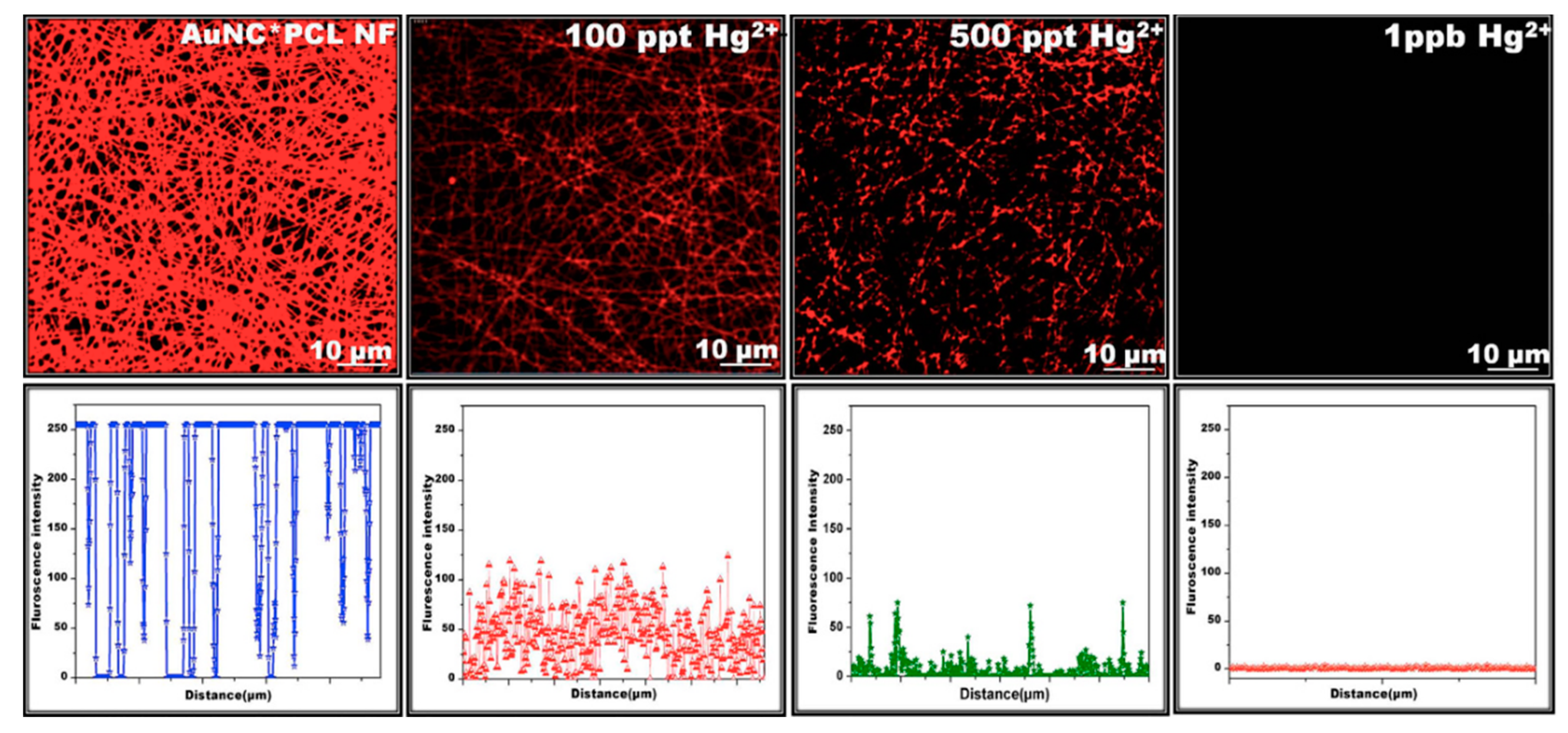
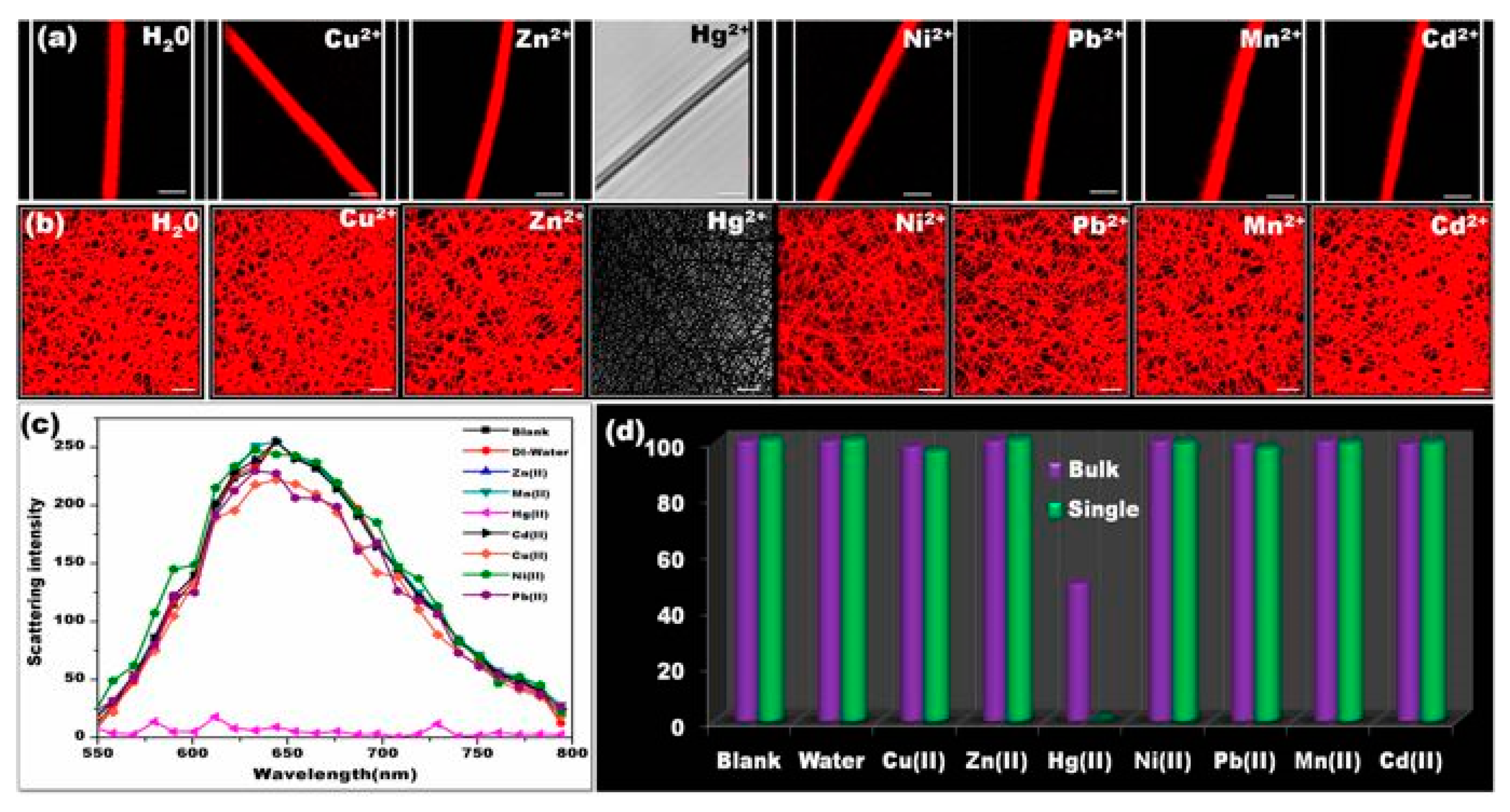
| Polycaprolactone | BSA-capped fluorescent gold nanoclusters (AuNC) | Surface functionalization | 50 ppt | 10 s | [117] |
| Polymer blends of hydroxyl monomers | Spirocyclic Rhodamine 6G phenyl-thiosemicarbazide derivative | Direct blending | 0.1 µM | 15 min | [118] |
| Ethyl cellulose | 2-(9-methyl-9H-carbazol-3-yl)-5-(pyridin-4-yl)- 1,3,4-oxadiazole (ODC-3) dye | Direct blending | 1.70 × 10⁻¹⁵ M | - | [119] |
| Polyacrylonitrile | Silver and Silica | Direct blending/ Surface decoration | - | - | [99] |
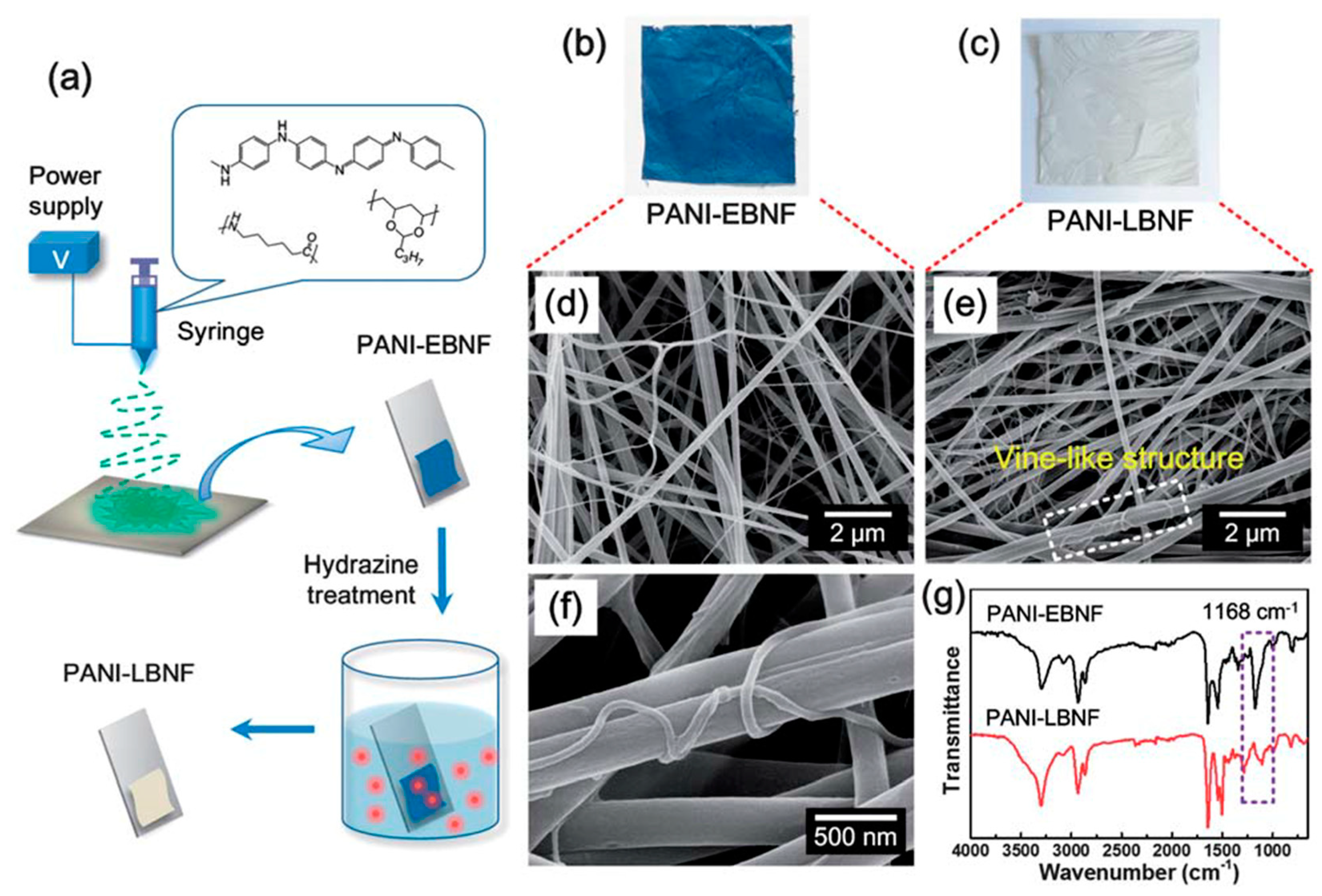
| Polyaniline | Polyaniline leucoemeraldine base (PANI-LB) | Direct blending | 5 nM | 20 min | [120] |
| Poly (MMA-co-ADMA) | Rhodamine–β-cyclodextrin | Surface functionalization | 6.0 × 10⁻⁵ mol L−1 | <1 min | [121] |
| Fibrous Matrix | Functional Molecule | Encapsulation Approach | Limit of Detection (LOD) | Response Time | Ref |
|---|---|---|---|---|---|
| Poly(acrylic acid) | Poly(pyrene methanol) | Direct blending | 1.1 × 10⁶ M−1 | - | [108] |
| Polymethyl methacrylate | Hexaphenylsilole (HPS) | Direct blending | - | - | [115] |
| Zein | Curcumin | Direct blending | 0.4 mg/L | 3 h | [135] |
| Zein | Curcumin | Direct blending | 0.3 mg/L | 0.5 h | [136] |
| Ethyl cellulose | N’-(4-cyanobenzylidene) isonicotinohydrazide (CBINH) | Direct blending | 0.07 pM | <30 s | [137] |
| Polycaprolactam | 1,10-phenanthroline | Direct blending | 1 µg/mL | - | [138] |
| Polycaprolactone | 4,4′-Fluoresceinoxy bisphthalonitrile | Direct blending | 2.9413 nM | 1 min | [139] |
| Polysuccinimide | Poly (aspartic acid) | Direct blending | 0.1 mg/L | - | [82] |
| Polyacrylonitrile | Silver and silica | Direct blending/ Surface functionalization | - | - | [99] |
| Poly(methyl methacrylate) and poly(vinyl chloride-co-vinyl acetate-co-vinyl alcohol) | Pyrene | Direct blending | - | - | [140] |
| Poly(2-hydroxyethyl methacrylate-co-N-methylolacrylamide-co-nitrobenzoxadiazolyl derivative) | Spirolactam rhodamine derivative | Direct blending | 10−4 M | 15 min | [141] |
| Poly (methyl methacrylate-co- rhodamine and quinolone) | Poly(1-pyrenemethylmethacrylate) | Direct blending | 2 × 10⁻⁵ M | - | [142] |
| Poly(methyl methacrylate-co- rhodamine and quinolone) | Rhodamine | Direct blending | 1.19 µM | <1 min | [143] |
| Poly(vinylbenzyl chloride) | 2-(2′-pyridyl)imidazole | Surface functionalization | 2 µg mL−1 | - | [144] |
| Polyacrylonitrile | Carbon quantum dots | Surface functionalization | 3.95 µM | - | [145] |
© 2020 by the authors. Licensee MDPI, Basel, Switzerland. This article is an open access article distributed under the terms and conditions of the Creative Commons Attribution (CC BY) license (http://creativecommons.org/licenses/by/4.0/).
Share and Cite
Balusamy, B.; Senthamizhan, A.; Uyar, T. Functionalized Electrospun Nanofibers as a Versatile Platform for Colorimetric Detection of Heavy Metal Ions in Water: A Review. Materials 2020, 13, 2421. https://doi.org/10.3390/ma13102421
Balusamy B, Senthamizhan A, Uyar T. Functionalized Electrospun Nanofibers as a Versatile Platform for Colorimetric Detection of Heavy Metal Ions in Water: A Review. Materials. 2020; 13(10):2421. https://doi.org/10.3390/ma13102421
Chicago/Turabian StyleBalusamy, Brabu, Anitha Senthamizhan, and Tamer Uyar. 2020. "Functionalized Electrospun Nanofibers as a Versatile Platform for Colorimetric Detection of Heavy Metal Ions in Water: A Review" Materials 13, no. 10: 2421. https://doi.org/10.3390/ma13102421