Effect of Samarium on the Microstructure and Corrosion Resistance of AZ91 Magnesium Alloy Treated by Ultrasonic Vibration


Abstract
:1. Introduction
2. Experimental
2.1. Material Preparation and Microstructural Observation
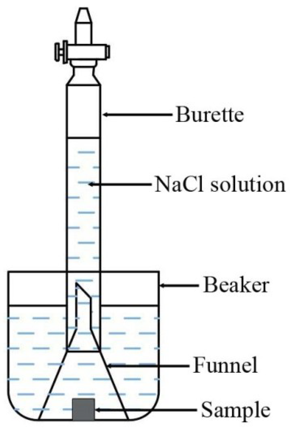
2.2. Immersion Testing
2.3. Electrochemical Testing
3. Results
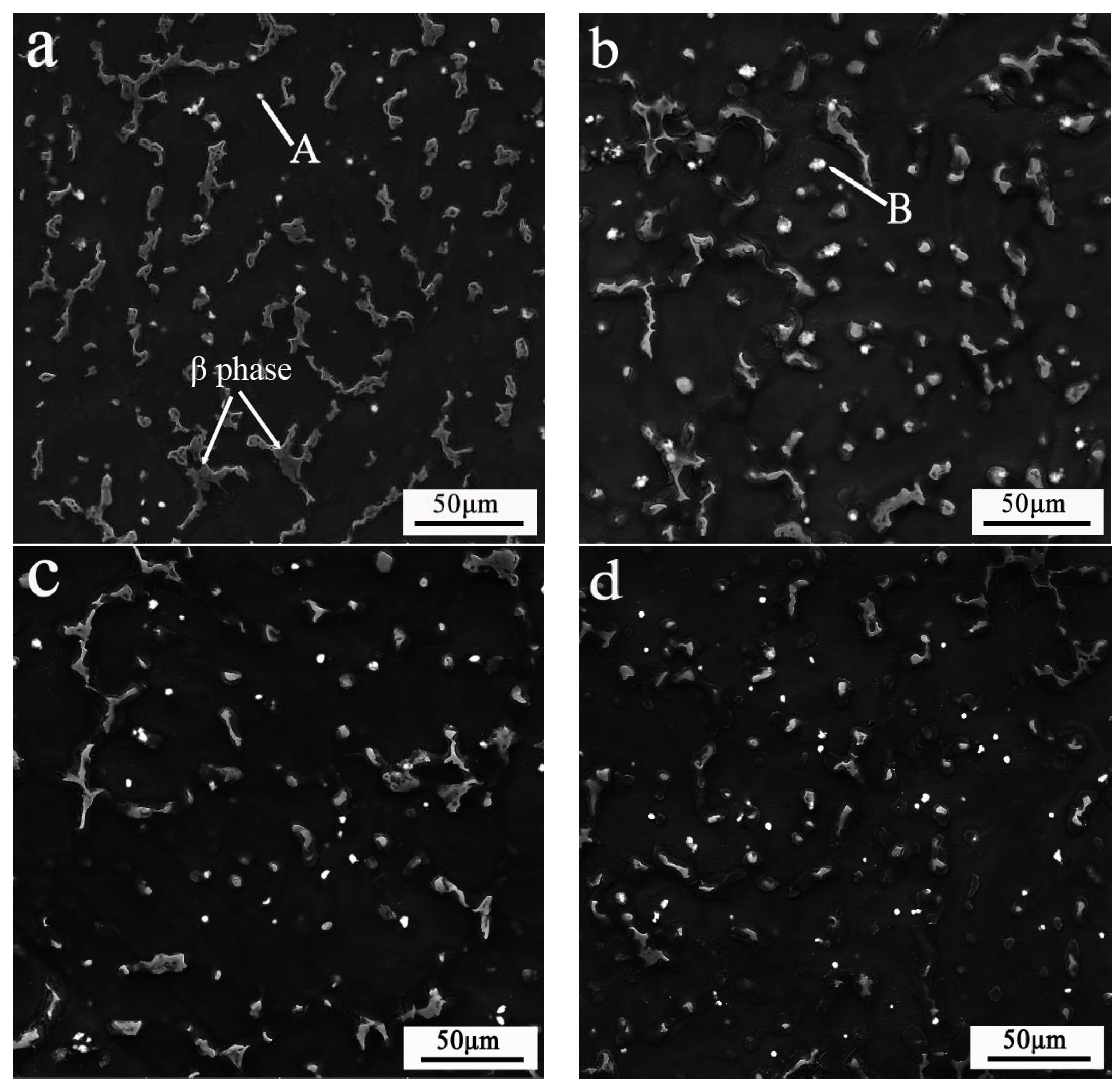
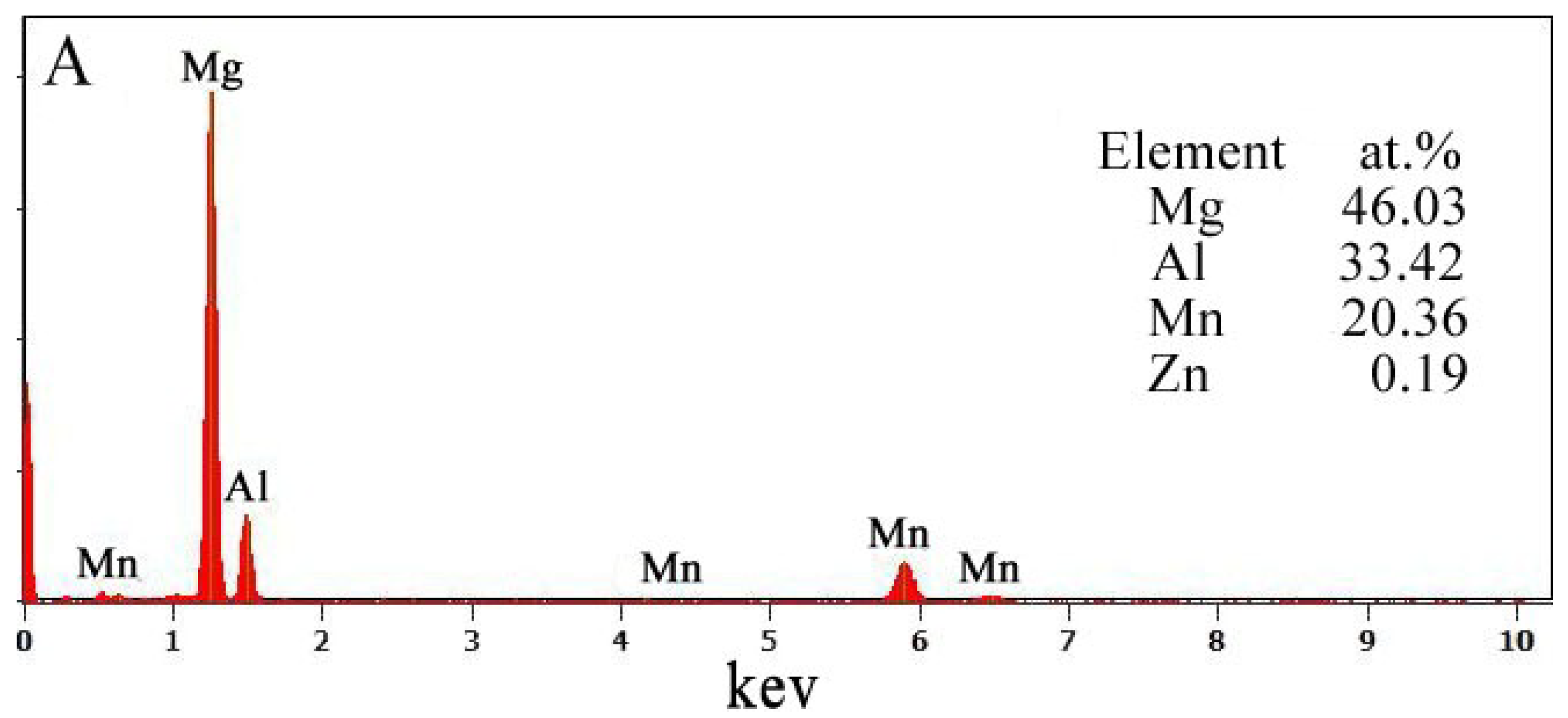
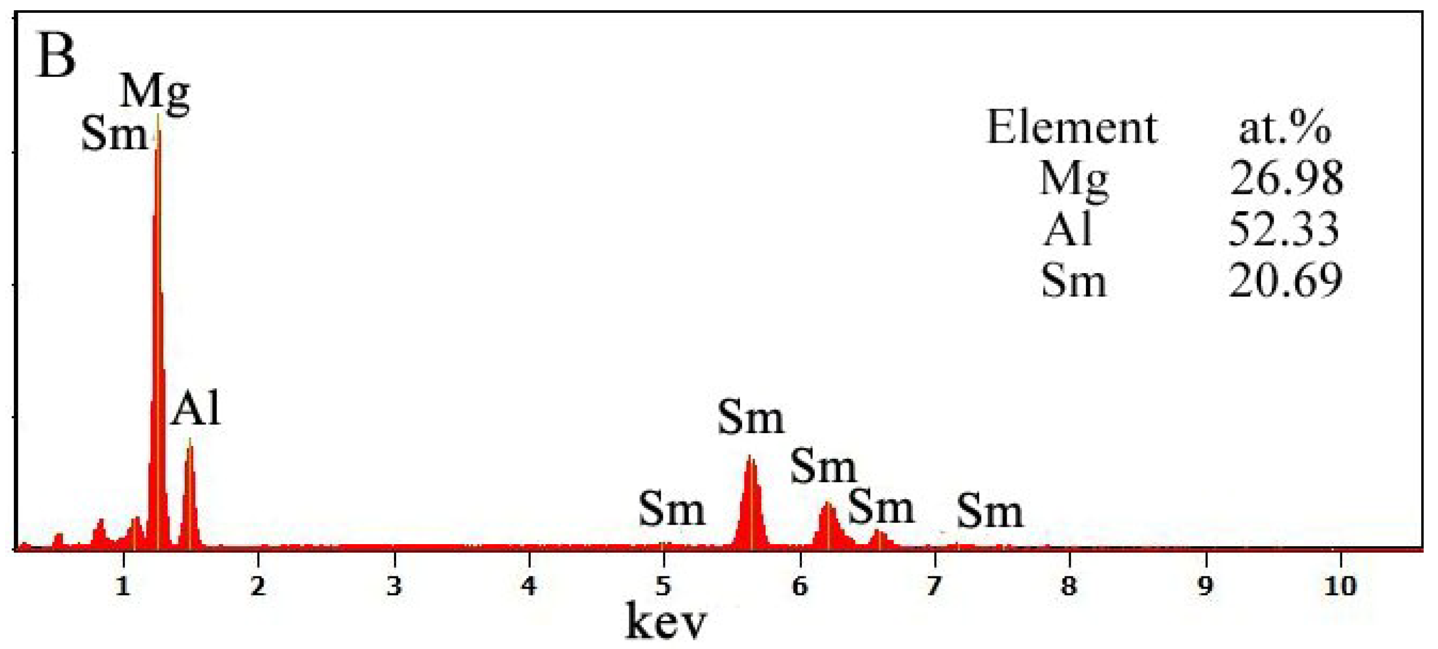
3.1. Microstructure
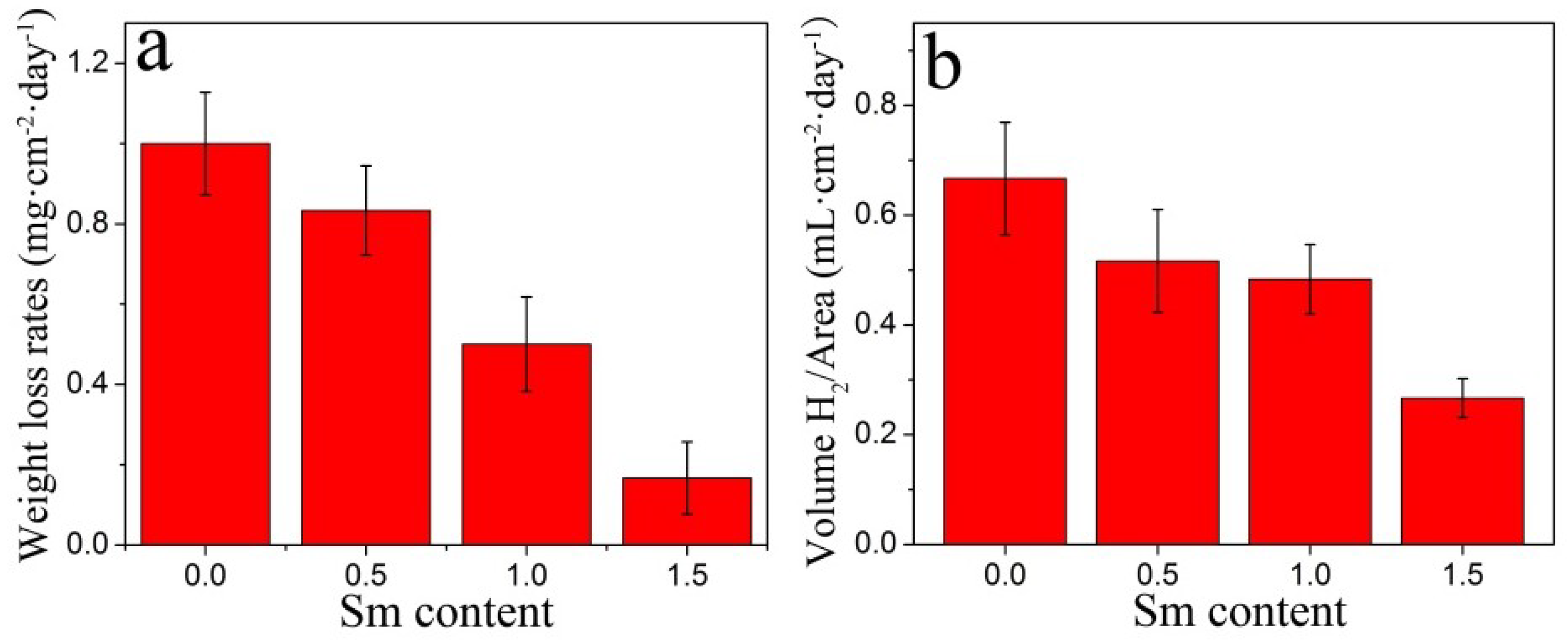
3.2. Weight Loss and Hydrogen Evolution
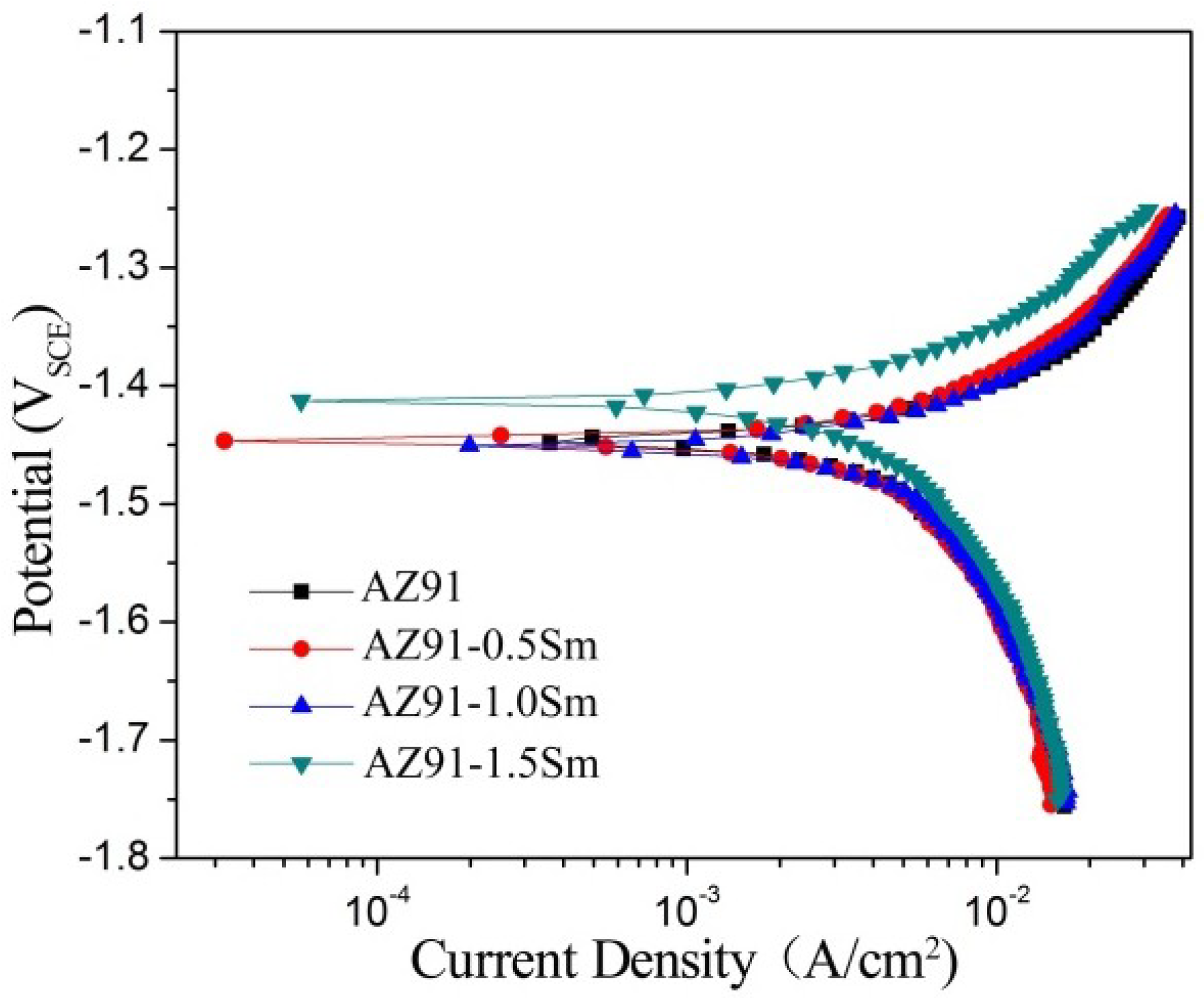
3.3. Potentiodynamic Polarization
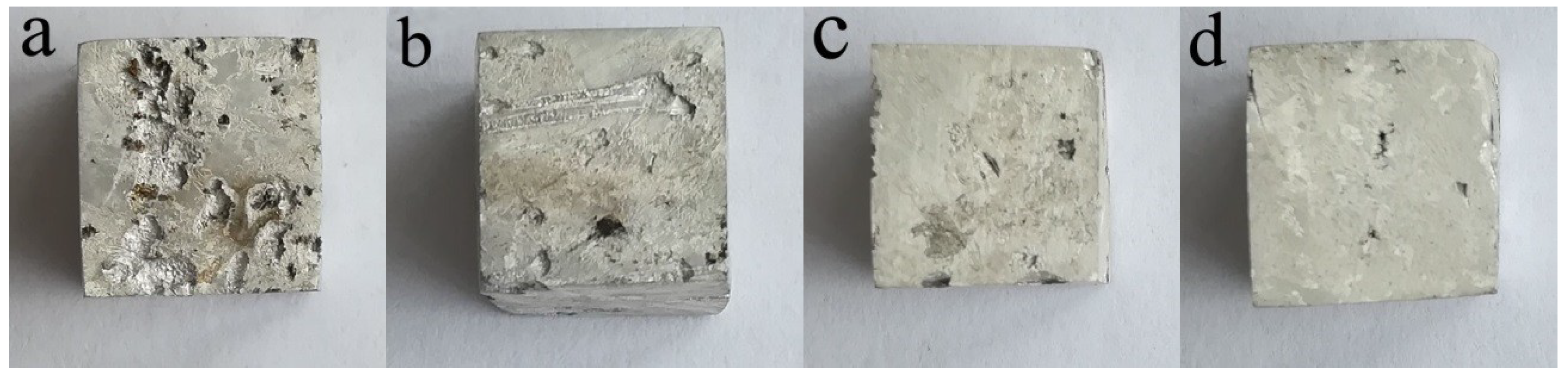
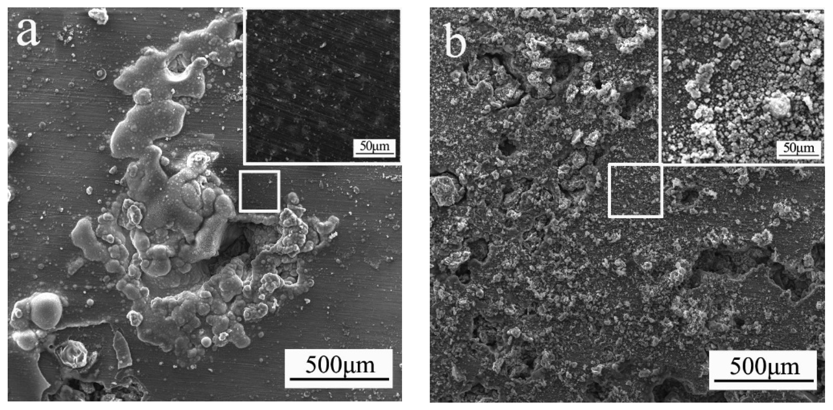
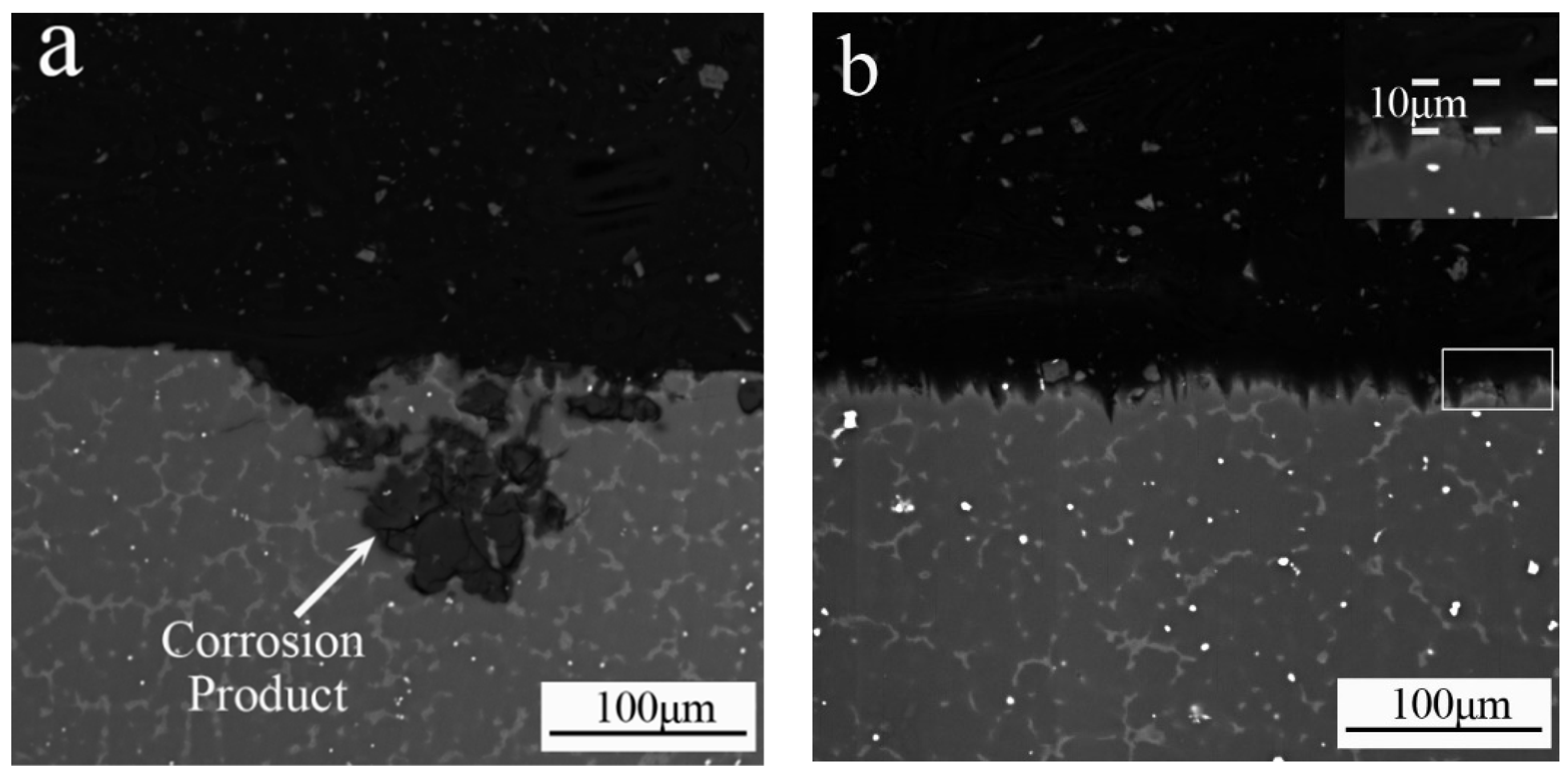
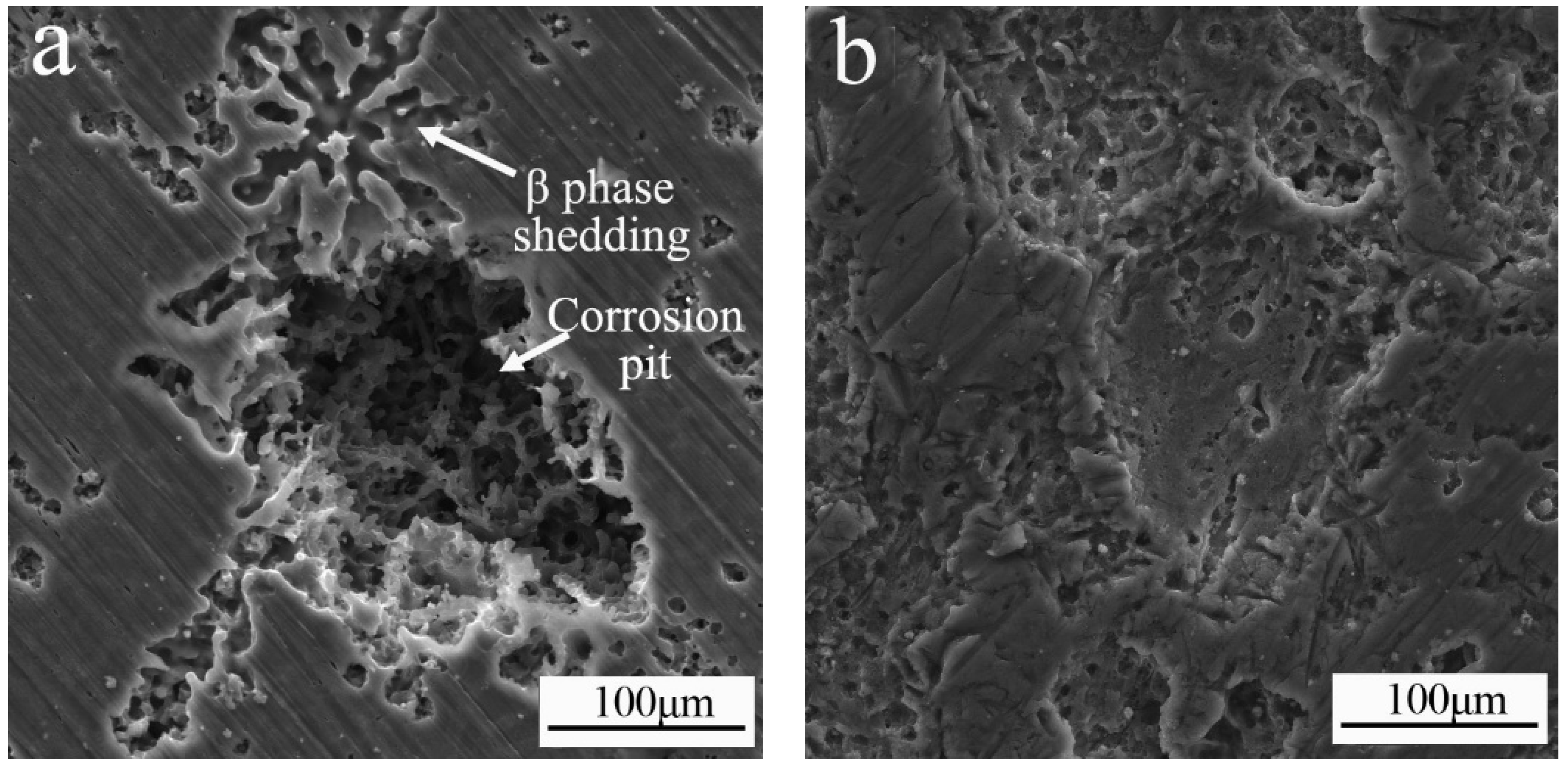
3.4. Corrosion Morphology
4. Discussion
5. Conclusions
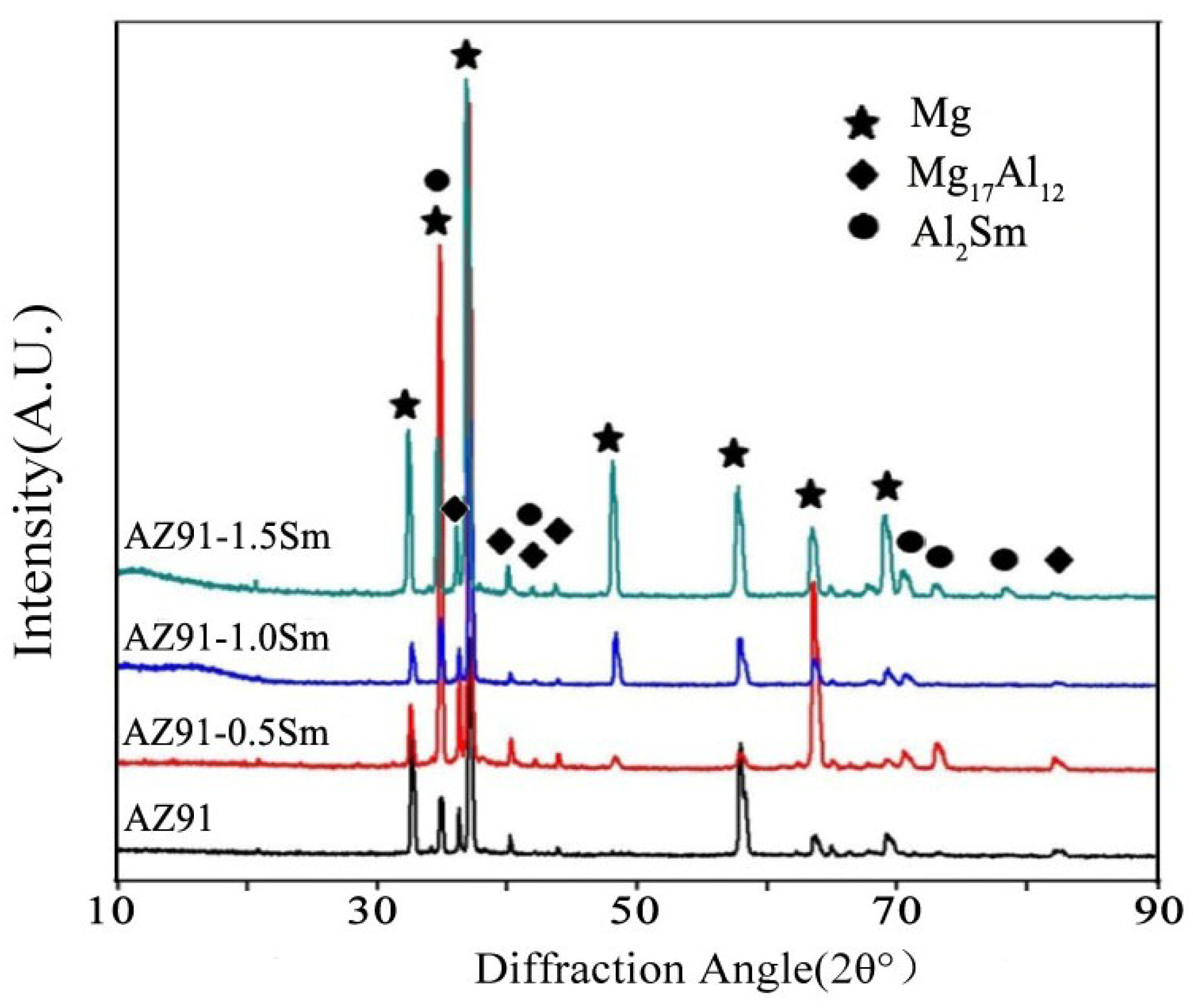
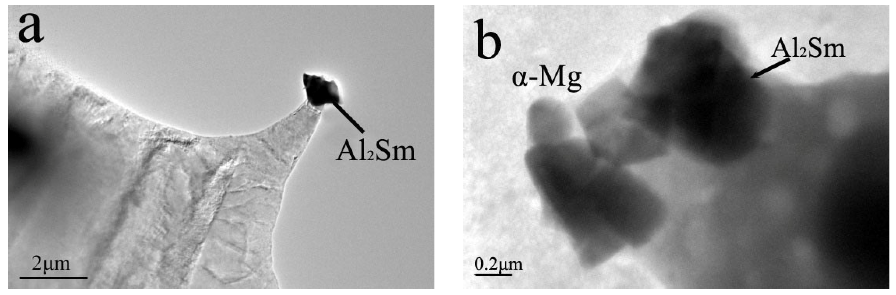
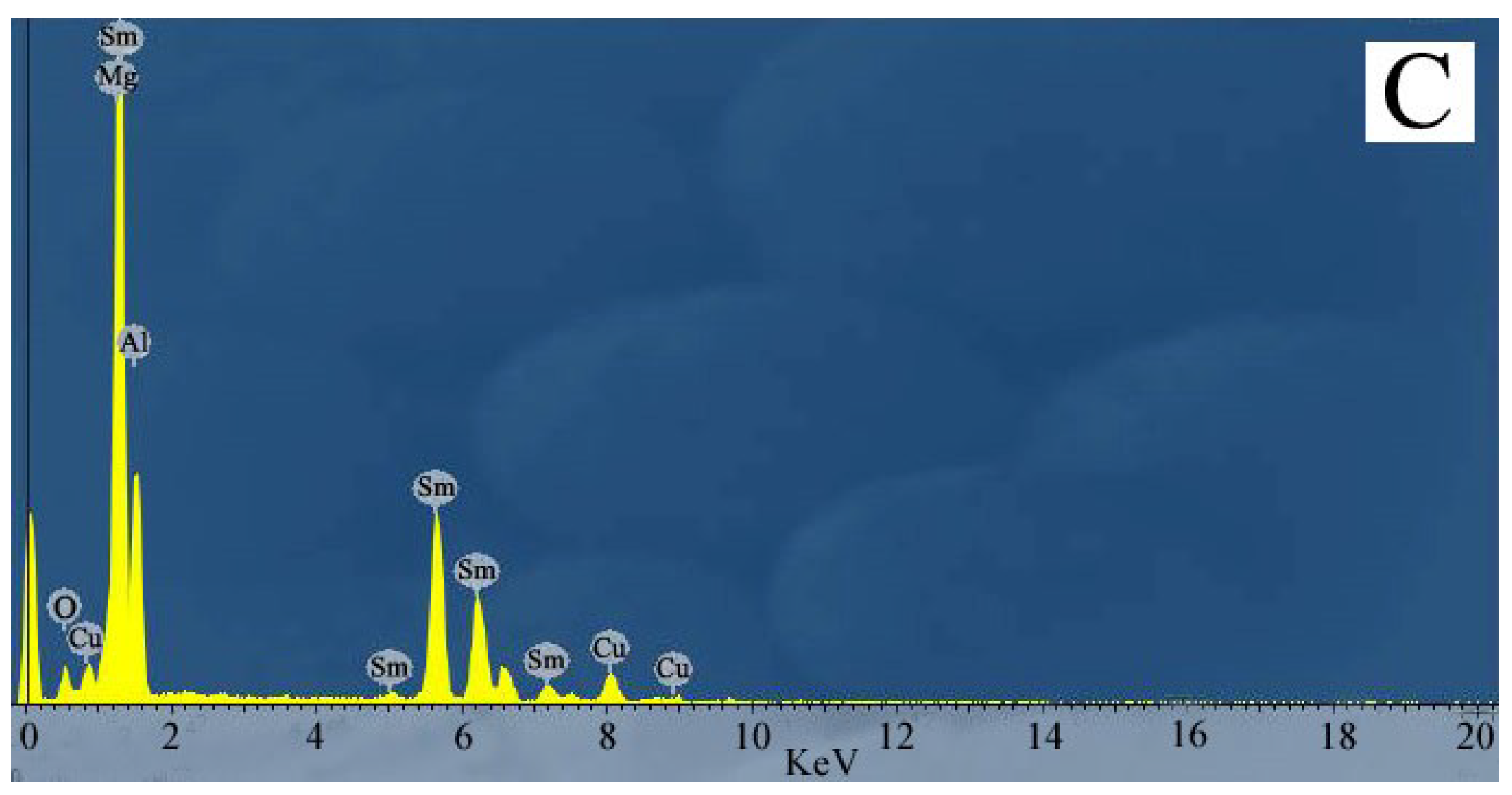
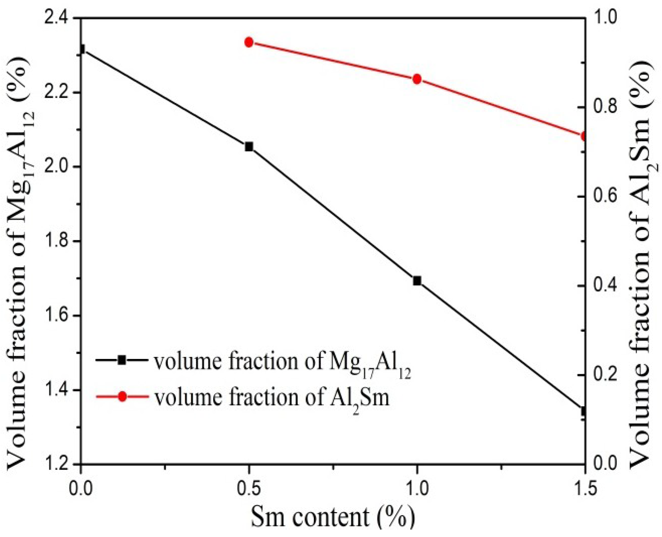
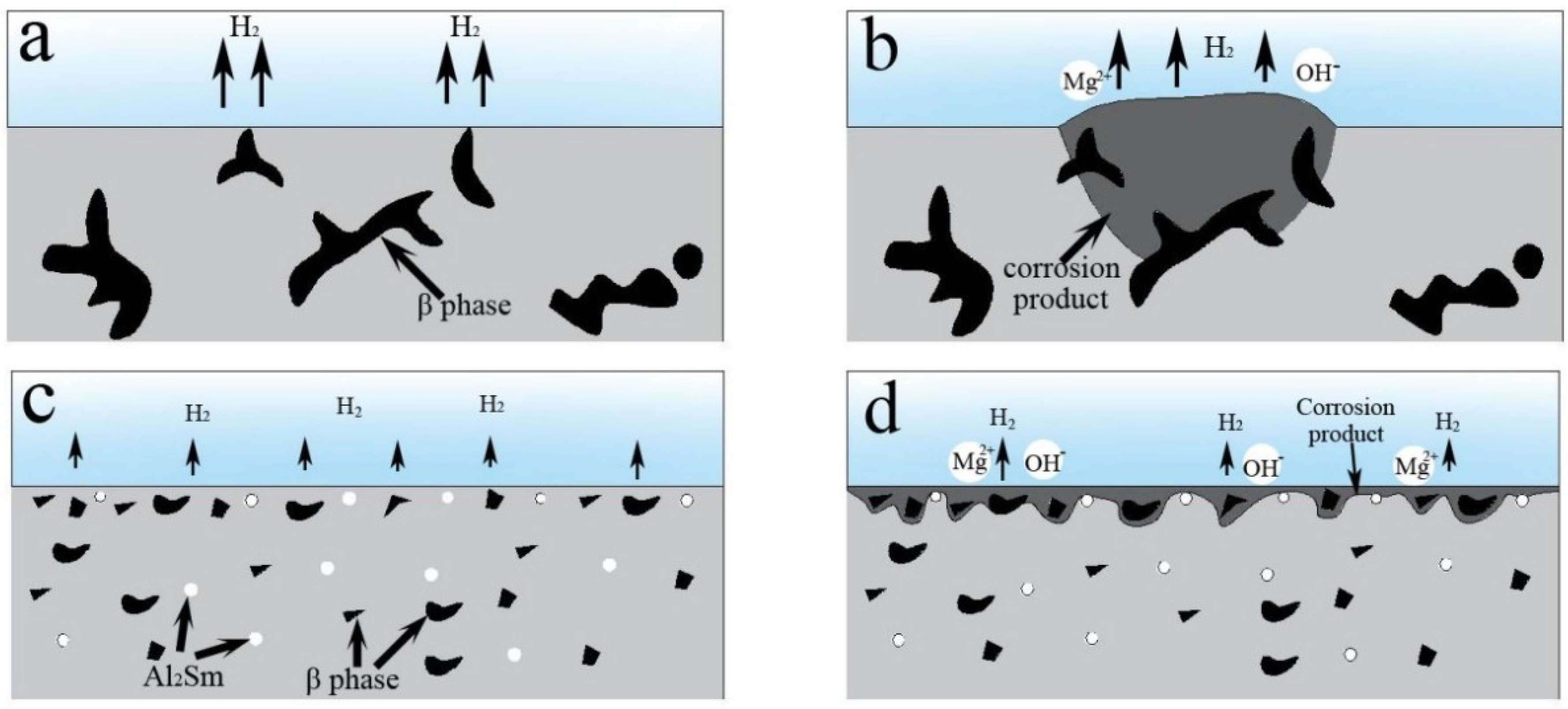
- The addition of Sm to the AZ91 alloy treated by ultrasonic vibration resulted in the formation of an Al2Sm phase about 2 μm in size, which reduced the volume fraction of the β-Mg17Al12 phase and changed the morphology of β-Mg17Al12 phase to fine granular.
- The weight loss, 0.167 mg·cm−2·day−1, and hydrogen evolution, 0.247 mL·cm−2·day−1, revealed that the ultrasonic AZ91–1.5Sm alloy was characterized by a relatively lower corrosion rate than the alloy prepared without Sm addition. Additionally, the electrochemical test also showed increased corrosion resistance of alloy specimens with increased Sm content.
- The size and morphology of the second phase had a key role in the micro-galvanic corrosion growth process. The locally coarse β phase accelerated the possibility of micro-galvanic corrosion growth into the matrix. The fine β and Al2Sm phases in the alloy reduced the probability of micro-galvanic corrosion growth, resulting in the formation of a uniform corrosion layer.
Author Contributions
Funding
Conflicts of Interest
References
- Song, Y.L.; Liu, Y.H.; Yu, S.R.; Zhu, X.Y.; Wang, S.H. Effect of neodymium on microstructure and corrosion resistance of AZ91 magnesium alloy. J. Mat. Sci. 2007, 42, 4435–4440. [Google Scholar] [CrossRef]
- Liang, C.; Wang, S.; Huang, N.; Zhang, Z.; Zhang, S.; Jing, R. Effects of Lanthanum and Cerium Mixed Rare Earth Metal on Abrasion and Corrosion Resistance of AM60 Magnesium Alloy. Rare Met. Mater. Eng. 2015, 44, 521–526. [Google Scholar]
- Seetharaman, S.; Blawert, C.; Ng, B.M.; Wong, W.L.E.; Goh, C.S.; Hort, N.; Gupta, M. Effect of erbium modification on the microstructure, mechanical and corrosion characteristics of binary Mg-Al alloys. J. Alloys Compd. 2015, 648, 759–770. [Google Scholar] [CrossRef]
- Thomas, S.; Medhekar, N.V.; Frankel, G.S.; Birbilis, N. Corrosion mechanism and hydrogen evolution on Mg. Curr. Opin. Solid State Mater. Sci. 2015, 19, 85–94. [Google Scholar] [CrossRef]
- Tkacz, J.; Minda, J.; Fintova, S.; Wasserbauer, J. Comparison of Electrochemical Methods for the Evaluation of Cast AZ91 Magnesium Alloy. Materials 2016, 9, 925. [Google Scholar] [CrossRef] [PubMed]
- Li, Q.A.; Li, X.; Zhang, Q.; Chen, J. Effect of rare-earth element Sm on the corrosion behavior of Mg-6Al-1.2Y-0.9Nd alloy. Rare Met. 2010, 29, 557–560. [Google Scholar] [CrossRef]
- Makar, G.L.; Kruger, J. Corrosion of magnesium. Met. Rev. 1993, 38, 138–153. [Google Scholar] [CrossRef]
- Birbilis, N.; Cavanaugh, M.K.; Sudholz, A.D.; Zhu, S.M.; Easton, M.A.; Gibson, M.A. A combined neural network and mechanistic approach for the prediction of corrosion rate and yield strength of magnesium-rare earth alloys. Corros. Sci. 2011, 53, 168–176. [Google Scholar] [CrossRef]
- Braszczyńskamalik, K.N. Some Mechanical Properties of Experimental Mg-Al-RE-Mn Magnesium Alloys. Arch. Foundry Eng. 2014, 14, 13–16. [Google Scholar] [CrossRef]
- Mi, J.S.; Chul, P.K.; Ho, K.B.; Kyun, P.S.; Ho, P.Y. Investigation on the Microstructure and Mechanical Properties of Mg-Al-Yb Alloys. Mater. Trans. 2011, 52, 1088–1095. [Google Scholar] [Green Version]
- Jiang, Z.; Feng, J.; Chen, Q.; Jiang, S.; Dai, J.; Jiang, B.; Pan, F. Preparation and Characterization of Magnesium Alloy Containing Al2Y Particles. Materials 2018, 11, 1748. [Google Scholar] [CrossRef] [PubMed]
- Zhang, L.; Jia, R.; Li, D.; Zhang, W.; Guo, F. Effect of Intermetallic Phases on Corrosion Initiation of AZ91 Alloy with Rare Earth Y Addition. J. Mater. Sci. Technol. 2015, 31, 504–511. [Google Scholar] [CrossRef]
- Wu, D.; Yan, S.; Wang, Z.; Zhang, Z.; Miao, R.; Zhang, X.; Chen, D. Effect of samarium on microstructure and corrosion resistance of aged as-cast AZ92 magnesium alloy. J. Rare Earth 2014, 32, 663–671. [Google Scholar] [CrossRef]
- Wang, C.; Dai, J.; Liu, W.; Zhang, L.; Wu, G. Effect of Al additions on grain refinement and mechanical properties of Mg–Sm alloys. J. Alloys Compd. 2015, 620, 172–179. [Google Scholar] [CrossRef]
- Gao, D.; Li, Z.; Han, Q.; Zhai, Q. Effect of ultrasonic power on microstructure and mechanical properties of AZ91 alloy. Mater. Sci. Eng. A 2009, 502, 2–5. [Google Scholar] [CrossRef]
- Baek, E.J.; Ahn, T.Y.; Jung, J.G.; Lee, J.M.; Cho, Y.R.; Euh, K. Effects of ultrasonic melt treatment and solution treatment on the microstructure and mechanical properties of low-density multicomponent Al70Mg10Si10Cu5Zn5 alloy. J. Alloys Compd. 2016, 696, 450–459. [Google Scholar] [CrossRef]
- Hu, Y.; Zheng, H. Effects of ultrasonic treatment on microstructure and mechanical properties of semisolid Sn-52Bi alloy. J. Wuhan Univ. Nat. Sci. Ed. 2016, 31, 1063–1067. [Google Scholar] [CrossRef]
- Jung, J.G.; Lee, S.H.; Cho, Y.H.; Yoon, W.H.; Ahn, T.Y.; Ahn, Y.S.; Lee, J.M. Effect of transition elements on the microstructure and tensile properties of Al–12Si alloy cast under ultrasonic melt treatment. J. Alloys Compd. 2017, 712, 277–287. [Google Scholar] [CrossRef]
- Xu, J.; Zhou, J.; Tan, W.; Huang, S.; Wang, S.; He, W. Study on laser surface melting of AZ31B magnesium alloy with different ultrasonic vibration amplitude. Corros. Eng. Sci. Technol. 2017, 53, 1–7. [Google Scholar] [CrossRef]
- Zhang, Z.; Qichi, L.E.; Cui, J. Effect of high-intensity ultrasonic field on process of semi-continuous casting for AZ80 magnesium alloy billets. Trans. Nonferrous Met. Soc. China 2010, 20, 376–381. [Google Scholar] [CrossRef]
- Zhang, Z.; Qichi, L.E.; Cui, J. Influence of high-intensity ultrasonic treatment on the phase morphology of a Mg-9.0wt.%Al binary alloy. Rare Met. 2009, 28, 86. [Google Scholar] [CrossRef]
- Sun, T.; Wang, Z.; Li, J.; Zhang, T. Effect of Ultrasonic Vibration Solidification Treatment on the Corrosion Behavior of AZ80 Magnesium Alloy. Int. J. Electrochem. Sci. 2013, 8, 7298–7319. [Google Scholar]
- Hu, Z.; Li, X.; Yan, H.; Wu, X.; Qun, H.; Lin, J. Effects of ultrasonic vibration on microstructure evolution and elevated-temperature mechanical properties of hot-extruded Mg-6Al-0.8Zn-2.0Sm wrought magnesium alloys. J. Alloys Compd. 2016, 685, 58–64. [Google Scholar] [CrossRef]
- Shi, Z.; Liu, M.; Atrens, A. Measurement of the corrosion rate of magnesium alloys using Tafel extrapolation. Corros. Sci. 2010, 52, 579–588. [Google Scholar] [CrossRef]
- Nam, N.D.; Min, J.K.; Jang, Y.W.; Kim, J.G. Effect of tin on the corrosion behavior of low-alloy steel in an acid chloride solution. Corros. Sci. 2010, 52, 14–20. [Google Scholar] [CrossRef]
- Zhong, G.; Wu, S.; Jiang, H.; An, P. Effects of ultrasonic vibration on the iron-containing intermetallic compounds of high silicon aluminum alloy with 2% Fe. J. Alloys Compd. 2010, 492, 482–487. [Google Scholar] [CrossRef]
- Liu, X.; Osawa, Y.; Takamori, S.; Mukai, T. Microstructure and mechanical properties of AZ91 alloy produced with ultrasonic vibration. Mater. Sci. Eng. A 2008, 487, 120–123. [Google Scholar] [CrossRef]
- Lin, C.; Shu-Sen, W.U.; Zhong, G.; Wan, L.; Ping, A.N. Effect of ultrasonic vibration on Fe-containing intermetallic compounds of hypereutectic Al–Si alloys with high Fe content. Trans. Nonferrous Met. Soc. China 2013, 23, 1245–1252. [Google Scholar] [CrossRef]
- Osawa, Y.; Takamori, S.; Kimura, T.; Minagawa, K.; Kakisawa, H. Morphology of Intermetallic Compounds in Al-Si-Fe Alloy and Its Control by Ultrasonic Vibration. Mater. Trans. 2007, 48, 2467–2475. [Google Scholar] [CrossRef] [Green Version]
- Lan, J.; Yang, Y.; Li, X. Microstructure and microhardness of SiC nanoparticles reinforced magnesium composites fabricated by ultrasonic method. Mater. Sci. Eng. A 2004, 386, 284–290. [Google Scholar] [CrossRef]
- Zhao, M.C.; Liu, M.; Song, G.; Atrens, A. Influence of the β-phase morphology on the corrosion of the Mg alloy AZ91. Corros. Sci. 2008, 50, 1939–1953. [Google Scholar] [CrossRef]
- Op’t Hoog, C.; Birbilis, N.; Zhang, M.X.; Estrin, Y. Surface Grain Size Effects on the Corrosion of Magnesium. Key Eng. Mater. 2008, 384, 229–240. [Google Scholar] [CrossRef]
- Chen, Y.; Yang, Y.; Zhang, W.; Zhang, T.; Wang, F. Influence of second phase on corrosion performance and formation mechanism of PEO coating on AZ91 Mg alloy. J. Alloys Compd. 2017, 718, 92–103. [Google Scholar] [CrossRef]
- Coy, A.E.; Viejo, F.; Skeldon, P.; Thompson, G.E. Susceptibility of rare-earth-magnesium alloys to micro-galvanic corrosion. Corros. Sci. 2010, 52, 3896–3906. [Google Scholar] [CrossRef]


| Alloy | Al | Mn | Zn | Sm | Mg |
|---|---|---|---|---|---|
| AZ91 | 9.23 | 0.29 | 0.67 | --- | Bal. |
| AZ91–0.5Sm | 9.04 | 0.35 | 0.82 | 0.44 | Bal. |
| AZ91–1.0Sm | 9.12 | 0.17 | 0.55 | 0.95 | Bal. |
| AZ91–1.5Sm | 8.87 | 0.26 | 0.90 | 1.39 | Bal. |
| Alloy | Ecorr (VSCE) | Icorr (mA/cm2) | βa (mV/Dec) | βc (mV/Dec) | Rp (Ω·cm2) |
|---|---|---|---|---|---|
| AZ91 | −1.447 | 28.8 | 324.4 | 1062.1 | 3.8 |
| AZ91–0.5Sm | −1.442 | 19.1 | 381.6 | 1574.5 | 7.0 |
| AZ91–1.0Sm | −1.452 | 20.6 | 398.1 | 1325.4 | 6.5 |
| AZ91–1.5Sm | −1.413 | 16.6 | 344.3 | 1681.1 | 7.5 |
© 2018 by the authors. Licensee MDPI, Basel, Switzerland. This article is an open access article distributed under the terms and conditions of the Creative Commons Attribution (CC BY) license (http://creativecommons.org/licenses/by/4.0/).
Share and Cite
Chen, Y.; Yin, Z.; Yan, H.; Zhou, G.-H.; Wu, X.-Q.; Hu, Z. Effect of Samarium on the Microstructure and Corrosion Resistance of AZ91 Magnesium Alloy Treated by Ultrasonic Vibration. Materials 2018, 11, 2331. https://doi.org/10.3390/ma11112331
Chen Y, Yin Z, Yan H, Zhou G-H, Wu X-Q, Hu Z. Effect of Samarium on the Microstructure and Corrosion Resistance of AZ91 Magnesium Alloy Treated by Ultrasonic Vibration. Materials. 2018; 11(11):2331. https://doi.org/10.3390/ma11112331
Chicago/Turabian StyleChen, Yang, Zheng Yin, Hong Yan, Guo-Hua Zhou, Xiao-Quan Wu, and Zhi Hu. 2018. "Effect of Samarium on the Microstructure and Corrosion Resistance of AZ91 Magnesium Alloy Treated by Ultrasonic Vibration" Materials 11, no. 11: 2331. https://doi.org/10.3390/ma11112331