Management of Parapharyngeal Space Tumors: Clinical Experience with a Large Sample and Review of the Literature

Abstract
:1. Introduction
2. Materials and Methods
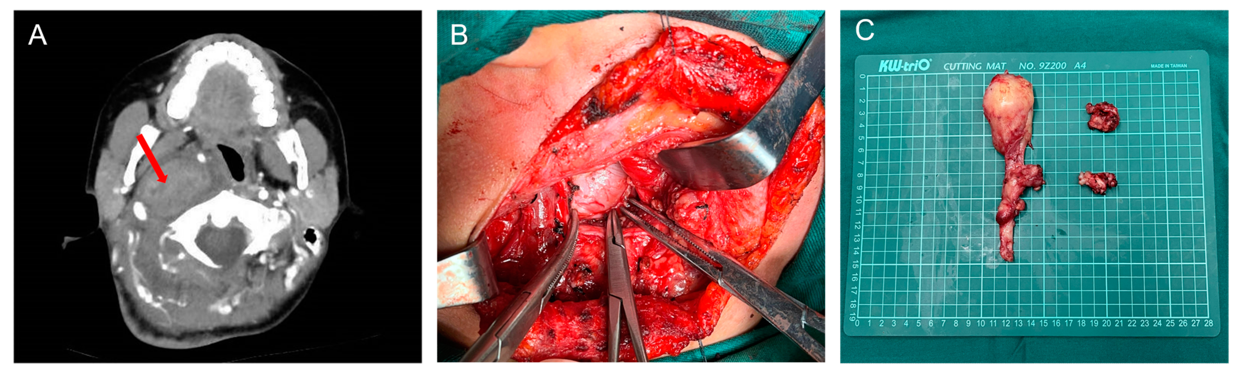
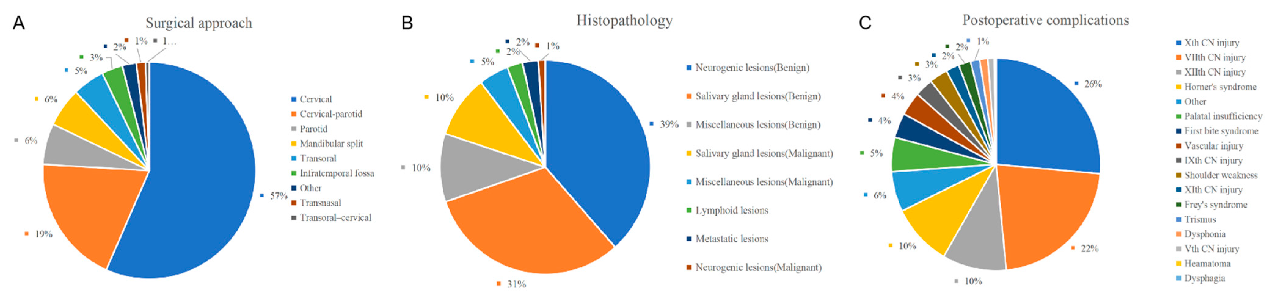
3. Results

| Hughes et al. 1995 [17] n = 172 | Cohen et al. 2005 [18] n = 166 | Sun et al. 2017 [19] n = 103 | Tao et al. 2018 [16] n = 188 | Zhao et al. 2020 [20] n = 214 | Our Center Cases n = 177 | |
|---|---|---|---|---|---|---|
| Symptoms | ||||||
| External or intraoral mass | 145 | - | - | 61 | - | 114 |
| Otalgia | 62 | - | - | - | - | 1 |
| Dysphagia | 22 | 12 | 10 | - | - | 3 |
| Dysphonia | 18 | 12 | 3 | 2 | - | - |
| Dyspnea | - | 1 | 1 | - | 2 | |
| Pain (facial) | 11 | 10 | - | 6 | - | - |
| Hearing loss | 19 | 6 | 1 | 33 d | 21 f | - |
| Foreign body sensation | - | - | 12 | 102 | 143 g | 15 |
| Tinnitus | 3 | 8 | - | 33 | - | 4 |
| Facial muscle weakness | 7 | - | - | - | - | |
| Trismus | - | - | - | 2 | - | - |
| Painful throat | 4 | - | - | 17 | - | 6 |
| Tongue parasthesias | 3 | - | - | 1 | - | 1 |
| Aspiration | 2 | - | - | - | - | - |
| Headaches | 2 | - | - | 2 | 5 | 1 |
| Free symptoms | - | 42 | 45 | 24 | 11 | 19 |
| Snoring | - | 1 | 8 | 15 | 15 | 3 |
| Other | 3 a | 3 b | 23 c | 24 e | 19 h | 14 i |
| Signs | ||||||
| Intraoral mass | 113 | 42 | 9 | 101 | 156 | 120 |
| External mass | 99 | 51 | 65 | 25 | 36 | 31 |
| Cranial nerve deficit | 22 | 38 | 5 | 6 | 11 | 4 |
| Palatal weakness | 9 | 14 | - | - | - | - |
| Pulsation over mass | 19 | - | - | - | - | - |
| Hearing loss | 15 | - | - | - | - | - |
| Horner’s syndrome | 3 | 3 | - | - | - | - |
| Trismus | 3 | - | - | 1 | - | - |
| Shoulder weakness | - | 7 | - | - | - | - |
| Serous otitis media | - | 6 | - | - | - | - |
| Other unspecified | - | 1 | 24 | 33 | 11 | 22 |
| Approach | Hughes et al. 1995 [17] n = 172 | Shahab et al. 2005 [21] n = 114 | Cohen et al. 2005 [18] n = 166 | Zhi et al. 2009 [22] n = 162 | Hong et al. 2015 [23] n = 112 | Sun et al. 2017 [19] n = 103 | Tao et al. 2018 [16] n = 188 | Lombardi et al. 2020 [24] n = 153 | Zhao et al. 2020 [20] n = 214 | Our Center Cases n = 177 |
|---|---|---|---|---|---|---|---|---|---|---|
| Cervical | 49 | 49 | 89 | 51 | 51 | 75 | 159 | 49 | 167 | 131 |
| Parotid | 56 | 27 | - | - | - | 11 | - | - | - | - |
| Cervical-parotid | 63 | - | 20 | 93 | 45 | - | 8 | 56 | - | 11 |
| Mandibular split | - | 27 | 3 | 18 | 10 | 9 | 8 | 4 | 8 | 3 |
| Transoral | 2 | - | - | - | 6 | 8 | 7 | 8 | 23 | 20 |
| Transoral–cervical | - | - | 3 | - | - | - | - | 3 | 3 | |
| Transnasal | - | - | - | - | - | - | 9 | 1 | 5 | 6 |
| Infratemporal fossa | - | 3 | 30 | - | - | - | 4 | 8 | 4 | - |
| Other | - | 8 | - | - | - | - | - | 22 | - | 3 |
| Histopathology | Pensak et al. 1994 [27] n = 123 | Hughes et al. 1995 [17] n = 172 | Shahab et al. 2005 [21] n = 114 | Cohen et al. 2005 [18] n = 166 | Zhi et al. 2009 [22] n = 162 | Hong et al. 2015 [23] n = 112 | Sun et al. 2017 [19] n = 103 | Tao et al. 2018 [16] n = 188 | Lombardi et al. 2020 [24] n = 153 | Zhao et al. 2020 [20] n = 214 | Our Center Cases n = 177 |
|---|---|---|---|---|---|---|---|---|---|---|---|
| Salivary gland lesions Benign | |||||||||||
| Pleomorphic adenoma | - | 68 | 34 | 33 | 57 | 39 | 16 | 61 | 53 | 36 | 26 |
| Warthin’s tumour | - | 1 | - | - | 3 | 10 | - | - | 3 | - | 1 |
| Basal cell adenoma | - | - | - | - | 5 | - | - | 7 | - | 3 | 1 |
| Lymphoepithelial lesion | - | 1 | - | - | - | - | - | 2 | - | 2 | |
| Myoepithelioma | - | - | - | 1 | - | - | - | - | - | 3 | 1 |
| Monomorphic adenoma | - | 1 | - | - | - | - | - | - | - | - | - |
| Granulomatous parotitis | - | - | - | - | - | - | - | - | - | - | - |
| Other/Unspecified | 39 | - | 8 | - | - | 3 | - | - | 1 | - | 4 |
| Malignant | |||||||||||
| Adenoid cystic carcinoma | - | 10 | 5 | 1 | 2 | 2 | - | - | 8 | 8 | - |
| Mucoepidermoid carcinoma | - | - | 3 | 1 | 3 | 4 | - | - | 2 | 5 | 3 |
| Squamous cell carcinoma | - | 3 | - | - | 1 | - | - | - | - | 27 | 1 |
| Carcinoma ex pleomorphic adenoma | - | 7 | - | 1 | - | 2 | - | - | 4 | 2 | - |
| Adenocarcinoma | - | 1 | - | 3 | 2 | - | - | 8 | 2 | - | 1 |
| Acinic cell carcinoma | - | 2 | - | 1 | 1 | - | - | - | - | 3 | - |
| Myoepithelial carcinoma | - | - | - | 3 | - | - | - | - | 1 | 3 | - |
| Undifferentiated carcinoma | - | - | - | 1 | - | - | - | - | - | - | - |
| Other/Unspecified | 12 | - | 4 | - | - | - | - | - | 4 | - | 1 |
| Neurogenic lesions | |||||||||||
| Benign | |||||||||||
| Vagal paraganglioma | 8 | 24 | 16 | 61 | - | - | - | - | 4 | - | - |
| Carotid body tumour | 9 | 9 | 17 | 2 | - | 4 | 8 | - | - | - | - |
| Glomus jugulare | 7 | 1 | - | - | - | - | - | - | - | - | - |
| Sympathetic paraganglioma | - | - | - | 4 | - | - | - | - | - | - | - |
| Paraganglioma not specified | - | - | 2 | - | 8 | - | - | 2 | 8 | 22 | 3 |
| Schwannoma | 6 | - | 11 | 16 | 36 | 20 | 34 | 77 | 12 | 39 | 88 |
| Neurofibroma | 2 | - | 3 | 7 | 18 | 9 | 4 | 1 | 1 | 4 | 7 |
| Other/Unspecified | - | 24 | - | - | - | - | - | 1 | 3 | - | - |
| Malignant | |||||||||||
| Unspecified malignant PNST a | - | 6 | - | - | 3 | 2 | - | - | 1 | - | - |
| Malignant paraganglioma | - | - | - | 2 | 3 | - | - | - | 1 | - | - |
| Miscellaneous lesions | |||||||||||
| Benign | |||||||||||
| Aneurysm | 2 | - | - | 6 | 4 | - | - | - | - | - | - |
| Branchial cleft cyst | 1 | - | - | 2 | 4 | 4 | - | 1 | 5 | - | - |
| Hemangioma | 1 | 1 | - | 1 | 5 | - | 5 | 10 | 2 | 2 | 5 |
| Meninigioma | - | 2 | 2 | 1 | - | - | - | - | - | 6 | - |
| Lipoma | 2 | - | - | 1 | - | 1 | 2 | 1 | 3 | 2 | 5 |
| Inflammatory pseudotumour | 4 | - | - | - | - | - | 5 | 3 | - | - | - |
| Cystic hygroma | 2 | 2 | - | 1 | - | - | - | - | - | - | 1 |
| Other/Unspecified | - | 2 | 5 | 7 | 4 | 3 | 14 | 2 | 4 | 8 | 16 |
| Malignant | |||||||||||
| Undifferentiated carcinoma | - | 1 | - | - | - | - | - | 1 | - | - | - |
| Chondrosarcoma | - | - | - | 1 | 3 | - | - | 2 | 2 | 2 | - |
| Sarcoma not specified | - | - | 2 | - | - | - | - | - | 2 | - | 3 |
| Hemangiopericytoma | 3 | - | - | 1 | - | - | - | - | - | 1 | - |
| Fibrosarcoma | 1 | - | - | - | - | - | - | - | - | 1 | - |
| Rhabdomyosarcoma | 3 | - | - | 1 | - | - | - | 2 | 1 | 8 | - |
| Chordoma | - | 1 | - | - | - | - | - | - | - | 4 | 1 |
| Malignant fibrous histiocytom | - | - | - | - | - | - | - | - | - | 1 | - |
| Other/Unspecified | 1 | 2 | - | 1 | - | - | 9 | 3 | 2 | 11 | - |
| Lymphoid lesions | |||||||||||
| Lymphoma | 4 | 3 | 2 | - | 4 | - | 4 | 4 | 1 | 1 | 1 |
| Lymphoid hyperplasia | - | - | - | - | - | - | - | - | - | 1 | 3 |
| Reactive lymphoid tissue | - | - | - | - | - | - | - | - | - | 2 | - |
| Castleman’s disease | - | - | - | 1 | - | - | - | - | - | - | 1 |
| Other/Unspecified | - | - | - | - | - | 8 | - | - | - | - | - |
| Metastatic lesions | |||||||||||
| Metastatic thyroid carcinoma | - | - | - | 4 | - | - | - | - | 4 | - | - |
| Metastatic squamous cell carcinoma | - | - | - | - | - | - | - | - | 8 | - | - |
| Other/Unspecified | 11 | - | - | 1 | - | 1 | - | - | 11 | - | - |
| Complications | Hughes et al. 1995 [17] n = 172 | Cohen et al. 2005 [18] n = 166 | Zhi et al. 2009 [22] n = 162 | Hong et al. 2015 [23] n = 112 | Sun et al. 2017 [19] n = 103 | Tao et al. 2018 [16] n = 188 | Lombardi et al. 2020 [24] n = 153 | Zhao et al. 2020 [20] n = 214 | Our Center Cases n = 177 |
|---|---|---|---|---|---|---|---|---|---|
| Vth CN injury | - | - | - | - | - | - | 6 | - | - |
| VIIth CN injury | 19 | 19 | 7 | 25 | 2 | 5 | 39 | 18 | 6 |
| Xth CN injury | 41 | 45 | - | 1 | 6 | 8 | 19 | 36 | 12 |
| IXth CN injury | 4 | - | - | - | - | - | 14 | - | - |
| XIth CN injury | 12 | - | - | - | - | - | 1 | - | - |
| XIIth CN injury | 15 | 15 | - | 3 | 1 | 3 | 7 | 13 | 6 |
| Horner’s syndrome | 11 | 12 | 5 | 2 | 3 | 4 | 10 | 11 | 2 |
| First bite syndrome | - | 18 | - | - | - | - | 6 | - | - |
| Shoulder weakness | - | 18 | - | - | - | - | - | - | - |
| Trismus | - | 6 | - | - | - | - | - | - | 3 |
| Heamatoma | - | 2 | - | - | - | - | - | - | - |
| Vascular injury | 6 | - | - | - | 1 | 4 | 8 | 2 | 2 |
| Dysphagia | - | - | - | - | - | - | - | - | - |
| Dysphonia | - | - | 8 | - | - | - | - | - | - |
| Palatal insufficiency | - | 33 | - | - | - | - | - | - | - |
| Frey’s syndrome | - | - | - | 4 | - | - | 8 | - | - |
| Other | - | 10 | - | 4 | 1 | 4 | 20 | - | - |
4. Discussion
5. Conclusions
Author Contributions
Funding
Institutional Review Board Statement
Informed Consent Statement
Data Availability Statement
Conflicts of Interest
References
- Stambuk, H.E.; Patel, S.G. Imaging of the Parapharyngeal Space. Otolaryngol. Clin. N. Am. 2008, 41, 77–101. [Google Scholar] [CrossRef] [PubMed]
- Strohl, M.P.; El-Sayed, I.H. Contemporary Management of Parapharyngeal Tumors. Curr. Oncol. Rep. 2019, 21, 103. [Google Scholar] [CrossRef] [PubMed]
- Riffat, F.; Dwivedi, R.C.; Palme, C.; Fish, B.; Jani, P. A Systematic Review of 1143 Parapharyngeal Space Tumors Reported over 20 Years. Oral Oncol. 2014, 50, 421–430. [Google Scholar] [CrossRef]
- Kuet, M.-L.; Kasbekar, A.V.; Masterson, L.; Jani, P. Management of Tumors Arising from the Parapharyngeal Space: A Systematic Review of 1,293 Cases Reported over 25 Years. Laryngoscope 2015, 125, 1372–1381. [Google Scholar] [CrossRef] [PubMed]
- Holcomb, A.J.; Richmon, J.D. Robotic and Endoscopic Approaches to Head and Neck Surgery. Hematol. Oncol. Clin. North Am. 2021, 35, 875–894. [Google Scholar] [CrossRef]
- Holsinger, F.C.; Sweeney, A.D.; Jantharapattana, K.; Salem, A.; Weber, R.S.; Chung, W.Y.; Lewis, C.M.; Grant, D.G. The Emergence of Endoscopic Head and Neck Surgery. Curr. Oncol. Rep. 2010, 12, 216–222. [Google Scholar] [CrossRef]
- Larson, A.R.; Ryan, W.R. Transoral Excision of Parapharyngeal Space Tumors. Otolaryngol. Clin. N. Am. 2021, 54, 531–541. [Google Scholar] [CrossRef]
- Locketz, G.D.; Horowitz, G.; Abu-Ghanem, S.; Wasserzug, O.; Abergel, A.; Yehuda, M.; Fliss, D.M. Histopathologic Classification of Parapharyngeal Space Tumors: A Case Series and Review of the Literature. Eur. Arch. Otorhinolaryngol. 2016, 273, 727–734. [Google Scholar] [CrossRef]
- Basaran, B.; Polat, B.; Unsaler, S.; Ulusan, M.; Aslan, I.; Hafiz, G. Parapharyngeal Space Tumours: The Efficiency of a Transcervical Approach without Mandibulotomy through Review of 44 Cases. Acta Otorhinolaryngol. Ital. 2014, 34, 310–316. [Google Scholar]
- Rzepakowska, A.; Osuch-Wójcikiewicz, E.; Żurek, M.; Durmaj, A.; Niemczyk, K. Tumor, host and surgery related factors predisposing to cranial nerve deficits after surgical treatment of parapharyngeal space tumors. Eur. Arch. Otorhinolaryngol. 2021, 278, 1973–1981. [Google Scholar] [CrossRef]
- Matsuki, T.; Okamoto, I.; Tada, Y.; Masubuchi, T.; Fushimi, C.; Kamata, S.; Miyamoto, S.; Yamashita, T.; Miura, K. Resection of Parapharyngeal Space Tumors Located in the Prestyloid Compartment: Efficacy of the Cervical Approach. Ann. Surg. Oncol. 2021, 28, 3066–3072. [Google Scholar] [CrossRef]
- Bulut, O.C.; Giger, R.; Alwagdani, A.; Aldabal, N.; Stenzinger, A.; Heimgartner, S.; Nisa, L.; Borner, U. Primary neoplasms of the parapharyngeal space: Diagnostic and therapeutic pearls and pitfalls. Eur. Arch. Otorhinolaryngol. 2021, 278, 4933–4941. [Google Scholar] [CrossRef]
- Lien, K.H.; Young, C.K.; Chin, S.C.; Liao, C.T.; Huang, S.F. Parapharyngeal space tumors: A serial case study. J. Int. Med. Res. 2019, 47, 4004–4013. [Google Scholar] [CrossRef] [PubMed] [Green Version]
- Chen, H.; Sun, G.; Tang, E.; Hu, Q. Surgical Treatment of Primary Parapharyngeal Space Tumors: A Single-Institution Review of 28 Cases. J. Oral Maxillofac. Surg. 2019, 77, 1520.e1–1520.e16. [Google Scholar] [CrossRef] [PubMed]
- Van Hees, T.; van Weert, S.; Witte, B.; René Leemans, C. Tumors of the parapharyngeal space: The VU University Medical Center experience over a 20-year period. Eur. Arch. Otorhinolaryngol. 2018, 275, 967–972. [Google Scholar] [CrossRef] [PubMed] [Green Version]
- Tao, L.; Shi, X.L.; Li, X.M.; Wu, H.T.; Chen, X.L.; Li, C.; Shen, Y.; Wei, C.S.; Wang, D.H.; Zhou, L. Retrospective analysis of 188 cases of parapharyngeal space tumors. Lin Chung Er Bi Yan Hou Tou Jing Wai Ke Za Zhi 2018, 32, 129–133. [Google Scholar] [CrossRef]
- Hughes, K.V.; Olsen, K.D.; McCaffrey, T.V. Parapharyngeal Space Neoplasms. Head Neck 1995, 17, 124–130. [Google Scholar] [CrossRef]
- Cohen, S.M.; Burkey, B.B.; Netterville, J.L. Surgical Management of Parapharyngeal Space Masses. Head Neck 2005, 27, 669–675. [Google Scholar] [CrossRef]
- Sun, F.; Yan, Y.; Wei, D.; Li, W.; Cao, S.; Liu, D.; Li, G.; Pan, X.; Lei, D. Surgical Management of Primary Parapharyngeal Space Tumors in 103 Patients at a Single Institution. Acta Otolaryngol. 2018, 138, 85–89. [Google Scholar] [CrossRef]
- Zhao, J.; Wen, Y.; Zong, L.; Chen, L.; Liu, M.; Huang, D.; Wang, J.; Wu, W.; Lv, P.; Feng, B. Retrospective analysis of primary parapharyngeal space tumors. Lin Chung Er Bi Yan Hou Tou Jing Wai Ke Za Zhi 2020, 34, 743–747. [Google Scholar] [CrossRef]
- Shahab, R.; Heliwell, T.; Jones, A.S. How we do it: A series of 114 primary pharyngeal space neoplasms. Clin. Otolaryngol. 2005, 30, 364–367. [Google Scholar] [CrossRef]
- Zhi, K.; Ren, W.; Zhou, H.; Wen, Y.; Zhang, Y. Management of Parapharyngeal-Space Tumors. J. Oral Maxillofac. Surg. 2009, 67, 1239–1244. [Google Scholar] [CrossRef] [PubMed]
- Hong, Y.; Hu, J.; Liang, Z. Analysis of clinical diagnosis and treatment of 112 cases of parapharyngeal space tumors. Lin Chung Er Bi Yan Hou Tou Jing Wai Ke Za Zhi 2015, 29, 994–997. [Google Scholar] [PubMed]
- Lombardi, D.; Ferrari, M.; Paderno, A.; Taboni, S.; Rampinelli, V.; Barbara, F.; Schreiber, A.; Mattavelli, D.; Tomasoni, M.; Farina, D.; et al. Selection of the Surgical Approach for Lesions with Parapharyngeal Space Involvement: A Single-Center Experience on 153 Cases. Oral Oncol. 2020, 109, 104872. [Google Scholar] [CrossRef]
- Li, L.; London, N.R.; Li, S.; Chen, X.; Carrau, R.L. Endoscopic Transoral Approach for Resection of Basal Cell Adenoma Arising in Parapharyngeal Space. J. Neurol. Surg. B Skull Base 2021, 82, 675–681. [Google Scholar] [CrossRef] [PubMed]
- Liu, Q.; Wang, H.; Zhao, W.; Song, X.; Sun, X.; Yu, H.; Wang, D.; Fernandez-Miranda, J.C.; Snyderman, C.H. Endoscopic Transnasal Transmaxillary Approach to the Upper Parapharyngeal Space and the Skull Base. Eur. Arch Otorhinolaryngol. 2020, 277, 801–807. [Google Scholar] [CrossRef] [Green Version]
- Pensak, M.L.; Gluckman, J.L.; Shumrick, K.A. Parapharyngeal space tumors: An algorithm for evaluation and management. Laryngoscope 1994, 104, 1170–1173. [Google Scholar] [CrossRef]
- Williamson, A.; Sutton, L.; Virk, J.; Clarke, P. Large Parapharyngeal Tumours: Operative Technique and Case Series of 17 Patients. Clin. Otolaryngol. 2019, 44, 865–870. [Google Scholar] [CrossRef]
- Xing, Y.; Pei, R.; Qu, J.; Wang, J.; Zhou, H.; Wang, Z.; Yan, W.; Sun, X.; Sun, T.; Li, L. Urban-Rural Differences in Factors Associated with Willingness to Receive Eldercare among the Elderly: A Cross-Sectional Survey in China. BMJ Open 2018, 8, e020225. [Google Scholar] [CrossRef] [Green Version]
- Brundisini, F.; Giacomini, M.; DeJean, D.; Vanstone, M.; Winsor, S.; Smith, A. Chronic Disease Patients’ Experiences with Accessing Health Care in Rural and Remote Areas: A Systematic Review and Qualitative Meta-Synthesis. Ont. Health Technol. Assess. Ser. 2013, 13, 1–33. [Google Scholar]
- López, F.; Suárez, C.; Vander Poorten, V.; Mäkitie, A.; Nixon, I.J.; Strojan, P.; Hanna, E.Y.; Rodrigo, J.P.; de Bree, R.; Quer, M.; et al. Contemporary Management of Primary Parapharyngeal Space Tumors. Head Neck 2019, 41, 522–535. [Google Scholar] [CrossRef] [Green Version]
- De Virgilio, A.; Costantino, A.; Mercante, G.; Di Maio, P.; Iocca, O.; Spriano, G. Trans-Oral Robotic Surgery in the Management of Parapharyngeal Space Tumors: A Systematic Review. Oral Oncol. 2020, 103, 104581. [Google Scholar] [CrossRef]
- Carrau, R.L.; Myers, E.N.; Johnson, J.T. Management of Tumors Arising in the Parapharyngeal Space. Laryngoscope 1990, 100, 583–589. [Google Scholar] [CrossRef]
- Carrau, R.L.; Johnson, J.T.; Myers, E.N. Management of Tumors of the Parapharyngeal Space. Oncology 1997, 11, 633–640, discussion 640, 642. [Google Scholar] [PubMed]
- Presutti, L.; Molteni, G.; Malvè, L.; Marchioni, D.; Ghidini, A.; Tassi, S.; Chiarini, L.; Alicandri-Ciufelli, M. Parapharyngeal Space Tumors without Mandibulotomy: Our Experience. Eur. Arch. Otorhinolaryngol. 2012, 269, 265–273. [Google Scholar] [CrossRef]
- Teng, M.S.; Genden, E.M.; Buchbinder, D.; Urken, M.L. Subcutaneous Mandibulotomy: A New Surgical Access for Large Tumors of the Parapharyngeal Space. Laryngoscope 2003, 113, 1893–1897. [Google Scholar] [CrossRef]
- Li, L.; London, N.R.; Gao, Y.; Carrau, R.L.; Chen, X. Endoscopic Transoral Approach for Resection of Retrostyloid Parapharyngeal Space Tumors: Retrospective Analysis of 16 Patients. Head Neck 2020, 42, 3531–3537. [Google Scholar] [CrossRef] [PubMed]
- Ansarin, M.; Tagliabue, M.; Chu, F.; Zorzi, S.; Proh, M.; Preda, L. Transoral Robotic Surgery in Retrostyloid Parapharyngeal Space Schwannomas. Case Rep. Otolaryngol. 2014, 2014, 296025. [Google Scholar] [CrossRef] [PubMed]
- Pilolli, F.; Giordano, L.; Galli, A.; Bussi, M. Parapharyngeal Space Tumours: Video-Assisted Minimally Invasive Transcervical Approach. Acta Otorhinolaryngol. Ital. 2016, 36, 259–264. [Google Scholar] [CrossRef] [PubMed]
- Duek, I.; Sviri, G.E.; Billan, S.; Gil, Z. Minimally Invasive Surgery for Resection of Parapharyngeal Space Tumors. J. Neurol. Surg. B Skull Base 2018, 79, 250–256. [Google Scholar] [CrossRef]
- Fang, Y.; Wu, H.; Tan, A.D.; Cheng, L. Transcervical Endoscopic Approach for Parapharyngeal Space: A Cadaver Study and Clinical Practice. Acta Otolaryngol. 2020, 140, 163–169. [Google Scholar] [CrossRef] [PubMed]
- Laccourreye, O.; Papon, J.-F.; Kania, R.; Crevier-Buchman, L.; Brasnu, D.; Hans, S. Intracordal Injection of Autologous Fat in Patients with Unilateral Laryngeal Nerve Paralysis: Long-Term Results from the Patient’s Perspective. Laryngoscope 2003, 113, 541–545. [Google Scholar] [CrossRef]
- Laccourreye, O.; Paczona, R.; Ageel, M.; Hans, S.; Brasnu, D.; Crevier-Buchman, L. Intracordal Autologous Fat Injection for Aspiration after Recurrent Laryngeal Nerve Paralysis. Eur. Arch. Otorhinolaryngol. 1999, 256, 458–461. [Google Scholar] [CrossRef] [PubMed]
- Steel, S.J.; Robertson, C.E. First Bite Syndrome: What Neurologists Need to Know. Curr. Pain Headache Rep. 2021, 25, 31. [Google Scholar] [CrossRef] [PubMed]
- Shaikh, N.E.; Jafary, H.A.; Behnke, J.W.; Turner, M.T. Botulinum Toxin A for the Treatment of First Bite Syndrome-a Systematic Review. Gland Surg. 2022, 11, 1251–1263. [Google Scholar] [CrossRef] [PubMed]
- House, J.W.; Brackmann, D.E. Facial Nerve Grading System. Otolaryngol. Head Neck Surg. 1985, 93, 146–147. [Google Scholar] [CrossRef] [PubMed]


Disclaimer/Publisher’s Note: The statements, opinions and data contained in all publications are solely those of the individual author(s) and contributor(s) and not of MDPI and/or the editor(s). MDPI and/or the editor(s) disclaim responsibility for any injury to people or property resulting from any ideas, methods, instructions or products referred to in the content. |
© 2023 by the authors. Licensee MDPI, Basel, Switzerland. This article is an open access article distributed under the terms and conditions of the Creative Commons Attribution (CC BY) license (https://creativecommons.org/licenses/by/4.0/).
Share and Cite
Jiang, C.; Wang, W.; Chen, S.; Liu, Y. Management of Parapharyngeal Space Tumors: Clinical Experience with a Large Sample and Review of the Literature. Curr. Oncol. 2023, 30, 1020-1031. https://doi.org/10.3390/curroncol30010078
Jiang C, Wang W, Chen S, Liu Y. Management of Parapharyngeal Space Tumors: Clinical Experience with a Large Sample and Review of the Literature. Current Oncology. 2023; 30(1):1020-1031. https://doi.org/10.3390/curroncol30010078
Chicago/Turabian StyleJiang, Chuanya, Wenqian Wang, Shanwen Chen, and Yehai Liu. 2023. "Management of Parapharyngeal Space Tumors: Clinical Experience with a Large Sample and Review of the Literature" Current Oncology 30, no. 1: 1020-1031. https://doi.org/10.3390/curroncol30010078