Article

The Marine Factor 3,5-Dihydroxy-4-methoxybenzyl Alcohol Represses Adipogenesis in Mouse 3T3-L1 Adipocytes In Vitro: Regulating Diverse Signaling Pathways

by
Masayoshi Yamaguchi
1,*,†,
Kenji Yoshiike
2,
Hideaki Watanabe
2 and
Mitsugu Watanabe
2,3,4,†
1
Cancer Biology Program, University of Hawaii Cancer Center, University of Hawaii at Manoa, 701 Ilalo Street, Hawaii, HI 96813, USA
2
Watanabe Oyster Laboratory Co., Ltd., 490-3 Shimoongata-cho, Hachioji 192-0154, Tokyo, Japan
3
Graduate School of Science and Engineering, Soka University, 1-236 Tangi-Machi, Hachioji 192-8577, Tokyo, Japan
4
Faculty of Health Sciences, Hokkaido University, Sapporo 060-0812, Japan
*
Author to whom correspondence should be addressed.
These authors contributed equally to this work.
Nutraceuticals 2023, 3(3), 366-379; https://doi.org/10.3390/nutraceuticals3030028
Submission received: 7 June 2023 / Revised: 11 July 2023 / Accepted: 26 July 2023 / Published: 1 August 2023
(This article belongs to the Special Issue Feature Papers in Nutraceuticals from Editorial Board Members)

Abstract

:
The augmentation of adipocytes in the adipose tissues brings disordered pathophysiological conditions, including type 2 diabetes, hyperlipidemia, hypertension, cardiovascular disease, and cancer. The phenolic antioxidant 3,5-dihydroxy-4-methoxybenzyl alcohol (DHMBA) prevents oxidative stress as radical scavenging in cells. However, the role of the disorder as a pharmacologic factor has been poorly understood. This study elucidates the regulatory effects of DHMBA on adipogenesis in mouse 3T3-L1 adipocytes in vitro. The 3T3-L1 preadipocytes were cultured in DMEM containing 10% calf fetal serum in the presence of DHMBA. Culturing with DHMBA repressed the growth of 3T3-L1 preadipocytes cultured in a medium without differentiation factors. Interestingly, when 3T3-L1 preadipocytes were cultured in a medium including differentiation factors containing insulin, DHMBA did not affect the number of cells with the differentiation process of adipogenesis. Culturing with DHMBA (1, 10, or 100 μM) inhibited lipid accumulation in adipocytes and repressed adipogenesis in 3T3-L1 cells. The potent inhibitory effects of DHMBA on adipogenesis were seen at the later stage of culture. Adipogenesis was inhibited by the presence of wortmannin, PD98059, or Bay 11-7082, which are inhibitors of pathways related to insulin signaling pathway. Notably, the suppressive effects of DHMBA on adipogenesis were expressed by the presence of these inhibitors. DHMBA treatment declined the levels of PPARy and C/EBPα related to preadipocyte differentiation and PI3 kinase 100α, Akt, MAPK, phosphor-MAPK, and mTOR implicated in the insulin signaling pathway, leading to adipogenesis promotion. Thus, DHMBA may inhibit adipogenesis via regulating diverse signaling pathways, providing a new strategy for the therapy of obesity.

Graphical Abstract

1. Introduction

Generally, obesity is caused by excess energy with the intake of high-calorie foods, leading to the accumulation of triglycerides via fatty acids in adipocytes. The augmentation of adipocytes in the adipose tissues brings the disordered elevation in the levels of hormones and cytokines secreted from its tissue, which leads to pathological conditions such as type 2 diabetes, hyperlipidemia, hypertension, cardiovascular disease, and cancer [1,2]. Nowadays, obesity is recognized as a risk factor for several diseases. Thus, adipose tissue may play a crucial role in maintaining pathophysiologic homeostasis.
Adipose tissue is composed of adipocytes, which are formed by the differentiation of preadipocytes from bone marrow mesenchymal stem cells [3,4]. Adipogenesis is an important process in adipocyte formation and lipid accumulation. The major transcription factors regulating this process are CCAAT/enhancer-binding protein alpha (C/EBPα), peroxisome proliferation-activated receptor gamma (PPARγ), sterol response element-binding protein-1c (SREBP-1c), and fatty-acid-binding protein (FABP4) [5,6]. C/EBPα and PPARγ are essential for the differentiation of precursor cells into mature adipocytes [7]. PPARγ participates in promoting adipogenesis in cells lacking C/EBP expression. SREBP is a supplementary regulator of adipogenesis and plays a crucial role in regulating lipid metabolism [5,6,7]. These transcription factors regulate the synthesis of fatty acids and triglycerides during adipogenesis [7]. Moreover, the mitogen-activated protein kinase (MAPK) and protein kinase B (AKT), which are linked to insulin signaling pathways, are known to activate adipogenesis in adipocytes [8].
The novel phenolic antioxidant 3,5-dihydroxy-4-methoxybenzyl alcohol (DHMBA) was originally found in the Pacific oyster Crassostrea gigas [9,10]. DHMBA has dual properties to prevent oxidative stress as radical scavenging in several cells [9,10,11,12,13,14]. Interestingly, DHMBA is an outstanding peroxyl radical scavenger, being about 15 times and four orders of magnitude better than Trolox for that purpose in lipid and aqueous media, respectively [15]. This compound reacts faster with HOO(•) than other known antioxidants such as resveratrol and ascorbic acid [15]. DHMBA may play an important nutritional factor in the regulation of cell function as an antioxidant [9,10,11,12,13,14,15].
More recently, we have demonstrated that DHMBA represses the growth of metastatic prostate cancer cells via targeting diverse signaling pathways, providing a new strategy for prostate cancer therapy with DHMBA [16]. Moreover, elucidating the pharmacologic effects of DHMBA may be significant in the preventing and treating of various diseases. Therefore, this study has been undertaken to elucidate whether DHMBA regulates adipogenesis in 3T3-L1 preadipocytes in vitro. Here, we demonstrate that the culture with DHMBA blocks the growth of 3T3-L1 preadipocytes in vitro, leading to diminishing cell numbers. Mechanistically, culturing with DHMBA was found to repress adipogenesis in the process of the differentiation from 3T3-L1 preadipocytes to adipocytes via regulating diverse signaling processes linked to insulin signaling. Our study may offer a useful therapeutic tool for the adipogenesis implicated in obesity.

2. Materials and Methods

2.1. Reagents

Dulbecco’s modification of Eagle’s medium (DMEM) with 4.5 g/L glucose, L-glutamine and sodium pyruvate and antibiotics (penicillin and streptomycin; P/S) (100 μg/mL penicillin and 100 μg/mL streptomycin; 1% P/S). was obtained from Corning (Mediatech, Inc., Manassas, VA, USA). Fetal bovine serum (FBS) was from Thermo Scientific HyClone (Logan, UT, USA). Bovine calf serum (BCS), dexamethasone, 3-isobutyl-1-methylxanthine (IBMX), wortmannin, Bay 11-7082, and all other reagents were purchased from the Sigma-Aldrich (St. Louis, MO, USA) unless otherwise specified.

2.2. 3,5-Dihydroxy-4-methoxybenzyl Alcohol

3,5-dihydroxy-4-methoxybenzyl alcohol (DHMBA), a novel amphipathic phenolic compound, was initially isolated from the Pacific oyster (Crassostrea gigas) with a characterization of antioxidant [9,10]. We used the synthesized DHMBA in the present study [12,13,14]. DHMBA was obtained from Watanabe Oyster Laboratory, Inc. (Hachioji, Tokyo, Japan). The structure of DHMBA is shown in Figure 1. The purity of synthesized DHMBA was 100% [12,13,14]. DHMBA was dissolved in 100% ethanol and stored at −20 °C until use.

2.3. 3T3-L1 Preadipocytes

The 3T3-L1 mouse embryo fibroblasts (preadipocytes) were obtained from American Type Culture Collection (Rockville, MD, USA). The cells were maintained in DMEM containing 10% BCS and 1% P/S at 37 °C under 95% 5% CO2 [17]. The culture medium was replaced every alternate day until 70–80% confluence was reached, and cells were subcultured every 3 days.

2.4. Assay of Cell Growth and Death

Firstly, to determine the effects of DHMBA on cell proliferation, 3T3-L1 preadipocytes (1 × 10⁵/mL per well) were cultured using 24-well plates in DMEM containing 10% BCS and 1% P/S in the presence of either vehicle (1% ethanol as a final concentration) or DHMBA (0.1, 1, 10, 100, or 1000 μM) for 3 days on reaching subconfluence in a water-saturated atmosphere containing 5% CO2 and 95% air at 37 °C [16,18] (Figure 2A).
In the next experiments to assay cell death, 3T3-L1 preadipocytes (1 × 10⁵/mL per well in 24-well plates) were cultured using 24-well plates in DMEM containing 10% BCS and 1% P/S for 3 days. Cells on reaching subconfluence were cultured for an additional 48 h in the presence of either vehicle (PBS or 1% ethanol as a final concentration) or DHMBA (0.1, 1, 10, 100, or 1000 μM) [16,19] (Figure 2B).
Moreover, to determine the effects of DHMBA on the growth of cells, 3T3-L1 preadipocytes (1 × 10⁵/mL per well) were cultured using 24-well plates in DMEM containing 10% BCS and 1% P/S in the presence of either vehicle (1% ethanol as a final concentration) or DHMBA (0.1, 1, 10,100, or 1000 μM) for 1, 2, 3, 4, or 5 days (Figure 2C). In this experiment, we chose 10 μM DHMBA, which did not have any effects on cell death.
After culturing, the cells were detached from each well by adding a sterile solution (0.1 mL per well) of 0.05% trypsin plus EDTA in Ca2+/Mg2+-free PBS (Thermo Fisher Scientific, Waltham, MA, USA) with incubation for 2 min at 37 °C, and then 0.9 mL of DMEM containing 10% FBS was added to each well [16,18,19]. To determine the number of living cells, the medium containing the suspended cells (0.1 mL) was mixed with 0.1 mL of 0.5% trypan blue staining solution, which can look like living cells but not dead cells. Viable cells were counted under a microscope (Olympus MTV-3) with a Hemocytometer (Sigma-Aldrich, St. Louis, MO, USA) by using a cell counter (Line Seiki H-102P, Tokyo, Japan). For each dish, we took the average of two counts. Cell numbers were shown as numbers per well.

2.5. Differentiation of 3T3-L1 Preadipocytes and Assay of Lipid Droplets

To elucidate the effects of DHMBA on the differentiation of 3T3-L1 preadipocytes, 3T3-L1 preadipocytes (5 × 10⁴ cells/mL per well in 24-well plates) were cultured in DMEM containing 10% bovine calf serum (BCS) and 1% P/S for 3 days on reaching subconfluence. After reaching subconfluence (day 0; early stage of differentiation), the cells were additionally cultured for 2 days in a differentiation medium of DMEM containing 10% FBS, 1% P/S, 0.5 mM IBMX, 1 μM dexamethasone, and insulin (10 μg/mL) [17,20,21]. On day 2 (later stage of differentiation), moreover, to promote adipogenesis, the fresh differentiation medium was replaced with DMEM containing 10% FBS and insulin (10 μg/mL), and then the cells were further cultured for 2 days. Firstly, to determine the effects of DHMBA on the number of cells on day 0 or 2, the differentiation medium included either vehicle (1% ethanol as a final concentration) or DHMBA (1 or 10 μM). After the culture of days 0–2 or days 2–4, the number of attached cells was counted as described in the previous section (Figure 3).
In further experiments to elucidate the effects of DHMBA on the lipid content in 3T3-L1 adipocytes from day 0 to 4, the differentiation medium included either vehicle (1% ethanol as a final concentration) or DHMBA (0.1, 1, 10, or 100 μM) (Figure 4). To determine lipid content (triglyceride) after cell culture, the cells in 24-well plates were gently washed twice with PBS and fixed with 4% formaldehyde in PBS for 30 min. Subsequently, the fixed cells were washed three times with PBS and stained for 60 min with Oil Red O solution (1.5% Oil Red O in 100% isopropanol) at room temperature [17]. After staining, the staining solution was removed and washed with PBS. Images of the stained lipid (triglyceride) droplets were observed and photographed under a microscope (40×) (Olympus IX71; Olympus Corporation, Tokyo, Japan). Furthermore, to quantify lipid contents, the Oil Red stain was extracted with the addition of isopropanol of 100% purity (300 μL) in each well by shaking for 30 min. The absorbance of each well was measured at 510 nm by using a spectrophotometer (μQuant, Bio-Tek Instruments).

2.6. Assay of Adipogenesis in 3T3-L1 Adipocytes

To investigate the effects of DHMBA on adipogenesis (Figure 5), 3T3-L1 preadipocytes (104 cells/0.2 mL per well in 96-well plates) were cultured in DMEM containing 10% BCS and 1% P/S for 3 days on reaching subconfluence [17,20,21]. After subconfluence (day 0), the cells were additionally cultured for 2 days (on day 2) in a differentiation medium with DMEM containing 10% FBS, 1% P/S, 0.5 mM IBMX, and 1 μM dexamethasone with either vehicle (1% ethanol as a final concentration) or DHMBA (0.1, 1, 10, or 100 μM). On day 2, the medium was replaced with DMEM containing 10% FBS and insulin (10 μg/mL) without DHMBA, and then the cells were further cultured for 2 days.
In separate experiments (Figure 5) on day 0, the cells were cultured in the differentiation medium without DHMBA. On day 2, the medium was replaced with DMEM containing 10% FBS and insulin (10 μg/mL) including either vehicle (1% ethanol as a final concentration) or DHMBA (0.1, 1, 10, or 100 μM), and then the cells were further cultured for 2 days.
In another experiment (Figure 6), to determine the involvement of intracellular signaling factors on day 2, the medium was replaced with DMEM containing 10% FBS and insulin (10 μg/mL) with either vehicle (1% ethanol) or DHMBA (1 or 10 μM) with or without various signaling factors, including genistein (1 or 10 μM), wortmannin (10 or 100 nM), PD98059 (1 or 10 μM), or Bay 11-7082 (1 or 10 nM), and then the cells were further cultured for 2 days. After cell culture, the lipid (triglyceride) content in the cells was determined using commercially available AdipoRed assay reagent according to the manufacturer’s instructions (Lonza, Walkersville, MD, USA) [20,21,22]. Fluorescence was measured with excitation at 485 and emission at 570 nm in a 96-well plate reader (μQuant, Bio-Tek Instruments).

2.7. Western Blotting

The 3T3-L1 preadipocytes (1 × 10⁶ cells/10 mL of 100 mm dishes) were cultured for 3 days in DMEM containing 10% BCS and 1% P/S on reaching subconfluence. After subconfluence (day 0), the cells were additionally cultured for 2 days in a differentiation medium with DMEM containing 10% FBS, 1% P/S, 0.5 mM IBMX, and 1 μM dexamethasone. On day 2, the medium was replaced with DMEM containing 10% FBS and insulin (10 μg/mL) with or without DHMBA (10 μM), and then the cells were further cultured for 2 days. After culture, the dishes were washed three times with cold PBS (10 mL) to exclude floating and dead cells and the attached cells were removed from the dish by scraping in cell lysis buffer (Cell Signaling Technology, Danvers, MA, USA) supplemented with protease and protein phosphatase inhibitors (Roche Diagnostics, Indianapolis, IN, USA) [16]. The cell lysates were then centrifuged at 17,000× g at 4 °C for 10 min. The concentration of protein in the supernatant was determined using the Bio-Rad Protein Assay Dye (Bio-Rad Laboratories, Inc., Hercules, CA, USA) with bovine serum albumin as a standard. The supernatant was stored at −80 °C until use.
As shown in our previous papers [16], the samples of forty micrograms of supernatant protein per lane were separated by SDS polyacrylamide gel electrophoresis (12% SDS-PAGE) and then transferred to PVDF membranes. Transferred membranes were immunoblotted using specific antibodies against various proteins obtained from Cell Signaling Technology (Danvers, MA, USA), including PPARy (C26H12) (cat. no. 2435, rabbit), C/EBPα (cat. no. 2295, rabbit), phosphoinositide 3-kinase p110α (PI3K; cat. no. 4255), Akt (cat. no. 9272, rabbit), mitogen-activated protein kinase (MAPK; cat. no. 4695, rabbit), phosphorylated-MAPK (cat. no. 4370, rabbit), mechanistic target of rapamycin (mTOR, cat. no. 4517, mouse), NF-κB p65 (cat. no. 3034, rabbit), and β-actin (cat. no. 3700, rabbit). Target proteins were incubated with one of the primary antibodies (1:1000) as described above, overnight at 4 °C. After incubation, the membranes were additionally incubated for 60 min at room temperature in horseradish peroxidase-conjugated secondary antibodies (Santa Cruz Biotechnology, Inc., mouse sc-2005 or rabbit sc-2305; diluted 1:1000) for 60 min at room temperature, and protein bands were detected using a Chemiluminescence substrate (cat. no. 34577, Thermo Scientific, Rockford, IL, USA) on X-ray film. A total of 3 or 4 films from 4 independent experiments on separate membranes were scanned on an Epson Perfection 1660 Photo scanner, and the bands were quantified using Image J2 software (National Institutes of Health, Bethesda, MD, USA). For immunoblotting with additional antibodies, we used the restore Western blot stripping buffer (cat. no. 21059; Thermo Scientific, Rockford, IL, USA) to remove the attached Chemiluminescence substrate (Thermo Scientific) by incubation at a room temperature for 30 min.

2.8. Statistical Analysis

Statistical significance was determined using GraphPad InStat version 3 for Windows XP (GraphPad Software Inc., La Jolla, CA, USA). Data are presented as the mean ± standard deviation (SD). Multiple comparisons were performed by one-way analysis of variance (ANOVA) with Tukey–Kramer multiple comparisons post-test for parametric data, as indicated. A p-value of <0.05 was considered statistically significant.

3. Results

3.1. Effects of DHMBA on the Proliferation, Death or Growth of 3T3-L1 Preadipocytes

First, we investigated whether DHMBA affects the proliferation of 3T3-L1 preadipocytes (Figure 2A). Cells (105 cells/mL per well in 24-well plates) were cultured for 3 days on reaching subconfluence in the presence of either vehicle (1% ethanol as a final concentration) or DHMBA (0.1, 1. 10, 100, or 1000 µM). Cell proliferation was repressed at a concentration in the range of 1–1000 µM DHMBA (Figure 2A). Next, to determine the effects of DHMBA on the death of 3T3-L1 preadipocyte, the cells were cultured in DMEM containing 10% FBS and 1% PS for 3 days on reaching subconfluence, and then the cells were additionally cultured for 2 days in DMEM containing 10% FBS and 1% P/S with or without DHMBA (0.1, 1, 10, 100, or 1000 µM) (Figure 2B). The number of cells was reduced by the presence of DHMBA (1000 µM), indicating that cell death is caused by a higher concentration of DHMBA. Moreover, we investigated whether DHMBA affects the growth of 3T3-L1 preadipocytes (Figure 2C). Cells (105 cells/mL per well in 24-well plates) were cultured for 1, 2, 3, 4, and 5 days in the presence of either vehicle (1% ethanol as a final concentration) or DHMBA (10 µM), which did not cause any effects on cell death. The growth of cells was repressed by culturing with DHMBA for 3–5 days. Thus, DHMBA was found to repress the growth of 3T3-L1 preadipocytes in vitro (Figure 2C).

3.2. Effects of DHMBA on Cell Growth with Differentiation Process of 3T3-L1 Preadipocytes

Next, we determined the effects of DHMBA on cell growth in the differentiation process of 3T3-L1 preadipocytes (Figure 3). We used DHMBA with 1 or 10 μM, which is a lower concentration without cytotoxicity. Cells reaching subconfluence (day 0) were further cultured for 2 days in a differentiation medium with DMEM containing 10% FBS, 1% P/S, 0.5 mM IBMX, 1 μM dexamethasone, and insulin (10 μg/mL) with or without DHMBA (1 or 10 μM) (Figure 3A). The number of cells was not altered by the presence of DHMBA (Figure 3B). Moreover, on day 2 in promoting adipogenesis, the fresh differentiation medium was replaced with DMEM containing 10% FBS, and insulin (10 μg/mL) with or without DHMBA (1 or 10 μM), and then the cells were further cultured for 2 days. Cell number was not altered by the presence of DHMBA (Figure 3C). These results indicate that the culture with DHMBA did not influence the number of cells in the process of differentiation of adipogenesis.

3.3. DHMBA Inhibits Lipid Accumulation in 3T3-L1 Adipocytes

To elucidate the effects of DHMBA on the lipid accumulation of 3T3-L1 adipocytes, cells were cultured on days 0 and 2 in the differentiation medium including either vehicle (1% ethanol as a final concentration) or DHMBA (0.1, 1, 10, or 100 μM). Photography with Oil Red O staining showed that culturing with DHMBA caused the decreased accumulation of lipids in 3T3-L1 adipocytes (Figure 4A) and the diminished lipid content in the cells (Figure 4B). These results suggest that culturing with DHMBA blocks the accumulation of lipids in adipocytes.

3.4. DHMBA Represses Adipogenesis in 3T3-L1 Adipocytes

We investigated whether adipogenesis in 3T3-L1 adipocytes is altered by culturing with DHMBA (Figure 5). Cells were cultured in DMEM containing 10% BCS and 1% P/S for 3 days on reaching subconfluence, and then the cells were cultured in the presence of DHMBA (0.1, 1, 10, or 100 μM) for 4 days (from day 0 to 4) (Figure 5A). Culturing with DHMBA (1, 10, or 100 μM) blocked the adipogenesis of 3T3L1 adipocytes (Figure 5A). Such repression was also seen in the culture of days 0–2 or 2–4 in the presence of DHMBA (1, 10, or 100 μM) (Figure 5B, C). Notably, the inhibition of adipogenesis in 3T3-L1 adipocytes cultured with DHMBA was greatly seen at the later stage (days 2–4) (Figure 5C). These results suggest that culturing of DHMBA potently inhibits the process of promoting adipogenesis linked to insulin signaling in 3T3-L1 adipocytes.

3.5. Effects of DHMBA on Adipogenesis in 3T3-L1 Adipocytes Cultured with the Inhibitor of Intracellular Signaling Pathway

To understand the underlying mechanism by which DHMBA suppresses adipogenesis in 3T3-L1 adipocytes, we investigated the involvement of intracellular signaling factors (Figure 6). On day 2, the medium was replaced with DMEM containing DHMBA (1 or 10 μM) with or without various signaling factors, including genistein (1 or 10 μM), wortmannin (10 or 100 nM), PD98059 (1 or 10 μM), or Bay 11-7082 (1 or 10 nM), and then the cells were further cultured for 2 days. Adipogenesis was not altered by the presence of genistein (Figure 6A). In the presence of genistein, DHMBA (1 or 10 μM) inhibited adipogenesis (Figure 6A). Notably, adipogenesis was inhibited by culturing with wortmannin (10 or 100 nM) (Figure 6B), PD98059 (1 or 10 μM) (Figure 6C), or Bay 11-7082 (1 or 10 nM) (Figure 6D), which are related to signaling of insulin and NF-κB. These inhibitions were further potentiated by the presence of DHMBA (1 or 10 μM), suggesting that DHMBA suppresses adipogenesis by inhibiting diverse pathways including insulin signaling.

3.6. Effects of DHMBA on the Levels of Proteins Related to Signaling Process of Adipogenesis

Furthermore, to understand the underlying mechanism, we elucidated whether culturing with DHMBA regulates the levels of key proteins, which are implicated in adipogenesis (Figure 7A,B). The treatment with DHMBA (10 μM) declined the levels of PPARy and C/EBPα, a transcription factor, which are related to the differentiation of preadipocytes. Moreover, DHMBA treatment caused a reduction in the levels of PI3 kinase 100α, Akt, MAPK, phosphor-MAPK, and mTOR, which are implicated in the insulin signaling pathway in enhancing adipogenesis. In addition, DHMBA treatment diminished NF-κB p65, which are transcription factors. Thus, culturing with DHMBA was demonstrated to diminish the levels of various protein molecules connected to adipogenesis.

4. Discussion

Adipose tissue plays a crucial role in maintaining pathophysiologic homeostasis, including type 2 diabetes, hyperlipidemia, hypertension, cardiovascular disease, and cancer [1,2,23]. Adipose tissue is composed of adipocytes, which are formed by the differentiation of preadipocytes from bone marrow mesenchymal stem cells [3,4]. It is evidenced that DHMBA has potential anti-oxidative stress as radical scavenging in cells [9,10,11,12,13,14]. Furthermore, we elucidated the effects of DHMBA on adipogenesis. This study found that culturing with DHMBA repressed the growth of 3T3-L1 preadipocytes and inhibited adipogenesis with differentiation of preadipocytes independent of its repressing effects on cell growth in vitro. This study supports the view that DHMBA has an anti-adipogenesis effect.
The treatment with DHMBA inhibited adipogenesis related to insulin signaling in 3T3-L1 adipocyte. DHMBA expressed a potent inhibitory effect on adipogenesis at the late stage of the differentiation of 3T3-L1 preadipocyte as compared with that of the early stage in the process of differentiation. Culture at the early stage was in medium containing differentiation factors with IBMX, dexamethasone, and insulin, while culture at the late stage included insulin only. Notably, DHMBA greatly inhibited adipogenesis at the late stage. Insulin has a potent stimulatory effect on adipogenesis and lipid accumulation in mature adipocytes [24]. Thus, DHMBA may regulate the process by which adipogenesis is potently enhanced by insulin.
There may be complexity in the mechanism of signaling pathways in obesity [25]. To elucidate the underlying mechanistic characterization by which DHMBA treatment suppresses adipogenesis in adipocytes, we used various inhibitors linked to signaling pathway of insulin, including genistein, an inhibitor of tyrosine kinase [26], wortmannin, an inhibitor of PI3K/Akt signaling pathway [27], PD98059, an inhibitor of ERK/MAP kinase-related to signaling pathway [28], and Bay 11-7089, an inhibitor of NF-κB [29]. Culturing with DHMBA caused a decrease in adipogenesis in the presence of genistein, wortmannin, PD98059, or Bay 11-7089. These results suggest that DHMBA potently inhibits adipogenesis in 3T3-L1 adipocyte via repressing diverse signaling processes linked to insulin signaling.
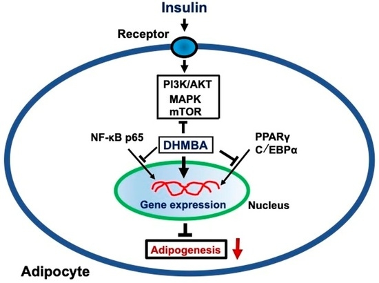
DHMBA treatment was found to diminish the levels of PPARy and C/EBAα, which are transcription factors related to the differentiation process of preadipocytes [5,6]. These results may support the view that DHMBA inhibits the process of differentiation from preadipocytes to adipocytes. Moreover, culturing with DHMBA decreased the levels of PI3K, Akt, MAPK, phosphor-MAPK, and mTOR, leading to promotion of adipogenesis related to insulin signaling. Activation of mTOR pathway promotes differentiation of 3T3-L1 preadipocytes [30]. These results suggest that DHMBA regulates the process of adipogenesis, which is enhanced by insulin, and that regulates mTOR activity implicated in the latter adipogenesis. Moreover, DHMBA diminished the levels of NF-κB p65 in adipocytes. Adipogenesis was depressed by the presence of Bay 11-7089, an inhibitor of NF-κB signaling [29]. DHMBA treatment inhibited adipogenesis in the presence of Bay 11-7089. DHMBA may also suppress adipogenesis which regulates the pathway of NF-κB signaling [31]. As summarized in Figure 8, DHMBA may express anti-adipogenic effects by regulating diverse signaling pathways in adipocytes.
In conclusion, we found that the treatment with DHMBA expressed a suppressive effect on adipogenesis related to insulin signaling in mouse 3T3-L1 adipocytes in vitro. The effects of DHMBA may be mediated via diverse pathways implicated in insulin signaling. Supplementation of DHMBA may play a role in the prevention and therapy of obesity by inhibiting adipogenesis in adipocytes.

Author Contributions

M.Y.: conceptualization, data curation, formal analysis, funding acquisition, investigation, methodology, project administration, resources, software, supervision, validation, visualization, writing—original draft, writing—review and editing. K.Y.: investigation, resources, writing—review and editing. H.W.: investigation, resources, writing—review and editing. M.W.: funding acquisition, investigation, resources, writing—review and editing. All authors have read and agreed to the published version of the manuscript.

Funding

This study was supported in part by funds provided by the University of Hawaii Cancer Center (DHMBA 129-7-4680-4; M.Y.) and the Foundation of Watanabe Oyster Laboratory (DHMBA Found; M.W.).

Institutional Review Board Statement

This study was approved by IBC Protocol (University of Hawaii System; No. 20-06-039-03).

Informed Consent Statement

Not applicable.

Data Availability Statement

The datasets used during the present study are available from the corresponding author upon reasonable request.

Conflicts of Interest

All authors declare that they have no known competing financial interest or personal relationship that could have appeared to influence the work reported in this paper.

References

  1. Wickelgren, I. Obesity: How Big a Problem? Science 1998, 280, 1364–1367. [Google Scholar] [CrossRef] [PubMed]
  2. Guh, D.P.; Zhang, W.; Bansback, N.; Amarsi, Z.; Birmingham, C.I.; Anis, A.H. The incidence of co-morbidities related to obesity and overweight: A systematic review and meta-analysis. BMC Public Health 2009, 9, 88. [Google Scholar] [CrossRef] [Green Version]
  3. Morison, R.I.; Farmer, S.R. Insight into the transcriptional control of adipocyte differentiation. J. Cell Biochem. 1999, 75, 59–67. [Google Scholar] [CrossRef]
  4. Laudes, M. Role of WNT signaling in the determination of human mesenchymal stem cells into preadipocytes. J. Mol. Endocrinol. 2001, 46, R65–R72. [Google Scholar]
  5. Green, H.; Kehinde, O. Spontaneous heritable changes leading to increased adipose conversion in 3T3 cells. Cell 1976, 7, 105–113. [Google Scholar] [CrossRef]
  6. Bernlohr, D.; Bolanowski, M.; Kelly, T.J.; Lane, M.D. Evidence for an increase in transcription of specific mRNA during differentiation of 3T3-L1 preadipocytes. J. Biol. Chem. 1985, 260, 5563–5567. [Google Scholar] [CrossRef]
  7. Tontonoz, P.; Hu, E.; Spiegelman, B.M. Stimulation of adipogenesis in fibroblasts by PPARγ2, a lipid-activated transcription factor. Cell 1994, 79, 1147–1156. [Google Scholar] [CrossRef]
  8. Al-Mansoori, L.; Al-Jaber, H.; Prince, M.S.; Elrayess, M.A. Role of inflammatory cytokines, growth factors and adipokines in adipogenesis and insulin resistance. Inflammation 2022, 45, 31–44. [Google Scholar] [CrossRef]
  9. Watanabe, M.; Fuda, H.; Jin, S.; Sakurai, T.; Ohkawa, F.; Hui, S.-P.; Takeda, S.; Watanabe, T.; Koike, T.; Chiba, H. Isolation and characterization of a phenolic antioxidant from the pacific oyster (Crassostrea gigas). J. Agric. Food Chem. 2012, 60, 830–835. [Google Scholar] [CrossRef]
  10. Fuda, H.; Watanabe, M.; Hui, S.-P.; Joko, S.; Okabe, H.; Jin, S.; Takeda, S.; Miki, E.; Watanabe, T.; Gigas, H. Anti-apoptotic effects of novel phenolic antioxidant isolated from the Pacific oyster (Crassostrea gigas) on cultured human hepatocytes under oxidative stress. Food Chem. 2015, 176, 226–233. [Google Scholar] [CrossRef]
  11. Joko, S.; Watanabe, M.; Fuda, H.; Takeda, S.; Furukawa, T.; Hui, S.-P.; Shrestha, R.; Chiba, H. Comparison of chemical structures and cytoprotection abilities between direct and indirect antioxidants. J. Funct. Foods 2017, 35, 245–255. [Google Scholar] [CrossRef]
  12. Tamano, H.; Shakushi, Y.; Watanabe, M.; Ohashi, K.; Uematsu, C.; Otsubo, T.; Ikeda, K.; Takeda, A. Preventive effect of 3,5-dihydroxy-4-methoxybenzyl alcohol (DHMBA) and zinc, components of the pacific oyster Crassostrea gigas, on glutamatergic neuron activity in the hippocampus. J. Agric. Food Chem. 2019, 67, 12844–12853. [Google Scholar] [CrossRef]
  13. Fukai, M.; Nakayabu, T.; Ohtani, S.; Shibata, K.; Shimada, S.; Sakamoto, S.; Fuda, H.; Furukawa, T.; Watanabe, M.; Hui, S.-P.; et al. The Phenolic Antioxidant 3,5-dihydroxy-4-methoxybenzyl Alcohol (DHMBA) prevents enterocyte cell death under oxygen-dissolving cold conditions through polyphyletic antioxidant actions. J. Clin. Med. 2021, 10, 1972. [Google Scholar] [CrossRef]
  14. Okabe, H.; Hui, S.-P.; Fuda, H.; Furukawa, T.; Takeda, S.; Shrestha, R.; Miura, Y.; Watanabe, M.; Chiba, H. Mass Spectrometric quantification of amphipathic, polyphenolic antioxidant of the pacific oyster (Crassostrea gigas). Anal. Sci. 2015, 31, 1341–1344. [Google Scholar] [CrossRef] [Green Version]
  15. Villuendas-Rey, Y.; Alvarer-Idaboy, J.R.; Galano, A. Assessing the protective activity of a recently discovered phenolic compound against oxidative stress using computational chemistry. J. Chem. Int. Model. 2015, 55, 2552–2561. [Google Scholar] [CrossRef] [PubMed]
  16. Yamaguchi, M.; Yoshiike, K.; Watanabe, H.; Watanabe, M. The marine factor 3,5-dihydroxy-4-methoxybenzyl alcohol suppresses growth, migration, and invasion and stimulates death of metastatic human prostate cancer cells: Targeting diverse signaling processes. Anticancer Drug 2022, 33, 424–436. [Google Scholar] [CrossRef] [PubMed]
  17. Murata, T.; Yamaguchi, M.; Kohno, S.; Takahashi, C.; Watanabe, R.; Hatori, K.; Hikita, K.; Kaneda, N. Regucalcin enhances adipocyte differentiation and attenuates inflammation in 3T3-L1 cells. FEBS Open Bio. 2020, 10, 1967–1984. [Google Scholar] [CrossRef]
  18. Misawa, H.; Inagaki, S.; Yamaguchi, M. Suppression of cell proliferation and deoxyribonucleic acid synthesis in cloned rat hepatoma H4-II-E cells overexpressing regucalcin. J. Cell Biochem. 2002, 84, 143–149. [Google Scholar] [CrossRef]
  19. Izumi, T.; Yamaguchi, M. Overexpression of regucalcin suppresses cell death in cloned rat hepatoma H4-II-E cells induced by tumor necrosis factor-α or thapsigargin. J. Cell Biochem. 2004, 92, 296–306. [Google Scholar] [CrossRef]
  20. Yamaguchi, M.; Weitzmann, M.N.; Baile, C.A.; Murata, T. Exogenous regucalcin suppresses osteoblastogenesis and stimulates adipogenesis in mouse bone marrow culture. Integ. Biol. 2012, 4, 1215–1222. [Google Scholar] [CrossRef]
  21. Yamaguchi, M.; Baile, C.A.; Zhu, S.; Shoji, M. Bioactive flavonoid p-hydroxycinnamic acid stimulates osteoblastogenesis and suppresses adipogenesis in bone marrow culture. Cell Tissue Res. 2013, 354, 743–750. [Google Scholar] [CrossRef] [PubMed] [Green Version]
  22. Greenspan, P.; Mayer, E.P.; Fowler, S.D. Nile red: A selective fluorescent stain for intracellular lipid droplets. J. Cell Biol. 1985, 100, 965–973. [Google Scholar] [CrossRef] [PubMed]
  23. Otley, M.O.C.; Sinal, C.J. Adipocyte-cancer cell interactions in the bone microenvironment. Front. Endocrinol. 2022, 13, 903926. [Google Scholar] [CrossRef] [PubMed]
  24. Hemati, N.; Ross, S.E.; Erickson, R.L.; Manson, M.M.; Walker, R.A.; Gescher, A. Signaling pathways through which insulin regulates CCAAT/enhance binding protein alpha (C/EBPalpha) phosphorylation and gene expression in 3T3-L1 adipocytes: Correlation with GLUT4 gene expression. J. Biol. Chem. 1997, 272, 25913–25919. [Google Scholar] [CrossRef] [Green Version]
  25. Wen, X.; Zhang, B.; Wu, B.; Xiao, H.; Li, Z.; Li, R.; Xu, X.; Li, T. Signaling pathways in obesity: Mechanism and therapeutic interventions. Signal. Transd. Target. Ther. 2022, 7, 298. [Google Scholar] [CrossRef]
  26. Si, H.; Liu, D. Phytochemical genistein in the regulation of vascular function: New insight. Curr. Med. Chem. 2007, 14, 2581–2589. [Google Scholar] [CrossRef]
  27. Peleck, S.L.; Charest, D.L.; Mordret, G.P.; Siow, Y.L.; Palaty, C.; Campbell, D.; Chaslton, L.; Samiei, M.; Sanghera, J.S. Networking with mitogen-activated protein kinases. Mol. Cell Biochem. 1993, 127, 157–169. [Google Scholar] [CrossRef]
  28. Serrano-Nascimento, C.; da Silva Teixeira, S.; Nicola, J.P.; Nachbar, R.T.; Masini-Repiso, A.M.; Nunes, M.T. The acute inhibitory effect of iodide excess on sodium/iodide symporter expression and activity involves the PI3K/Akt signaling pathway. Endocrinology 2014, 155, 1145–1156. [Google Scholar] [CrossRef] [Green Version]
  29. Keller, S.A.; Schattner, E.J.; Cesarman, E. Inhibition of NF-kappaB induces apoptosis of KSHV-infected primary effusion lymphoma cells. Blood 2000, 96, 2537–2542. [Google Scholar] [CrossRef]
  30. Guo, Y.; Tan, J.; Xiong, W.; Chen, S.; Fan, L.; Li, Y. Notch3 promotes 3T3-L1 pre adipocytes differentiation by up-regulating the expression of LARS to activate the mTOR pathway. J. Cell Mol. Med. 2020, 24, 1116–1127. [Google Scholar] [CrossRef] [Green Version]
  31. Grancieri, M.; Martino, H.S.D.; Gonzalex de Mejia, E. Protein digests and pure peptides from chia seed prevented adipogenesis and inflammation by inhibiting PPARγ and NF-κB pathways in 3T3-L1 adipocytes. Nutrients 2021, 13, 176. [Google Scholar] [CrossRef] [PubMed]
Figure 1. The chemical structure of 3,5-dihydroxy-4-methoxybenzyl alcohol (DHMBA). The molecular formula of DHMBA is C8H10O4 and its molecular weight is 170.164.
Nutraceuticals 03 00028 g001
Figure 2. Effects of the marine factor 3,5-dihydroxy-4-methoxybenzyl alcohol (DHMBA) on the proliferation, death, or growth of 3T3-L1 mouse embryo fibroblasts cells (preadipocytes) in vitro. (A) To determine the effects of DHMBA on the cell proliferation, cells (1 × 10⁵ cells/mL per well in 24-well plates) were cultured in DMEM containing 10% bovine calf serum (BCS) and 1% P/S for 3 days on reaching subconfluence in the presence of either vehicle (1% ethanol as a final concentration) or DHMBA (0.1, 1, 10, 100, or 1000 µM). (B) To determine the effects of DHMBA on cell death, cells (1 × 10⁵ cells/mL per well in 24-well plates) were cultured for 3 days on reaching subconfluence, and then they were additionally cultured in the presence of either vehicle (1% ethanol as a final concentration) or DHMBA (0.1, 1, 10, 100, or 1000 µM). (C) To determine the effects of DHMBA on cell growth, cells (1 × 10⁵ cells/mL per well in 24-well plates) were cultured for 1, 2, 3, 4, or 5 days in the presence of DHMBA (10 µM). After the culture, the number of cells attached to the dish was counted. Data are presented as the mean ± SD of the value obtained from 8 wells in a total of 2 replicate plates by using different cell preparations. * p < 0.001 versus the control group without DHMBA (grey bar). One-way ANOVA, Tukey–Kramer post-test.
Nutraceuticals 03 00028 g002
Figure 3. Effects of the marine factor 3,5-dihydroxy-4-methoxybenzyl alcohol (DHMBA) on the number of 3T3-L1 mouse preadipocytes in the process of cell differentiation in vitro. (A) Process of the culture of cells is shown. (B) Cells (1 × 10⁵ cells/mL per well in 24-well plates) were cultured in DMEM containing 10% BCS and 1% P/S without DHMBA for 3 days on reaching subconfluence, and then they were additionally cultured for 2 days (an early stage of differentiation) in the above medium containing 0.5 mM IBMX, 1 μM dexamethasone, and insulin (10 μg/mL) with either vehicle (1% ethanol as a final concentration) or DHMBA (1 or 10 µM). (C) After further culture for 2 days (a later stage of differentiation), the cells were additionally cultured in a medium replaced with insulin (10 μg/mL) without IBMX and dexamethasone in the presence of DHMBA (1 or 10 μM). After the culture, the number of cells attached to the dish was counted. (B) shows the number of cells with early differentiation, (C) shows the number of cells with later differentiation. Data are presented as the mean ± SD of the value obtained from 8 wells in a total of 2 replicate plates by using different cell preparations. Not significant versus the control group without DHMBA (grey bar). One-way ANOVA, Tukey–Kramer post-test.
Nutraceuticals 03 00028 g003
Figure 4. Effects of the marine factor 3,5-dihydroxy-4-methoxybenzyl alcohol (DHMBA) on lipid droplets in 3T3-L1 mouse preadipocytes in vitro. The 3T3-L1 preadipocytes (5 × 10⁴ cells/mL per well in 24-well plates) were cultured in DMEM containing 10% BCS and 1% P/S for 3 days on reaching subconfluence. After subconfluence (day 0), the cells were additionally cultured for 2 days in a differentiation medium with DMEM containing 10% fetal bovine serum (FBS), 1% P/S, 0.5 mM IBMX, 1 μM dexamethasone, and insulin (10 μg/mL). On day 2, to promote adipogenesis, the fresh differentiation medium was replaced with DMEM containing 10% FBS and insulin (10 μg/mL), and then the cells were further cultured for 2 days. To determine the effects of DHMBA on adipogenesis of 3T3-L1 cells on days 0 and 2, the differentiation medium included either vehicle (1% ethanol as a final concentration) or DHMBA (0.1, 1, 10, or 100 μM). After cell culture, the cells in 24-well plates were gently washed twice with PBS and fixed with 4% formaldehyde in PBS for 30 min. Subsequently, the fixed cells were washed three times with PBS and stained for 60 min with Oil Red O solution (1.5% Oil Red O in isopropanol). (A) After staining, images of the stained lipid droplets were observed and photographed under a microscope. (B) To quantify lipid (triglyceride) contents, the Oil Red stain was extracted with isopropanol. The absorbance of each well was measured at 510 nm by using a spectrophotometer (μQuant, Bio-Tek Instruments). Data are presented as the mean ± SD of the value obtained from 8 wells in a total of 2 replicate plates by using different cell preparations. * p < 0.001 versus the control group without DHMBA (grey bar). One-way ANOVA, Tukey–Kramer post-test.
Nutraceuticals 03 00028 g004
Figure 5. Effects of the marine factor 3,5-dihydroxy-4-methoxybenzyl alcohol (DHMBA) on adipogenesis of 3T3-L1 mouse preadipocytes in vitro. 3T3-L1 preadipocytes (1 × 10⁴ cells/mL per well in 96-well plates) were cultured in DMEM containing 10% BCS and 1% P/S for 3 days on reaching subconfluence. (A) After subconfluence (day 0), the cells were additionally cultured for 2 days in a differentiation medium with DMEM containing 10% fetal bovine serum (FBS), 1% P/S, 0.5 mM IBMX, 1 μM dexamethasone, and insulin (10 μg/mL) with either vehicle (1% ethanol as a final concentration) or DHMBA (0.1, 1, 10, or 100 μM). On day 2, the fresh differentiation medium was replaced with DMEM containing 10% FBS and insulin (10 μg/mL) without DHMBA, and then the cells were further cultured for 2 days. (B) After subconfluence (day 0), the cells were additionally cultured for 2 days in a differentiation medium with DMEM containing 10% fetal bovine serum (FBS), 1% P/S, 0.5 mM IBMX, 1 μM dexamethasone, and insulin (10 μg/mL) without DHMBA. On day 2, the fresh differentiation medium was replaced with DMEM containing 10% FBS, and insulin (10 μg/mL) with DHMBA (0.1, 1, 10, or 100 μM), and then the cells were further cultured for 2 days. (C) After cell culture, lipid content in the cells was determined using commercially available AdipoRed assay reagent according to the manufacturer’s instructions (Lonza, Walkersville, MD, USA). Fluorescence was measured with excitation at 485 and emission at 570 nm in a 96-well plate reader. Data are presented as the mean ± SD of the value obtained from 8 wells in a total of 2 replicate plates by using different cell preparations. * p < 0.001 versus the control group without DHMBA (grey bar). One-way ANOVA, Tukey–Kramer post-test.
Nutraceuticals 03 00028 g005
Figure 6. Effects of the marine factor 3,5-dihydroxy-4-methoxybenzyl alcohol (DHMBA) on adipogenesis of 3T3-L1 mouse preadipocytes in the presence of various inhibitors of signaling pathways. 3T3-L1 preadipocytes (1 × 10⁴ cells/mL per well in 96-well plates) were cultured in DMEM containing 10% BCS and 1% P/S for 3 days on reaching subconfluence. After subconfluence (day 0), the cells were additionally cultured for 2 days in a differentiation medium with DMEM containing 10% FBS, 1% P/S, 0.5 mM IBMX, 1 μM dexamethasone, and insulin (10 μg/mL) without DHMBA. On day 2, the fresh differentiation medium was replaced with DMEM containing 10% FBS and insulin (10 μg/mL), including genistein (1 or 10 μM) (A), wortmannin (10 or 100 nM) (B), PD98059 (1 or 10 μM) (C), or Bay 11-7082 (1 or 10 nM) (D), in the presence of either vehicle (1% ethanol of a final concentration) or DHMBA (1 or 10 μM), and then the cells were further cultured for 2 days. After cell culture, lipid triglyceride content in the cells was determined using commercially available AdipoRed assay reagent according to the manufacturer’s instructions (Lonza, Walkersville, MD, USA). Fluorescence was measured with excitation at 485 and emission at 570 nm in a 96-well plate reader. Data are presented as the mean ± SD of the value obtained from 8 wells in a total of 2 replicate plates by using different cell preparations. * p < 0.001 versus the control group without DHMBA (grey bar). # p < 0.001 versus the wortmannin group without DHMBA (black bar). One-way ANOVA, Tukey–Kramer post-test.
Nutraceuticals 03 00028 g006
Figure 7. Effects of the marine factor 3,5-dihydroxy-4-methoxybenzyl alcohol (DHMBA) on the levels of various proteins linked to signaling of adipogenesis in adipocytes. The 3T3-L1 preadipocytes (1 × 10⁶ cells/10 mL of 100 mm dishes) were cultured for 3 days in DMEM containing 10% BCS and 1% P/S on reaching subconfluence. After subconfluence (day 0), the cells were additionally cultured for 2 days in a differentiation medium with DMEM containing 10% FBS, 1% P/S, 0.5 mM IBMX, and 1 μM dexamethasone. On day 2, the medium was replaced with DMEM containing 10% FBS and insulin (10 μg/mL) with or without DHMBA (10 μM), and then the cells were further cultured for 2 days. After culture, the cell lysate was prepared. Samples of 40 micrograms of supernatant protein per lane were used for Western blot analysis. (A) Representative data are presented. (B) The band was presented as a fold of control. Data are presented as the mean ± SD obtained from a total of 4 dishes per data set using different cell preparations. * p < 0.01 versus control. One-way ANOVA, Tukey–Kramer post-test.
Nutraceuticals 03 00028 g007
Figure 8. A possible mechanism by which 3,5-dihydroxy-4-methoxybenzyl alcohol (DHMBA) inhibits adipogenesis in 3T3 L1 adipocytes. DHMBA treatment diminished the levels of PPARy and C/EBPα, which are transcription factors related to the differentiation process of preadipocytes. Moreover, DHMBA decreased the levels of PI3K, Akt, MAPK, phosphor-MAPK, and mTOR in enhancing the promotion of adipogenesis related to insulin signaling. DHMBA may also suppress adipogenesis which regulates the pathway of NF-κB signaling, Thus, DHMBA may express anti-adipogenic effects by regulating diverse signaling pathways in adipocytes.
Nutraceuticals 03 00028 g008
Disclaimer/Publisher’s Note: The statements, opinions and data contained in all publications are solely those of the individual author(s) and contributor(s) and not of MDPI and/or the editor(s). MDPI and/or the editor(s) disclaim responsibility for any injury to people or property resulting from any ideas, methods, instructions or products referred to in the content.