Article

Protection against Microglia Senescence by the Dietary Supplement Dekosilhue® in BV2 Cells: A New Perspective for Obesity and Related Complications

1
Department of Neuroscience, Psychology, Drug Research and Child Health (NEUROFARBA), Section of Pharmacology and Toxicology, University of Florence, Viale G. Pieraccini 6, 50139 Florence, Italy
2
Department of Biomedical Sciences, University of Padua, Via Marzolo, 3, 35131 Padua, Italy
*
Author to whom correspondence should be addressed.
Nutraceuticals 2023, 3(2), 250-261; https://doi.org/10.3390/nutraceuticals3020020
Submission received: 20 March 2023 / Revised: 17 May 2023 / Accepted: 19 May 2023 / Published: 23 May 2023
(This article belongs to the Special Issue Functional Foods as a New Therapeutic Strategy 2.0)

Abstract

:
Growing evidence indicates chronic low-grade systemic inflammation as a major pathophysiological mechanism of obesity. Systemic inflammation provokes an immune response in the brain through the activation of microglia that results in the development of neuroinflammation, cellular senescence, and occurrence of neurological dysfunction. In the efforts to identify an innovative intervention with potential efficacy on obesity and associated complications, our aim was to study the capability of the dietary supplement Dekosilhue® (DKS), successfully used for improving the control of body weight, to attenuate microglia senescent phenotype. Microglia senescence was induced by intermittent stimulation of BV2 cells with LPS 500 ng/mL every 72 h for 4 h/day, over a period of 10 days. DKS (100 µg/mL) treatment reduced ß-galactosidase activity and expression, the formation of senescence-associated heterochromatin foci to control levels, and increased cell viability of senescent BV2 (2 folds of control). DSK reduced the expression of Nuclear Factor-kB (NF-kB) (20% lower than control), a key molecule involved in the acquisition of the senescence-associated secretory phenotype (SASP). DKS promoted a neuroprotective effect by increasing the cell viability of SH-SY5Y neuronal cells exposed to the senescent BV2 conditioned medium to values of non-senescent cells. In conclusion, DKS attenuated the senescent microglia phenotype, showing senotherapeutic activity that might be further investigated as adjunctive intervention for obesity and obesity-related neurological disturbances.

Graphical Abstract

1. Introduction

Obesity affects nearly one-third of the entire population; its incidence continues to rise [1] and is not effectively controlled by current interventions [2]. Obesity, rather than being simply the result of an energy imbalance between calorie intake and expenditure, is a complex, chronic multifactorial disorder associated with a wide range of metabolic and neurological disorders. Growing evidence indicates chronic low-grade systemic inflammation as a major pathophysiological mechanism underlying obesity [3]. Obesity causes a phenotypic modification of adipocytes which become inflamed and secrete proinflammatory cytokines, leading to an elevation of the peripheral inflammatory response [4,5]. In spite of its low-grade nature, inflammation of the adipose tissue is mechanistically linked to metabolic disease and remote organ complications of obese individuals [6,7]. Since the peripheral and central innate immune systems are in constant communication [8], systemic inflammation provokes an immune response in the brain [9], leading obesity to promote the development of neuroinflammation [10,11] and the occurrence of neurological disturbances [12].
On a cellular level, a main trait of neuroinflammation is the activation of the resident immune cells in the brain, the microglia, whose primary function is to maintain the CNS homeostasis. Upon stimulation, these cells rapidly become activated and undergo morphological and molecular changes [13]. Microglia alter their morphology and functional activity in response to the increased pro-inflammatory response from the circulation, creating a less neurotrophic tissue environment that may contribute to neurodegeneration [14]. In an unresolved chronic inflammatory situation, microglia may not cope with this continuous stimulus and lose their homeostatic activity, becoming senescent [15]. The senescent microglia are characterized by morphological changes, impaired phagocytic activity, and huge release of inflammatory mediators [16].
Senescent cell abundance increases with obesity in peripheral tissues (adipose, hepatic, pancreatic) and the brain in experimental animals and obese humans [17], and recent studies have shown that eliminating senescent cells can alleviate obesity-induced metabolic dysfunction in several tissues [18,19]. Interestingly, cellular senescence of CNS cells is taking on an increasingly interesting role in the study of new therapeutic strategies in neurodegenerative diseases [20,21]. Thus, targeting senescent cells with natural or synthetic agents is a promising new therapeutic approach for chronic diseases [18,19,20,21,22]. Studies suggest that, for treating complex inflammatory disorders with multifactorial natures, a balanced modulation of more than one target can be a more efficient strategy than the single target modulation [23]. In this frame, plant species often serve as good sources for identifying these kinds of multitarget intervention. Bioactive principles present in the herbal remedy may act synergistically among them and also modulate the activity of other bioactive constituents from other plant sources [24]. In the efforts devoted to identifying innovative interventions with potential efficacy on obesity and associated complications, the aim of this study was to investigate the effectiveness of nutraceutical interventions. Dekosilhue® (DKS) is a phytocomplex composed of an association of thirteen plant extracts, used as dietary supplement to promote carbohydrate and lipid metabolism, that has recently been reported to possess anti-neuroinflammatory activity [25]. On these bases, we examine the effectiveness of DKS in attenuating the microglia senescent phenotype in an in vitro model of microglial senescence.

2. Materials and Methods

2.1. Reagents

The commercially available food supplement Dekosilhue® (DKS) (expiration date 30 September 2025; kindly provided by Gianluca Mech S.p.A., Vicenza, Italy) was used in the study. The list of constituents is reported in Table 1.
DKS was obtained using a decoction process. Briefly, the raw material (root, leaf, cortex, fruit, aerial part, stem, according to each plant) is cooked several times in water at temperatures between 97 and 99 °C. For the in vitro tests, DKS was diluted in distilled water and then diluted in media at a concentration of 100 μg/mL, as previously optimized [25]. Bacterial lipopolysaccharide (LPS) from Gram-(Salmonella enteridis) was purchased from Sigma-Aldrich (Milan, Italy). All chemicals and solvents used in the present study were of analytical grade or with purity above 95%.

2.2. Cell Cultures

BV2 (immortalized murine microglial cells, C57BL/6 Tema Ricerca, Genova, Italy) were kept in culture in a 75 cm2 flask (Sarstedt, Verona, Italy) in medium containing RPMI (Sigma-Aldrich, Milan, Italy) with 10% heat-inactivated fetal bovine serum (56 °C, 30 min) (FBS, Gibco, Milan, Italy), 1% glutamine, and 1% penicillin-streptomycin solution (Merck, Milan, Italy).
A human neuroblastoma cell line (SH-SY5Y), kindly donated by Prof. Lorenzo Corsi (University of Modena and Reggio Emilia, Italy), was cultured in DMEM (Sigma-Aldrich) and F12 Ham’s nutrients mixture (Sigma-Aldrich), containing 10% heat-inactivated FBS (Sigma-Aldrich, Milan, Italy), 1% L-glutamine (Sigma-Aldrich, Milan, Italy), and 1% penicillin-streptomycin solution (Sigma-Aldrich, Milan, Italy) until confluence (70–80%). The cells were grown in a humidified atmosphere with 5% CO2 at 37 °C. EDTA-trypsin solution (Sigma-Aldrich, Milan, Italy) was used for detaching the cells from flasks, and cell counting was performed using a hemocytometer via Trypan blue staining.

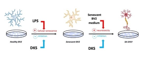
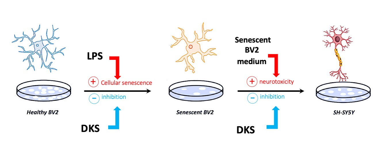
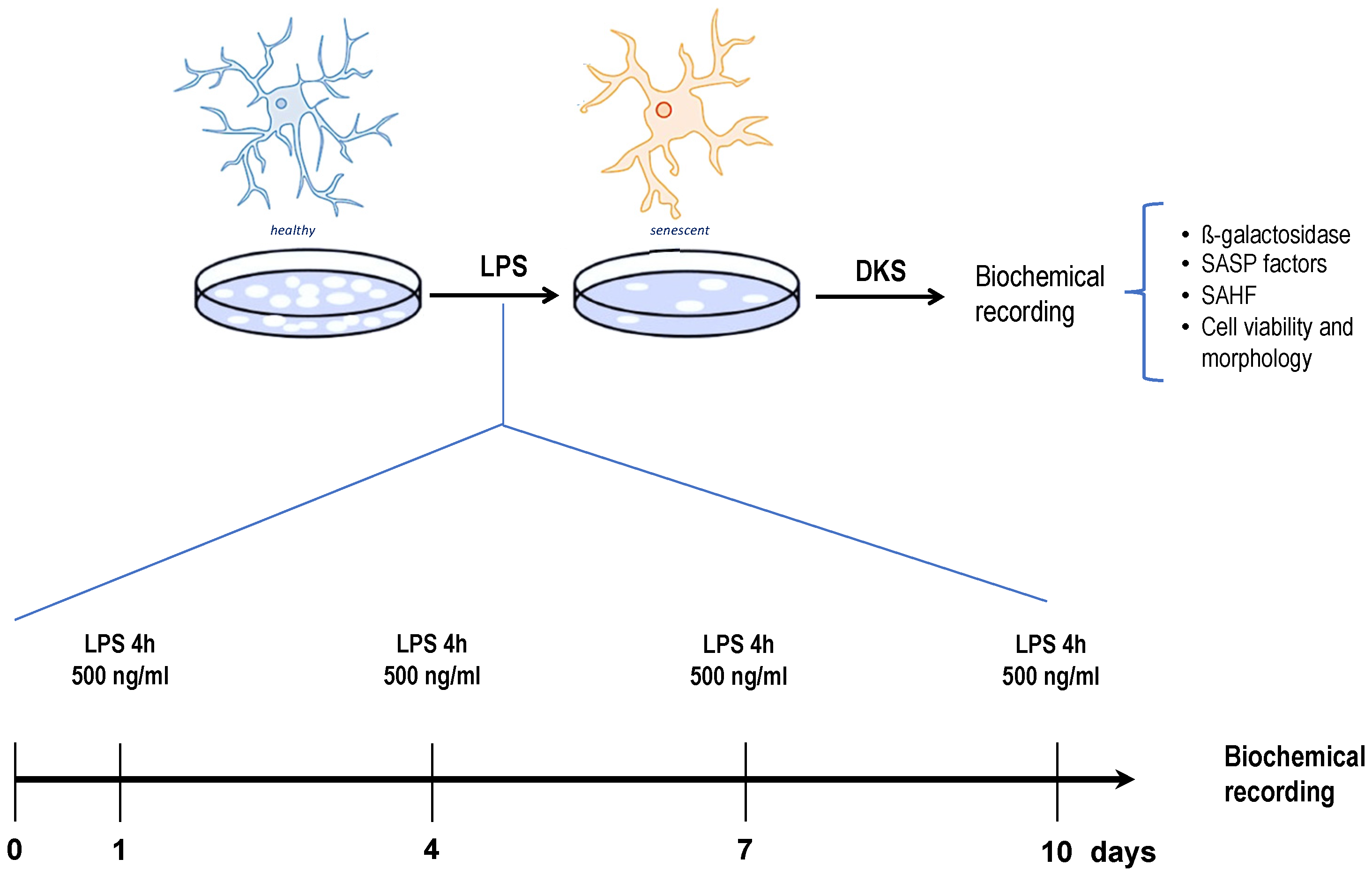
2.3. Senescent Microglia Model

The senescent model was performed as previously reported [26]. BV2 cells were treated with the LPS (Merck, Darmstadt, Germany) 4 times, for 4 h/day for a total of 10 days, at a concentration of 500 ng/mL in minimal medium (RPMI with 3% FBS), as reported in Figure 1. DKS (100 μg/mL) was added for 24 h after the last LPS stimulation.

2.4. Sulforhodamine B (SRB) Assay

The SRB test was performed to assess cell viability [27]. Briefly, cells were seeded in 96-well plates (2.0 × 10⁴ cells per well) and fixed in 50% trichloroacetic acid (TCA, Merck, Darmstadt, Germany) at 4 °C for 1 h. Then, they were treated with SRB 0.4% in acetic acid 1% for 30 min at rt. Finally, Tris-HCl pH = 10 was used and absorbance at 570 nm was recorded using a multiplate reader (Biorad, Milan, Italy). Three independent experiments (n = 3) were carried out to evaluate the effect of each treatment. Cell viability values were normalized to the mean of the control.

2.5. Senescence-Associated Heterochromatin Foci Analysis (SAHF)

Immortalized murine microglial cells (BV2) were seeded in 24-well plates (1.0 × 10⁵ cells per well) containing previously sterilized slides at the bottom of the wells. Following treatments, cells were fixed with 4% PFA for 30 min at 4 °C. After 3 washes with PBS, the slides were treated with a solution containing DAPI in mounting medium (90% glycerol + PBS) and images were taken (OLYMPUS BX63F fluorescence microscope connected to a PC). Three independent experiments (n = 3) were carried out to evaluate the effects of treatments. The DAPI intensity values were normalized to the mean of the control [26].

2.6. ß-Galactosidase Activity Assay

In a 96-well plate, 25 μL of fresh cell lysate, 80 μL of solution containing 76 μL of “buffer A” (NaH2PO4 100 mM, KCl 10 mM, MgSO4 1 mM) and 4 μL of ß-mercaptoethanol were added to each well. The plate was left in an oven at 37 °C for 5 min. Then, 25 μL of the chromogenic substrate, o-nitrophenyl ß-D-galactopyranoside (ONPG) 4 mg/mL in NaH2PO4 buffer (pH = 7.5), was added for each well, and the plate was placed back in the stove at 37 °C for 2 h until a light-yellow color was obtained. After the required time had elapsed, 45 μL of stop solution (Na2CO3 1 M) was added to each well and a spectrophotometer reading was taken at 405–450 nm.

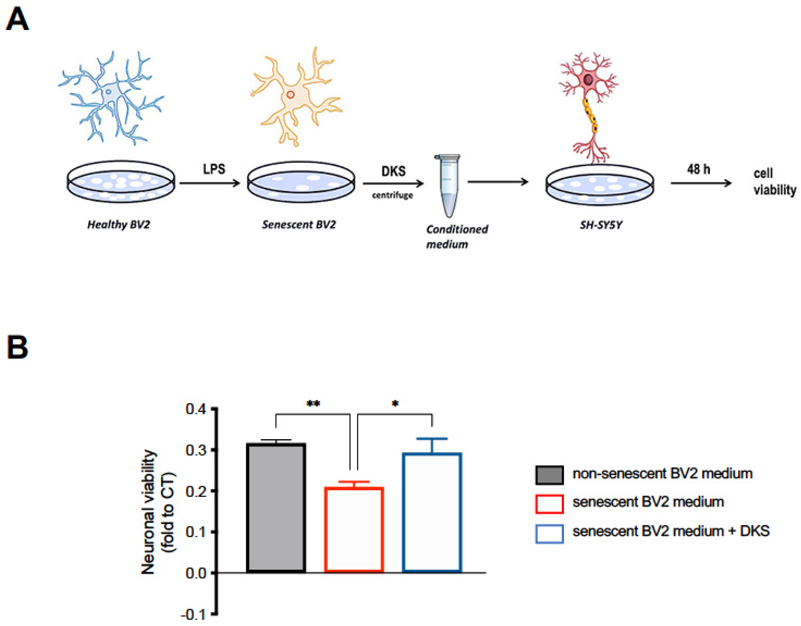
2.7. Neuroprotection Model

On day 10 after LPS intermittent stimulation, DKS (100 μg/mL) was added to senescent BV2 cells. The conditioned medium was collected and centrifuged (1000× g for 10 min, 37 °C). The pellet was discarded, and the supernatant was stored at −80 °C. The cell viability of the SH-SY5Y cells treated with the conditioned medium from untreated or DKS-treated senescent BV2 for 48 h was assessed to evaluate the neuroprotective activity of DKS. Unstimulated BV2 medium was used as a control [28].

2.8. Western Blot (WB)

Protein samples (30 μg of protein/lane) were separated by SDS_PAGE on 10% minigels and thereafter transferred to nitrocellulose membranes for 90 min at 110 V. Membranes were blocked for 90 min in PBST (PBS with 0.1% Tween) containing 5% non-fat dry milk. After blocking membranes were incubated overnight at 4 °C with primary antibodies: anti-ß-galactosidase (Santa Cruz Biotechnology, Santa Cruz, CA, USA, Cat# sc-65670, RRID: AB_831022IBA1), anti-p-NFκB p65 (Santa Cruz Biotechnology, Santa Cruz, CA, USA, Cat# sc-136548). Blots were then rinsed three times with PBST and incubated at rt with HRP-conjugated secondary antibodies for 2 h, and then detected via Colorimetric detection Kit (Opti-4CNTM Substrate Kit, BIO-RAD). Signal intensity (pixels/mm2) was quantified using ImageJ (NIH). For each sample, the signal intensity was normalized to that of total protein stained by Ponceau S. The treatments were carried out in three independent experiments (n = 3), and protein expression was calculated by normalizing the values to the mean of the control.

2.9. Statistical Analysis

The data are presented as the mean ± SEM. One-way or two-way ANOVA, followed by Tukey or Bonferroni post hoc tests, was performed. p < 0.05 was considered statistically significant. The software GraphPad Prism (version 9.5, San Diego, CA, USA) was used.

3. Results

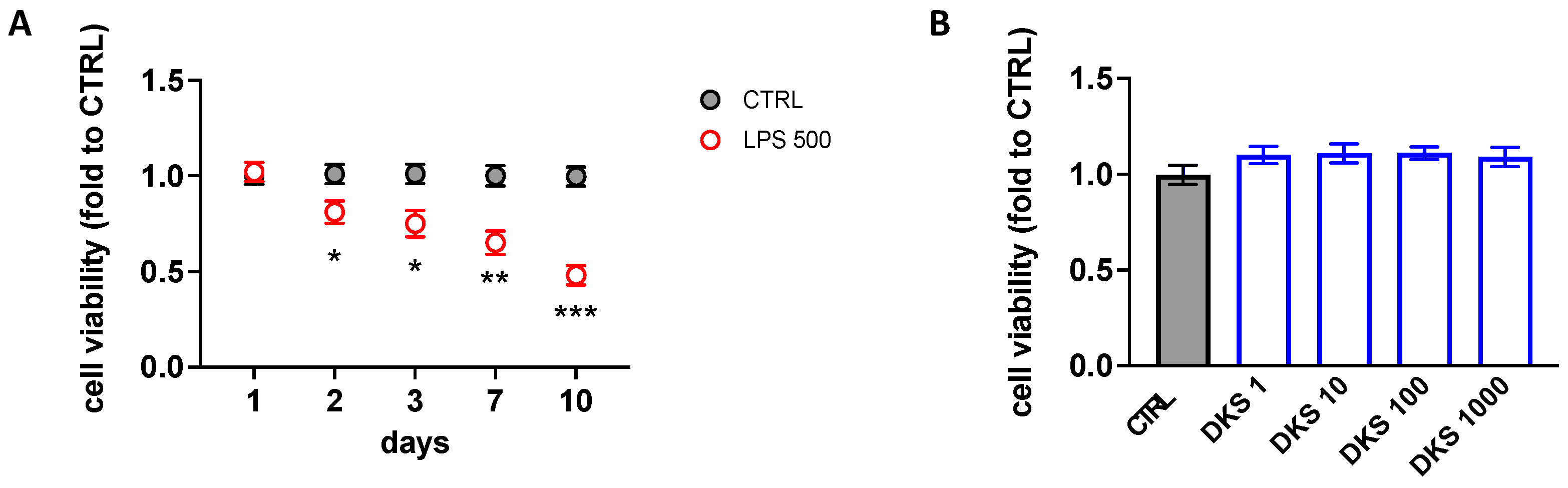
3.1. Effects of DKS on Microglial Senescent Cells

Repeated intermittent exposition of BV2 cells to low doses of the inflammatory agent LPS (LPS 500 ng/mL every 72 h for 4 h/day) promoted the development of a senescent phenotype. Cell viability was progressively reduced, starting from 2 days of treatment up to 10 days of intermittent stimulation, when a highly significant reduction of cell viability was observed (Figure 2A). Lower doses of LPS were ineffective. DKS (1–100 µg/mL) did not alter cell viability of unstimulated BV2 cells (Figure 2B). Thus, the experimental condition of 10 days of intermittent LPS treatment was chosen to conduct the study. For treatment effect experimentations, BV2 cells were treated overnight with DKS 100 µg/mL on day 10, after the last inflammatory stimulus, and the effects on main senescence parameters (i.e., ß-galactosidase, SAHF, cell viability, SASP) were evaluated.

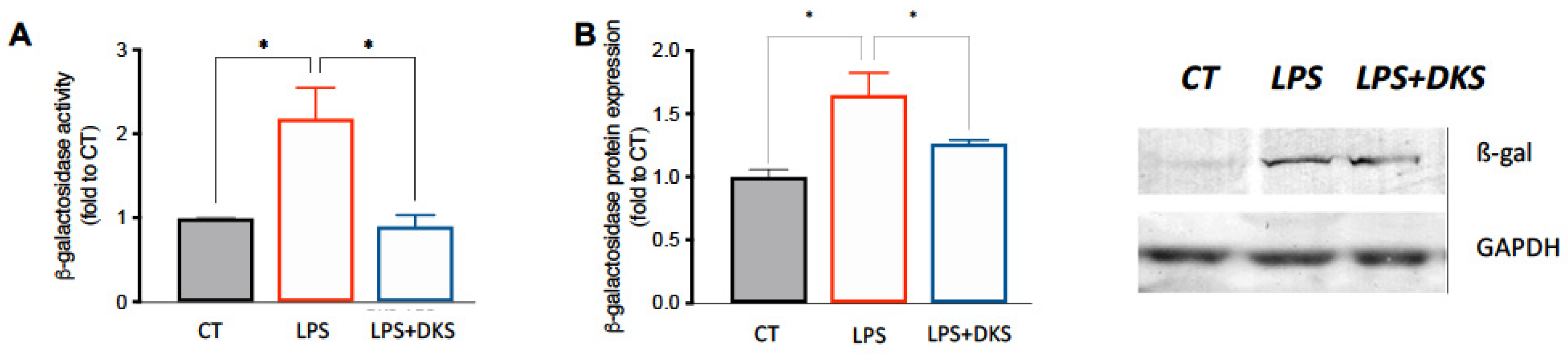
3.2. Effect on ß-Galactosidase Activity and Expression

ß-galactosidase (ß-gal) represents one of the main markers of cellular senescence [29]. In Figure 3A, we can observe a significant increase in ß-gal activity compared with control cells, which were not stimulated with LPS. DKS significantly reduced this activity bringing the values closer to those of the control (CT) group. This result was then confirmed by evaluating the expression of the enzyme by Western blot (WB) analysis. In Figure 3B, we can observe the increase of ß-gal protein expression induced by LPS and the reversal of this up-regulation by DKS, obtaining CTRL-like levels.

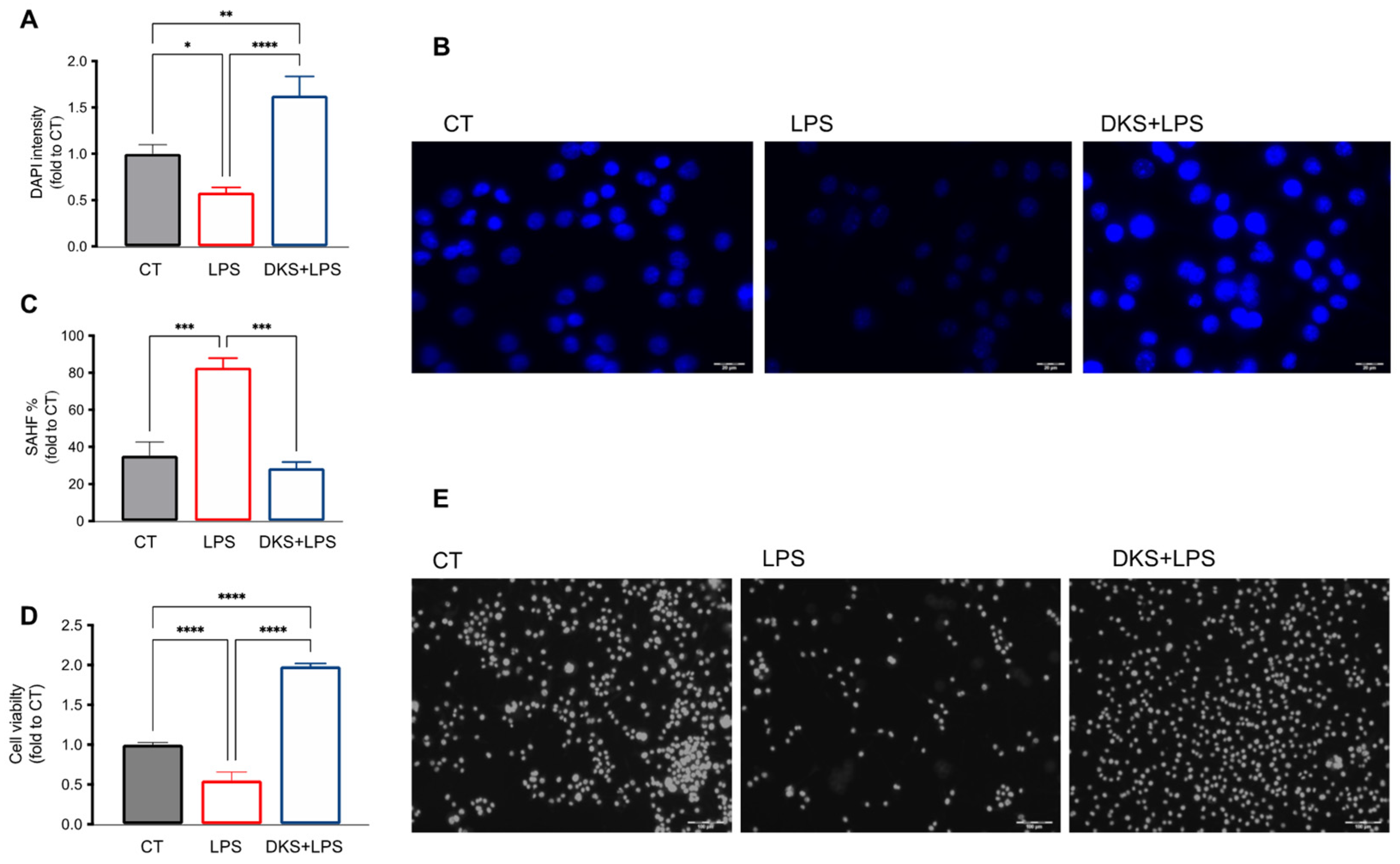
3.3. Reduction of SAHF Formation

Senescent cells tend to form clusters of heterochromatin, resulting in a reduction in the expression of genes involved in cell replication processes, leading to cell death [30]. In senescent cells, as can be seen from the results shown in Figure 4A,B, a lower intensity in the staining of the nuclear marker DAPI and an increase in the expression of heterochromatin foci (Figure 4C) was observed compared with control cells. DKS treatment increased the intensity of DAPI staining and significantly reduced the number of heterochromatin foci compared with senescent cells.
From Figure 4D,E, we can observe that DSK is able to significantly increase cell viability compared with senescent cells treated with LPS, suggesting a reduction in the apoptotic mechanism and an increase in cell replication.

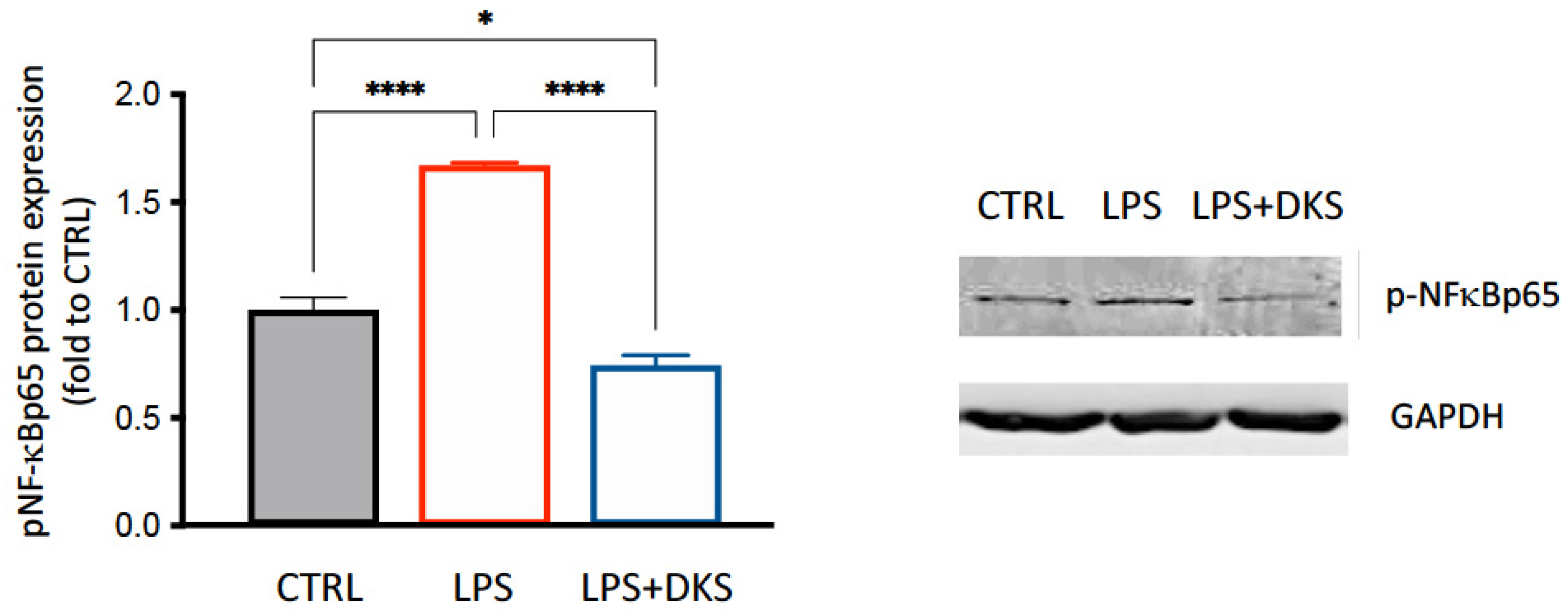
3.4. DKS Reduced SASP Inflammatory Markers

The senescent cells take on new characteristics, including an increase in the production and secretion of cytokines, growth factors, and proteolytic enzymes, leading to the acquisition of a proinflammatory phenotype defined as “secretory phenotype associated with senescence” (SASP) [31,32]. The production of these factors appears to be controlled by complex molecular mechanisms involving the transcription factor NF-kB [33], which also represents the main driver of pro-inflammatory microglia activation. LPS-senescent microglia cells had higher levels of phosphorylated NF-kB p65 subunit protein expression, and DKS treatment counteracted this increase to values comparable to that observed for the CT group (Figure 5). These results suggested a reduction of microglial activity in the inflammatory process associated with the development of the senescent phenotype.

3.5. Neuroprotective Activity of DKS

SH-SY5Y cells were stimulated with medium from senescent BV2 cells, taken on day 10 from the beginning of the LPS intermittent stimulation (Figure 6A). After 48 h stimulation, neuronal cells showed a reduction of cell viability, evaluated by SRB test, compared with neurons treated with non-senescent BV2 medium (Figure 6B). SH-SY5Y cells stimulated with DKS-treated senescent BV2 medium showed a higher percentage of living cells, highlighting a neuroprotective effect of treatment on neurotoxicity induced by microglia aging (Figure 6B).

4. Discussion

The present study investigated the capability of the food supplement DKS to attenuate microglia senescence in an in vitro model. The results obtained showed the senotherapeutic properties of DKS and illustrated its efficacy in promoting neuroprotection against the senescent microglia-associated neurotoxicity.
Obesity is associated with profound changes in cellular function, showing a pathophysiology that recapitulates a chronic condition similar to the aging process. Indeed, obesity is a condition in which the burden of senescent cells is particularly high. An exaggerated and abnormal accumulation of senescent cells in tissues drastically increases the secretion of SASP factors, fostering the development of a persistent low-grade chronic inflammation [34]. Components of the SASP secreted by senescent adipocytes have been suggested to confer and exacerbate insulin resistance [35,36]. Several reports indicate that senescent cells accumulate in adipose tissue of obese and diabetic humans and mice [37,38] and in other organs, including the brain [17]. Thus, targeting senescent cells by means of senotherapeutics has been postulated as a new strategy to alleviate obesity and to improve obesity-associated complications [12,18].
With the aim of searching for an innovative senotherapeutic treatment potentially useful in the therapy of obesity and associated neurological complications, we investigated the antisenescence activity of DKS, a product containing polyphenol-based herbal extracts with antioxidant and anti-inflammatory properties, currently used as adjuvant in hypocaloric diet to stimulate the metabolism of lipids and carbohydrates. Indeed, natural compounds are increasingly attracting scientific interest for their beneficial effects and high tolerability. Many natural constituents, especially polyphenols from commonly used herbal preparations, are widely known to possess antioxidant and anti-inflammatory activities and their anti-obesity effect has been documented [39]. Specifically, treatments endowed with antioxidant and anti-inflammatory activity are promising candidates to mitigate cellular senescent. Of note, oxidative stress, increased in obesity [37], plays an important role in the generation of senescent cells [40] and concurs to the activation of microglia, key cells that play a prominent role not only in the immune and inflammatory response, but also in neuroprotection, food intake, and energy expenditure [41]. Indeed, activation of microglia is a key event in obesity, and in mice fed with high-fat diet, the degree of microgliosis parallels the degree of weight gain [42]. Furthermore, obesity-associated neuroinflammation may be involved in neurodegenerative alteration observed in obese subjects [43].
Exposure of BV2 immortalized murine microglia cells to LPS has been broadly used as a model of neuroinflammation. We have recently shown that, by prolonging the time of exposure and reducing the concentration of LPS, BV2 cells produced a model of microglia senescence [26]. In this model, DKS was able to attenuate ß-gal activity and expression. Increased senescence-associated ß-gal activity was the first marker used for the revelation of cellular senescence in tissues in situ [44] and it is presently recognized as one of the prominent markers of cellular senescence. DKS was also able to reduce SAHF formation, which are heterochromatin domains that induce silencing of various genes involved in the promotion of proliferation and represent a well-known marker for senescent cells [45]. Importantly, treatment of BV2 senescent cells with DKS largely reduced the expression of the phosphorylated p65 subunit of the transcription factor NF-kB, a key event in the acquisition of the SASP phenotype, restoring the basal levels of the protein. NF-kB represents one of the main regulators of proinflammatory processes, as well as driving of microglia activation and the activation of genes that could contribute to cellular senescence and SASP acquisition [46]. Thus, NF-kB represents a key target for both anti-inflammatory and anti-senescence activity. Of note, SASP activity is increased in murine senescent microglia in in vitro and in vivo models, whereas in microglia from the aged mouse brain, SASP is unaltered [16]. This evidence further correlates the microglia senescent phenotype that has acquired SASP activity with pathological conditions rather than with physiological aging.
Senotherapeutics comprise drugs that promote a selective cell death of senescent cells, termed senolytics, and drugs that suppress markers of senescence, in particular the SASP, termed senomorphics [47]. Senomorphic drugs are emerging as alternatives to target senescence-associated diseases [48]. Indeed, the SASP is involved in the secretion of cytokines, chemokines, growth factors, and reactive oxygen species that can lead to detrimental effects on neighboring tissues. Senescence then spreads, since SASP factors can convert adjacent cells to senescence. At the molecular level, senomorphics act by targeting the most important transcription factors for inflammatory mediators, i.e., NF-kB, indicating a promising senomorphic activity for DKS. Multiple commonly used drugs, including metformin [49], have been classified as senomorphics. These compounds usually improve longevity in animal models, confirming the therapeutic value of reducing the effects of senescent cells.
Neurological disturbances are associated with obesity [12]. Neuroinflammation has emerged as key risk to the development of neurological disturbances and microglia dominate as contributors to neuroinflammation [50]. Senescent glial cells are found in obese laboratory animals, and their removal attenuates anxiety-related behaviors [51]. Previous findings showed anti-neuroinflammatory properties for DKS by reducing the levels of proinflammatory mediators [25], and we thus investigated the capability of DKS to protect neurons in the presence of microglia senescent phenotype. We observed that treatment with DKS counteracted the decrease of cell viability of neuronal cells exposed to the neurotoxic insult induced by senescent BV2 medium, showing a potential neuroprotective activity. Growing evidence indicates the potential role of glucagon-like peptide-1 receptor (GLP-1R) activation in the management of obesity and obesity-related brain disorders [52]. GLP-1R activation in microglia suppress their inflammatory response and prevent neurodegeneration in obese mice, making the modulation of this pathway a promising therapeutic strategy. On these bases, we cannot exclude an involvement of GLP-1R mediated effects in the activity of DKS.
Senomorphic treatment represents a safer approach to target senescent cells, as it reduces the generation of SASP factors without causing the death of senescent cells [47]. Obesity causes a continuous production of new senescent cells, given ongoing metabolic insults, and anti-SASP strategies may require a continual treatment to maintain efficacy, given that senescent cells are not eliminated from tissue. Of course, effectiveness, tolerability, and safety must be carefully considered further for use in clinical practice. DKS contains extracts from more than ten medicinal plants, obtained through an extraction process with water at high temperatures. This phytocomplex fosters a synergistic activity between the various components, allowing the use of lower doses that limit the occurrence of potential side effects. In addition, DKS represent a multitarget approach that has been postulated to represent a more effective therapeutic strategy than single target therapies in multifactorial disorders, including obesity [53].

5. Conclusions

Obesity, an impending global threat, is not being effectively controlled by current intervention. The prevalence of obesity is rising, creating an urgent need for efficacious therapies. Growing evidence illustrates cellular senescence as a causal factor in obesity-related inflammation and metabolic derangements. Removing or blunting the effects of senescent cells can alleviate obesity-induced organ and tissue dysfunction. Our results showed the senotherapeutic activity of DKS on senescent microglia, and its neuroprotective effect toward the senescent microglia-associated neurotoxicity. Thus, DKS might hold promise as an adjunctive intervention to control inflammation-related mechanisms of obesity and obesity-related complications.

Author Contributions

Conceptualization, N.G. and V.B.; methodology, formal analysis, and data curation V.B., C.S., M.M. and N.G.; original draft preparation, N.G., C.S., M.M. and V.B.; review and editing, N.G., V.B. and L.C.; All authors have read and agreed to the published version of the manuscript.

Funding

This research received no external funding.

Institutional Review Board Statement

Not applicable.

Informed Consent Statement

Not applicable.

Data Availability Statement

The data presented in this study are available on request from the corresponding author.

Acknowledgments

Thanks to all the technical team of Gianluca Mech Srl for providing Dekosilhue.

Conflicts of Interest

The authors declare no conflict of interest.

References

  1. Chooi, Y.C.; Ding, C.; Magkos, F. The epidemiology of obesity. Metabolism 2019, 92, 6–10. [Google Scholar] [CrossRef]
  2. Velazquez, A.; Apovian, C.M. Updates on obesity pharmacotherapy. Ann. N. Y. Acad. Sci. 2018, 1411, 106–119. [Google Scholar] [CrossRef] [PubMed]
  3. Asghar, A.; Sheikh, N. Role of immune cells in obesity induced low grade inflammation and insulin resistance. Cell. Immunol. 2017, 315, 18–26. [Google Scholar] [CrossRef] [PubMed]
  4. Kawai, T.; Autieri, M.V.; Scalia, R. Adipose tissue inflammation and metabolic dysfunction in obesity. Am. J. Physiol. Cell Physiol. 2021, 320, C375–C391. [Google Scholar] [CrossRef] [PubMed]
  5. Sindhu, S.; Thomas, R.; Kochumon, S.; Wilson, A.; Abu-Farha, M.; Bennakhi, A.; Al-Mulla, F.; Ahmad, R. Increased adipose tissue expression of interferon regulatory factor (IRF)-5 in obesity: Association with metabolic inflammation. Cells 2019, 8, 1418. [Google Scholar] [CrossRef]
  6. Lumeng, C.N.; Saltiel, A.R. Inflammatory links between obesity and metabolic disease. J. Clin. Investig. 2011, 121, 2111–2117. [Google Scholar] [CrossRef]
  7. Hotamisligil, G.S. Foundations of Immunometabolism and Implications for Metabolic Health and Disease. Immunity 2017, 47, 406–420. [Google Scholar] [CrossRef]
  8. Dantzer, R. Neuroimmune interactions: From the brain to the immune system and vice versa. Physiol. Rev. 2018, 98, 477–504. [Google Scholar] [CrossRef]
  9. Rivest, S. Regulation of innate immune responses in the brain. Nat. Rev. Immunol. 2009, 9, 429–439. [Google Scholar] [CrossRef]
  10. Lai, K.S.P.; Liu, C.S.; Rau, A.; Lanctôt, K.L.; Köhler, C.A.; Pakosh, M.; Carvalho, A.F.; Herrmann, N. Peripheral inflammatory markers in Alzheimer’s disease: A systematic review and meta-analysis of 175 studies. J. Neurol. Neurosurg. Psychiatry 2017, 88, 876–882. [Google Scholar] [CrossRef]
  11. Guillemot-Legris, O.; Muccioli, G.G. Obesity-Induced Neuroinflammation: Beyond the Hypothalamus. Trends Neurosci. 2017, 40, 237–253. [Google Scholar] [CrossRef] [PubMed]
  12. Fulton, S.; Décarie-Spain, L.; Fioramonti, X.; Guiard, B.; Nakajima, S. The menace of obesity to depression and anxiety prevalence. Trends Endocrinol. Metab. 2022, 33, 18–35. [Google Scholar] [CrossRef]
  13. Kim, H.J.; Cho, M.H.; Shim, W.H.; Kim, J.K.; Jeon, E.-Y.; Kim, D.-H.; Yoon, S.-Y. Deficient autophagy in microglia impairs synaptic pruning and causes social behavioral defects. Mol. Psychiatry 2017, 22, 1576–1584. [Google Scholar] [CrossRef]
  14. Alexaki, V.I. The impact of obesity on microglial function: Immune, metabolic and endocrine perspectives. Cells 2021, 10, 1584. [Google Scholar] [CrossRef] [PubMed]
  15. Subhramanyam, C.S.; Wang, C.; Hu, Q.; Dheen, S.T. Microglia-mediated neuroinflammation in neurodegenerative diseases. Semin. Cell Dev. Biol. 2019, 94, 112–120. [Google Scholar] [CrossRef] [PubMed]
  16. Stojiljkovic, M.R.; Ain, Q.; Bondeva, T.; Heller, R.; Schmeer, C.; Witte, O.W. Phenotypic and functional differences between senescent and aged murine microglia. Neurobiol. Aging 2019, 74, 56–69. [Google Scholar] [CrossRef]
  17. Palmer, A.K.; Tchkonia, T.; Kirkland, J.L.; Jensen, M.D. Senescence in obesity. In Cellular Senescence in Disease; Academic Press: Cambridge, MA, USA, 2022; pp. 289–308. [Google Scholar]
  18. Palmer, A.K.; Xu, M.; Zhu, Y.; Pirtskhalava, T.; Weivoda, M.M.; Hachfeld, C.M.; Prata, L.G.; Dijk, T.H.; Verkade, E.; Casaclang-Verzosa, G.; et al. Targeting senescent cells alleviates obesity-induced metabolic dysfunction. Aging Cell 2019, 18, e12950. [Google Scholar] [CrossRef]
  19. Narasimhan, A.; Flores, R.R.; Camell, C.D.; Bernlohr, D.A.; Robbins, P.D.; Niedernhofer, L.J. Cellular Senescence in Obesity and Associated Complications: A New Therapeutic Target. Curr. Diab. Rep. 2022, 22, 537–548. [Google Scholar] [CrossRef]
  20. Van Deursen, J.M. The role of senescent cells in ageing. Nature 2014, 509, 439–446. [Google Scholar] [CrossRef]
  21. Kumar, A.; Bano, D.; Ehninger, D. Cellular senescence in vivo: From cells to tissues to pathologies. Mech. Ageing Dev. 2020, 190, 111308. [Google Scholar] [CrossRef]
  22. Lagoumtzi, S.M.; Chondrogianni, N. Senolytics and senomorphics: Natural and synthetic therapeutics in the treatment of aging and chronic diseases. Free. Radic. Biol. Med. 2021, 171, 169–190. [Google Scholar] [CrossRef]
  23. Mathew, G.; Unnikrishnan, M.K. Multi-target drugs to address multiple checkpoints in complex inflammatory pathologies: Evolutionary cues for novel “first-in-class” anti-inflammatory drug candidates: A reviewer’s perspective. Inflamm. Res. 2015, 64, 747–752. [Google Scholar] [CrossRef] [PubMed]
  24. Wagner, H.; Ulrich-Merzenich, G. Synergy research: Approaching a new generation of phytopharmaceuticals. Phytomedicine 2009, 16, 97–110. [Google Scholar] [CrossRef] [PubMed]
  25. Borgonetti, V.; Cenci, L.; Galeotti, N. Anti-Inflammatory and Neuroprotective Effect of the Anti-Obesity Dietary Supplement Dekosilhue® in an In Vitro Model of Neuroinflammation. Nutraceuticals 2022, 2, 22–31. [Google Scholar] [CrossRef]
  26. Borgonetti, V.; Galeotti, N. Rosmarinic Acid Reduces Microglia Senescence: A Novel Therapeutic Approach for the Management of Neuropathic Pain Symptoms. Biomedicines 2022, 10, 1468. [Google Scholar] [CrossRef] [PubMed]
  27. Wu, F.Y.H.; Liao, W.C.; Chang, H.M. Comparison of antitumor activity of vitamins K1, K2 and K3 on human tumor cells by two (MTT and SRB) cell viability assays. Life Sci. 1993, 52, 1797–1804. [Google Scholar] [CrossRef] [PubMed]
  28. Borgonetti, V.; Governa, P.; Biagi, M.; Pellati, F.; Galeotti, N. Zingiber officinale Roscoe rhizome extract alleviates neuropathic pain by inhibiting neuroinflammation in mice. Phytomedicine 2020, 78, 153307. [Google Scholar] [CrossRef]
  29. Ng, P.Y.; McNeely, T.L.; Baker, D.J. Untangling senescent and damage-associated microglia in the aging and diseased brain. FEBS J. 2021, 290, 1326–1339. [Google Scholar] [CrossRef]
  30. Aird, K.M.; Zhang, R. Detection of senescence-associated heterochromatin foci (SAHF). Methods Mol. Biol. 2013, 965, 185–196. [Google Scholar] [CrossRef]
  31. Burton, D.G.A.; Stolzing, A. Cellular senescence: Immunosurveillance and future immunotherapy. Ageing Res. Rev. 2018, 43, 17–25. [Google Scholar] [CrossRef]
  32. Hernandez-Segura, A.; de Jong, T.V.; Melov, S.; Guryev, V.; Campisi, J.; DeMaria, M. Unmasking Transcriptional Heterogeneity in Senescent Cells. Curr. Biol. 2017, 27, 2652–2660.e4. [Google Scholar] [CrossRef] [PubMed]
  33. Ohanna, M.; Giuliano, S.; Bonet, C.; Imbert, V.; Hofman, V.; Zangari, J.; Bille, K.; Robert, C.; Paillerets, B.B.-D.; Hofman, P.; et al. Senescent cells develop a parp-1 and nuclear factor-κB-associated secretome (PNAS). Genes Dev. 2011, 25, 1245–1261. [Google Scholar] [CrossRef] [PubMed]
  34. Franceschi, C.; Campisi, J. Chronic inflammation (Inflammaging) and its potential contribution to age-associated diseases. J. Gerontol. Ser. A Biol. Sci. Med. Sci. 2014, 69, S4–S9. [Google Scholar] [CrossRef] [PubMed]
  35. Ahima, R.S. Connecting obesity, aging and diabetes. Nat. Med. 2009, 15, 996–997. [Google Scholar] [CrossRef] [PubMed]
  36. Xu, M.; Palmer, A.K.; Ding, H.; Weivoda, M.M.; Pirtskhalava, T.; A White, T.; Sepe, A.; O Johnson, K.; Stout, M.B.; Giorgadze, N.; et al. Targeting senescent cells enhances adipogenesis and metabolic function in old age. Elife 2015, 4, e12997. [Google Scholar] [CrossRef] [PubMed]
  37. Minamino, T.; Orimo, M.; Shimizu, I.; Kunieda, T.; Yokoyama, M.; Ito, T.; Nojima, A.; Nabetani, A.; Oike, Y.; Matsubara, H.; et al. A crucial role for adipose tissue p53 in the regulation of insulin resistance. Nat. Med. 2009, 15, 1082–1087. [Google Scholar] [CrossRef] [PubMed]
  38. Schafer, M.J.; Miller, J.D.; LeBrasseur, N.K. Cellular senescence: Implications for metabolic disease. Mol. Cell. Endocrinol. 2017, 455, 93–102. [Google Scholar] [CrossRef]
  39. Wang, S.; Moustaid-Moussa, N.; Chen, L.; Mo, H.; Shastri, A.; Su, R.; Bapat, P.; Kwun, I.; Shen, C.-L. Novel insights of dietary polyphenols and obesity. J. Nutr. Biochem. 2014, 25, 1–18. [Google Scholar] [CrossRef]
  40. Hernandez-Segura, A.; Nehme, J.; Demaria, M. Hallmarks of Cellular Senescence. Trends Cell Biol. 2018, 28, 436–453. [Google Scholar] [CrossRef]
  41. García-Cáceres, C.; Balland, E.; Prevot, V.; Luquet, S.; Woods, S.C.; Koch, M.; Horvath, T.L.; Yi, C.-X.; Chowen, J.A.; Verkhratsky, A.; et al. Role of astrocytes, microglia, and tanycytes in brain control of systemic metabolism. Nat. Neurosci. 2019, 22, 7–14. [Google Scholar] [CrossRef]
  42. De Luca, S.N.; Miller, A.A.; Sominsky, L.; Spencer, S.J. Microglial regulation of satiety and cognition. J. Neuroendocr. 2020, 32, e12838. [Google Scholar] [CrossRef]
  43. Woo, A.; Botta, A.; Shi, S.S.W.; Paus, T.; Pausova, Z. Obesity-Related Neuroinflammation: Magnetic Resonance and Microscopy Imaging of the Brain. Int. J. Mol. Sci. 2022, 23, 8790. [Google Scholar] [CrossRef] [PubMed]
  44. Kurz, D.J.; Decary, S.; Hong, Y.; Erusalimsky, J.D. Senescence-associated ß-galactosidase reflects an increase in lysosomal mass during replicative ageing of human endothelial cells. J. Cell Sci. 2000, 113, 3613–3622. [Google Scholar] [CrossRef] [PubMed]
  45. Narita, M.; Nũnez, S.; Heard, E.; Narita, M.; Lin, A.W.; Hearn, S.A.; Spector, D.L.; Hannon, G.J.; Lowe, S.W. Rb-mediated heterochromatin formation and silencing of E2F target genes during cellular senescence. Cell 2003, 113, 703–716. [Google Scholar] [CrossRef] [PubMed]
  46. Salminen, A.; Kauppinen, A.; Kaarniranta, K. Emerging role of NF-kB signaling in the induction of senescence-associated secretory phenotype (SASP). Cell Signal. 2012, 24, 835–845. [Google Scholar] [CrossRef]
  47. Zhang, L.; Pitcher, L.E.; Prahalad, V.; Niedernhofer, L.J.; Robbins, P.D. Targeting cellular senescence with senotherapeutics: Senolytics and senomorphics. FEBS J. 2022, 290, 1362–1383. [Google Scholar] [CrossRef]
  48. Birch, J.; Gil, J. Senescence and the SASP: Many therapeutic avenues. Genes Dev. 2020, 34, 1565–1576. [Google Scholar] [CrossRef]
  49. Kanigur Sultuybek, G.; Soydas, T.; Yenmis, G. NF-kB as the mediator of metformin’s effect on ageing and ageing-related diseases. Clin. Exp. Pharmacol. Physiol. 2019, 46, 413–422. [Google Scholar] [CrossRef]
  50. Wang, H.; He, Y.; Sun, Z.; Ren, S.; Liu, M.; Wang, G.; Yang, J. Microglia in depression: An overview of microglia in the pathogenesis and treatment of depression. J. Neuroinflammation 2022, 19, 132. [Google Scholar] [CrossRef]
  51. Ogrodnik, M.; Zhu, Y.; Langhi, L.G.P.; Tchkonia, T.; Krüger, P.; Fielder, E.; Victorelli, S.; Ruswhandi, R.A.; Giorgadze, N.; Pirtskhalava, T.; et al. Obesity-Induced Cellular Senescence Drives Anxiety and Impairs Neurogenesis. Cell Metab. 2019, 29, 1061–1077.e8. [Google Scholar] [CrossRef]
  52. Cui, Q.N.; Stein, L.M.; Fortin, S.M.; Hayes, M.R. The role of glia in the physiology and pharmacology of glucagon-like peptide-1: Implications for obesity, diabetes, neurodegeneration and glaucoma. Br. J. Pharmacol. 2022, 179, 715–726. [Google Scholar] [CrossRef] [PubMed]
  53. Camilleri, M.; Acosta, A. Combination Therapies for Obesity. Metab. Syndr. Relat. Disord. 2018, 16, 390–394. [Google Scholar] [CrossRef] [PubMed]
Figure 1. Schematic representation of the senescent microglia model, administration, and tests schedule.
Nutraceuticals 03 00020 g001
Figure 2. LPS and DKS effects on cell viability by the SRB test. (A) Time-course evaluation of cell viability in BV2 cells intermittently exposed to LPS (500 ng/mL). Two-way ANOVA * p < 0.05, ** p < 0.01, *** p < 0.001 in comparison with unstimulated control (CTRL) group. (B) Dose–response effect on cell viability of unstimulated BV2 cells by DSK (1, 10, 100, and 1000 µg/mL). Vertical lines represent SEM.
Nutraceuticals 03 00020 g002
Figure 3. Effect of DKS on ß-galactosidase. (A) ß-galactosidase (ß-gal) activity is increased on fresh cell lysate. DKS treatment counteracted this activity. (B) ß-galactosidase (ß-gal) protein expression and representative blots. One-way ANOVA * p < 0.05.
Nutraceuticals 03 00020 g003
Figure 4. Effect of DKS on senescent-associated heterochromatin foci (SAHF). DAPI staining of nuclei (quantification analysis (A); representative image of DAPI intensity (B)) and SAHF formation (C) in BV2 senescent cells at 10 days after intermittent stimulus with LPS (500 ng/mL). DSK (100 μg/mL) reverted both LPS-induced senescence markers. (D) Decrease of cell viability by LPS intermittent stimulus and reversal by DSK (100 μg/mL). (E) Representative image of cell viability. One-way ANOVA * p < 0.05, ** p < 0.01, *** p < 0.001, **** p < 0.0001.
Nutraceuticals 03 00020 g004
Figure 5. Effect of DKS on NF-kB pathway activation. Reversal by DKS (100 µg/mL) of increased pNF-kBp65 protein expression induced by LPS (500 ng/mL) intermittent stimulation. Representative blots are reported. One-way ANOVA * p < 0.05, **** p < 0.0001.
Nutraceuticals 03 00020 g005
Figure 6. Neuroprotective effect of DKS. (A) Schematic representation of the experimental protocol. (B). Evaluation of neuronal viability (SRB test) in SH−SY5Y cells exposed to non−senescent medium, senescent medium or senescent medium followed by DKS (100 µg/mL) treatment. One-way ANOVA * p < 0.05, ** p < 0.01.
Nutraceuticals 03 00020 g006
Table 1. Composition of Dekosilhue® (DKS). The content of each constituent (aqueous extract) is expressed as grams of extract in 1 L of product (g/L).
DKS Constituentg/L
Cinnamon (Cinnamomum zeylanicum Blume) bark90
Orthosiphon (Orthosiphon stamineus Benth) leaf90
Green tea (Camellia sinensis (L.) Kuntze) leaf90
Mate (Ilex paraguariensis A.St.Hill) leaf70
Gymnema (Gymnema sylvestre R. Br.) leaf70
Bean (Phaseolus vulgaris L.) pod60
Pineapple (Ananas comosus (L.) Merr.) stem40
Common gromwell (Lithospermum officinale L.) seeds40
Horsetail (Equisetum arvense L.) herb40
Curly dock (Rumex crispus L.) root30
Asparagus (Asparagus officinalis L.) root30
Fennel (Foeniculum vulgare Miller) fruit30
Birch (Betuta pendolo Roth.) leaf20
Disclaimer/Publisher’s Note: The statements, opinions and data contained in all publications are solely those of the individual author(s) and contributor(s) and not of MDPI and/or the editor(s). MDPI and/or the editor(s) disclaim responsibility for any injury to people or property resulting from any ideas, methods, instructions or products referred to in the content.

