Communication

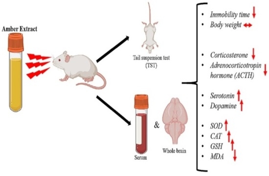
Behavioral and Biochemical Evaluation of Anti-Depressive and Oxidative Stress-Ameliorating Effects of Amber Extract in Adult Male ICR Mice

1
Tsukuba Life Science Innovation Program (T-LSI), University of Tsukuba, Tennodai 1-1-1, Tsukuba 305-8577, Japan
2
Faculty of Life and Environmental Sciences, University of Tsukuba, Tsukuba 305-8572, Japan
3
Kohaku Bio Technology Co., Ltd., Tsukuba 305-8572, Japan
*
Author to whom correspondence should be addressed.
Nutraceuticals 2023, 3(2), 222-230; https://doi.org/10.3390/nutraceuticals3020017
Submission received: 8 March 2023 / Revised: 14 April 2023 / Accepted: 20 April 2023 / Published: 24 April 2023

Abstract

:
Amber, a plant resin, exhibits an anti-stress effect and is used in traditional medicine. Recently, it has been speculated that amber may possess an anti-depressive effect. However, there is no evidence to support this efficacy. Thus, this study investigated the anti-depressive and oxidative-stress-ameliorating effects of amber extract in mice subjected to restraint stress. Mice were treated with amber extract (25 and 50 mg/kg, p.o.) and bupropion (10 mg/kg, p.o.) as positive control. Mice were then subjected to a tail suspension test, and their immobility time, body weight before and after stress, levels of stress-related hormones and neurotransmitters, and oxidative stress parameters were assessed. Amber supplementation did not affect the body weight of mice in any of the groups. Amber extract (25 and 50 mg/kg) demonstrated an anti-depressive effect by significantly decreasing the immobility time and adrenocorticotropin-hormone and corticosterone-hormone levels. Moreover, amber extract at a dose of 25 mg/kg increased the levels of dopamine and serotonin. Additionally, superoxide dismutase, catalase, and glutathione levels increased, whereas the malondialdehyde content decreased with amber supplementation. These findings confirm that amber may possess an anti-depressive effect and hence can be a useful alternative therapy for preventing and managing depression.

Graphical Abstract

1. Introduction

Mood is a description of an individual’s internal state, which is reflected in the external behavior. Individuals may occasionally feel sad or anxious regarding their lives. However, extreme and persistent feelings of sadness and anxiety may result in mood disorders, particularly depression. Depression or depressive disorders have become a global menace, costing economies huge sums of money and productivity [1]. This includes, though is not limited to, feelings of guilt, irritability, lack of motivation, appetite changes, suicidal thoughts, loss of interest, and sleep changes. These symptoms disrupt daily activities and affect quality of life. Several factors, such as psychological and environmental events, may contribute to the occurrence of depression. The brain plays a crucial role in the development of depressive disorders. It regulates chemical messengers and thus can prevent or manage the progression of depressive disorders. Furthermore, owing to its constant metabolic activity, it is highly susceptible to oxidative damage [2]. Alterations in brain function have been shown to cause mood disorders, such as post-traumatic stress disorder (PTSD) and depression [2,3,4].
Chronic stress activates the hypothalamus-pituitary-adrenal (HPA) axis to release cortisol (in mammals) or corticosteroids (in rodents); their excessive levels may be detrimental to the health of an individual [4]. Moreover, chronic stress increases the accumulation of free radicals in the body. The inability of the body to scavenge these reactive species results in lipid peroxidation, DNA damage, cell death, and eventually several diseases and disorders, including depressive disorders. Thus, oxidative stress markers, such as malondialdehyde (MDA), are crucial in determining the mental health of individuals. Several reports have highlighted the association between depression and oxidative stress [1,5,6]. Oxidative-stress-induced lipid peroxidation products such as MDA are present in depressive disorders [5].
Over the years, researchers have extensively discussed the importance of natural products such as dietary supplements, particularly for stress management. Traditional antidepressants, such as selective serotonin reuptake inhibitors (SSRIs) and serotonin-norepinephrine reuptake inhibitors (SNRIs), have been shown to be effective in managing stress and have therefore been the mainstream drugs of choice for treating depressive disorders and other mood disorders. However, compliance with these drugs is a challenge owing to their cost and associated side effects, such as gastrointestinal upset, sexual dysfunction, dependency, and weight gain. Alternative therapeutic approaches, such as the use of herbal supplements, are as effective as traditional medicines and have fewer side effects, rapid action, and a lower cost [6]. Some well-known herbal products currently reported as effective alternative medicines and traditionally used for stress management include ashwagandha [7,8], Ginkgo biloba [9], and chamomile [10].
Amber, a terpenoid-containing resin from the pine tree, has been reported to possess several biological activities, such as anti-stress [11], anti-Alzheimer’s or neuroprotective [12], anti-inflammatory [13], and lipid-lowering [14] effects. It contains agathic acid, which is known to exert an antibacterial effect [15]. However, there are no reports on its antidepressant effect. We previously demonstrated that amber might exhibit an anti-stress effect through the regulation of HPA hyperactivity [11]. Thus, we hypothesized that amber possesses an antidepressant effect via the regulation of hormones in the HPA axis and factors contributing to oxidative stress.
Hence, the present study aimed to investigate the protective effects of amber extract against depression and oxidative-stress damage using a tail suspension test [TST] in Institute of Cancer Research [ICR] mice to induce psychological stress. The body weight of mice before and after stress was determined, in addition to the measurement of the immobility time of tail-suspended mice. Furthermore, the HPA axis activity, depression-related neurotransmitters, and oxidative-stress parameters were investigated.

2. Materials and Methods

2.1. Amber Extract and Other Reagents

Amber was crushed, powdered, and extracted twice with 50% ethanol at 40 °C for 1 h. The filtrate was freeze-dried to obtain a powder. The samples were then dissolved in ethanol to form a paste. The samples were dissolved in 100% ethanol and diluted with 5% ethanol in water to obtain the required concentration for further experiments.
Bupropion hydrochloride was obtained from Sigma Aldrich, Co. (St. Louis, MO, USA).

2.2. Animals

Twenty-five (five-week-old) male ICR mice were obtained from Tokyo Laboratory Animals Science Co., Ltd. (Tokyo, Japan) and housed in a temperature-controlled room. The animals were randomly divided into five groups, with five mice per group: CT (NS) group representing control group of non-stressed mice, CT (S) group representing control group of stressed mice, BUP group representing positive control group of mice treated with bupropion (10 mg/kg body weight), AMB 25 group representing mice treated with 25 mg/kg body weight of amber extract, and AMB 50 group representing mice treated with 50 mg/kg body weight amber extract. CT (NS) and CT (S) groups received Milli-Q water. All mice had easy access to food and water and were kept under a controlled 12-h day-light schedule. Before treatment initiation, they were allowed to acclimatize for one week. Animal experiments were approved by the Animal Care and Use Committee of the University of Tsukuba. Drug treatment was carried out as shown in Figure S1.

2.3. Sample Collection

Thirty minutes after the final TST, mice were euthanized by spinal dislocation and immediately sacrificed, and blood samples (1.5 mL) were collected from the heart into tubes. After 1 h, the samples were centrifuged at 3000× g for 10 min at 4 °C. Serum was collected and immediately stored at −80 °C until further analysis.
Mice brains were harvested into 5 mL tubes and immediately frozen in liquid nitrogen. Brain samples were later stored at −80 °C until further analysis.

2.4. Tissue Homogenization and Protein Quantification

Frozen brain samples were thawed in RIPA buffer containing a phosphatase inhibitor and homogenized on ice using Power Masher II (Nippi Inc., Tokyo, Japan). The samples were then centrifuged at 12,000× g at 4 °C for 20 min, and supernatants were transferred to 1.5 mL tubes. Proteins were quantified using TaKaRa BCA Protein Assay Kit (TaKaRa, Japan), and samples were stored at −80 °C until further use.

2.5. Tail Suspension Test (TST)

All mice except those in the CT(NS) group were subjected to TST, which was conducted 60 min after treatment. Mice were suspended by the tail attached to a hanging pin in the cage. The test was performed for 6 min, during which mice movements or idleness were recorded using a camera. During the final 3 min, the immobility time, the time depicting stress resistance and during which no movements were observed in suspended mice, was measured. This test was performed twice more, as planned, and the last TST data were used for analysis.

2.6. Determination of the Levels of Hormones and Neurotransmitters

Corticosterone and serotonin levels in mice serum were determined using commercially available ELISA kits (Cayman Chemical Company, Ann Arbor, MI, USA; Enzo Life Sciences, Inc., Farmingdale, NY, USA, respectively), according to the manufacturers’ instructions. Whole mice brains (collected and quantified as described previously) were used to assess dopamine (ImmuSmol, Bordeaux, France) and adrenocorticotropin hormone (ACTH) (Elabscience, Houston, TX, USA) levels, according to the manufacturer’s instructions.

2.7. Determination of Oxidative Stress Parameters

Mice serum samples were used for the assessment of the antioxidant enzymes superoxide dismutase (SOD) and catalase (CAT) (Invitrogen, Thermo Fisher Scientific, Waltham, MA, USA), according to the manufacturer’s instructions. Brain samples of mice were used to assess the levels of the lipid peroxidation product MDA (Invitrogen, Thermo Fisher Scientific, Waltham, MA, USA) and the activities of GSH (Dojindo Laboratories, Kumamoto, Japan) using commercial kits, according to the manufacturers’ instructions.

2.8. Statistical Analysis

All graphs and statistics were generated using GraphPad Prism software 8.0, and the results were represented as mean ± standard deviation (SD). Statistical analysis of data was performed using one-way analysis of variance (ANOVA) and two-way ANOVA, as appropriate. Statistical significance was set at p ≤ 0.05.

3. Results

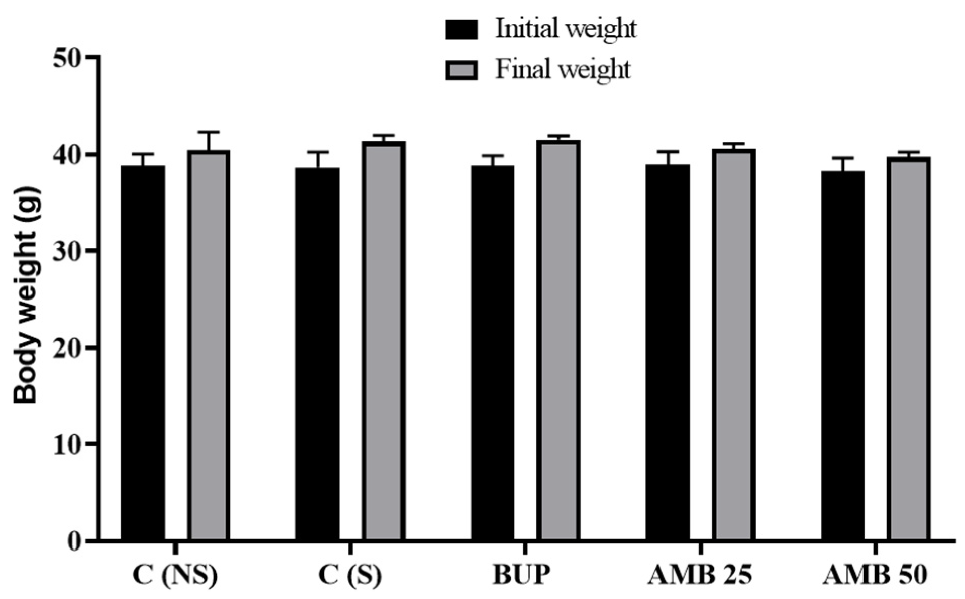
3.1. Amber Treatment Did Not Affect the Body Weight of Mice

The body weight of mice was measured before and after TST to determine the effects of stress and amber supplementation. There was no significant difference in the body weight of mice in all groups before and after the stress test. Further, amber supplementation did not affect the body weight of mice (Figure 1).

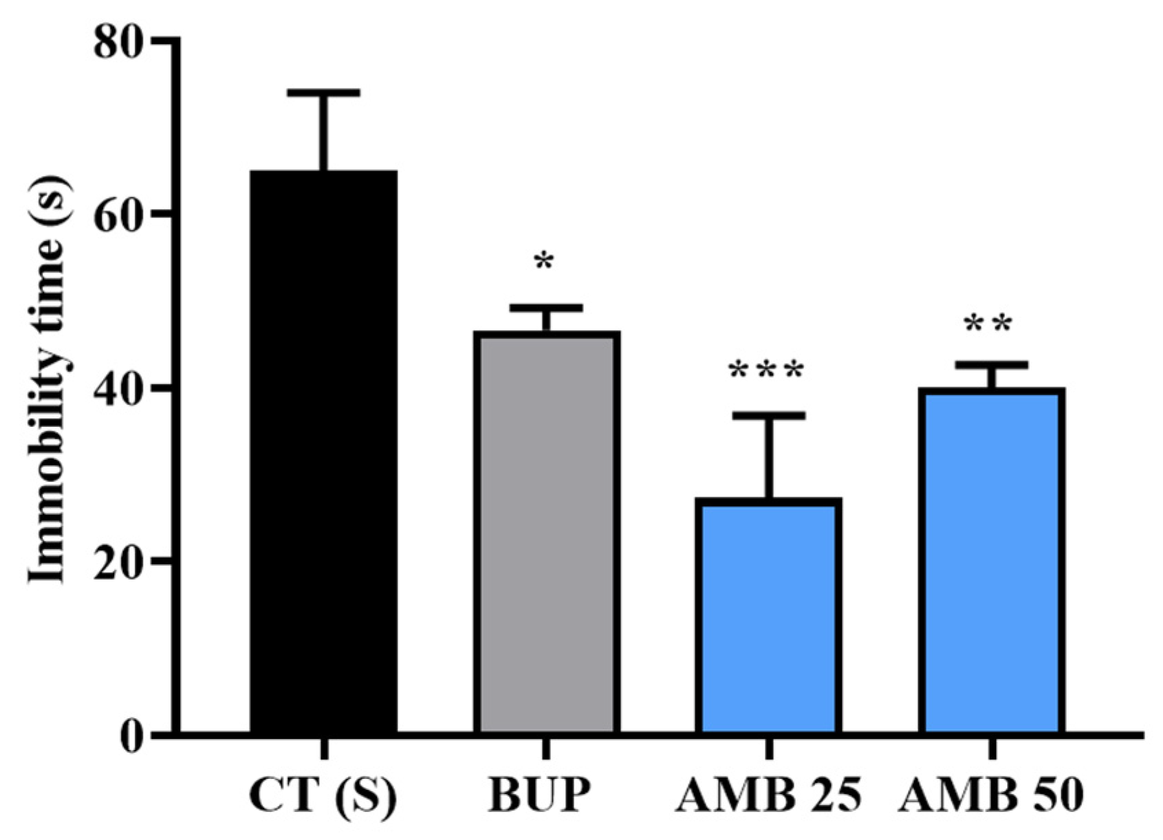
3.2. Amber Supplementation Showed Antidepressant Effect in Tail-Suspended Mice

The psychological stress test, TST, conducted on mice showed that amber (25 and 50 mg/kg)-treated mice exhibited a significant reduction in immobility time compared to the control group, CT (S) (Figure 2). The BUP group (positive-control mice treated with 10 mg/kg bupropion) also showed a significant reduction in immobility time, thus indicating that amber may possess an antidepressant effect.

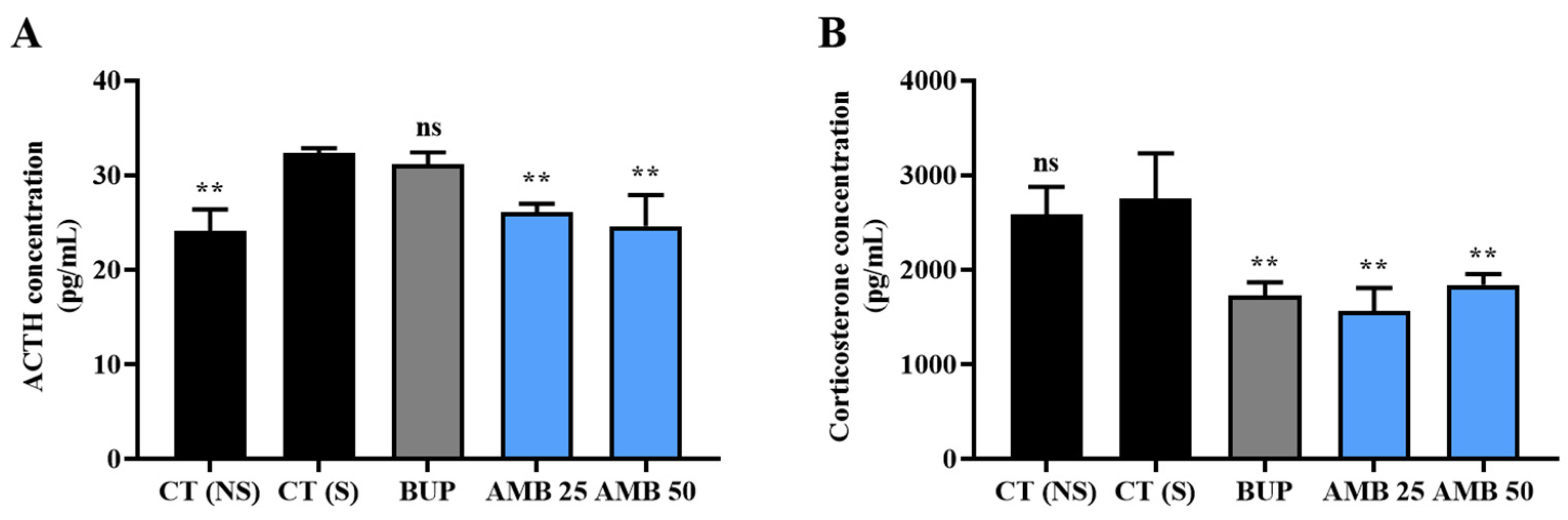
3.3. Amber Positively Regulated Hormones in the HPA Axis System

Hyperactivity of the HPA axis is a prominent cause of several diseases and disorders, including depression. In our study, TST was conducted to induce hyperactivity of the HPA axis. As seen in Figure 3A, amber supplementation (25 and 50 mg/kg) significantly reduced ACTH levels compared to CT (S) mice, similar to the levels in of the control non-stressed mice, CT (NS). However, the effect of bupropion was not significant. A significant reduction in corticosterone levels was observed in both amber (25 and 50 mg/kg)- and bupropion-treated mice when compared to CT (S) mice. No significant reduction was observed in the CT (NS) mice (Figure 3B). This indicates that amber treatment can decrease the HPA axis hyperactivity to regulate the release of stress hormones, thereby reducing the risk of developing depression and other disorders.

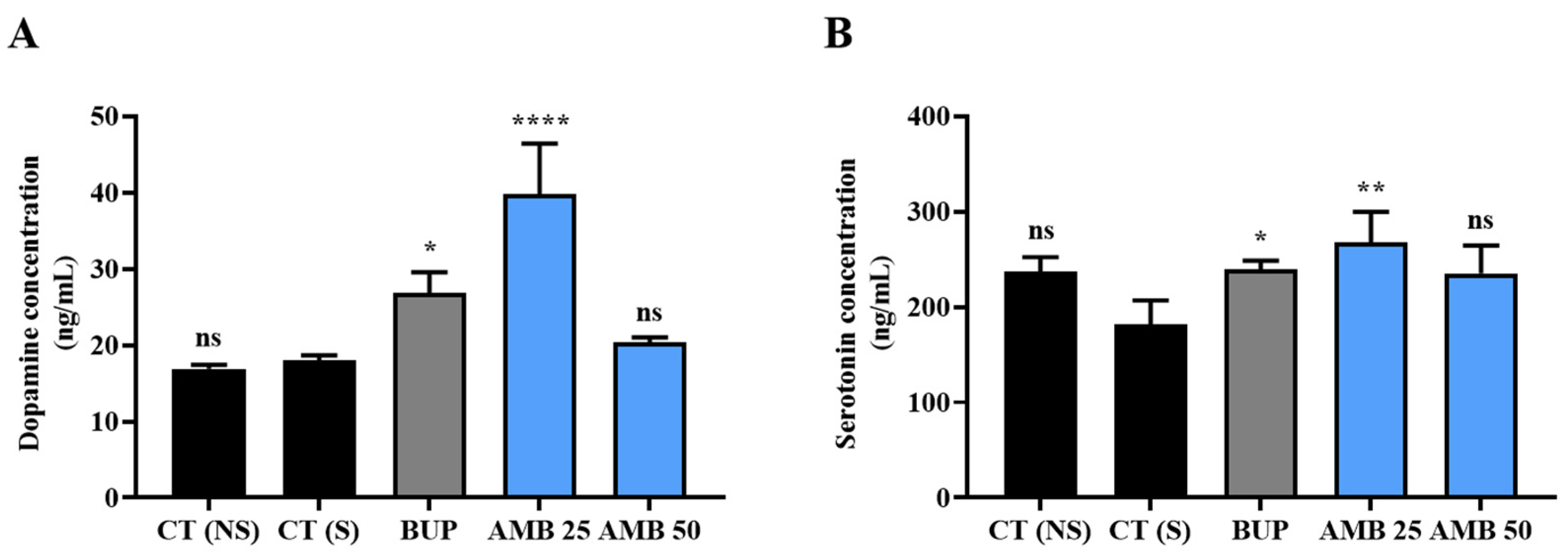
3.4. Amber Treatment Positively Controlled Antidepressant Hormones

Amber (25 mg/kg) and bupropion treatment significantly increased the levels of dopamine (Figure 4A) and serotonin (Figure 4B) in mice. However, amber (50 mg/kg) failed to significantly increase the levels of both hormones compared to CT (S) mice. CT (NS) mice showed no significant difference in hormone levels compared to CT (S) mice.

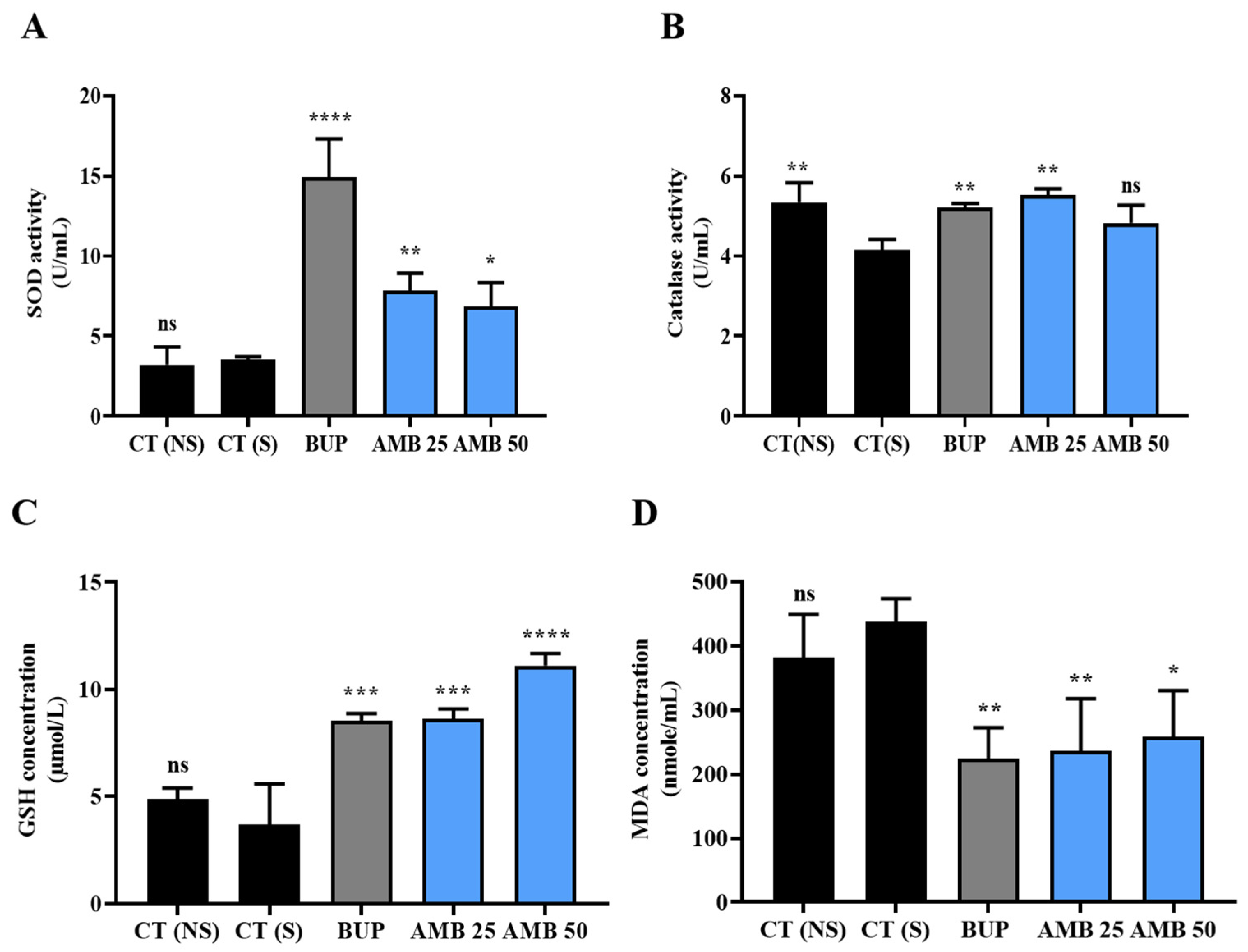
3.5. Amber Ameliorated Oxidative Stress in Mice

In animals treated with amber extract (25 and 50 mg/kg) and bupropion, a significant increase in SOD (Figure 5A) and GSH activity (Figure 5C) was observed compared to CT (S) mice. However, CT (NS) mice showed no significant difference in these parameters. As shown in Figure 5B, mice treated with 25 mg/kg, but not 50 mg/kg, of amber extract showed a significant increase in CAT activity, similar to the BUP and CT (NS) groups. Regarding lipid peroxidation, a substantial reduction in MDA levels was observed following amber (25 and 50 mg/kg) and bupropion treatment (Figure 5D). However, MDA levels were high in both CT (S) and CT (NS) groups.

4. Discussion

Depression is a complex mental condition that may be caused by environmental factors and dysfunction in biological systems. A current study evaluated the effect of amber extract on stress-associated depression by investigating how amber regulates hormones in the HPA axis, monoamines associated with depression, and oxidative-stress parameters because abnormality in their levels can lead to depression. Control of the HPA axis and certain brain neurotransmitters have been reported to help alleviate depression [16]. Thus, it is important to study how compounds control these stress-response systems to manage or prevent depressive episodes. Adverse effects of traditional medications for depression have prompted the quest for alternative treatment options.
TST is a behavioral experiment conducted on mice to investigate the antidepressant potential of compounds [17]. Several compounds under investigation have shown a reduction in the immobility time of tail-suspended mice [16,18], including bupropion, a commercial antidepressant drug. Consistently, a reduced immobility time in TST observed in this study indicates that amber possesses an antidepressant effect.
Generally, long-term exposure to stress leads to hyperactivity of the HPA axis, which is the main cause of several health disorders, including depression. An increase in ACTH levels in the HPA axis increases the release of corticosterone into the bloodstream. As the body attempts to adapt to these changes, several normal physiological balances are disrupted, thereby affecting health [19]. In this study, one of the restraint stress tests, TST, elevated the levels of ACTH and corticosterone, and this effect was attenuated by amber treatment, thus suggesting that amber can regulate HPA axis hyperactivity to regulate the increased secretion of ACTH and glucocorticoids. This antidepressant activity of amber may involve modulating hormones in the HPA axis. Similarly, adult mice supplemented with green-tea polyphenols showed a reduced secretion of both ACTH and corticosterone when subjected to the forced swimming test, another type of restraint stress test [18].
The monoamines dopamine and serotonin have long been reported to be associated with depression and depressive episodes [19]. It is known that the depletion of these happy hormones causes loss of motivation, reduced interest, mood changes, etc. In our study, amber increased dopamine and serotonin levels, thus counteracting the stress-induced depletion of these hormones. An increase in the levels of these neurotransmitters can switch negative emotions into positive feelings and alleviate depression. Several studies have reported that increasing the levels of these monoamines plays a vital role in the management of depression and other related disorders [20,21]. In 2003, Vaswani et al. reported that increasing serotonin levels or activity by inhibiting its reuptake is helpful in reducing depression, which is the underlying mechanism of the antidepressant action of SSRIs, the first-line therapeutic approach for managing depression [22]. This implies that amber can be considered a potent antidepressant in the future.
In the brain, hyperactivity of the HPA axis due to chronic stress leads to oxidative stress caused by ROS accumulation. This is largely due to high levels of glucocorticoids (cortisol or corticosterone) circulating in the blood. Persistent oxidative stress can contribute to depression [19]. In this study, amber positively controlled oxidative-stress parameters. Stress caused the depletion of SOD and CAT in CT (S) mice (Figure 3A,B); however, the activity of these antioxidant enzymes was greatly enhanced by amber supplementation, thus improving free-radical scavenging. During the scavenging of free radicals, SOD first converts free radicals, such as superoxide ions (O-), into hydrogen peroxide (H2O2), which is then decomposed into water by CAT and GSH through different pathways [23]. Thus, increased GSH levels after amber treatment also played an important role in reducing oxidative stress. When mice were subjected to psychological stress through TST, an increased accumulation of the lipid-peroxidation product MDA was observed because of oxidative damage, which was significantly reduced by amber supplementation. These findings confirmed the antioxidant effect of amber observed in our previous study on Caenorhabditis elegans, in which amber treatment significantly protected worms from H2O2-induced oxidative stress and increased the expression of the antioxidant genes sod-3, gst-4, and ctl-1 [11].
Interestingly, mice supplemented with 25 mg/kg of amber extract performed better in this study than those supplemented with 50 mg/kg amber. Although the exact reason for this could not be explored, both doses (25 and 50 mg/kg) were considered safe for animals, based on a previous experiment conducted in our laboratory (unpublished data). No physical or extreme behavioral changes were observed in mice treated with 50 mg/kg amber to depict signs of rejection or toxicity. Thus, we conclude that amber at a dose of 25 mg/kg is more effective than at 50 mg/kg.
Taken together, the study findings have shown that amber can prevent or control stress-induced depression by positively regulating the HPA axis system and reducing the levels of key stress hormones, such as ACTH and corticosterone. Additionally, amber treatment led to enhanced levels of anti-depressive neurotransmitters (dopamine and serotonin) along with the regulation of oxidative stress markers, including SOD, CAT, GSH, and MDA. The findings suggest that amber is a safe dietary supplement or alternative therapy for the management of stress and stress-induced depression. Further studies should be performed on human subjects to determine a more effective and safer dose of amber and reveal the exact active ingredient(s) responsible for its antidepressant effect.

Supplementary Materials

The following supporting information can be downloaded at: https://www.mdpi.com/article/10.3390/nutraceuticals3020017/s1, Figure S1: Experimental design.

Author Contributions

Conceptualization, K.S.; Methodology, K.S., S.S.-A. and M.B.O.; Data curation, S.S.-A., M.B.O. and K.S.; Sample preparation, R.T., K.O. and M.S.; Analysis, S.S.-A. and K.S.; Writing-Original draft preparation, S.S.-A.; Writing-Review and Editing, S.S.-A. and K.S.; Supervision, K.S.; Project Administration, K.S.; Funding acquisition, K.S. All authors have read and agreed to the published version of the manuscript.

Funding

This work was supported in part by Grants-in-Aid for Scientific Research and Education from the University of Tsukuba, Japan, Kohaku Bio Technology Co., Ltd., Tsukuba, Japan, and the Japan Science and Technology, SPRING (Grant no. JPMJSP2124). The sponsors had no role in the design, execution, interpretation, or writing of the study.

Institutional Review Board Statement

All animal experiment in this study were approved by the Animal Experimental Ethics Committee of the University of Tsukuba (16-042).

Informed Consent Statement

Not applicable.

Data Availability Statement

Data is contained within the article or Supplementary Materials.

Acknowledgments

We appreciate Kohaku Bio Technology Co., Ltd., Tsukuba, Japan, for providing us with the amber samples. We thank Abraham Akonnor, John Fetse, and Enoch Appiah for their valuable input in the data discussion.

Conflicts of Interest

The authors declare no conflict of interest.

Abbreviations

ACTH, Adrenocorticotropin hormone; CAT, Catalase; GSH, Glutathione; H2O2, Hydrogen peroxide; HPA, Hypothalamus-Pituitary-Adrenal; ICR, Institute of Cancer Research; MDA, Malondialdehyde; O, Superoxide ions; PTSD, Post-traumatic stress disorder; SNRI, Serotonin-norepinephrine reuptake inhibitors; SOD, Superoxide dismutase; SSRI, Selective serotonin reuptake inhibitors.

References

  1. Bhatt, S.; Nagappa, A.N.; Patil, C.R. Role of oxidative stress in depression. Drug Discov. Today 2020, 25, 1270–1276. [Google Scholar] [CrossRef] [PubMed]
  2. Bajpai, A.; Verma, A.K.; Srivastava, M.; Srivastava, R. Oxidative stress and major depression. J. Clin. Diagn. Res. 2014, 8, CC04–CC07. [Google Scholar] [CrossRef] [PubMed]
  3. Sannino, G.; Pasqualini, L.; Ricciardelli, E.; Montilla, P.; Soverchia, L.; Ruggeri, B.; Falcinelli, S.; Renzi, A.; Ludka, C.; Kirchner, T.; et al. Acute stress enhances the expression of neuroprotection- and neurogenesis-associated genes in the hippocampus of a mouse restraint model. Oncotarget 2016, 7, 8455–8465. [Google Scholar] [CrossRef] [PubMed]
  4. Dhama, K.; Latheef, S.K.; Dadar, M.; Samad, H.A.; Munjal, A.; Khandia, R.; Karthik, K.; Tiwari, R.; Yatoo, M.I.; Bhatt, P.; et al. Biomarkers in stress related diseases/disorders: Diagnostic, prognostic, and therapeutic values. Front. Mol. Biosci. 2019, 6, 91. [Google Scholar] [CrossRef]
  5. Michel, T.M.; Pülschen, D.; Thome, J. The role of oxidative stress in depressive disorders. Curr. Pharm. Des. 2012, 18, 5890–5899. [Google Scholar] [CrossRef]
  6. Sulakhiya, K.; Patel, V.K.; Saxena, R.; Dashore, J.; Srivastava, A.K.; Rathore, M. Effect of Beta vulgaris Linn. leaves extract on anxiety–and depressive-like behavior and oxidative stress in mice after acute restraint stress. Pharmacogn. Res. 2016, 8, 1–7. [Google Scholar] [CrossRef]
  7. Salve, J.; Pate, S.; Debnath, K.; Langade, D. Adaptogenic and anxiolytic effects of ashwagandha root extract in healthy adults: A double-blind, randomized, placebo-controlled clinical study. Cureus 2019, 11, e6466. [Google Scholar] [CrossRef]
  8. Bhattacharya, S.K.; Muruganandam, A.V. Adaptogenic activity of Withania somnifera: An experimental study using a rat model of chronic stress. Pharmacol. Biochem. Behav. 2003, 75, 547–555. [Google Scholar] [CrossRef]
  9. Rai, D.; Bhatia, G.; Sen, T.; Palit, G. Anti-stress effects of Ginkgo biloba and Panax ginseng: A comparative study. J. Pharmacol. Sci. 2003, 93, 458–464. [Google Scholar] [CrossRef]
  10. Amsterdam, J.D.; Li, Q.S.; Xie, S.X.; Mao, J.J. Putative antidepressant effect of chamomile (Matricaria chamomilla L.) oral extract in subjects with comorbid generalized anxiety disorder and depression. J. Altern. Complement. Med. 2020, 26, 813–819. [Google Scholar] [CrossRef]
  11. Somuah-Asante, S.; Sakamoto, K. Stress buffering and longevity effects of amber extract on Caenorhabditis elegans (C. elegans). Molecules 2022, 27, 3858. [Google Scholar] [CrossRef] [PubMed]
  12. Luo, Y.; Zhou, S.; Haeiwa, H.; Takeda, R.; Okazaki, K.; Sekita, M.; Yamamoto, T.; Yamano, M.; Sakamoto, K. Role of amber extract in protecting SHSY5Y cells against amyloid β1-42-induced neurotoxicity. Biomed. Pharmacother. 2021, 141, 111804. [Google Scholar] [CrossRef] [PubMed]
  13. Tian, Y.; Zhou, S.; Takeda, R.; Okazaki, K.; Sekita, M.; Sakamoto, K. Anti-inflammatory activities of amber extract in lipopolysaccharide-induced RAW 264.7 macrophages. Biomed. Pharmacother. 2021, 141, 111854. [Google Scholar] [CrossRef] [PubMed]
  14. Sogo, E.; Zhou, S.; Haeiwa, H.; Takeda, R.; Okazaki, K.; Sekita, M.; Yamamoto, T.; Yamano, M.; Sakamoto, K. Amber extract reduces lipid content in mature 3T3-L1 adipocytes by activating the lipolysis pathway. Molecules 2021, 26, 4630. [Google Scholar] [CrossRef] [PubMed]
  15. Al-Tamimi, W.H.; Al-Saadi, S.A.A.M.; Burghal, A.A. Antibacterial activity and GC-MS analysis of baltic amber against pathogenic bacteria. Int. J. Adv. Sci. Technol. 2020, 29, 611–618. [Google Scholar]
  16. Villareal, M.O.; Ikeya, A.; Sasaki, K.; Arfa, A.B.; Neffati, M.; Isoda, H. Anti-stress and neuronal cell differentiation induction effects of Rosmarinus officinalis L. essential oil. BMC Complement. Altern. Med. 2017, 17, 549. [Google Scholar] [CrossRef]
  17. Can, A.; Dao, D.T.; Terrillion, C.E.; Piantadosi, S.C.; Bhat, S.; Gould, T.D. The tail suspension test. J. Vis. Exp. 2012, 59, e3769. [Google Scholar] [CrossRef]
  18. Zhu, W.-L.; Shi, H.-S.; Wei, Y.-M.; Wang, S.-J.; Sun, C.-Y.; Ding, Z.-B.; Lu, L. Green tea polyphenols produce antidepressant-like effects in adult mice. Pharmacol. Res. 2012, 65, 74–80. [Google Scholar] [CrossRef]
  19. Juszczyk, G.; Mikulska, J.; Kasperek, K.; Pietrzak, D.; Mrozek, W.; Herbet, M. Chronic stress and oxidative stress as common factors of the pathogenesis of depression and Alzheimer’s disease: The role of antioxidants in prevention and treatment. Antioxidants 2021, 10, 1439. [Google Scholar] [CrossRef]
  20. Garrone, G.; Dick, P. Monoamine oxidase inhibitors in the treatment of depressive states. Psychiatr. Neurol. 1960, 140, 107–114. [Google Scholar] [CrossRef]
  21. Haenisch, B.; Bönisch, H. Depression and antidepressants: Insights from knockout of dopamine, serotonin or noradrenaline re-uptake transporters. Pharmacol. Ther. 2011, 129, 352–368. [Google Scholar] [CrossRef] [PubMed]
  22. Vaswani, M.; Linda, F.K.; Ramesh, S. Role of selective serotonin reuptake inhibitors in psychiatric disorders: A comprehensive review. Prog. Neuropsychopharmacol. Biol. Psychiatry 2003, 27, 85–102. [Google Scholar] [CrossRef] [PubMed]
  23. Wang, Z.H.; Ah Kang, K.; Zhang, R.; Piao, M.J.; Jo, S.H.; Kim, J.S.; Kang, S.S.; Lee, J.S.; Park, D.H.; Hyun, J.W. Myricetin suppresses oxidative stress-induced cell damage via both direct and indirect antioxidant action. Environ. Toxicol. Pharmacol. 2010, 29, 12–18. [Google Scholar] [CrossRef] [PubMed]
Figure 1. Effect of amber treatment on the body weight of mice before and after stress. Mice were weighed before and after every TST. Data represent mean ± SD (n = 3). Statistical significance was assessed by two-way ANOVA.
Nutraceuticals 03 00017 g001
Figure 2. Effect of amber treatment on immobility time of tail-suspended mice. Mice were subjected to a tail suspension test after amber supplementation three alternating times. The immobility time of mice during the final 3 min of the 6-min TST was determined. Data represent the average of the last TST conducted on mice as mean ± SD (n = 3). * p ≤ 0.05, ** p ≤ 0.01, and *** p ≤ 0.001 by one-way ANOVA, compared to the CT (S) group.
Nutraceuticals 03 00017 g002
Figure 3. Effect of amber extract on the HPA axis hyperactivity. (A) ACTH and (B) corticosterone levels were assessed by ELISA, as described in the Materials and Methods section. Each data represents mean ± SD (n = 3). ** p ≤ 0.01 by one-way ANOVA, compared to the CT (S) group. ns indicates non-significant.
Nutraceuticals 03 00017 g003
Figure 4. Effect of amber treatment on depression-related hormones. (A) Dopamine and (B) serotonin levels in the brain and serum, respectively, of mice were assessed by ELISA, as described in the Materials and Methods section. Each data represents mean ± SD (n = 3). * p ≤ 0.05, ** p ≤ 0.01, and **** p ≤ 0.0001 by one-way ANOVA, compared to the CT (S) group. ns indicates non-significant.
Nutraceuticals 03 00017 g004
Figure 5. Effect of amber treatment on oxidative stress parameters. (A) SOD, (B) CAT, (C) GSH activity, and (D) MDA levels in mice were assessed using specific kits, according to the manufacturer’s instructions. Each data represents mean ± SD (n = 3). * p ≤ 0.05, ** p ≤ 0.01, *** p ≤ 0.001, and **** p ≤ 0.0001 by one-way ANOVA, compared to the CT (S) group. ns indicates non-significant.
Nutraceuticals 03 00017 g005
Disclaimer/Publisher’s Note: The statements, opinions and data contained in all publications are solely those of the individual author(s) and contributor(s) and not of MDPI and/or the editor(s). MDPI and/or the editor(s) disclaim responsibility for any injury to people or property resulting from any ideas, methods, instructions or products referred to in the content.

