Review

Cyanobacteria: A Promising Source of Antifungal Metabolites

by
Samuel Cavalcante do Amaral
1,*,
Luciana Pereira Xavier
1,
Vítor Vasconcelos
2,3 and
Agenor Valadares Santos
1,*
1
Laboratory of Biotechnology of Enzymes and Biotransformation, Biological Sciences Institute, Federal University of Pará, Belém 66075-110, Brazil
2
CIIMAR/CIMAR, Interdisciplinary Centre of Marine and Environmental Research, Terminal de Cruzeiros do Porto de Leixões, University of Porto, 4450-208 Matosinhos, Portugal
3
Departamento de Biologia, Faculdade de Ciências, Universidade do Porto, Rua do Campo Alegre, Edifício FC4, 4169-007 Porto, Portugal
*
Authors to whom correspondence should be addressed.
Mar. Drugs 2023, 21(6), 359; https://doi.org/10.3390/md21060359
Submission received: 21 April 2023 / Revised: 23 May 2023 / Accepted: 27 May 2023 / Published: 14 June 2023
(This article belongs to the Section Marine Pharmacology)

Abstract

:
Cyanobacteria are a rich source of secondary metabolites, and they have received a great deal of attention due to their applicability in different industrial sectors. Some of these substances are known for their notorious ability to inhibit fungal growth. Such metabolites are very chemically and biologically diverse. They can belong to different chemical classes, including peptides, fatty acids, alkaloids, polyketides, and macrolides. Moreover, they can also target different cell components. Filamentous cyanobacteria have been the main source of these compounds. This review aims to identify the key features of these antifungal agents, as well as the sources from which they are obtained, their major targets, and the environmental factors involved when they are being produced. For the preparation of this work, a total of 642 documents dating from 1980 to 2022 were consulted, including patents, original research, review articles, and theses.

1. Introduction

The fungi kingdom is one of the most diverse on the planet, containing eukaryotic heterotrophs with distinct life cycles, morphologies, and physiologies. Even though they are known for their importance in the ecosystem as decomposers, as well as for their biotechnological potential in pharmaceutical and food industries, many species have been associated with several diseases both in plants and animals, causing a significant number of deaths and major economic losses [1,2]. The incidence of such illnesses has considerably increased worldwide. Several factors have contributed to this scenario, among them, is the excessive use of immune system-modifying drugs and the drastic increase in hematopoietic stem cell transplants [3,4]. Global warming also seems to be one of the elements involved with the rise in diseases caused by fungi since it has contributed to the selection of organisms more resistant to temperature variations, which acts as a barrier against pathogens in animal bodies [5,6].
The antifungal agents are only effective in some appropriate contexts since they possess certain limitations, such as elevated toxicity to patients [7]. Moreover, the low availability of drugs for human use, combined with the limited number of cellular targets of these compounds, have facilitated the emergence of multidrug-resistant strains. Currently, it is possible to observe microorganisms that are resistant to all groups of available antifungal agents. Due to these limitations, the discovery of new fungicidal compounds with different targets is crucial [8].
As a result, cyanobacteria have attracted considerable attention. These photosynthetic microorganisms are very diverse and widely distributed in nature, including in extreme environments with high temperatures, radiation, and salt concentrations [9,10]. Survival in such environments is guaranteed by the production of a variety of secondary metabolites, which have been extensively investigated due to their ecological relevance and biotechnological value [11,12,13]. Some of these substances exhibit a broad spectrum of action, targeting both eukaryotic and prokaryotic cells, whereas others are quite specific with regard to their targets, only inhibiting the growth of certain organisms [14,15].
The metabolites that cyanobacteria produce are known to have peculiar structural characteristics that are associated with increased bioactivity and stability [16,17,18]. Only a small portion of these compounds have been investigated for their antifungal properties. Within this group, there are many promising molecules with inhibitory potential that are superior to commercial antifungals and capable of killing multi-drug-resistant strains. Filamentous cyanobacteria have been the main source of fungicidal agents. The metabolic pathways employed for the production of such substances are relatively complex, involving several steps and different enzymes [19].
Although the antifungal activity of cyanobacteria has been explored in some reviews, most of them do not address this issue in depth [20]. Usually, some examples of metabolites are given without many details about their origin and regulation [21]. Most of the works focused on the description of antifungal compounds in these microorganisms are limited to their chemical nature and activity [19]. The mechanism of action of these metabolites is generally overlooked, is little discussed, or when explored, it is restricted to a chemical class [22]. This review is dedicated to describing the main antifungal components obtained from cyanobacteria, as well as their cell targets, and factors involved in their regulation, in order to comprehensively cover this topic. A total of 642 publications were consulted for the preparation of this study, dating from 1980 to 2022. Science Direct and Google Scholar were employed as search engines, utilizing the key words “cyanobacteria” AND “fungicide OR antifungal”. Publications involving the identification and isolation of natural products of cyanobacterial origin were prioritized. Reviews were utilized to complement the data.

2. Methodology

For the preparation of this work, a total of 642 documents from 1980 to 2022 were obtained and preselected from Science Direct and Google Scholar; the key words “cyanobacteria” AND “fungicide OR antifungal” were used as search terms. In some cases, the names of the individual compounds were separately investigated. Publications focused on the discovery of new secondary metabolites, their action mechanisms, and regulation; these publications were prioritized over those that possessed too many limitations for use in an evaluation. Data concerning metabolite structure, target organisms, activity, identification and source of the producing cyanobacterium, regulatory factors, and cellular effect were recorded from these documents.
In the analysis of the main genus responsible for antifungal compound production, primary metabolites, such as phycobiliproteins, enzymes, and polysaccharides, were not included since the production of such metabolites is a common feature among all cyanobacteria.

Antifungal Metabolites from Cyanobacteria

From the last century onward, the fungicidal synthesis of cyanobacteria has been reported and it has been found to contribute to the maintenance of living beings on planet Earth. Many of the interactions of cyanobacteria with other organisms are mediated through these metabolites, which may act as defensive mechanisms against predators and parasites [23]. Fungi are known to be one of the main groups responsible for the population control of cyanobacteria, mainly those belonging to the chytrid group [24]. The ability of this pathogen to infect these photosynthetic microorganisms can be attributed to the presence of chemotactic zoospores and the development of rhizoids that are capable of finding the target and extracting the nutrients, respectively [25]. The molecular repertoire of each cyanobacterial strain can explain the different susceptibility levels in the phylum [26]. Some of these compounds can inhibit vital processes of the fungi, thus preventing the infection. Moreover, they can also target other organisms, such as some bacteria and cyanobacteria, which can act as predators and competitors, respectively [27].
Over the course of our research project, we found a total of 106 secondary metabolites of cyanobacterial origin with antifungal activity, together with some enzymes, phycobiliproteins, and polysaccharides. Terrestrial strains were the main source of these compounds (Figure 1). The majority of the molecules identified in these cyanobacteria were hapalindole-type alkaloids. These nitrogen-containing compounds never occur alone. They are produced as a mixture of various other alkaloids belonging to the same group. Thus, it is very common to observe the production of various hapalindole-type alkaloids in a single cyanobacterium [28]. The terrestrial ecosystems, compared with others, usually show a higher level of fluctuation, which is representative of a remarkable stress level. The addition of new gene families is a usual strategy employed by cyanobacteria that are isolated from these places, and it contributes to a larger genome in relation to marine and freshwater strains. Certain incorporated genes are associated with the production of natural products [29]. Norharmane was the only product detected in the three types of environments. Cyanobacteria from freshwater and terrestrial environments shared the production of five metabolites, which included members of the Carbamidocyclophane, Scytophycin, and Laxaphycin families. Of the 31 molecules obtained from marine strains, only 2 and 1 are common with terrestrial and freshwater strains, respectively.
The metabolites identified here belong to 11 different groups: peptides, phycobiliproteins, enzymes, carbohydrates, fatty acids, alkaloids, polyketides, macrolides, phenolic compounds, terpenoids, and polymers. Peptides and alkaloids were the dominant classes with 40 and 36 representatives, respectively. The comparison between the activity of each metabolite with the control group allowed us to identify various peptides and macrolides that exhibited superior activity to the commercial drugs. Carbohydrates and phycobiliproteins showed reduced bioactivity. Taking only secondary metabolites into account, approximately 27 genera, 17 families, and 6 orders (Synechococcales, Oscillatoriales, Nostocales, Chroococcales, Leptolyngbyales, Geitlerinematales) were documented as producers of antifungal metabolites. The genus Fischerella and Scytonema produced the highest number of metabolites (Figure 2).

3. Chemical Classes of Metabolites

3.1. Peptides

The majority of antifungal metabolites originating from cyanobacteria are produced in a nucleic-acid-free environment wherein modular multienzyme complexes, known as nonribosomal peptide synthetases (NRPSs) and polyketide synthases (PKS), are present [13,30]. These pathways are capable of producing an enormous variety of structures that differ significantly in terms of their biological activity. Cyanobacterial peptides created from the combination of these pathways, or that are individually produced by NRPSs, normally possess atypical features, such as some chemically modified amino acids produced via methylation [18], cyclization [31], halogenation [17], and dehydration [11,25], as well as the presence of β- and D- amino acids [11].

3.1.1. Peptides with β-Amino Acids

The incorporation of β-amino acids into the peptide chain can improve biological activity and enhance resistance to hydrolysis and temperature variations [32]. These modified amino acids have been encountered in some fungicidal cyclopeptides extracted from cyanobacteria, such as the metabolites Schizotrin A (Schiz A) and Pahayokolides A and B (Pahakos A and B) (Figure 3) [33,34]. Both metabolite groups share the β-amino acid 3-amino-2,5,7,8-tetrahydroxy-10-methyl decanoic acid (Aound/Athmu) [33,34]. Other metabolites with this modified amino acid include the algaecides Portoamides (Figure 3), which can be found in the biomass and exudate of the strain, Oscillatoria sp. LEGE 05292 [35]. Schiz A is an undecapeptide isolated from the soil cyanobacterium, Schizothrix sp. IL-89-2 (Table 1); it possesses the ability to precisely work against the fungi Saccharomyces cerevisiae, Candida albicans, Candida tropicalis, Rhodotorula mucilaginosa, Sclerotium rolfsii, Rhizoctonia solani, Fusarium oxysporum, and Colletotrichum gloeosporioides (Table 2) [33]. The Proline residue (Pro) segment connected to the Athmu motive in this oligopeptide seems to be required for biological activity to occur [33]. Proline residue attached to a β-amino acid is also a common feature of the oligopeptides, Puwainaphycin C (Puwa C) and Calophycin (Figure 4) [36,37], the latter of which is a decapeptide obtained from the freshwater strain Calothrix fusca EU-10-1. Its action spectrum is quite broad, exerting an inhibitory effect against a variety of fungi (Table 2). Its Pro residue is linked to 3-amino-2-hydroxy-4-methylpalmitic acid (Hamp) [36].
Pahakos (Figure 3) are larger cyclopeptides obtained from the freshwater cyanobacterium, Lyngbya sp. 15-2 (Table 1) [38]. To date, only four Pahakos (A–D) have been identified. Pahako B has been identified as one of the minor constituents of the extract, and it has the same polyketide skeleton as Pahako A, differing only in terms of its N-acetyl-N-methyl leucine unit, which is removed from its structure through the cleavage of an ester bond. Pahakos C and D are conformers of Pahako A, which is similar to Portoamide A; they also exhibit anti-algal activity and cytotoxicity in a varied number of cancer cell lines. Regarding its antifungal potential, only S. cerevisiae was tested as an indicator. A paper disk loaded with 30 µg of Pahako A provided a 20 mm zone of inhibition for this yeast [38].
Muscotoxins A–C (Muscos) (Figure 3) are additional lipopeptides with elevated similarities to Schiz A. They are formed when a variant of a β-amino acid (3-amino-2,5-dihydroxydecanoic acid) links with an aliphatic chain composed of ten residues, of which, seven are identical to those found in Schizotrin A, whereas the other three possess an equivalent polarity (Figure 3) [39]. Although the differences between the Muscos normally occur in only one amino acid, they significantly impact the minimal dosage required for the manifestation of antifungal activity against the phytopathogen, Sclerotinia sclerotiorum. For instance, the substitution of isoleucine (Ile) with valine (Val) at the sixth position in Muscotoxin C. Furthermore, the remaining Muscos results in the total abolishment of antifungal activity, and the methylation of a proline residue in Muscotoxin B is responsible for the increase in activity. Due to the low yield of Muscos B–C, only Muscotoxin A has a determined spectrum of activity, demonstrating moderate and strong antifungal activity against a variety of fungi (MIC ranged from 0.58 to 75 µg mL−1) and restricted bioactivity in bacteria [40].
Initially isolated from the Hawaiian terrestrial cyanobacterium, Anabaena sp. BQ-16-1, Puwainaphycins (Puwas) (Figure 4) are a cyclic decapeptides class formed when a β-amino fatty acid is connected to a nine-membered peptide ring. The elements belonging to this family can be clearly distinguished by variances in the length and functional unit occurrences (such as ketone and chlorine) in the fatty acid chain [37]. Their names are derived from the site of the Punchbowl National Cemetery, which is mainly known as Puowaina in Hawaiian [112]. Since the discovery of Puwainaphycins A–E by Moore and his research group, other analogous compounds have been described in freshwater cyanobacteria, such as those Aphanizomenon gracile strains (PMC638, 644, and 649) investigated by Halary and colleagues, and the Brazilian Aliinostoc strains (535 and 548) isolated from the alkaline lake, Salina Verde [113,114].
The soil cyanobacterium, C. alatosporum CCALA 988, is currently the model organism for the study of Puwas. In addition to Puwas F (Figure 4) and G (the only difference between them is situated in the fourth position), the microorganism harbors nearly 25 Puwa F and G congeners [41,115,116]. The production of some of these congeners is shared by other Cylindrospermum strains (CCALA 993 and 994) and Anabaena sp. UHCC-0399 [115]. The synthesis of various Puwas with a single strain, containing only one operon for oligomers, can be justified with the promiscuity of the fatty acyl-AMP ligase (FAAL) starter unit and the multi-specificity of several domains [41]. Puwa F bioactivity has been tested against a panel of microorganisms, which produced no effects against either gram-negative or gram-positive bacterial strains. Moreover, it exerted antagonistic activity for the yeasts, C. albicans HAMBI 261 and S. cerevisiae HAMBI 1164, with a MIC (Minimum inhibitory concentration) of 5.5 µM (6.3 µg mL−1) (Table 2) [79].
Nostofungicidine (Figure 5) is another antifungal cyclic peptide of cyanobacterial origin with a β-amino acid. This lipopeptide is obtained from the methanolic extract of the field-grown terrestrial cyanobacterium, N. commune (Table 1), and it exhibited bioactivity against A. candidus (Table 2). In addition to the β-amino acid 3-amino-6-hydroxy stearic acid (AHS), its structure harbors various hydroxy-derivatives (Figure 5) [45].
Anabaenolysins (Abls) are structurally comparable to Nostofungicidine, displaying a long tail formed by a β-amino fatty acid connected to a peptidic macrocycle composed of two proteinogenic amino acids and the unusual amino acid 2-(3-amino-5-oxytetrahydrofuran-2-yl)-2-hydroxyacetic acid (AOFHA) (Figure 5) [46]. An initial screening for cytolytic substances, involving nearly 30 Baltic Sea benthic cyanobacteria, led to the acquisition of Abls A–B from two strains of Anabaena (XPORK 15F and XSPORK 27C) [46]. A structural comparison of both demonstrates that those molecules are distinguishable only because of an unsaturated β-amino acid at C-15, which confers the presence of a conjugated dienic structure to Ana A, which is not found in Anabaenolysin B. The strain XPORK 15F harbors eight additional Abls; the main differences between them are the length and number of double bonds present in the hydroxyamino fatty acid. Due to the abundance of these lipopeptides in the Anabaena biomass, it is believed that they are of great importance for the survival of producing cyanobacteria [46].
Abls produce little antifungal activity against C. albicans, whereas the crude extract containing these lipopeptides has been shown to have improved efficacy in this respect. This contrast is due to the presence of cyclodextrins (Figure 5) in the extract, which are co-produced with Abls and act in synergy with this lipopeptide group [47]. The mechanism by which cyclodextrins act, in terms of its ability to enhance Abls’ productivity, has not been fully elucidated. However, the presence of various types of cyclodextrins with distinct properties in the extract suggests the existence of multiple pathways [47]. The co-occurrence of a hydrophilic external surface formed by the hydroxyl groups and an internal hydrophobic portion makes the cyclodextrins excellent solubilizers with enormous potential as carriers in the pharmaceutical and food industries [117]. Alpha-dextrin is the most recurrent dextrin of the Anabaena that are isolated from the Baltic Sea; it is likely to be involved in increasing the solubility of Abls through the formation of an inclusion complex with a hydrocarbon chain. Collectively, the small amount of β-dextrin, a by-product from Abls-producing Anabaena strains, is most likely to be involved in the fungal ergosterol capture from the plasmatic membrane, thus resulting in the loss of membrane integrity [47].
Synergistic interactions have also been documented in cyclopeptides belonging to the Laxaphycins (Laxas) family, which were originally extracted from the non-polar extract of the terrestrial cyanobacterium A. laxa UH FK-1-2 (Table 1) [48,118]. The joint antifungal effects of several Laxas are more pronounced than the individual action of each one. In certain situations, the biological activity of the HPLC fraction containing the separated peptide is absent [48,118]. Interestingly, synergism occurs mainly in fractions containing Laxas (Table 2) with distinct molecular weights, such as Laxas A and B, which are the main peptides present in the extract. The antiproliferative property of Laxas was initially demonstrated for the fungi A. oryzae, C. albicans, P. notatum, S. cerevisiae, and T. mentagrophytes, as well as the tumor cell lines, KB (epidermoid carcinoma) and LoVo (colorectal adenocarcinoma) [118]. Among these microorganisms, A. oryzae was the most sensitive to the action of these cyanopeptides. The purified Laxa B exhibits a MIC value of 45.8 µM mL−1 for this fungus; however, when combined with 8.1 µM of Laxa A, only 4.6 µM is needed to produce the same effect [118]. A similar phenomenon was also reported by Bonnard and co-workers in 1997 for C. albicans using Laxa A and B [50]. In contrast with the above results, Pennings and colleagues demonstrated that the purified Laxa A was bioactive, and capable of deterring the parrotfish, Scarus schlegeli, the sea urchin, Diadema savignyi, and the crabs, Leptodius sp. from feeding [119]. These data are consistent with those of Dussault and colleagues, who demonstrated the bioactivity of Laxa A using various Gram-positive bacteria [120].
The structural investigation of Laxas allowed us to split the members of this family into two classes based on the amino acid distribution and number. The former group includes all those Laxas that are structurally related to Laxa A (Figure 5), a cyclic undecapeptide whose sequences reveal an interesting segregation between hydrophobic and hydrophilic residues [48]. The second group is formed by Laxas originating from the dodecapeptide Laxa B, which, in contrast to Laxa A, contains alternating hydrophobic and hydrophilic amino acids. Currently, the Laxas family comprises nearly 42 lipopeptides [49,52,53,121,122,123,124,125]. All are characterized by a diversity of exotic amino acids, such as β-amino acids with a short linear chain of 8 or 10 carbons, and 4-hydroxyproline, hydroxythreonine, and hydroxyasparagine [53].
Laxas have been documented in cyanobacteria of diverse origin, which indicates its horizontal transfer among members of the phylum or the presence of a common ancestor. Among the species reported as producers are L. majuscula [50], A. torulosa [117], and H. enteromorphoides [119]. Its biotransformation was primarily reported by Alvariño and co-workers (2020) in the herbivorous gastropod Stylocheilus striatus, and it is involved in amino acid deletion and ring-opening [124]. Recently, the Laxas biosynthetic pathway was elucidated using the genome of the cyanobacterium, S. hofmannii PCC 7110 [52]. S. hofmannii PCC 7110 Laxas are called Scytocyclamides, whereas those extracted from Nostoc sp. UHCC 0702 receive the name of Heinamides [52,53]. Both have been tested using a panel of bacteria and fungi (Table 2), and they demonstrated a very precise antagonistic activity against the fungus A. flavus FBCC 2467 (Table 2) [52,53].
At the same time that Laxas were discovered, Gerwick and his research group elucidated the total structure of the cyclopeptide, Hormothamnin A (Hormo A) (Figure 5), and they isolated it from the marine cyanobacterium, H. enteromorphoides [54]. The peptide has a similar structure to Laxa A, differing only in the stereochemistry of the dehydrohomoalanine residue. Interestingly, Hormo A also synergistically works with Laxa B to provide powerful antimicrobial activity [126]. Regarding the antifungal property of Hormo A, the metabolite failed to inhibit C. albicans and T. mentagrophytes, whereas the HPLC fraction containing Hormos C, D, G, J, or K individuals displayed moderate biological activity against C. albicans [77]. Contrary to Laxa A, Hormo A is highly cytotoxic in a diversity of solid cancer cell lines [77].
Thus far, Microcolins (Figure 6) are one of the few antifungal lipopeptides with an acyclic structure that are documented in cyanobacteria [56]. The discovery of Microcolins A–B occurred during an investigation of the immunosuppressive activity of an ethanolic extract in a Venezuelan sample containing the cyanobacterium, L. majuscula [56]. Both manifest a very modest antifungal activity in relation to the known antifungal compound, amphotericin B. Their median lethal dose values (LD50) for the two strains of marine fungus Dendryphiella salina are greater than 200 µg mL−1 (250 µM) [56]. In contrast, amphotericin B is capable of reducing the growth of this strain by 100%, at a concentration of 3.13 µg mL−1 (3.4 µM) (Table 2) [55].

3.1.2. Peptides with Thiazole and Oxazole Rings

Thiazole and oxazole rings are part of certain antifungal peptides identified in cyanobacteria. Metabolites bearing these moieties have been extensively investigated due to their significant pharmacological relevance [127]. Oxazole displays a ring structure with five members, where the first position is occupied by an oxygen atom and the third position is occupied by a nitrogen atom (Figure 7). It can be biologically obtained from the cyclization and oxidation of the following amino acids: threonine and serine (Figure 7) [128]. Thiazole is an oxazole analog that originates from the cysteine amino acid (Figure 7). The oxygen atom in oxazole is replaced by a sulfur atom in thiazole. Both are found mainly in marine organisms [128]. The incorporation of these elements not only modifies the peptide backbone connectivity, but also the interaction with the target molecules [129].
Examples of cyclic hexapeptides containing two thiazole residues and one oxazole residue, that are linked by three peptide units, are Nostocyclamide and Nostocyclamide M (Figure 8). These were isolated by Todorova and his research group, and they were classified as allelochemicals from the freshwater cyanobacterium, Nostoc sp. 31 (Table 1) [61,62]. A chemical comparison of these metabolites demonstrates that both are almost structurally identical, diverging only by one amino acid residue. In addition to its significant algaecide effect, especially for cyanobacteria, Nostocyclamide has demonstrated very restricted antifungal activity against S. cerevisiae (Table 2), some rotifers, and crustaceans [62,130]. Of note, this cyanopeptide has been also found in the cyanobacterium, A. halophila, which has a very distinct lifestyle compared with Nostoc sp. 31 [131].
The chlorinated lipopeptide, Hectochlorin (Figure 8), obtained from the Jamaican L. majuscula, also occupies the motif thiazole [132]. This metabolite has been extensively studied due to its moderate activity against a panel of cell lines originating from various tissues, mainly colon, melanoma, and ovarian; it showed an average IC50 value of 5.1 µM. Its remarkable antimicrobial potential has been confirmed for the yeast, C. albicans ATCC 14053, and several fungi related to crop diseases [76,132]. At a dose of 100 and 10 µg/disk, the lipopeptide provokes a 16- and 11-mm zone of inhibition, respectively, against C. albicans (Table 2). Bacteria treated with Hectochlorin were unaffected in terms of [76].
The members of the Lyngbyabellins (Figure 8) family are structurally related to Hectochlorin and the natural product, dolabellin, which was first reported in the sea hare Dolabella auricularia, a generalist herbivore. With exception of Lyngbyabellin B, all other 17 elements belonging to this group share two thiazole units and one chlorinated β-hydroxy acid [133]. Their antifungal property has hardly been explored; only Lyngbyabellin B has been tested, exhibiting an inhibition zone of 10.5 mm to C. albicans ATCC 14053 at 100 µg (Table 2) [76]. Neither the gram-positive nor gram-negative bacteria that were used as targets had their growth blocked in the compound’s presence [76]. In addition to the Lyngbya genus, only cyanobacteria belonging to the Moorea, Okeania, and Perforafilum genera. All Oscillatoriales have been documented as sources of these peptides [134,135,136,137]. Considering the relationship between biological activity and structure, the cyclization, the number of chlorine atoms, and the side chain seem to favor the cytotoxicity of Lyngbyabellins [133,135,136]. However, this is not a general rule as some acyclic Lyngbyabellins may still exhibit pronounceable anti-tumor activity; this is likely due to their ability to conform, similarly to the cyclic forms when they interact with the target molecule. Furthermore, in contrast to the results documented for cytotoxicity, the number of side chains appears to slightly diminish the anti-fouling effects of Lyngbyabellins. The evaluation of the potential effects of these molecules, including their antifungal properties, as well as the discovery of new representatives, can better clarify such a relationship [134,136,138].

3.1.3. Lipoglycopeptides

In 2005, Neuhoff and coworkers reported a new structural type of antifungal agent that was extracted from the epilithic cyanobacterium, Hassallia sp. B02-07 (Table 2). The compound received the name Hassallidin A (Has A) (Figure 9), and it consists of an esterified eight-residue cyclic peptide linked to an amino acid, an α, β-dihydroxytetradecanoic acid, and a mannose. Spectrum analyses revealed the presence of the following nonproteinogenic amino acids within the peptide moiety: dehydroaminobutyric acid, D-glutamine, D-Tyrosine, D-Threonine, and D-allo-threonine [68]. One year later, Has B (Figure 9) was discovered in the same strain. It differs from the first family member only in that there is rhamnose in the fatty acid. This modification was responsible for conferring better water solubility to the molecule, but little altered in terms of its anti-fungal properties, which were tested against a panel composed of 16 Candida strains [67]. Subsequently, the genome sequencing analysis of Anabaena sp. SYKE748A enabled the discovery of various other variants, not only from this cyanobacterium, which produces at least 40 distinct analogs, but also from other 20 Anabaena strains; these were selected from a screening process involving 99 species of the genus. Has C and D were the most abundant compounds. Both shared the same amino acid backbone as Has A and B, differing only in position 10, which was occupied either by glutamine (Has C) or tyrosine (Has D). In this same study, Has biosynthesis was observed in other heterocystous cyanobacteria, such as C. raciborskii ATCC 9502 and CS-505, A. gracile Heaney/Camb 1986 140 1/1, Nostoc sp. 159 and 113.5, and Tolypothrix sp. PCC 9009. Contrary to the Has identified among Anabaena strains, which exhibit significant structural variation in terms of sugar composition and fatty acid chain length, the Has detected in these strains barely diverged among them. A similar analysis was performed by Shishido and coworkers, who documented the occurrence of 14, 9, 10, and 9 Has variants in Nostoc sp. CENA 219, Anabaena sp. BIR JV1, Nostoc calcicula 6 sf Calc, and Anabaena sp. HAN7/1, respectively [69]. Has E was one of the newest members discovered in the benthic cyanobacterium, P. serta PCC 8927 (Table 1) [70]. Regarding the antifungal properties of these new congeners, Has D demonstrated a MIC value of 2.8 µg mL−1 for C. albicans and C. krusei, and its linear form gave a value of 36 µg mL−1 (Table 2); therefore, this demonstrates the importance of the ring structure in terms of the inhibitory potential of these molecules. A superior MIC value was obtained for Has E (MIC: 32 µg mL−1) which was tested using C. albicans CBS 562, C. neoformans H99, C. parapsilosis ATCC 22019, and C. krusei ATCC 6258 [69,70]. The absence of an acetylated sugar, or the presence of a very long fatty acid chain, could be a reason for the reduced activity [69,70].
Hassallidin gene clusters have been documented in the total genome of several cyanobacteria [70,139,140,141,142]. Some of these clusters are located in the plasmids, as are those encountered in the cyanobacteria, Aulosira laxa NIES-50 and T. tenuis PCC 7101 [143]. However, the presence of such gene clusters does not guarantee the biosynthesis of these metabolites since some mutations can occur within genes, thus preventing their activity [144]. For example, Anabaena sp. 90 harbors the Has gene cluster, but is not capable of producing the metabolite due to a 526 bp deletion in the has V gene that occurred between 2003 and 2006. Curiously, through the inactivation of the anabaenopeptilide gene cluster, a genetically modified Anabaena sp. 90 maintained the genes necessary for the production of intact Has, though it demonstrated inferior growth when compared with the wild strain. The event indicates that a lower metabolic burden favors cell growth [144]. The cryptic genes possess an important evolutive role due to their potential to increase the adaptative ability of microorganisms from point of activation; this can occur either via mutation, recombination, or insertion, and it can enable the compound [144]. On the other hand, there are certain situations where although the cyanobacteria carry the intact gene cluster for Has, their production is not observed [81]. Some secondary metabolites are only generated in very specific situations or are present in minuscule quantities, making it difficult to detect [30,145].
Several C. raciborskii strains have also proven to be a potential source of Has. Despite the organizational similarity of their gene cluster in terms of Has production, each strain exhibits a peculiarity in the number and type of tailoring enzymes, thus indicating the biosynthesis of structurally diverse molecules [146]. The Hassallidin E gene cluster in PCC 8927 is the most divergent when compared with the orthologous gene clusters investigated in heterocystous cyanobacteria; these are the smallest gene clusters, comprising only 16 genes, and 13 are in concordance with the reference cluster of Anabaena sp. SYKE748. The existence of such gene clusters in this strain can be explained by a horizontal gene transference event as the region where the Has genes are situated is flanked by 48 genes; these are absent in the other 14 available Planktothrix genomes. Additionally, the GC content of this region considerably diverges from the whole genome [70].
The Has family also harbors the Balticidins (Figure 10), which were discovered in the Baltic Sea cyanobacterium, A. cylindrica Bio33. The Balticidins, as well as the Has, are glycosylated lipopeptides; however, they differ in some respects. The occurrence of a dihydroxyhexadecanoic acid side chain, connected to a disaccharide formed by arabinose and galacturonic acid, is found exclusively in Balticidins. Another feature that can be observed in some Balticidins, but is absent among Has, is the presence of chloride in the fatty acid residue. Balticidins B and D are analogs, and they possess circular structures, sharing the same core as Has A and B, differing only in that Thr is substituted with β-HO Tyr. In the disk diffusion assay, it was observed that Balticidins A–D (10 µg) display an inhibitory activity against C. maltosa SBUG700 (Table 2) [71].
Recently, a new glycosylated lipopeptides family named Desmamides was discovered in the plant facultative symbiotic cyanobacterium D. muscorum LEGE 12446 (Figure 10) [147]. The microorganism was isolated from the coralloid root of the cycad, Cycas revoluta. Despite some structural similarities with some lipoglycopeptides, the members of this group possess some particularities that distinguish them from any other family [147]. Different from the Has, the fatty acyl residue in Desmamides A–C forms part of the macrocycle. The connection is guaranteed due to an amide bond with a N-terminal amino acid, and an ester bond with a C-terminus in the peptide chain [147]. When in the Has, the sugar moiety occurs in the nonproteinogenic amino acid, N-Methyl-Threonine, and/or in the 3-hydroxy group of the fatty acyl moiety. O-glycosylation of Desmamides A–B occurs in the Tyrosine amino acid. Both compounds were tested against several Gram-positive and Gram-negative bacterial and pathogenic fungi at 100 μg mL−1. Only the plant pathogenic bacterium Xanthomonas campestris had its growth significantly affected, showing an IC50 value of 48 and 34 μM, respectively [147].

3.1.4. Extracellular Peptides

The fungicidal activity of oligopeptides found in the cyanobacterial exudate, has been investigated to a lesser extent. Typically, these compounds are referred to as allelochemicals and they have been associated with the defensive mechanism of cyanobacteria [148]. The cyclic tridecapeptides, Tolybyssidins A and B, belong to this category (Figure 11). Both are extracted from the culture medium of the cyanobacterium, T. byssoidea EAWAG 195 (Table 1). Moderate antifungal activity against C. albicans was observed in these metabolites, showing a MIC of 32 and 64 µg mL−1 (21.8 and 42.9 µM), respectively. The moiety, dehydrohomoalanine (Dhha), is present in both peptides, whereas D-amino acids are restricted to Tolybyssidin A [72]. Another peptide that is also obtained from a supernatant is the cyclic depsipeptide, Cryptophycin A (Figure 11). This peptide was first isolated from the marine sponge, Dysidea arenaria, and then identified as a product of cyanobacterial origin, as it was detected in the symbiotic cyanobacterium, Nostoc sp. GSV 224 (Table 1), which is currently known by the code, ATCC 53789 [149,150]. To date, over 28 analogs of Cryptophycin have been described, and all have been obtained exclusively from cyanobacteria belonging to the Nostoc genus [73,74,151,152,153,154,155,156,157]. The name Cryptophycin was conceived due to its ability to strongly inhibit the growth of various strains of C. neoformans (Table 2). The disk diffusion assay has demonstrated that in addition to Cryptococcus species, Cryptophycin A also inhibits 16 species of fungi (Table 2) [152]. Currently, metabolites belonging to this cyanopeptides family have mainly been obtained from cyanobacterial biomass. Due to their elevated cytotoxicity, the majority of investigations have focused on their application as chemotherapeutic agents in cancer treatment [73].

3.2. Phycobiliproteins

A protein group that exhibits antimicrobial activity is the phycobiliproteins group. These natural water-soluble pigments are mainly encountered in cyanobacteria, and the chloroplasts of algae belong to the Rhodophyta phylum [158]. They are the major pigments present in the antennae of these organisms, which are responsible for capturing light energy, and transferring it to the chlorophyll molecules. Phycobiliproteins can be classified, based on their spectral properties, into the following groups: phycoerythrin (red), phycocyanin (blue), and allophycocyanin (bluish-green) [159]. A considerable number of phycocyanins have demonstrated fungicidal properties against human and phytopathogenic fungi [160]. Arthrospira is one of the most investigated genera among cyanobacteria [161,162]. Purified phycocyanins in this group have demonstrated inhibitory properties against the fungi A. niger, A. flavus, Penicillium sp., Rhizopus, and C. albicans [161]. Their growth was also inhibited by the same type of pigment extracted from the thermotolerant cyanobacterium, Synechocystis sp. R10 [162] as well as those obtained from four Tolypothrix species isolated by Rao and coworkers [163]. The Phycocyanin of these filamentous cyanobacteria are also capable of impeding the growth of other fungi species such as C. guilliermondii, A. niger, and A. fumigatus without presenting a negative effect on Eri silkworm development [163].
The first phycoerythrin that originated from cyanobacteria with antifungal properties was reported in 2018 by Hemlata and colleagues [164] from the cyanobacterium, Michrochaete. At 200 µg mL−1, this peptide displays moderate and weak inhibition for C. albicans and A. niger, respectively. Antifungal phycoerythrin was also found in the strain, Nostoc sp. A5, which was obtained from a cliff face [160]. The high light intensity and low water availability of the isolation source of this cyanobacterium were associated with elevated phycoerythrin stability, and it maintained its antimicrobial properties for 10 days [160]. Recently, phycobiliproteins isolated from Arthrospira platensis presented antifungal properties for the phytopathogen B. cinerea [165]. This fungus occurs worldwide, it is capable of infecting various plant species, including those with high commercial value, and it can cause gray mold disease. In the presence of phycobiliproteins, this phytopathogen’s growth and sporulation were reduced compared with the control group. Furthermore, tomatoes previously treated with this pigment when artificially infected with Botrytis cinerea show a higher level of resistance [165].

3.3. Enzymes

An enormous variety of prokaryotic and eukaryotic organisms are well known for their ability to synthesize a considerable number of hydrolytic enzymes with antifungal properties [166,167,168]. In cyanobacteria, this feature has been poorly explored. There are few publications focusing on antifungal enzymes that originate from these microorganisms. The photosynthetic nature of cyanobacteria, which emit an organic carbon source, has contributed to differing views on the biocide potential of their enzymes. However, it is known that these living organisms employ diverse protective mechanisms against some protozoa, bacteria, and fungi, in which hydrolytic enzymes can be involved [26,169,170].
The hydrolases primarily act in the fungus’s cell wall, which, in addition to being quite accessible, plays an important role in cell integrity. This barrier essentially consists of glycoproteins, glucans, and chitin, and in some situations, melanin is also observed [171,172]. The proteins of glycoproteins are covalently bound to carbohydrates either through their amide group, which is present in the side chain of asparagine, or through its oxygen atom, which is located in the side chain of threonine or serine [173]. They participate in various biologic processes, such as adhesion, protection against various types of molecules, and absorption of certain substances. Glucans are structural polysaccharides formed by glucose monomers that vary in terms of their glycosidic bond position, whereas chitins are polymers of N-acetylglucosamine, which are deposited nearby to the plasmatic membrane (Figure 12). All of these biomolecules have been investigated extensively as therapeutic targets [174].
Glucanases are the hydrolases responsible for breaking the fungal cell wall through the cleaving of glycosidic linkages. They have been reported in diverse organisms, including cyanobacteria [175,176,177]. An example is a terrestrial strain, Calothrix elenkinii RPC1, that produces a fungicidal β-1,4-endoglucanase belonging to the peptidase M20 superfamily. This enzyme exerts inhibitory activity against the oomycete, Pythium aphanidermatum, thus causing the disintegration of its mycelia. The open reading frame responsible for its production encodes a chain composed of 348 amino acid residues, which includes a signal peptide that reveals its extracellular nature. Different from endoglucanase, which is encountered in other aerobic microorganisms that possess a typical structure constructed by a mature protein with a catalytic domain bound to a cellulose-binding domain (CBD) through a peptide sequence rich in proline/threonine/serine, the endoglucanase secreted by C. elenkinii RPC1 contain neither the CBD nor the linker peptide sequence. Thus, it shows a higher similarity with endonucleases synthesized by invertebrates [178]. A. laxa RPAN 8 also produces fungicidal endonucleases that target P. aphanidermatum. Analyses performed by Gupta and colleagues revealed a mixture of two glucanases in this strain, one with 38 kDa (end 1), which exhibits β-1,4 endonuclease properties, and another with 73.89 kDa (end 2), whose activity was detected in both β-1,4 and β-1,3 endonuclease. Both enzymes exhibit tolerance to pH value variation, maintaining their activity for 12 h in a range of 5.0 to 7.0 for end1, and 5.0 to 9.0 for end 2. The tridimensional structure of their active site confirmed the presence of the thiol group in the catalytic site, and it demonstrated the importance of residues in a signal peptide with regard to antifungal activity [179,180].
The chitinases are glycosyl hydrolases that degrade chitin. As this polymer is the second most abundant on the Earth’s surface, these enzymes fulfil a varied number of functions in nature [181,182]; for instance, they participate in the defense mechanisms of some plants and other organisms in response to infections caused by pathogenic fungi [183,184]. Chitin’s chemical degradation can lead to the production of its deacetylated form, the polysaccharide chitosan, which is further hydrolyzed by chitosanases (Figure 13). These hydrolases also assist in the digestion of the fungal cell wall [185]. In cyanobacteria, the occurrence of the homologs of chitosanases was initially reported in three strains of Anabaena: A. laxa RPAN8, Anabaena iyengarii RPAN9, and A. fertilissima RPAN1 [186,187]. Their production was positively correlated with the fungicidal properties observed in the supernatant of these microorganisms [186]. The chitosanase of A. fertilissima RPAN1 (Cho), in accordance with its catalytic site, is associated with the GH3-like family, and can degrade chitotetrose and longer chitosan oligosaccharides into dimers and trimers, but it does not significantly act on chitobiose and chitotriose. These data indicate the endo-type essence of Cho. Minor variations in its amino acid sequence, acquired from site-directed mutagenesis, reveal that Glu-121 and Glu-141 residues are crucial for their ability to restrict the growth of the fungus, F. oxysporum. Inhibition via glutathione, β-mercaptoethanol, dithiothreitol, silver, and mercuric cation suggests the involvement of a thiol group in the active catalytic site, whereas the enhanced activity after the addition of Cu2+ and Zn2+ indicates a possible interaction between these metals and two imidazole nitrogens obtained from histidine, which is a very common amino acid in chitosanases. The elevated pH levels and temperature stability of RPAN1 chitosanase makes it extremely promising when controlling phytopathogenic fungi in different soil types [188].

3.4. Carbohydrates and Their Derivatives

Polysaccharides exhibit enormous structural and functional diversity, which is mainly attributed to monomer composition, linkage types, ramification size, and connection with other chemical groups [189,190]. In cyanobacteria, they act in a very diversified manner, occupying the role of storage molecules, such as glycogen [191] and starch, and they integrate the cell envelopes in the form of lipopolysaccharides, which contribute to 70–75% of the surface of the cyanobacterial cell [192]. Some polysaccharides in cyanobacteria are secreted into the extracellular matrix, which is important for the defense of these microorganisms against toxins and predators. Furthermore, they can also act in communication with other microorganisms and during biofilm development [193,194]. An underexplored property of these polymers is their ability to inhibit bacteria and fungi.
Cyanobacterial polysaccharides may bear unusual features in comparison with other prokaryotes [195]. The terrestrial cyanobacteria, N. commune (Table 3), produces a polysaccharide that exhibits biocidal activity against the following microorganisms: Bacillus subtilis, Escherichia coli, and A. niger. The major constituents of this polymer are as follows: acyl-amino, hydroxyl, and pyranose. Its content can represent about 15% of N. commune’s biomass [83]. Belhaja and coworkers [81] identified a polysaccharide exhibiting inhibitory activity against T. rhizoctonia (MIC: 78 µg mL−1), F. solani (MIC: 39 µg mL−1), F. oxysporum (MIC: 78 µg mL−1), A. niger (MIC: 19 µg mL−1), and C. albicans (MIC: 78 µg mL−1) (Table 2) in the Tunisian Phormidium versicolor NCC 466 (Table 3). Chemical analysis revealed a mixture of different monomers, including arabinose, xylose, ribose, galactose, glucose, mannose, and rhamnose, among others. In addition to this heterogeneity, there was a greater amount of uronic acid and more sulfated groups, which are associated with the antimicrobial and antioxidant capacities of some polysaccharides [81,196].
The polysaccharide isolated from the strain, Anabaena sp. BEA 0300B, shows an inhibitory effect against the fungus B. cinerea, both in vitro and in vivo, using strawberries. The reduction in symptoms of the infected fruits was not only correlated with the direct action of the Anabaena sp. BEA 0300B polysaccharide on the phytopathogen, but also to its impact on the activation of the plant defense system, as it activated various signaling pathways [82]. Some authors have documented the ability of some algal polysaccharides to trigger plant defense mechanisms, as illustrated by the polysaccharide Laminarin, which is extracted and purified from the brown alga Laminaria digitata. At a dose of 200 µg mL−1, this polymeric carbohydrate induces hydrogen peroxide release in a few minutes in tobacco. After some hours, the cells of this plant exhibit an enhanced expression of the enzymes phenyl ammonia-lyase, caffeic acid O-methyltransferase, and lipoxygenase. All are somehow related to plant defense mechanisms [197].
Recently, Brilisauer and collaborators have examined antimetabolites in their search for an alternative to control the growth of plants and pathogenic microorganisms. These compounds are known for their ability to inhibit enzymatic activity by mimicking the enzyme substrate. The Brilisauer study discovered the uncommon sugar, 7-deoxy-sedoheptulose (7dSh), in the methanolic extract of a supernatant of a S. elongatus culture in the stationary phase. This antimetabolite acts as an inhibitor of the 3-dehydroquinate synthase, which is a crucial enzyme in the shikimate pathway. The enzymes belonging to this pathway cannot be substituted by any other alternative enzyme, and they are exclusively found in cyanobacteria, fungi, and plants; they are not present in mammal cells, which make them very promising targets. The end-products of the shikimate pathway are aromatic amino acids. The growth of S. cerevisiae is only affected by 7dSh when cultivated in the YNB minimal medium. In the YPD complex medium, the yeast growth was not altered. One plausible explanation for this difference is that the YPD medium offers an environment rich in nutrients and is therefore capable of reducing the deficiency of aromatic amino acids. A similar antifungal effect was observed for the herbicide glyphosate, but at a tenfold higher concentration than that documented for 7dSh [80,198].

3.5. Fatty Acids and Their Derivatives

Cyanobacterial genomes harbor a suite of gene clusters that are associated with the incorporation of fatty acid-derived moieties [199]. This group of molecules can present diverse functions in the cell and they have mainly been investigated due to their biocidal properties and for the production of biofuel [200,201]. The first fatty acid derivatives with antifungal properties that were identified in cyanobacteria were the cytotoxic compounds, isonitrile mirabilenes A-F (Figure 14); these were obtained from the lipophilic extract of the cyanobacterium, S. mirabile BY-8-1 (Table 4). A. oryzae and P. chrysogenum had their growth weakly inhibited when exposed to this toxin [86].
Tanikolide (Figure 14) is another fatty acid derivative that exhibits antifungal activity. One hundred micrograms of this metabolite produced a zone of inhibition with a 13-mm diameter for C. albicans. It was first isolated from the lipophilic extract of the marine species, L. majuscula, as a result of its strong toxic effects against the brine shrimp, Artemia salina. Structurally, Tanikolide is composed of a lactone connected to a hydroxymethylene group, and an undecyl chain through the C-5 (Figure 14) [88]. One related compound is the Malyngolide (Figure 14), which is also formed by the same units, but its alkyl chain is slightly shorter and it has an opposite stereoconfiguration. In contrast to Tanikolide, Malyngolide (Figure 14) exhibits no anti-candidiasis activity; however, it exhibits anti-bacterial properties [88].
In addition to the lactone ring, modified fatty acids obtained from cyanobacteria, mainly those from the marine environment, can also contain some halogens, such as chlorine and bromine, in their structures. Such replacements can alter the biological activity of their natural products. MacMillan and Molinski (2005) [85] extracted a brominated cyclopropyl fatty acid from a marine cyanobacterial mat assemblage, called Majusculoic Acid (Figure 14). For the fungi, C. albicans ATCC 14503 and C. glabrata, this compound showed a MIC of 8 µM and 19.3 µM (Table 2), respectively. Moreover, it was incapable of limiting the growth of the fluconazole-resistant strain, C. albicans UCD-FR1 [85].
Recently, a novel series of esters of chlorinated lauric acid and lactic acid, called Chlorosphaerolactylates A−D (Figure 14), have been reported in the cyanobacterium, Sphaerospermopsis sp. LEGE 00249 [84]. All members of this group displayed weak antimicrobial and antibiofilm activity against multidrug-resistant clinical isolates, including the strain C. parapsilosis SMI416 (Table 2). Chlorosphaerolactylates B–D possess the same molecular skeleton as Chlorosphaerolactylate A; they only differ in terms of position and/or the quantity of chlorine atoms linked to the lauryl moiety. The pathway responsible for their production has been reported by Abt et al., 2021 [202]. The structure of the Chlorosphaerolactylates resembles that of commercial lactylates, which are broadly employed as emulsifying agents in the cosmetic and food industries. Among the interesting features of this group of emulsifiers are their elevated biodegradability and low toxicity for humans.

3.6. Alkaloids

Many bioactive molecules found in cyanobacteria belong to a group of alkaloids [28]. These compounds possess alkali-like properties and are structurally characterized by the presence of at least one nitrogen atom in a heterocyclic ring [203]. The genes responsible for their biosynthesis are widespread in nature and are found in higher plants and other microorganisms, as well as in many marine animals, amphibians, and arthropods [204]. Due to the presence of different active functional groups in the same molecule, most alkaloids can interact with multiple cellular components and can perform more than one biological function [28,205].
A variety of alkaloids belonging to the same family can be identified in a single organism, since such compounds are usually produced as a mixture of a few majors and various minors [28]. The fungicidal properties of many cyanobacterial extracts is attributed to the presence of certain alkaloids. The best-known representatives that possess this property are those belonging to the group of hapalindole-type alkaloids and Tjipanazoles, which were reviewed here, along with the individual compounds, Carriebowlinol, Norharmane, and Nostocarboline.

3.6.1. Hapalindole-Type Alkaloids

Hapalindole-like indole alkaloids belong to one of the largest groups of bioactive compounds originating from cyanobacteria (Figure 15) [206]. Members of this family have only been detected among those members of the Stigonematales order, and they are distributed into four classes: Ambiguines, Hapalindoles, Fischerindoles, and Welwitindoliones (Figure 15) [206,207,208]. The Stigonematalean cyanobacteria that are known for producing such natural products have been mainly isolated from freshwater and terrestrial environments. A single strain can produce various hapalindole-type alkaloids with remarkable structural diversity [209,210]. These metabolites are known due to their bioactivity against diverse organisms, including bacteria, insects, fungi, and cancer cells. The main differences between them concern the number and connection patterns of the rings, which are generated by variations in the chlorination, cyclization, and oxidation/reduction steps [92,209,211].
Hapalindole is the most predominant group, with approximately 31 members [206]. They normally exhibit a polycyclic system formed by four or three rings. These alkaloids were partially responsible for the antimicrobial activity of the lipophilic extract of the soil strain, H. fontinalis ATCC 39694 (Table 2) [89,95,212]. Moreover, similarly to Fischerindoles (tetracyclic), they possess stereocenters at C-10, C-11, C-12, and C-15, which guarantee a spectrum of stereoisomers. Both groups are also known for the incorporation of halogens and hydroxyl groups in their structure. The main difference between them is the key connection between the rings [206]. In Fischerindoles, the key connection is between the carbons located at positions 2 and 16, whereas in the tetracyclic Hapalindoles, this connection is between the carbons situated at positions 4 and 16 (Figure 15A,B) [206].
Ambiguines arose from the screening work of Smitka and coworkers using the terrestrial strains F. ambigua UTEX 1903, H. hibernicus BZ-3-1, and W. prolifica EN-3-1 against several fungi (Table 5). They differ from the Hapalindoles in that they have an additional isoprene unit attached to the C-2 of the indole moiety. Certain ambiguines have their isoprenyl group fused to the isonitrile-bearing carbon [89,90,92]. In 1998, Huber, Moore, and Patterson discovered ambiguine G in the cyanobacterium, H. delicatulus IC-13-1 (Table 5). This was the first documented ambiguine with a nitrile moiety, instead of the hallmark isonitrile [213]. In addition to their antimicrobial properties, ambiguines also target cancer cells and can suppress the root growth of lettuce [211,214]. The teratogenic effect has been also reported for the variant, 12-epi-ambiguine B nitrile, in zebrafish embryos [215].
The welwitindoliones are the most distinct hapalindole-like indole alkaloids, compared with the other groups. The defining feature of such compounds is the presence of an oxidized carbon in the second position and the bicyclo [4.3.1] decane ring system, which, with the exception of the welwitindolinone A isonitrile, is present in all other members [206,219,220]. Welwitindolinone A isonitrile and N-Methylwelwitindolinone were the first members of the group to be discovered in the terrestrial strain, H. welwitschii IC-52-3 (Table 5), and they were responsible for conferring fungicidal and larvicidal properties onto this strain, [99].

3.6.2. Tjipanazoles

Tjipanazoles (Tjipas) are polyhalogenated natural products belonging to the group of compounds possessing the indolo [2,3−a] carbazole pentacyclic system in their structure [98] (Figure 16). Members of this family have been explored as therapeutics due to their biological versatility. T. tjipanasensis DB-1-1 (Table 5) was the first identified source of these metabolites [98]. An investigation into the fungicidal properties of its lipophilic extract led to the obtainment of Tjipas Al, A2, B, Cl, C2, C3, C4, D, E, Fl, F2, Gl, G2, I. Furthermore, J. Tjipas Al and A2 display considerable fungicidal activity against rice blast and leaf rust wheat infections [98]; however, as with the other Tjipas, these compounds offer no in vivo protection against disseminated candidiasis in mice, and they possess a low level of toxicity when combatting leukemia and solid tumor cell lines [98].
Recently, Chilczuk and coworkers reported new Tjipas variants (K, L, and M), along with those known (D and I) in the terrestrial strain, F. ambigua 108b [221]. Tjipanazoles J, K, L, and M are the only ones in the class with the pyrrolo[3,4-c] ring. Such a moiety is present in analogous rebeccamycin, arcyriaflavin A, and K252-d. Similar to the variants J, K, L, and M, Tjipas D and I do not incorporate any sugar moiety into their structure [221]. Structural differences between these metabolites are related to their ability to inhibit the ABCG2 transporter (ATP-binding cassette superfamily G member 2). The pyrrolo [3,4-c] ring and chlorination are essential attributes in terms of biological activity [222]. The biosynthetic pathway of Tjipas has been elucidated, and it utilizes Two L-tryptophan residues to act as precursors. The transformation of these amino acids into Tjipanazoles D occurs through the actions of five enzymes (an L-tryptophan halogenase, an L-tryptophan oxidase, a chromopyrrolic acid synthase-like protein, a CYP 450 enzyme, and a FAD-binding monooxygenase) [222].
Recently, based on a polyphasic analysis involving morphological information and taxonomic data that were established from the study of 16S rRNA, a 16S–23S internal transcribed spacer (ITS), and the protein-coding gene rbcLX, F. ambigua 108b/CCAP1427/4/97.28 was reclassified as a member of the Symphyonemataceae family, receiving the name Symphyonema bifilamentata sp. nov. 97.28 [223]. Such information corroborates with the data obtained from a bioinformatic screening of homologous genes associated with Tjipanazoles biosynthesis across all genomes available in the NCBI database. Fischerella genomes are devoid of these genes [223].

3.6.3. Individual Compounds

Carriebowlinol

Carriebowlinol is a quinoline alkaloid analog that is extracted from a cyanobacterial mat using Lyngbic acid; it is abundantly distributed on a coral reef located near Carrie Bow Cay in Belize (Figure 17). The components of this mat are two unidentified filamentous cyanobacteria. The most abundant species on this mat morphologically resembles L. majuscula, displaying thick and long filaments. A phylogenetic analysis of this microorganism revealed that it has an independent lineage whose evolutionary process is very dissimilar from any known cyanobacterial groups. The second biocomponent is closely related to those species of the genus Hormoscilla, which are frequently associated with other marine cyanobacteria. Due to the difficulty in separating one cyanobacterium from another, the exact source of the metabolite has not yet been determined. The antimicrobial potential of Carriebowlinol includes fungi of marine origin, including L. thalassiae, D. salina, and Fusarium sp., various bacteria belonging to the Vibrio genus, and the species Paramoritella alkaliphila and Pseudoalteromonas mariniglutinosa. The anti-proliferative properties of Lyngbic acid were demonstrated in all the aforementioned fungi, showing IC50 values that were tenfold higher than those obtained for Carriebowlinol (Table 2). The presence of both metabolites can justify the absence of microbial biofilm on the walls of these cyanobacteria [87].

Norharmane

Norharmane (9H-pyrido(3,4-b)indole) is known as the simplest example of β-Carboline. Norharmane (Figure 17) has not only been described in cyanobacteria, but also in various other organisms, such as some higher plants [224], algaecide bacteria [225], and in a few marine species, some dinoflagellates and sponges [226]. Its antimicrobial spectrum is ample, harboring bacteria, cyanobacteria, and fungi. Structurally, this alkaloid is formed by a pyridine ring fused to an indole skeleton. In 2006, Volk and Furkert were the first to document Norharmane production in the culture medium of a cyanobacterium (N. harveyana). The compound exerted a fungicidal effect on the growth of C. albicans ATCC 10231, with a MIC of 40 µg mL−1, and it behaved as an allelochemical which exhibited inhibitory action against cyanobacteria and pathogenic bacteria [97]. Norharmane is also one of the main constituents of the antifungal extracellular extract of N. muscorum, which exhibits a significant ability to control the proliferation of the fungus, A. porri, which is the etiological agent of the purple blotch disease [227]. The release of Norharmane into the environment has also been described for other cyanobacteria such as S. aquatilis, A. cylindrica, A. inaequalis, A. siamensis, C. minutus, N. carneum, N. commune, and P. foveolarum in concentrations that vary from 0.24 to 524.70 μg. L−1 [217,218].

Nostocarboline

Nostocarboline is a β-carboline alkaloid with an elevated similarity to Norharmane (Figure 17). It was primarily obtained from the aqueous methanolic extract of the freshwater cyanobacterium, Nostoc sp. 78-12A. Although this strain was initially highlighted by Flores and Wolk in 1996 as a propitious source for the acquisition of anticyanobiotics, the first work examining this alkaloid focuses mainly on its enzymatic inhibitory properties, which originally included acetylcholinesterase (IC50: 13.2 μM), and in a subsequent study, trypsin (IC50: 2.8 μM) [228,229].
Other attributes of Nostocarboline include its algaecidal properties, which exhibit inhibitory activities in the toxic cyanobacterium, M. aeruginosa PCC 7806, the nontoxic cyanobacterium, Synechococcus sp. PCC 6911, and the eukaryotic green alga, Kirchneriella contorta SAG 11.81, at a MIC of 1 µM [230,231]. Interestingly, the metabolite also has a negative impact on the growth of the producing strain, but at a much higher concentration. This significant difference in toxicity can offer an ecological advantage through the release of the compound [232].
Based on the ability of Nostocarboline to target the photosynthetic apparatus, its deleterious effect has been extended to some protozoans; this is because some of these eukaryotic organisms contain apicoplast, which is one plastid-like organelle that is inherited from a secondary endosymbiotic relationship with an alga [233]. At a nanomolar concentration, the compound exhibits a specificity for Plasmodium falciparum K1, and it is inactive when used with Trypanosoma brucei rhodesiense STIB 900, Trypanosoma cruzi Tulahuen C2C4, and Leishmania donovani MHOMET-67/L82. The recorded IC50 value for the L6 rat myoblast cell line is nearly 620-fold higher than that observed for Plasmodium falciparum K1, therefore demonstrating considerable selectivity [234].
Although the bioactivity of this anticyanobiotic that works against fungi and bacteria is modest or absent, all ten dimers constructed from this metabolite demonstrate antimicrobial properties. Dimers with larger linker chains exhibit a better antagonistic activity. Among these, only the largest linking chain presented an inhibitory action against the yeasts, S. cerevisiae A-136 and C. albicans T-3419 (Table 2) [96].

3.7. Polyketides

Despite the enormous diversity in identified polyketides, there are few antifungal polyketides in cyanobacteria. The extract of the cyanobacterium, Leptolyngbya sp. (Table 6), from the screening assay was pinpointed as being a promising source of antineoplastic agents. Further purification steps of this material led to the isolation of two pyrone-containing polyketides: Kalkipyrones (Kalkis) A and B (Figure 18). They differ only in the presence of an unsaturated bond between C-10 and C-11 in the alkyl chain. Such a difference does not seem to determine how toxic they are to the fungus, S. cerevisiae ABC16-Monster, since both exhibit a moderate activity against this microorganism, with IC50 values that are very close together (14.6 and 13.4 µM, respectively). In contrast, their bioactivity against H-460 human lung cancer cells showed a considerable difference in bioactivity. The unsaturated bond led to a 10-fold decrease in activity [101]. Kalki A had also been detected in an assemblage of the marine L. majuscula and Tolypothrix sp., obtained from a splash zone. It demonstrated elevated toxicity against Artemia salina (LD50 1 µg mL−1), and ichthyotoxicity against goldfish (LD50 2 µg mL−1). Attempts to determine which associated species were the source of this polyketide were inconclusive [235].
Another polyketide with antifungal properties that work against S. cerevisiae was isolated from an environmental sample containing the cyanobacterium, Schizothrix sp. The compound is known as Yoshinone A, and it has been detected in other organisms (Figure 18) [236]. Its structure bears various resemblances to Kalki A. The only difference between them is the occurrence of a methoxy subunit at C-11 of Yoshinone A, which is absent in Kalki A. The presence of this additional subunit was determined as being the main cause of lower bioactivity in Yoshinone A compared with Kalkis [101].
Cyclophanes are distinct polyketides formed by an aromatic ring system that is connected with alkyl chains. The rigidity of these compounds can be attributed to the presence of rings, whereas their flexibility is determined by aliphatic units [237]. Carbamidocyclophanes (Carbami) are cyclophanes produced by certain cyanobacteria, and they exhibit strong cytotoxic activity against cancer cell lines. Additionally, they exhibit antimicrobial potential, mainly for gram-positive bacteria (Figure 19) [238,239]. Their fungicidal properties have not been well-emphasized. Among the Carbamidocyclophanes identified, only Carbamidocyclophanes A, B, and F have demonstrated appreciable anti-Candida activity, presenting MIC values of 5.5, 1.3, and 2.9 µM (Table 2), respectively [102]. Freshwater and terrestrial species belonging to the genera, Cylindrospermum and Nostoc, have been the exclusive sources of these substances (Table 7) [240,241].

3.8. Macrolides

Several macrolides are responsible for the fungicidal properties of a considerable number of cyanobacterial extracts. Scytophycins (Scytos) and Tolytoxin are the best-known antifungal and cytotoxic macrolides associated with these photosynthetic microorganisms (Figure 20) [243]. Scytos A and B (Figure 20) were two of the first members of this family to be documented in the terrestrial strain, S. pseudohofmanni ATCC 53141 [103]. Scyto B exhibits elevated toxicity levels in mice, with a minimum lethal dose value of 650 µg/kg, and similarly to Scyto B, it displays moderate activity against distinct types of cancer cell lines, both in vivo and in vitro. The antifungal properties of both, as well as of variants of Scytos C–E, were demonstrated for the fungi S. pastorianus, N. crassa, C. albicans, P. ultimum, R. solani, and S. homoeocarpa (Table 2) [103]. Treatment of Scyto B with acetic acid and ethanol leads to the formation of an aldehyde, which, once reduced with sodium borohydride, produces a primary alcohol. Both derivatives (the aldehyde and the primary alcohol) also exhibit antifungal properties [244].
Scytos are patented as a fungicidal solution that will enable the control of phytopathogens, either through their application in soil or directly on the plant’s surface [245]. Other cyanobacteria have demonstrated the ability to synthesize these macrolides. From an antifungal screening, comprising a total of 194 cyanobacterial strains isolated from distinct environments in Finland, Brazil, the United States, Australia, the Czech Republic, Sweden, Switzerland, Denmark, and Bermuda, two Scytos producing Anabaena strains were detected: Anabaena sp. HAN21/1, collected from a lake in Denmark, and Anabaena cf. cylindrica PH133, obtained from an aquatic gastropod. The same study also observed Scytos production in the benthic strains, Scytonema sp. HAN3/2 and Nostoc sp. HAN11/1 [14]. C. muscicola and Nostoc sp. 5/96 are the other two sources of these fungicidal metabolites, which have the ability to produce an enormous diversity of variants [246,247]. The main difference among the members of the Scytos family concerns the positions C-6, -7, -16, -19, -23, and -27 [14].
Tolytoxin A (Figure 20) belongs to the Scytos group, and it is described as 6-hydroxy-7-O-methyl-scytophycin B. The name refers to the first cyanobacterium that was identified as a producer of this substance (T. conglutinate). Its structure is very similar to Scytophycin A and B [244]. Its antifungal potential is comparable to that of the commercial antimycotic, Nystatin, exhibiting MICs in the ranges of 0.25 to 8 nM, which works against yeasts and filamentous fungi. Regarding its antibacterial potential, this Scyto fails to produce any type of activity that works against various tested bacteria at a 1 nM concentration [104].
A 42-carbon ring macrolide, known as Swinholide A (Swin A) (Figure 21), which shares significant similarities with Scytos, was identified in a cyanobacterium that also belongs to the genus, Scytonema (Figure 21) [248]. Swinholide A (Swin A) was first detected in the Red Sea sponge, Theonella swinhoei, in 1985, by Kashman and Carmely [105]. Its absolute stereostructure was only determined five years later [249]. It was first documented as a monomer, but it was later described as a dimer [249]. In cyanobacteria, its only occurrence was observed in 2005 by Andrianasolo and coworkers. These authors detected two additional glycosylated variants of (Swin A) in the cyanobacterium, Geitlerinema sp., which were isolated from the Nosy Mitsio Island; they were given the name Ankaraholides (Ankara) A and B (Figure 21) [248]. Ankara A and B exhibit the same structure, differing uniquely in that there is an additional methyl group linked at C-16, which is positioned in the sugar moiety (Figure 21). Ankara A displays strong cytotoxicity toward a variety of tumor cells, with IC50 values ranging from 8.9 nM to 262 nM. Recently, the same research group documented a series of Swinholide-related compounds, called samholides, using an environmental sample containing the cyanobacterium, Phormidium sp. They diverge as the methylation and esterification patterns present in the sugars, and via double-bond geometry. All nine Samholides that were isolated from the sample were cytotoxic to the H-460 human lung carcinoma cell line at nanomolar concentrations [250], except for Swin A, whose antifungal activity was detected, but scarcely explored; none of the compounds that were related to this macrolide and isolated from cyanobacteria had their antimicrobial properties investigated. However, these molecules are very promising with regard to antimicrobial properties, since they exhibit elevated levels of cytotoxicity and are extremely similar to Scyto family members [105,248].
Amantelides A and B are polyhydroxylated macrolides extracted from the biomass of a Guamanian marine cyanobacterium belonging to the Oscillatoriales family (Figure 22). The broad-spectrum activity of Amantelide A includes cytotoxicity toward HT29 and HeLa cancer cell lines. It also includes antimicrobial activity against the marine fungi D. salina, L. thalassiae, and Fusarium sp. (Table 2), the gram-positive bacterium S. aureus, and the gram-negative bacterium Pseudomonas aeruginosa at submicromolar concentrations. A comparison of its activity with Amantelide B and Peracetyl-Amantelide A suggests that the hydroxyl group at C-33 plays an important role in enhancing its cytotoxic abilities. Monoacetylation at this position, as visualized in Amantelide B, results in an increase in IC50 values by 14- and 11-fold for HT29 and HeLa cells, respectively [106]. The consequences of these modifications on antifungal properties were demonstrated to be more severe for the fungi S. cerevisiae and Schizosaccharomyces pombe, wherein the compound activity was totally abolished [251].
Structure−activity relationship studies have also been applied to the macrolide, Sacrolide A (Sacro A), a member of the oxylipins, obtained from the ethanolic extract of the freshwater cyanobacterium, Aphanothece sacrum (Table 8), along with its congeners 9-epi-sacrolide A and 15,16-dihydrosacrolide A (Figure 23) [107,108]. Sacro A possesses antimicrobial properties that work against the gram-positive Bacillus subtilis, Micrococcus luteus, Staphylococcus aureus, Streptomyces lividans, and the fungi C. albicans, S. cerevisiae, and P. chrysogenum (Table 2). This metabolite displays superior toxicity against the fungus P. chrysogenum, and the bacteria S. aureus and S. lividans in its variants. Compared with its congener 15,16-dihydrosacrolide, Sacrolide A shows an eightfold improvement in its activity against P. chrysogenum. The variant 15,16-dihydrosacrolide differs from Sacro A only because of the presence of an unsaturated bond at C-15 in the aliphatic chain, whereas the 9-epi-sacrolide A differs due to an epimerization of C-9. The latter does not exhibit bioactivity against any of the abovementioned microorganisms. This finding suggests that the hydroxyl group at this position also exerts a crucial role in toxicity modulation [107,108].

3.9. Phenolic Compounds

Phenolic compounds are widely distributed in nature, occupying a remarkable place in terms of diversity and abundance. Some works have associated the antiproliferative properties of cyanobacterial extract with the high relative quantity of these antioxidant compounds [253,254,255]. They are normally produced in response to a stress condition or as a defense mechanism against pathogens and competitors [256]. The cyanobacterium, Nostoc sp. 54.79, releases the polyphenolic metabolite 4,4′-dihydroxybiphenyl into the environment (Figure 24). The exometabolite has algaecidal and antifungal effects, thus exerting an inhibitory activity on the growth of the yeast, C. albicans, with a MIC of 32 µg mL−1 (171.8 µM) (Table 2). Phenol compounds with antagonistic properties against fungi and bacteria have also been detected in the supernatants and biomasses of two distinct strains of Nostoc muscorum [253,257].
Ambigols (Ambi) (Figure 24) fail in the class of phenolic compounds with fungicidal properties. This family was first found in the terrestrial cyanobacterium, S. bifilamentata sp. nov. 97.28. It was found to be bioactive for Bacillus subtilis, Biomphalaria glabrata, and P. oxalicum. Some members of this family can also stop the action of the cyclooxygenase and HIV reverse transcriptase enzymes from performing their required actions [258]. Structurally, they resemble the polyhydroxylated phenol, Vidalol A, and some Phlorotannins, which are characteristic metabolites of the red algae, Vidalia obtusiloba, and certain brown algae, respectively. In contrast to such algal metabolites, Ambi are formed by three aromatic rings with multiple chlorine substituents [258,259]. Both Ambigols A and C reveal selectivity as their bacterial target, and they do not enforce any significant impact on the growth of gram-negative bacteria. The former, as compared with Ambigols B–C, is more interesting in biotechnological and ecological terms, since it exerts considerable deleterious effects on a variety of living beings, including the filamentous fungi M. violaceum, E. repens, F. oxysporum, and M. microspora. Of these, only M. microspora was also inhibited by Ambigol C [109,260]. In 2020, two further Ambis were documented (Ambis D–E), and their inhibitory activity was confirmed in Serratia sp. ATCC 39006. The biosynthetic pathway of Ambigols has recently been elucidated, and it involves enzymes belonging to the shikimate pathway, which is responsible for aromatic amino acid production [261,262].

3.10. Other

An unrelated metabolite is the cyclic polymer, Parsiguine (Figure 25), which can be isolated and identified from the supernatant of the terrestrial strain, Fischerella ambigua PCC 1635 (Table 9). At least 20 µg mL−1 of this substance is required to inhibit the growth of the fungus, C. krusei ATCC 44507 [110]. Another polymer of a non-identified nature was extracted from the spent culture medium of a cyanobacterium from the genus, Nostoc. It exerted a negative effect on the development of the fungi C. gloeosporioides, Fusarium verticillioides, B. cinerea, and Fusarium sp. The polymer was applied to the production of a biofilm with very promising features for the food industry once it was capable of providing protection against some microorganisms, and when it had improved the nutritional value of the foodstuff [263]. Recently, notable attention has been paid to terpenoids that are extracted from cyanobacteria due to their enormous biotechnological potential [264]. The majority of studies focusing on the antimicrobial properties of these molecules have mainly focused on their antibacterial properties, with a scarce number of investigations focusing on their antimycotic potential [19]. Scytoscalarol (Figure 25) was the first sesterterpene documented in cyanobacteria and the first sesterterpene with a guanidine moiety isolated from a natural source. This was achieved via an antimicrobial screening that used the crude extract of the terrestrial strain, Scytonema sp. UTEX 1163. The bioactivity of Scytoscalarol was demonstrated for B. anthracis, S. aureus, Escherichia coli, C. albicans, and M. tuberculosis, showing MIC values in the range of 2 to 110 μM [111]. Recently, two other antimicrobial sesterterpenes bearing a guanidino group were extracted from the strain, Nostoc sp. BEA-0956, which was isolated from the wall of a cave located in Montañón Negro (Spain). The compounds have received the names Cybastacines A and B (Figure 25), and they have demonstrated moderate antibiotic activity. The guanidine group is considered to play a crucial role in the biological activity of both compounds since the hydrogen atoms present in its structure must facilitate the communication between molecules and the target protein. Furthermore, the protonated form of this residue, which is predominantly found at the physiological pH, seems to favor the interaction between antibiotics and the plasmatic membrane of bacteria [265].

4. Major Targets

4.1. Cell Membrane

The cell membrane of mammalians and fungi exhibits a high degree of similarity; they differ mainly in terms of lipid composition. Mammalian cells have a cell membrane composed of cholesterol whereas fungal cells have a membrane enriched with ergosterol. Both play similar functional and structural roles, acting on the fluidity and integrity of the membrane [266,267]. Such a contrast between the cells makes ergosterol a very promising target. The metabolites originated from cyanobacteria, and they have the ability to perturb the membranes of organisms. Their action mechanism has been elucidated, principally by employing mammalian cells. One of the main features of these compounds is their rapid cytotoxic effect, which can induce cell death within minutes [268]. Some act as a detergent, solubilizing the lipid component, whereas others induce pore formation (Figure 26). Abls A and B are inserted into the former group, specifically affecting those membranes with a significant quantity of cholesterol. Consequently, they are incapable of targeting the mitochondrial membrane, even at concentrations superior to those necessary to provoke cell lysis since such a membrane approximately contains only 5% cholesterol. The Abls activity spectrum also extends to anucleate cells, as erythrocytes [269]. During the rupturing of these cells, morphological transformations occur, wherein both discocytes and echinocytes form, accompanied by the release of adenine nucleotide and hemoglobin. The behavior of Abls is similar to that of the detergent, digitonin, which also possesses an amphipathic nature and has a neutral polar head at a physiological pH. The absence of a positive charge seems to be related to cholesterol dependency [269]. Curiously, at sub-lytic concentrations, Abls facilitate the entrance of various compounds without causing any type of injury at the membrane level. One example is the toxin nodularin, which is not internalized by kidney epithelial cells that are isolated from normal rats (NRK); however, when combined with Abl A, its entry is facilitated, which therefore leads to cell apoptosis. The absorption of plasmids that encode green fluorescent proteins into human embryonic kidney 293 cells (HEK 293) is also promoted by the activity of Abl A [269].
Sterol dependence may explain the selectivity of some compounds for eukaryotic cells, as noted in Has, which are not capable of altering the bacterial growth, but are nevertheless known for inducing cell death in both normal and malignant cells with very similar EC50 values [270]. These peptides are required in a slightly higher concentrations for the disruption of ergosterol liposomes, compared with cholesterol liposomes. Combined with the presence of a cell wall composed of chitin, this may explain the higher EC50 value recorded for C. albicans. Despite the fact that molecules of sterols do not directly interact with Has, they are responsible for offering a better organized and structured membrane for peptide insertion and conformational stability [270]. The first contact that Has has with the cell membrane occurs through its amino acids. The lipid tail is only introduced into the cholesterol shortly afterwards. Once inserted into the plasma membrane, the polar head of the Has molecules produces a conical geometry that modifies the membrane curvature to a more convex form, thus leading to its disruption [270].
The plasmatic membrane is also the target of the lipopeptide, Musco A. At a dosage of 25 μM, this undecapeptide rapidly increases the outer cell membrane’s permeability, thus leading to the entrance of small ions, and consequently, increasing the Ca2+ concentration inside the cell. At a temperature of 25 °C, its action on the cell membrane can occur in the absence of non-phospholipidic components, such as sugars and proteins; however, at 37 °C, the toxicity of the compound is limited to the presence of cholesterol and sphingomyelin [39]. The suggested mechanism of action is associated with its ability to diminish the fluidity of the cell membrane, whereas the structural abnormalities in the mitochondria are allied with the significant quantity of Ca2+ in the cytoplasm. Curiously, the membrane of this organelle, and the cytoskeleton of the cell remain intact during exposure [39]. Similarly to Muscotoxin A, the Puwas F and G are also responsible for provoking a strong and sudden increase in the intracellular concentration of calcium after causing damage to the plasmatic membrane. However, different to Muscotoxin A, these cyclic decapeptides alter the cytoskeletal structures, thus promoting the translocation of the cortical actin to an unusual location—the nuclear envelope. This phenomenon is unique and extremely specific, requiring more advanced studies for better elucidation [271]. The cytotoxic effect of Puwa F has also been demonstrated in human colorectal adenocarcinoma caco-2 cells through the determination of the total protein concentration of the cell’s lysate and lactate dehydrogenase enzymes. A model of the intestinal barrier built from these cells, as well as an evaluation of the expression of tight junction proteins, revealed that although Puwa F in non-cytotoxic concentrations is not capable of penetrating the monolayer, it can cause significant dysfunction from a biological point of view, as it increases the production levels of the inflammatory cytokine, interleukin 8 [272].
Rat fibroblastic cells treated with Sacro A at a dosage of 13 μM rapidly loses its ability to bind to the surface. This event is followed by blebs formation on the cellular surface. The anchorage loss caused cell death. Since these alterations occurred in a very short period of time, and as the cell does not initiate the cell cycle, procedures linked to DNA, RNA, and protein synthesis does not seem to be the target of this macrolide. Despite some phosphatase inhibitors of a lipophilic nature being known by their ability to form pronounced protuberances on the cell surface, this is not the mechanism employed here; this is because this enzyme is not inactivated by Sacrolide A [108].

4.2. Cytoskeleton

Many antifungal metabolites extracted from cyanobacteria can cause damage to one or more cytoskeleton components. The cytoskeleton is one of the main structures responsible for the maintenance of the shape and internal organization of cells [76,273,274]. Its major constituents are the microtubules, actin filaments, and intermediate filaments. As the name suggests, microtubules possess a tubular structure that is formed from a combination of 13 protofilaments composed of polymers of αβ-tubulin heterodimers. The side on the cell’s periphery is called the plus end since it grows faster than the opposite side; the opposite side is known as the minus end, which is anchored at the centrosome-containing microtubule-organizing center (MTOC), near the cell nucleus [275,276]. The microtubule’s dynamism is guaranteed by a diverse range of factors. Among them are GTP hydrolysis using β-tubulin, the production of different tubulin isotypes, as well as post-translational modifications. Such microtubule movements play a crucial role in chromosome migration during mitosis through their interaction with the kinetochores of chromosomes [277].
Microtubule-targeting agents have not only been researched due to their fungicidal potential, but also (and mainly) because of their anticancer properties. Many of these metabolites have received FDA approval and are all-natural products; alternatively, they have been derived from compounds initially identified in nature [278]. The diversity of mechanisms employed by such compounds allowed their classification into two groups: microtubule destabilizers or stabilizers (Figure 27A). Molecules belonging to the former group provoke microtubule depolymerization, and consequently, they diminish the density of the material within the cell. In the second group, agents capable of intensifying tubulin polymerization and increasing its density within the cell are present, thus resulting in the formation of intracellular microtubule bundles [279]. Several compounds of diverse structures and origins are known as they promote the stabilization of microtubules. These agents include taxanes, epothilones, and the polyketide, discodermolide. Agents that destabilize microtubules consist of vinblastine, colchicine, vinca alkaloids, dolastatins, halichondrin B, and cryptophycins [280]. One main difference between them lies in the tubulin-binding sites. Currently, four sites have been identified for microtubule depolymerizers, namely, colchicine, vinca, maytansine, and the pironetin domain [278]. Cryptophycins bind at a site that may overlap with the vinca binding site [281]. This finding is based on the study conducted by Mooberry, Taoka, and Busquets in 1996 [282] They evaluated the changes in the tubulin structure using a proteolysis pattern with trypsin and chymotrypsin. The latter enzyme, hydrolase β-tubulin at tyrosine 281, formed an amino-terminal sequence with an apparent mass of 34 kDa, and a carboxyl-terminal portion with an apparent mass of 21 kDa. Colchicine exposure prevents the construction of such segments through the induction of a local unfolding around arginine 390 in the carboxyl terminus. This change alters the cleavage site of chymotrypsin to phenylalanine 389, and consequently, it leads to the formation of a segment with an apparent molecular mass of 43 kDa, which is designated as β-Col, and it is a minor cleavage product. Different from Colchicine, and similar to the vinca alkaloid vinblastine, Cryptophycin 1 causes opposite effects. Its contact with β-tubulin inhibits the production of the minor tryptic cleavage product, and it results in the non-detection of β-Col. Another shared feature between vinblastine and Cryptophycin 1, as well as other vinca alkaloids, consists of their ability to inhibit the cleavage of α-tubulin by trypsin [282].
Actin filaments are one of the major targets of cyanometabolites. In a similar manner to the microtubules, they exhibit a polymeric nature, being formed by monomers of globular actin (G-actin). The helical aggregation of these proteins occurs after their binding with ATP molecules, and it results in the formation of short chains of F-actin, which, with the aid of proteins, acquire stabilization [283]. Tolytoxin exhibits cytostatic effects against various cell types; among them are breast, human epidermoid, and ovarian carcinoma cells, as well as neural cells [74,273]. Its action mechanism involves the inhibition of actin filament formation and the depolymerization of preformed filaments, showing no effect on intermediary filamentous and microtubules. The core moiety of the macrolide directly interacts with the actin filaments; however, its tail affects the globular actin [284]. Swin, which displays a high degree of structural similarity to Tolytoxin, also exerts its toxicity by disrupting the actin cytoskeleton. The dimer is capable of binding to two G-actin molecules at the same time, thus leading to the formation of a tertiary complex. The sequestration of such proteins inhibits actin filament synthesis [248,285]. Conversely, Hectochlorin and Lyngbyabellin B promote actin polymerization [76].

4.3. Other Targets

The members of the Laxas family are known for showing significant differences in the action mechanism. Some are very cytotoxic, activating the apoptotic pathway through topoisomerase enzyme inhibition, whereas others, although not capable of blocking cell proliferation, can act by inducing autophagy, which can lead to the elimination of dysfunctional proteins or damaged organelles [124,286]. Laxaphycins B and B3 are among those with deleterious effects on the cell. Neuroblastoma cells exposed to these peptides have their Annexin A5 and Caspase 03 levels considerably enhanced. The expression of the former protein on the cell surface is related to the promotion of pro-apoptotic mechanisms, whereas the production of the second protein can lead to the activation of several apoptotic substrates that result in cell death [123]. L-Val8-laxaphycin A, D-Val9-laxaphycin A, [des-Gly] acyclolaxaphycin, acyclolaxaphycins B and B3, and [des-(Ala4-Hle5)] acyclolaxaphycins B and B3 have been described as inducers of autophagy. Mitochondria seem to be the primary target of these peptides since the initial cell response to these metabolites involves the reduction of ATP and reactive oxygen species production; this subsequently results in the activation of the AMP-activated protein kinase, whose main role is to maintain energy homeostasis. Once activated, the enzyme is likely to be responsible for ATP recovery after 24 h by inducing autophagic flux; this provides energy and substrates to the cell through catabolic processes [123,124].
Apart from topoisomerase, the fungicidal cyanometabolites can inhibit other enzyme groups, such as some proteases involved in pathogenesis, and it can inhibit the virulence of various fungi. Certain members of the Candida genus are known for releasing a battery of hydrolytic enzymes, which are responsible for digesting cell membranes, cells, and components of the immune system. Furthermore, they are capable of facilitating tissue adhesion and invasion [287]. Aspartyl proteinases are one of the most significant extracellular hydrolytic enzymes secreted by these microorganisms, and therefore, they have been the object of study for many researchers [287]. In accordance with the computational model generated by Manivannan and Muralitharan, the ABC transporter permease proteins of the cyanobacterium, Microcoleus chthonoplastes PCC 7420, effectively interact with the secreted aspartyl protease 5 of C. albicans, and thus, it can be utilized for the control of this pathogen [288]. Other cyanobacterial components have been reported as ligands of aspartyl proteases through docking analysis, such as the formic acid and nanonic acid identified in the ethanolic extract of the cyanobacteria, M. aeruginosa and P. corium, respectively [289]. Phenolic compounds extracted from Spirulina sp. LEB-18 dramatically diminished the amylase and protease content of the F. graminearum, and they were responsible for inhibiting strain growth [290].
The metabolite, Ambigol C, possesses an unknown mechanism in eukaryotic cells, but it is known that its addition to the cultivation media of Serratia sp. ATCC 39006 enhances prodiginine production through the increase in concentration of malonyl-CoA, and the uptake of L-proline, which act as precursors of this molecule [291]. Despite the low bioactivity of Ambigol C against fungi, some prodiginine analogs have been recognized for their antifungal and anti-algal activities [292,293]. Hence, the construction of microbial consortiums based on these properties could serve as a good strategy with regard to fungal growth control [291].

5. Regulation

5.1. Light

Cyanobacteria have established several strategies that permit their growth under distinct light conditions [294]. Currently, there are few studies that demonstrate the light intensity effect, nor the effect of light quality and photoperiods on the antifungal properties of such photosynthetic microorganisms. Each cyanobacterial strain can respond in a contrary or similar manner, with regard to alterations of these parameters (Figure 28) [295]. One example is light intensity, which, even though it is normally considered a limiting factor in the growth of various cyanobacteria, it has a reduced impact in some strains. Such differences are guaranteed by the morphological and physiological diversity that is present among members of this phylum [296,297].
The photoperiod (Figure 28B) has a strong influence on the behavior of cyanobacteria, affecting their growth and secondary metabolite biosynthesis [298]. As illustrated by Patterson and Bollis in 1993 [299], the influence of several abiotic factors on the production of Scytophycins by the cyanobacterium, S. ocellatum ATCC 55232, indicates that time in the light favors the biosynthesis of these peptides in such a way that the quantity of Tolytoxin that was produced was fivefold higher when the cyanobacterium was incubated in constant light; it was cultivated during an 8 h period of light. Cryptophycin content per unit of biomass also diminishes as the photoperiod decreases. The continuous use of light yields nearly 50 µg of the peptide per mg of dried biomass of the strain Nostoc sp. ATCC 53789, whereas only about 30 µg of the same material was obtained during 16 h of light [300]. Likewise, continuous light incubation (24-L) offers the best conditions for the production of the fungicide Majusculamide C, from A. laxa, followed by 16 h of light and 8 h of dark (L: D-8:16), 8 h of light, 16 h of dark (L: D-8:16), and 24 h of darkness (24-D) [301]. One plausible explanation for this correlation lies in the fact that the increase in the length of the light period affords a higher quantity of quantum available for photosynthesis; this results in a higher carbohydrate accumulation that can act as a carbon and energy source for the construction of fungicidal biomolecules [302]. These observations differ from those reported by Gupta and his research group [187] who detected a positive contribution during the dark period with regard to anti-fungal chitinase production from A. fertilissima RPAN1. Growth retardation and photosynthetic efficiency reduction were also observed in this strain when the light period was shortened, and the aforementioned effects are likely to be related to this event [187]. Similarly, Ibraheem and coworkers found that the L: D-16:8 cycle favors the release of a higher quantity of bioactive compounds against C. albicans IMRU 3669, A. alternata, A. flavus IMI 111023, F. solani, and Pythium sp. from the cyanobacterium, C. minutus, as compared with the conditions of 24-L and 24-D [303].
The light quality (Figure 28C) is another decisive factor in cyanobacterial physiology, as it is crucial for the equilibrium between photosystems II and I. Both apparatuses utilize an antenna formed by pigments to arrest light energy; however, they can be distinguished by the diversity and abundance of these dyes. Hence, some wavelengths are preferentially absorbed by the determined photosystem. Blue light, for example, is better captured by photosystem I inasmuch as this complex is composed of 96 chlorophyll molecules, whereas photosystem II only has 35. On the other hand, orange light is favorably absorbed by photosystem II since its antenna is mainly formed from phycobiliproteins. Regarding the red light, both chlorophyll and phycobilisome possess the ability to capture it [304]. A study of the model cyanobacterium, Synechocystis sp. PCC 6803, has shown a negative impact on cell growth under blue light due to an imbalance between the two photosystems, which is caused by an energy surplus at PSI and a deficiency in the electron transfer chain [305]. The orange-red light is clearly preferred for obtaining the best growth rate for Nostoc sp. ATCC 53789, as well as the production of Cry, compared with the blue light, which offered the worst conditions for the production of the peptide [300]. Incubation with blue and green light does not coincide with the optimal growth of S. ocellatum ATCC 55232, but it has no significant effect on Scytos production [299].
The light intensity (Figure 28A) is the most investigated light parameter with regard to the expression of antifungal metabolites by cyanobacteria. Elevated light intensity has been correlated with the deceleration of growth in several cyanobacteria, and it is responsible for provoking cell death in certain situations following oxidative stress. From medium to very high light intensity (80–200 µmol photon m−2 s−1), Nostoc sp. ATCC 53789 exhibited considerably reduced growth, and the biosynthesis of Cry was also reduced [300]. Norharmane production in C. minutus is also determined by light intensity. At both 15 °C and 35 °C, incubation with 43.61 µmol photon m−2 s−1 ensures a better expression of the alkaloid in comparison with 98.9 µmol photon m−2 s−1 [306]. Similar light intensities were found to be responsible for increasing the antimicrobial potential of another strain of C. minutus [303].

5.2. Nutrients

Some bioproducts of cyanobacteria are dependent on the limitations of one or more nutrients [307,308,309,310,311]. For instance, the Scyto accumulation in S. ocellatum ATCC 55232 noticeably varies depending on the growing medium. The BG-11 and Allen media offer the cyanobacteria more favorable conditions for the production of macrolides, whereas cultivation with Gerloff, A3M7, C, and Volvox media results in a low yield [299]. With this in mind, Patterson and Bolis, in a subsequent investigation, evaluated the effects of the source and concentration of the BG-11 medium elements on Scyto formation using this strain [312]. Of the potassium phosphate quantity examined, concentrations above 1.7 mM resulted in a lower dry biomass yield and higher 6-Hydroxy-7-O-methylscytophycin E and 19-O-demethylscytophycin C production without causing any alterations to Tolytoxin accumulation [312]. Regarding the sulfur source, sulfite, sulfate, or thiosulfate were incorporated into the medium at equimolar concentrations, which led to a similar amount biomass and Scyto being produced [312]. However, the complete deprivation of this element culminated in the reduced production of macrolides, and it did not significantly alter the growth of the cyanobacterium [312]. On the other hand, both the nitrogen source and concentration had an impact on biomass and Scyto accumulation. Of the nitrogen sources evaluated, nitrate provided the greatest biomass and Scyto yields, whereas the use of urea, amino acids, and indole occasionally reduced growth and Scyto formation [312]. These data match with those reported for another Scytonema strain’s (TISTR 8208) growth on polyurethane foam, wherein nitrate was the best nitrogen source, and its concentration was positively correlated with antibiotic production [308]. The cultivation of TISTR 8208 in the BG11 medium resulted in the improved production of the antagonistic compound in comparison to the Allen and BGA media. However, the supplementation of the latter medium with nitrogen elicited greater antimicrobial production, even exceeding the yield obtained in the BG11 medium [308]. The enrichment of the same medium, also enriched with 1.5 g L−1 of nitrate and phosphate, was responsible for boosting the antagonistic activity of the nitrogen-fixing cyanobacterium, Calothrix sp. TISTR 8906, against the phytopathogen, Macrophomina phaseolina [313]. The optimization of such activity allowed the efficient use of the cyanobacterial extract in the M. phaseolina control, which targeted mung beans at a concentration that was similar to that reported for the commercial fungicide, mancozeb [313].
The BG-11 medium was also the preferred medium for producing one inhibitory compound against A. niger and A. flavous using N. muscorum, as compared with Benecke’s solution and Rhode and Chu media [257]. The maximum production of this metabolite was acquired after doubling the nitrate concentration and reducing the quantity of the remaining constituents by half. Conversely, in the opposite situation, wherein the nitrate quantity was diminished by half and the other elements increased by 100%, the antimicrobial activity reached its minimum production value [257]. A similar finding was observed for Symphyonema bifilamentata sp. nov. 97.28. Its initial cultivation in a Z-medium led to the detection of Ambis A–B, whereas the utilization of the BG11 medium allowed for Ambis A, C, D, and E identification [109,221,258]. In the same sense, the full replacement of chlorin, either via bromine or iodine atoms through the alteration of the BG-11 medium’s composition, resulted in the total loss of Ambigols. These experimental data suggest that the halogenase enzyme involved in the production of such phenolic compounds is highly specific to chlorine [109]. The influence of halide ions has also been the object of study for Carbamidocyclophanes biosynthesis. Different from Ambigols, the halogenase enzyme involved in the Carbamis’ formation seems to have low substrate specificity; this is due to the cultivation of the cyanobacterium, Nostoc sp. CAVN2, in a KBr or KCL-enriched medium, which resulted in the identification of brominated and chlorinated Carbamis, respectively. The average total content of the two types of halogenated Carbamis was nearly the same. However, the cultivation of the strain in a medium supplemented with both halogens at equimolar concentrations demonstrated the microorganism’s particular preference for chloride, whereas the absence of these halogens resulted in a 50-fold reduction in the Carbamis’ total content [242].
The cyanobacterial siderophores are also very sensitive to nutritional alterations that occur in the environment. These chelators are known for their low molecular weight and high affinity with iron; thus, they play an essential role in the survival of cyanobacteria, which, due to their photosynthetic nature, require such elements in much larger quantities than those documented for a non-photosynthetic organism. Some cyanobacteria use siderophores as antibiotics [314]. The thermotolerant cyanobacterium, Phormidium sp., investigated by Fish and Codd, produces siderophore-like metabolites with antifungal properties against C. albicans. Although temperature, nitrate, and nitrilotriacetic acid concentrations had been identified as one of the main regulators involved in the production of these compounds, the iron (III) chloride concentration exerted the greatest effect [309].
In addition to the abiotic factors mentioned above, Tolytoxin production can be up-regulated in a dose-dependent manner with the addition of fungal extracts from P. notatum and C. spathiphyllum in the medium. Individual analyses of the extract components of the Tolytoxin accumulation allowed the identification of chitin, carboxymethylcellulose, and pectin as the components responsible for the elicitor activity. The presence of acetyl or carboxymethyl functional groups seems to be correlated with this property since the chitosan and cellulose, which are, respectively, deacetylated and dealkylated forms of chitin and carboxymethylcellulose, display weak or no elicitor potential. None of the evaluated monosaccharides, such as glucose and mannose, were capable of altering the expression of the macrolide. This study was important so that a comparison of Tolytoxin’s ecological role, with the reported ecological role of phytoalexin, whose production occurs in response to fungal attacks, can be conducted [315]. In opposition to the previous finding, Nowruzi and coworkers demonstrated that glucose supplementation, combined with high light intensity, has a positive impact on the antimicrobial properties of the FSN_E and ASN_M Nostoc strains against A. niger ATCC 16404 and C. albicans ATCC 10231. While growth under autotrophic or mixotrophic conditions using sucrose does not favor the production of antimicrobials [316].

5.3. pH

Little is known about how pH disturbs the antifungal properties of cyanobacteria. In certain situations where there is a change in the composition of the medium, this parameter has been ignored. Therefore, it is sometimes unclear as to whether the change in cyanobacterial bioactivity is a direct response to the supplementation or replacement of the given element, or a combined effect that occurs with pH changes. An increase in Norharmane production due to the cyanobacterium, C. minutus, under neutral pH conditions, was observed by Karan and coworkers. At pH values of 5 and 9, the synthesis of the metabolite undergoes a reduction of about 98.35 and 63.8%, respectively [306]. Chaudhary, Prasanna, and Bhatnagar (2012) [317] demonstrated that the growth of Anabaena strains differ under distinct pH conditions, thus resulting in significant changes in the fungicidal potential of the cell-free filtrate obtained from these cyanobacteria. Using P. debaryanum and F. oxysporum during the experiment, it was noted that such consequences vary depending on the target microorganism utilized. For example, although a pH of 5.5 provides the best conditions for the acquisition of antimicrobial metabolites from the RP69 strain that works against P. debaryanum, the same pH has no elicitor effect on the antagonistic activity that works against F. oxysporum, compared with the other tested pH values (7.5 and 9.5) [317]. On some occasions, the optimum pH for cyanobacterial growth offered the most favorable environment for the synthesis of the biocide compounds, S. maxima and S. ocellatum; indeed, their best growth and inhibitory compound production occurred between the pH values of 8.0–8.5 [299,318].

5.4. Temperature

Various studies have documented the modulating effect of temperature on cyanobacterial behavior [319,320,321,322]. Low temperatures are responsible for reducing photosynthetic electron transportation, whereas high temperatures significantly modify carbon dioxide and nitrogen fixations [323]. The effects of such events, in addition to deeply affecting the primary metabolism, can alter the secondary metabolism, which is involved in toxin biosynthesis [324,325]. Although some compounds belong to the same family, their production may be differently affected when the cyanobacterium is exposed to temperature variations, such as the Carbamidocyclophanes obtained from Nostoc sp. CAVN10. The maximum accumulation of the highly cytotoxic variants A–C and E is observed between 24 to 28 °C, whereas the production of the low-active variant D is not significantly affected by temperatures ranging from 24 to 33 °C. On the other hand, the optimum temperature for strain growth is considerably higher (33 °C) [240]. The contrast between the best temperature for growth and fungicide production has been reported in another study wherein the exopolysaccharide quantity and anti-Candida activity, extracted from the thermophilic cyanobacterium Gloeocapsa sp., decreased progressively with temperature increases and the increased growth rate [326]. The generation of Tolytoxin using S. ocellatum FF-66-3 is also not directly proportional to the growth and temperature parameters. Although the strain archived its maximum growth at 20 °C, the metabolite achieved a better accumulation at 25 °C with 6-Hydroxy-7-O-methylscytophycin E. The aforementioned bacteria, as well as 19-O-demethylscytophycin C, have their production drastically diminished above 35 °C [299]. In different circumstances, the rise in temperature simultaneously favors Norharmane production and the growth of C. minutus [327].

6. Final Considerations

The fungicidal compounds extracted from cyanobacteria are very structurally diverse, and they have been detected in distinct ecosystems. They are mostly peptides, and their main source is filamentous cyanobacteria. Some of these substances are exclusively produced by a certain group of cyanobacteria, such as the cryptophycins, which have only been encountered in cyanobacteria of the genus, Nostoc. Lifestyle also seems to be a determining factor in the production of certain compounds; for example, the hapalindole-type alkaloids, which are mainly found in stigonematalean cyanobacteria that are isolated from freshwater and terrestrial environments.
The production of these fungicides is affected by environmental factors. The study of such parameters has not only aided the optimization of the production of these compounds at a level that allows for their preclinical and clinical investigation, but it has also made their ecological role clearer. Among the investigated parameters are the concentration of certain nutrients, the light quality, quantity, temperature, and pH. The rich enzymatic apparatus, as well as the low-substrate tolerance of some enzymes, have justified the existence of several variants from the same group in just a single strain. The biosynthesis of a diversity of congeners has been proposed as an adaptive strategy against parasites with a rapid evolution.
The mechanism of action employed by such antifungal agents can be very diverse, even among members belonging to the same family. The majority of studies have utilized mammalian cells to elucidate the effect of these metabolites since they have also been evaluated for their anticancer properties. Cell membrane and cytoskeleton components have been one of the main targets investigated. Despite some of these agents sharing the same target, they can exhibit distinct pathways to kill the cells. The majority of antifungal metabolites of cyanobacterial origin do not have their action mechanism elucidated, and thus, more studies are required.

Author Contributions

Conceptualization, S.C.d.A., L.P.X., V.V. and A.V.S. The investigation, S.C.d.A. Writing—original draft preparation, S.C.d.A. Writing—review and editing, S.C.d.A., L.P.X., V.V. and A.V.S. Supervision, L.P.X., V.V. and A.V.S. All authors have read and agreed to the published version of the manuscript.

Funding

This study was financed in part by Coordenação de Aperfeiçoamento de Pessoal de Nível Superior—Brazil (CAPES)—Finance Code 001 and Fundação Amazônia Paraense de Amparo a Estudos e Pesquisas (FAPESPA)—03/2019.

Data Availability Statement

Not applicable.

Acknowledgments

The authors would like to thank Pró−Reitoria de Pesquisa e Pós−Graduação da Universidade Federal do Pará (PROPESP/UFPA). CIIMAR acknowledges FCT via UIDB/04423/2020 and UIDP/04423/2020.

Conflicts of Interest

The authors declare no conflict of interest.

References

  1. Kendrick, B. Fungi: Ecological Importance and Impact on Humans. eLS 2011, 1–5. [Google Scholar] [CrossRef]
  2. Friedman, D.Z.P.; Schwartz, I.S. Emerging Fungal Infections: New Patients, New Patterns, and New Pathogens. J. Fungi 2019, 5, 67. [Google Scholar] [CrossRef] [PubMed] [Green Version]
  3. Lockhart, S.R.; Guarner, J. Emerging and Reemerging Fungal Infections. Semin. Diagn. Pathol. 2019, 36, 177–181. [Google Scholar] [CrossRef] [PubMed]
  4. Jenks, J.D.; Cornely, O.A.; Chen, S.C.A.; Thompson, G.R.; Hoenigl, M. Breakthrough Invasive Fungal Infections: Who Is at Risk? Mycoses 2020, 63, 1021–1032. [Google Scholar] [CrossRef] [PubMed]
  5. Garcia-Solache, M.A.; Casadevall, A. Global Warming Will Bring New Fungal Diseases for Mammals. mBio 2010, 1, e00061-10. [Google Scholar] [CrossRef] [Green Version]
  6. Fausto, A.; Rodrigues, M.L.; Coelho, C. The Still Underestimated Problem of Fungal Diseases Worldwide. Front. Microbiol. 2019, 10, 214. [Google Scholar] [CrossRef] [Green Version]
  7. Pianalto, K.M.; Alspaugh, J.A. New Horizons in Antifungal Therapy. J. Fungi. 2016, 2, 26. [Google Scholar] [CrossRef] [Green Version]
  8. Rauseo, A.M.; Coler-reilly, A.; Larson, L.; Spec, A. Hope on the Horizon: Novel Fungal Treatments in Development. Open. Forum. Infect. Dis. 2020, 7, ofaa016. [Google Scholar] [CrossRef] [Green Version]
  9. Perera, I.; Subashchandrabose, S.R.; Venkateswarlu, K.; Naidu, R.; Megharaj, M. Consortia of Cyanobacteria/Microalgae and Bacteria in Desert Soils: An Underexplored Microbiota. Appl. Microbiol. Biotechnol. 2018, 102, 7351–7363. [Google Scholar] [CrossRef]
  10. Patel, A.; Matsakas, L.; Rova, U.; Christakopoulos, P. A Perspective on Biotechnological Applications of Thermophilic Microalgae and Cyanobacteria. Bioresour. Technol. 2019, 278, 424–434. [Google Scholar] [CrossRef]
  11. Welker, M.; Von Döhren, H. Cyanobacterial Peptides—Nature’s Own Combinatorial Biosynthesis. FEMS Microbiol. Rev. 2006, 30, 530–563. [Google Scholar] [CrossRef] [PubMed] [Green Version]
  12. Morone, J.; Lopes, G.; Preto, M.; Vasconcelos, V.; Martins, R. Exploitation of Filamentous and Picoplanktonic Cyanobacteria for Cosmetic Applications: Potential to Improve Skin Structure and Preserve Dermal Matrix Components. Mar. Drugs 2020, 18, 486. [Google Scholar] [CrossRef] [PubMed]
  13. Calteau, A.; Fewer, D.P.; Latifi, A.; Coursin, T.; Laurent, T.; Jokela, J.; Kerfeld, C.A.; Sivonen, K.; Piel, J.; Gugger, M. Phylum-Wide Comparative Genomics Unravel the Diversity of Secondary Metabolism in Cyanobacteria. BMC Genom. 2014, 15, 977. [Google Scholar] [CrossRef] [PubMed] [Green Version]
  14. Shishido, T.K.; Humisto, A.; Jokela, J.; Liu, L.; Wahlsten, M.; Tamrakar, A.; Fewer, D.P.; Permi, P.; Andreote, A.P.D.; Fiore, M.F.; et al. Antifungal Compounds from Cyanobacteria. Mar. Drugs 2015, 13, 2124–2140. [Google Scholar] [CrossRef] [Green Version]
  15. Nowruzi, B.; Porzani, S.J. Toxic Compounds Produced by Cyanobacteria Belonging to Several Species of the Order Nostocales: A Review. J. Appl. Toxicol. 2021, 41, 510–548. [Google Scholar] [CrossRef] [PubMed]
  16. Kumar, M.; Singh, P.; Tripathi, J.; Srivastava, A.; Tripathi, M.K.; Ravi, A.K.; Asthana, R.K. Identification and structure elucidation of antimicrobial compounds from Lyngbya aestuarii and Aphanothece bullosa. Cell. Mol. Biol. 2014, 60, 82–89. [Google Scholar] [PubMed]
  17. Moosmann, P.; Ueoka, R.; Gugger, M.; Piel, J. Aranazoles: Extensively Chlorinated Nonribosomal Peptide-Polyketide Hybrids from the Cyanobacterium Fischerella sp. PCC 9339. Org. Lett. 2018, 20, 5238–5241. [Google Scholar] [CrossRef]
  18. Zervou, S.K.; Gkelis, S.; Kaloudis, T.; Hiskia, A.; Mazur-Marzec, H. New Microginins from Cyanobacteria of Greek Freshwaters. Chemosphere 2020, 248, 125961. [Google Scholar] [CrossRef]
  19. Swain, S.S.; Paidesetty, S.K.; Padhy, R.N. Antibacterial, Antifungal and Antimycobacterial Compounds from Cyanobacteria. Biomed. Pharmacother. 2017, 90, 760–776. [Google Scholar] [CrossRef]
  20. Ali Shah, S.A.; Akhter, N.; Auckloo, B.N.; Khan, I.; Lu, Y.; Wang, K.; Wu, B.; Guo, Y.W. Structural Diversity, Biological Properties and Applications of Natural Products from Cyanobacteria. A Review. Mar. Drugs 2017, 15, 354. [Google Scholar] [CrossRef] [Green Version]
  21. Asimakis, E.; Shehata, A.A.; Eisenreich, W.; Acheuk, F.; Lasram, S.; Basiouni, S.; Emekci, M.; Ntougias, S.; Taner, G.; May-Simera, H.; et al. Algae and Their Metabolites as Potential Bio-Pesticides. Microorganisms 2022, 10, 307. [Google Scholar] [CrossRef] [PubMed]
  22. Fewer, D.P.; Jokela, J.; Heinilä, L.; Aesoy, R.; Sivonen, K.; Galica, T.; Hrouzek, P.; Herfindal, L. Chemical Diversity and Cellular Effects of Antifungal Cyclic Lipopeptides from Cyanobacteria. Physiol. Plant. 2021, 173, 639–650. [Google Scholar] [CrossRef] [PubMed]
  23. Leão, P.N.; Engene, N.; Antunes, A.; Gerwick, W.H.; Vasconcelos, V. The Chemical Ecology of Cyanobacteria. Nat. Prod. Rep. 2012, 29, 372–391. [Google Scholar] [CrossRef] [PubMed] [Green Version]
  24. Gerphagnon, M.; Latour, D.; Colombet, J.; Sime-Ngando, T. Fungal Parasitism: Life Cycle, Dynamics and Impact on Cyanobacterial Blooms. PLoS ONE 2013, 8, e60894. [Google Scholar] [CrossRef] [Green Version]
  25. Amaral, S.C.D.; Monteiro, P.R.; Neto, J.d.S.P.; Serra, G.M.; Gonçalves, E.C.; Xavier, L.P.; Santos, A.V. Current Knowledge on Microviridin from Cyanobacteria. Mar. Drugs 2021, 19, 17. [Google Scholar] [CrossRef] [PubMed]
  26. Rohrlack, T.; Christiansen, G.; Kurmayer, R. Putative Antiparasite Defensive System Involving Ribosomal and Nonribosomal Oligopeptides in Cyanobacteria of the Genus Planktothrix. Appl. Environ. Microbiol. 2013, 79, 2642–2647. [Google Scholar] [CrossRef] [Green Version]
  27. Demay, J.; Bernard, C.; Reinhardt, A.; Marie, B. Natural Products from Cyanobacteria: Focus on Beneficial Activities. Mar. Drugs 2019, 17, 320. [Google Scholar] [CrossRef] [Green Version]
  28. Vasas, G.; Borbely, G.; Nanasi, P.; Nanasi, P.P. Alkaloids from Cyanobacteria with Diverse Powerful Bioactivities. Mini. Rev. Med. Chem. 2010, 10, 946–955. [Google Scholar] [CrossRef]
  29. Chen, M.Y.; Teng, W.K.; Zhao, L.; Hu, C.X.; Zhou, Y.K.; Han, B.P.; Song, L.R.; Shu, W.S. Comparative Genomics Reveals Insights into Cyanobacterial Evolution and Habitat Adaptation. ISME J. 2021, 15, 211–227. [Google Scholar] [CrossRef]
  30. Jones, M.R.; Pinto, E.; Torres, M.A.; Dörr, F.; Mazur-Marzec, H.; Szubert, K.; Tartaglione, L.; Dell’Aversano, C.; Miles, C.O.; Beach, D.G.; et al. CyanoMetDB, a Comprehensive Public Database of Secondary Metabolites from Cyanobacteria. Water Res. 2021, 196, 117017. [Google Scholar] [CrossRef]
  31. Monteiro, P.R.; Do Amaral, S.C.; Siqueira, A.S.; Xavier, L.P.; Santos, A.V. Anabaenopeptins: What We Know so Far. Toxins 2021, 13, 522. [Google Scholar] [CrossRef]
  32. Cabrele, C.; Martinek, T.A.; Reiser, O.; Berlicki, Ł. Peptides Containing β-Amino Acid Patterns: Challenges and Successes in Medicinal Chemistry. J. Med. Chem. 2014, 57, 9718–9739. [Google Scholar] [CrossRef] [PubMed]
  33. Pergament, I.; Carmeli, S. Schizotrin A; a Novel Antimicrobial Cyclic Peptide from a Cyanobacterium. Tetrahedron Lett. 1994, 35, 8473–8476. [Google Scholar] [CrossRef]
  34. An, T.; Kumar, T.K.S.; Wang, M.; Liu, L.; Lay, J.O.; Liyanage, R.; Berry, J.; Gantar, M.; Marks, V.; Gawley, R.E.; et al. Structures of Pahayokolides A and B, Cyclic Peptides from a Lyngbya sp. J. Nat. Prod. 2007, 70, 730–735. [Google Scholar] [CrossRef] [PubMed] [Green Version]
  35. Leão, P.N.; Pereira, A.R.; Liu, W.T.; Ng, J.; Pevzner, P.A.; Dorrestein, P.C.; König, G.M.; Vasconcelos, V.M.; Gerwick, W.H. Synergistic Allelochemicals from a Freshwater Cyanobacterium. Proc. Natl. Acad. Sci. USA 2010, 107, 11183–11188. [Google Scholar] [CrossRef] [PubMed] [Green Version]
  36. Moon, S.S.; Lu Chen, J.; Moore, R.E.; Patterson, G.M.L. Calophycin, a Fungicidal Cyclic Decapeptide from the Terrestrial Blue-Green Alga Calothrix fusca. J. Org. Chem. 1992, 57, 1097–1103. [Google Scholar] [CrossRef]
  37. Gregson, J.M.; Chen, J.; Patterson, G.M.L.; Moore, R.E. Structures of Puwainaphycins A–E. Tetrahedron 1992, 48, 3727–3734. [Google Scholar] [CrossRef]
  38. Berry, J.P.; Gantar, M.; Gawley, R.E.; Wang, M.; Rein, K.S. Pharmacology and Toxicology of Pahayokolide A, a Bioactive Metabolite from a Freshwater Species of Lyngbya Isolated from the Florida Everglades. Comp. Biochem. Physiol. C. Toxicol. Pharmacol. 2004, 139, 231–238. [Google Scholar] [CrossRef] [Green Version]
  39. Tomek, P.; Hrouzek, P.; Kuzma, M.; Sýkora, J.; Fišer, R.; Černý, J.; Novák, P.; Bártová, S.; Šimek, P.; Hof, M.; et al. Cytotoxic Lipopeptide Muscotoxin A, Isolated from Soil Cyanobacterium Desmonostoc muscorum, Permeabilizes Phospholipid Membranes by Reducing Their Fluidity. Chem. Res. Toxicol. 2016, 28, 216–224. [Google Scholar] [CrossRef]
  40. Cheel, J.; Hájek, J.; Kuzma, M.; Saurav, K.; Smýkalová, I.; Ondráčková, E.; Urajová, P.; Vu, D.L.; Faure, K.; Kopecký, J.; et al. Application of HPCCC Combined with Polymeric Resins and HPLC for the Separation of Cyclic Lipopeptides Muscotoxins A-C and Their Antimicrobial Activity. Molecules 2018, 23, 2653. [Google Scholar] [CrossRef] [Green Version]
  41. Mareš, J.; Jek, J.H.; Urajová, P.; Kopecký, J.; Hrouzek, P. A Hybrid Non-Ribosomal Peptide/Polyketide Synthetase Containing Fatty-Acyl Ligase (Faal) Synthesizes the b- Amino Fatty Acid Lipopeptides Puwainaphycins in the Cyanobacterium Cylindrospermum alatosporum. PLoS ONE 2014, 9, e111904. [Google Scholar] [CrossRef] [PubMed] [Green Version]
  42. Ferreira, L.; Morais, J.; Preto, M.; Silva, R.; Urbatzka, R.; Vasconcelos, V.; Reis, M. Uncovering the Bioactive Potential of a Cyanobacterial Natural Products Library Aided by Untargeted Metabolomics. Mar. Drugs 2021, 19, 633. [Google Scholar] [CrossRef] [PubMed]
  43. Hájek, J.; Bieringer, S.; Voráčová, K.; Macho, M.; Saurav, K.; Delawská, K.; Divoká, P.; Fišer, R.; Mikušová, G.; Cheel, J.; et al. Semi-Synthetic Puwainaphycin/Minutissamide Cyclic Lipopeptides with Improved Antifungal Activity and Limited Cytotoxicity. RSC Adv. 2021, 11, 30873–30886. [Google Scholar] [CrossRef]
  44. Kang, H.S.; Krunic, A.; Shen, Q.; Swanson, S.M.; Orjala, J. Minutissamides A-D, Antiproliferative Cyclic Decapeptides from the Cultured Cyanobacterium Anabaena minutissima. J. Nat. Prod. 2011, 74, 1597–1605. [Google Scholar] [CrossRef] [PubMed] [Green Version]
  45. Kajiyama, S.I.; Kanzaki, H.; Kawazu, K.; Kobayashi, A. Nostofungicidine, an Antifungal Lipopeptide from the Field-Grown Terrestrial Blue-Green Alga Nostoc commune. Tetrahedron Lett. 1998, 39, 3737–3740. [Google Scholar] [CrossRef]
  46. Jokela, J.; Oftedal, L.; Herfindal, L.; Permi, P.; Wahlsten, M.; Døskeland, S.O.; Sivonen, K. Anabaenolysins, Novel Cytolytic Lipopeptides from Benthic Anabaena Cyanobacteria. PLoS ONE 2012, 7, e41222. [Google Scholar] [CrossRef] [Green Version]
  47. Shishido, T.K.; Jokela, J.; Kolehmainen, C.T.; Fewer, D.P.; Wahlsten, M.; Wang, H.; Rouhiainen, L.; Rizzi, E.; De Bellis, G.; Permic, P.; et al. Antifungal Activity Improved by Coproduction of Cyclodextrins and Anabaenolysins in Cyanobacteria. Proc. Natl. Acad. Sci. USA 2015, 112, 13669–13674. [Google Scholar] [CrossRef] [Green Version]
  48. Frankmölle, W.P.; Knübel, G.; Moore, R.E.; Patterson, G.M. Antifungal Cyclic Peptides from the Terrestrial Blue-Green Alga Anabaena laxa. J. Antibiot. 1992, 45, 1458–1466. [Google Scholar] [CrossRef] [Green Version]
  49. Cai, W.; Matthew, S.; Chen, Q.; Paul, V.J.; Luesch, H. Discovery of New A- and B-Type Laxaphycins with Synergistic Anticancer Activity. Bioorg. Med. Chem. 2018, 26, 2310–2319. [Google Scholar] [CrossRef]
  50. Bonnard, I.; Rolland, M.; Francisco, C.; Banaigs, B. Total Structure and Biological Properties of Laxaphycins A and B, Cyclic Lipopeptides from the Marine Cyanobacterium Lyngbya majuscula. Int. J. Pept. Res. Ther. 1997, 4, 289–292. [Google Scholar] [CrossRef]
  51. Bonnard, I.; Rolland, M.; Salmon, J.; Debiton, E.; Barthomeuf, C.; Banaigs, B. Total Structure and Inhibition of Tumor Cell Proliferation of Laxaphycins. J. Med. Chem. 2007, 50, 1266–1279. [Google Scholar] [CrossRef]
  52. Heinilä, L.M.P.; Fewer, D.P.; Jokela, J.K.; Wahlsten, M.; Jortikka, A.; Sivonen, K. Shared PKS Module in Biosynthesis of Synergistic Laxaphycins. Front. Microbiol. 2020, 11, 578878. [Google Scholar] [CrossRef] [PubMed]
  53. Heinilä, L.M.P.; Fewer, D.P.; Jokela, J.K.; Wahlsten, M.; Ouyang, X.; Permi, P.; Jortikka, A.; Sivonen, K. The Structure and Biosynthesis of Heinamides A1–A3 and B1–B5, Antifungal Members of the Laxaphycin Lipopeptide Family. Org. Biomol. Chem. 2021, 19, 5577–5588. [Google Scholar] [CrossRef]
  54. Gerwick, W.H.; Jiang, Z.D.; Agarwal, S.K.; Farmer, B.T. Total Structure of Hormothamnin A, A Toxic Cyclic Undecapeptide from the Tropical Marine Cyanobacterium Hormothamnion enteromorphoides. Tetrahedron 1992, 48, 2313–2324. [Google Scholar] [CrossRef]
  55. Meickle, T.; Matthew, S.; Ross, C.; Luesch, H.; Paul, V. Bioassay-Guided Isolation and Identification of Desacetylmicrocolin B from Lyngbya Cf. Polychroa. Planta Med. 2009, 75, 1427–1430. [Google Scholar] [CrossRef] [PubMed] [Green Version]
  56. Koehn, F.E.; Longley, R.E.; Reed, J.K. Microcolins A and B, New Immunosuppressive Peptides from the Blue-Green Alga Lyngbya majuscula. J. Nat. Prod. 1992, 55, 613–619. [Google Scholar] [CrossRef]
  57. Yu, H.B.; Glukhov, E.; Li, Y.; Iwasaki, A.; Gerwick, L.; Dorrestein, P.C.; Jiao, B.H.; Gerwick, W.H. Cytotoxic Microcolin Lipopeptides from the Marine Cyanobacterium Moorea producens. J. Nat. Prod. 2019, 82, 2608–2619. [Google Scholar] [CrossRef]
  58. Engene, N.; Choi, H.; Esquenazi, E.; Byrum, T.; Villa, F.A.; Cao, Z.; Murray, T.F.; Dorrestein, P.C.; Gerwick, L.; Gerwick, W.H. Phylogeny-Guided Isolation of Ethyl Tumonoate A from the Marine Cyanobacterium Cf. Oscillatoria Margaritifera. J. Nat. Prod. 2011, 74, 1737–1743. [Google Scholar] [CrossRef] [Green Version]
  59. Macmillan, J.B.; Ernst-russell, M.A.; Ropp, J.S.D.; Molinski, T.F. Lobocyclamides A–C, Lipopeptides from a Cryptic Cyanobacterial Mat Containing Lyngbya confervoides. J. Org. Chem. 2002, 271, 8210–8215. [Google Scholar] [CrossRef] [PubMed]
  60. Macmillan, J.B.; Molinski, T.F. Lobocyclamide B from Lyngbya confervoides. Configuration and Asymmetric Synthesis of -Hydroxy- r -Amino Acids by (−)-Sparteine-Mediated Aldol Addition. Org. Lett. 2002, 4, 1883–1886. [Google Scholar] [CrossRef]
  61. Jüttner, F.; Todorova, A.K.; Walch, N.; von Philipsborn, W. Nostocyclamide M: A Cyanobacterial Cyclic Peptide with Allelopathic Activity from Nostoc sp. 31. Phytochemistry 2001, 57, 613–619. [Google Scholar] [CrossRef] [PubMed]
  62. Todorova, A.K.; Juettner, F.; Linden, A.; Pluess, T.; von Philipsborn, W. Nostocyclamide: A New Macrocyclic, Thiazole-Containing Allelochemical from Nostoc sp. 31 (Cyanobacteria). J. Org. Chem. 1995, 60, 7891–7895. [Google Scholar] [CrossRef]
  63. Ramaswamy, A.V.; Sorrels, C.M.; Gerwick, W.H. Cloning and Biochemical Characterization of the Hectochlorin Biosynthetic Gene Cluster from the Marine Cyanobacterium Lyngbya majuscula. J. Nat. Prod. 2007, 70, 1977–1986. [Google Scholar] [CrossRef] [PubMed]
  64. Kleigrewe, K.; Gerwick, L.; Sherman, D.H.; Gerwick, W.H. Unique Marine Derived Cyanobacterial Biosynthetic Genes for Chemical Diversity. Nat. Prod. Rep. 2016, 33, 348–364. [Google Scholar] [CrossRef] [Green Version]
  65. Luesch, H.; Yoshida, W.Y.; Moore, R.E.; Paul, V.J.; Mooberry, S.L. Isolation, Structure Determination, and Biological Activity of Lyngbyabellin A from the Marine Cyanobacterium Lyngbya majuscula. J. Nat. Prod. 2000, 63, 611–615. [Google Scholar] [CrossRef]
  66. Thornburg, C.C.; Cowley, E.S.; Sikorska, J.; Shaala, L.A.; Ishmael, J.E.; Youssef, D.T.A.; McPhail, K.L. Apratoxin H and Apratoxin A Sulfoxide from the Red Sea Cyanobacterium Moorea producens. J. Nat. Prod. 2013, 76, 1781–1788. [Google Scholar] [CrossRef] [Green Version]
  67. Neuhof, T.; Schmieder, P.; Seibold, M.; Preussel, K.; von Döhren, H. Hassallidin B-Second Antifungal Member of the Hassallidin Family. Bioorg. Med. Chem. Lett. 2006, 16, 4220–4222. [Google Scholar] [CrossRef]
  68. Neuhof, T.; Schmieder, P.; Preussel, K.; Dieckmann, R.; Pham, H.; Bartl, F.; Von Döhren, H. Hassallidin A, a Glycosylated Lipopeptide with Antifungal Activity from the Cyanobacterium Hassallia Sp. J. Nat. Prod. 2005, 68, 695–700. [Google Scholar] [CrossRef]
  69. Vestola, J.; Shishido, T.K.; Jokela, J.; Fewer, D.P.; Aitio, O.; Permi, P.; Wahlsten, M.; Wang, H.; Rouhiainen, L.; Sivonen, K. Hassallidins, Antifungal Glycolipopeptides, are Widespread among Cyanobacteria and are the End-Product of a Nonribosomal Pathway. Proc. Natl. Acad. Sci. USA 2014, 111, 1909–1917. [Google Scholar] [CrossRef] [Green Version]
  70. Pancrace, C.; Jokela, J.; Sassoon, N.; Ganneau, C.; Desnos-Ollivier, M.; Wahlsten, M.; Humisto, A.; Calteau, A.; Bay, S.; Fewer, D.P.; et al. Rearranged Biosynthetic Gene Cluster and Synthesis of Hassallidin E in Planktothrix serta PCC 8927. ACS Chem. Biol. 2017, 12, 1796–1804. [Google Scholar] [CrossRef] [Green Version]
  71. Bui, T.H.; Wray, V.; Nimtz, M.; Fossen, T.; Preisitsch, M.; Schröder, G.; Wende, K.; Heiden, S.E.; Mundt, S. Balticidins A-D, Antifungal Hassallidin-like Lipopeptides from the Baltic Sea Cyanobacterium Anabaena cylindrica Bio33. J. Nat. Prod. 2014, 77, 1287–1296. [Google Scholar] [CrossRef] [PubMed]
  72. Jaki, B.; Zerbe, O.; Heilmann, J.; Sticher, O. Two Novel Cyclic Peptides with Antifungal Activity from the Cyanobacterium Tolypothrix byssoidea (EAWAG 195). J. Nat. Prod. 2001, 64, 154–158. [Google Scholar] [CrossRef] [PubMed]
  73. Weiss, C.; Figueras, E.; Borbely, A.N.; Sewald, N. Cryptophycins: Cytotoxic Cyclodepsipeptides with Potential for Tumor Targeting. J. Pept. Sci. 2017, 23, 514–531. [Google Scholar] [CrossRef] [PubMed] [Green Version]
  74. Smith, C.D.; Zhang, X.; Mooberry, S.L.; Patterson, G.M.L.; Moore, R.E. Cryptophycin: A New Antimicrotubule Agent Active against Drug-Resistant Cells. Cancer Res. 1994, 54, 3779–3784. [Google Scholar] [PubMed]
  75. Neuhof, T.; Seibold, M.; Thewes, S.; Laue, M.; Han, C.O.; Hube, B.; von Döhren, H. Comparison of Susceptibility and Transcription Profile of the New Antifungal Hassallidin A with Caspofungin. Biochem. Biophys. Res. Commun. 2006, 349, 740–749. [Google Scholar] [CrossRef]
  76. Marquez, B.L.; Watts, K.S.; Yokochi, A.; Roberts, M.A.; Verdier-Pinard, P.; Jimenez, J.I.; Hamel, E.; Scheuer, P.J.; Gerwick, W.H. Structure and Absolute Stereochemistry of Hectochlorin, a Potent Stimulator of Actin Assembly. J. Nat. Prod. 2002, 65, 866–871. [Google Scholar] [CrossRef]
  77. Gerwick, W.H.; Mrozek, C.; Moghaddam, M.F.; Agarwal, S.K. Novel Cytotoxic Peptides from the Tropical Marine CyanobacteriumHormothamnion Enteromorphoides 1. Discovery, Isolation and Initial Chemical and Biological Characterization of the Hormothamnins from Wild and Cultured Material. Experientia 1989, 45, 115–121. [Google Scholar] [CrossRef]
  78. Luesch, H.; Yoshida, W.Y.; Moore, R.E.; Paul, V.J. Isolation and Structure of the Cytotoxin Lyngbyabellin B and Absolute Configuration of Lyngbyapeptin a from the Marine Cyanobaeterium Lyngbya majuscula. J. Nat. Prod. 2000, 63, 1437–1439. [Google Scholar] [CrossRef]
  79. Mareš, J.; Hájek, J.; Urajová, P.; Kust, A.; Jokela, J.; Saurav, K.; Galica, T.; Čapková, K.; Mattila, A.; Haapaniemi, E.; et al. Alternative Biosynthetic Starter Units Enhance the Structural Diversity of Cyanobacterial Lipopeptides. Appl. Environ. Microbiol. 2019, 85, e02675-18. [Google Scholar] [CrossRef] [Green Version]
  80. Brilisauer, K.; Rapp, J.; Rath, P.; Schöllhorn, A.; Bleul, L.; Weiß, E.; Stahl, M.; Grond, S.; Forchhammer, K. Cyanobacterial Antimetabolite 7-Deoxy-Sedoheptulose Blocks the Shikimate Pathway to Inhibit the Growth of Prototrophic Organisms. Nat. Commun. 2019, 10, 545. [Google Scholar] [CrossRef] [Green Version]
  81. Belhaj, D.; Frikha, D.; Athmouni, K.; Jerbi, B.; Ahmed, M.B.; Bouallagui, Z.; Kallel, M.; Maalej, S.; Zhou, J.; Ayadi, H. Box-Behnken Design for Extraction Optimization of Crude Polysaccharides from Tunisian Phormidium versicolor Cyanobacteria (NCC 466): Partial Characterization, in Vitro Antioxidant and Antimicrobial Activities. Int. J. Biol. Macromol. 2017, 105, 1501–1510. [Google Scholar] [CrossRef] [PubMed]
  82. Righini, H.; Baraldi, E.; Fernández, Y.G.; Quintana, A.M.; Roberti, R. Different Antifungal Activity of Anabaena sp., Ecklonia sp., and Jania sp. Against Botrytis Cinerea. Mar. Drugs 2019, 17, 15–17. [Google Scholar] [CrossRef] [PubMed] [Green Version]
  83. Diao, Y.; Han, H.; Li, Y.; Zhou, J.; Yang, Z. Extraction, Infrared Spectral Analysis and the Antimicrobial Activity on Polysaccharide within Nostoc commune Vauch. Int. Proc. Chem. Biol. Environ. Eng. 2013, 51, 6. [Google Scholar] [CrossRef]
  84. Gutiérrez-Del-Río, I.; de Fraissinette, N.B.; Castelo-Branco, R.; Oliveira, F.; Morais, J.; Redondo-Blanco, S.; Villar, C.J.; JoséIglesias, M.; Soengas, R.; Cepas, V.; et al. Chlorosphaerolactylates A−D: Natural Lactylates of Chlorinated Fatty Acids Isolated from the Cyanobacterium Sphaerospermopsis sp. Lege 00249. J. Nat. Prod. 2020, 83, 1885–1890. [Google Scholar] [CrossRef] [PubMed]
  85. MacMillan, J.B.; Molinski, T.F. Majusculoic Acid, a Brominated Cyclopropyl Fatty Acid from a Marine Cyanobacterial Mat Assemblage. J. Nat. Prod. 2005, 68, 604–606. [Google Scholar] [CrossRef]
  86. Carmeli, S.; Moore, R.E.; Patterson, G.M.L.; Mori, Y.; Suzuki, M. Isonitriles from the Blue-Green Alga Scytonema mirabile. J. Org. Chem. 1990, 55, 4431–4438. [Google Scholar] [CrossRef]
  87. Soares, A.R.; Engene, N.; Gunasekera, S.P.; Sneed, J.M.; Paul, V.J. Carriebowlinol, an Antimicrobial Tetrahydroquinolinol from an Assemblage of Marine Cyanobacteria Containing a Novel Taxon. J. Nat. Prod. 2015, 78, 534–538. [Google Scholar] [CrossRef]
  88. Singh, I.P.; Milligan, K.E.; Gerwick, W.H. Tanikolide, a Toxic and Antifungal Lactone from the Marine Cyanobacterium Lyngbya majuscula. J. Nat. Prod. 1999, 62, 1333–1335. [Google Scholar] [CrossRef]
  89. Smitka, T.A.; Bonjouklian, R.; Doolin, L.; Jones, N.D.; Deeter, J.B.; Yoshida, W.Y.; Prinsep, M.R.; Moore, R.E.; Patterson, G.M.L. Ambiguine Isonitriles, Fungicidal Hapalindole-Type Alkaloids from Three Genera of Blue-Green Algae Belonging to the Stigonemataceae. J. Org. Chem. 1992, 57, 857–861. [Google Scholar] [CrossRef]
  90. Raveh, A.; Carmeli, S. Antimicrobial Ambiguines from the Cyanobacterium Fischerella sp. Collected in Israel. J. Nat. Prod. 2007, 70, 196–201. [Google Scholar] [CrossRef]
  91. Mo, S.; Krunic, A.; Santarsiero, B.D.; Franzblau, S.G.; Orjala, J. Hapalindole-Related Alkaloids from the Cultured Cyanobacterium Fischerella ambigua. Phytochemistry 2010, 71, 2116–2123. [Google Scholar] [CrossRef] [PubMed] [Green Version]
  92. Mo, S.; Krunic, A.; Chlipala, G.; Orjala, J. Antimicrobial Ambiguine Isonitriles from the Cyanobacterium Fischerella ambigua. J. Nat. Prod. 2009, 72, 894–899. [Google Scholar] [CrossRef] [PubMed] [Green Version]
  93. Kim, H.; Lantvit, D.; Hwang, C.H.; Kroll, D.J.; Swanson, S.M.; Franzblau, S.G.; Orjala, J. Indole Alkaloids from Two Cultured Cyanobacteria, Westiellopsis sp. and Fischerella muscicola. Bioorg. Med. Chem. 2012, 20, 5290–5295. [Google Scholar] [CrossRef] [Green Version]
  94. Zhu, Q.; Hillwig, M.L.; Doi, Y.; Liu, X. Aliphatic Halogenase Enables Late-Stage C-H Functionalization: Selective Synthesis of a Brominated Fischerindole Alkaloid with Enhanced Antibacterial Activity. ChemBioChem 2016, 17, 466–470. [Google Scholar] [CrossRef] [PubMed]
  95. Moore, Richard Elliott Patterson Leon, G.M. Hapalindoles. EP 0171283A2, 6 August 1985.
  96. Locher, H.H.; Ritz, D.; Pfaff, P.; Gaertner, M.; Knezevic, A.; Sabato, D.; Schroeder, S.; Barbaras, D.; Gademann, K. Dimers of Nostocarboline with Potent Antibacterial Activity. Chemotherapy 2010, 56, 318–324. [Google Scholar] [CrossRef] [Green Version]
  97. Volk, R.B.; Furkert, F.H. Antialgal, Antibacterial and Antifungal Activity of Two Metabolites Produced and Excreted by Cyanobacteria during Growth. Microbiol. Res. 2006, 161, 180–186. [Google Scholar] [CrossRef]
  98. Bonjouklian, R.; Smitka, T.A.; Doolin, L.E.; Molloy, R.M.; Debono, M.; Shaffer, S.A.; Moore, R.E.; Stewart, J.B.; Patterson, G.M.L. Tjipanazoles, New Antifungal Agents from the Blue-Green Alga Tolypothrix tjipanasensis. Tetrahedron 1991, 47, 7739–7750. [Google Scholar] [CrossRef]
  99. Stratmann, K.; Moore, R.E.; Patterson, G.M.L.; Bonjouklian, R.; Deeter, J.B.; Shaffer, S.; Smitka, T.A.; Smith, C.D. Welwitindolinones, Unusual Alkaloids from the Blue-Green Algae Hapalosiphon welwitschii and Westiella intricata. Relationship to Fischerindoles and Hapalinodoles. J. Am. Chem. Soc. 1994, 116, 9935–9942. [Google Scholar] [CrossRef]
  100. Gross, E.M.; Wolk, C.P.; Juttner, F. Fischerellin, A New Allelochemical From The Freshwater Cyanobacterium Fischerella muscicola. J. Phycol. 1991, 27, 686–692. [Google Scholar] [CrossRef] [Green Version]
  101. Bertin, M.J.; Demirkiran, O.; Navarro, G.; Moss, N.A.; Lee, J.; Goldgof, G.M.; Vigil, E.; Winzeler, E.A.; Valeriote, F.A.; Gerwick, W.H. Kalkipyrone B, a Marine Cyanobacterial γ-Pyrone Possessing Cytotoxic and Anti-Fungal Activities. Phytochemistry 2016, 122, 113–118. [Google Scholar] [CrossRef] [Green Version]
  102. Luo, S.; Kang, H.S.; Krunic, A.; Chlipala, G.E.; Cai, G.; Chen, W.L.; Franzblau, S.G.; Swanson, S.M.; Orjala, J. Carbamidocyclophanes F and G with Anti-Mycobacterium Tuberculosis Activity from the Cultured Freshwater Cyanobacterium Nostoc sp. Tetrahedron Lett. 2014, 55, 686–689. [Google Scholar] [CrossRef] [PubMed] [Green Version]
  103. Ishibashi, M.; Moore, R.E.; Patterson, G.M.; Xu, C.; Clardy, J. Scytophycins, Cytotoxic and Antimycotic Agents from the Cyanophyte Scytonema pseudohofmanni. J. Org. Chem. 1986, 51, 5300–5306. [Google Scholar] [CrossRef]
  104. Patterson, G.M.L.; Carmeli, S. Biological Effects of Tolytoxin (6-Hydroxy-7-O-Methyl-Scytophycin b), a Potent Bioactive Metabolite from Cyanobacteria. Arch. Microbiol. 1992, 157, 406–410. [Google Scholar] [CrossRef] [PubMed]
  105. Carmely, S.; Kashman, Y. Structure of Swinholide-a, a New Macrolide from the Marine Sponge Theonella swinhoei. Tetrahedron Lett. 1985, 26, 511–514. [Google Scholar] [CrossRef]
  106. Salvador-Reyes, L.A.; Sneed, J.; Paul, V.J.; Luesch, H. Amantelides A and B, Polyhydroxylated Macrolides with Differential Broad-Spectrum Cytotoxicity from a Guamanian Marine Cyanobacterium. J. Nat. Prod. 2015, 78, 1957–1962. [Google Scholar] [CrossRef] [Green Version]
  107. Oku, N.; Hana, S.; Matsumoto, M.; Yonejima, K.; Tansei, K.; Isogai, Y.; Igarashi, Y. Two New Sacrolide-Class Oxylipins from the Edible Cyanobacterium Aphanothece sacrum. J. Antibiot. 2017, 70, 708–709. [Google Scholar] [CrossRef] [Green Version]
  108. Oku, N.; Matsumoto, M.; Yonejima, K.; Tansei, K.; Igarashi, Y. Sacrolide A, a New Antimicrobial and Cytotoxic Oxylipin Macrolide from the Edible Cyanobacterium Aphanothece sacrum. Beilstein J. Org. Chem. 2014, 10, 1808–1816. [Google Scholar] [CrossRef]
  109. Wright, A.D.; Papendorf, O.; König, G.M. Ambigol C and 2,4-Dichlorobenzoic Acid, Natural Products Produced by the Terrestrial Cyanobacterium Fischerella ambigua. J. Nat. Prod. 2005, 68, 459–461. [Google Scholar] [CrossRef]
  110. Ghasemi, Y.; Tabatabaei Yazdi, M.; Shafiee, A.; Amini, M.; Shokravi, S.; Zarrini, G. Parsiguine, a Novel Antimicrobial Substance from Fischerella ambigua. Pharm. Biol. 2004, 42, 318–322. [Google Scholar] [CrossRef]
  111. Mo, S.; Krunic, A.; Pegan, S.D.; Franzblau, S.G.; Orjala, J. An Antimicrobial Guanidine-Bearing Sesterterpene from the Cultured Cyanobacterium Scytonema sp. J. Nat. Prod. 2009, 72, 2043–2045. [Google Scholar] [CrossRef] [Green Version]
  112. Moore, R.E.; Bornemann, V.; Niemczura, W.P.; Gregson, J.M.; Chen, J.; Norton, T.R.; Patterson, G.M.L.; Helms, G.L. Puwainaphycin C, a cardioactive cyclic peptide from the blue-green alga Anabaena BQ-16-1. Use of two-dimensional carbon-13-carbon-13 and carbon-13-nitrogen-15 correlation spectroscopy in sequencing the amino acid units. J. Am. Chem. Soc. 1989, 9, 6128–6132. [Google Scholar] [CrossRef]
  113. Halary, S.; Duperron, S.; Kim Tiam, S.; Duval, C.; Dhenaim, E.; Bernard, C.; Marie, B. Unexpected Micro-Spatial Scale Genomic Diversity of the Bloom-Forming Cyanobacterium Aphanizomenon Gracile and Its Phycosphere. PrePrint 2021, 1–31. [Google Scholar]
  114. Shishido, T.K.; Popin, R.V.; Jokela, J.; Wahlsten, M.; Fiore, M.F.; Fewer, D.P.; Herfindal, L.; Sivonen, K. Dereplication of Natural Products with Antimicrobial and Anticancer Activity from Brazilian Cyanobacteria. Toxins 2019, 12, 12. [Google Scholar] [CrossRef] [PubMed] [Green Version]
  115. Urajová, P.; Hájek, J.; Wahlsten, M.; Jokela, J.; Galica, T.; Fewer, D.P.; Kust, A.; Zapomělová-Kozlíková, E.; Delawská, K.; Sivonen, K.; et al. A Liquid Chromatography-Mass Spectrometric Method for the Detection of Cyclic β-Amino Fatty Acid Lipopeptides. J. Chromatogr. A 2016, 1438, 76–83. [Google Scholar] [CrossRef]
  116. Cheel, J.; Urajová, P.; Hájek, J.; Hrouzek, P.; Kuzma, M.; Bouju, E.; Faure, K.; Kopecký, J. Separation of Cyclic Lipopeptide Puwainaphycins from Cyanobacteria by Countercurrent Chromatography Combined with Polymeric Resins and HPLC. Anal. Bioanal. Chem. 2017, 409, 917–930. [Google Scholar] [CrossRef] [PubMed]
  117. Tan, L.T. Marine Cyanobacteria: A Treasure Trove of Bioactive Secondary Metabolites for Drug Discovery. In Studies in Natural Products Chemistry; Elsevier, B.V.: Amsterdam, The Netherlands, 2012; Volume 232, pp. 67–110. ISBN 9780444538369. [Google Scholar]
  118. Frankmölle, W.P.; Larsen, L.K.; Caplan, F.R.; Patterson, G.M.L.; Knubel, G.; Levine, I.A.; Moore, R.E. Blue-Green Alga Anabaena laxa I. Isolation and Biological Properties. J. Antibiot. 1992, 45, 1451–1457. [Google Scholar] [CrossRef]
  119. Pennings, S.C.; Pablo, S.R.; Paul, V.J. Chemical Defenses of the Tropical, Benthic Marine Cyanobacterium Hormothamnion enteromorphoides: Diverse Consumers and Synergisms. Limnol. Oceanogr. 1997, 42, 911–917. [Google Scholar] [CrossRef] [Green Version]
  120. Dussault, D.; Dang, K.; Vansach, T.; Horgen, F.D.; Lacroix, M. Antimicrobial Effects of Marine Algal Extracts and Cyanobacterial Pure Compounds against Five Foodborne Pathogens. Food Chem. 2016, 199, 114–118. [Google Scholar] [CrossRef]
  121. Bornancin, L.; Boyaud, F.; Mahiout, Z.; Bonnard, I.; Mills, S.C.; Banaigs, B.; Inguimbert, N. Isolation and Synthesis of Laxaphycin B—Type Peptides: A Case Study and Clues to Their Biosynthesis. Mar. Drugs 2015, 13, 7285–7300. [Google Scholar] [CrossRef] [Green Version]
  122. Luo, S.; Kang, H.; Krunic, A.; Chen, W.; Yang, J.; Woodard, J.L.; Fuchs, J.R.; Hyun, S.; Franzblau, S.G.; Swanson, S.M.; et al. Bioorganic & Medicinal Chemistry Trichormamides C and D, Antiproliferative Cyclic Lipopeptides from the Cultured Freshwater Cyanobacterium Cf. Oscillatoria Sp. UIC 10045. Bioorg. Med. Chem. 2015, 23, 3153–3162. [Google Scholar] [CrossRef] [Green Version]
  123. Bornancin, L.; Alonso, E.; Alvariño, R.; Inguimbert, N.; Bonnard, I.; Botana, L.M.; Banaigs, B. Bioorganic & Medicinal Chemistry Structure and Biological Evaluation of New Cyclic and Acyclic Laxaphycin-A Type Peptides. Bioorg. Med. Chem. 2019, 27, 1966–1980. [Google Scholar] [CrossRef]
  124. Alvariño, R.; Alonso, E.; Bornancin, L.; Bonnard, I.; Inguimbert, N.; Banaigs, B.; Botana, L.M. Biological Activities of Cyclic and Acyclic B-Type Laxaphycins in SH-SY5Y Human Neuroblastoma Cells. Mar. Drugs 2020, 18, 364. [Google Scholar] [CrossRef] [PubMed]
  125. Sullivan, P.; Krunic, A.; Burdette, J.E.; Orjala, J. Laxaphycins B5 and B6 from the Cultured Cyanobacterium UIC 10484. J. Antibiot. 2020, 73, 526–533. [Google Scholar] [CrossRef] [PubMed]
  126. Gerwick, W.H.; Tan, L.I.K.T. Nitrogen-Containing Metabolites from Marine Cyanobacteria. Alkaloids Chem. Biol. 2001, 57, 75–184. [Google Scholar] [CrossRef]
  127. Guerrero-Pepinosa, N.Y.; Cardona-Trujillo, M.C.; Garzón-Castaño, S.C.; Veloza, L.A.; Sepúlveda-Arias, J.C. Antiproliferative Activity of Thiazole and Oxazole Derivatives: A Systematic Review of in Vitro and in Vivo Studies. Biomed. Pharmacother. 2021, 138, 111495. [Google Scholar] [CrossRef]
  128. Mhlongo, J.T.; Brasil, E.; de la Torre, B.G.; Albericio, F. Naturally Occurring Oxazole-Containing Peptides. Mar. Drugs 2020, 18, 203. [Google Scholar] [CrossRef] [Green Version]
  129. Urda, C.; Fern, R.; Rodr, J.; Marta, P.; Jim, C.; Cuevas, C. Bistratamides M and N, Oxazole-Thiazole Containing Cyclic Hexapeptides Isolated from Lissoclinum bistratum Interaction of Zinc (II) with Bistratamide K. Mar. Drugs 2017, 15, 209. [Google Scholar] [CrossRef] [Green Version]
  130. Todorova, A.; Jüttner, F. Ecotoxicological Analysis of Nostocyclamide, a Modified Cyclic Hexapeptide from Nostoc. Phycologia 1996, 35, 183–188. [Google Scholar] [CrossRef]
  131. Vishwakarma, R.; Rai, A.K. Separation of Bioactive Metabolites from Aphanothece halophytica Through HPLC and Characterization of the Analytes Through ESI-MS and NMR. Nat. Prod. J. 2013, 3, 151–157. [Google Scholar] [CrossRef]
  132. Cetusic, J.R.P.; Green, F.R.; Graupner, P.R.; Oliver, M.P. Total Synthesis of Hectochlorin. Org. Lett. 2002, 4, 1307–1310. [Google Scholar] [CrossRef]
  133. Hai, Y.; Wei, M.Y.; Wang, C.Y.; Gu, Y.C.; Shao, C.L. The Intriguing Chemistry and Biology of Sulfur-Containing Natural Products from Marine Microorganisms (1987–2020). Mar. Life Sci. Technol. 2021, 3, 488–518. [Google Scholar] [CrossRef]
  134. Petitbois, J.G.; Casalme, L.O.; Lopez, J.A.V.; Alarif, W.M.; Abdel-Lateff, A.; Al-Lihaibi, S.S.; Yoshimura, E.; Nogata, Y.; Umezawa, T.; Matsuda, F.; et al. Serinolamides and Lyngbyabellins from an Okeania sp. Cyanobacterium Collected from the Red Sea. J. Nat. Prod. 2017, 80, 2708–2715. [Google Scholar] [CrossRef]
  135. Sweeney-Jones, A.M.; Gagaring, K.; Antonova-Koch, J.; Zhou, H.; Mojib, N.; Soapi, K.; Skolnick, J.; McNamara, C.W.; Kubanek, J. Antimalarial Peptide and Polyketide Natural Products from the Fijian Marine Cyanobacterium Moorea producens. Mar. Drugs 2020, 18, 167. [Google Scholar] [CrossRef]
  136. Fathoni, I.; Petitbois, J.G.; Alarif, W.M.; Abdel-Lateff, A.; Al-Lihaibi, S.S.; Yoshimura, E.; Nogata, Y.; Vairappan, C.S.; Sholikhah, E.N.; Okino, T. Bioactivities of Lyngbyabellins from Cyanobacteria of Moorea and Okeania Genera. Molecules 2020, 25, 3986. [Google Scholar] [CrossRef] [PubMed]
  137. Zimba, P.V.; Shalygin, S.; Huang, I.S.; Momčilović, M.; Abdulla, H. A New Boring Toxin Producer–Perforafilum tunnelli Gen. & sp. Nov. (Oscillatoriales, Cyanobacteria) Isolated from Laguna Madre, Texas, USA. Phycologia 2021, 60, 10–24. [Google Scholar] [CrossRef]
  138. Han, B.; McPhail, K.L.; Gross, H.; Goeger, D.E.; Mooberry, S.L.; Gerwick, W.H. Isolation and Structure of Five Lyngbyabellin Derivatives from a Papua New Guinea Collection of the Marine Cyanobacterium Lyngbya majuscula. Tetrahedron 2005, 61, 11723–11729. [Google Scholar] [CrossRef]
  139. Fuentes-Valdés, J.J.; Soto-Liebe, K.; Pérez-Pantoja, D.; Tamames, J.; Belmar, L.; Pedrós-Alió, C.; Garrido, D.; Vásquez, M. Draft Genome Sequences of Cylindrospermopsis raciborskii strains CS-508 and MVCC14, Isolated from Freshwater Bloom Events in Australia and Uruguay. Stand. Genom. Sci. 2018, 13, 26. [Google Scholar] [CrossRef] [PubMed]
  140. Österholm, J.; Popin, R.V.; Fewer, D.P.; Sivonen, K. Phylogenomic Analysis of Secondary Metabolism in the Toxic Cyanobacterial Genera Anabaena, Dolichospermum and Aphanizomenon. Toxins 2020, 12, 248. [Google Scholar] [CrossRef] [PubMed] [Green Version]
  141. Pancrace, C.; Barny, M.A.; Ueoka, R.; Calteau, A.; Scalvenzi, T.; Pédron, J.; Barbe, V.; Piel, J.; Humbert, J.F.; Gugger, M. Insights into the Planktothrix Genus: Genomic and Metabolic Comparison of Benthic and Planktic Strains. Sci. Rep. 2017, 7, 41181. [Google Scholar] [CrossRef] [PubMed] [Green Version]
  142. Zhang, X.; Ye, X.; Chen, L.; Zhao, H.; Shi, Q.; Xiao, Y.; Ma, L.; Hou, X.; Chen, Y.; Yang, F. Functional Role of Bloom-Forming Cyanobacterium Planktothrix in Ecologically Shaping Aquatic Environments. Sci. Total Environ. 2020, 710, 136314. [Google Scholar] [CrossRef]
  143. Popin, R.V.; Alvarenga, D.O.; Castelo-Branco, R.; Fewer, D.P.; Sivonen, K. Mining of Cyanobacterial Genomes Indicates Natural Product Biosynthetic Gene Clusters Located in Conjugative Plasmids. Front. Microbiol. 2021, 12, 684565. [Google Scholar] [CrossRef] [PubMed]
  144. Wang, H.; Sivonen, K.; Rouhiainen, L.; Fewer, D.P.; Lyra, C.; Rantala-Ylinen, A.; Vestola, J.; Jokela, J.; Rantasärkkä, K.; Li, Z.; et al. Genome-Derived Insights into the Biology of the Hepatotoxic Bloom-Forming Cyanobacterium Anabaena sp. strain 90. BMC Genom. 2012, 13, 613. [Google Scholar] [CrossRef] [PubMed] [Green Version]
  145. Crnkovic, C.M.; May, D.S.; Orjala, J. The Impact of Culture Conditions on Growth and Metabolomic Profiles of Freshwater Cyanobacteria. J. Appl. Phycol. 2018, 30, 375–384. [Google Scholar] [CrossRef] [PubMed]
  146. Abreu, V.A.C.; Popin, R.V.; Alvarenga, D.O.; Schaker, P.D.C.; Hoff-Risseti, C.; Varani, A.M.; Fiore, M.F. Genomic and Genotypic Characterization of Cylindrospermopsis raciborskii: Toward an Intraspecific Phylogenetic Evaluation by Comparative Genomics. Front. Microbiol. 2018, 9, 306. [Google Scholar] [CrossRef] [PubMed]
  147. Freitas, S.; Castelo-Branco, R.; Wenzel-Storjohann, A.; Vasconcelos, V.M.; Tasdemir, D.; Leão, P.N. Structure and Biosynthesis of Desmamides A–C, Lipoglycopeptides from the Endophytic Cyanobacterium Desmonostoc muscorum LEGE 12446. J. Nat. Prod. 2022, 85, 1704–1714. [Google Scholar] [CrossRef]
  148. Leão, P.N.; Vasconcelos, M.T.S.D.; Vasconcelos, V.M. Allelopathy in Freshwater Cyanobacteria Allelopathy in Freshwater Cyanobacteria. Crit. Rev. Microbiol. 2009, 35, 271–282. [Google Scholar] [CrossRef]
  149. Hirsch, C.F.; Liesch, J.M.; Salvatore, M.J.; Schwartz, R.E.; Sesin, D.F. Antifungal Fermentation Product and Method. US Patent 4946835A, 15 July 1988. [Google Scholar]
  150. Trimurtulu, G.; Ohtani, I.; Patterson, G.M.L.; Moore, R.E.; Corbett, T.H.; Valeriote, F.A.; Demchik, L. Total Structures of Cryptophycins, Potent Antitumor Depsipeptides from the Blue-Green Alga Nostoc sp. strain GSV 224. J. Am. Chem. Soc. 1994, 116, 4729–4737. [Google Scholar] [CrossRef]
  151. Schwartz, R.E.; Hirsch, C.F.; Sesin, D.F.; Flor, J.E.; Chartrain, M.; Fromtling, R.E.; Harris, G.H.; Salvatore, M.J.; Liesch, J.M.; Yudin, K. Pharmaceuticals from Cultured Algae. J. Ind. Microbiol. 1990, 5, 113–123. [Google Scholar] [CrossRef]
  152. Golakoti, T.; Ogino, J.; Heltzel, C.E.; Husebo, T.L.; Jensen, C.M.; Larsen, L.K.; Patterson, G.M.L.; Moore, R.E.; Mooberry, S.L.; Corbett, T.H.; et al. Structure Determination, Conformational Analysis, Chemical Stability Studies, and Antitumor Evaluation of the Cryptophycins. Isolation of 18 New Analogs from Nostoc sp. strain GSV 224. J. Am. Chem. Soc. 1995, 117, 12030–12049. [Google Scholar] [CrossRef]
  153. Martinelli, M.J.; Vaidyanathan, R.; Khau, V.V.; Staszak, M.A. Reaction of Cryptophycin 52 with Thiols. Tetrahedron Lett. 2002, 43, 3365–3367. [Google Scholar] [CrossRef]
  154. Chaganty, S.; Golakoti, T.; Heltzel, C.; Moore, R.E.; Yoshida, W.Y. Isolation and Structure Determination of Cryptophycins 38, 326, and 327 from the Terrestrial Cyanobacterium Nostoc sp. GSV 224. J. Nat. Prod. 2004, 67, 1403–1406. [Google Scholar] [CrossRef] [PubMed]
  155. Choi, H.; Mevers, E.; Byrumb, T.; Valeriote, F.A.; Gerwick, W.H. Lyngbyabellins K-N from Two Palmyra Atoll Collections of the Marine Cyanobacterium Moorea bouillonii. Eur. J. Org. Chem. 2012, 27, 5141–5150. [Google Scholar] [CrossRef] [PubMed]
  156. Ghosh, A.K.; Bischoff, A. Asymmetric Syntheses of Potent Antitumor Macrolides Cryptophycin B and Arenastatin A. Eur. J. Org. Chem. 2004, 2004, 2131–2141. [Google Scholar] [CrossRef] [PubMed]
  157. Nowruzi, B.; Khavari-Nejad, R.-A.; Sivonen, K.; Kazemi, B.; Najafi, F.; Nejadsattari, T. Identification and Toxigenic Potential of a Nostoc sp. ALGAE 2012, 27, 303–313. [Google Scholar] [CrossRef]
  158. Pagels, F.; Guedes, A.C.; Amaro, H.M.; Kijjoa, A.; Vasconcelos, V. Phycobiliproteins from Cyanobacteria: Chemistry and Biotechnological Applications. Biotechnol. Adv. 2019, 37, 422–443. [Google Scholar] [CrossRef]
  159. Pagels, F.; Pereira, R.N.; Vicente, A.A.; Guedes, A.C. Extraction of Pigments from Microalgae and Cyanobacteria-a Review on Current Methodologies. Appl. Sci. 2021, 11, 5187. [Google Scholar] [CrossRef]
  160. Nowruzi, B.; Fahimi, H.; Lorenzi, A.S. Recovery of Pure C-Phycoerythrin from a Limestone Drought Tolerant Cyanobacterium Nostoc sp. and Evaluation of Its Biological Activity. An. Biol. 2020, 42, 115–128. [Google Scholar] [CrossRef]
  161. Murugan, T.; Radhamadhavan, T. Screening for Antifungal and Antiviral Activity of C-Phycocyanin from Spirulina platensis. J. Pharm. Res. 2011, 4, 4161–4163. [Google Scholar]
  162. Najdenski, H.M.; Gigova, L.G.; Iliev, I.I.; Pilarski, P.S.; Lukavský, J.; Tsvetkova, I.V.; Ninova, M.S.; Kussovski, V.K. Antibacterial and Antifungal Activities of Selected Microalgae and Cyanobacteria. Int. J. Food Sci. Technol. 2013, 48, 1533–1540. [Google Scholar] [CrossRef]
  163. Digamber Rao, B.; Shamitha, G.; Renuka, G.; Ramesh Babu, M. Action of C-Phycocyanin Pigment and Cell Extracts of Tolypothrix sp. on the Biochemical Activity of Eri Silkworm and Their Antifungal Activity. Nat. Environ. Pollut. Technol. 2011, 10, 351–356. [Google Scholar]
  164. Hemlata; Afreen, S.; Fatma, T. Extraction, Purification and Characterization of Phycoerythrin from Michrochaete and Its Biological Activities. Biocatal. Agric. Biotechnol. 2018, 13, 84–89. [Google Scholar] [CrossRef]
  165. Righini, H.; Francioso, O.; Di Foggia, M.; Quintana, A.M.; Roberti, R. Preliminary Study on the Activity of Phycobiliproteins against Botrytis cinerea. Mar. Drugs 2020, 18, 600. [Google Scholar] [CrossRef] [PubMed]
  166. Schlumbaum, A.; Mauch, F.; Vögeli, U.; Boller, T. Plant Chitinases Are Potent Inhibitors of Fungal Growth. Nature 1986, 324, 365–367. [Google Scholar] [CrossRef]
  167. Veliz, E.A.; Martínez-Hidalgo, P.; Hirsch, A.M. Chitinase-Producing Bacteria and Their Role in Biocontrol. AIMS Microbiol. 2017, 3, 689–705. [Google Scholar] [CrossRef]
  168. Xie, X.H.; Fu, X.; Yan, X.Y.; Peng, W.F.; Kang, L.X. A Broad-Specificity Chitinase from Penicillium Oxalicum K10 Exhibits Antifungal Activity and Biodegradation Properties of Chitin. Mar. Drugs 2021, 19, 356. [Google Scholar] [CrossRef]
  169. Urrutia-Cordero, P.; Agha, R.; Cirés, S.; Lezcano, M.á.; Sánchez-Contreras, M.; Waara, K.O.; Utkilen, H.; Quesada, A. Effects of Harmful Cyanobacteria on the Freshwater Pathogenic Free-Living Amoeba Acanthamoeba castellanii. Aquat. Toxicol. 2013, 130–131, 9–17. [Google Scholar] [CrossRef]
  170. Agha, R.; Quesada, A. Oligopeptides as Biomarkers of Cyanobacterial Subpopulations. Toward an Understanding of Their Biological Role. Toxins 2014, 6, 1929–1950. [Google Scholar] [CrossRef] [Green Version]
  171. Lima, S.L.; Colombo, A.L.; de Almeida Junior, J.N. Fungal Cell Wall: Emerging Antifungals and Drug Resistance. Front. Microbiol. 2019, 10, 2573. [Google Scholar] [CrossRef] [Green Version]
  172. Ibe, C.; Munro, C.A. Fungal Cell Wall: An Underexploited Target for Antifungal Therapies. PLoS Pathog. 2021, 17, e1009470. [Google Scholar] [CrossRef]
  173. Gonzalez, M.; de Groot, P.W.J.; Klis, F.M.; Lipke, P.N. Glycoconjugate Structure and Function in Fungal Cell Walls. In Microbial Glycobiology; Elsevier: Amsterdam, The Netherlands, 2010; pp. 169–183. ISBN 9780123745460. [Google Scholar]
  174. Bowman, S.M.; Free, S.J. The Structure and Synthesis of the Fungal Cell Wall. BioEssays 2006, 28, 799–808. [Google Scholar] [CrossRef]
  175. Ait-Lahsen, H.; Soler, A.; Rey, M.; De La Cruz, J.; Monte, E.; Llobell, A. An Antifungal Exo-α-1,3-Glucanase (AGN13.1) from the Biocontrol Fungus Trichoderma harzianum. Appl. Environ. Microbiol. 2001, 67, 5833–5839. [Google Scholar] [CrossRef] [PubMed] [Green Version]
  176. Baiyee, B.; Ito, S.i.; Sunpapao, A. Trichoderma asperellum T1 Mediated Antifungal Activity and Induced Defense Response against Leaf Spot Fungi in Lettuce (Lactuca Sativa L.). Physiol. Mol. Plant. Pathol. 2019, 106, 96–101. [Google Scholar] [CrossRef]
  177. Chen, H.; Ju, H.; Wang, Y.; Du, G.; Yan, X.; Cui, Y.; Yuan, Y.; Yue, T. Antifungal Activity and Mode of Action of Lactic Acid Bacteria Isolated from Kefir against Penicillium expansum. Food Control. 2021, 130, 108274. [Google Scholar] [CrossRef]
  178. Natarajan, C.; Gupta, V.; Kumar, K.; Prasanna, R. Molecular Characterization of a Fungicidal Endoglucanase from the Cyanobacterium Calothrix elenkinii. Biochem. Genet. 2013, 51, 766–779. [Google Scholar] [CrossRef]
  179. Gupta, V.; Natarajan, C.; Kumar, K.; Prasanna, R. Identification and Characterization of Endoglucanases for Fungicidal Activity in Anabaena laxa (Cyanobacteria). J. Appl. Phycol. 2011, 23, 73–81. [Google Scholar] [CrossRef]
  180. Gupta, V.; Prasanna, R.; Chaudhary, V.; Nain, L. Biochemical, Structural and Functional Characterization of Two Novel Antifungal Endoglucanases from Anabaena laxa. Biocatal. Agric. Biotechnol. 2012, 1, 338–347. [Google Scholar] [CrossRef]
  181. Zhang, X.; Yuan, J.; Li, F.; Xiang, J. Chitin Synthesis and Degradation in Crustaceans: A Genomic View and Application. Mar. Drugs 2021, 19, 153. [Google Scholar] [CrossRef]
  182. Ali, M.A.; Ren, H.; Ahmed, T.; Luo, J.; An, Q.; Qi, X.; Li, B. Antifungal Effects of Rhizospheric Bacillus Species Against Bayberry Twig Blight Pathogen Pestalotiopsis Versicolor. Agronomy 2020, 10, 1811. [Google Scholar] [CrossRef]
  183. Poveda, J.; Francisco, M.; Cartea, M.E.; Velasco, P. Development of Transgenic Brassica Crops against Biotic Stresses Caused |by Pathogens and Arthropod Pests. Plants 2020, 9, 1664. [Google Scholar] [CrossRef]
  184. Filyushin, M.A.; Anisimova, O.K.; Kochieva, E.Z.; Shchennikova, A.V. Genome-wide Identification and Expression of Chitinase Class I Genes in Garlic (Allium sativum l.) Cultivars Resistant and Susceptible to Fusarium proliferatum. Plants 2021, 10, 720. [Google Scholar] [CrossRef]
  185. Jiang, X.; Chen, D.; Chen, L.; Yang, G.; Zou, S. Purification, Characterization, and Action Mode of a Chitosanase from Streptomyces roseolus Induced by Chitin. Carbohydr. Res. 2012, 355, 40–44. [Google Scholar] [CrossRef]
  186. Prasanna, R.; Nain, L.; Tripathi, R.; Gupta, V.; Chaudhary, V.; Middha, S.; Joshi, M.; Ancha, R.; Kaushik, B.D. Evaluation of Fungicidal Activity of Extracellular Filtrates of Cyanobacteria—Possible Role of Hydrolytic Enzymes. J. Basic. Microbiol. 2008, 48, 186–194. [Google Scholar] [CrossRef] [PubMed]
  187. Gupta, V.; Prasanna, R.; Natarajan, C.; Srivastava, A.K.; Sharma, J. Identification, Characterization, and Regulation of a Novel Antifungal Chitosanase Gene (Cho) in Anabaena sp. Appl. Environ. Microbiol. 2010, 76, 2769–2777. [Google Scholar] [CrossRef] [PubMed] [Green Version]
  188. Gupta, V.; Prasanna, R.; Srivastava, A.K.; Sharma, J. Purification and Characterization of a Novel Antifungal Endo-Type Chitosanase from Anabaena Fertilissima. Ann. Microbiol. 2012, 62, 1089–1098. [Google Scholar] [CrossRef]
  189. Ahmad, N.H.; Mustafa, S.; Man, Y.B.C. Microbial Polysaccharides and Their Modification Approaches: A Review. Int. J. Food Prop. 2015, 18, 332–347. [Google Scholar] [CrossRef] [Green Version]
  190. Wang, P.; Guo, H.; Yi, W.; Song, J. Current Understanding on Biosynthesis of Microbial Polysaccharides. Curr. Top. Med. Chem. 2008, 8, 141–151. [Google Scholar] [CrossRef]
  191. Cano, M.; Holland, S.C.; Artier, J.; Burnap, R.L.; Ghirardi, M.; Morgan, J.A.; Yu, J. Glycogen Synthesis and Metabolite Overflow Contribute to Energy Balancing in Cyanobacteria. Cell. Rep. 2018, 23, 667–672. [Google Scholar] [CrossRef]
  192. Bhatnagar, M.; Bhatnagar, A. Diversity of Polysaccharides in Cyanobacteria. In Microbial Diversity in Ecosystem Sustainability and Biotechnological Applications; Springer: Singapore, 2019; pp. 447–496. ISBN 1557527318. [Google Scholar]
  193. Maeda, K.; Okuda, Y.; Enomoto, G.; Watanabe, S.; Ikeuchi, M. Biosynthesis of a Sulfated Exopolysaccharide, Synechan, and Bloom Formation in the Model Cyanobacterium Synechocystis Sp. Strain Pcc 6803. Elife 2021, 10, e66538. [Google Scholar] [CrossRef]
  194. Parwani, L.; Bhatt, M.; Singh, J. Potential Biotechnological Applications of Cyanobacterial Exopolysaccharides. Braz. Arch. Biol. Technol. 2021, 64, 1–13. [Google Scholar] [CrossRef]
  195. Cruz, D.; Vasconcelos, V.; Pierre, G.; Michaud, P.; Delattre, C. Exopolysaccharides from Cyanobacteria: Strategies for Bioprocess Development. Appl. Sci. 2020, 10, 3763. [Google Scholar] [CrossRef]
  196. Pierre, G.; Sopena, V.; Juin, C.; Mastouri, A.; Graber, M.; Maugard, T. Antibacterial Activity of a Sulfated Galactan Extracted from the Marine Alga Chaetomorpha aerea against Staphylococcus aureus. Biotechnol. Bioprocess. Eng. 2011, 16, 937–945. [Google Scholar] [CrossRef]
  197. Klarzynski, O.; Plesse, B.; Joubert, J.M.; Yvin, J.C.; Kopp, M.; Kloareg, B.; Fritig, B. Linear β-1,3 Glucans are Elicitors of Defense Responses in Tobacco. Plant. Physiol. 2000, 124, 1027–1037. [Google Scholar] [CrossRef] [PubMed] [Green Version]
  198. Rapp, J.; Rath, P.; Kilian, J.; Brilisauer, K.; Grond, S.; Forchhammer, K. A Bioactive Molecule Made by Unusual Salvage of Radical SAM Enzyme Byproduct 5-Deoxyadenosine Blurs the Boundary of Primary and Secondary Metabolism. J. Biol. Chem. 2021, 296, 1–31. [Google Scholar] [CrossRef] [PubMed]
  199. Figueiredo, S.A.C.; Preto, M.; Moreira, G.; Martins, T.P.; Abt, K.; Melo, A.; Vasconcelos, V.M.; Leão, P.N. Discovery of Cyanobacterial Natural Products Containing Fatty Acid Residues. Angew. Chem. Int. Ed. 2021, 60, 10064–10072. [Google Scholar] [CrossRef]
  200. Zheng, C.J.; Yoo, J.S.; Lee, T.G.; Cho, H.Y.; Kim, Y.H.; Kim, W.G. Fatty Acid Synthesis is a Target for Antibacterial Activity of Unsaturated Fatty Acids. FEBS Lett. 2005, 579, 5157–5162. [Google Scholar] [CrossRef] [Green Version]
  201. Kwan, J.C.; Meickle, T.; Ladwa, D.; Teplitski, M.; Paul, V.; Luesch, H. Lyngbyoic Acid, a “Tagged” Fatty Acid from a Marine Cyanobacterium, Disrupts Quorum Sensing in P. aeruginosa. Mol. Biosyst. 2011, 7, 1205–1216. [Google Scholar] [CrossRef]
  202. Abt, K.; Castelo-Branco, R.; Leão, P.N. Biosynthesis of Chlorinated Lactylates in Sphaerospermopsis sp. LEGE 00249. J. Nat. Prod. 2021, 84, 278–286. [Google Scholar] [CrossRef]
  203. Taylor, S.L.; Hefle, S.L. Naturally Occurring Toxicants in Foods. In Foodborne Diseases, 3rd ed.; Elsevier Inc.: Amsterdam, The Netherlands, 2017; pp. 327–344. ISBN 9780123850072. [Google Scholar]
  204. Qiu, S.; Sun, H.; Zhang, A.H.; Xu, H.Y.; Yan, G.L.; Han, Y.; Wang, X.J. Natural Alkaloids: Basic Aspects, Biological Roles, and Future Perspectives. Chin. J. Nat. Med. 2014, 12, 401–406. [Google Scholar] [CrossRef]
  205. Walton, K.; Berry, J.P. Indole Alkaloids of the Stigonematales (Cyanophyta): Chemical Diversity, Biosynthesis, and Biological Activity. Mar. Drugs 2016, 14, 73. [Google Scholar] [CrossRef] [Green Version]
  206. Hohlman, R.M.; Sherman, D.H. Recent Advances in Hapalindole-Type Cyanobacterial Alkaloids: Biosynthesis, Synthesis, and Biological Activity. Nat. Prod. Rep. 2021, 38, 1567–1588. [Google Scholar] [CrossRef]
  207. Awakawa, T.; Abe, I. Molecular Basis for the Plasticity of Aromatic Prenyltransferases in Hapalindole Biosynthesis. Beilstein J. Org. Chem. 2019, 15, 1545–1551. [Google Scholar] [CrossRef]
  208. Wang, J.; Chen, C.C.; Yang, Y.; Liu, W.; Ko, T.P.; Shang, N.; Hu, X.; Xie, Y.; Huang, J.W.; Zhang, Y.; et al. Structural Insight into a Novel Indole Prenyltransferase in Hapalindole-Type Alkaloid Biosynthesis. Biochem. Biophys. Res. Commun. 2018, 495, 1782–1788. [Google Scholar] [CrossRef]
  209. Hillwig, M.L.; Zhu, Q.; Liu, X. Biosynthesis of Ambiguine Indole Alkaloids in Cyanobacterium Fischerella ambigua. ACS Chem. Biol. 2014, 9, 372–377. [Google Scholar] [CrossRef] [PubMed]
  210. Bhat, V.; Dave, A.; MacKay, J.A.; Rawal, V.H. The Chemistry of Hapalindoles, Fischerindoles, Ambiguines, and Welwitindolinones. In Alkaloids: Chemistry and Biology; Elsevier Inc.: Amsterdam, The Netherlands, 2014; Volume 73, pp. 65–160. ISBN 9780124115651. [Google Scholar]
  211. Acuña, U.M.; Zi, J.; Orjala, J.; Carcache de Blanco, E.J. Ambiguine I Isonitrile from Fischerella ambigua Induces Caspase-Independent Cell Death in MCF-7 Hormone Dependent Breast Cancer Cells. Int. J. Cancer Res. 2015, 49, 1655–1662. [Google Scholar]
  212. Moore, R.E.; Cheuk, C.; Patterson, G.M.L. Hapalindoles: New Alkaloids from the Blue-Green Alga Hapalosiphon fontinalis. J. Am. Chem. Soc. 1984, 106, 6456–6457. [Google Scholar] [CrossRef]
  213. Huber, U.; Moore, R.E.; Patterson, G.M.L. Isolation of a Nitrile-Containing Indole Alkaloid from the Terrestrial Blue-Green Alga Hapalosiphon delicatulus. J. Nat. Prod. 1998, 61, 1304–1306. [Google Scholar] [CrossRef] [PubMed]
  214. Koodkaew, I.; Sunohara, Y.; Matsuyama, S.; Matsumoto, H. Isolation of Ambiguine D Isonitrile from Hapalosiphon sp. and Characterization of Its Phytotoxic Activity. Plant. Growth Regul. 2012, 68, 141–150. [Google Scholar] [CrossRef]
  215. Walton, K.; Gantar, M.; Gibbs, P.D.L.; Schmale, M.C.; Berry, J.P. Indole Alkaloids from Fischerella Inhibit Vertebrate Development in the Zebrafish (Danio rerio) Embryo Model. Toxins 2014, 6, 3568–3581. [Google Scholar] [CrossRef] [Green Version]
  216. Bonjouklian, R.; Moore, R.E.; Patterson, G.M.L.; Smitka, T.A. Hapalindole Alkaloids as Antifungal and Antitumor Agents. European Patent Office EP0543516A1, 22 November 1992. [Google Scholar]
  217. Volk, R.B. Screening of Microalgae for Species Excreting Norharmane, a Manifold Biologically Active Indole Alkaloid. Microbiol. Res. 2008, 163, 307–313. [Google Scholar] [CrossRef] [PubMed]
  218. Mohamed, Z.A. Allelopathic Activity of the Norharmane-Producing Cyanobacterium Synechocystis aquatilis against Cyanobacteria and Microalgae. Oceanol. Hydrobiol. Stud. 2013, 42, 1–7. [Google Scholar] [CrossRef]
  219. Avendano, C.; Menendez, J. Synthetic Studies on N-Methylwelwitindolinone C Isothiocyanate (Welwistatin) and Related Substructures. Curr. Org. Synth. 2005, 1, 65–82. [Google Scholar] [CrossRef]
  220. Brailsford, J.A.; Lauchli, R.; Shea, K.J. Synthesis of the Bicyclic Welwitindolinone Core via an Alkylation/Cyclization Cascade Reaction. Org. Lett. 2009, 11, 5330–5333. [Google Scholar] [CrossRef] [PubMed] [Green Version]
  221. Chilczuk, T.; Schäberle, T.F.; Vahdati, S.; Mettal, U.; El Omari, M.; Enke, H.; Wiese, M.; König, G.M.; Niedermeyer, T.H.J. Halogenation-Guided Chemical Screening Provides Insight into Tjipanazole Biosynthesis by the Cyanobacterium Fischerella ambigua. ChemBioChem 2020, 21, 2170–2177. [Google Scholar] [CrossRef] [PubMed]
  222. Voldoire, A.; Moreau, P.; Sancelme, M.; Matulova, M.; Léonce, S.; Pierré, A.; Hickman, J.; Pfeiffer, B.; Renard, P.; Dias, N.; et al. Analogues of Antifungal Tjipanazoles from Rebeccamycin. Bioorg. Med. Chem. 2004, 12, 1955–1962. [Google Scholar] [CrossRef] [Green Version]
  223. Jung, P.; D’agostino, P.M.; Büdel, B.; Lakatos, M. Symphyonema bifilamentata Sp. Nov., the Right Fischerella ambigua 108b: Half a Decade of Research on Taxonomy and Bioactive Compounds in New Light. Microorganisms 2021, 9, 745. [Google Scholar] [CrossRef]
  224. Bourke, C.A.; Stevens, G.R.; Carrigan, M.J. Locomotor Effects in Sheep of Alkaloids Identified in Australian Tribulus terrestris. Aust. Vet. J. 1992, 69, 163–165. [Google Scholar] [CrossRef]
  225. Kodani, S.; Imoto, A.; Mitsutani, A.; Murakami, M. Isolation and Identification of the Antialgal Compound from Algicidal Bacterium Pseudomonas sp. K44-1. Fish. Sci. 2002, 68, 623–624. [Google Scholar] [CrossRef] [Green Version]
  226. Inoue, S.; Okada, K.; Tanino, H.; Kakoi, H.; Goto, T. Trace Characterization of the Fluorescent Substances of a Dinoflagellate, Noctiluca miliaris. Chem. Lett. 1980, 9, 297–298. [Google Scholar] [CrossRef] [Green Version]
  227. Abdel-Hafez, S.I.I.; Abo-Elyousr, K.A.M.; Abdel-Rahim, I.R. Fungicidal Activity of Extracellular Products of Cyanobacteria against Alternaria porri. Eur. J. Phycol. 2015, 50, 239–245. [Google Scholar] [CrossRef]
  228. Becher, P.G.; Beuchat, J.; Gademann, K.; Jüttner, F. Nostocarboline: Isolation and Synthesis of a New Cholinesterase Inhibitor from Nostoc 78-12A. J. Nat. Prod. 2005, 68, 1793–1795. [Google Scholar] [CrossRef]
  229. Becher, P.G.; Baumann, H.I.; Gademann, K.; Jüttner, F. The Cyanobacterial Alkaloid Nostocarboline: An Inhibitor of Acetylcholinesterase and Trypsin. J. Appl. Phycol. 2009, 21, 103–110. [Google Scholar] [CrossRef] [Green Version]
  230. Blom, J.F.; Brütsch, T.; Barbaras, D.; Bethuel, Y.; Locher, H.H.; Hubschwerlen, C.; Gademann, K. Potent Algicides Based on the Cyanobacterial Alkaloid Nostocarboline. Org. Lett. 2006, 8, 737–740. [Google Scholar] [CrossRef]
  231. Gademann, K. Cyanobacterial Natural Products for the Inhibition of Biofilm Formation and Biofouling. Chimia (Aarau) 2007, 61, 373–377. [Google Scholar] [CrossRef] [Green Version]
  232. Gademann, K. Natural Product Hybrids. Chimia (Aarau) 2006, 60, 841–845. [Google Scholar] [CrossRef] [Green Version]
  233. Lim, L.; McFadden, G.I. The Evolution, Metabolism and Functions of the Apicoplast. Philos. Trans. R. Soc. B Biol. Sci. 2010, 365, 749–763. [Google Scholar] [CrossRef] [Green Version]
  234. Barbaras, D.; Kaiser, M.; Brun, R.; Gademann, K. Potent and Selective Antiplasmodial Activity of the Cyanobacterial Alkaloid Nostocarboline and Its Dimers. Bioorg. Med. Chem. Lett. 2008, 18, 4413–4415. [Google Scholar] [CrossRef] [Green Version]
  235. Graber, M.A.; Gerwick, W.H. Graber, Gerwick, 1998, Kalkipyrone, a Toxic γ -Pyrone from an Assemblage of the Marine Cyanobacteria Lyngbya majuscula and Tolypothrix sp. J. Nat. Prod. 1998, 16, 677–680. [Google Scholar] [CrossRef]
  236. Koyama, T.; Kawazoe, Y.; Iwasaki, A.; Ohno, O.; Suenaga, K.; Uemura, D. Anti-Obesity Activities of the Yoshinone A and the Related Marine γ-Pyrone Compounds. J. Antibiot. 2016, 69, 348–351. [Google Scholar] [CrossRef]
  237. Shibata, T. Asymmetric Cycloaddition Reactions. In Comprehensive Inorganic Chemistry II.; Elsevier: Amsterdam, The Netherlands, 2013; Volume 6, pp. 249–269. ISBN 9780080965291. [Google Scholar]
  238. Bui, H.T.N.; Jansen, R.; Pham, H.T.L.; Mundt, S. Carbamidocyclophanes A−E, Chlorinated Paracyclophanes with Cytotoxic and Antibiotic Activity from the Vietnamese Cyanobacterium Nostoc sp. J. Nat. Prod. 2007, 70, 499–503. [Google Scholar] [CrossRef]
  239. Preisitsch, M.; Harmrolfs, K.; Pham, H.T.L.; Heiden, S.E.; Füssel, A.; Wiesner, C.; Pretsch, A.; Swiatecka-Hagenbruch, M.; Niedermeyer, T.H.J.; Müller, R.; et al. Anti-MRSA-Acting Carbamidocyclophanes H-L from the Vietnamese Cyanobacterium Nostoc sp. CAVN2. J. Antibiot. 2015, 68, 165–177. [Google Scholar] [CrossRef]
  240. Preisitsch, M.; Bui, H.T.N.; Bäcker, C.; Mundt, S. Impact of Temperature on the Biosynthesis of Cytotoxically Active Carbamidocyclophanes A–E in Nostoc sp. CAVN10. J. Appl. Phycol. 2016, 28, 951–963. [Google Scholar] [CrossRef]
  241. Pérez, V.T.; Ticona, L.A.; Cabanillas, A.H.; Corral, S.M.; Perles, J.; Valencia, D.F.R.; Quintana, A.M.; Domenech, M.O.; Sánchez, Á.R. Antitumoral Potential of Carbamidocyclophanes and Carbamidocylindrofridin A Isolated from the Cyanobacterium Cylindrospermum stagnale BEA 0605B. Phytochemistry 2020, 180, 112529. [Google Scholar] [CrossRef]
  242. Preisitsch, M.; Heiden, S.E.; Beerbaum, M.; Niedermeyer, T.H.J.; Schneefeld, M.; Herrmann, J.; Kumpfmüller, J.; Thürmer, A.; Neidhardt, I.; Wiesner, C.; et al. Effects of Halide Ions on the Carbamidocyclophane Biosynthesis in Nostoc sp. CAVN2. Mar. Drugs 2016, 14, 21. [Google Scholar] [CrossRef] [Green Version]
  243. Moore, R.E.; Patterson, G.M.L.; Mynderse, J.S.; Barchi, J.; Norton, T.R.; Furusawa, E.; Furusawa, S. Toxins from Cyanophytes Belonging to the Scytonemataceae. Pure Appl. Chem. 1986, 58, 263–271. [Google Scholar] [CrossRef]
  244. Carmeli, S.; Moore, R.E.; Patterson, G.M.L. Tolytoxin and New Scytophycins from Three Species of Scytonema. J. Nat. Prod. 1990, 53, 1533–1542. [Google Scholar] [CrossRef]
  245. Moore, R.E.; Furusawa, E.; Norton, T.R.; Patterson, G.M.L.; Mynderse, J.S. Scytophycins. United States Patent US 4996229A, 31 August 1989. [Google Scholar]
  246. Jung, J.H.; Moore, R.E.; Patterson, G.M.L. Scytophycins from a Blue-Green Alga Belonging to the Nostocaceae. Phytochemistry 1991, 30, 3615–3616. [Google Scholar] [CrossRef]
  247. Tomsickova, J.; Ondrej, M.; Cerny, J.; Hrouzek, P.; Kopecky, J. Analysis and Detection of Scytophycin Variants by HPLC-ESI-MS. Chem. Nat. Compd. 2014, 49, 1170–1171. [Google Scholar] [CrossRef]
  248. Andrianasolo, E.H.; Gross, H.; Goeger, D.; Musafija-Girt, M.; McPhail, K.; Leal, R.M.; Mooberry, S.L.; Gerwick, W.H. Isolation of Swinholide A and Related Glycosylated Derivatives from Two Field Collections of Marine Cyanobacteria. Org. Lett. 2005, 7, 1375–1378. [Google Scholar] [CrossRef]
  249. Kobayashi, M.; Tanaka, J.i.; Katori, T.; Matsuura, M.; Kitagawa, I. Structure of Swinholide A, a Potent Cytotoxic Macrolide from the Okinawan Marine Sponge Theonella swinhoei. Tetrahedron Lett. 1989, 30, 2963–2966. [Google Scholar] [CrossRef]
  250. Tao, Y.; Li, P.; Zhang, D.; Glukhov, E.; Gerwick, L.; Zhang, C.; Murray, T.F.; Gerwick, W.H. Samholides, Swinholide-Related Metabolites from a Marine Cyanobacterium Cf. Phormidium sp. J. Org. Chem. 2018, 83, 3034–3046. [Google Scholar] [CrossRef] [Green Version]
  251. Elsadek, L.A.; Matthews, J.H.; Nishimura, S.; Nakatani, T.; Ito, A.; Gu, T.; Luo, D.; Salvador-Reyes, L.A.; Paul, V.J.; Kakeya, H.; et al. Genomic and Targeted Approaches Unveil the Cell Membrane as a Major Target of the Antifungal Cytotoxin Amantelide A. ChemBioChem 2021, 22, 1790–1799. [Google Scholar] [CrossRef] [PubMed]
  252. Crnkovic, C.M.; Braesel, J.; Krunic, A.; Eustáquio, A.S.; Orjala, J. Scytodecamide from the Cultured Scytonema Sp. UIC 10036 Expands the Chemical and Genetic Diversity of Cyanobactins. ChemBioChem 2020, 21, 845–852. [Google Scholar] [CrossRef]
  253. Cano, M.M.S.D.; De Mul, M.C.Z.; De Caire, G.Z.; De Halperin, D.R. Inhibition of Candida albicans and Staphylococcus aureus by Phenolic Compounds from the Terrestrial Cyanobacterium Nostoc muscorum. J. Appl. Phycol. 1990, 2, 79–81. [Google Scholar] [CrossRef]
  254. Abdel-Raouf, N.; Ibraheem, I.B.M.; Abdel-Tawab, S.; Naser, Y.A.G. Antimicrobial and Antihyperlipidemic Activities of Isolated Quercetin from Anabaena aequalis. J. Phycol. 2011, 47, 955–962. [Google Scholar] [CrossRef] [PubMed]
  255. Martelli, F.; Cirlini, M.; Lazzi, C.; Neviani, E.; Bernini, V. Edible Seaweeds and Spirulina Extracts for Food Application: In Vitro and in Situ Evaluation of Antimicrobial Activity towards Foodborne Pathogenic Bacteria. Foods 2020, 9, 1442. [Google Scholar] [CrossRef]
  256. Boudet, A.M. Evolution and Current Status of Research in Phenolic Compounds. Phytochemistry 2007, 68, 2722–2735. [Google Scholar] [CrossRef]
  257. El-Sheekh, M.M.; Osman, M.E.H.; Dyab, M.A.; Amer, M.S. Production and Characterization of Antimicrobial Active Substance from the Cyanobacterium Nostoc muscorum. Environ. Toxicol. Pharmacol. 2006, 21, 42–50. [Google Scholar] [CrossRef]
  258. Falch, B.S.; König, G.M.; Wright, A.D.; Sticher, O.; Röegger, H.; Bernardinelli, G. Ambigol A and B: New Biologically Active Polychlorinated Aromatic Compounds from the Terrestrial Blue-Green Alga Fischerella ambigua. J. Org. Chem. 1993, 58, 6570–6575. [Google Scholar] [CrossRef]
  259. Milzarek, T.M.; Gulder, T.A.M. Total Synthesis of the Ambigols: A Cyanobacterial Class of Polyhalogenated Natural Products. Org. Lett. 2021, 23, 102–106. [Google Scholar] [CrossRef]
  260. Wright, A.D.; Papendorf, O.; König, G.M.; Oberemm, A. Effects of Cyanobacterium Fischerella ambigua Isolates and Cell Free Culture Media on Zebrafish (Danio Rerio) Embryo Development. Chemosphere 2006, 65, 604–608. [Google Scholar] [CrossRef]
  261. Duell, E.R.; Milzarek, T.M.; El Omari, M.; Linares-Otoya, L.J.; Schäberle, T.F.; König, G.M.; Gulder, T.A.M. Identification, Cloning, Expression and Functional Interrogation of the Biosynthetic Pathway of the Polychlorinated Triphenyls Ambigol A-C from Fischerella ambigua 108b. Org. Chem. Front. 2020, 7, 3193–3201. [Google Scholar] [CrossRef]
  262. Kresna, I.D.M.; Linares-Otoya, L.; Milzarek, T.; Duell, E.R.; Mir Mohseni, M.; Mettal, U.; König, G.M.; Gulder, T.A.M.; Schäberle, T.F. In Vitrocharacterization of 3-Chloro-4-Hydroxybenzoic Acid Building Block Formation in Ambigol Biosynthesis. Org. Biomol. Chem. 2021, 19, 2302–2311. [Google Scholar] [CrossRef] [PubMed]
  263. Morales-Jiménez, M.; Gouveia, L.; Yañez-Fernandez, J.; Castro-Muñoz, J.; Barragan-Huerta, B.E. Microalgae-Based Biopolymer as a Potential Bioactive Film. Coatings 2020, 10, 120. [Google Scholar] [CrossRef] [Green Version]
  264. Pattanaik, B.; Lindberg, P. Terpenoids and Their Biosynthesis in Cyanobacteria. Life 2015, 5, 269–293. [Google Scholar] [CrossRef] [PubMed] [Green Version]
  265. Cabanillas, A.H.; Tena Pérez, V.; Maderuelo Corral, S.; Rosero Valencia, D.F.; Martel Quintana, A.; Ortega Doménech, M.; Rumbero Sánchez, Á. Cybastacines A and B: Antibiotic Sesterterpenes from a Nostoc sp. Cyanobacterium. J. Nat. Prod. 2018, 81, 410–413. [Google Scholar] [CrossRef]
  266. Mouritsen, O.G.; Zuckermann, M.J. What’s so Special about Cholesterol? Lipids 2004, 39, 1101–1113. [Google Scholar] [CrossRef] [PubMed]
  267. Dupont, S.; Lemetais, G.; Ferreira, T.; Cayot, P.; Gervais, P.; Beney, L. Ergosterol Biosynthesis: A Fungal Pathway for Life on Land? Evolution 2012, 66, 2961–2968. [Google Scholar] [CrossRef]
  268. Li, Y.; Li, Y.; Mengist, H.M.; Shi, C.; Zhang, C.; Wang, B.; Li, T.; Huang, Y.; Xu, Y.; Jin, T. Structural Basis of the Pore-Forming Toxin/Membrane Interaction. Toxins 2021, 13, 128. [Google Scholar] [CrossRef]
  269. Oftedal, L.; Myhren, L.; Jokela, J.; Gausdal, G.; Sivonen, K.; Doskeland, S.O.; Herfindal, L. The Lipopeptide Toxins Anabaenolysin A and B Target Biological Membranes in a Cholesterol-Dependent Manner. Biochim. Biophys. Acta Biomembr. 2012, 1818, 3000–3009. [Google Scholar] [CrossRef] [Green Version]
  270. Humisto, A.; Jokela, J.; Teigen, K.; Wahlsten, M.; Permi, P.; Sivonen, K.; Herfindal, L. Characterization of the Interaction of the Antifungal and Cytotoxic Cyclic Glycolipopeptide Hassallidin with Sterol-Containing Lipid Membranes. Biochim. Biophys. Acta Biomembr. 2019, 1861, 1510–1521. [Google Scholar] [CrossRef]
  271. Hrouzek, P.; Kuzma, M.; Černyý, J.; Novák, P.; Fisšer, R.; Sšimek, P.; Lukesšová, A.; Kopeckyý, J. The Cyanobacterial Cyclic Lipopeptides Puwainaphycins F/G Are Inducing Necrosis via Cell Membrane Permeabilization and Subsequent Unusual Actin Relocalization. Chem. Res. Toxicol. 2012, 25, 1203–1211. [Google Scholar] [CrossRef] [PubMed]
  272. Vašíček, O.; Hájek, J.; Bláhová, L.; Hrouzek, P.; Babica, P.; Kubala, L.; Šindlerová, L. Cyanobacterial Lipopeptides Puwainaphycins and Minutissamides Induce Disruptive and Pro-Inflammatory Processes in Caco-2 Human Intestinal Barrier Model. Harmful Algae 2020, 96, 101849. [Google Scholar] [CrossRef] [PubMed]
  273. Patterson, G.M.L.; Smith, C.D.; Kimura, L.H.; Britton, B.A.; Carmeli, S. Action of Tolytoxin on Cell Morphology, Cytoskeletal Organization, and Actin Polymerization. Cell. Motil. Cytoskelet. 1993, 24, 39–48. [Google Scholar] [CrossRef] [PubMed]
  274. Koiso, Y.; Morita, K.; Kobayashi, M.; Wang, W.; Ohyabu, N.; Iwasaki, S. Effects of Arenastatin A and Its Synthetic Analogs on Microtubule Assembly. Chem. Biol. Interact. 1996, 102, 183–191. [Google Scholar] [CrossRef]
  275. Parker, A.L.; Kavallaris, M.; McCarroll, J.A. Microtubules and Their Role in Cellular Stress in Cancer. Front. Oncol. 2014, 4, 153. [Google Scholar] [CrossRef] [Green Version]
  276. Brouhard, G.J.; Rice, L.M. Microtubule Dynamics: An Interplay of Biochemistry and Mechanics. Nat. Rev. Mol. Cell. Biol. 2018, 19, 451–463. [Google Scholar] [CrossRef]
  277. De Forges, H.; Bouissou, A.; Perez, F. Interplay between Microtubule Dynamics and Intracellular Organization. Int. J. Biochem. Cell. Biol. 2012, 44, 266–274. [Google Scholar] [CrossRef]
  278. Kaul, R.; Risinger, A.L.; Mooberry, S.L. Microtubule-Targeting Drugs: More than Antimitotics. J. Nat. Prod. 2019, 82, 680–685. [Google Scholar] [CrossRef]
  279. Steinmetz, M.O.; Prota, A.E. Microtubule-Targeting Agents: Strategies To Hijack the Cytoskeleton. Trends Cell. Biol. 2018, 28, 776–792. [Google Scholar] [CrossRef]
  280. Wordeman, L.; Vicente, J.J. Microtubule Targeting Agents in Disease: Classic Drugs, Novel Roles. Cancers 2021, 13, 5650. [Google Scholar] [CrossRef]
  281. Smith, C.D.; Zhang, X. Mechanism of Action of Cryptophycin: Interaction with the Vinca Alkaloid Domain of Tubulin. J. Biol. Chem. 1996, 271, 6192–6198. [Google Scholar] [CrossRef] [PubMed] [Green Version]
  282. Mooberry, S.L.; Taoka, C.R.; Busquets, L. Cryptophycin 1 Binds to Tubulin at a Site Distinct from the Colchicine Binding Site and at a Site That May Overlap the Vinca Binding Site. Cancer Lett. 1996, 107, 53–57. [Google Scholar] [CrossRef] [PubMed]
  283. Liebman, C.; McColloch, A.; Rabiei, M.; Bowling, A.; Cho, M. Mechanics of the Cell: Interaction Mechanisms and Mechanobiological Models. In Current Topics in Membranes; Elsevier Inc.: Amsterdam, The Netherlands, 2020; Volume 86, pp. 143–184. ISBN 9780128210215. [Google Scholar]
  284. Klenchin, V.A.; Allingham, J.S.; King, R.; Tanaka, J.; Marriott, G.; Rayment, I. Trisoxazole Macrolide Toxins Mimic the Binding of Actin-Capping Proteins to Actin. Nat. Struct. Biol. 2003, 10, 1058–1063. [Google Scholar] [CrossRef]
  285. Klenchin, V.A.; King, R.; Tanaka, J.; Marriott, G.; Rayment, I. Structural Basis of Swinholide A Binding to Actin. Chem. Biol. 2005, 12, 287–291. [Google Scholar] [CrossRef] [PubMed] [Green Version]
  286. Gbankoto, A.; Vigo, J.; Dramane, K.; Banaigs, B.; Aina, E.; Salmon, J. Cytotoxic Effect of Laxaphycins A and B on Human Lymphoblastic Cells (CCRF-CEM) Using Digitised Videomicrofluorometry. Vivo 2005, 582, 577–582. [Google Scholar]
  287. Schaller, M.; Borelli, C.; Korting, H.C.; Hube, B. Hydrolytic Enzymes as Virulence Factors of Candida albicans. Mycoses 2005, 48, 365–377. [Google Scholar] [CrossRef]
  288. Manivannan, P.; Muralitharan, G. Molecular Modeling of Abc Transporter System—Permease Proteins from Microcoleus chthonoplastes PCC 7420 for Effective Binding against Secreted Aspartyl Proteinases in Candida albicans—A Therapeutic Intervention. Interdiscip. Sci. 2014, 6, 63–70. [Google Scholar] [CrossRef]
  289. Madhumathi, V.; Vijayakumar, S. Identification of Novel Cyanobacterial Compounds for Oral Disease through in Vitro and Insilico Approach. Biomed. Aging Pathol. 2014, 4, 223–228. [Google Scholar] [CrossRef]
  290. Pagnussatt, F.A.; Kupski, L.; Darley, F.T.; Filoda, P.F.; Ponte, É.M.D.; Garda-Buffon, J.; Badiale-Furlong, E. Fusarium graminearum Growth Inhibition Mechanism Using Phenolic Compounds from Spirulina sp. Food Sci. Technol. 2013, 33, 75–80. [Google Scholar] [CrossRef] [Green Version]
  291. Niedermeyer, T.H.J.; Chilczuk, T.; Monson, R.; Schmieder, P.; Christov, V.; Enke, H.; Salmond, G. Ambigols from the Cyanobacterium Fischerella ambigua Increase Prodigiosin Production in Serratia sp. ACS Chem. Biol. 2020, 15, 2929–2936. [Google Scholar] [CrossRef]
  292. Hazarika, D.J.; Gautom, T.; Parveen, A.; Goswami, G.; Barooah, M.; Modi, M.K.; Boro, R.C. Mechanism of Interaction of an Endofungal Bacterium Serratia marcescens D1 with Its Host and Non-Host Fungi. PLoS ONE 2020, 15, e0224051. [Google Scholar] [CrossRef] [PubMed] [Green Version]
  293. Habash, S.S.; Brass, H.U.C.; Klein, A.S.; Klebl, D.P.; Weber, T.M.; Classen, T.; Pietruszka, J.; Grundler, F.M.W.; Schleker, A.S.S. Novel Prodiginine Derivatives Demonstrate Bioactivities on Plants, Nematodes, and Fungi. Front. Plant. Sci. 2020, 11, 579807. [Google Scholar] [CrossRef] [PubMed]
  294. Wiltbank, L.B.; Kehoe, D.M. Diverse Light Responses of Cyanobacteria Mediated by Phytochrome Superfamily Photoreceptors. Nat. Rev. Microbiol. 2019, 17, 37–50. [Google Scholar] [CrossRef]
  295. Muhetaer, G.; Asaeda, T.; Jayasanka, S.M.D.H.; Baniya, M.B.; Abeynayaka, H.D.L.; Rashid, M.H.; Yan, H.Y. Effects of Light Intensity and Exposure Period on the Growth and Stress Responses of Two Cyanobacteria Species: Pseudanabaena galeata and Microcystis aeruginosa. Water 2020, 12, 407. [Google Scholar] [CrossRef] [Green Version]
  296. Vonshak, A.; Chanawongse, L.; Bunnag, B.; Tanticharoen, M. Light Acclimation and Photoinhibition in Three Spirulina platensis (Cyanobacteria) Isolates. J. Appl. Phycol. 1996, 8, 35–40. [Google Scholar] [CrossRef]
  297. Zheng, T.; Zhou, M.; Yang, L.; Wang, Y.; Wang, Y.; Meng, Y.; Liu, J.; Zuo, Z. Effects of High Light and Temperature on Microcystis aeruginosa Cell Growth and β-Cyclocitral Emission. Ecotoxicol. Environ. Saf. 2020, 192, 110313. [Google Scholar] [CrossRef]
  298. Klepacz-Smółka, A.; Pietrzyk, D.; Szeląg, R.; Głuszcz, P.; Daroch, M.; Tang, J.; Ledakowicz, S. Effect of Light Colour and Photoperiod on Biomass Growth and Phycocyanin Production by Synechococcus PCC 6715. Bioresour. Technol. 2020, 313, 123700. [Google Scholar] [CrossRef]
  299. Patterson, G.M.L.; Bolis, C.M. Regulation of Scytophycin Accumulation in Cultures of Scytonema ocellatum. I. Physical Factors. Appl. Microbiol. Biotechnol. 1993, 40, 375–381. [Google Scholar] [CrossRef]
  300. Polyzois, A.; Kirilovsky, D.; Dufat, T.H.; Michel, S. Effects of Modification of Light Parameters on the Production of Cryptophycin, Cyanotoxin with Potent Anticancer Activity, in Nostoc sp. Toxins 2020, 12, 809. [Google Scholar] [CrossRef]
  301. Gupta, V.; Prasanna, R.; Singh, S.; Dureja, P.; Nageena, R.; Sharma, J. Enhancing the Production of an Antifungal Compound from Anabaena laxa through Modulation of Environmental Conditions and Its Characterization. Process Biochem. 2013, 48, 768–774. [Google Scholar] [CrossRef]
  302. Khajepour, F.; Hosseini, S.A.; Ghorbani Nasrabadi, R.; Markou, G. Effect of Light Intensity and Photoperiod on Growth and Biochemical Composition of a Local Isolate of Nostoc calcicola. Appl. Biochem. Biotechnol. 2015, 176, 2279–2289. [Google Scholar] [CrossRef] [PubMed]
  303. Ibraheem, I.; Abdel-Raouf, N.; Hammouda, O.; Abdel-Wahab, N. The Potential for Using Culture Filtrate of Chroococcus minutus As Fungicidal Agent Against Phytopathogenic Pythium sp. Egypt. J. Phycol. 2008, 9, 99–114. [Google Scholar] [CrossRef]
  304. Cramer, W.A.; Singh, S.K. A Structure Perspective on Organelle Bioenergetics. In Encyclopedia of Cell Biology; Elsevier: Amsterdam, The Netherlands, 2016; Volume 3, pp. 298–308. ISBN 9780123944474. [Google Scholar]
  305. Luimstra, V.M.; Schuurmans, J.M.; Verschoor, A.M.; Hellingwerf, K.J.; Huisman, J.; Matthijs, H.C.P. Blue Light Reduces Photosynthetic Efficiency of Cyanobacteria through an Imbalance between Photosystems I and II. Photosynth. Res. 2018, 138, 177–189. [Google Scholar] [CrossRef] [PubMed] [Green Version]
  306. Karan, T.; Kayir, O.; Altuner, Z.; Erenler, R. Growth and Norharmane Production of Chroococcus minutus under Various Stress Conditions. Int. J. Chem. Technol. 2018, 2, 10–15. [Google Scholar] [CrossRef] [Green Version]
  307. Bloor, S.; England, R.R. Antibiotic Production by the Cyanobacterium Nostoc muscorum. J. Appl. Phycol. 1989, 1, 367–372. [Google Scholar] [CrossRef]
  308. Chetsumon, A.; Fujieda, K.; Hirata, K.; Yagi, K.; Miura, Y. Optimization of Antibiotic Production by the Cyanobacterium Scytonema sp. TISTR 8208 Immobilized on Polyurethane Foam. J. Appl. Phycol. 1993, 5, 615–622. [Google Scholar] [CrossRef]
  309. Codd, G.A.; Fish, S.A. Analysis of Culture Conditions Controlling the Yield of Bioactive Material Produced by the Thermotolerant Cyanobacterium (Blue-Green Alga) Phormidium. Eur. J. Phycol. 1994, 29, 261–266. [Google Scholar] [CrossRef] [Green Version]
  310. Abdel Hameed, M.S.; Hassan, S.H.; Mohammed, R.; Gamal, R. Isolation and Characterization of Antimicrobial Active Compounds from the Cyanobacterium Nostoc commune Vauch. J. Pure Appl. Microbiol. 2013, 7, 109–116. [Google Scholar] [CrossRef]
  311. Gradíssimo, D.G.; Oliveira da Silva, V.C.; Xavier, L.P.; do Nascimento, S.V.; Valadares, R.B.D.S.; Faustino, S.M.M.; Schneider, M.P.C.; Santos, A.V. Glucosidase Inhibitors Screening in Microalgae and Cyanobacteria Isolated from the Amazon and Proteomic Analysis of Inhibitor Producing Synechococcus sp. GFB01. Microorganisms 2021, 9, 1593. [Google Scholar] [CrossRef]
  312. Patterson, G.M.L.; Bolis, C.M. Regulation of Scytophycin Accumulation in Cultures of Scytonema ocellatum. II. Nutrient Requirements. Appl. Microbiol. Biotechnol. 1995, 43, 692–700. [Google Scholar] [CrossRef]
  313. Mahakhant, A.; Padungwong, P.; Arunpairojana, V.; Atthasampunna, P. Control of the Plant Pathogenic Fungus Macrophomina phaseolina in Mung Bean by a Microalgal Extract. Phycol. Res. 1998, 46, 3–7. [Google Scholar] [CrossRef]
  314. Årstøl, E.; Hohmann-Marriott, M.F. Cyanobacterial Siderophores—Physiology, Structure, Biosynthesis, and Applications. Mar. Drugs 2019, 17, 281. [Google Scholar] [CrossRef] [PubMed] [Green Version]
  315. Patterson, G.M.L.; Bolis, C.M. Fungal Cell-Wall Polysaccharides Elicit an Antifungal Secondary Metabolite (Phytoalexin) in the Cyanobacterium Scytonema ocellatum. J. Phycol. 1997, 33, 54–60. [Google Scholar] [CrossRef]
  316. Nowruzi, B. A Gene Expression Study on Strains of Nostoc (Cyanobacteria) Revealing Antimicrobial Activity under Mixotrophic Conditions. Afr. J. Biotechnol. 2012, 11, 11296–11308. [Google Scholar] [CrossRef]
  317. Chaudhary, V.; Prasanna, R.; Bhatnagar, A.K. Influence of Phosphorus and PH on the Fungicidal Potential of Anabaena Strains. J. Basic. Microbiol. 2013, 53, 201–213. [Google Scholar] [CrossRef]
  318. Battah, M.G.; Ibrahim, H.A.H.; El-Naggar, M.M.; Abdel_Gawad, F.K.; Amer, M.S. Antifungal Agent from Spirulina maxima: Extraction and Characterization. Glob. J. Pharmacol. 2014, 8, 228–236. [Google Scholar]
  319. Lürling, M.; Eshetu, F.; Faassen, E.J.; Kosten, S.; Huszar, V.L.M. Comparison of Cyanobacterial and Green Algal Growth Rates at Different Temperatures. Freshw. Biol. 2013, 58, 552–559. [Google Scholar] [CrossRef]
  320. Zhang, D.; Dechatiwongse, P.; del Rio-Chanona, E.A.; Maitland, G.C.; Hellgardt, K.; Vassiliadis, V.S. Modelling of Light and Temperature Influences on Cyanobacterial Growth and Biohydrogen Production. Algal Res. 2015, 9, 263–274. [Google Scholar] [CrossRef] [Green Version]
  321. Huisman, J.; Codd, G.A.; Paerl, H.W.; Ibelings, B.W.; Verspagen, J.M.H.; Visser, P.M. Cyanobacterial Blooms. Nat. Rev. Microbiol. 2018, 16, 471–483. [Google Scholar] [CrossRef]
  322. Mantzouki, E.; Lürling, M.; Fastner, J.; de Senerpont Domis, L.; Wilk-Woźniak, E.; Koreivienė, J.; Seelen, L.; Teurlincx, S.; Verstijnen, Y.; Krztoń, W.; et al. Temperature Effects Explain Continental Scale Distribution of Cyanobacterial Toxins. Toxins 2018, 10, 156. [Google Scholar] [CrossRef] [Green Version]
  323. Sakamoto, T.; Bryant, D.A. Growth at Low Temperature Causes Nitrogen Limitation in the Cyanobacterium Synechococcus sp. PCC 7002. Arch. Microbiol. 1997, 169, 10–19. [Google Scholar] [CrossRef] [PubMed]
  324. Alghanmi, H.A.; Alkam, F.M.; Al-Taee, M.M. Effect of Light and Temperature on New Cyanobacteria Producers for Geosmin and 2-Methylisoborneol. J. Appl. Phycol. 2018, 30, 319–328. [Google Scholar] [CrossRef]
  325. Jacinavicius, F.R.; De Carvalho, L.R.; Carneiro, R.L.; Sant’Anna, C.L. The Influence of Temperature on Radiocystis Fernandoi Strain (Cyanobacteria) Growth and Microcystin Production. Rev. Bras. De Bot. 2018, 41, 675–680. [Google Scholar] [CrossRef]
  326. Gacheva, G.; Gigova, L.; Ivanova, N.; Iliev, I.; Toshkova, R.; Gardeva, E.; Kussovski, V.; Najdenski, H. Suboptimal Growth Temperatures Enhance the Biological Activity of Cultured Cyanobacterium Gloeocapsa Sp. J. Appl. Phycol. 2013, 25, 183–194. [Google Scholar] [CrossRef]
  327. Erenler, R.; Karan, T.; Altuner, Z. Growth and Metabolite Production of Chroococcus Minutus under Different Temperature and Light Conditions. J. New Results Sci. 2017, 6, 47–52. [Google Scholar]
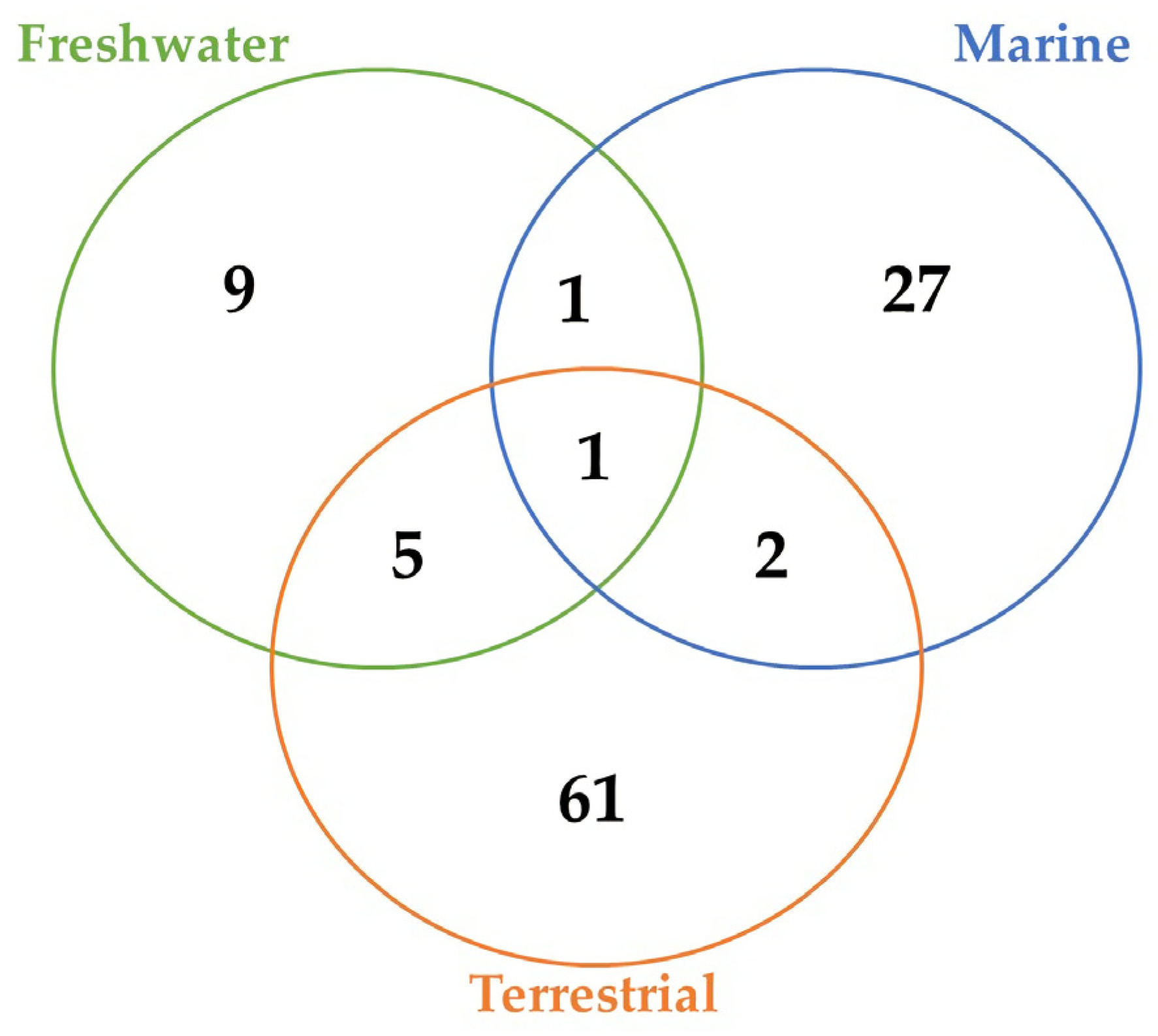
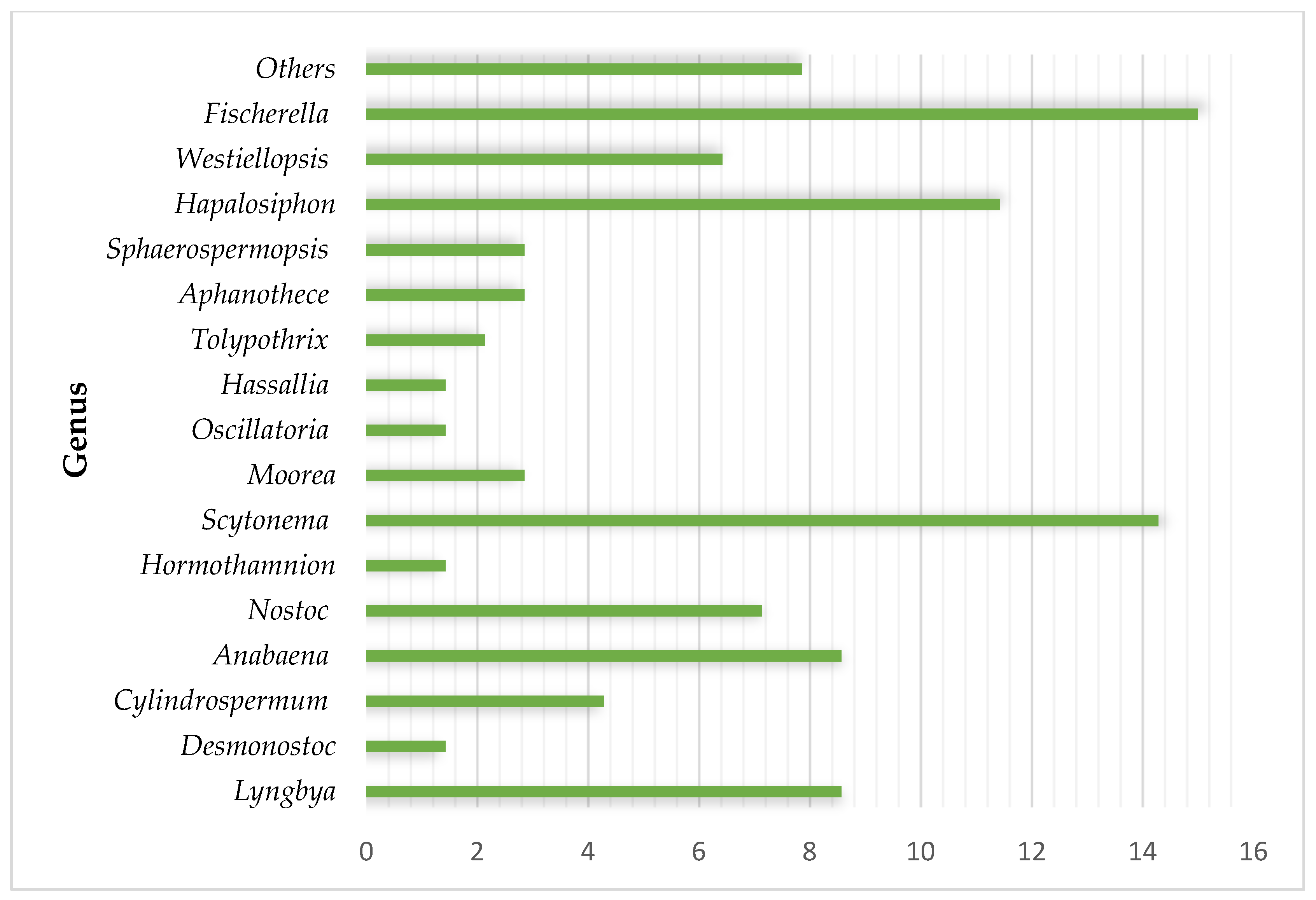
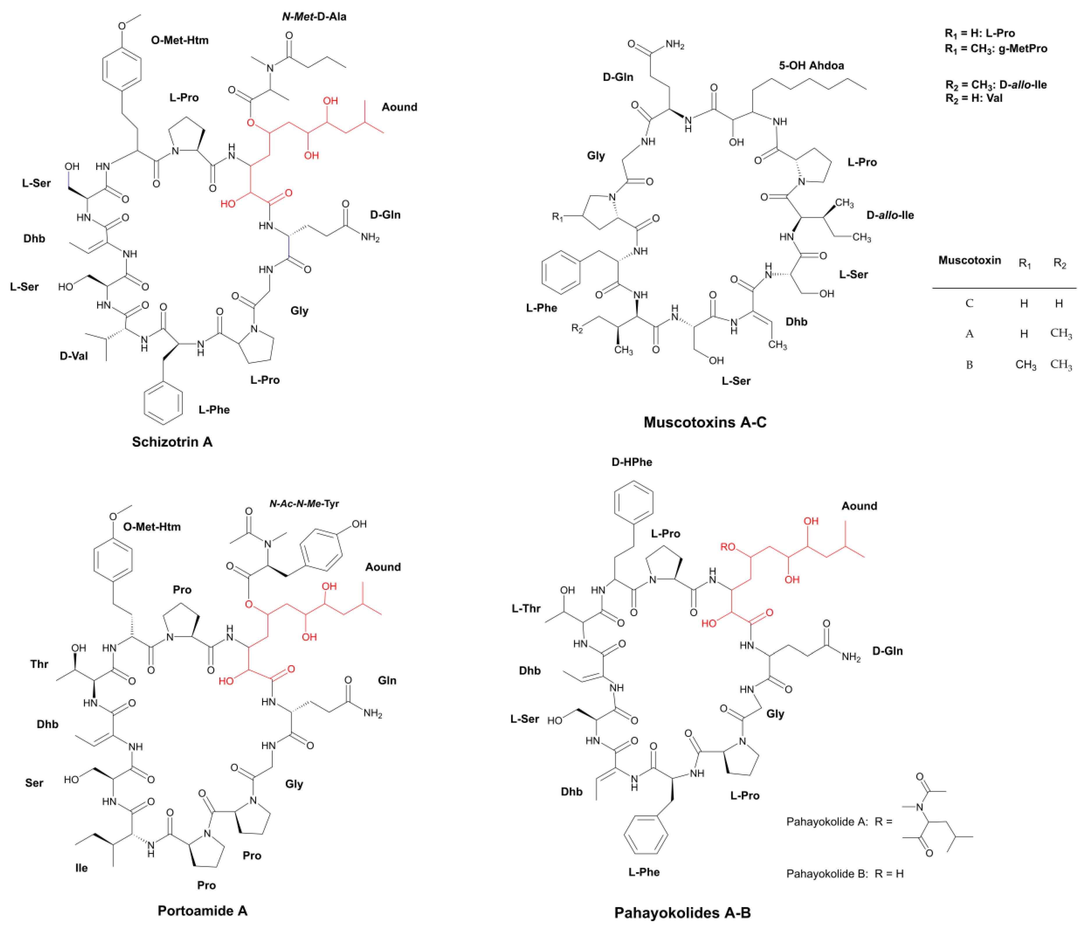
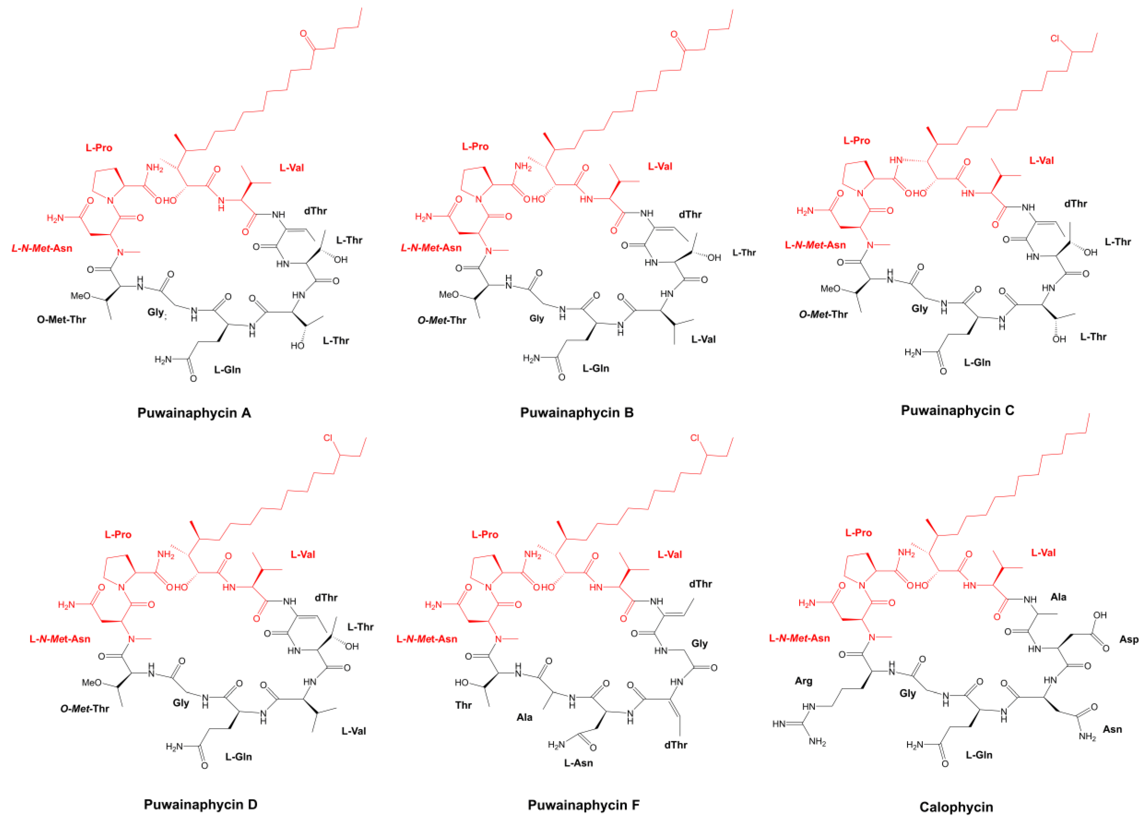
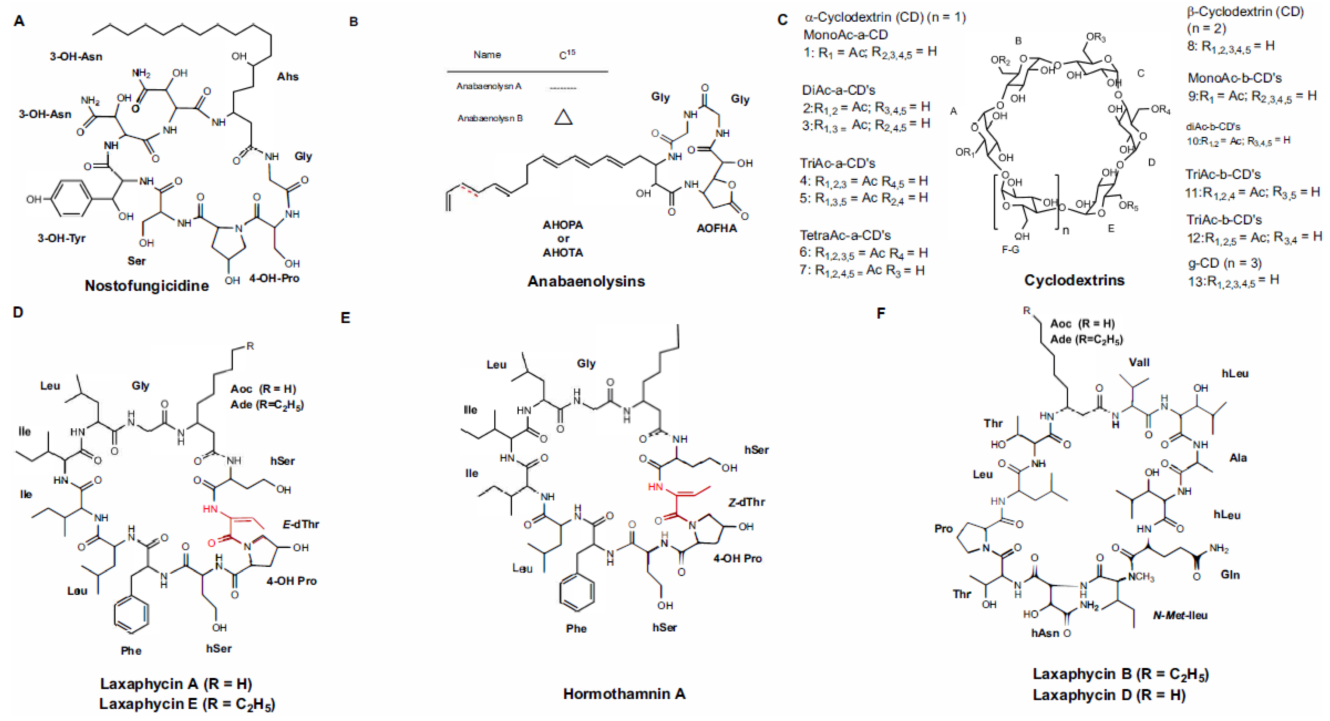
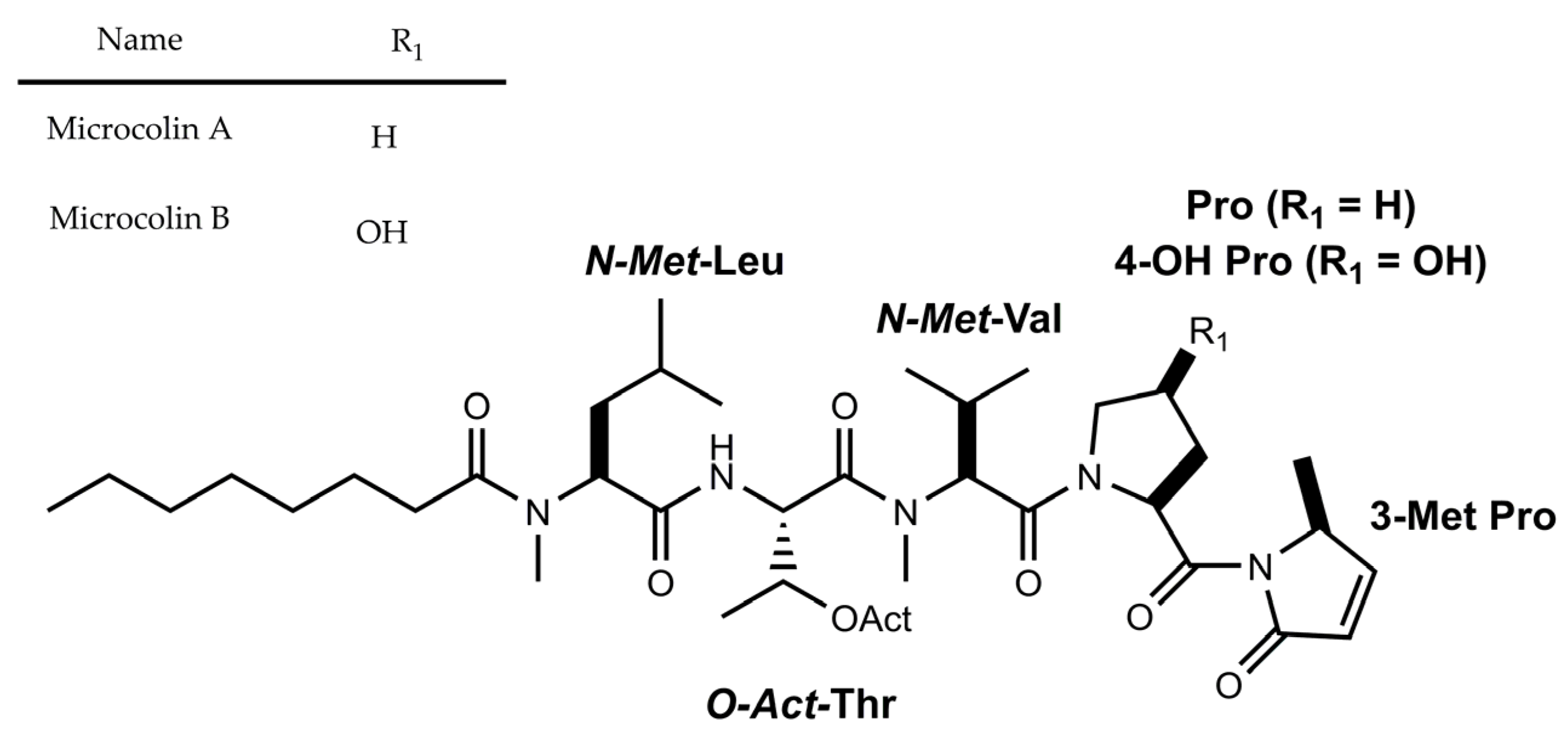
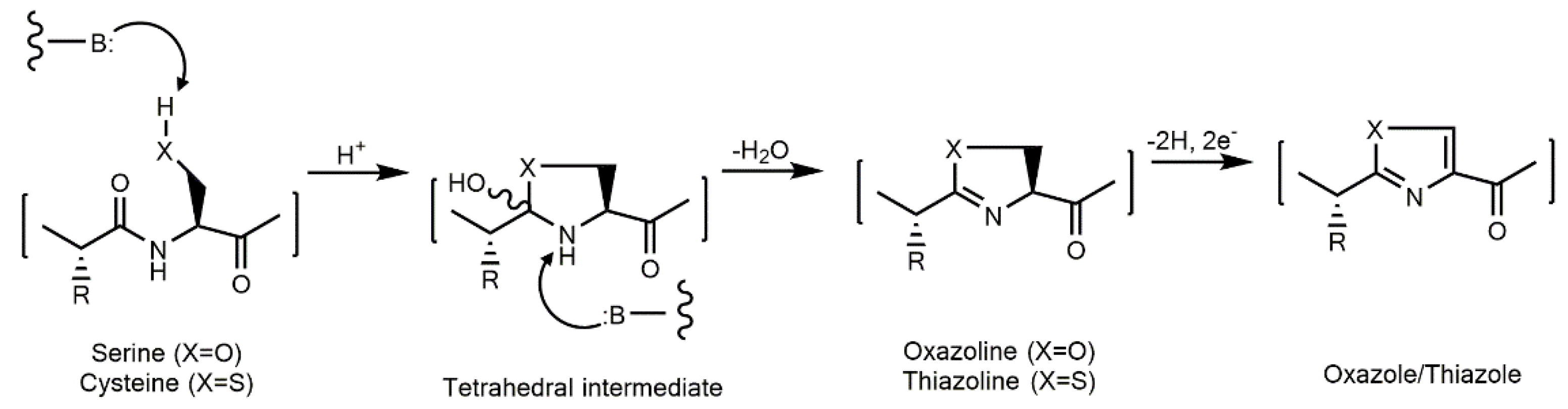
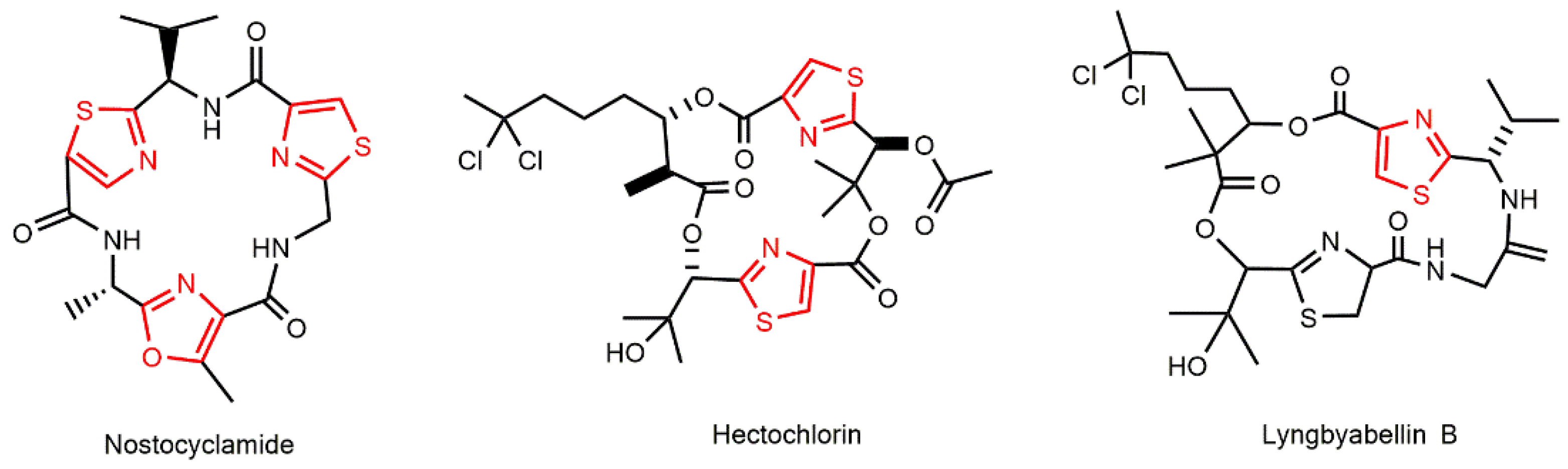
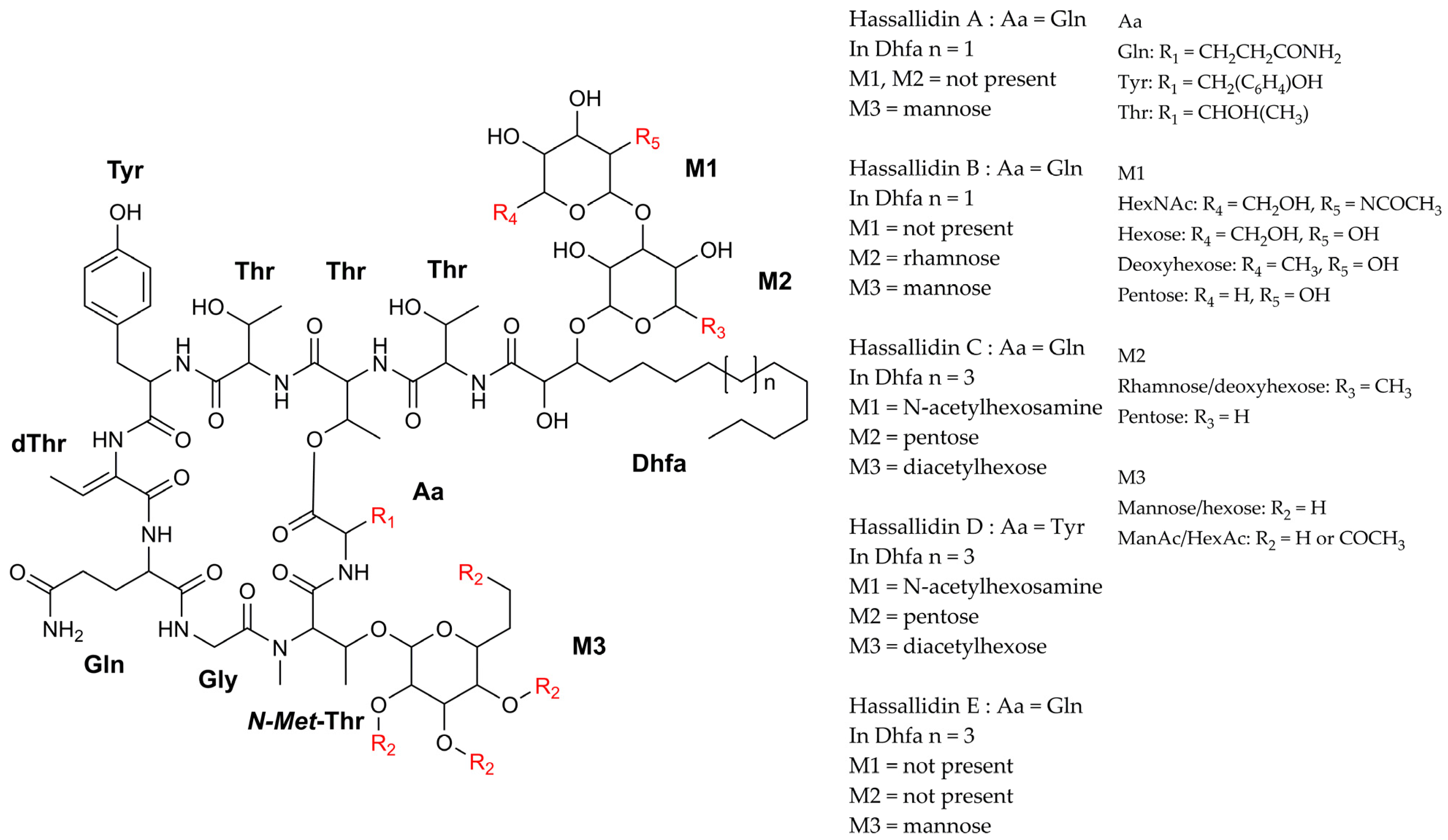
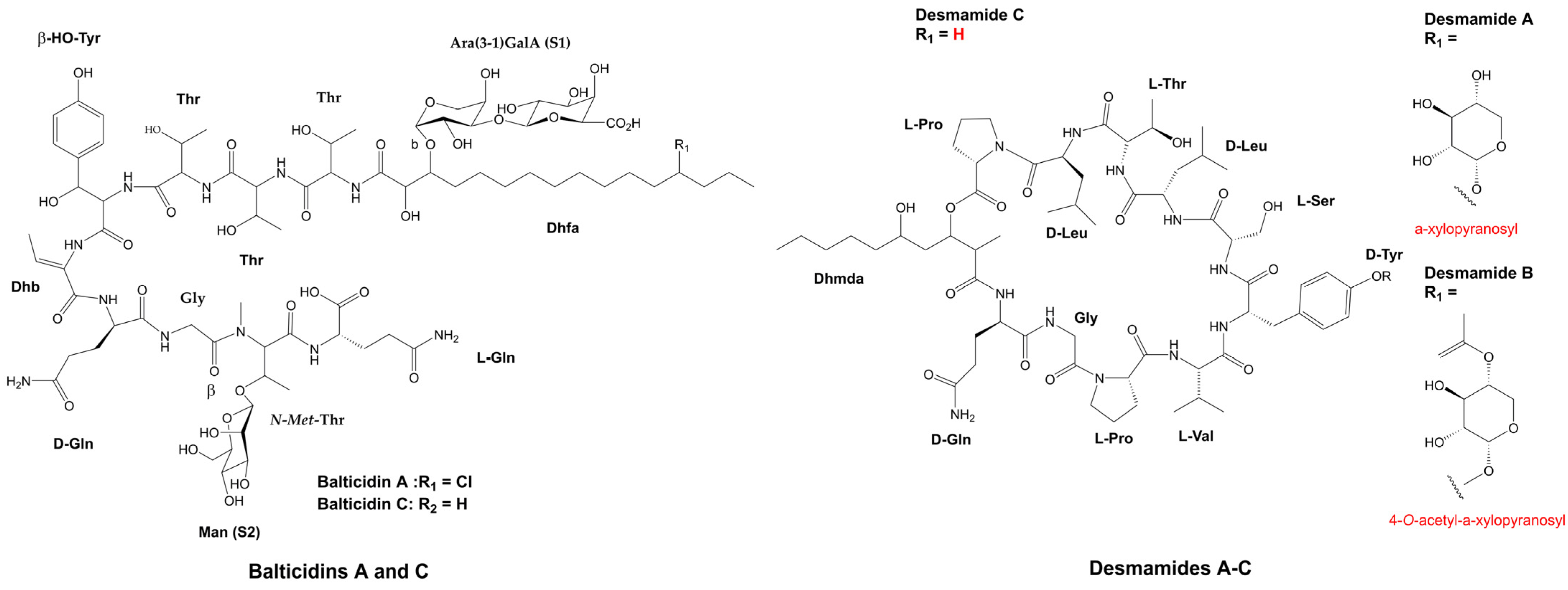
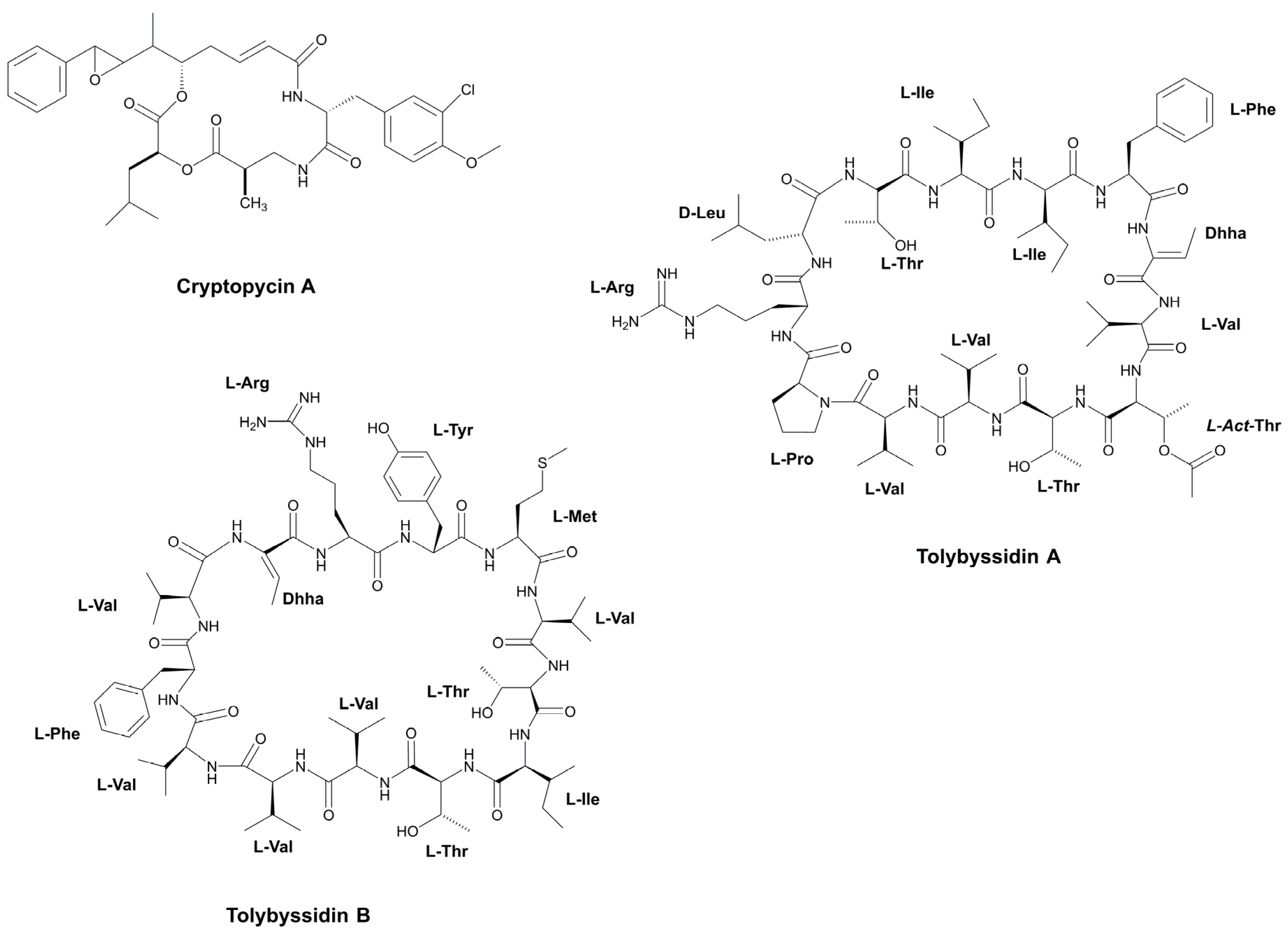
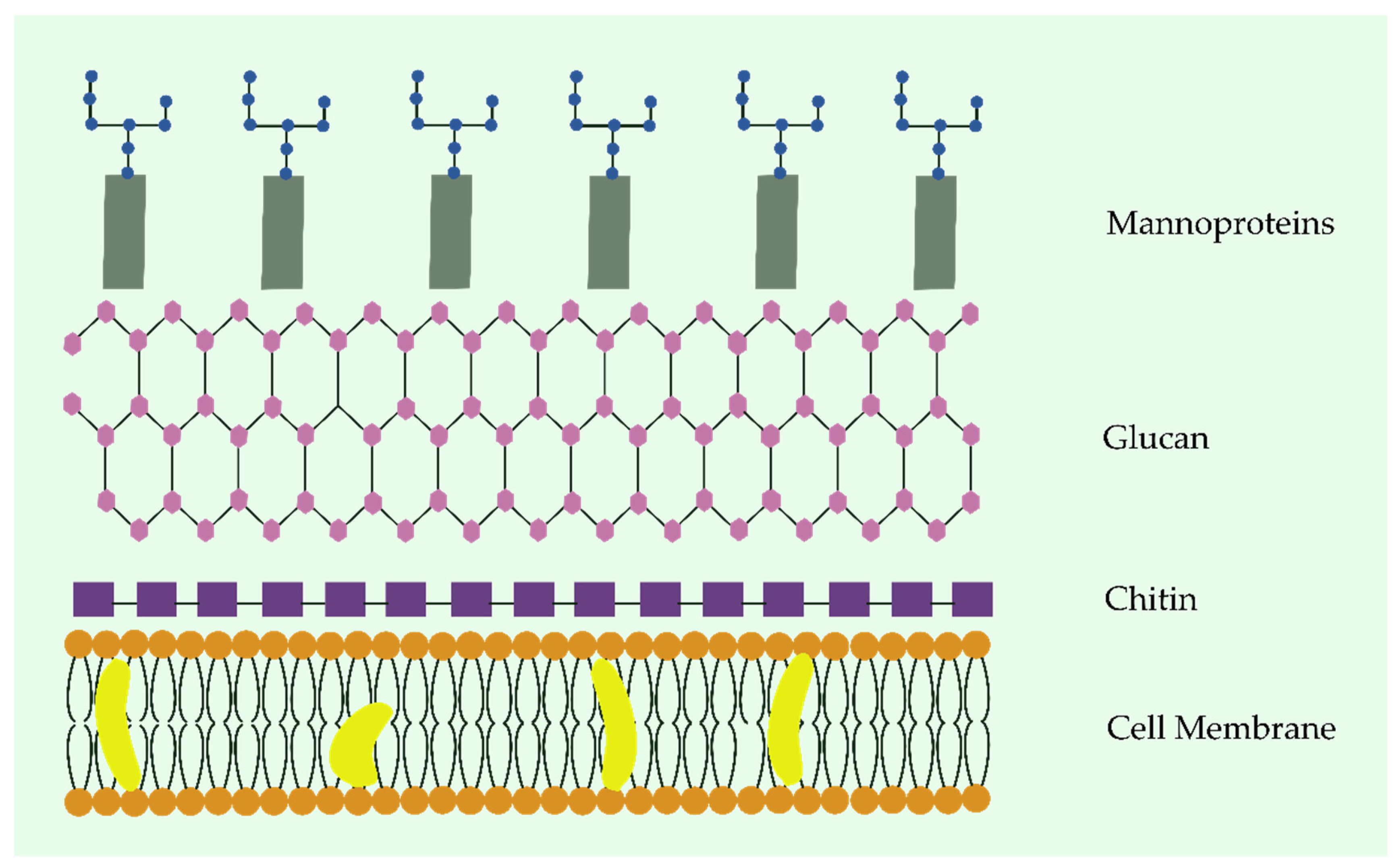
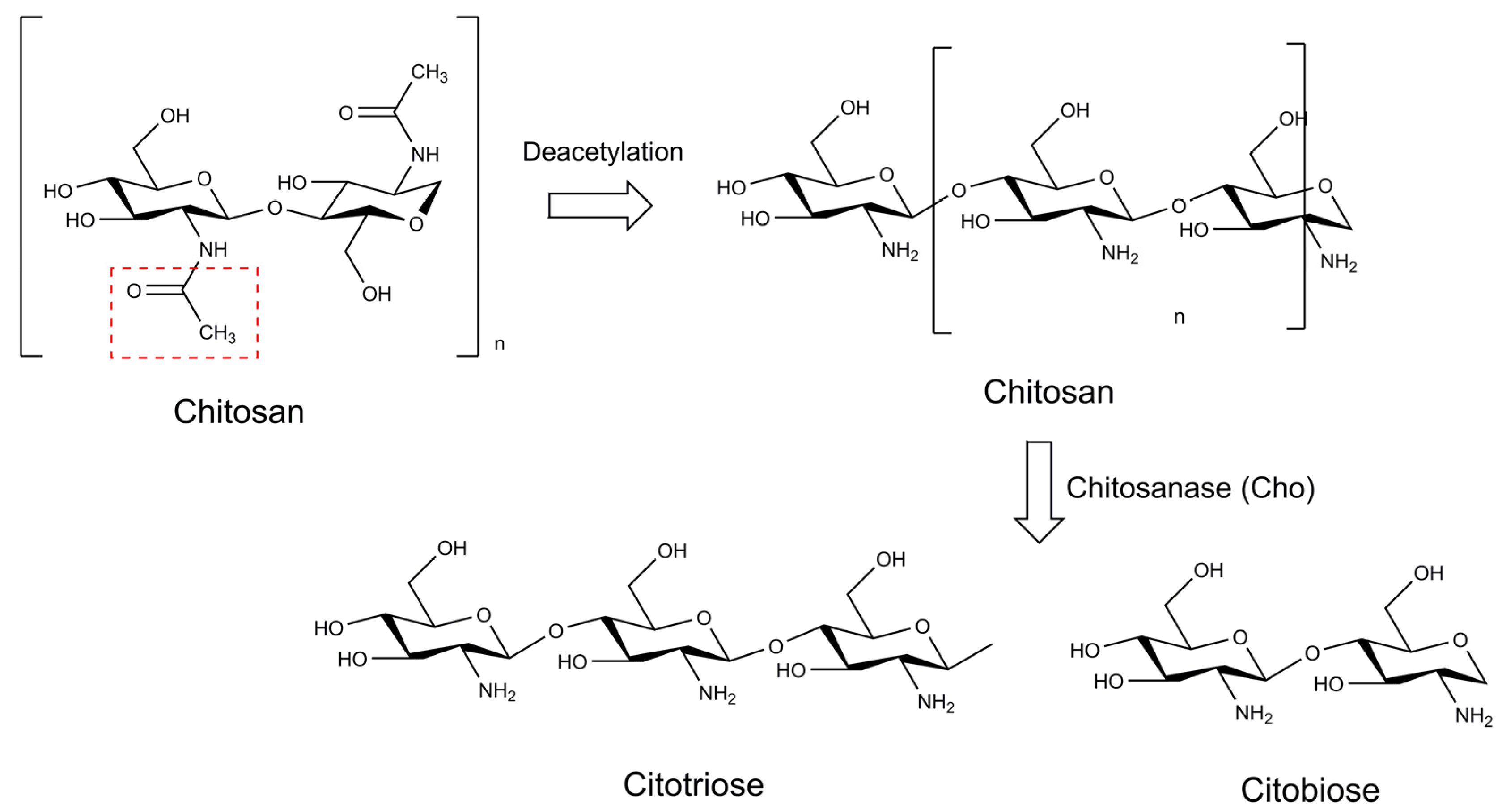
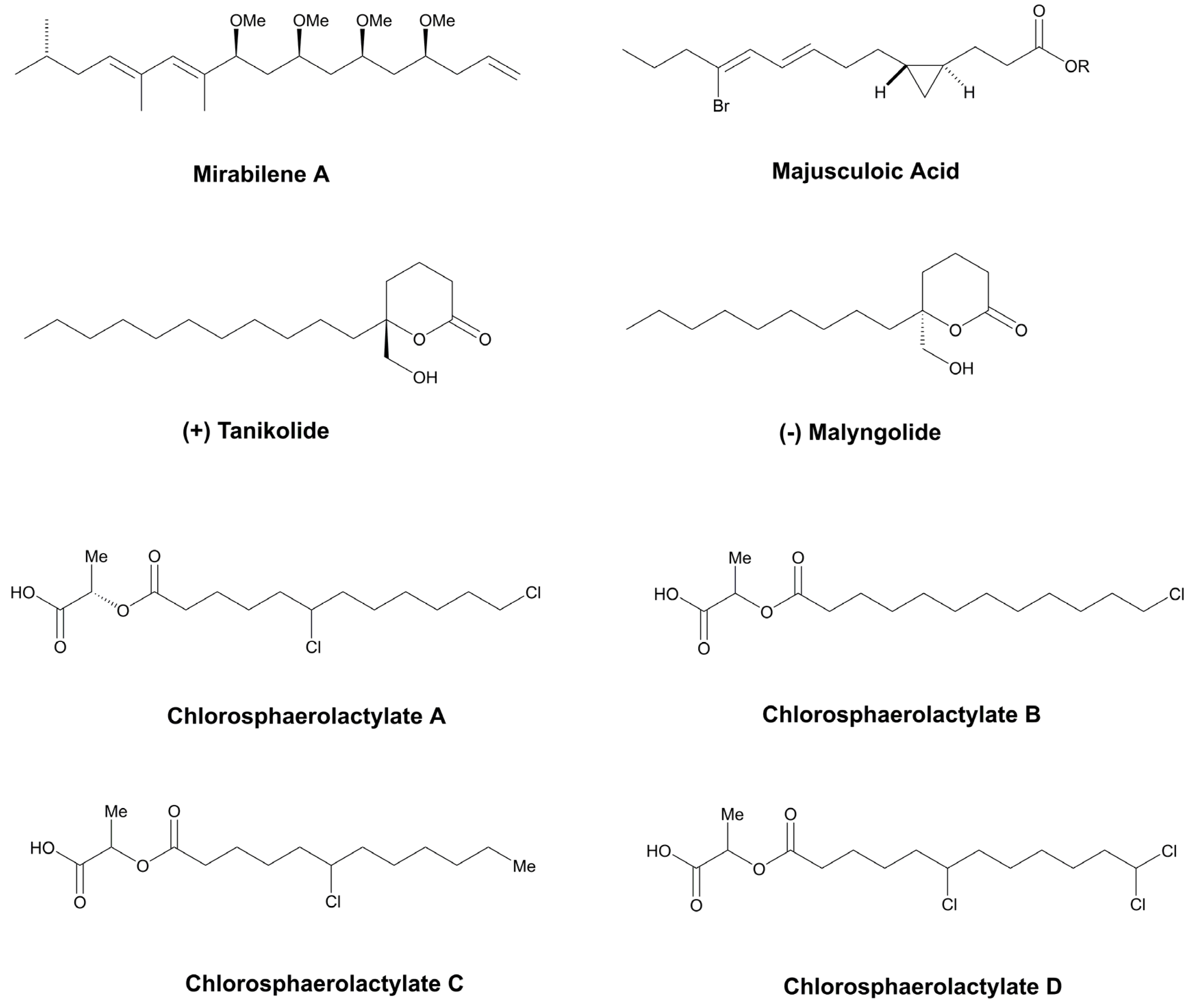
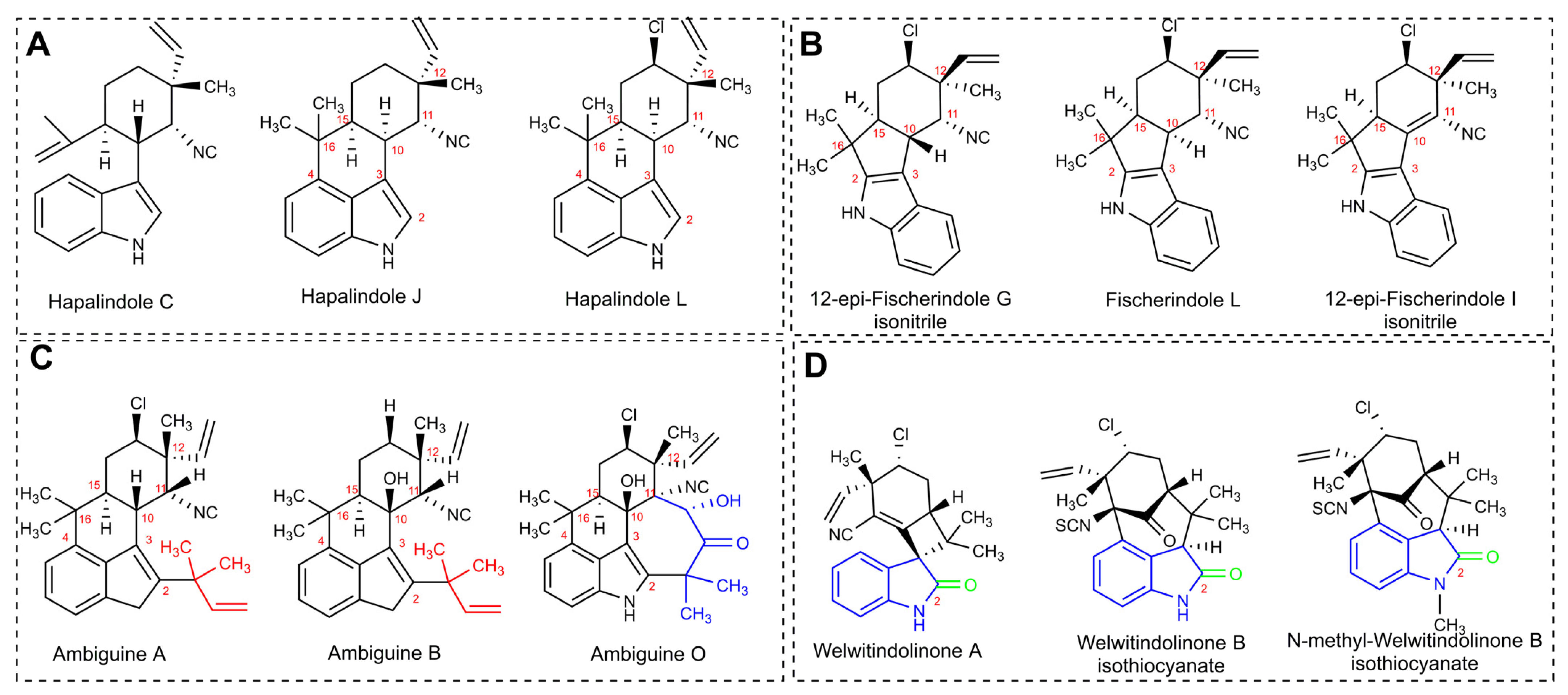
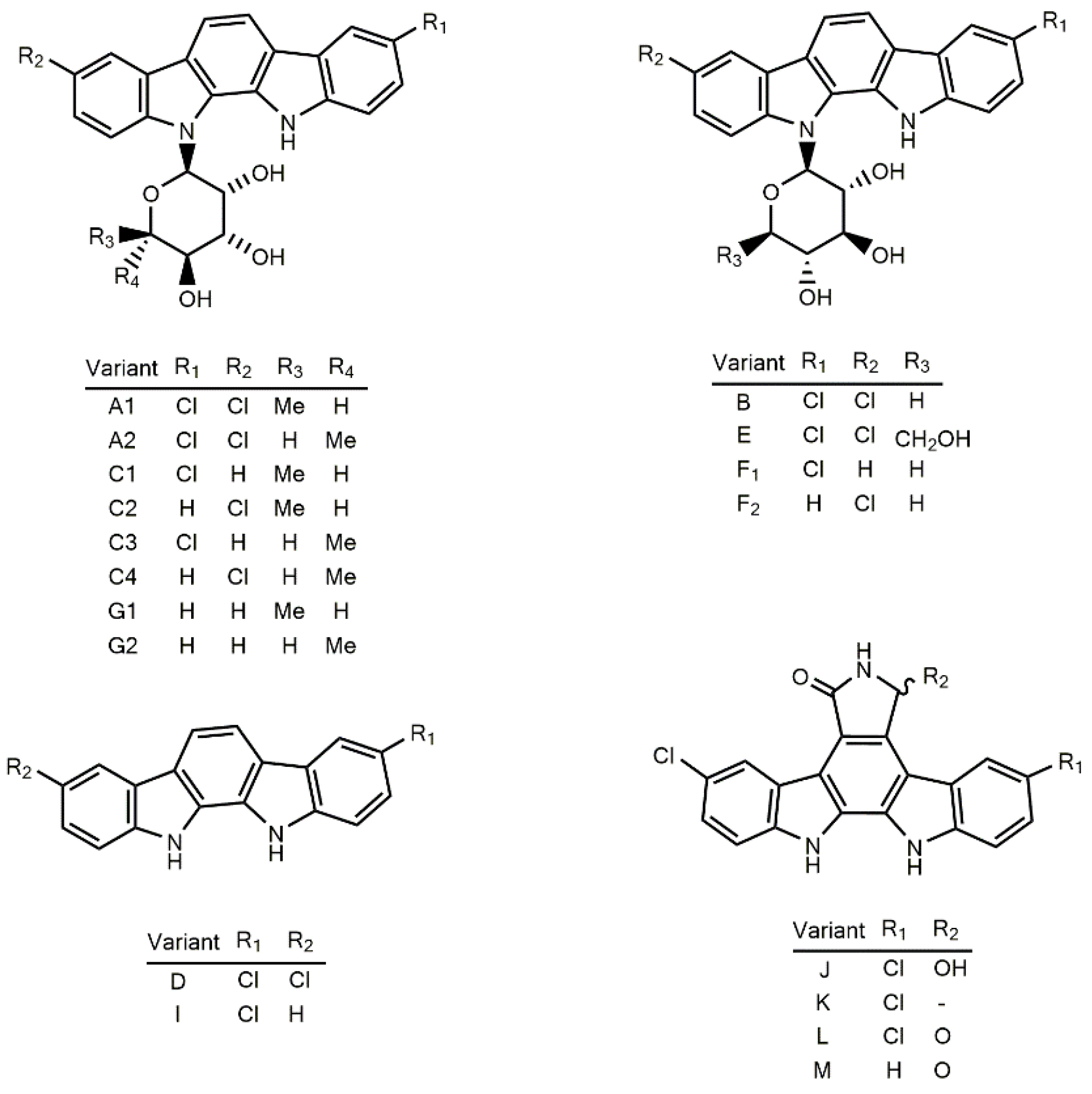
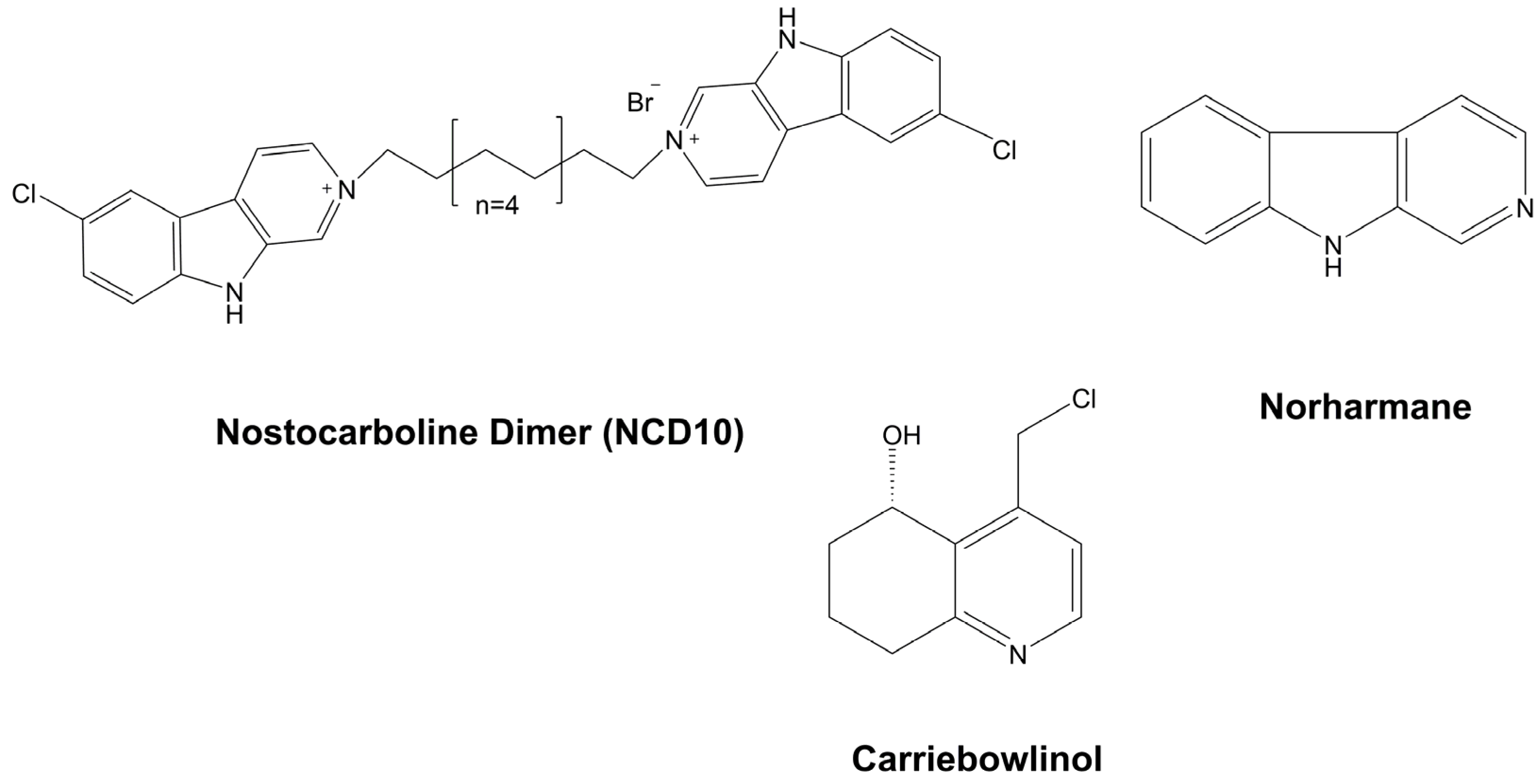
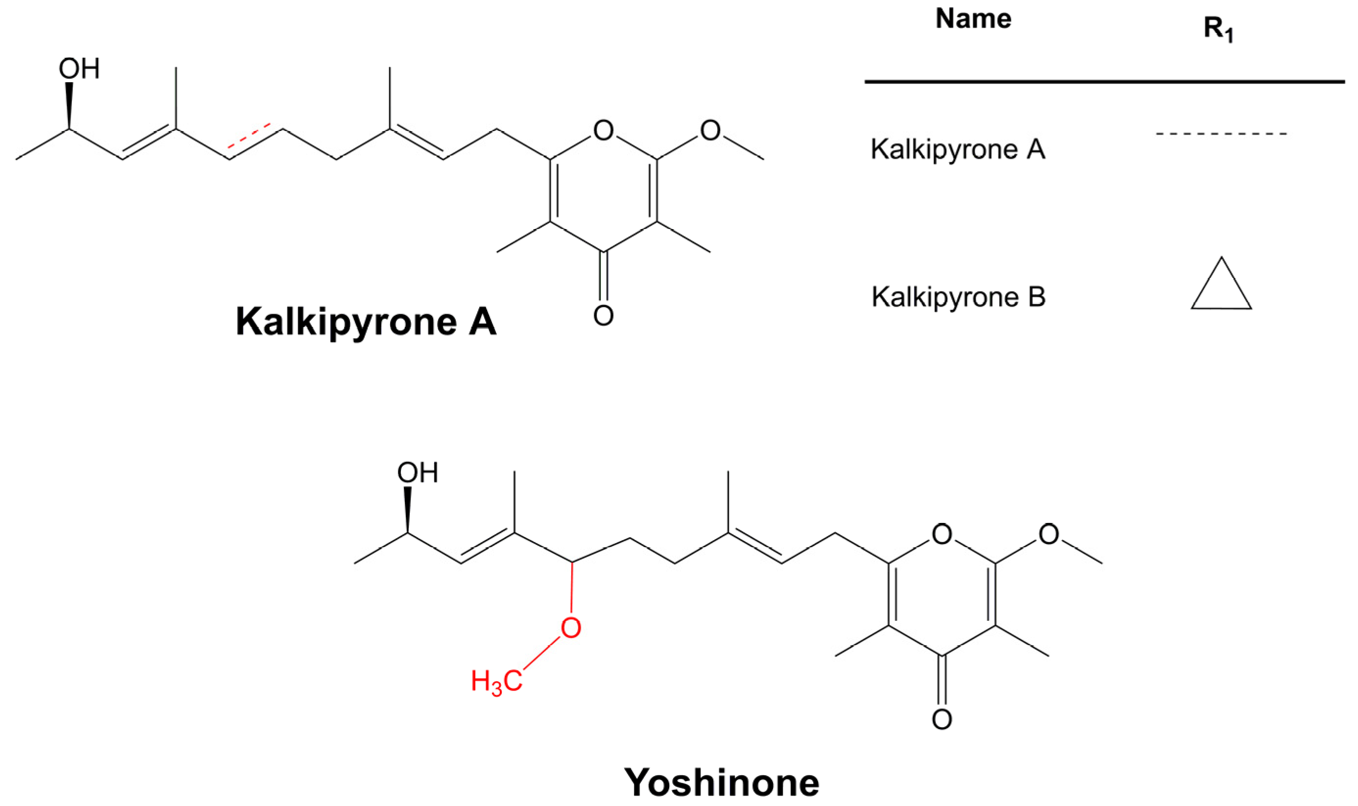
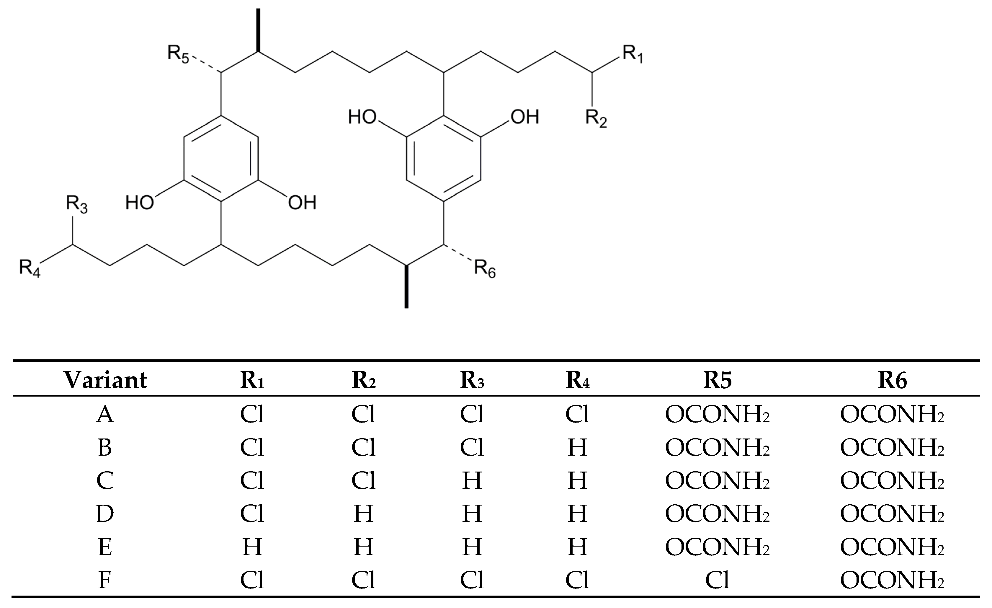
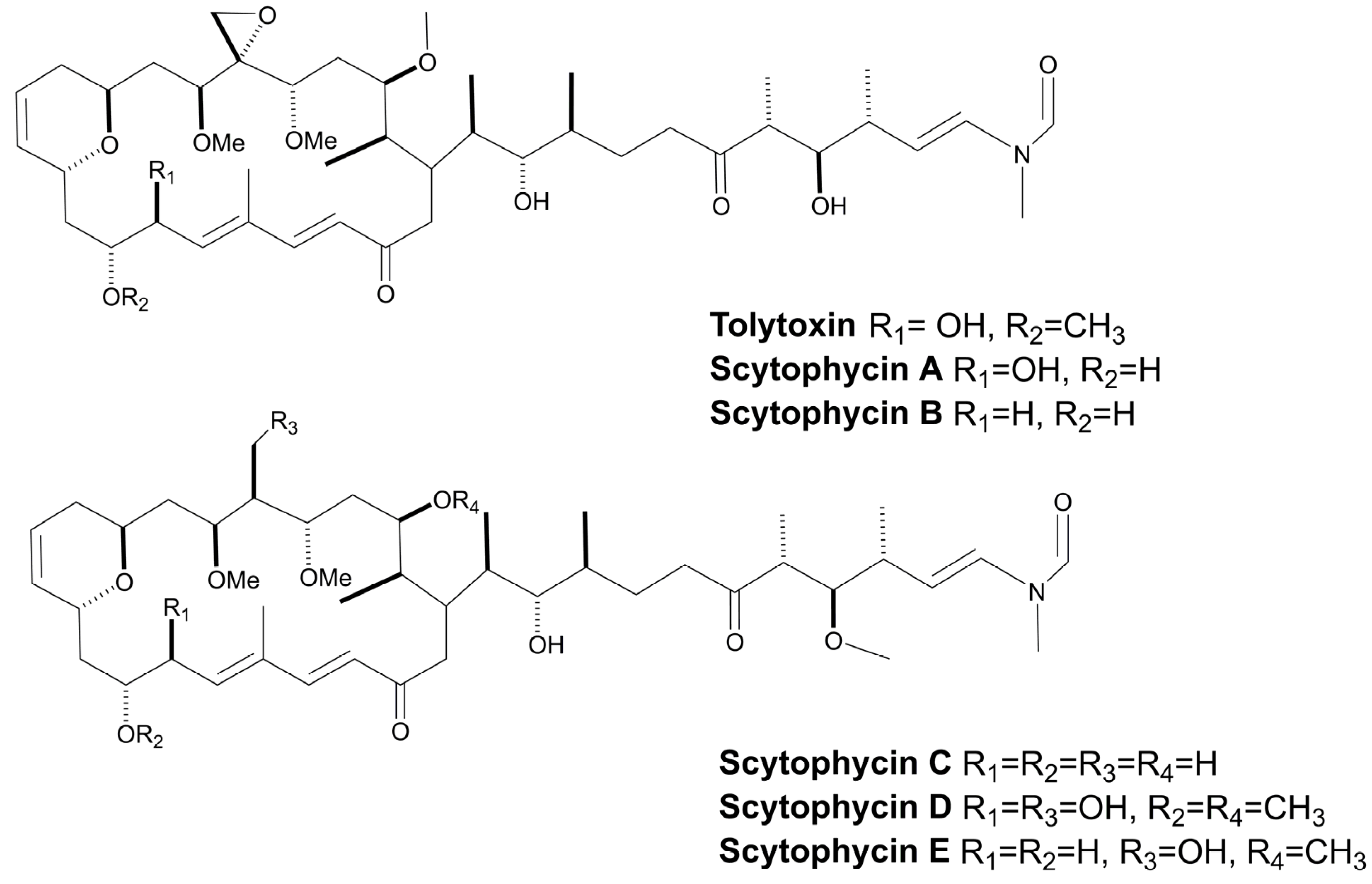
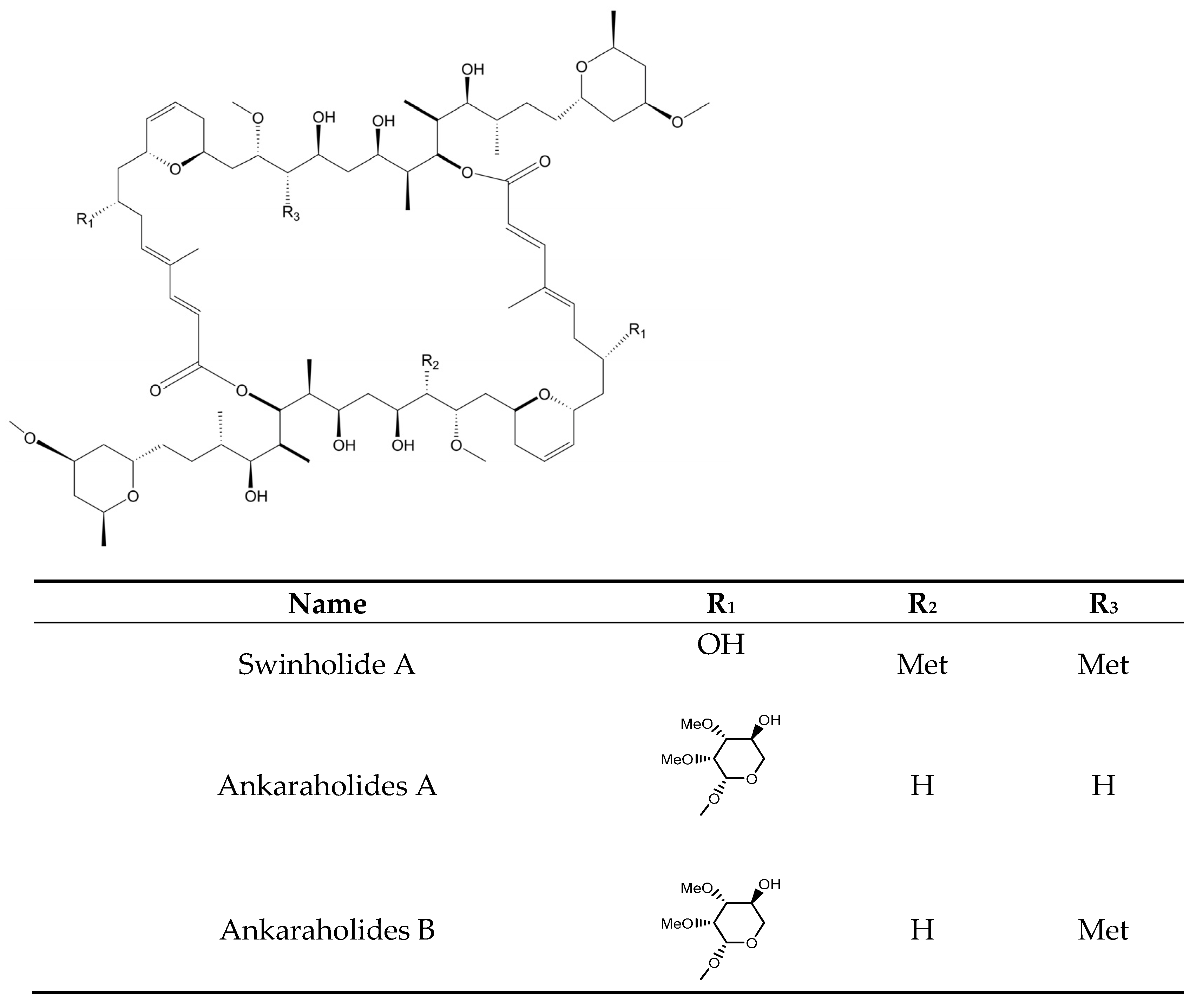
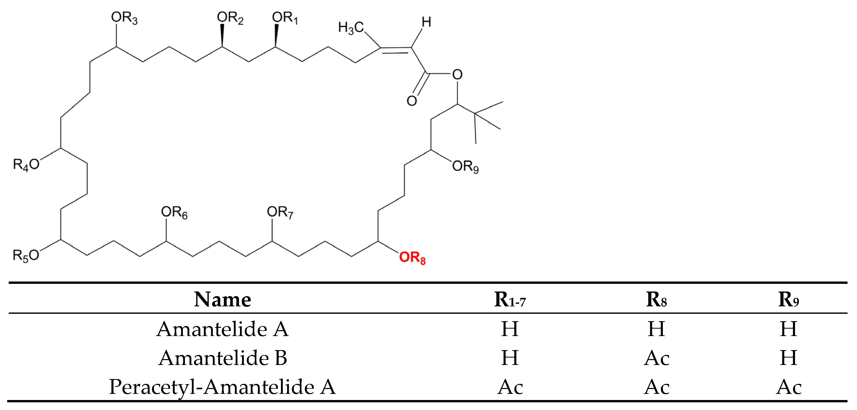
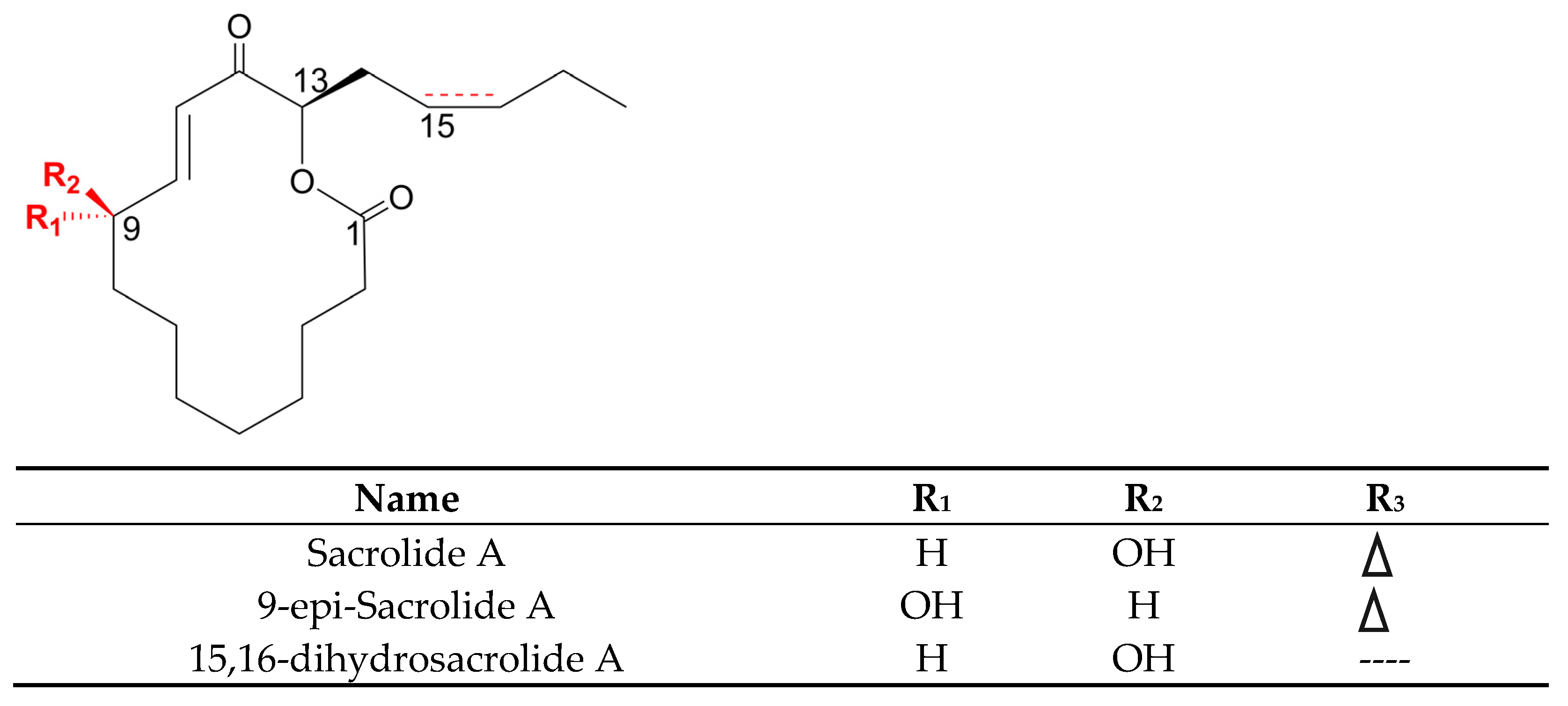
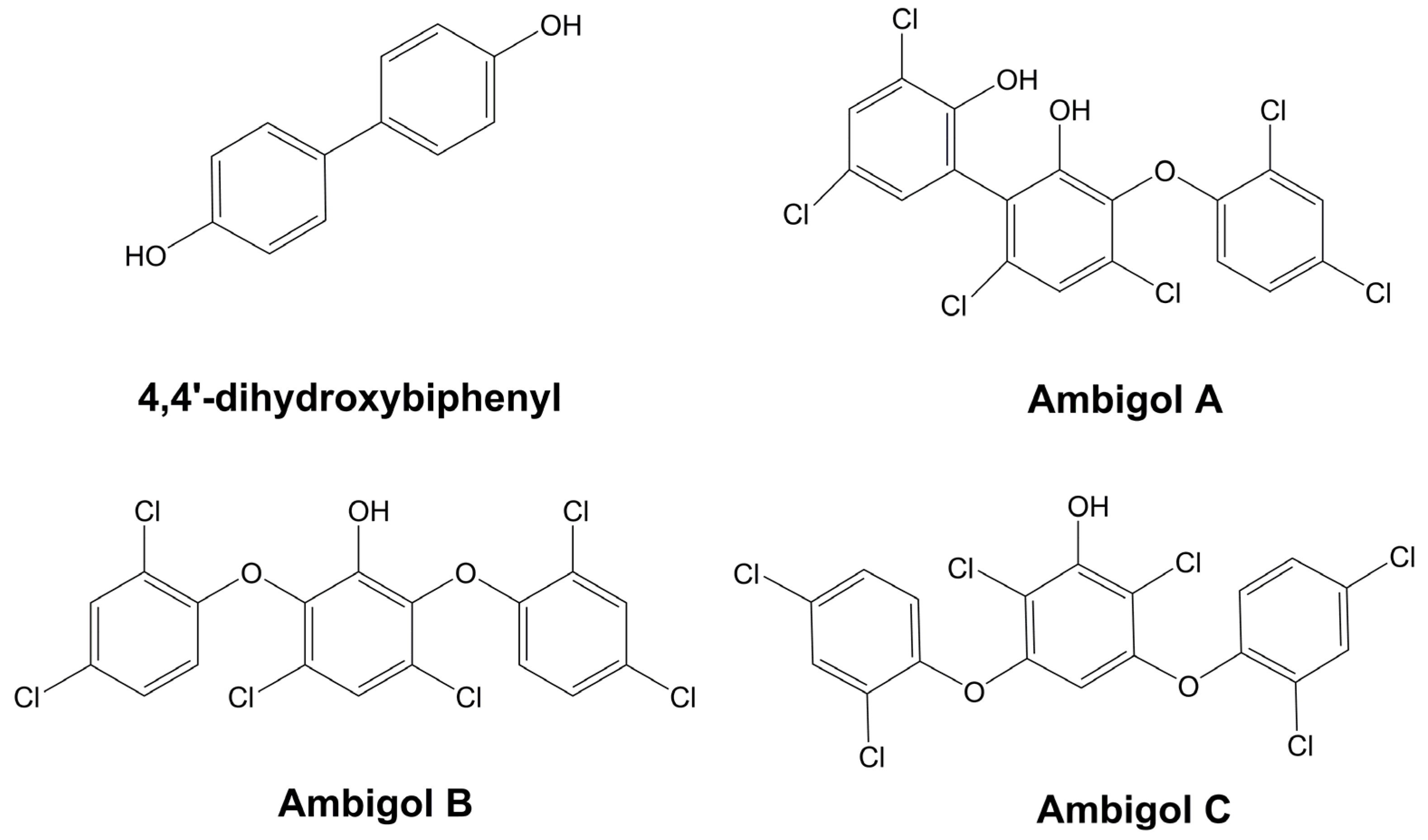
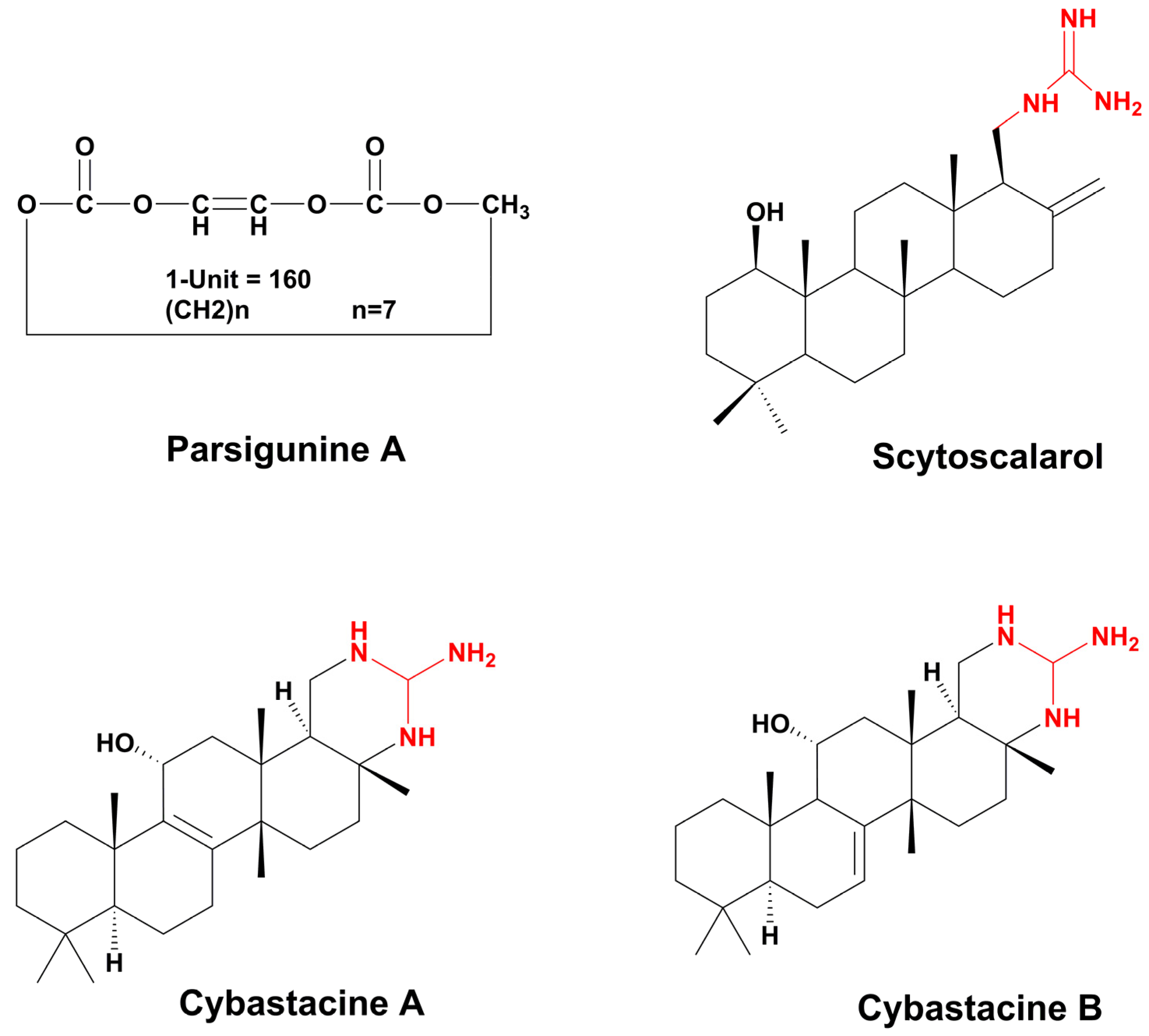
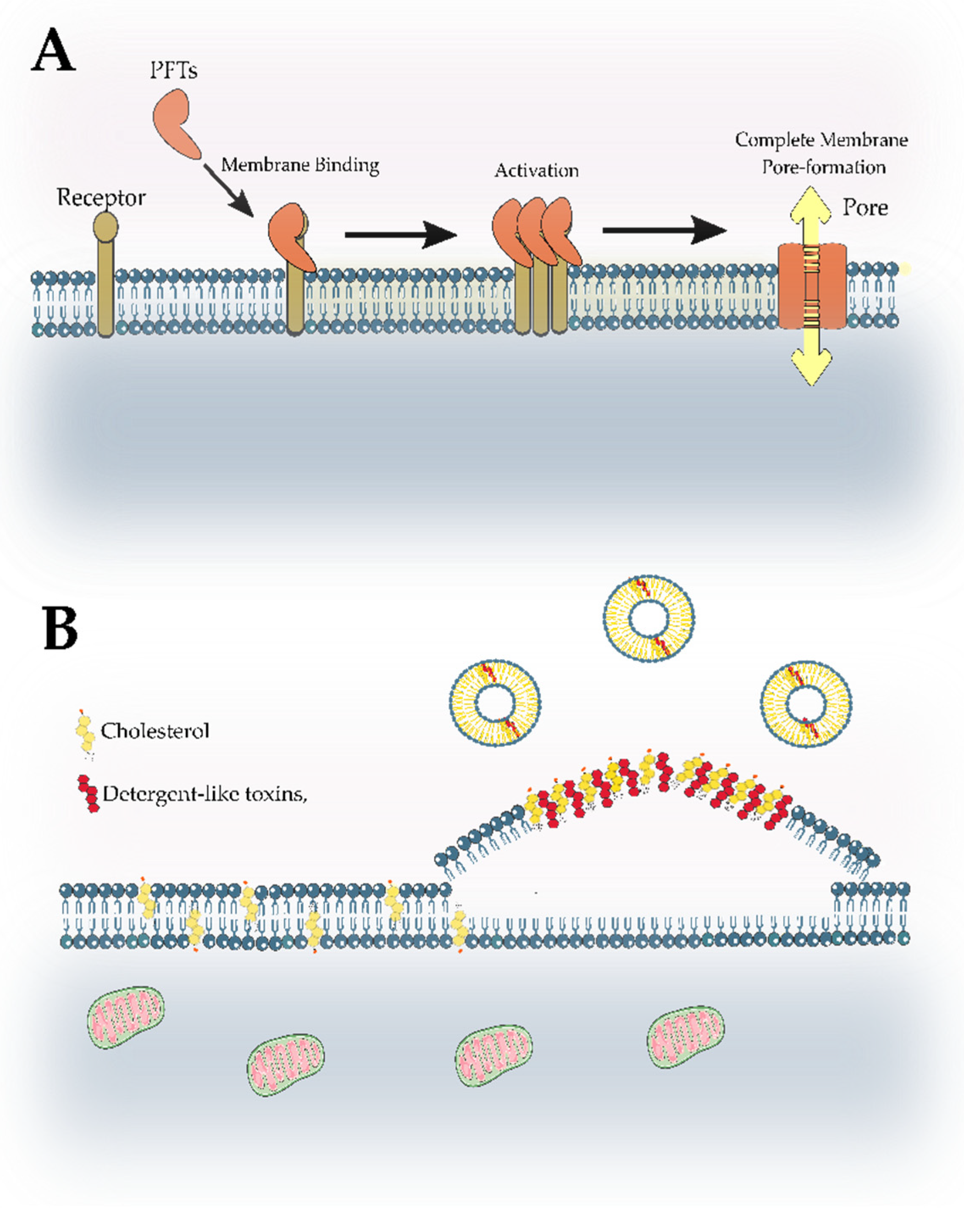
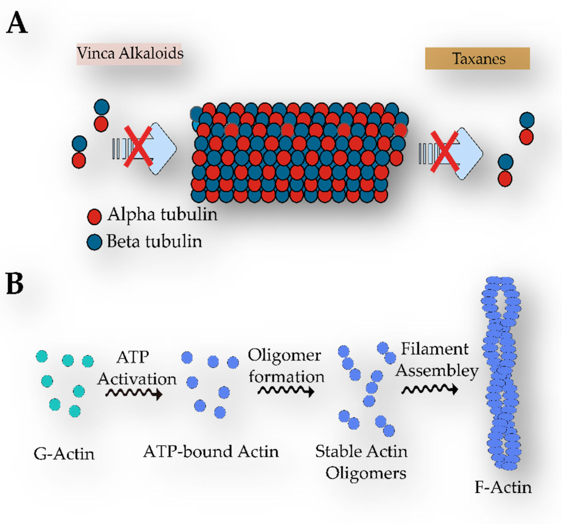
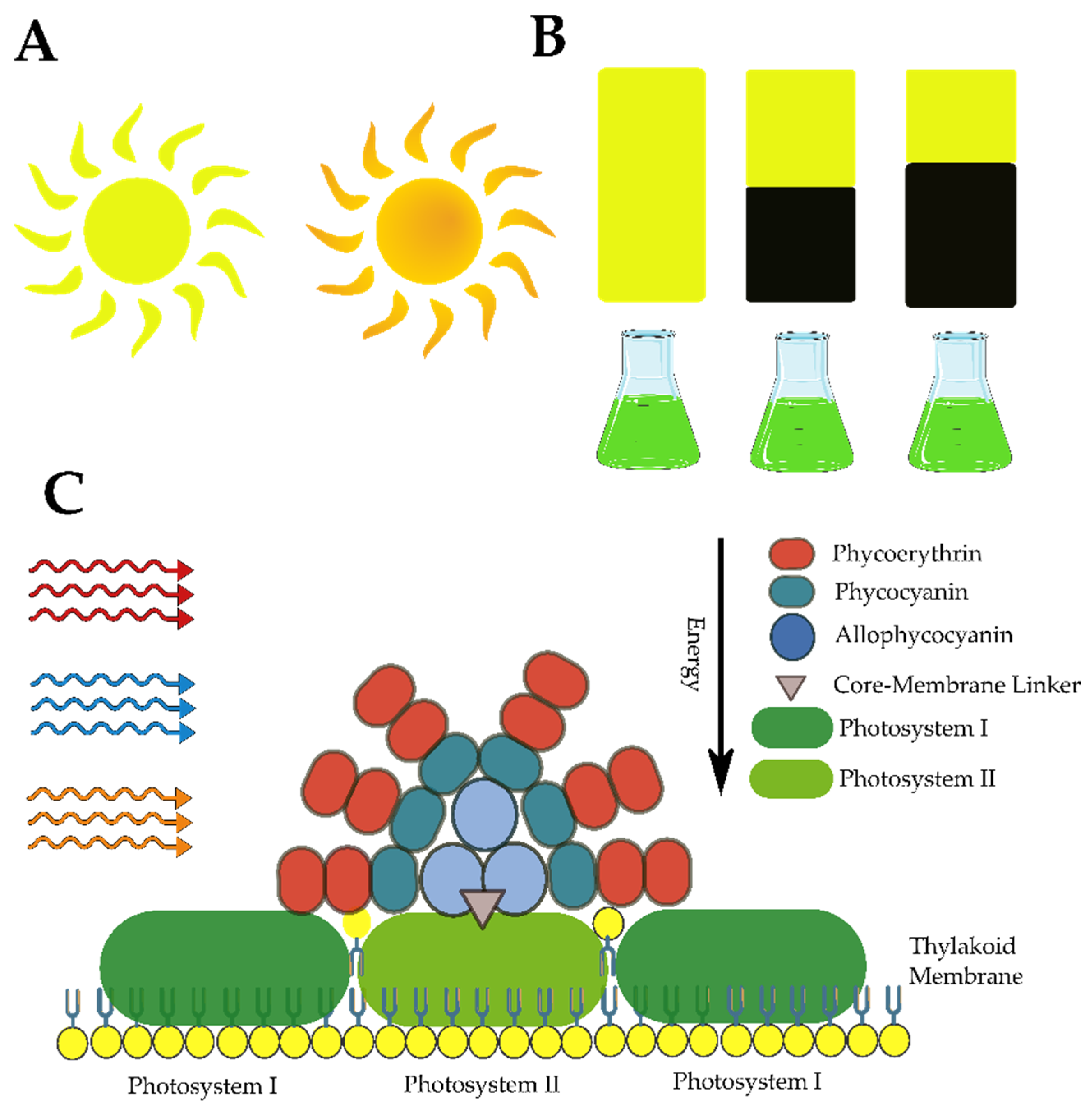
Figure 1. The number of metabolites with antifungal properties identified and shared among strains of cyanobacteria from different environments, including environmental samples.
Marinedrugs 21 00359 g001
Figure 2. Percentage of compound in each genus.
Marinedrugs 21 00359 g002
Figure 3. Chemical structure of some antifungal cyanopeptides with β-amino acids.
Marinedrugs 21 00359 g003
Figure 4. Similarity (red) and differences between the members of the Puwainaphycin family and the cyclopeptide, Calophycin.
Marinedrugs 21 00359 g004
Figure 5. Fungicide isolated from cyanobacteria. (A) Nostofungicidine. (B) Anabaenolysins A and B. (C) Cyclodextrin variants at different acetylation levels. (D) Laxaphycins A and E. (E) Hormothamnin A. (F) Laxaphycins B and D.
Marinedrugs 21 00359 g005
Figure 6. Structure of the peptides, Microcolins A and B.
Marinedrugs 21 00359 g006
Figure 7. Biosynthesis of oxazole and thiazole rings from the amino acids, Serine and Cysteine, respectively.
Marinedrugs 21 00359 g007
Figure 8. Cyanopeptides with fungicidal properties bearing thiazole and oxazole rings.
Marinedrugs 21 00359 g008
Figure 9. The structure of Hassallidins A–E. Abbreviations: Dhfa (dihydroxy fatty acid), dihydroxy tetradecanoic acid in hassallidin A and B, and dihydroxy hexadecanoic acid in hassallidin C, D, and E.
Marinedrugs 21 00359 g009
Figure 10. Chemical structure of Balticidin A and C, and the recently discovered Desmamides A–C.
Marinedrugs 21 00359 g010
Figure 11. Example of fungicidal cyanopeptides released or excreted into the supernatant. Abbreviations: L-Arg (L-arginine), L-Tyr (L-tyrosine), L-Met (L-methionine), L-Val (L-valine), L-Thr (L-threonine), L-Ile (L-isoleucine), L-Phe (L-phenylalanine), Dhha (dehydrohomoalanine), L-Pro (L-proline), D-Leu (D-leucine), O-Ac-Thr (O-Acetyl-L-Threonine).
Marinedrugs 21 00359 g011
Figure 12. Fungal cell wall components. The fungal cell wall is composed of a membrane enriched with ergosterol (in yellow) and some proteins. It is also possible to observe the presence of a protective layer of chitin, as well as glucans, and mannoproteins on the surface.
Marinedrugs 21 00359 g012
Figure 13. Hydrolysis of chitosan by the enzyme chitosanase (Cho), which is produced and released by the strain, Anabaena fertilissima RPAN1.
Marinedrugs 21 00359 g013
Figure 14. Fatty acids and their derivatives which possess fungicidal activity, isolated from cyanobacteria.
Marinedrugs 21 00359 g014
Figure 15. Hapalindole-type alkaloids. (A). Some examples of tricycle hapalindoles (Hapalindole C) and tetracyclo hapalindoles (J and C). In the tetracyclic hapalindoles, the key connection occurs between C-4 and C-16. (B). Chemical structure of some Fischerindoles, in which the key connection is between C-2 and C-16 (C). Chemical structure of Ambiguines A, B, and C. In red, the isoprene unit is attached to C-2, and in blue, the isoprenyl group is fused to the isonitrile-bearing carbon. (D) Some examples of welwitindoliones. In green, the oxidized carbon is in the second position, and in blue, the bicyclo is shown [4.3.1].
Marinedrugs 21 00359 g015
Figure 16. Chemical structure of members of the Tjipanazoles family.
Marinedrugs 21 00359 g016
Figure 17. Chemical structure of alkaloids, with fungicidal properties, obtained from cyanobacteria.
Marinedrugs 21 00359 g017
Figure 18. Chemical structure of the Polyketides, Kalkipyrones A–B, and Yoshinone. In red, the difference between Kalkipyrone A and Yoshinone is shown.
Marinedrugs 21 00359 g018
Figure 19. Carbamidocyclophanes’ (A–F) chemical structures.
Marinedrugs 21 00359 g019
Figure 20. Chemical structure of Scytophycins.
Marinedrugs 21 00359 g020
Figure 21. Chemical structure of the macrolides Swinholide A and Ankaraholides A–B.
Marinedrugs 21 00359 g021
Figure 22. Chemical structure of Amantelides A–B and the Peracetyl-Amantelide A.
Marinedrugs 21 00359 g022
Figure 23. Structure of Sacrolide A and its congeners 9-epo-Sacrolide A and 15,16-dihydrosacrolide A.
Marinedrugs 21 00359 g023
Figure 24. Phenolic compounds with antifungal activity of cyanobacterial origin.
Marinedrugs 21 00359 g024
Figure 25. Structure of the polymer Parsiguine A, terpenoid Scytoscalarol, and Cybastacines A–B. In red, the guanidine group is shown.
Marinedrugs 21 00359 g025
Figure 26. Molecular mechanisms of membrane-targeting compounds. (A) Pathways of pore-forming toxins (PFTs). Soluble PFTs move to plasmatic membrane and bind to receptors molecules. Then, they oligomerize on the surface of the membrane and produce transmembrane pores. (B) General mechanism of detergent-like toxins. These molecules normally bind to the external monolayer of the plasmatic membrane containing cholesterol and promote the vesicles formation and lateral phase separation.
Marinedrugs 21 00359 g026
Figure 27. Structure of a Microtubule and the Actin Filaments. (A) Mechanism of action of vinca alkaloids and taxanes. Although taxanes promote microtubule stabilization by avoiding the release of dimers of tubulin, vinca alkaloids block microtubule polymerization. (B) F-actin formation.
Marinedrugs 21 00359 g027
Figure 28. Light parameters normally investigated in studies concerning cyanobacterial growth and the production of secondary metabolites. (A) Light intensity, (B) Photoperiod, (C) Wavelength.
Marinedrugs 21 00359 g028
Table 1. List of antifungal peptides reported in cyanobacteria.
CompoundMolecular WeightCyanobacterial Strain
(Family)
SourceLocationRef
Schizotrin A1490.71Schizothrix sp. IL-89-2
(Schizotrichaceae)
TerrestrialKfar Azar (Israel)[33]
Pahayokolides A1472.68Lyngbya sp. 15-2
(Oscillatoriaceae)
FreshwaterEverglades (Florida, U.S)[38]
Muscotoxins A1211.41Desmonostoc muscorum
CCALA 125
(Nostocaceae)
TerrestrialDlouhá ves
(Chelcice, Czech Republic)
[39,40]
Muscotoxins B1225.45
Calophycin1249.48Calothrix fusca EU-10-1
(Rivulariaceae)
FreshwaterIsland of Oahu
(Hawaii, U.S)
[36]
Puwainaphycin F1146.34Cylindrospermum alatosporum CCALA 988
(Aphanizomenonaceae)
TerrestrialRiding Mountain National Park (Manitoba, Canada)[41]
Minutissamide A1118.28Nostocales LEGE 17548FreshwaterLagoa de Mira
(Mira, Portugal)
[42]
Anabaena minutissima
UTEX 1613
(Aphanizomenonaceae)
TerrestrialSouth Texas, USA[43,44]
Nostofungicidine885.57Nostoc commune
(Nostocaceae)
TerrestrialGulf of Finland
(Porkkala, Finland)
[45]
Anabaenolysin A558.63Benthic Anabaena strains
(Aphanizomenonaceae)
MarineIsland of Oahu
(Hawaii, U.S)
[46,47]
Anabaenolysin B560.64
Laxaphycin A1196.48Anabaena laxa FK-1-2
(Aphanizomenonaceae)
TerrestrialIsland of Oahu
(Hawaii, U.S)
[48]
Hormothamnion
Enteromorphoides
(Nostocaceae)
MarineKey West
(Florida, U.S)
[49]
Lyngbya majuscula
(Oscillatoriaceae)
MarineMoorea atoll
(French Polynesia)
[50]
Anabaena torulosa
(Aphanizomenonaceae)
MarineMoorea atoll
(French Polynesia)
[51]
Laxaphycin B1395.70A. laxa FK-1-2
(Aphanizomenonaceae)
TerrestrialIsland of Oahu
(Hawaii, U.S)
[48]
L. majuscula
(Oscillatoriaceae)
MarineMoorea atoll
(French Polynesia)
[49]
A. torulosa
(Aphanizomenonaceae)
MarineMoorea atoll
(French Polynesia)
[50]
Laxaphycin C1379.69A. laxa FK-1-2
(Aphanizomenonaceae)
TerrestrialIsland of Oahu
(Hawaii, U.S)
[48]
Scytocyclamide A1223.52Scytonema hofmannii
PCC 7110
(Scytonemataceae)
TerrestrialBermuda
(United Kingdom)
[52]
Scytocyclamide A21208.51
Scytocyclamide B1367.65
Scytocyclamide B21352.64
Scytocyclamide B31336.64
Scytocyclamide C1351.65
Heinamides1186.65–1518.85Nostoc sp. UHCC 0702
(Nostocaceae)
FreshwaterVillähteen Kukkanen
(Nastola, Finland)
[53]
Hormothamnin A1196.48H. Enteromorphoides
(Nostocaceae)
MarineLa Parguera
(Puerto Rico Caribbean)
[54]
Microcolin A747.96Lyngbya cf. polychroa
(Oscillatoriaceae)
MarineCoast of Hollywood
(Florida, U.S)
[55]
Microcolin B731.96L. majuscula
(Oscillatoriaceae)
MarineLa Blanquilla Island
(Venezuela)
[56]
Moorea producens
(Oscillatoriaceae)
MarinePlaya Kalki (Curaçao)[57]
Oscillatoria sp. LP16
(Oscillatoriaceae)
MarineBroward County
(Florida, U.S)
[58]
Lobocyclamide A1198.45Lyngbya confervoides
(Oscillatoriaceae)
MarineCay Lobos
(Bahamas, Caribbean)
[59,60]
Lobocyclamide B1398.70
Lobocyclamide C1370.65
Nostocyclamide474.55Nostoc sp. 31
(Nostocaceae)
FreshwaterNew Jersey (United States[61,62]
Hectochlorin665.59L. majuscula
UOG VP417
(Oscillatoriaceae)
MarineApra Harbor (Guam)[63]
M. producens JHB
(Oscillatoriaceae)
Hector Bay (Jamaica)[64]
Lyngbyabellin B665.59L. majuscula JHB-22
(Oscillatoriaceae)
MarineHector Bay
(Portland, Jamaica)
[65]
M. producens RS05
(Oscillatoriaceae)
Sharm el-Sheikh (Egypt)[66]
Hassallidin A1382.52Hassallia sp. B02-07
(Tolypothrichaceae)
MarineOrrido Clough
(Bellano, Italy)
[67,68]
Hassallidin B1528.66
Hassallidin D1865.00Anabaena sp. SYKE748A
(Aphanizomenonaceae)
MarineTuusulanjarvi Lake
(Tuusulanjarvi, Finland)
[69]
Hassallidin E1410.58Planktothrix serta PCC 8927
(Microcoleaceae)
MarineBerre le Clos (França)[70]
Balticidin A1787.28Anabaena cylindrica Bio33
(Aphanizomenonaceae)
MarineBaltic Sea
(Rügen Island, Germany)
[71]
Balticidin B1769.26
Balticidin C1752.83
Balticidin D1734.81
Tolybyssidin A1465.80Tolypothrix byssoidea
EAWAG 195
(Tolypothrichaceae)
MarinePokhara (Nepal)[72]
Tolybyssidin B1491.86
Cryptophycin A655.19Nostoc sp. ATCC 53789
(Nostocaceae)
TerrestrialIsle of Arran (Scotland)[73,74]
Table 2. Comparison of activity between cyanobacterial antifungals and commercial drugs. The activity is presented in minimum inhibitory concentration (µM/mM/µg mL−1/mg mL−1), Half-maximal inhibitory concentration (IC50), diameter of the inhibition halo (mm), or percentage inhibition of radial growth (%).
MetaboliteTargetActivityRef
MetaboliteControl
Anabaenolysin AC. albicans HAMBI 261--[47]
Anabaenolysin BC. albicans HAMBI 261--
Balticidin ACandida maltosa SBUG70012 mm-[71]
Balticidin B15 mm-
Balticidin C9 mm-
Balticidin D18 mm-
Calophycin
1.2 µg
Aspergillus oryzae13 mmNo activity
Amphotericin B
[36]
Penicillium notatum12 mmNo activity
Amphotericin B
S. cerevisiae12 mm9 mm
Amphotericin B
CalophycinTrichophyton mentagrophytes2 µM-
Aspergillus fumigatus1 µM1.35 µM
Amphotericin B
C. albicans1µM0.67 µM
Amphotericin B
Cryptophycin ARhizonucor miehei, Scopulariopsis communes, Trichoderma lignorum, Verticilium serrae, Cryptococcus albidus, Cryptococcus laurentii, Ustilago zea, and Cercospora beticola.10–15 mm-[74]
A. fumigatus, Cephalosporium sp., F. oxysporum, and Ceratocystis ulmi16–20 mm-
Alternaria solani, Aspergillus flavus, Aspergillus niger, Botrytis ali, Penicillium sp., Cochliobolus miyabeanus, Phona sp.20–25 mm-
Cryptococcus neoformans MY1051 and MY1146≤0.1 µM<1.1 µM
Amphotericin B
C. neoformans strains0.046 µM
Hassallidin AC. albicans strains
Candida guillermondii
ATCC 90877
C. tropicalis ATCC 750
2.9 µM0.23–0.91 µM
Caspofungin
[67,68,75]
Cryptococcus neoformans
Trichosporon asahii
Trichosporon mucoides
7.3 -29.3 µM
Caspofungin
Candida glabrata strains
Candida parapsilosis strains
C. tropicalis strains
Candida krusei strains
5.2 µM0.23–0.91 µM
Caspofungin
Aspergillus niger
Ustilago maydis
Penicillium sp.
Fusarium sambucium.
--
Hassallidin BC. parapsilosis ATCC 22019 C. krusei ATCC 6258
C. albicans strains
C. tropicalis ATCC 90874
5.2 µM0.23–0.91 µM
Caspofungin
C. neoformans strains7.3–29.3 µM
Caspofungin
C. albicans ATCC 24433
C. glabrata strains
C. parapsilosis ATCC 90018
C. tropicalis ATCC 750
C. krusei ATCC 90878
10.5 µM0.23–0.91 µM
Caspofungin
Aspergillus niger
Ustilago maydis
Penicillium sp.
Fusarium sambucium.
--
A. fumigatus and C. albicans3.1 µM-
Hassallidin DC. albicans ATCC 11006
C. albicans ATCC 10231
C. krusei ATCC 6258
1.5 µM-[69]
Hassallidin D
10 μg
Cryptococcus albidus
ATCC 10666
17 mm-[69]
Filobasidiella neoformans ATCC 1022611 mm-
Hassallidin D
Linear Form
C. albicans ATCC 1100620 µM-
Hassallidin EC. albicans CBS56222.7 µM-[70]
C. neoformans H99-
C. parapsilosis ATCC22019-
C. krusei ATCC6258-
Hectochlorin
100 µg
C. albicans16 mm-[76]
HeinamidesAspergillus flavus
FBCC 2467
--[53]
Hormothamnins
(10 µg)
C. albicans7–11 mm-[77]
Laxaphycins A + B
(25 + 25 µg)
A. oryzae29 mm-[48]
C. albicans23 mm-
P. notatum30 mm-
S. cerevisiae22 mm-
T. mentagrophytes30 mm-
Laxaphycin B
(50 µg)
A. oryzae19 mm
45.8 µM
4.6 µM
(8.1 µM of Laxa A)
C. albicans8 mm-
S. cerevisiae12 mm-
T. mentagrophytes9 mm-
Laxaphycin C
(50 µg)
A. oryzae14 mm-
C. albicans9 mm-
P. notatum11 mm-
S. cerevisiae12 mm-
Lobocyclamide A
(150 µg)
C. albicans 96-4897 mm-[59]
Lobocyclamide B
(150 µg)
C. albicans 96-4898 mm-
C. glabrata6 mm-
Lobocyclamide C
(150 µg)
C. albicans 96-48910 mm-
C. glabrata8 mm-
Lyngbyabellin B
(100 µg)
C. albicans ATCC 1405310.5 mm-[78]
Microcolin ADendryphiella salina SIO
D. salina EBGJ
>250 µM3.4 µM
Amphotericin B
[55]
Microcolin B
Minutissamide AA. fumigatus37.5 µM-[43]
Alternaria alternata75 µM-
Muscotoxins ACandida friedrichii61.9 µM26.1 µM
Fluconazole
[40]
Trichoderma harzianum31.5 µM13.1 µM
Fluconazole
Bipolaris sorokinianaNANA
Fluconazole
Alternaria alternata0.5 µM6.5 µM
Fluconazole
Monographella cucumerina1.9 µM6.5 µM
Fluconazole
A. fumigatus1.9 µM104.5 µM
Fluconazole
Chaetomium globosum15.5 µM6.5 µM
Fluconazole
Fusarium oxysporum61.9 µM26.1 µM
Fluconazole
S. sclerotiorum41.3 µg *-
Muscotoxins BS. sclerotiorum20.4 µg *-
NostocyclamideS. cerevisiae--[62]
NostofungicidineAspergillus candidus1.8 µM-[45]
Pahayokolides AS. cerevisiae20 mm-[38]
Puwainaphycin FC. albicans HAMBI 2615.5 µM-[79]
S. cerevisiae HAMBI 1164-
Schizotrin A
(13.4–16.7 nM)
S. cerevisiae
C. albicans
7 mm-[33]
C. tropicalis9 mm-
R. rubra8 mm-
S. rolfsii
R. solani
25–28%-
C. gloeosporioides47%-
Schizotrin A
33.5 nM
F. oxysporum37%-
Scytocyclamide A
200 µg
A. flavus FBCC 246710 mm-[52]
Scytocyclamide A2
200 µg
7 mm-
Scytocyclamide B
600 µg
23 mm-
Scytocyclamide B2
85 µg
10 mm-
Scytocyclamide B3
85 µg
20 mm-
Scytocyclamide C
160 µg
22 mm-
Scytocyclamide A + B
100 + 300 µg
36 mm-
Scytocyclamide A + C
100 + 80 µg
33 mm-
Scytocyclamide A2 + B2
100 + 43 µg
24 mm-
Scytocyclamide A2 + B3
100 + 43 µg
25 mm-
Scytocyclamide B + C
300 + 80 µg
23 mm-
Tolybyssidin ACandida albicans21.8 µM19.2 µM
Miconazole
[72]
Tolybyssidin B42.9 µM
7-DeoxysedoheptuloseS. cerevisiae50 µM590 μM
Glyphosate
[80]
Polysaccharide
10 µg
T. rhizoctonia15 mm23 mm Amphotericin[81]
F. solani16 mm27 mm Amphotericin
F. oxysporum15 mm28 mm Amphotericin
A. niger17 mm25 mm Amphotericin
C. albicans ATCC 9002815 mm20 mm Amphotericin
PolysaccharideBotrytis cinerea1064 mg mL−1 (EC50)-[82]
PolysaccharideA. niger707 mg mL−1-[83]
Chlorosphaerolactylate AC. parapsilosis SMI4163 mM [84]
Chlorosphaerolactylates
B–C
3.3 mM-
Chlorosphaerolactylate D5.5 mM-
Majusculoic AcidC. albicans ATCC 145038 µM1 µM
Fluconazole
[85]
C. glabrata19.3 µM-
Mirabilenes A–F
10 μg
P. notatum10–15 mm-[86]
A. oryzae
Lyngbic AcidFusarium sp.2.1 µMIC50-[87]
Lindra thalassiae2.7 µMIC50-
D. salina2.9 µMIC50-
Tanikolide
100 μg
C. albicans13 mm-[88]
Ambiguine AC. albicans6.1 µM0.34 µM
Amphotericin B
[89,90,91]
T. mentagrophytes24.6 µM0.06 µM
Tolnaftate
A. fumigatus196.6 µM2.3 µM
Amphotericin B
Ambiguine BC. albicans5.9 µM0.34 µM
Amphotericin B
T. mentagrophytes5.9 µM0.06 µM
Tolnaftate
A. fumigatus47.3 µM2.3 µM
Amphotericin B
Ambiguine CC. albicans3.2 µM0.34 µM
Amphotericin B
T. mentagrophytes1.6 µM0.06 µM
Tolnaftate
A. fumigatus>20.6 µM2.3 µM
Amphotericin B
Ambiguine DC. albicans2.8 µM0.34 µM
Amphotericin B
T. mentagrophytes1.4 µM0.06 µM
Tolnaftate
A. fumigatus176.6 µM2.3 µM
Amphotericin B
Ambiguine EC. albicans5.7 µM0.34 µM
Amphotericin B
T. mentagrophytes5.7 µM0.06 µM
Tolnaftate
A. fumigatus>183.1 µM2.3 µM
Amphotericin B
Ambiguine FC. albicans2.7 µM0.34 µM
Amphotericin B
T. mentagrophytes2.7 µM0.06 µM
Tolnaftate
A. fumigatus>175.8 µM2.3 µM
Amphotericin B
Ambiguine GC. albicans>100 µM0.03 µM
Amphotericin B
Ambiguine IS. cerevisiae 1.5 µM 0.57 µM
Puromycin
C. albicans ATCC 90028 1.5 µM 1.7 µM
Amphotericin B
Ambiguine KC. albicans <0.9 µM 0.03 µM
Ketoconazole
[92]
Ambiguine L <1.0 µM
Ambiguine M 1.1 µM
Ambiguine N <1.0 µM
Ambiguine O <1.0 µM
Ambiguine P 32.9 µM
Anhydrohapaloxidole A C. albicans1.9 µM0.12 µM
Amphotericin B
[93]
CarriebowlinolFusarium sp.0.2 µMIC50-[87]
L. thalassiae0.4 µM IC50-
D. salina0.5 µM IC50-
Fischerindole L C. albicans 1.2 µM 0.12 µM
Amphotericin B
[93]
12-epi-fischerindole U C. albicans SC5314 1.2 µM -[94]
12-epi-fischerindole G 1.6 µM -
13R-Bromo 12-epi-fischerindole U 2.5 µM -
Fischambiguine AC. albicans15.3 µM0.12 µM
Amphotericin B
[92]
Hapalindole AC. albicans3.7 µM-[95]
T. mentagrophytes3.7 µM-
Hapalindole BC. albicans>53.9 µM-
T. mentagrophytes>53.9 µM-
Hapalindole CC. albicans2.1 µM-
T. mentagrophytes2.1 µM-
Hapalindole DC. albicans59.4 µM-
T. mentagrophytes59.4 µM-
Hapalindole EC. albicans0.9 µM
T. mentagrophytes1.8 µM-
Hapalindole FC. albicans53.9 µM-
T. mentagrophytes53.9 µM-
Hapalindole GC. albicans29.5 µM-
T. mentagrophytes7.4 µM-
Hapalindole H C. albicans 32.9 µM-
T. mentagrophytes 4.1 µM-
C. albicans <0.6 µM -
Hapalindole A C. albicans1.2 µM0.12 µM
Amphotericin B
[93]
Hapalindole J 0.7 µM0.12 µM
Amphotericin B
Hapalindole X 2.5 µM0.12 µM
Amphotericin B
Hapalonamide H <0.6 µM0.12 µM
Amphotericin B
NostocarbolineS. cerevisiae A-1364.6 µM7.9 µM
Chlorhexidine
[96]
C. albicans T-34192.3 µM
NorharmaneC. albicans ATCC 10231237 µM-[97]
Tjipanazole AC. albicans--[98]
T. mentagrophytes--
A. flavus--
Welwitindolinone A
isonitrile
A. oryzae--[99]
P. notatum--
S. cerevisiae--
Fischerellin A
611.9 µM
U. appendiculatus100%-[100]
Fischerellin A
2.44 µM
Erysiphe graminis100%-
Phytophthora infestans80%-
Pyricularia oryzae80%-
Monilinia fructigena80%-
Pseudocercosporella herpotrichoides30%-
Kalkipyrone AS. cerevisiae ABC16-Monster14.6 µMIC50-[101]
Kalkipyrone B13.4 µMIC50-
Yoshinone A63.8 µMIC50-
Carbamidocyclophane AC. albicans5.5 µM-[102]
Carbamidocyclophane B1.3 µM-
Carbamidocyclophane F2.9 µM-
Scytophycin ASaccharomyces pastorianus17 mm-[103]
Neurospora crassa25 mm-
Candida albicans19 mm-
Pythium ultimum>30 mm-
R. solani23 mm-
Sclerotina homoeocarpa30 mm-
Scytophycin BS. pastorianus20 mm-
N. crassa27 mm-
C. albicans22 mm-
P. ultimum30 mm-
R. solani>30 mm-
S. homoeocarpa>30 mm-
Scytophycin CS. pastorianus17 mm-
N. crassa30 mm-
C. albicans22 mm-
P. ultimum12 mm-
R. solani30 mm-
S. homoeocarpa32 mm-
Scytophycin DS. pastorianus12 mm-
N. crassa25 mm-
C. albicans19 mm-
P. ultimum27 mm-
R. solani30 mm-
S. homoeocarpa26 mm-
Scytophycin ES. pastorianus23 mm-
N. crassa36 mm-
C. albicans21 mm-
P. ultimum40 mm-
R. solani46 mm-
S. homoeocarpa35 mm-
TolytoxinC. albicans A26
T. mentagrophytes A23
8 nM0.12–1 nM
Nystatin
[104]
S. cerevisiae
Phytophtora nicotianae H729 A. alternata 1715
Colletotrichum eoecodes 1809
4 nM
Bipolaris incurvata 2118 Caloneetria critalarae 18092 nM
Sclerotium rolfsii 2133 Thielaviopsis paradoxa 12151 nM
A. oryzae
Phyllosticta capitalensis
0.5 nM
P. notatum
R. solani 1165
0.25 nM
Swinholide A---[105]
Amantelide A
7.9 µM
D. salina0%100%
Amphotericin B
(67.6 µM)
[106]
L. thalassiae40%21%
Amphotericin B
(67.6 µM)
Fusarium sp.35%6%
Amphotericin B
(67.6 µM)
Sacrolide APenicillium chrysogenum3.2 µM-[107,108]
S. cerevisiae25.9 µM-
C. albicans25.9 µM-
9-epi-sacrolide AS. cerevisiae≦ 25.9 µM-
P. chrysogenum≦ 25.9 µM-
15,16-dihydrosacrolide AS. cerevisiae25.8 µM-
P. chrysogenum25.8 µM-
Ambigol A
50 µg
Microbotryum violaceum7 mm20 mm
Miconazole
[109]
Eurotium repens7 mm
F. oxysporum8 mm
Mycotypha microspora4 mm
Ambigol C
50 µg
M. violaceum5 mm
4,4′-dihydroxybiphenylC. albicans ATCC 10231171.8 µM-[97]
ParsiguineC. krusei ATCC 4450720 µg mL−1-[110]
ScytoscalarolC. albicans4 µM-[111]
*, the lowest dose to manifest its activity (µg).
Table 3. List of carbohydrates and their derivatives found in cyanobacteria.
CompoundMolecular WeightCyanobacterial Strain
(Family)
SourceLocationRef
Polysaccharide-Nostoc Commune
(Nostocaceae)
TerrestrialNanbu County
(SiChuan, China)
[83]
-Phormidium versicolor
NCC 466
(Oscillatoriaceae)
TerrestrialSfax (Tunisia)[81]
-Anabaena sp. BEA 0300B
(Aphanizomenonaceae)
TerrestrialAjuy
(Gran Canaria, Spain)
[82]
7-Deoxysedoheptulose194.18Synechococcus elongatus
PCC 7942
(Synechococcaceae)
FreshwaterSan Francisco Bay
(California, U.S)
[80]
Table 4. Fatty acids and their derivatives found in cyanobacteria.
CompoundMolecular WeightCyanobacterial Strain
(Family)
SourceLocationRef
Mirabilene A isonitrile407.59Scytonema mirabile BY-8-1
(Scytonemataceae)
TerrestrialTantalus (Hawaii, U.S)[86]
Mirabilene B isonitrile405.57
Mirabilene C isonitrile
Mirabilene D isonitrile
Mirabilene E isonitrile
Mirabilene F isonitrile465.67
Tanikolide284.44L. majuscula MNT-6
(Oscillatoriaceae)
MarineTanikeli Island
(Madagascar)
[88]
Majusculoic Acid315.25Environmental SampleMarineSweetings Cay (Bahamas)[16,85]
Aphanothece bullosa
(Microcystaceae)
FreshwaterBanaras Hindu University (India)
Chlorosphaerolactylate A341.27Sphaerospermopsis sp. LEGE 00249
(Aphanizomenonaceae)
FreshwaterMaranhão Dam Reservoir
(Montargil, Portugal)
[84]
Chlorosphaerolactylate B–C306.83Freshwater
Chlorosphaerolactylate D375.72Freshwater
Table 5. List of antifungal alkaloids found in cyanobacteria.
CompoundMolecular WeightCyanobacterial Strain
(Family)
SourceLocationRef
Hapalindole A338.87Hapalosiphon fontinalis
ATCC 39694
(Hapalosiphonaceae)
TerrestrialMarshall Islands[95]
Hapalindole B370.94
Hapalindole C304.43
Hapalindole D336.49
Hapalindole E338.87
Hapalindole F370.94
Hapalindole G338.87
Hapalindole H304.43
Hapalindole X304.43Westiellopsis sp.
SAG 20.93
(Hapalosiphonaceae)
TerrestrialMae Hong Son
(Thailand)
[93]
Hapalonamide H336.43
Hapalindole J304.43
Anhydrohapaloxindole A352.86Fischerella muscicola UTEX LB1829
(Hapalosiphonaceae)
Terrestrial-[93]
Fischerindole L338.87
12-epi-fischerindole U304.43Chemical Synthesis--[94]
12-epi-fischerindole G338.87
13R-Bromo 12-epi-fischerindole U-
Fischambiguine A386.53F. ambigua UTEX 1903
(Hapalosiphonaceae)
Terrestrial-[91]
Ambiguine A isonitrile406.99F. ambigua UTEX 1903
Hapalosiphon hibernicus BZ-3-1
Westiellopsis prolifica EN-3-1
(Hapalosiphonaceae)
Terrestrial-[89,216]
Ambiguine B isonitrile422.99
Ambiguine C isonitrile388.55Maui Island
(Hawaii, U.S)
Ambiguine D isonitrile452.97
Ambiguine E isonitrile436.98-
Ambiguine F isonitrile454.99
Ambiguine G isonitrile402.97H. delicatulus
IC-13-1
(Hapalosiphonaceae)
TerrestrialAustralia[213]
Ambiguine H isonitrile372.55Fischerella sp.
TAU IL-199-3-1
(Hapalosiphonaceae)
TerrestrialThe Cactus Nursery
(Herzliya, Israel)
[90]
Ambiguine I isonitrile402.54
Ambiguine K isonitrile420.98F. ambigua
UTEX 1903
(Hapalosiphonaceae)
Terrestrial-[91,92]
Ambiguine L isonitrile386.54
Ambiguine M isonitrile438.99
Ambiguine N isonitrile404.55
Ambiguine O isonitrile452.98
Ambiguine P isonitrile359.51
Welwitindolinone A isonitrile588.98Hapalosiphon welwitschii
UH IC-52-3
(Hapalosiphonaceae)
TerrestrialAustralia[99]
Tjipanazole A471.33Tolypothrix tjipanasensis
DB-1-1
(Tolypothrichaceae)
TerrestrialVero Beach
(Florida, U.S)
[98]
Carriebowlinol197.66L. majuscula-Hormoscilla sp. ConsortiumMarineCoral reefs
(Carrie Bow Cay, Belize)
[87]
Norharmane168.2N. harveyana 44.85
(Nodulariaceae)
MarineUnited Kingdom[97,217]
Synechocystis aquatilis
(Microcystaceae)
FreshwaterSaudi Arabia[218]
Anabaena cylindrica
SAG 1403-2
(Aphanizomenonaceae)
FreshwaterUnited Kingdom[97,217]
Anabaena inaequalis
SAG 1403-10
(Aphanizomenonaceae)
FreshwaterNetherlands
Cylindrospermum
Siamensis B 11.82
(Aphanizomenonaceae)
TerrestrialThailand
Chroococcus minutus
SAG 41.79
(Chroococcaceae)
FreshwaterRomania
Nostoc carneum
(Nostocaceae)
--
Phormidium foveolarum
UTEX 427
(Oscillatoriaceae)
--
Nostoc commune
SAG 1453-5
(Nostocaceae)
--
Table 6. List of polyketides identified in cyanobacteria.
CompoundMolecular WeightCyanobacterial Strain
(Family)
SourceLocationRef
Kalkipyrone A332.44cf. Leptolyngbya sp.
(Leptolyngbyaceae)
MarineFagasa Bay
(American Samoa)
[101,235]
L. majuscula and
Tolypothrix sp. assemblage
Curaçao
Kalkipyrone B334.45cf. Leptolyngbya sp.
(Leptolyngbyaceae)
MarineFagasa Bay
(American Samoa)
Yoshinone A364.48cf. Schizothrix sp.
(Schizotrichaceae)
MarinePanama[101]
Leptolyngbya sp.
(Leptolyngbyaceae)
Okinawa (Japan)[236]
Table 7. List of Carbamidocyclophanes that exhibit antifungal activity and are isolated from cyanobacteria.
CompoundMolecular WeightCyanobacterial StrainSourceLocationRef
Carbamidocyclophane A808.66Nostoc sp. CAVN10TerrestrialVietnam[240]
Nostoc sp. UIC 10274FreshwaterDes Plaines
(Illinois, U.S)
[102]
Nostoc sp. CAVN02FreshwaterVietnam[239,242]
Cylindrospermum
stagnale BEA 0605B
FreshwaterCanary Islands (Spain)[241]
Carbamidocyclophane B774.21Nostoc sp. CAVN10TerrestrialVietnam[240]
Nostoc sp. UIC 10274FreshwaterDes Plaines
(Illinois, U.S)
[102]
Nostoc sp. CAVN2FreshwaterVietnam[242]
Carbamidocyclophane F765.63Nostoc sp. UIC 10274FreshwaterDes Plaines
(Illinois, U.S)
[102]
Nostoc sp. CAVN2FreshwaterVietnam[242]
Cylindrospermum
stagnale BEA 0605B
FreshwaterCanary Islands (Spain)[241]
Table 8. List of macrolides obtained from cyanobacteria.
CompoundMolecular WeightCyanobacterial Strain
(Family)
SourceLocationRef
Scytophycin A822.08S. pseudohofmanni ATCC 53141
(Scytonemataceae)
TerrestrialIsland of Oahu
(Hawaii, U.S)
[103]
Scytophycin B820.07Cylindrospermum muscicola GO-17-1TerrestrialIsland of Kauai
(Hawaii, U.S)
S. pseudohofmanni ATCC 53141TerrestrialIsland of Oahu
(Hawaii, U.S)
Scytophycin C806.09S. pseudohofmanni ATCC 53141TerrestrialIsland of Oahu
(Hawaii, U.S)
[103]
Scytonema sp. UIC 10036FreshwaterHomestead
(Florida, U.S)
[252]
Scytophycin D822.09S. pseudohofmanni ATCC 53141TerrestrialIsland of Oahu
(Hawaii, U.S)
[103]
Scytophycin E822.09C. muscicola GO-17-1TerrestrialIsland of Kauai
(Hawaii, U.S)
[103]
S. pseudohofmanni ATCC 53141TerrestrialIsland of Oahu
(Hawaii, U.S)
[103]
Tolytoxin850.09Scytonema ocellatum FF-66-3-South Pasture Pond (Illinois, U.S)[104]
S. ocellatum FF-65-1-Columbia
(Missouri, U.S)
S. ocellatum DD-8-1-University of Guam
(Guam)
S. mirabile BY-8-1TerrestrialIsland of Oahu
(Hawaii, U.S)
S. burmanicum DO-4-1-Moon Beach
(Okinawa, Japan)
Scytonema sp. UIC 10036FreshwaterHomestead
(Florida, U.S)
[252]
Swinholide A1389.89Symploca cf. sp.
(Phormidiaceae)
MarineFiji Islands[248]
Geitlerinema sp.
(Coleofasciculaceae)
Nosy Mitsio
(Madagascar)
Amantelide A789.14A Member of Oscillatoriales familyMarinePuntan dos Amates
(Guam)
[106]
Sacrolide A308.41A. sacrum
(Microcystaceae)
FreshwaterKyushu District
(Japan)
[107,108]
9-epi-sacrolide A308.41
15,16-dihydrosacrolide A310.43
Table 9. Phenolic compounds and other metabolites exhibiting antifungal activity from cyanobacteria.
CompoundMolecular WeightCyanobacterial Strain
(Family)
SourceLocationRef
4,4′-dihydroxybiphenyl186.21Nostoc insulare SAG 54.79
(Nostocaceae)
Terrestrial-[97]
Ambigol A484.97Symphyonema bifilamentata sp. nov. 97.28
(Symphyonemataceae)
TerrestrialMellingen (Switzerland)[109,258]
Ambigol C484.97
Scytoscalarol415.66Scytonema sp. UTEX 1163
(Scytonemataceae)
Terrestrial-[111]
Parsiguine160.00F. ambigua
(Hapalosiphonaceae)
TerrestrialNoushahr (Iran)[110]
Disclaimer/Publisher’s Note: The statements, opinions and data contained in all publications are solely those of the individual author(s) and contributor(s) and not of MDPI and/or the editor(s). MDPI and/or the editor(s) disclaim responsibility for any injury to people or property resulting from any ideas, methods, instructions or products referred to in the content.

