Article

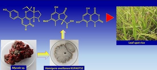
Pentaketides and 5-p-Hydroxyphenyl-2-pyridone Derivative from the Culture Extract of a Marine Sponge-Associated Fungus Hamigera avellanea KUFA0732

1
ICBAS-Instituto de Ciências Biomédicas Abel Salazar, Rua de Jorge Viterbo Ferreira 228, 4050-313 Porto, Portugal
2
Department of Plant Pathology, Faculty of Agriculture, Kasetsart University, Bangkok 10240, Thailand
3
Interdisciplinary Centre of Marine Environmental Research (CIIMAR), Terminal de Cruzeiros do Porto de Leixões, Av. General Norton de Matos s/n, 4450-208 Matosinhos, Portugal
4
Instituto de Biologia Molecular e Celular (i3S-IBMC), Universidade do Porto, Rua de Jorge Viterbo Ferreira 228, 4050-313 Porto, Portugal
5
Faculty of Pharmaceutical Sciences, Burapha University, 169 Long Had Bangsaen Rd, Chonburi 20131, Thailand
6
HEJ Research Institute of Chemistry, International Center for Chemical and Biological Sciences, University of Karachi, Karachi 75270, Pakistan
7
Laboratório de Química Orgânica e Farmacêutica, Departamento de Ciências Químicas, Faculdade de Farmácia, Universidade do Porto, Rua de Jorge Viterbo Ferreira 228, 4050-313 Porto, Portugal
8
Department of Chemistry, University of Leicester, University Road, Leicester LE 7 RH, UK
9
Departamento de Química & QOPNA, Universidade de Aveiro, 3810-193 Aveiro, Portugal
*
Authors to whom correspondence should be addressed.
Mar. Drugs 2023, 21(6), 344; https://doi.org/10.3390/md21060344
Submission received: 2 May 2023 / Revised: 31 May 2023 / Accepted: 31 May 2023 / Published: 2 June 2023

Abstract

:
Five undescribed pentaketide derivatives, (R)-6,8-dihydroxy-4,5-dimethyl-3-methylidene-3,4-dihydro-1H-2-benzopyran-1-one (1), [(3S,4R)-3,8-dihydroxy-6-methoxy-4,5-dimethyl-1-oxo-3,4-dihydro-1H-isochromen-3-yl]methyl acetate (2), (R)-5, 7-dimethoxy-3-((S)-(1-hydroxyethyl)-3,4-dimethylisobenzofuran-1(3H)-one (4b), (S)-7-hydroxy-3-((S)-1-hydroxyethyl)-5-methoxy-3,4-dimethylisobenzofuran 1(3H)-one (5), and a p-hydroxyphenyl-2-pyridone derivative, avellaneanone (6), were isolated together with the previously reported (R)-3-acetyl-7-hydroxy-5-methoxy-3,4-dimethylisobenzofuran-1(3H)-one (3), (R)-7-hydroxy-3-((S)-1-hydroxyethyl)-5-methoxy-3,4-dimethylisobenzofuran-1(3H)-one (4a) and isosclerone (7), from the ethyl acetate extract of a culture of a marine sponge-derived fungus, Hamigera avellanea KUFA0732. The structures of the undescribed compounds were elucidated using 1D and 2D NMR, as well as high-resolution mass spectral analyses. The absolute configurations of the stereogenic carbons in 1, 4b, 5, and 6 were established by X-ray crystallographic analysis. The absolute configurations of C-3 and C-4 in 2 were determined by ROESY correlations and on the basis of their common biosynthetic origin with 1. The crude fungal extract and the isolated compounds 1, 3, 4b, 5, 6, and 7 were assayed for their growth inhibitory activity against various plant pathogenic fungi viz. Alternaria brassicicola, Bipolaris oryzae, Colletotrichum capsici, C. gloeosporiodes, Curvularia oryzae, Fusarium semitectum, Lasiodiplodia theobromae, Phytophthora palmivora, Pyricularia oryzae, Rhizoctonia oryzae and Sclerotium rolfsii.

Graphical Abstract

1. Introduction

The fungi of the genus Hamigera (Family Aspergillaceae, Order Eurotiales) are widespread and common soil fungi, despite one species being associated with beetles [1]. Although many species of Eurotiales are rich sources of secondary metabolites due to the high number of secondary metabolite gene clusters present in their genomes, only a few chemical classes of secondary metabolites have been reported from the genus Hamigera [2].
Yamazaki et al. isolated two new cyclic pentapeptides, avellanins A and B, from Hamigera avellanea Stolk and Samson [3] while Breinholt et al. described the isolation of (Z, Z)-N,N′-[1-[(4-hydroxyphenyl)methylene]-2-[(4-methoxyphenyl)methylene]-1,2-ethanediyl]bis-formamide from an ethanol extract of the mycelium of H. avellanea (CBS 501.94) [4]. Breinholt’s group later noticed the inhibitory activity of the culture filtrates of the same fungus on the growth of the rice blast fungus, Pyricularia oryzae, so they used bioassay-guided fractionation to isolate two polyketides, hamigerone, and dihydrohamigerone. Hamigerone and dihydrohamigerone were tested for the in vitro growth inhibitory activity against plant pathogenic fungi, P. oryzae and Venturia inaequalis, and the results showed that a growth inhibitory activity of hamigerone against both pathogenic fungi was comparable to the commercial fungicides Prochloraz® and Bitertanol ® whereas dihydrohamigerone exhibited only marginal activity. However, when hamigerone was tested for its ability to protect rice plants against subsequent infection with P. oryzae, it was found to be far inferior to commercial fungicides [5]. Isaka et al. have described the isolation of two novel cyclopropyl diketones, hamavellones A and B, and two new 14-membered macrolactones, hamigeromycins A and B, together with a resorcyclic lactone, 89-250904-F1 (radicicol analog A), a spiro-heterocyclic γ-lactam alkaloid, pseurotin A, and anthraquinone pigments emodin, ϖ-hydroxyemodin, and emodin bianthrones from the ethyl acetate (EtOAc) extract of the culture broth and the mycelium of H. avellanea BCC 17816, isolated from a soil sample collected in Chiangmai, Thailand [6]. The Isaka group later isolated from the same fungus, hamigeromycins A and C–G [7].
On the other hand, in an attempt to use biological control as an alternative to commercial synthetic fungicides used in conventional farming, a member of our group (T. Dethoup) has evaluated the antagonistic activity of crude extracts of several marine-derived fungi against many plant pathogens that are causative agents of diseases of the economic crops [8], especially those that cause rice brown spot, rice dirty panicle [9] and sheath blight diseases [10].
Due to the in vitro inhibitory activity of hamigerone, isolated from H. avellanea (CBS 501.94), against the rice blast fungus, P. oryzae, we have examined the growth inhibitory activities of the crude EtOAc extract of a marine-derived H. avellanea KUFA0732, isolated from the marine sponge Mycale sp., which was collected from the coral reef at Samaesan Island in Chonburi province, Thailand, as well as some of the isolated compounds against various plant pathogenic fungi, viz. Bipolaris oryzae (brown spot of rice), Curvularia oryzae (leaf spot of rice), Fusarium semitectum (dirty particle of rice), P. oryzae (rice blast), Rhizoctonia oryzae (sheath blight of rice), Alternaria brassicicola (black spot of Chinese kale), Colletotrichum capsici (anthracnose of chili), C. gloeosporiodes (anthracnose of mango), Lasiodiplodia theobromae (fruit rot of mangosteen), Phytophthora palmivora (root and stem rot of durian), and Sclerotium rolfsii (stem rot of bean).
Fractionation of the EtOAc extract of the culture H. avellanea KUFA0732 using silica gel column chromatography, followed by purification using preparative TLC, a Sephadex LH-20 column, and crystallization, led to the isolation of the undescribed (R)-6,8-dihydroxy-4,5-dimethyl-3-methylidene-3,4-dihydro-1H-2-benzopyran-1-one (1), [(3S,4R)-3,8-dihydroxy-6-methoxy-4,5-dimethyl-1-oxo-3,4-dihydro-1H-isochromen-3-yl]methyl acetate (2), (R)-5,7-dimethoxy-3-((S)-(1-hydroxyethyl)-3,4-dimethylisobenzofuran-1(3H)-one (4b), (S)-7-hydroxy-3-((S)-1-hydroxyethyl)-5-methoxy-3,4-dimethylisobenzofuran 1(3H)-one (5), and avellaneanone (6), together with the previously reported (R)-3-acetyl-7-hydroxy-5-methoxy-3,4-dimethylisobenzofuran-1(3H)-one (3) [11,12,13], (R)-7-hydroxy-3-((S)-1-hydroxyethyl)-5-methoxy-3,4-dimethylisobenzofuran-1(3H)-one (4a) [13] and isosclerone (7) [14,15,16] (Figure 1). The structures of the undescribed compounds were established based on an extensive analysis of the 1D and 2D NMR spectra as well as HRMS data. In the case of 1, 4b, 5, and 6, the absolute configurations of their stereogenic carbons were established by X-ray analysis, while the absolute configurations of the stereogenic carbons in 2 were determined by ROESY correlations and comparison of its 1H NMR data with those of 1.

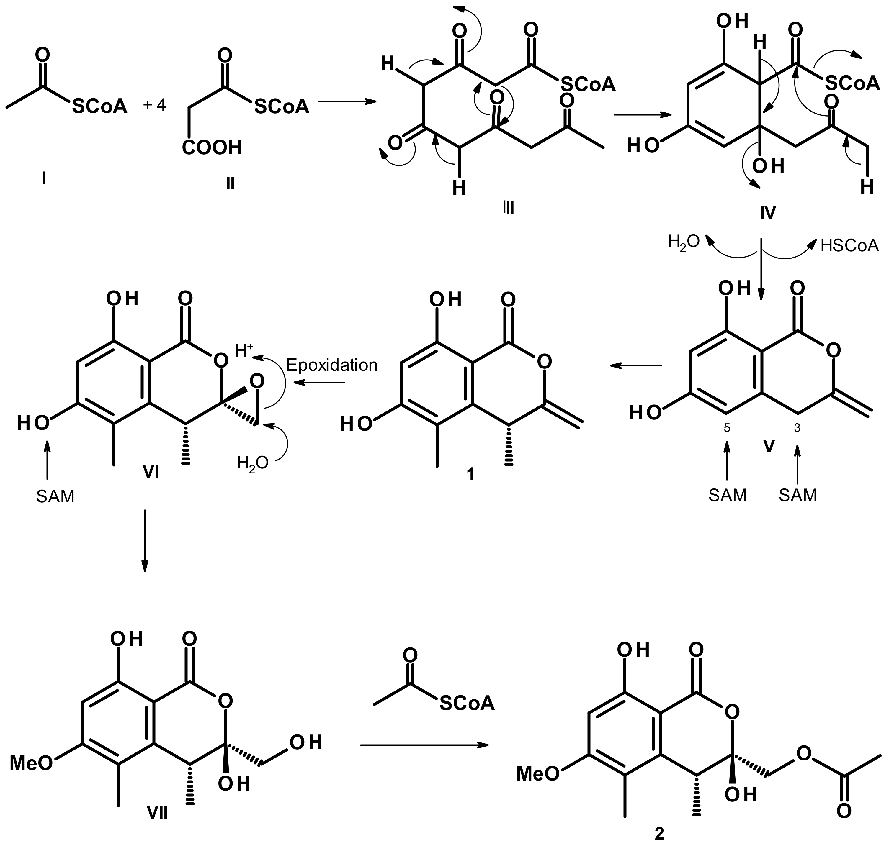
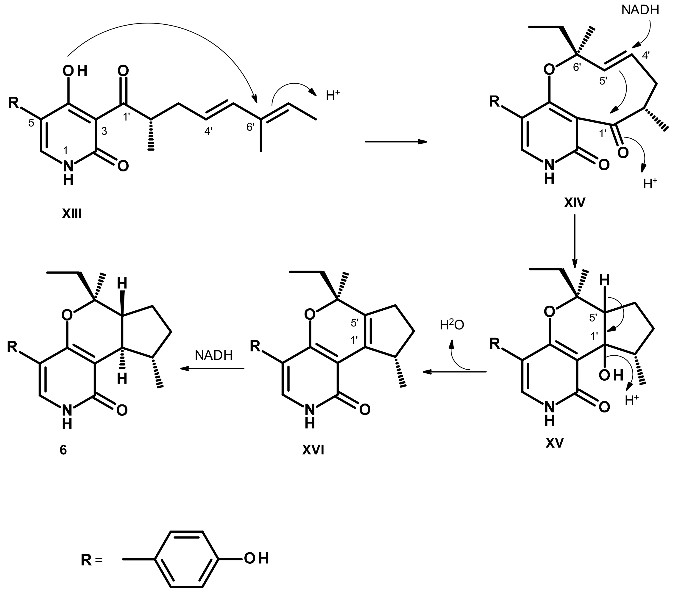
2. Results and Discussions

The structures of (R)-3-acetyl-7-hydroxy-5-methoxy-3,4-dimethylisobenzofuran-1(3H)-one (3) [11,12,13], (R)-7-hydroxy-3-((S)-1-hydroxyethyl)-5-methoxy-3,4-dimethylisobenzofuran-1(3H)-one (4a) [13] and isosclerone (7) [14,15,16] were elucidated by analysis of their 1D and 2D NMR spectra (Figures S14–S23 and S42–S46, Tables S1, S2 and S7) and by comparison of their NMR spectral data and the sign of their optical rotations with those reported in the literature.
Compound 1 was isolated as white crystals (mp = 217–218 °C), and its molecular formula C12H12O4 was established based on (+)-HRESIMS m/z 221.0814 [M + H]+ (calculated for C12H13O4, 221.0814) (Figure S6), requiring seven degrees of unsaturation. The 13C NMR spectrum (Table 1, Figure S2) exhibited twelve carbon signals which, in combination with DEPT and HSQC spectra (Figure S4), can be classified as one conjugated lactone carbonyl (δC 166.3), six non-protonated sp2C 164.1, 161.8, 157.4, 143.6, 113.2, 97.7), one protonated sp2C 101.0), one methylene sp2C 96.2), one methine sp3C 34.2) and two methyl (δC 22.3 and 10.2) carbons. The 1H NMR spectrum (Table 1, Figure S1) displayed a broad singlet of a hydrogen-bonded hydroxyl proton at δH 10.70, one aromatic singlet at δH 6.33, two doublets of the olefinic protons at δH 4.75 (J = 1.6 Hz) and 4.73 (J = 1.6 Hz), one methine quartet at δH 4.02 (J = 7.2 Hz), one methyl singlet at δH 2.02 and one methyl doublet at δH 1.27 (J = 7.0 Hz). The type and chemical shift values of the carbon atoms revealed that 1 is a 3, 4, 5, 6, 8-pentasubstituted chromone. That the hydroxyl group at δH 10.70 was on C-8, the methyl group at δH 2.02, s (δC 22.3, Me-11) was on C-5, and another hydroxyl group was on C-6 was evidenced by the chemical shift value of the hydrogen-bonded OH-8 as well as by the HMBC correlations (Table 1, Figure 2 and Figure S5) from H3-11 to C-5 (δC 113.2), C-4a (δC 143.6), 164.1 (C-6), and H-7 (δH 6.33, s) to C-5, C-6, C-8 (δC 161.8) and C-8a (δC 97.7). The COSY correlation from the methyl doublet at δH 1.27 (J = 7.0 Hz, Me-10) to the quartet at δH 4.02 (J = 7.2 Hz/δH 34.2) (Table 1, Figure 2 and Figure S3) revealed that Me-10 was on C-4. This was supported by HMBC correlations from H3-10 to C-4, C-4a, and C-3 (δC 157.4), and H-4 to C-4a, C-5, C-8a, and C-11. That the methylidene group was on C-3 was supported by HMBC correlations from the two doublets of the olefinic protons at δH 4.73 (J = 1.6 Hz/δC 96.2) and δH 4.75 (J = 1.6 Hz/δH 96.2) to C-3 and C-4 (Table 1, Figure 2 and Figure S5). Taking together the HRMS and 1D and 2D NMR data, a planar structure of 1 was elucidated as 6,8-dihydroxy-4,5-dimethyl-3-methylidene-3,4-dihydro-1H-2-benzopyran-1-one.
Compound 1 has one stereogenic center (C-4), and it is, therefore, necessary to determine the absolute configuration of this carbon. Since 1 could be obtained as a suitable crystal for X-ray analysis using a diffractometer equipped with CuKα radiation, its stereostructure was obtained. The Ortep view of 1, shown in Figure 3, revealed that the configuration of C-4 is R. Therefore, the absolute structure of 1 was elucidated as (R)-6,8-dihydroxy-4,5-dimethyl-3-methylidene-3,4-dihydro-1H-2-benzopyran-1-one.
A literature search revealed that 1 has never been previously reported; however, a structurally similar compound, but with opposite stereochemistry at C-4, i.e. (S)-8-hydroxy-6-methoxy-4,5-dimethyl-3-methyleneisochromen-1-one, was reported by Tayone et al. [17] from the EtOAc extract of the culture broth of Leptosphaeria sp. KTC 727, which was collected from woody debris in Rebun Island, Hokkaido, Japan. The absolute configuration at C-4 was determined as S by comparison of the calculated and experimental ECD spectra. Later, the same compound was also isolated from the mycelium and culture broth extracts of the fungus Xylaria sp. BL321, isolated from the leaves of the mangrove plant Acanthus ilicifolius L., was collected in Guangdong, China. The absolute configuration at C-4 was also determined as S by comparison of the calculated and experimental ECD spectra [18].
Compound 2 was isolated as a pale yellow viscous mass. The molecular formula C15H18O7 of 2 was obtained from the (+)-HRESIMS m/z 311.1133 [M + H]+ (calculated for C15H19O7, 311.1131) (Figure S13), indicating seven degrees of unsaturation. The 13C NMR spectrum (Table 2, Figure S8) displayed 15 carbon signals which, in conjunction with DEPT and HSQC spectra (Figure S10), can be classified as one ester carbonyl (δC 170.7), one conjugated ester carbonyl (δC 168.2), two oxygen-bearing sp2C 164.9 and 163.1), four non-protonated sp2C 141.5, 115.5, 102.2 and 99.0), one pronated sp2C 97.5), one oxymethylene sp3C 65.9), one methoxy (δC 55.9), one methine sp3C 35.9) and three methyl (δC 20.8, 16.2, and 10.1) carbons. The 1H NMR spectrum (Table 2, Figure S7), in combination with the HSQC spectrum (Figure S10), exhibited a singlet of a hydrogen-bonded hydroxyl proton at δH 11.14, one aromatic singlet at δH 6.37 (δC 97.5), two terminally coupled doublets at δH 4.25 (J = 11.8 Hz/δC 65.9) and 4.59 (J = 11.8 Hz/δC 65.9), one methoxyl singlet at δH 3.85 (δC 55.9), one quartet at δH 4.33 (J = 7.1 Hz/δC 35.9), two methyl singlets at δH 2.19 (δC 20.8) and 2.08 (δC 10.1) and one methyl doublet at δH 1.19 (J = 7.1 Hz/δC 16.2). The presence of a 4,5-dimethyl-8-hydroxy-6-methoxy-3,4-dihydro-1H-2-benzopyran-2-one was substantiated by HMBC correlations from the singlet at δH 11.14 (OH-8) to the carbons at δC 163.1 (C-8) and 97.5 (C-7), the singlet at δHz/6.37 (H-7) to C-8, the carbons at δC 115.5 (C-5) and 99.0 (C-8a), a methyl singlet at δH 2.08 (H3-11) to the carbon at δC 164.9 (C-6), 141.5 (C-4a) and 115.5 (C-5), and the methyl doublet at δH 1.19 (H3-10) to C-4a, the carbons at δC 102.2 (C-3) and 35.9 (C-4) (Table 2, Figure 4 and Figure S11). The chemical shift value of C-4 is characteristic of a hemiketal carbon, and thus one of its substituents must be a hydroxyl group. Another substituent on C-3 was an acetoxymethyl group, supported by the HMBC correlations from H2-9 (δH 4.25, d, J = 11.8 Hz) and 4.59, d, J = 11.8 Hz) to C-3 and the carbonyl of the acetyl group at δC 170.7 (C-12) and the methyl protons of the acetyl group (δH 2.19) to C-12 (Table 2, Figure 4 and Figure S11).
Therefore, the planar structure of 2 was elucidated as (3,8-dihydroxy-6-methoxy-4,5-dimethyl-1-oxo-3,4-dihydro-1H-isochromen-3-yl)methyl acetate.
Compound 2 has two stereogenic carbons (C-3 and C-4); therefore, it is necessary to establish their absolute configurations. Since 2 is a viscous mass and could not be obtained as a suitable crystal for X-ray diffraction, the ROESY spectrum was obtained. The ROESY spectrum (Table 2, Figure S12) showed strong correlations from H-4 to H3-10 and H3-11, confirming that H-4 is β and in the equatorial position while Me-10 is α and in the axial position. Thus, the stereochemistry at C-4 in 2 is the same as in 1. Moreover, H3-10 also showed a strong correlation to H-9a and H-9b, while H-9a only showed a weak correlation to H-4. These ROESY correlations revealed that CH2-9 is α and OH-3 is β. Therefore, based on the absolute configuration at C-4 of 1, the absolute configurations at C-3 and C-4 in 2 are established as 3S, 4R.
Biosynthetically, 1 and 2 are pentaketides, and 2 could be considered to derive directly from 1 according to the proposed pathways shown in Figure 5. A Claisen condensation of one acetyl CoA (I) with four malonyl CoA (II) gives a pentaketide (III), which undergoes cyclization and is followed by enolization to give IV, and further dehydration of IV gives rise to V. Stereospecific methylation at C-3 and C-5 of V by S-adenosyl methionine (SAM) gives rise to 1. Epoxidation of the exocyclic double bond of 1 gives an epoxide in VI. Enzymatic hydrolysis of the epoxide and methylation of a phenolic hydroxyl group in VI produces VII, which undergoes acetylation by acetyl CoA to give 2. Consequently, the absolute configuration at C-4 in 2 must be the same as that of C-4 in 1, i.e., 4R.
Interestingly Tayone et al. [17] have described the isolation of a mixture of two diastereomers of 3,8-dihydroxy-3-hydroxymethyl-6-methoxy-4,5-dimethylisochroman-1-one, whose structures are similar to VII, except for the stereochemistry at C-4 which is opposite to that of VII, from the EtOAc extract of the culture broth of Leptosphaeria sp. KTC 727. These authors have detected the difference in the 1H and 13C NMR chemical shift values between the major component (3R, 4S) and the minor component (3S, 4S) in the mixture but were not able to isolate them individually. On the contrary, the 1H and 13C NMR spectra (Figures S7 and S8) of 2 displayed clearly only one set of signals, implying that 2 is not a mixture of two diastereomers.
1H and 13C NMR data (Table S2, Figures S19 and S20) of 4a resemble those of (R)-7-hydroxy-3-((R)-1-hydroxyethyl)-5-methoxy-3,4-dimethylisobenzofuran-1(3H)-one, isolated from the culture extract of a marine mangrove-associated fungus, Xylaria sp. BL321 [18] as well as (R)-7-hydroxy-3-((S)-1-hydroxyethyl)-5-methoxy-3,4-dimethylisobenzofuran 1(3H)-one, isolated from the culture extract of the fungus, Leptosphaeria sp. KTC 727, collected from woody debris at Akaiwa, Rebun island in Hokkaido, Japan [13]. Table S3 compared 1H and 13C NMR data of 4a with those of both (R)-7-hydroxy-3-((R)-1-hydroxyethyl)-5-methoxy-3,4-dimethylisobenzofuran 1(3H)-one and (R)-7-hydroxy-3-((S)-1-hydroxyethyl)-5-methoxy-3,4-dimethylisobenzofuran 1(3H)-one. From the 1H and 13C NMR chemical shift values in Table S3, it is not possible to determine if the absolute configurations at C-3 and C-8 in 4a are 3R, 8R, or 3R, 8S. However, the two stereoisomers differ in the sign of the optical rotations. While (R)-7-hydroxy-3-((R)-1-hydroxyethyl)-5-methoxy-3,4-dimethylisobenzofuran 1(3H)-one is dextrorotatory ([α]25D + 355.4, c 0.05, MeOH), (R)-7-hydroxy-3-((S)-1-hydroxyethyl)-5-methoxy-3,4-dimethylisobenzofuran 1(3H)-one is levorotatory ([α]25D −27, c 0.02, CDCl3) [13]. Since 4a is levorotatory ([α]25D −73, c 0.06, MeOH), we concluded that 4a is (R)-7-hydroxy-3-((S)-1-hydroxyethyl)-5-methoxy-3,4-dimethylisobenzofuran 1(3H)-one.
Compound 4b was isolated as white crystals (mp = 174–176 °C), and its molecular formula C14H18O5 was determined based on the (+)-HRESIMS m/z 267.1233 [M + H]+ (calculated for C14H19O5, 267.1232) (Figure S29), requiring six degrees of unsaturation. The 1H and 13C NMR spectra (Table 3, Figures S24 and S25) are quite similar to those of 4a, except for the presence of an extra methoxyl substituent (δH 3.96, s/δC 56.1) on the aromatic ring and the lack of a broad signal of the phenolic hydroxyl group around 7 ppm. Thus, the structure of 4b was deduced as a methoxy derivative of 4a. The correlations observed in the COSY, HSQC, and HMBC spectra (Table 3, Figures S26–S28) allowed the establishment of the planar structure of 4b as 5, 7-dimethoxy-3-(1-hydroxyethyl)-3,4-dimethylisobenzofuran 1(3H)-one.
Since 4b is also levorotatory ([α]25D −300, c 0.05, MeOH) similar to 4a ([α]25D −73, c 0.06, MeOH), we concluded that the absolute configurations at C-3 and C-8 in 4b are 3R and 8S also. After many attempts, we were finally able to obtain 4b as a suitable crystal and an X-ray analysis was performed. The Ortep view of 4b (Figure 6) confirmed the stereochemistry of C-3 and C-8 as 3R and 8S. A literature search revealed that 4b has never been previously reported.
Compound 5 was isolated as a white crystal (mp = 116–118 °C). The (+)-HRESIMS spectrum showed the [M + H]+ peak at m/z 253.1077 (calcd for C13H17O5, 253.1076) (Figure S35), revealing the same molecular formula as 4a, i.e., C13H16O5. Therefore, 5 is an isomer of 4a. The 1H and 13C NMR spectra (Table 4, Figures S30 and S31) of 5 resembled those of 4a; however, the protons of the three methyl groups in 5 showed different chemical shift values from those in 4a. Analysis of the COSY, HSQC, and HMBC spectra (Table 4, Figures S32–S34) revealed that the planar structure of 5 is the same as that of 4a.
Curiously, the proton chemical shift value of the doublet of Me-9 (δH 1.40, J = 6.4 Hz) in 5 is higher than that of 4aH 0.89, J = 6.5 Hz) around 0.5 ppm while the proton chemical shift value of its Me-10 singlet (δH 1.67) was lower than that of 4aH 1.78) just around 0.1 ppm. The differences in chemical shift values of the methyl protons of Me-9 and Me-10 can be attributed to the anisotropic effect of the benzene ring on both Me-9 and Me-10. As the chemical shift value of Me-10 in 5 is higher than that of its counterpart in 4a, while Me-10 in 5 is lower than that of its counterpart in 4a, the absolute configuration at C-3 in 5 must be opposite to that at C-3 in 4a, i.e., 3S. Since 5 could be obtained in a suitable crystal, an X-ray analysis was performed. The Ortep view of 5, shown in Figure 7, not only confirmed the 3S configuration but also determined the configuration at C-8 as 8S. Therefore, a complete structure of 5 is (S)-7-hydroxy-3-((S)-1-hydroxyethyl)-5-methoxy-3,4-dimethylisobenzofuran 1(3H)-one, which is the enantiomer of (R)-7-hydroxy-3-((R)-1-hydroxyethyl)-5-methoxy-3,4-dimethylisobenzofuran 1(3H)-one ([α]25D +355.4, c 00.05, MeOH) [18]. This was also supported by the fact that 5 is levorotatory ([α]25D −320, c 0.05, MeOH). From the Ortep view (Figure 7), the methyl protons of Me-10 of the α-axial 1-hydroxyethyl substituent should experience a paramagnetic field of the benzene ring, while the protons of the β-equatorial Me-9 should experience a slight diamagnetic effect of the benzene ring. A literature search revealed that 5 also has never been reported.
Analysis of the structures of 3, 4a, 4b, and 5 revealed that they all derived from a cyclization of a linear pentaketide, similar to that of 1 and 2. Figure 8 depicts proposed biosynthetic pathways for 3, 4a, 4b, and 5.
The intermediate VIII, derived from a cyclization and aromatization of a linear pentaketide III, through IV, undergoes stereospecific methylation at the benzyl carbon and a methylation at the aromatic carbon and the phenolic hydroxyl group to give IX. Stereospecific hydroxylation of the benzyl carbon of IX gives rise to X, which undergoes a lactonization to give 3. Stereospecific reduction of the carbonyl ketone of the side chain of 3 produces 4a. Methylation of another phenolic hydroxyl group of 4a gives rise to 4b.
The route leading to the formation of 5 also begins with intermediate VIII. In this route, the stereospecific methylation of the benzyl carbon to form XI occurs on the oppo face to the methylation to form IX. Oxidation of the benzyl carbon and lactonization of XI gives rise to XII, which, after reduction of the ketone carbonyl of the side chain, leads to the formation of 5.
Compound 6 was isolated as colorless crystals (mp = 209–210 °C), and its molecular formula C21H25NO3 was established on the basis of (+)-HRESIMS m/z 362.1734 [M + Na]+ (calculated for C21H25NO3Na, 362.1732) (Figure S41), requiring ten degrees of unsaturation. The 13C NMR spectrum (Table 5, Figure S37) displayed 19 carbon signals which, in combination with DEPT and HSQC spectra (Figure S39), can be categorized as one conjugated amide carbonyl (δC 162.4), two oxygen-bearing sp2C 160.2 and 156.6), three non-protonated sp2C 110.4, 113.6 and 126.7), five protonated sp2C 115.1 (2C), 130.6 (2C), 131.5), one oxyquaternary sp3C 82.2), three methine sp3C 51.9, 43.8, 35.3), three methylene sp3C 34.5, 33.8, 25.2) and three methyl (δC 25.1, 18.6, 7.7) carbons. The 1H NMR spectrum (Table 5, Figure S36), in conjunction with the HSQC spectrum (Figure S39), showed two broad singlets at δH 9.38 and 10.93, two doublets of four ortho-coupled aromatic protons at δH 6.73 (J = 8.6 Hz, 2H/δC 115.1) and 7.15 (J = 8.6 Hz, 2H/δC 130.6), one olefinic singlet at δH 6.98 (δC 131.5), a double doublet at δH 2.06 (J = 12.0, 8.7 Hz/δC 43.8), a double of double doublet at δH 1.71 (J = 12.0, 12.0, 7.0 Hz/δC 51.9), a quartet at δH 1.55 (J = 7.1 Hz/δC 33.8), one methyl singlet at δH 1.11 (δC 18.6), one methyl doublet at δH 1.47 (J = 6.2 Hz/δC 25.1) and one methyl triplet at δH 0.85 (J = 7.3 Hz/δC 7.7), in addition to various overlapped multiplets. The presence of a 4-hydroxy phenyl moiety was evidenced by the COSY correlation from the doublet at δH 7.15 (J = 8.6 Hz/H-18, H-22) to the doublet at δH 6.73 (J = 8.6 Hz/H-19, H-21) (Figure S38) as well as the HMBC correlations from H-18 to C-20 (δC 156.6) and C-22 (δC 130.6), H-22 to C-18 (δC 130.6), and C-20, H-19 to C-17 (δC 126.7), and C-21 (δC 115.1), and H-21 to C-17, and C-19 (Figure S40). That another portion of 6 consists of a 3,4,5-trisubstituted 2-pyridone was substantiated by the HMBC correlations from H-6 (δH 6.98, s) to C-2 (δC 162.4), C-4 (δC 160.2), and C-5 (δC 113.6). Since H-18 and H-22 showed HMBC cross peaks to C-5 while H-6 showed cross peaks to C-17, the p-hydroxyphenyl group was on C-5 of the 2-pyridone ring. That the last portion of the molecule was 1-ethyl-1,5-dimethyl-1,4a,5,6,7,7a-hexahydrocyclopenta[c]pyran was supported by the COSY correlations from H3-16 (δH 0.85, t, J = 7.3 Hz/δC 7.7) to H-15 (δH 1.55, q, J = 7.1 Hz/δC 33.8), H3-12 (δH 1.47, d, J = 6.2 Hz/δC 25.1) to H-11 (δH 2.15, m/δC 35.3), and a coupling system from H3-12 through H-11/H-7 (δH 2.06, dd, J = 12.0, 8.7 Hz/δC 43.8)/H-8 (δH 1.71, ddd, J = 12.0, 12.0, 7.0 Hz/δC 51.9)/H-9 (δH 1.56, m/δC 25.2) and H-10 (δH 1.39, m and 1.99, m/δC 34.5) (Table 5, Figure 8 and Figure S40). This was corroborated by HMBC correlations from H3-16 to C-13 (δC 82.2) and C-15, H3-14 (δH 1.11, s/δC 18.6) to C-8, C-13, C-15, H3-12 to C-7, C-10 and C-11, H-7 to C-11 and C-12, H-8 to C-13 and C-14 (Table 5, Figure 8 and Figure S40). That the 3,4-dihydro-2H-pyran is fused with the 2-pyridone ring at C-3 and C-4 was supported by the HMBC correlation from H-7 to H-3 and the chemical shift values of C-4 and C-13 (Table 5, Figure 8 and Figure S40). Taking together the molecular formula, the COSY, and HMBC correlations, the planar structure of 6 was elucidated, as shown in Figure 9.
Compound 6 possesses four stereogenic centers viz. C-7, C-8, C-11 and C-13. Therefore, it is challenging to determine the absolute configurations of these carbons. Since we were able to obtain a suitable crystal of 6, its X-ray analysis was carried out using an X-ray diffractometer equipped with CuKα radiation. The Ortep view of 6 is shown in Figure 10.
The Ortep view showed clearly that the absolute configuration at C-7, C-8, C-12, and C-13 are 7R, 8R, 12S, and 13S. Since 6 has never been reported, we named it avellaneanone.
5-p-Hydroxy phenyl-2-pyridones with alkyl or alkenyl side chains on C-3 have been previously isolated from different fungal strains. Examples of these are tenellin [19] and desmethylbassianin, isolated from the entomopathogenic fungus, Beauveria bassiana [20], and aspyridone-A from Aspergillus nidulans [21]. Moreover, the alkyl and alkenyl side chains can undergo cyclization to form a tetrahydropyran ring such as sambutoxin, isolated from the wheat culture of Fusarium sambucinum which was obtained from a rot potato tuber [22], and a decalin ring system such as illicicolin H from the fermentation of Cylindrocladium ilicicola MFC 870 [23]. Additionally, the alkenyl side chain can form a pyran ring of a hexahydro-1H-2-benzopyran in trichodin A, isolated from a marine-derived Trichoderma sp. MF106, which was obtained from a Greenland Sea (Fram Strait) sample. However, only the relative configurations of the stereogenic carbons of the hexahydro-1H-2-benzopyran in trichodin A were established by analysis of NOESY correlations [24].
The 5-p-Hydroxyphenyl-2-pyridone derivatives are polyketide synthase (PKS)-non-ribosomal peptide synthetase (NRPS) metabolites, which were reported to be synthesized from ring expansion of tetrameric acid intermediates catalyzed by cytochrome P450 monooxygenases [25]. Analysis of the structure of 6 revealed that the positions of the methyl groups in its doubly methylated pentaketide are different from those of tenellin [26], suggesting that H. avellanea KUFA 0732 must possess a different C-methylation domain (CMeT) from other fungi. Thus we propose that the biosynthetic pathways of 6 should start from the intermediate XIII, which is derived from a tetramic acid derivative formed by the condensation of tyrosine with a doubly methylated pentaketide. The nucleophilic addition of the hydroxyl group of the 2-pyridone ring to C-6′ of the side chain leads to the formation of the intermediate XIV. Reduction of C-4′ in XIV, with a concomitant nucleophilic addition of C-5′ to the carbonyl group (C-1′), leads to the formation of a cyclopentane ring in XV. Dehydration of XV renders a double bond between C-1′ and C-5′ in XVI. Stereospecific reduction of the double bond between C-1′ and C-5′ in XVI by dehydrogenase gives rise to 6 (Figure 11).
The crude EtOAc extract and the isolated compounds 1, 3, 4b, 5, 6, and 7 were assayed for the growth inhibitory activity against various plant pathogenic fungi that cause diseases for economic crops, viz. Alternaria brassicicola (black spot of Chinese Kale), Bipolaris oryzae (brown spot of rice), Colletotrichum capsici (anthracnose of chili), C. gloeosporiodes (anthracnose of mango), Curvularia oryzae (leaf spot of rice), Fusarium semitectum (dirty panicle of rice), Lasiodiplodia theobromae (fruit rot of mangosteen), Phytophthora palmivora (root and stem rot of durian), Pyricularia oryzae (rice blast), Rhizoctonia oryzae (sheath blight of rice) and Sclerotium rolfsii (stem rot of bean). The crude extract showed complete mycelial growth inhibition of all plant pathogenic fungi at 10 g/L, except for F. semitectum, A. brassicicola and B. oryzae. However, at a concentration of 1 g/L, the crude extract exhibited significant (p < 0.05) inhibition of the mycelial growth of R. oryzae, C. capsici, C. gloeosporiodes, and C. oryzae, causing more than 50% inhibition but displayed less than 50% inhibition for the rest of the pathogens tested (Table 6).
Interestingly, 3, 5, and 6 exhibited growth inhibition on C. oryzae, a causative agent of leaf spot of rice, while 1, 2, 4b, and 7 were inactive (MIC > 500 µg/mL). Compounds 3 and 5 displayed strong inhibitory activity, with MIC values of 125 µg/mL, which is comparable to the positive control, mancozeb (MIC = 125 µg/Ml), while 6 only showed moderate activity, with MIC value of 250 µg/Ml. All the tested compounds were not active (MIC > 500 µg/Ml) against the rest of the plant pathogens (Table 7).

3. Methods and Materials

3.1. General Experimental Procedures

The melting points were determined on a Stuart Melting Point Apparatus SMP3 (Bibby Sterilin, Stone, Staffordshire, UK) and are uncorrected. Optical rotations were measured on an ADP410 Polarimeter (Bellingham + Stanley Ltd., Tunbridge Wells, Kent, UK). The 1H and 13C NMR spectra were recorded at ambient temperature on a Bruker AMC instrument (Bruker Biosciences Corporation, Billerica, MA, USA) operating at 300 and 75 MHz, respectively. High-resolution mass spectra were measured with a Waters Xevo QToF mass spectrometer (Waters Corporations, Milford, MA, USA) coupled to a Waters Aquity UPLC system. A Merck (Darmstadt, Germany) silica gel GF254 was used for preparative TLC, and Merck Si gel 60 (0.2–0.5 mm) was used for column chromatography. LiChroprep silica gel and Sephadex LH 20 were used for column chromatography.

3.2. Fungal Material

The fungus was isolated from a marine sponge Mycale sp., which was collected by scuba diving at a depth of 5-10 m from the coral reef at Samaesan Island (12°34′36.64″ N 100°56′59.69″ E), in the Gulf of Thailand, Chonburi Province, in May 2018. The sponge was washed with 0.01% sodium hypochlorite solution for 1 min, followed by sterilized seawater three times, and then dried on a sterile filter paper under sterile aseptic condition. The sponge was cut into small pieces (ca. 5 × 5 mm) and placed on Petri dish plates containing 15 mL potato dextrose agar (PDA) medium mixed with 300 mg/L of streptomycin sulfate and incubated at 28 °C for 7 days. The hyphal tips emerging from sponge pieces were individually transferred onto a PDA slant and maintained as pure cultures at Kasetsart University Fungal Collection, Department of Plant Pathology, Faculty of Agriculture, Kasetsart University, Bangkok, Thailand. The fungal strain KUFA 0732 was identified as Hamigera avellanea based on morphological characteristics such as colony growth rate and growth pattern on standard media, namely Czapek’s agar, Czapek yeast autolysate agar, and malt extract agar. Microscopic characteristics, including the size, shape, and ornamentation of conidiophores and spores, were examined under a light microscope. This identification was confirmed by molecular techniques using internal transcribed spacer (ITS) primers. DNA was extracted from young mycelia following a modified Murray and Thompson method [27]. The universal primer pairs, ITS1 and ITS4, were used for ITS gene amplification [28]. PCR reactions were conducted on Thermal Cycler, and the amplification process consisted of initial denaturation at 95 °C for 5 min, 34 cycles at 95 °C for 1 min (denaturation), at 55 °C for 1 min (annealing), and at 72 °C for 1.5 min (extension), followed by the final extension at 72 °C for 10 min. PCR products were examined by agarose gel electrophoresis (1% agarose with 1 × Tris-Borate-EDTA (TBE) buffer) and visualized under UV light after staining with ethidium bromide. DNA sequencing analyses were performed using the dideoxyribonucleotide chain termination method [29] by Macrogen Inc. (Seoul, Republic of Korea). The DNA sequences were edited using FinchTV software and submitted to the BLAST program for alignment and compared with that of fungal species in the NCBI database (http://www.ncbi.nlm.nih.gov/ accessed on 1 May 2023). Its gene sequences were deposited in GenBank with the accession number OQ520878.

3.3. Extraction and Isolation

The fungus was cultured for one week at 28 °C in five Petri dishes (i.d. 90 mm) containing 20 mL of PDA per dish. The mycelial plugs (5 mm in diameter) were transferred to two 500 mL Erlenmeyer flasks containing 200 mL of potato dextrose broth and incubated on a rotary shaker at 120 rpm at 28 °C for one week. Fifty 1000 mL Erlenmeyer flasks, each containing 300 g of cooked rice, were autoclaved at 121 °C for 15 min. After cooling to room temperature, 20 mL of a mycelial suspension of the fungus was inoculated per flask and incubated at 28 °C for 30 days, after which 500 mL of EtOAc was added to each flask of the moldy rice and macerated for 7 days, and then filtered with a Whatman No. 1 filter paper. The EtOAc solutions were combined and concentrated under reduced pressure to yield 184.1 g of a crude EtOAc extract, which was dissolved in 500 mL of CHCl3, washed with H2O (3 × 500 mL), and dried with anhydrous Na2SO4, filtered and evaporated under reduced pressure to give 153.5 g of a crude CHCl3 extract. The crude CHCl3 extract (55.5 g) was applied on a silica gel column (350 g) and eluted with mixtures of petrol-CHCl3 and CHCl3-Me2CO, wherein 250 mL fractions (frs) were collected as follows: frs 1–146 (petrol-CHCl3, 1:1), 147–204 (petrol-CHCl3, 3:7), 205–234 (petrol-CHCl3, 1:9), 235–250 (CHCl3), 251–322 (CHCl3-Me2CO, 9:1), 323–382 (CHCl3-Me2CO, 7:3). Frs 30–31 were combined (1.43 g) and applied over a Sephadex LH-20 column (10g) and eluted with MeOH, wherein 14 subfractions (sfrs) of 2 mL were collected. Sfrs 1–2 were combined (275.4 mg) and purified by a preparative TLC (silica gel G254, CHCl3: petrol: Me2CO: HCO2H, 90:5:5:0.1) to give 12.5 mg of 3. Frs 39–46 were combined (397 mg) and recrystallized in CHCl3 to give 24.8 mg of 1. The mother liquor of frs 39–46 (360.2 mg) was combined with frs 47–57 (305.0 mg) and purified by preparative TLC (silica gel G254, CHCl3: Me2CO: HCO2H, 90:10:0.1) to give additional 93.6 mg of 1. Frs 62–79 were combined (710.4 mg) and applied over a Sephadex LH-20 column (10 g), and eluted with MeOH, wherein 12 sfrs of 2 mL were collected. Sfrs 2–8 were combined (540.8 mg) and applied over another Sephadex LH-20 column (10 g) and eluted with MeOH, wherein 28 sub-subfractions (ssfrs) of 2 mL were collected. Ssfrs 11–22 were combined (281.4 mg) and purified by preparative TLC (silica gel G254, CHCl3:Me2CO: HCO2H, 90:10:0.1) to give 38.5 mg of 7 and 22.4 mg of 2. Frs 87–107 were combined (1.23 g), and part of it (543.2 mg) was purified by preparative TLC (silica gel G254, CHCl3: petrol: Me2CO: HCO2H, 90:5:5:0.1) to give 375.3 mg of a mixture of compounds which was then applied on a Sephadex LH-20 column (10 g) and eluted with MeOH, wherein 16 sfrs were collected. Sfrs 3-16 were combined to give 80 mg of 4a. Another part of the combined frs 87–109 (670 mg) was purified by a preparative TLC (silica gel G254, CHCl3: petrol: Me2CO: HCO2H, 90:5:5:0.1) to give 19.3 mg of 4a. Frs 117–149 were combined (264.1 mg) and crystallized in CHCl3 to give 15 mg of 5. Frs 150–166 were combined (584.8 mg) and applied on a silica gel column (10 g), and eluted with mixtures of petrol-CHCl3, CHCl3, CHCl3-Me2CO, wherein 100 mL sfrs were collected as follows: sfrs 1–12 (petrol-CHCl3, 1:1), 13–21 (petrol-CHCl3, 3:7), 22–30 (petrol-CHCl3, 1:9), 31–73 (CHCl3), 74–81 (CHCl3-Me2CO, 9:1). Sfrs 35-39 were combined (143.9 mg) and purified by a preparative TLC (silica gel G254, CHCl3: Me2CO: HCO2H, 90:10:0.1) to give 18 mg of 4b and 10 mg of 4a. Sfrs 60–81 were combined (228.2 mg) and purified by a preparative TLC (silica gel G254, CHCl3: Me2CO: HCO2H, 90:10:0.1) to give 15.2 mg of 4b and 5.6 mg of 4a. Frs 326–349 were combined (1.01 g) and applied on a silica gel column (10 mg) and eluted with mixtures of petrol-CHCl3, CHCl3, CHCl3-Me2CO, wherein 100 mL sfrs were collected as follows: Sfrs 1–11 (petrol-CHCl3, 1:1), 12–16 (petrol-CHCl3, 3:7), 17–26 (CHCl3), 27-54 (CHCl3-Me2CO, 9:1), 55–61 (CHCl3-Me2CO, 7:3). Sfrs 33–35 were combined (154.8 mg) and crystallized in CHCl3 and Me2CO to give 64.2 mg of 6.

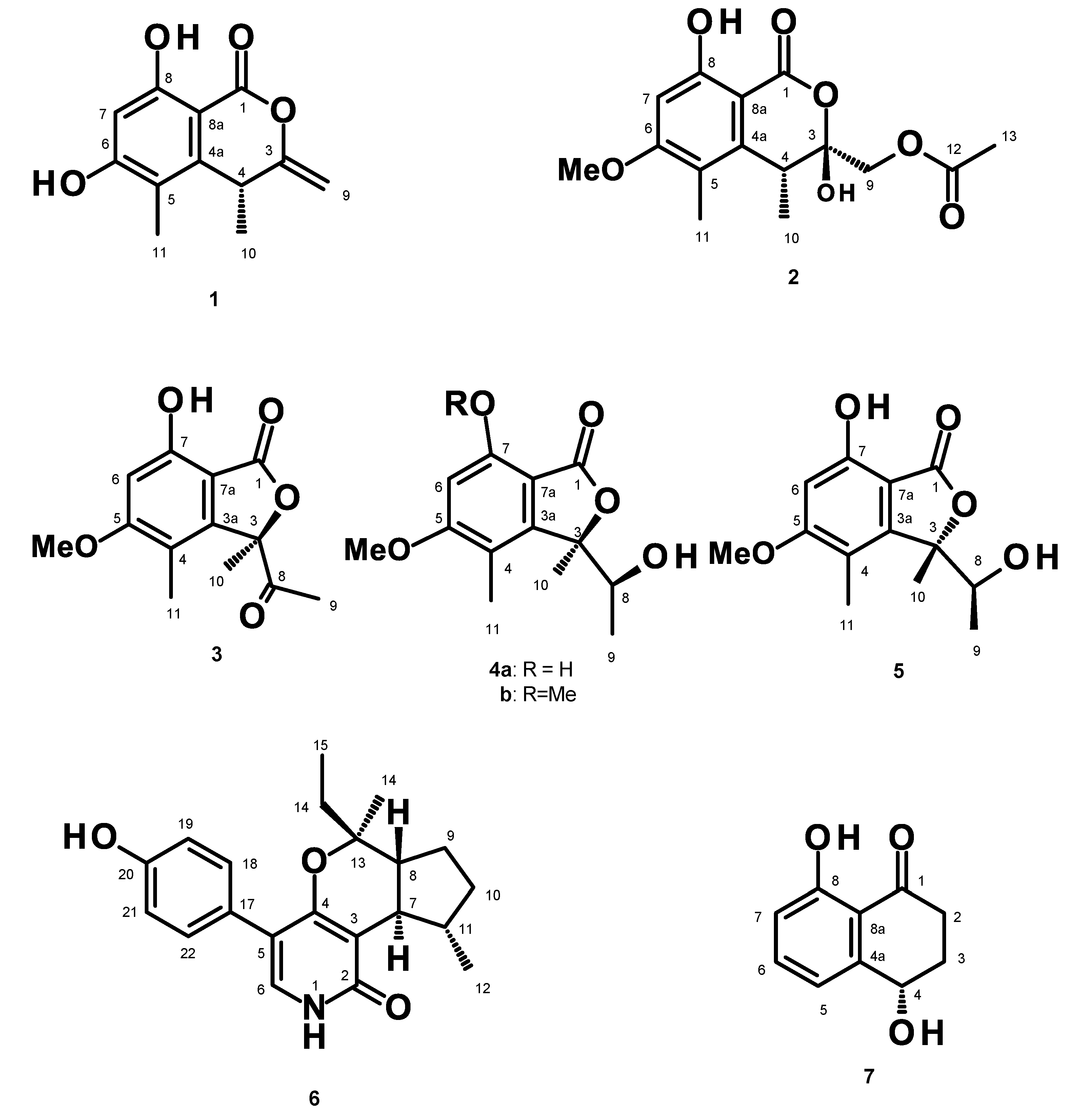
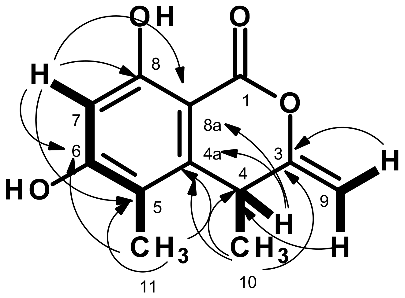
3.3.1. (R)-6,8-Dihydroxy-4,5-dimethyl-3-methylidene-3,4-dihydro-1H-2-benzopyran-1-one (1)

White crystal. mp 217–218 °C. [α ] D 20 −140 (c 0.05, MeOH); For 1H and 13C spectroscopic data (DMSO-d6, 300 and 75 MHz), see Table 1; (+)-HRESIMS m/z 221.0814 [M + H]+ (calcd for C12H13O4, 221.0814).

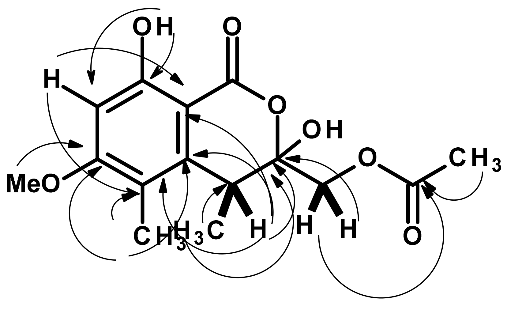
3.3.2. (3S,4R)-3,8-Dihydroxy-6-methoxy-4,5-dimethyl-1-oxo-3,4-dihydro-1H-2-benzopyran-3-yl)methyl acetate (2)

Pale yellow viscous mass. [α ] D 20 −104 (c 0.05, MeOH); For 1H and 13C spectroscopic data (CDCl3, 300 and 75 MHz), see Table 2; (+)-HRESIMS m/z 311.1133 [M + H]+ (calcd for C15H19O7, 311.1131) and 333.0948 [M + Na]+ (calculated for C15H18O7Na, 333.0950).

3.3.3. (R)-5, 7-Dimethoxy-3-((S)-(1-hydroxyethyl)-3,4-dimethylisobenzofuran 1(3H)-one (4b)

White crystals. mp 174–176 °C. [α ] D 20 −300 (c 0.05, MeOH); For 1H and 13C spectroscopic data (CDCl3, 300 and 75 MHz), see Table 3; (+)-HRESIMS m/z 267.1233 [M + H]+ (calcd for C14H19O5, 267.1232).

3.3.4. (S)-7-Hydroxy-3-((S)-1-hydroxyethyl)-5-methoxy-3,4-dimethylisobenzofuran 1(3H)-one (5)

White crystal. mp 116–118 °C. [α ] D 20 −320 (c 0.05, MeOH); For 1H and 13C spectroscopic data (CDCl3, 300 and 75 MHz), see Table 4; (+)-HRESIMS m/z 253.1077 [M + H]+ (calcd for C13H17O5, 253.1076).

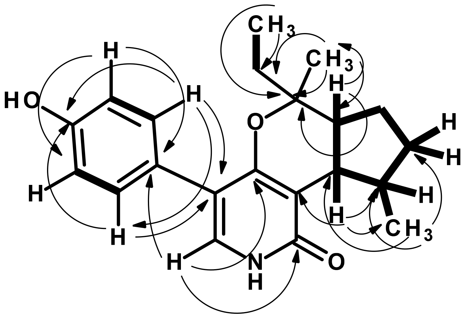
3.3.5. Avellaneanone (6)

White crystal. mp 219–210 °C. [α ] D 20 + 240 (c = 0.05, MeOH) (c 0.05, MeOH); For 1H and 13C spectroscopic data (DMSO-d6, 300 and 75 MHz), see Table 5; (+)-HRESIMS m/z 362.1734 [M + Na]+ (calcd for C21 H25 NO3Na, 362.1732).

3.4. X-ray Crystal Structures

Single crystals were mounted on a cryo-loop using paratone. X-ray CuKα radiation (λ = 1.54184 Å). The structures were resolved by direct methods using SHELXS-97 and refined with SHELXL-97 [30].
Full details of data collection and refinement and tables of atomic coordinates, bond lengths, angles, and torsion angles have been deposited with the Cambridge Crystallographic Data Centre.

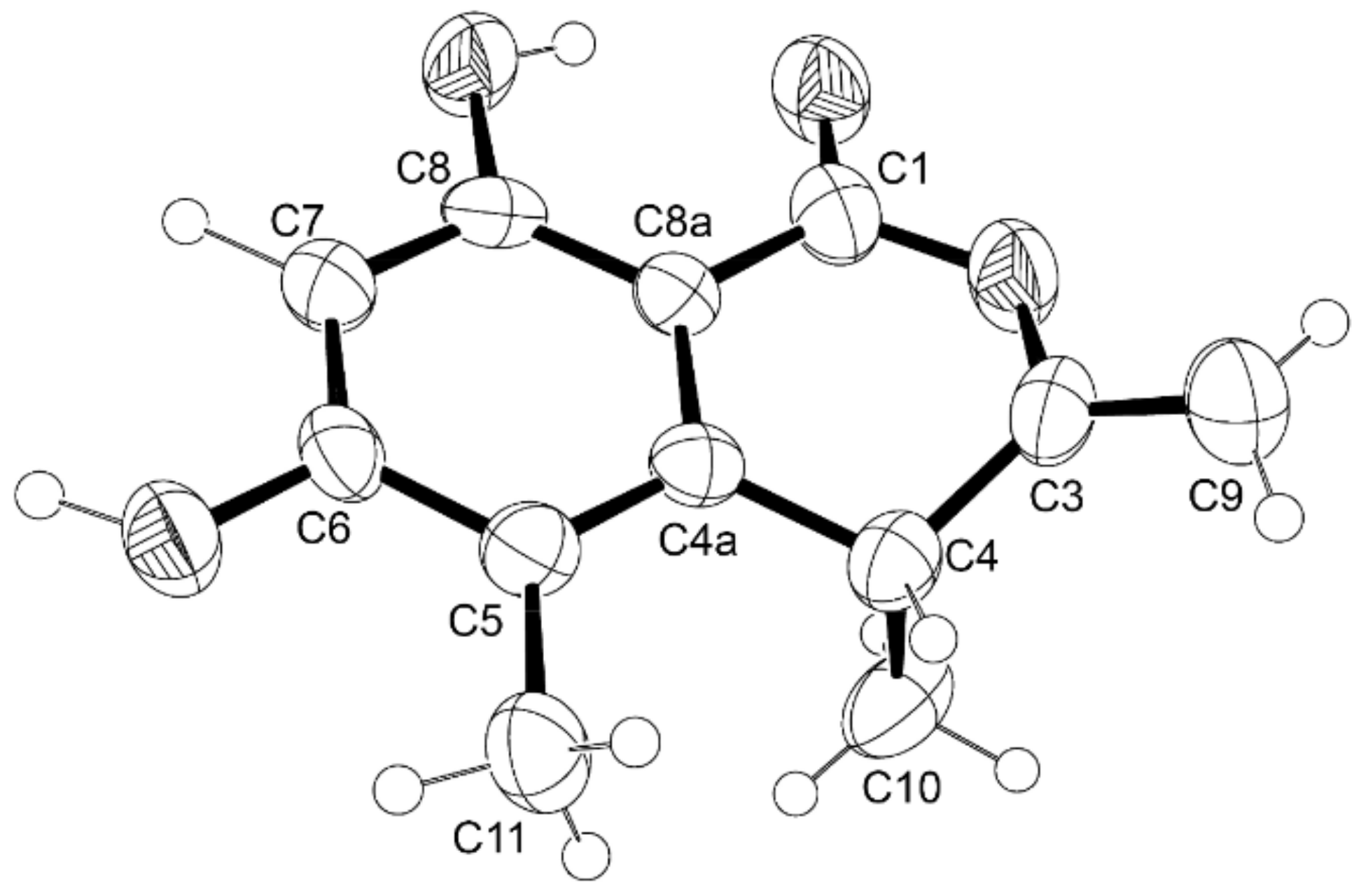
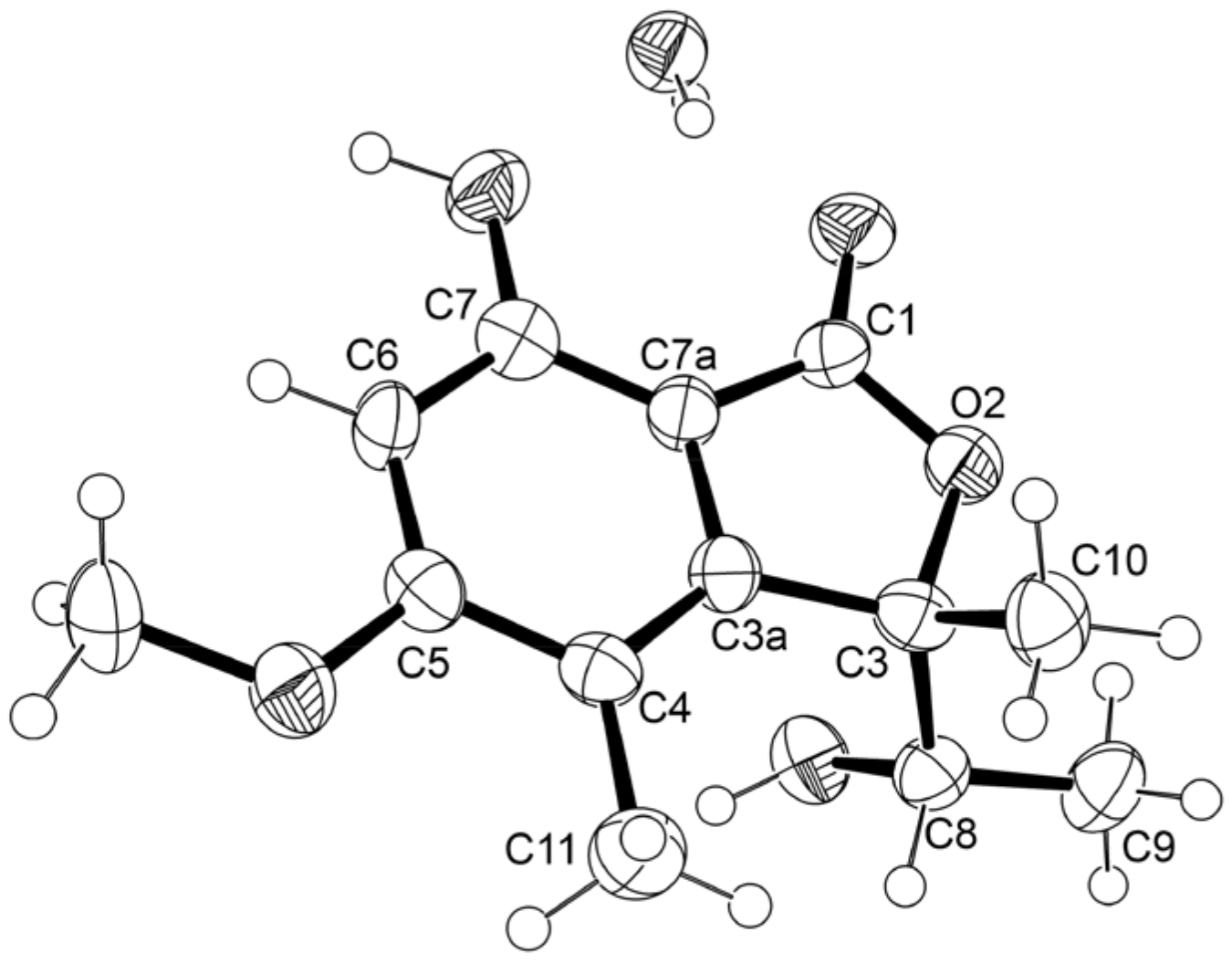
3.4.1. X-ray Crystal Structure of 1

The crystal was orthorhombic, space group P212121, cell volume 1050.3(2) Å3 and unit cell dimensions a = 5.2675(7) Å, b = 13.5239(16) Å, c = 14.7440(14) Å (uncertainties in parentheses). The calculated crystal density is 1.393 g.cm−3. Non-hydrogen atoms were refined anisotropically. Hydrogen atoms were directly found from different Fourier maps and refined freely with isotropic displacement parameters or placed at their idealized positions using appropriate HFIX instructions in SHELXL and included in subsequent refinement cycles. The refinement converged to R (all data) = 13.26% and wR2 (all data) = 15.30%. CCDC deposition number 2256003.

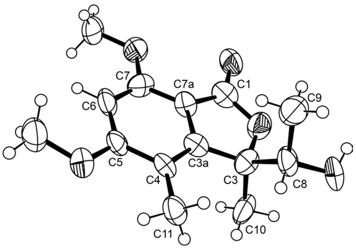
3.4.2. X-ray Crystal Structure of 4b

The crystal was triclinic, space group P1, cell volume 662.30(14) Å3 and unit cell dimensions a = 7.3973(9) Å, b = 8.4697(12) Å, c = 11.2567(13) Å and angles α = 107.628(12)°, β = 92.120(11)° and γ = 98.481(11))° (uncertainties in parentheses). The calculated crystal densities were 1.393 g.cm−3 and 1.335 g.cm−3. Two molecules were found in the asymmetric unit. Non-hydrogen atoms were refined anisotropically. Hydrogens were placed at their idealized positions using appropriate HFIX instructions in SHELXL and included in subsequent refinement cycles. The refinement converged to R (all data) = 19.00% and wR2 (all data) = 47.69%. CCDC deposition number 2260970.

3.4.3. X-ray Crystal Structure of 5

The crystal was triclinic, space group P-1, cell volume 655.52(16) Å3 and unit cell dimensions a = 7.8395(11) Å, b = 9.2206(12) Å, c = 9.8904(14) Å and angles α = 87.687(11)°, β = 85.728(11)° and γ = 66.857(13)° (uncertainties in parentheses). The calculated crystal density is 1.452 g.cm−3. One water molecule was found in the asymmetric unit. Non-hydrogen atoms were refined anisotropically. Hydrogens were directly found from different Fourier maps and refined freely with isotropic displacement parameters. The refinement converged to R (all data) = 10.99% and wR2 (all data) = 19.13%. CCDC deposition number 2256016.

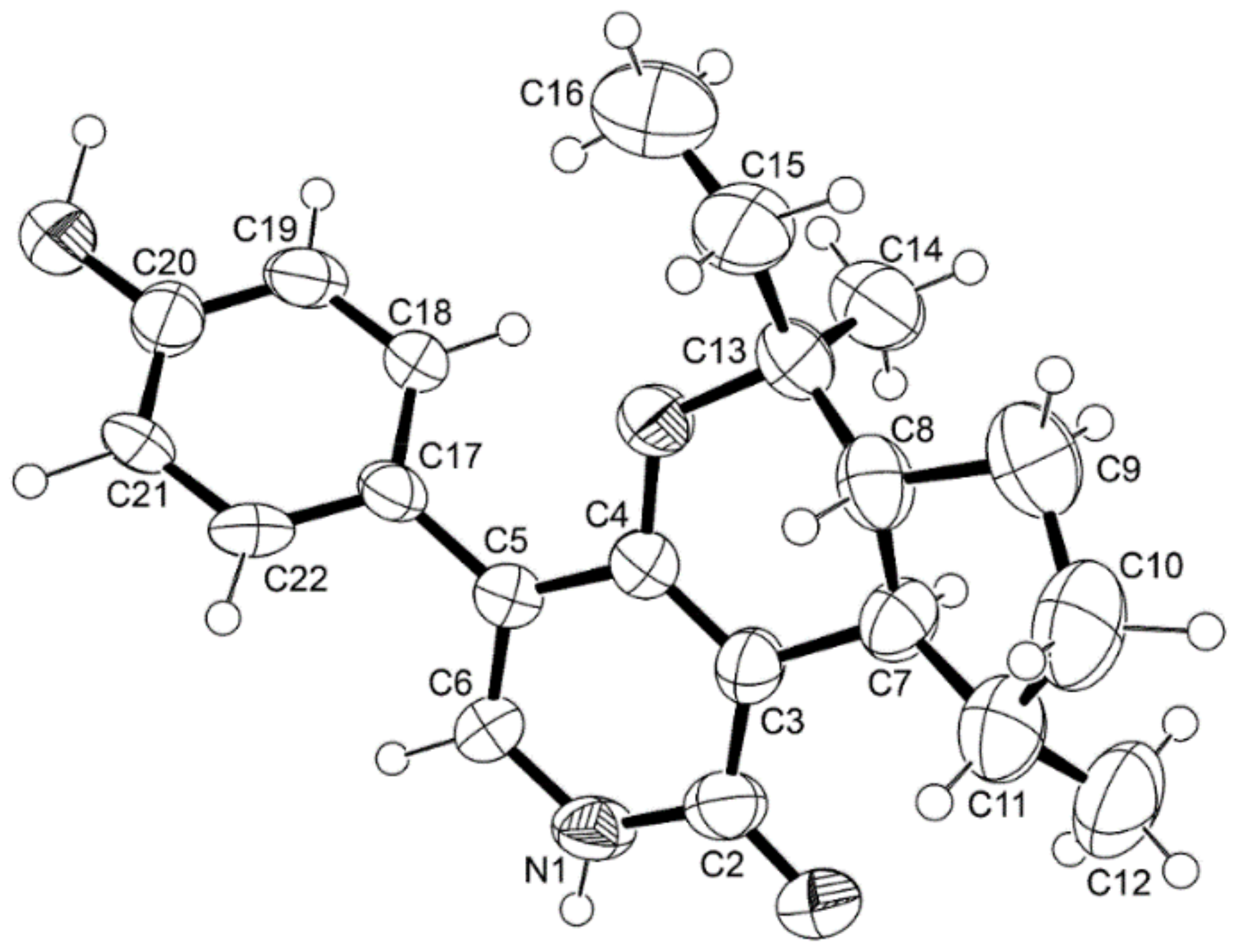
3.4.4. X-ray Crystal Structure of 6

The crystal was monoclinic, space group I2, cell volume 1880.5(3) Å3 and unit cell dimensions a = 8.9429(8) Å, b = 6.4805(6) Å, c = 32.453(3) Å and β = 91.065(8)° (uncertainties in parentheses). The calculated crystal density is 1.199 g.cm−3. Non-hydrogen atoms were refined anisotropically. Hydrogen atoms were directly found from different Fourier maps and refined freely with isotropic displacement parameters or placed at their idealized positions using appropriate HFIX instructions in SHELXL and included in subsequent refinement cycles. The refinement converged to R (all data) = 7.81% and wR2 (all data) = 12.53%. CCDC deposition number 2256402.

3.5. Antifungal Activity Bioassays

3.5.1. Antifungal Activity of the Crude Extract against Plant Pathogenic Fungi

The in vitro growth inhibitory activity of the crude EtOAc extract of H. avellanea KUFA0732 against eleven plant pathogenic fungi was evaluated using the poison food technique described previously by Dethoup et al. [9]. The crude EtOAc extract (1 g) was dissolved in 1 mL of DMSO and serially diluted with 9 mL of H2O to obtain stock solutions of 100 and 10 g/L. A volume of 1 mL of each stock solution was added to 9 mL of warm PDA, thoroughly mixed by a vortex mixer, and then poured into Petri dishes to give medium plates amended with crude extract with concentrations of 10g/L and 1 g/L, respectively. Each pathogen strain was cultured on a PDA for 7 days at 28 ± 2 °C, and a mycelial plug of 0.5 cm in diameter of each pathogen was placed on the center of the crude extract-containing PDA plates and incubated at room temperature for 14 days. A PDA plate void of the crude extract was used as a negative control. The inhibition levels were calculated using the formula: [(xy)/x] × 100, where x = colony radius of the plant pathogenic fungi in the negative control, and y = colony radius of the plant pathogenic fungi in the presence of the crude extract. Each treatment was performed with five replications and repeated three times independently. The mean inhibition levels and standard deviations were calculated from five replications and three repetitions. Data were subjected to analysis of variance, and subsequently, means were compared using Duncan’s multiple range test (p ˂ 0.05) in the SPSS version 19 statistical program (IBM Corporation, Somers, NY, USA).

3.5.2. Antifungal Activity of Isolated Compounds against Plant Pathogenic Fungi

The minimum inhibitory concentrations (MICs) for the antifungal activity of the isolated compounds on the plant pathogenic fungi were determined according to CLSI recommendation [31]. Each pathogen strain was cultured on PDA for 14 days at 28 ± 2 °C, and its spores were gently scraped by a sterile loop from the mycelium and then added to the PDA. A mass of 1 mg of each compound was dissolved in 100 µL of 10% DMSO, and then 900 µL of distilled H2O was added to prepare a stock solution of 1000 µg/L. Two-fold serial dilutions of the stock solution by potato dextrose broth mixed with a spore suspension of each pathogen at 106 spores/mL provided the tested concentrations at 500, 250, and 125 µg/L. Each treatment was loaded at 200 µL per well into the 96-well U-shaped untreated polystyrene plates with five wells per treatment, and then the plate was incubated for 7 days at 25 °C. The MIC was determined as the lowest concentration of the compound that inhibited visible growth. Mancozeb (OXA-Sigma-Aldrich, St. Louis, MI, USA), at the same concentration, was used as a positive control.

4. Conclusions

The crude EtOAc extract of a solid-rice culture of a marine sponge-associated fungus, Hamigera avellanea KUFA0732, isolated from the marine sponge Mycale sp., which was collected from the Gulf of Thailand, was found to have the potential to inhibit the growth of various plant pathogenic fungi. Chemical study of the crude extract of the culture of this marine-derived fungus led to the isolation of five undescribed specialized metabolites, viz. (R)-6,8-dihydroxy-4,5-dimethyl-3-methylidene-3,4-dihydro-1H-2-benzopyran-1-one (1), (3S, 4R)-3,8-dihydroxy-6-methoxy-4,5-dimethyl-1-oxo-3,4-dihydro-1H-isochromen-3-yl]methyl acetate (2), (R)-5, 7-dimethoxy-3-((S)-(1-hydroxyethyl)-3,4-dimethylisobenzofuran-1(3H)-one (4b), (S)-7-hydroxy-3-((S)-1-hydroxyethyl)-5-methoxy-3,4-dimethylisobenzofuran 1(3H)-one (5), and avellaneanone (6), as well as three previously reported (R)-3-acetyl-7-hydroxy-5-methoxy-3,4-dimethylisobenzofuran-1(3H)-one (3), (R)-7-hydroxy-3-((S)-1-hydroxyethyl)-5-methoxy-3,4-dimethylisobenzofuran-1(3H)-one (4a) and isosclerone (7). The common biosynthetic pathways of the pentaketide derivatives 1 and 2, as well as of 3, 4a, 4b, and 5, were determined. The biosynthesis of the PKS-NRPS hybrid product, avellaneanone (6), was also proposed based on the previously proposed pathways for other analogs. Finally, it is important to point out that although the crude EtOAc extract of H. avellanea KUFA0732 was able to completely inhibit the growth of many plant pathogenic fungi tested, only 3 and 5 displayed strong inhibitory activity (MIC = 125 µg/mL) comparable to the positive control, mancozeb, against Curvularia oryzae, while 6 exhibited only moderate activity (MIC = 250 µg/mL). These results give a new perspective on the potential application of marine-derived fungi and their specialized metabolites as biocontrol agents in agriculture.

Supplementary Materials

The following are available online at https://www.mdpi.com/article/10.3390/md21060344/s1, Figures S1–S5, S7–S12, S14–S28, S30–S34, S36–S40, S42–S46: 1D and 2D NMR spectra of compounds 17. Figures S6, S13, S29, S35, S41: HRMS data for compounds 1, 2, 4b, 5, 6. Table S1: 1H and 13C NMR data (CDCl3, 300 and 75 MHz), COSY and HMBC for 3. Table S2: 1H and 13C NMR (300 and 75 MHz, CDCl3), COSY, and HMBC of 4a. Table S3: Comparison of 1H and 13C NMR data of 4a (300 and 75 MHz, CDCl3) with those of (R)-7-hydroxy-3-((R)-1-hydroxyethyl)-5-methoxy-3,4-dimethylisobenzofuran-1(3H)-one (400 and 100 MHz, CDCl3) and (R)-7-hydroxy-3-((S)-1-hydroxyethyl)-5-methoxy-3,4-dimethylisobenzofuran-1(3H)-one (500 and 125 MHz, CDCl3). Table S4: 1H and 13C NMR data (DMSO-d6, 300 and 75 MHz), COSY, and HMBC for 7.

Author Contributions

A.K. and T.D. conceived and designed the experiment and elaborated the manuscript; R.K. performed the isolation and purification of the compounds; F.P.M. and D.K. assisted in the isolation and purification of some compounds; S.H.G. assisted in the structure elucidation of some compounds; T.D. collected, isolated, identified, cultured the fungus and performed bioassays of the extract and compounds; L.G. performed X-ray analysis; S.M. provided HRMS; A.M.S.S. provided NMR spectra; E.S. assisted in structure elucidation and preparation of the manuscript. All authors have read and agreed to the published version of the manuscript.

Funding

This work is partially supported by the national infrastructure PT-OPENSCREEN (NORTE-01-0145-FEDER-085468) and the national funds through the FCT—Foundation for Science and Technology with the scope of UIDB/04423/2020 and UIDP/04423/2020, and by the Office of the Ministry of Higher Education, Science, Research and Innovation; and the Thailand Science Research and Innovation.

Institutional Review Board Statement

Not applicable.

Data Availability Statement

The data presented in this study are available on request from the corresponding author.

Acknowledgments

R.K. thanks the Office of the Ministry of Higher Education, Science, Research and Innovation; and the Thailand Science Research and Innovation through the Kasetsart University Reinventing University Program 2021 for the scholarship.

Conflicts of Interest

The authors declare no conflict of interest.

References

  1. Peterson, S.W.; Jurjevic, Z.; Bills, G.F.; Stchigel, A.M.; Guarro, J.; Vega, F.E. Genus Hamigera, six new species and multilocus DNA sequence based phylogeny. Mycologia 2010, 102, 847–864. [Google Scholar] [CrossRef]
  2. Igarashia, Y.; Hanafusa, T.; Gohda, F.; Peterson, S.; Bills, G. Species-level assessment of secondary metabolite diversity among Hamigera species and a taxonomic note on the genus. Mycology 2014, 5, 102–109. [Google Scholar] [CrossRef]
  3. Yamazaki, M.; Horie, Y.; Bae, K.; Maebayashi, Y.; Jisai, Y.; Fujimoto, H. New fungal metabolites avellanins A and B from Hamigera avellanea, with pressure effect. Chem. Pharm. Bull. 1987, 35, 2122–2124. [Google Scholar] [CrossRef] [PubMed] [Green Version]
  4. Breinholt, J.; Kjaer, A.; Olsen, C.E.; Rassing, B.R. A bis-formamidodiphenylbutadiene from the fungus Hamigera avellanea. Acta Chem. Scand. 1996, 50, 643–645. [Google Scholar] [CrossRef]
  5. Breinholt, J.; Kjaer, A.; Olsen, C.E.; Rassing, B.R.; Rosendahl, C.N. Hamigerone and dihydrohamigerone: Two acetate-derived, antifungal metabolites from Hamigera avellanea. Acta Chem. Scand. 1997, 51, 1241–1244. [Google Scholar] [CrossRef] [Green Version]
  6. Isaka, M.; Chinthanom, P.; Veeranondha, S.; Supothina, S.; Luangsa-ard, J.J. Novel cyclopropyl diketones and 14-membered macrol ides from the soil fungus Hamigera avellanea BCC 17816. Tetrahedron 2008, 64, 11028–11033. [Google Scholar] [CrossRef]
  7. Isaka, M.; Chinthanom, P.; Kongthong, S.; Supothina, S.; Ittiworapong, S. Hamigeromycins C–G, 14-membered macrolides from the fungus Hamigera avellanea BCC 17816. Tetrahedron 2010, 66, 955–961. [Google Scholar] [CrossRef]
  8. Chalearmsrimuang, T.; Ismail, S.I.; Mazlan, N.; Suasaard, S.; Dethoup, T. Marine-derived fungi: A promising source of halotolerant biological control agents against plant pathogenic fungi. J. Pure Appl. Microbiol. 2019, 13, 209–223. [Google Scholar] [CrossRef] [Green Version]
  9. Dethoup, T.; Kaewsalong, N.; Songkumorn, P.; Jantasorn, A. Potential application of a marine-derived fungus, Talaromyces tratensis KUFA 0091 against rice diseases. Biol. Control 2018, 119, 1–6. [Google Scholar] [CrossRef]
  10. Klaram, R.; Jantasorn, A.; Dethoup, T. Efficacy of marine antagonist, Trichoderma spp. as halo-tolerant biofungicide in controlling rice diseases and yield improvement. Biol. Control 2022, 172, 104985. [Google Scholar] [CrossRef]
  11. Chinworrungsee, M.; Kittakoop, P.; Isaka, M.; Chanphen, R.; Tanticharoen, M.; Thebtaranonth, Y. Halorosellins A and B, unique isocoumarin glucosides from the marine fungus Halorosellinia oceanica. J. Chem. Soc. Perkin Trans. 1 2002, 2473–2476. [Google Scholar] [CrossRef]
  12. Bi, Y.M.; Bi, X.B.; Fang, A.; Zhao, Q.R. Metabolites from the fungus Cephalosporium sp. AL031. Arch. Pharm. Res. 2007, 30, 267–269. [Google Scholar] [CrossRef] [PubMed]
  13. Tayone, W.C.; Honma, M.; Kanamaru, S.; Noguchi, S.; Tanaka, K.; Nehira, T.; Hashimoto, M. Stereochemical investigations of isochromenones and isobenzofuranones isolated from Leptosphaeria sp. KTC 727. J. Nat. Prod. 2011, 74, 425–429. [Google Scholar] [CrossRef] [PubMed]
  14. Morita, T.; Aoki, R. Isosclerone, a new metabolite of Sclerotinia sclerotiorum (LIB.) DE BARY. Agric. Biol. Chem. 1974, 38, 1501–1505. [Google Scholar] [CrossRef]
  15. Evidente, A.; Sparapano, L.; Anna AndolfiI, A.; Bruno, G. Two naphthalenone pentaketides from liquid cultures of Phaeoacremonium aleophilum, a fungus associated with esca of grapevine. Phytopathol. Mediterr. 2000, 39, 162–168. [Google Scholar]
  16. Husain, S.M.; Müller, M. Fungal dihydroxynaphthalene-melanin: Diversity-Oriented biosynthesis through enzymatic and non-enzymatic transformations. Synlett 2017, 28, 2360–2372. [Google Scholar] [CrossRef]
  17. Tayone, W.C.; Kanamaru, S.; Honma, M.; Tanaka, K.; Nehira, T.; Hashimoto, M. Absolute stereochemistry of novel isochromanone derivatives from Leptosphaeria sp. KTC 727. Biosci. Biotechnol Biochem. 2011, 75, 2390–2393. [Google Scholar] [CrossRef] [Green Version]
  18. Song, Y.X.; Wang, J.; Li, S.W.; Cheng, B.; Li, L.; Chen, B.; Liu, L.; Lin, Y.C.; Gu, Y.C. Metabolites of the mangrove fungus Xylaria sp. BL321 from the South China Sea. Planta Med. 2012, 78, 172–176. [Google Scholar] [CrossRef] [Green Version]
  19. Wat, C.-K.; Mcinnes, A.G.; Smith, D.G.; Wright, J.L.C.; Vining, L.C. The yellow pigments of Beauveria species. Structures of tenellin and bassianin. Can. J. Chem. 1977, 55, 4090–4098. [Google Scholar] [CrossRef] [Green Version]
  20. Heneghan, M.N.; Yakasai, A.A.; Williams, K.; Kadir, K.A.; Wasil, Z.; Bakeer, W.; Fisch, K.M.; Bailey, A.M.; Simpson, T.J.; Cox, R.J.; et al. The programming role of trans-acting enoyl reductases during the biosynthesis of highly reduced fungal polyketides. Chem. Sci. 2011, 2, 972–979. [Google Scholar] [CrossRef]
  21. Bergmann, S.; Schümann, J.; Scherlach, K.; Lange, C.; Brakhage, A.A.; Hertweck, C. Genomics-driven discovery of PKS-NRPS hybrid metabolites from Aspergillus Nidulans. Nat. Chem. Biol. 2007, 3, 213–217. [Google Scholar] [CrossRef] [PubMed]
  22. Kim, J.-C.; Lee, Y.-W.; Tamura, H.; Yoshizawa, T. Sambutoxin: A new mycotoxin isolated from Fusarium sambucinum. Tetrahedron Lett. 1995, 36, 1047–1050. [Google Scholar] [CrossRef]
  23. Matsumoto, M.; Minato, H. Structure of ilicicolin H, an antifungal antibiotic. Tetrahedron Lett. 1976, 42, 3827–3830. [Google Scholar] [CrossRef]
  24. Wu, B.; Oesker, V.; Wiese, J.; Schmaljohann, R.; Imhoff, J.F. Two new antibiotic pyridones produced by a marine fungus, Trichoderma sp. Strain MF106. Mar. Drugs 2014, 12, 1208–1219. [Google Scholar] [CrossRef] [Green Version]
  25. Guo, Y.; Contesini, F.J.; Wang, X.; Ghidinelli, S.; Tornby, D.S.; Andersen, T.E.; Mortensen, U.F.; Larsen, T.O. Biosynthesis of calipyridone A represents a fungal 2-pyridone formation without ring expansion in Aspergillus californicus. Org. Lett. 2022, 24, 804–808. [Google Scholar] [CrossRef]
  26. Fisch, K.M.; Bakeer, W.; Yakasai, A.A.; Song, Z.; Pedrick, J.; Wasil, Z.; Bailey, A.M.; Lazarus, C.M.; Simpson, T.J.; Cox, R.J. Rational domain swaps decipher programming in fungal highly reducing polyketide synthases and resurrect an extinct metabolite. J. Am. Chem. Soc. 2011, 133, 16635–16641. [Google Scholar] [CrossRef]
  27. Murray, M.G.; Thompson, W.F. Rapid isolation of high molecular weight plant DNA. Nucleic Acids Res. 1980, 8, 4321–4325. [Google Scholar] [CrossRef] [Green Version]
  28. White, T.J.; Bruns, T.; Lee, S.; Taylor, J. Amplification and direct sequencing of fungal ribosomal RNA genes for phylogenetics. In PCR Protocols: A Guide to Methods and Applications; Innis, M.A., Gelfand, D.H., Sninsky, J.J., White, T.J., Eds.; Academic Press: New York, NY, USA, 1990; pp. 315–322. [Google Scholar]
  29. Sanger, F.; Nicklen, S.; Coulson, A.R. DNA sequencing with chain-terminating inhibitors. Proc. Natl. Acad. Sci. USA 1977, 72, 5463–5467. [Google Scholar] [CrossRef] [Green Version]
  30. Sheldrick, G.M. A short history of SHELX. Acta Cryst. 2008, A64, 112–122. [Google Scholar] [CrossRef] [Green Version]
  31. CLSI. Methods for Determining Bactericidal Activity of Antimicrobial Agents; Approved Guideline; CLSI Document M26-A; Clinical and Laboratory Standards Institute: Wayne, PA, USA, 1999. [Google Scholar]
Figure 1. Structures of 17.
Marinedrugs 21 00344 g001
Figure 2. Key COSY (bold line) and HMBC (arrow) correlations in 1.
Marinedrugs 21 00344 g002
Figure 3. Ortep view of 1.
Marinedrugs 21 00344 g003
Figure 4. Key COSY (bold line) and HMBC (arrow) correlations in 2.
Marinedrugs 21 00344 g004
Figure 5. Common biosynthetic pathways of 1 and 2.
Marinedrugs 21 00344 g005
Figure 6. Ortep diagram of 4b.
Marinedrugs 21 00344 g006
Figure 7. Ortep diagram of 5.
Marinedrugs 21 00344 g007
Figure 8. Proposed biosynthetic pathways for 3, 4a, 4b, and 5.
Marinedrugs 21 00344 g008
Figure 9. Key COSY (bold lines) and HMBC (arrow) correlations in 6.
Marinedrugs 21 00344 g009
Figure 10. Ortep view of 6.
Marinedrugs 21 00344 g010
Figure 11. Proposed biosynthetic pathways of 6.
Marinedrugs 21 00344 g011
Table 1. 1H and 13C NMR data (DMSO-d6, 300 and 75 MHz), COSY, and HMBC for 1.
PositionδC, TypeδH, (J in Hz)COSYHMBC
1166.3, C
3157.4, C
434.2, CH4.02 q (7.2)H3-10C-4a, 5, 8a, 11
4a143.6, C
5113.2, C
6164.1, C
7101.0, CH6.33,sH3-11C-5, 6, 8, 8a
8161.8, C
8a97.7, C
996.2, CH24.75, d (1.6)
4.73, d (1.6)
C-3, 4
1010.2, CH31.27, d (7.0)H-4C-3, 4, 4a
1122.3 CH32.02, sH-7C-4a, 5, 6
OH-8-10.70, brs
Table 2. 1H and 13C NMR data (DMSO-d6, 300 and 75 MHz), COSY, HMBC, and ROESY for 2.
PositionδC, TypeδH, (J in Hz)COSYHMBCROESY
1168.2, CO
3102.2, C
435.9, CH4.33, q (7.1)H3-10, C-3, 4a, 5, 8a, 10H3-10, H-11
4a141.5, C
5115.5, C
6164.9, C
797.5, CH6.37, s C-3, 5, 8, 8a, 12OMe-6
8163.1, C
8a99.0, C
9a
b
65.9, CH24.25, d (11.8)
4.59, d (11.8)
H-9b
H-9a
C-12
C-3, 12
H3-10
H3-10
1016.2, CH31.19, d (7.1)H-4C-3, 4, 4aH-9a, 9b
1110.1, CH32.08, s C-3, 4a, 6H-4
12170.7, CO
1320.8, CH32.19, s C-12
OMe-655.9, CH33.85, s C-6H-7
OH-8-11.14, s C-7, 8
Table 3. 1H and 13C NMR data (DMSO-d6, 300 and 75 MHz), COSY, and HMBC for 4b.
PositionδC, TypeδH, (J in Hz)COSYHMBC
1168.1, CO
388.7, C
3a152.8, C
4111.7, C
5164.5, C
694.5, CH6.04, sOMe-5, H3-11C-1, 4, 5, 7, 7a
7158.3, C
7a105.4, C
870.9, CH4.20, mH3-9
917.8, CH30.86, d (6.5)H-8C-3, 8
1021.5, CH31.74, s C-3, 3a, 8
1111.2, CH32.10, s C-3a, 4, 5
OMe-556.0, CH33.91, s C-5
OMe-756.1, CH33.96, s C-7
Table 4. 1H and 13C NMR data (DMSO-d6, 300 and 75 MHz), COSY and HMBC for 5.
PositionδC, TypeδH, (J in Hz)COSYHMBC
1171.1, CO
391.6, C
3a149.8, C
4112.4, C
5165.3, C
698.2, CH6.43, sH3-11, OMe-5C-1, 4,7, 7a
7156.3, C
7a103.7, C
870.5, CH4.26, mH3-9
917.9, CH31.40, d (6.4) C-3, 8
1021.1, CH31.67, s C-3, 3a, 8
1111.1, CH32.17,sH-6C-3a, 4, 5
OMe-556.3, CH33.87, sH-6C-5
OH-77.67, brs
Table 5. 1H and 13C NMR data (DMSO-d6, 300 and 75 MHz), COSY and HMBC for 6.
PositionδC, TypeδH, (J in Hz)COSYHMBC
NH-1-10.93, brs
2162.4 CO
3110.4 C
4160.2, C
5113.6, C
6131.5, CH6.98,s C-2, 4.5, 17
743.8, CH2.06, dd (12.0, 8.7)H-8, 11C-3, 11, 12
851.9, CH1.71, ddd (12.0, 12.0, 7.0)H-7, 9C-13, 14
925.2 CH21.56, mH-8, 10
1034.5, CH21.39, m
1.99, m
H-9, 11C-9, 11
1135.3, CH2.15, mH3-12
1225.1, CH31.47, d (6.2)H-11C-7, 10, 11
1382.2, C-
1418.6, CH31.11, s C-8, 13, 15
1533.8, CH21.55, q (7.1)H3-16
167.7, CH30.85, t (7.3)H-15C-13, 15
17126.7, C-
18130.6, CH7.15, d (8.6)H-19H-5, 20, 22
19115.1, CH6.73, d (8.6)H-18H-17, 20, 21
20156.6, C-
21115.1, CH6.73, d (8.6)H-22H-17, 19, 20
22130.6, CH7.15, d (8.6)H-21H-5, 18, 20
23OH-209.38, brs
Table 6. Effects of the crude EtOAc extract of H. avellanea KUFA0732 on the growth of plant pathogenic fungi.
Plant Pathogen% Mycelial Growth Inhibition at Concentrations
10 g/L1 g/L
Alternaria brassicicola (black spot of Chinese Kale)68.67 ± 2.27 c47.33 ± 1.62 d
Bipolaris oryzae (brown spot of rice)65.21 ± 3.12 d36.09 ± 1.14 f
Colletotrichum capsici (anthracnose of chili)100 ± 0 a58.67 ± 2.08 c
C. gloeosporiodes (anthracnose of mango)100 ± 0 a62.32 ± 3.58 b
Curvularia oryzae (leaf spot of rice)100 ± 0 a63.67 ± 2.15 b
Fusarium semitectum (dirty panicle of rice)85.34 ± 3.61 b38.04 ± 1.36 e
Lasiodiplodia theobromae (fruit rot of mangosteen)100 ± 0 a37.42 ± 1.08 e f
Phytophthora palmivora (root and stem rot of durian)100 ± 0 a0 ± 0 h
Pyricularia oryzae (rice blast)100 ± 0 a46.25 ± 2.01 d
Rhizoctonia oryzae (sheath blight of rice)100 ± 0 a68.34 ± 3.20 a
Sclerotium rolfsii (stem rot of bean)100 ± 0 a33.47 ± 1.06 g
Means ± standard derivations followed by the same letter in each column are not significantly different at p < 0.05 when analyzed using Duncan’s test of one-way ANOVA.
Table 7. Effects of 1, 3, 4b, 5, 6, and 7 on the growth of plant pathogenic fungi.
Plant PathogenPlant DiseaseMIC (µg/Ml)
134b567Mancozeb
Alternaria brassicicolablack spot of Chinese Kale>500>500>500>500>500>500125
Bipolaris oryzaebrown spot of rice>500>500>500>500>500>500125
Colletotrichum capsicianthracnose of chili>500>500>500>500>500>500125
C. gloeosporiodesanthracnose of mango>500500>500>500>500>500125
Curvularia oryzaeleaf spot of rice>500125>500125250>500125
Fusarium semitectumdirty panicle of rice>500>500>500>500>500>500125
Lasiodiplodia theobromaefruit rot of mangosteen>500>500>500>500>500>500250
Phytophthora palmivoraroot and stem of durian>500>500>500>500>500>500125
Pyricularia oryzaerice blast>500>500>500>500>500>500125
Rhizoctonia oryzaesheath blight of rice>500>500>500>500>500>500250
Sclerotium rolfsiistem rot of bean>500>500>500>500>500>500250
Disclaimer/Publisher’s Note: The statements, opinions and data contained in all publications are solely those of the individual author(s) and contributor(s) and not of MDPI and/or the editor(s). MDPI and/or the editor(s) disclaim responsibility for any injury to people or property resulting from any ideas, methods, instructions or products referred to in the content.

