Article

Biochemical Properties of a Cold-Active Chitinase from Marine Trichoderma gamsii R1 and Its Application to Preparation of Chitin Oligosaccharides

1
Shenzhen Raink Ecology & Environment Co., Ltd., Shenzhen 518102, China
2
School of Food Science and Engineering, South China University of Technology, Guangzhou 510640, China
*
Author to whom correspondence should be addressed.
Mar. Drugs 2023, 21(6), 332; https://doi.org/10.3390/md21060332
Submission received: 4 May 2023 / Revised: 23 May 2023 / Accepted: 25 May 2023 / Published: 29 May 2023

Abstract

:
The enzymatic degradation of different chitin polymers into chitin oligosaccharides (COSs) is of great significance given their better solubility and various biological applications. Chitinase plays a pivotal role in the enzymatic preparation of COSs. Herein, a cold-adapted and efficient chitinase (ChiTg) from the marine Trichoderma gamsii R1 was purified and characterized. The optimal temperature of ChiTg was 40 °C, and the relative activity at 5 °C was above 40.1%. Meanwhile, ChiTg was active and stable from pH 4.0 to 7.0. As an endo-type chitinase, ChiTg exhibited the highest activity with colloidal chitin, then with ball-milled and powdery chitin. In addition, ChiTg showed high efficiency when hydrolyzing colloidal chitin at different temperatures, and the end products were mainly composed of COSs with one to three degrees of polymerization. Furthermore, the results of bioinformatics analysis revealed that ChiTg belongs to the GH18 family, and its acidic surface and the flexible structure of its catalytic site may contribute to its high activity in cold conditions. The results of this study provide a cold-active and efficient chitinase and ideas for its application regarding the preparation of COSs from colloidal chitin.

1. Introduction

It is estimated that about 9.3 billion tons of crustaceans, such as crab, shrimp, and lobster, are produced annually, of which approximately 60% are inedible parts discarded as solid waste [1]. Previous studies have demonstrated that crustacean waste is mainly composed of chitin, protein, and minerals [2,3]. As an important constituent of crustacean waste, chitin is a linear polysaccharide that is composed of N-acetyl-D-glucosamine (GlcNAc) units linked by β-1,4 glycoside bonds. Meanwhile, chitin is also an important component of the cell walls of fungi and the exoskeletons of insects. Although chitin appears in abundance in the natural world, its poor solubility limits its further application [2,3]. Research studies have indicated that the derivatives of chitin, especially chitin oligosaccharides (COSs), exhibit better solubility and various kinds of biological activities such as antioxidant, immune-enhancing, and antimicrobial activities, which show great potential for application in different industries [4,5,6]. Chemical methods are usually chosen for the degradation of chitin or crustacean waste due to their simplicity and economic viability [7]. However, the disadvantages of these chemical methods, such as environmental pollution, harsh reaction conditions, and lack of control over the molecular structure of COSs, limit their further application [8]. Thus, it is essential to develop a specific, sustainable, and ecofriendly way to prepare COSs from chitin.
Recently, several studies have indicated that the enzymatic degradation of chitin displays the potential to replace the chemical methods owing to its mild reaction conditions, environmental friendliness, and providing the ability to control the process and final product [9,10]. As an important biocatalyst, chitinase can hydrolyze chitin by cleaving β-1,4 glycoside bonds and converting it into COSs with different degrees of polymerization. Previous studies have shown that chitinases have great potential value in various industries, with applications such as the preparation of COSs as biocontrol agents against pathogenic fungi and chitinous waste management [11,12]. Many chitinases have been isolated, purified, and characterized according to the origins from various organisms, including bacteria, fungi, and plants [11,12]. Generally, the chitinases from microorganisms are more suitable for industry applications than those from plants due to their excellent biochemical properties, extracellular secretion, and ease of cultivation [13]. In summary, the chitinases from microorganisms are mainly classified into two glycoside hydrolase (GH) families using the CAZy database: GH18 and GH19. Meanwhile, the chitinases from microorganisms can also be grouped as cold-adapted, mesophilic, and thermophilic based on their optimum reaction temperatures. Generally, cold-adapted enzymes offer some advantages for applications in different biotechnological processes. On one hand, cold-adapted enzymes exhibit high activity at low temperatures, so they can effectively reduce the energetic costs and risk of bacterial contamination. On the other hand, most cold-adapted enzymes are temperature-sensitive and easily inactivated by moderate heat. Compared with mesophilic or thermophilic chitinases, cold-active chitinases can remain active at low temperatures (0 to 10 °C) and exhibit several advantages such as the preparation of COSs at low temperatures, decreasing energy consumption, and biocontrol of microbial spoilage in refrigerated food, situations in which mesophilic or thermophilic chitinases often fail [14,15,16]. To date, several cold-active chitinases have been isolated and characterized, and most of them have usually been isolated from bacteria. It has been reported that chitinases EaChi39, Chi21702, AChi48, and ChiA from marine bacteria Exiguobacterium antarcticum DW2 [15], Sanguibacter antarcticus [17], Pseudoalteromonas sp. [18], and Annona cherimola [19] show high activity and efficiently catalyze at low temperatures. However, until now, few cold-adapted chitinases have been purified and characterized from fungi.
Trichoderma spp. are well-known fungi that have been used as effective biocontrol agents against plant pathogens in agriculture due to their ability to secrete different types of extracellular enzymes, especially chitinases [20]. To date, many Trichoderma species have been identified as efficient producers for chitinases [20,21]. According to the literature, Trichoderma chitinases exhibit high efficiency toward pretreated chitin, suggesting their suitability for applications in the preparation of COSs [20,22]. Nevertheless, there are few reports regarding the purification and characterization of cold-adapted chitinase from Trichoderma species.
In this work, a cold-adapted chitinase (ChiTg) from T. gamsii R1 was purified and characterized. Meanwhile, the gene encoding ChiTg was cloned and analyzed. To the best of our knowledge, this is the first cold-adapted chitinase to be characterized from T. gamsii, and it has great application value for the preparation of COSs.

2. Results and Discussion

2.1. Purification and Identification of Chitinase from T. gamsii R1

The fermentation conditions of T. gamsii R1 cultivated in shake flasks were optimized based on a single-factor experiment, and the results are shown in Supplementary Materials Figure S1. The culture supernatant with chitinase activity was centrifuged and further purified. As shown in Figure 1, a protein estimated at approximately 42 kDa was purified to apparent homogeneity after ultrafiltration and QSFF chromatography. In addition, the specific chitinase activity of a sample was 36.6 U/mg (Table 1). To date, several chitinases have been isolated from Trichoderma species such as Chit46 from T. harzianum GIM 3.442 [20], Ta-CHI42 from T. asperellum SH1 [23], chit42 from T. aureoviride M, etc. [24]. Briefly, the molecular masses of Trichoderma chitinases are in the range of 33 to 70 kDa, especially centered around 42 kDa [25,26]. Moreover, the Trichoderma chitinases with a molecular weight of 42 kDa have been extensively characterized and exhibit high-efficiency against pretreated chitin.
The obtained purified protein was further investigated via mass spectrometry. Several sequences of peptides were obtained, which included P1 (NFQPQNLVASDITHVIYS FMNFQADGTVVSGDAYADYQK), P2 (NLGLGGSMFWEASADKTGSDSLIGTSH R), P3 (VMLSIGGWTWSTNFPSAASTDANR), P4 (AGATVQYDSVAQAYYS YDPSSK), P5 (HYDDDSWNDVGNNAYGCVK), P6 (ASGYANAVYFTNWGIYGR), P7 (NLGLGG S MFWEASADK), and P8 (IVLGMPIYGR). The results of NCBIblastp revealed that these eight peptides exhibited high identity with the putative chitinase from T. gamsii. Therefore, the obtained purified protein was a chitinase and named ChiTg.

2.2. Substrate Specificity and Kinetic Parameters of ChiTg

The substrate preferences of ChiTg were determined with various substrates. As shown in Table 2, the preferred substrate for ChiTg was colloidal chitin, followed by ball-milled and powdery chitin. Due to pretreatment with hydrochloric acid, colloidal chitin is less crystalline and more easily accessible to chitinase than ball-milled and powdery chitin. It is particularly noteworthy that the preferred substrate for most of the characterized chitinases is colloidal chitin [9,20,27,28,29]. Meanwhile, ChiTg showed little activity against colloidal chitosan, with 85 to 95% DDA. In addition, ChiTg exhibited no activity with xylan or microcrystalline cellulose.
Based on the substrate specificity, colloidal chitin was chosen as the substrate for the analysis of the kinetic parameters of ChiTg. The values of Km and Vmax were 0.47 mg/mL and 41.2 μmoL/min/mg, respectively. The values of Km from different Trichoderma chitinases widely vary. For instance, the Km of chitinases Chit46, ECH42, Tv-ECH1, and Chit42 from T. harzianum GIM 3.442 [20], T. atroviride [30], and T. harzianum CECT2413 [22] are 0.65, 1.9, and 1.7 mg/mL, respectively.

2.3. Effects of Temperature and pH on Activity and Stability of ChiTg

The purified ChiTg showed the maximum activity at 40 °C, which remained above 40.1% relative activity in the range of 5 to 45 °C (Figure 2A). Generally, the optimum temperatures of chitinases from different fungi are in the range of 30 to 70 °C. For instance, the chitinases from T. harzianum CECT2413, M. thermophila C1, and H. grisea ITCC 10,360.16 show maximum activity at 35, 55, and 70 °C, respectively [21,31,32]. As depicted in Figure 2A, the purified ChiTg was still active at 5 °C, and the relative activity was 40.2%, indicating ChiTg is a cold-active chitinase. It is particularly noteworthy that few chitinases from fungi have been reported to still exhibit high activity as the reaction temperature drops below 10 °C (Table 3). To date, cold-active chitinases have usually been isolated from marine bacteria, and few cold-active chitinases have been isolated from fungi [11]. Thus, it was meaningful to isolate and characterize ChiTg. Compared with its mesophilic and thermophilic counterparts, ChiTg is effective at quite low temperatures, so can reduce the energy cost for the preparation of COSs.
As shown in Figure 2B, the residual activities of ChiTg incubation for 30 min at different temperatures (35 to 65 °C) were 95.3, 87.6, 83.2, 31.2, 8.1, 3.2, and 1.1%. In addition, the stability of ChiTg at 40 and 45 °C under different incubation times was further determined. The purified ChiTg was quite stable at 40 °C, and the residual activity was above 53.5%, even when the incubation time was 3 h (Figure 2C). Generally, cold-adapted chitinases display poor thermal stability due to their inherent structural flexibility, so they easily denature and lose activity as the temperature increases above 40 °C [38]. For instance, the chitinase ChiC from Pseudoalteromonas sp. DL-6 is only stable in the range of 10 to 30 °C, and the residual activity of chitinase ChiC rapidly decreases as the temperature increases above 40 °C [39]. Another chitinase ChiA, also isolated from Pseudoalteromonas sp. DL-6, retains above 50% of its maximum activity as the temperature falls below 40 °C [18]. Different from the above-mentioned chitinases, a cold-adapted chitinase EaChi39 from Exiguobacterium antarcticum DW2 exhibited excellent thermal stability even when the reaction temperature reached 60 °C [15]. The thermal stability results revealed that the reaction temperature for further application of ChiTg should be below 45 °C.
The effects of the pH on the activity and stability of ChiTg were also determined. ChiTg showed maximum activity at pH 5.0 and maintained a high relative activity in the range of pH 4.0 to 8.0 (Figure 2D). In addition, ChiTg was very stable when the reaction pH was in the range of 5.0 to 8.0, and the residual activity remained above 80%. When the reaction pH changed to 3.0 or above 9.0, the residual activity of ChiTg rapidly decreased (Figure 2E). The pH profile shows that ChiTg is suitable for the preparation of COSs from colloidal chitin because the process of the transformation of chitin to colloidal chitin occurs under acidic conditions.

2.4. Effects of Different Metal Ions on Stability of ChiTg

The relative activities of ChiTg treated with various metal ions were determined, and the results are shown in Supplementary Materials Table S1. ChiTg was slightly activated by Ca2+ and Zn2+, and the relative activities of ChiTg treated with those two metal ions were 104.3 and 106.5%, respectively. Meanwhile, the metal ions Mg2+, K+, Na+ and Cu2+ exhibited minimal inhibition on the activity of ChiTg, and the relative activities of ChiTg treated with those metal ions remained above 80.1%. Different from the above-mentioned metal ions, the activity of ChiTg was inhibited by Al3+ and Mn2+, which showed relative activities of 46.3 and 58.4%, respectively. Generally, the mineral salts of crustacean shells are mainly composed of Ca2+, Zn2+, and Mg2+. In this study, ChiTg was active and stable in the presence of Mg2+, Ca2+, and Zn2+, which would be helpful for further application for the recovery COSs from crustacean shells.

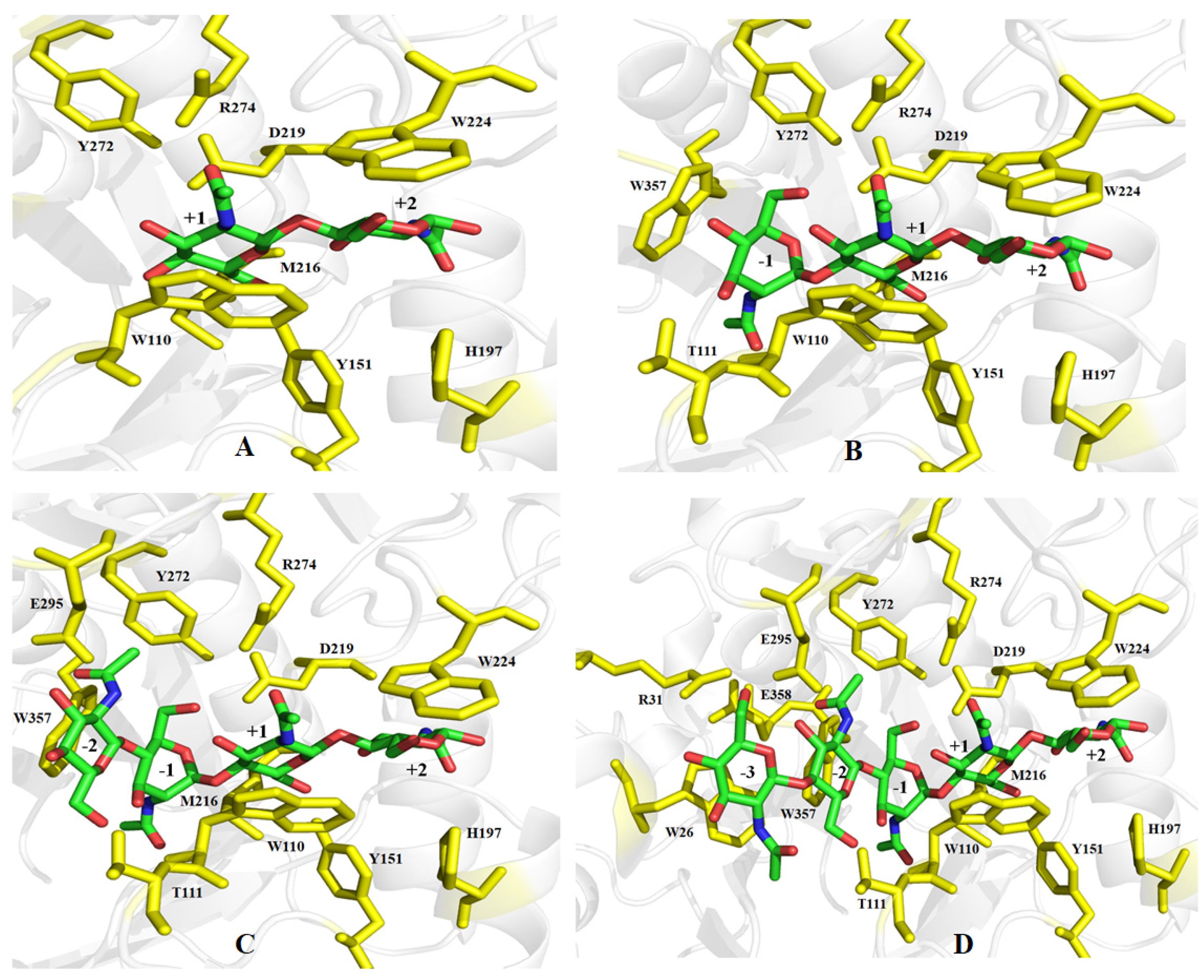
2.5. Hydrolytic Pattern of ChiTg

The hydrolysis property of ChiTg was determined by using different COSs (DP 2 to 5) as substrates. ChiTg exhibited no activity toward (GlcNAc)2, and (GlcNAc)2 was not transformed into smaller products by ChiTg even after 120 min of reaction (Figure 3A). Conversely, ChiTg hydrolyzed (GlcNAc)3 at a very fast rate, and most of the (GlcNAc)3 was cleaved and transformed to (GlcNAc)2 and GlcNAc as after 20 min of reaction (Figure 3B). In addition, ChiTg displayed high efficiency in hydrolyzing (GlcNAc)4 and (GlcNAc)5, which were almost fully decomposed and converted to smaller products when the reaction time reached 20 min. The reaction of ChiTg with (GlcNAc)4 generated (GlcNAc)2 as the end product (Figure 3C). (GlcNAc)5 was hydrolyzed and converted to (GlcNAc)2 and (GlcNAc)3 at a reaction time of 20 min (Figure 3D). When the reaction time reached 40 min, (GlcNAc)3 was decomposed and converted into (GlcNAc)2 and GlcNAc (Figure 3D). The end products of (GlcNAc)5 included (GlcNAc)2 and GlcNAc.
The hydrolysis ability of ChiTg indicated that it had no hydrolytic activity against COSs with a DP below three, which is similar to that of previously reported chitinases from the GH18 or GH19 family, such as PxCHi52 from Paenibacillus xylanexedens Z2-4 [29], SaChiB from Streptomyces alfalfa [40], and CsCHiE from Chitiniphilus shinanonensis [41]. In addition, the different COSs hydrolyzed by ChiTg revealed that ChiTg is an endo-type chitinase. Furthermore, the hydrolytic pattern of ChiTg revealed its potential for application in the preparation of those COSs.

2.6. Preparation of COSs from Colloidal Chitin

Recently, COSs have attracted considerable attention due to their versatile functional properties, including their antioxidant, immunomodulatory, antimicrobial, and antitumor abilities [42,43]. Regarding substrate specificity, ChiTg displayed highest activity toward colloidal chitin. Therefore, colloidal chitin was used as the substrate for the preparation of COSs. One-single factor experiment was performed, and the results are depicted in Figure 4. The conversion rate gradually increased with the increasing addition of ChiTg, reaching the highest value as the ratio of enzyme to substrate was 150 U/g (Figure 4A). Additionally, the concentration of substrate was optimized, and the conversion rates of groups 1 to 4% (w/v) were 80.1, 83.2, 76.5, and 68.5%, respectively (Figure 4B). Furthermore, the effects of reaction pH, time, and temperature on the conversion rate were investigated. As shown in Figure 4C, ChiTg exhibited the best hydrolysis efficiency on colloidal chitin when the reaction pH was 5.0, followed by when the pH was 6.0 and 4.0, which is in accordance with the pH profile of ChiTg. As can be seen from Figure 4D, the conversion rate quickly increases from 0.5 to 2 h of reaction time. However, the conversion rate slowly increased when the reaction duration was above 2 h. As depicted in Figure 4E, the optimal temperature for the ChiTg degradation of colloidal chitin to COSs was 35 °C, followed by 40 and 30 °C.
Based on the results of the above one-single factor experiment, a large-scale reaction was performed under the optimum reaction conditions. Most of the insoluble colloidal chitin was converted to soluble COSs after 2 h of reaction. The hydrolysates mainly included GlcNAc, (GlcNAc)2, and (GlcNAc)3; and the concentrations of those three COSs were 1.73, 11.62, and 1.92 mg/mL, respectively (Figure 4F). The HPLC charts of the hydrolysates are shown in Supplementary Materials Figure S2. In addition, the conversion rate was 84.2% after 2 h of reaction. Recently, several chitinases from different GH families have been used to prepare COSs from different chitin polymers. For instance, a chitinase, rChit46, from T. harzianum GIM 3.442 hydrolyzed 80.5% of 2% (w/v) colloidal chitin into GlcNAc, (GlcNAc)2, (GlcNAc)3, and (GlcNAc)4 after 3 h reaction at 45 °C in the presence of 200 U/mL of rChit46 [20]. In addition, Song et al. (2020) reported a chitinase, ChiA, from Bacillus licheniformis WX-02 can hydrolyze 10% (w/v) colloidal chitin into COSs at 50 °C and the conversion rate is 89% after 12 h reaction [44]. Zhang et al. (2021) found that a chitinase ChiA from Paenibacillus xylanexedens Z2-4 could convert about 60% of 1% (w/v) colloidal chitin into GlcNAc and (GlcNAc)2 after 24 h reaction at 45 °C [29]. In this study, ChiTg efficiently prepared COSs from colloidal chitin at lower temperatures (20 to 35 °C) with a higher conversion rate, indicating that ChiTg has great potential and is competitive for the preparation of COSs.

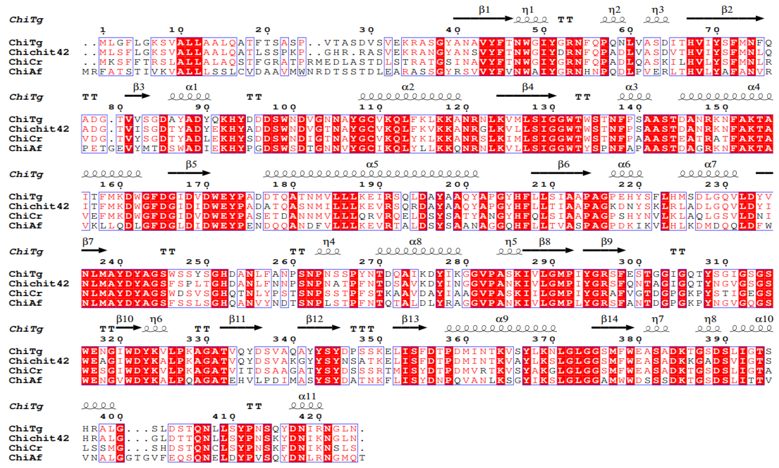
2.7. Gene Cloning and Bioinformatics Analysis of ChiTg

In order to better understand ChiTg, the gene encoding ChiTg was obtained via RT-PCR, and the bioinformatics analysis of ChiTg was performed. The results of the sequencing and prediction of the open reading frame indicated that chitg is 1275 bp in length, encoding 424 amino acids. Based on the results of NCBIblastn, we deduced that ChiTg belongs to the GH18 family due to its high sequence identity with other GH18 family chitinases from T. harzianum (accession No. AAA98644.1) and T. atroviride (accession No. BAB40593.1). In addition, the results of ProtParam analysis showed that 36 and 19 residues of ChiTg were negatively (Asp and Glu) and positively charged (Arg and Lys), respectively. Moreover, the first 27 amino acid residues of ChiTg were signal peptides. The results of the multiple alignment of ChiTg against already crystallized chitinase indicated that ChiTg also contains the classic motif (DXDXE) of the GH18 family. Meanwhile, three amino acids (D146, D148, and E150) were considered as the catalytic residues (Figure 5).
The results of NCBIblastn revealed that the sequence identity of ChiTg with chitinase Chit42 from T. harzianum CECT2413 is 82.5%. Then, the three-dimensional structure of ChiTg was obtained by homology modeling using already crystallized chitinase Chit42 from T. harzianum CECT2413 as a template (PDB deposition: 6ylj.1). As shown in Figure 6, the overall structure of ChiTg is characterized as an (α/β) TIM-barrel topology, which is mainly composed of eight β strands (β1 to β8) and eight α helices (α1 to α8). Different from the already reported chitinases, ChiTg contains an additional chitinase insertion domain that plays crucial roles in substrate binding and catalysis. Meanwhile, the docking results revealed that hydrogen bonds and hydrophobic interaction were the main forces through which ChiTg recognizes, binds, and catalyzes different substrates. In addition, several amino acids (W26, R52, G30, R31, D63, W110, T111, D146, D148, E150, Y151, H197, Y218, D219, Y220, W224, Y246, Y272, R274, E295, and I298) may be related to the catalytic hydrolysis of the substrate (Figure 7A–D). Furthermore, the docking results displayed that (GlcNAc)3 is the smallest substrate for ChiTg, and (GlcNAc)2 is the main end product, which is in accordance with the results of the hydrolytic pattern of ChiTg (Figure 3).
It is particularly noteworthy that the ability of cold-adapted enzymes to remain active at cold conditions is related to their structure [11,14]. Generally, the molecular mechanisms of cold-adapted enzymes are achieved through their highly acidic surface, low levels of rigidifying residues, small number of hydrogen bonds, low salt bridge content, having fewer hydrophobic interactions, and the flexible structure of catalytic site [16,38,45,46,47,48,49]. The cold adaptation ability of ChiTg may relate to its acidic surface and the flexible structure of its catalytic site (Figure 8A,B). A total of 36 residues of ChiTg are negatively charged (Asp and Glu), most of which are located at the surface (Figure 8A). A previous study indicated that acidic surfaces are helpful for retaining the solvation of cold-adapted enzymes, as water tends to be more viscous at lower temperature [45]. Additionally, the B factors showed that the additional chitinase insertion domain of ChiTg is highly flexible (Figure 8B), which is beneficial for substrate binding at low temperatures. Generally, the catalytic region of cold-adaptive enzymes is more flexible than their mesophilic and thermophilic counterparts. Research has shown that the higher flexibility of the catalytic site of cold-adapted enzymes is helpful for the formation of enzyme–substrate complexes at lower energy at lower temperatures [45,50]. Moreover, the tertiary structure alignment between ChiTg and a thermostable chitinase, Chit1 [37], from Thermomyces lanuginosus revealed that amino acid substitution may also be related to cold tolerance (Figure 8C). Compared with ChiTg, the surface of thermostable chitinase Chit1 has more proline, which is helpful for increasing thermal stability (Figure 8C).

3. Materials and Methods

3.1. Strains and Materials

T. gamsii R1 was isolated from shrimp shell waste and identified via ITS sequencing and then conserved in our laboratory. E. coli Dh5α competent cells were purchased from Tiangen Biotechnology (Beijing, China). PrimeSTAR® HS (Premix) DNA polymerase and PrimeScript™ RT-PCR Kits were purchased from Takara Biotechnology (Beijing, China). COSs with different degrees of polymerization (DPs), including N-acetyl-D-glucosamine (DP1), N-acetyl-chitobiose (DP2), N-acetyl-chitotriose (DP3), N-acetyl-chitotetraose (DP4), and N-acetyl-chitopentaose (DP5), were purchased from Qingdao BZ Oligo Biotech (Qingdao, China). Furthermore, GlcNAc, (GlcNAc)2, (GlcNAc)3, (GlcNAc)4, and (GlcNAc)5 represent N-acetyl-D-glucosamine, N-acetyl-chitobiose, N-acetyl-chitotriose, N-acetyl-chitotetraose, and N-acetyl-chitopentaose, respectively. Powdery chitin, xylan, microcrystalline cellulose, and chitosan with a degree of deacetylation (DDA) of 85 to 95% were purchased from Yuanye Biotechnology (Shanghai, China). The colloidal and ball milled-chitin were prepared according to a previously described method [51].

3.2. Production of Chitinase from T. gamsii R1 in Shake Flasks

A single-factor design was used to optimize the production of chitinase from T. gamsii R1, which included different nitrogen sources (yeast extract, peptone, or ammonium sulfate), concentrations of colloidal chitin (0.5 to 2%, w/v), fermentation temperatures (25 to 35 °C), and times (3 to 5 d). After single-factor experiment optimization, T. gamsii R1 was inoculated into 100 mL of fermentation medium (1.5% peptone, 1.0% colloidal chitin, 0.1% MgSO4•7H2O, 0.05%, FeSO4•7H2O, and 0.3% K2HPO4, w/v) in 500 mL flasks and incubated at 200 rpm and 30 °C for 4 d. The fermentation culture was centrifuged at 10,000× g and 6 °C for 10 min, and the obtained supernatant was used for a chitinase activity assay and purification.

3.3. Enzyme Activity Assay

The chitinase activity was determined according to a method described in a previous study [9]. We used 1% (w/v) colloidal chitin (pH 5.0) as the substrate. In the first step, 200 µL of diluted enzyme was added to a 2 mL test tube containing 200 µL of colloidal chitin and incubated at 40 °C for 30 min. In the second step, the reaction was stopped by the addition of 600 µL of 3,5-dinitrosalicylic acid (DNS), which was then incubated at 100 °C for 10 min. Finally, the cool-down solution was centrifuged at 11,000× g for 5 min, and the supernatant was monitored at 540 nm with a spectrophotometer. The reactions with heat-denatured enzyme were used as the control. One unit of enzyme activity was defined as the amount of enzyme that releases 1 µmol of reducing sugars per minute.

3.4. Enzyme Purification and Peptide Sequence Analysis

The process for enzyme purification was similar to that described in a previous study [15]. To begin, the obtained supernatant was concentrated via ultrafiltration with a membrane with a 10 kDa cut-off (Merck Millipore, Billerica, MA, USA). Then, the obtained protein samples were purified via Q Sepharase Fast Flow chromatography (GE life science, Fairfield, CT, USA). Finally, the homogenous fractions were pooled and concentrated via ultrafiltration with a membrane with a 10 kDa cut-off. The purified protein samples were analyzed with a chitinase activity assay and SDS-PAGE. The purified protein sample with chitinase activity was named ChiTg.
The partial amino acid sequence of ChiTg was analyzed via LC-MS/MS with a Q Exactive Orbitrap HF mass spectrometer with a nanoelectrospray ionization source (ThermoFisher Scientific, Waltham, MA, USA) and an LC-20AD nano high-performance liquid chromatography (HPLC) system equipped with a MonoCap C18 trap column (0.2 × 50 mm, 5 μm; Shimadzu, Kyoto, Japan). The detailed protocol is provided in the Supplementary Materials.

3.5. Kinetic Parameters of Purified ChiTg

The method for the determination of the kinetic parameters was similar to a previously reported method [52]. Different concentrations of colloidal chitin (1 to 10 mg/mL) were used as the substrate. The values of Km and Vmax were calculated with GraphPad Prism 8.0.2.

3.6. Effects of pH and Temperature on Activity and Stability of ChiTg

The method we used for the determination of the effects of pH and temperature on the activity and stability of the purified ChiTg was same as that reported in a previous study [53]. For the optimum pH, the activity of purified ChiTg was detected from pH 4.0 to 7.0, and the pH with the highest activity was set to 100%. For pH stability, the purified ChiTg was diluted in different buffers with pH ranging from 4.0 to 10.0, and the sample without any treatment was set as the control.
The optimum temperature of the purified ChiTg was determined within 30 to 70 °C, and the temperature with the highest activity was set to 100%. Regarding thermal stability, the purified ChiTg was incubated at temperatures ranging from 40 to 60 °C for 30 min, and the sample without any treatment was set as the control. In addition, the thermal stability of purified ChiTg at 40 and 45 °C treated for different durations was also determined.

3.7. Effects of Different Metal Ions on Stability of ChiTg

The effects of different metal ions (Mg2+, K+, Co2+, Cu2+, Zn2+, Ca2+, Na+, Mn2+, and Al3+) on the on the stability of purified ChiTg were determined by incubating enzyme samples for 4 h at 25 °C. The enzyme activity of purified ChiTg without metal ions was considered as 100%.

3.8. Hydrolysis Properties of ChiTg

The hydrolysis ability of recombinant ChiTg was determined based on the methods in a previous study using different COSs as the substrate [29]. The reaction was carried out as follows: to begin with, the purified recombinant ChiTg was mixed with different COSs (1%, w/v). Then, the mixture was incubated at 40 °C for different durations. Finally, the samples withdrawn at different times were incubated at 80 °C for 5 min and used for thin-layer chromatography (TLC) analysis. The process for TLC analysis was same as that previously reported [40].

3.9. Substrate Specificity of ChiTg

The method followed for the determination of substrate specificity of ChiTg was the same as that previously reported [41]. Colloidal chitosan with different DDAs (85 and 95%); colloidal, ball- milled, and powdery chitin; pretreated shrimp shell powder; xylan; and microcrystalline cellulose were used as the substrates for the activity assay. The substrate with highest activity was set to 100%.

3.10. Preparation of COSs from Colloidal Chitin

A single-factor design was used to optimize the preparation of COSs from colloidal chitin. Different ratios of enzyme to substrate (50, 100, 150, and 200 U/g), concentrations of substrate (1, 2, 3, and 4%, w/v), reaction pH values (4, 5, 6, and 7), reaction times (0.5, 1, 1.5, 2, 2.5, and 3 h), and reaction temperatures (20, 25, 30, 35, 40, and 45 °C) were selected for optimization. Reactions were carried out in a 50 mL flask containing 10 mL of colloidal chitin, which were incubated in a shaker at 150 rpm. The reaction was ended by incubating at 70 °C for 5 min and then centrifuging at 10,000× g and 10 °C for 10 min. The residual colloidal chitin was dried out at 90 °C. The conversion rate was calculated according to the method described in a previous study [20].
A large-scale reaction (1 L flask containing 200 mL of colloidal chitin) for the preparation of COSs was performed under the optimum reaction conditions. The ratio of enzyme to substrate; concentration of substrate; reaction pH, time, and temperature of the large-scale reaction were 150 U/g, 2%, 5.0, 2 h, and 35 °C, respectively. After 2 h of reaction, the end products were identified via high-performance liquid chromatography (HPLC). The HPLC system was the same as that in a previous study [54]. The mobile phase was composed of acetonitrile and water (70:30, v/v), and the flow rate was 0.8 mL/ min. Finally, the concentrations of different COSs were quantified by integrating the peak areas according to the respective standard curve.

3.11. Gene Cloning and Bioinformatics Analysis of ChiTg

T. gamsii R1 was cultivated on fermentation medium at 30 °C for 72 h. Then, the mycelia of T. gamsii R1 were collected and used for the extraction of total RNA. The total RNA was isolated with an E.Z.N.A.TM Fungal RNA Kit (Omega, Guangzhou, China). After that, the cDNA was synthesized with a PrimeScript 1st-strand cDNA synthesis kit (Takara, Beijing, China). Based on the hypothetical chitinase sequence from T. gamsii T6085 (GenBank: XM_018810913), two primers (Tg-fw: 5′-ATGTTGGGTTTCCTCGGA AAG-3′ and Tg-rev: 5′-TTAGTTAAGACCGTTTCGGAT-3′) were designed and used for PCR amplification. The obtained PCR product was purified, ligated to pMD20T, and sequenced.
NCBIblastn and NCBIblastp were used to analyze the sequence identity of ChiTg and ChiTg against different chitinases. The physical and chemical parameters of ChiTg were predicted with ProtParam (1.0). The prediction of the signal peptides of ChiTg was performed with the SignalP 5.0 server. The homology modeling of ChiTg was performed with SWISS-MODEL. The interaction between different COSs (DP 2 to 5) and ChiTg was performed with Autodock vina. Pymol1.8.x was used to analyze of the three-dimensional structure of ChiTg.

3.12. Statistical Analysis

All measurements were performed in triplicate. The data were analyzed with Statistical Package for the Social Sciences (SPSS) software (version 18.0, SPSS Inc., Amunk, NY, USA), and the results are expressed as mean ± standard deviation (SD).

4. Conclusions

In conclusion, a cold-active and efficient chitinase from T. gamsii R1 was successfully purified and characterized. In addition, the gene encoding ChiTg was cloned, and the bioinformatics analysis of ChiTg was performed. ChiTg belongs to the GH18 family and remains active and stable at temperatures of 5 to 40 °C and pH values of 4.0 to 7.0. As an endo-type chitinase, ChiTg displays activity toward different chitin polymers such as colloidal, ball mill, and powdery chitin. Moreover, ChiTg highly efficiently hydrolyzes colloidal chitin in the preparation of COSs. Overall, the excellent properties of ChiTg reveal its high value for the future bioconversion of different chitin polymers into COSs.

Supplementary Materials

The following supporting information can be downloaded at: https://www.mdpi.com/article/10.3390/md21060332/s1, Figure S1: Single-factor optimization for production of chitinase from T. gamsii R1; Figure S2: HPLC charts of hydrolysates; Table S1: Effects of different metal ions on stability of ChiTg.

Author Contributions

Conceptualization, J.W. and M.Z.; methodology, P.W.; software, P.W.; formal analysis, W.C.; data curation, J.W.; writing—original draft preparation, M.Z. and W.C.; writing—review and editing, J.W.; project administration, J.W. All authors have read and agreed to the published version of the manuscript.

Funding

This work was supported by the Key Science and Technology Foundation of Shenzhen (20201024145915001).

Institutional Review Board Statement

Not applicable.

Data Availability Statement

Data are contained within the article or Supplementary Material.

Conflicts of Interest

The authors declare no conflict of interest.

References

  1. Amiri, H.; Aghbashlo, M.; Sharma, M.; Gaffey, J.; Manning, L.; Moosavi Basri, S.M.; Kennedy, J.F.; Gupta, V.K.; Tabatabaei, M. Chitin and chitosan derived from crustacean waste valorization streams can support food systems and the UN Sustainable Development Goals. Nat. Food 2022, 3, 822–828. [Google Scholar] [CrossRef]
  2. Arnold, N.D.; Bruck, W.M.; Garbe, D.; Bruck, T.B. Enzymatic Modification of Native Chitin and Conversion to Specialty Chemical Products. Mar. Drugs 2020, 18, 93. [Google Scholar] [CrossRef] [PubMed]
  3. Schmitz, C.; Auza, L.G.; Koberidze, D.; Rasche, S.; Fischer, R.; Bortesi, L. Conversion of Chitin to Defined Chitosan Oligomers: Current Status and Future Prospects. Mar. Drugs 2019, 17, 452. [Google Scholar] [CrossRef]
  4. Giraldo, J.D.; Garrido-Miranda, K.A.; Schoebitz, M. Chitin and its derivatives: Functional biopolymers for developing bioproducts for sustainable agriculture—A reality? Carbohydr. Polym. 2023, 299, 120196. [Google Scholar] [CrossRef]
  5. Li, K.; Xing, R.; Liu, S.; Li, P. Chitin and Chitosan Fragments Responsible for Plant Elicitor and Growth Stimulator. J. Agric. Food Chem. 2020, 68, 12203–12211. [Google Scholar] [CrossRef]
  6. Tabassum, N.; Ahmed, S.; Ali, M.A. Chitooligosaccharides and their structural-functional effect on hydrogels: A review. Carbohydr. Polym. 2021, 261, 117882. [Google Scholar] [CrossRef] [PubMed]
  7. Qiu, S.; Zhou, S.; Tan, Y.; Feng, J.; Bai, Y.; He, J.; Cao, H.; Che, Q.; Guo, J.; Su, Z. Biodegradation and Prospect of Polysaccharide from Crustaceans. Mar. Drugs 2022, 20, 310. [Google Scholar] [CrossRef] [PubMed]
  8. Therien, J.P.D.; Hammerer, F.; Friscic, T.; Auclair, K. Mechanoenzymatic Breakdown of Chitinous Material to N-Acetylglucosamine: The Benefits of a Solventless Environment. ChemSusChem 2019, 12, 3481–3490. [Google Scholar] [CrossRef] [PubMed]
  9. Gao, L.; Sun, J.; Secundo, F.; Gao, X.; Xue, C.; Mao, X. Cloning, characterization and substrate degradation mode of a novel chitinase from Streptomyces albolongus ATCC 27414. Food Chem. 2018, 261, 329–336. [Google Scholar] [CrossRef]
  10. Mukherjee, S.; Behera, P.K.; Madhuprakash, J. Efficient conversion of crystalline chitin to N-acetylglucosamine and N,N′-diacetylchitobiose by the enzyme cocktail produced by Paenibacillus sp. LS1. Carbohydr. Polym. 2020, 250, 116889. [Google Scholar] [CrossRef]
  11. Tamadoni Jahromi, S.; Barzkar, N. Marine bacterial chitinase as sources of energy, eco-friendly agent, and industrial biocatalyst. Int. J. Biol. Macromol. 2018, 120, 2147–2154. [Google Scholar] [CrossRef]
  12. Doan, C.T.; Tran, T.N.; Nguyen, V.B.; Nguyen, A.D.; Wang, S.L. Production of a Thermostable Chitosanase from Shrimp Heads via Paenibacillus mucilaginosus TKU032 Conversion and its Application in the Preparation of Bioactive Chitosan Oligosaccharides. Mar. Drugs 2019, 17, 217. [Google Scholar] [CrossRef]
  13. Mathew, G.M.; Madhavan, A.; Arun, K.B.; Sindhu, R.; Binod, P.; Singhania, R.R.; Sukumaran, R.K.; Pandey, A. Thermophilic Chitinases: Structural, Functional and Engineering Attributes for Industrial Applications. Appl. Biochem. Biotechnol. 2021, 193, 142–164. [Google Scholar] [CrossRef] [PubMed]
  14. Bruno, S.; Coppola, D.; di Prisco, G.; Giordano, D.; Verde, C. Enzymes from Marine Polar Regions and Their Biotechnological Applications. Mar. Drugs 2019, 17, 544. [Google Scholar] [CrossRef] [PubMed]
  15. Fu, X.; Guo, Y.; Jin, Y.; Ma, M. Bioconversion of chitin waste using a cold-adapted chitinase to produce chitin oligosaccharides. Lwt 2020, 133, 109863. [Google Scholar] [CrossRef]
  16. Yang, G.; Mozzicafreddo, M.; Ballarini, P.; Pucciarelli, S.; Miceli, C. An In-Silico Comparative Study of Lipases from the Antarctic Psychrophilic Ciliate Euplotes focardii and the Mesophilic Congeneric Species Euplotes crassus: Insight into Molecular Cold-Adaptation. Mar. Drugs 2021, 19, 67. [Google Scholar] [CrossRef]
  17. Park, H.J.; Kim, D.; Kim, I.H.; Lee, C.-E.; Kim, I.-C.; Kim, J.Y.; Kim, S.J.; Lee, H.K.; Yim, J.H. Characteristics of cold-adaptive endochitinase from Antarctic bacterium Sanguibacter antarcticus KOPRI 21702. Enzym. Microb. Technol. 2009, 45, 391–396. [Google Scholar] [CrossRef]
  18. Wang, X.; Zhao, Y.; Tan, H.; Chi, N.; Zhang, Q.; Du, Y.; Yin, H. Characterisation of a chitinase from Pseudoalteromonas sp. DL-6, a marine psychrophilic bacterium. Int. J. Biol. Macromol. 2014, 70, 455–462. [Google Scholar] [CrossRef]
  19. Goni, O.; Sanchez-Ballesta, M.T.; Merodio, C.; Escribano, M.I. Two cold-induced family 19 glycosyl hydrolases from cherimoya (Annona cherimola) fruit: An antifungal chitinase and a cold-adapted chitinase. Phytochemistry 2013, 95, 94–104. [Google Scholar] [CrossRef]
  20. Deng, J.J.; Shi, D.; Mao, H.H.; Li, Z.W.; Liang, S.; Ke, Y.; Luo, X.C. Heterologous expression and characterization of an antifungal chitinase (Chit46) from Trichoderma harzianum GIM 3.442 and its application in colloidal chitin conversion. Int. J. Biol. Macromol. 2019, 134, 113–121. [Google Scholar] [CrossRef]
  21. Abdel Wahab, W.A.; Abd El Aty, A.A.; Mostafa, F.A. Improvement of catalytic, thermodynamics and antifungal activity of constitutive Trichoderma longibrachiatum KT693225 exochitinase by covalent coupling to oxidized polysaccharides. Int. J. Biol. Macromol. 2018, 112, 179–187. [Google Scholar] [CrossRef]
  22. Kidibule, P.E.; Santos-Moriano, P.; Jimenez-Ortega, E.; Ramirez-Escudero, M.; Limon, M.C.; Remacha, M.; Plou, F.J.; Sanz-Aparicio, J.; Fernandez-Lobato, M. Use of chitin and chitosan to produce new chitooligosaccharides by chitinase Chit42: Enzymatic activity and structural basis of protein specificity. Microb. Cell Fact. 2018, 17, 47. [Google Scholar] [CrossRef]
  23. Luong, N.N.; Tien, N.Q.D.; Huy, N.X.; Tue, N.H.; Man, L.Q.; Sinh, D.D.H.; Van Thanh, D.; Chi, D.T.K.; Hoa, P.T.B.; Loc, N.H. Expression of 42 kDa chitinase of Trichoderma asperellum (Ta-CHI42) from a synthetic gene in Escherichia coli. FEMS Microbiol. Lett. 2021, 368, fnab110. [Google Scholar] [CrossRef] [PubMed]
  24. Jinzhu, S.; Qian, Y.; Beidong, L.; Dianfu, C. Expression of the chitinase gene from Trichoderma aureoviride in Saccharomyces cerevisiae. Appl. Microbiol. Biotechnol. 2005, 69, 39–43. [Google Scholar] [CrossRef] [PubMed]
  25. de la Cruz, J.; Hidalgo-Gallego, A.; Lora, J.M.; Benitez, T.; Pintor-Toro, J.A.; Llobell, A. Isolation and characterization of three chitinases from Trichoderma harzianum. Eur. J. Biochem. 1992, 206, 859–867. [Google Scholar] [CrossRef]
  26. Jimenez-Ortega, E.; Kidibule, P.E.; Fernandez-Lobato, M.; Sanz-Aparicio, J. Structure-Function Insights into the Fungal Endo-Chitinase Chit33 Depict its Mechanism on Chitinous Material. Int. J. Mol. Sci. 2022, 23, 7599. [Google Scholar] [CrossRef] [PubMed]
  27. Wu, Y.L.; Wang, S.; Yang, D.F.; Yang, L.Y.; Wang, Q.Y.; Yu, J.; Li, N.; Pan, L.X. The Discovery, Enzymatic Characterization and Functional Analysis of a Newly Isolated Chitinase from Marine-Derived Fungus Aspergillus fumigatus df347. Mar. Drugs 2022, 20, 520. [Google Scholar] [CrossRef]
  28. Zhang, W.; Liu, Y.; Ma, J.; Yan, Q.; Jiang, Z.; Yang, S. Biochemical characterization of a bifunctional chitinase/lysozyme from Streptomyces sampsonii suitable for N-acetyl chitobiose production. Biotechnol. Lett. 2020, 42, 1489–1499. [Google Scholar] [CrossRef]
  29. Zhang, W.; Ma, J.; Yan, Q.; Jiang, Z.; Yang, S. Biochemical characterization of a novel acidic chitinase with antifungal activity from Paenibacillus xylanexedens Z2-4. Int. J. Biol. Macromol. 2021, 182, 1528–1536. [Google Scholar] [CrossRef]
  30. Perez-Martinez, A.S.; De Leon-Rodriguez, A.; Harris, L.J.; Herrera-Estrella, A.; Barba de la Rosa, A.P. Overexpression, purification and characterization of the Trichoderma atroviride endochitinase, Ech42, in Pichia pastoris. Protein. Expr. Purif. 2007, 55, 183–188. [Google Scholar] [CrossRef]
  31. Loc, N.H.; Huy, N.D.; Quang, H.T.; Lan, T.T.; Thu Ha, T.T. Characterisation and antifungal activity of extracellular chitinase from a biocontrol fungus, Trichoderma asperellum PQ34. Mycology 2020, 11, 38–48. [Google Scholar] [CrossRef] [PubMed]
  32. Liu, T.; Han, H.; Wang, D.; Guo, X.; Zhou, Y.; Fukamizo, T.; Yang, Q. Potent Fungal Chitinase for the Bioconversion of Mycelial Waste. J. Agric. Food Chem. 2020, 68, 5384–5390. [Google Scholar] [CrossRef] [PubMed]
  33. Sharma, V.; Salwan, R.; Sharma, P.N.; Kanwar, S.S. Molecular cloning and characterization of ech46 endochitinase from Trichoderma harzianum. Int. J. Biol. Macromol. 2016, 92, 615–624. [Google Scholar] [CrossRef] [PubMed]
  34. Krolicka, M.; Hinz, S.W.A.; Koetsier, M.J.; Joosten, R.; Eggink, G.; van den Broek, L.A.M.; Boeriu, C.G. Chitinase Chi1 from Myceliophthora thermophila C1, a Thermostable Enzyme for Chitin and Chitosan Depolymerization. J. Agric. Food Chem. 2018, 66, 1658–1669. [Google Scholar] [CrossRef]
  35. Kumar, M.; Brar, A.; Vivekanand, V.; Pareek, N. Process optimization, purification and characterization of a novel acidic, thermostable chitinase from Humicola grisea. Int. J. Biol. Macromol. 2018, 116, 931–938. [Google Scholar] [CrossRef]
  36. Xie, X.H.; Fu, X.; Yan, X.Y.; Peng, W.F.; Kang, L.X. A Broad-Specificity Chitinase from Penicillium oxalicum k10 Exhibits Antifungal Activity and Biodegradation Properties of Chitin. Mar. Drugs 2021, 19, 356. [Google Scholar] [CrossRef]
  37. Zhang, M.; Puri, A.K.; Govender, A.; Wang, Z.; Singh, S.; Permaul, K. The multi-chitinolytic enzyme system of the compost-dwelling thermophilic fungus Thermomyces lanuginosus. Process Biochem. 2015, 50, 237–244. [Google Scholar] [CrossRef]
  38. Duarte, A.W.F.; Dos Santos, J.A.; Vianna, M.V.; Vieira, J.M.F.; Mallagutti, V.H.; Inforsato, F.J.; Wentzel, L.C.P.; Lario, L.D.; Rodrigues, A.; Pagnocca, F.C.; et al. Cold-adapted enzymes produced by fungi from terrestrial and marine Antarctic environments. Crit. Rev. Biotechnol. 2018, 38, 600–619. [Google Scholar] [CrossRef]
  39. Wang, X.; Chi, N.; Bai, F.; Du, Y.; Zhao, Y.; Yin, H. Characterization of a cold-adapted and salt-tolerant exo-chitinase (ChiC) from Pseudoalteromonas sp. DL-6. Extremophiles 2016, 20, 167–176. [Google Scholar] [CrossRef]
  40. Lv, C.; Gu, T.; Ma, R.; Yao, W.; Huang, Y.; Gu, J.; Zhao, G. Biochemical characterization of a GH19 chitinase from Streptomyces alfalfae and its applications in crystalline chitin conversion and biocontrol. Int. J. Biol. Macromol. 2021, 167, 193–201. [Google Scholar] [CrossRef]
  41. Rani, T.S.; Madhuprakash, J.; Podile, A.R. Chitinase-E from Chitiniphilus shinanonensis generates chitobiose from chitin flakes. Int. J. Biol. Macromol. 2020, 163, 1037–1043. [Google Scholar] [CrossRef] [PubMed]
  42. Zheng, J.; Cheng, G.; Li, Q.; Jiao, S.; Feng, C.; Zhao, X.; Yin, H.; Du, Y.; Liu, H. Chitin Oligosaccharide Modulates Gut Microbiota and Attenuates High-Fat-Diet-Induced Metabolic Syndrome in Mice. Mar. Drugs 2018, 16, 66. [Google Scholar] [CrossRef]
  43. Baharlouei, P.; Rahman, A. Chitin and Chitosan: Prospective Biomedical Applications in Drug Delivery, Cancer Treatment, and Wound Healing. Mar. Drugs 2022, 20, 460. [Google Scholar] [CrossRef]
  44. Song, W.; Zhang, N.; Yang, M.; Zhou, Y.; He, N.; Zhang, G. Multiple strategies to improve the yield of chitinase a from Bacillus licheniformis in Pichia pastoris to obtain plant growth enhancer and GlcNAc. Microb. Cell Fact. 2020, 19, 181. [Google Scholar] [CrossRef] [PubMed]
  45. Noby, N.; Auhim, H.S.; Winter, S.; Worthy, H.L.; Embaby, A.M.; Saeed, H.; Hussein, A.; Pudney, C.R.; Rizkallah, P.J.; Wells, S.A.; et al. Structure and in silico simulations of a cold-active esterase reveals its prime cold-adaptation mechanism. Open Biol. 2021, 11, 210182. [Google Scholar] [CrossRef] [PubMed]
  46. Siddiqui, K.S.; Cavicchioli, R. Cold-adapted enzymes. Annu. Rev. Biochem. 2006, 75, 403–433. [Google Scholar] [CrossRef]
  47. Yang, G.; Yao, H.; Mozzicafreddo, M.; Ballarini, P.; Pucciarelli, S.; Miceli, C. Rational Engineering of a Cold-Adapted Amylase from the Antarctic Ciliate Euplotes focardii for Simultaneous Improvement of Thermostability and Catalytic Activity. Appl. Environ. Microbiol. 2017, 83, e00449-17. [Google Scholar] [CrossRef]
  48. Yang, G.; De Santi, C.; de Pascale, D.; Pucciarelli, S.; Pucciarelli, S.; Miceli, C. Characterization of the first eukaryotic cold-adapted patatin-like phospholipase from the psychrophilic Euplotes focardii: Identification of putative determinants of thermal-adaptation by comparison with the homologous protein from the mesophilic Euplotes crassus. Biochimie 2013, 95, 1795–1806. [Google Scholar]
  49. Yang, G.; Yang, G.; Aprile, L.; Turturo, V.; Pucciarelli, S.; Pucciarelli, S.; Miceli, C. Characterization and comparative analysis of psychrophilic and mesophilic alpha-amylases from Euplotes species: A contribution to the understanding of enzyme thermal adaptation. Biochem. Biophys. Res. Commun. 2013, 438, 715–720. [Google Scholar] [CrossRef]
  50. van der Ent, F.; Lund, B.A.; Svalberg, L.; Purg, M.; Chukwu, G.; Widersten, M.; Isaksen, G.V.; Brandsdal, B.O.; Aqvist, J. Structure and Mechanism of a Cold-Adapted Bacterial Lipase. Biochemistry 2022, 61, 933–942. [Google Scholar] [CrossRef]
  51. Lv, J.; Zhang, Y.; Ma, M.; Oh, D.H.; Fu, X. Characterization of chitinase from Exiguobacterium antarcticum and its bioconversion of crayfish shell into chitin oligosaccharides. Food Res. Int. 2022, 158, 111517. [Google Scholar] [CrossRef] [PubMed]
  52. Wang, J.; Wang, P.; Zhu, M.; Chen, W.; Yu, S.; Zhong, B. Overexpression and Biochemical Properties of a GH46 Chitosanase from Marine Streptomyces hygroscopicus R1 Suitable for Chitosan Oligosaccharides Preparation. Front. Microbiol. 2021, 12, 816845. [Google Scholar] [CrossRef] [PubMed]
  53. Wang, J.; Li, X.; Chen, H.; Lin, B.; Zhao, L. Heterologous Expression and Characterization of a High-Efficiency Chitosanase from Bacillus mojavensis SY1 Suitable for Production of Chitosan Oligosaccharides. Front. Microbiol. 2021, 12, 781138. [Google Scholar] [CrossRef] [PubMed]
  54. Chen, H.; Lin, B.; Zhang, R.; Gong, Z.; Wen, M.; Su, W.; Zhou, J.; Zhao, L.; Wang, J. Controllable preparation of chitosan oligosaccharides via a recombinant chitosanase from marine Streptomyces lydicus S1 and its potential application on preservation of pre-packaged tofu. Front. Microbiol. 2022, 13, 1007201. [Google Scholar] [CrossRef] [PubMed]
Figure 1. SDS-PAGE analysis of purified ChiTg. M: protein marker, 1: crude culture, 2: sample from ultrafiltration, and 3: purified ChiTg.
Marinedrugs 21 00332 g001
Figure 2. The characterization of purified ChiTg. Optimum temperature (A), thermostability (B), stability of ChiTg at 40 and 45 °C (C), optimum pH (D), and pH stability (E).
Marinedrugs 21 00332 g002
Figure 3. TLC analysis of the hydrolytic process of ChiTg toward (GlcNAc)2 (A), (GlcNAc)3 (B), (GlcNAc)4 (C), and (GlcNAc)5 (D).
Marinedrugs 21 00332 g003
Figure 4. Preparation of COSs from colloidal chitin. Optimization of different ratios of enzyme to substrate (A), concentration of substrate (B), reaction pH (C), reaction time (D), and reaction temperature (E). HPLC analysis of hydrolysates from large-scale reaction (F).
Marinedrugs 21 00332 g004
Figure 5. Sequence alignment of ChiTg with already crystallized chitinase. The listed sequences include Chit42 from Trichoderma harzianum (S78423.1), ChiCr from Clonostachys rosea (ABV57861.1), and ChiAf from Aspergillus fumigatus Af293 (XP_747065.1).
Marinedrugs 21 00332 g005
Figure 6. The overall structure of ChiTg.
Marinedrugs 21 00332 g006
Figure 7. Homology modeling structure of ChiTg. Interactions between ChiTg and (GlcNAc)2 (A), (GlcNAc)3 (B), (GlcNAc)4 (C), and (GlcNAc)5 (D). The amino acids residues colored in yellow play an important role in catalytic hydrolysis of substrates.
Marinedrugs 21 00332 g007
Figure 8. The potential factors related to the cold adaptation of ChiTg. Surface electrostatic potential of ChiTg (A). The surface coloring is based on the electrostatic potential, with a gradient from red (electronegative) to blue (electropositive). B-factor changes over the backbone of ChiTg (B). Tertiary structure alignment between ChiTg and thermostable chitinase Chit1 from Thermomyces lanuginosu s(C). The amino acid residues on the surface colored in red and yellow are from Chit1 and ChiTg, respectively.
Marinedrugs 21 00332 g008
Table 1. Purification of ChiTg from T. gamsii R1.
Purification StepTotal Activity (U)Protein
(mg)
Specific
Activity (U/mg)
Purification
Factor (fold)
Recovery
(%)
Crude culture1076.8 ± 42.5468.2 ± 22.12.3 ± 0.11100
Ultrafiltration764.2 ± 35.2201.1 ± 9.53.8 ± 0.21.6571
QSFF chromatography204.6 ± 12.35.6 ± 0.336.6 ± 1.616.819
Table 2. The substrate specificity of ChiTg.
SubstrateActivity (U/mg)
Powdery chitin5.7 ± 0.2
Colloidal chitin36.6 ± 1.8
ball milled chitin27.9 ± 1.6
xylanND *
microcrystalline celluloseND
Colloidal chitosan with 85% DDA1.1 ± 0.1
Colloidal chitosan with 90% DDA1.3 ± 0.1
Colloidal chitosan with 95% DDA1.3 ± 0.1
pretreated shrimp shell powder4.5 ± 0.3
* ND, enzyme activity was not detected.
Table 3. Temperature properties of chitinases from different fungi.
MicroorganismOptimum
Temperature
(°C)
Relative
Activity at 5 °C
(%)
Relative
Activity at 10 °C (%)
Relative Activity at 30 °C
(%)
Reference
T. gamsii R14040.250.182.1This study
T. harzianum GIM 3.44245NR *NRabout 48%[20]
T. harzianum CECT241335NRNRabout 88%[22]
T. asperellum SH1645NRNRabout 62%[23]
T. asperellum PQ3445NRNRabout 60%[33]
A. niger CBS 513.8840NRNRabout 83%[34]
T. harzianum ThHP360about 20%about 23%about 62%[31]
Myceliophthora thermophila C155NRNRabout 31%[32]
H. grisea ITCC 10,360.1670NRNRNR[35]
Penicillium oxalicum k1040NRNRabout 62%[36]
Aspergillus fumigatus df34745NRNRabout 47%[27]
Thermomyces lanuginosus SSBP50NRNRNR[37]
* NR, relative activity at this temperature was not reported.
Disclaimer/Publisher’s Note: The statements, opinions and data contained in all publications are solely those of the individual author(s) and contributor(s) and not of MDPI and/or the editor(s). MDPI and/or the editor(s) disclaim responsibility for any injury to people or property resulting from any ideas, methods, instructions or products referred to in the content.

