
Chitosan Nanoparticles-Based Cancer Drug Delivery: Application and Challenges

 ,
,  , , , , ,
  , , and
Abstract
:1. Introduction
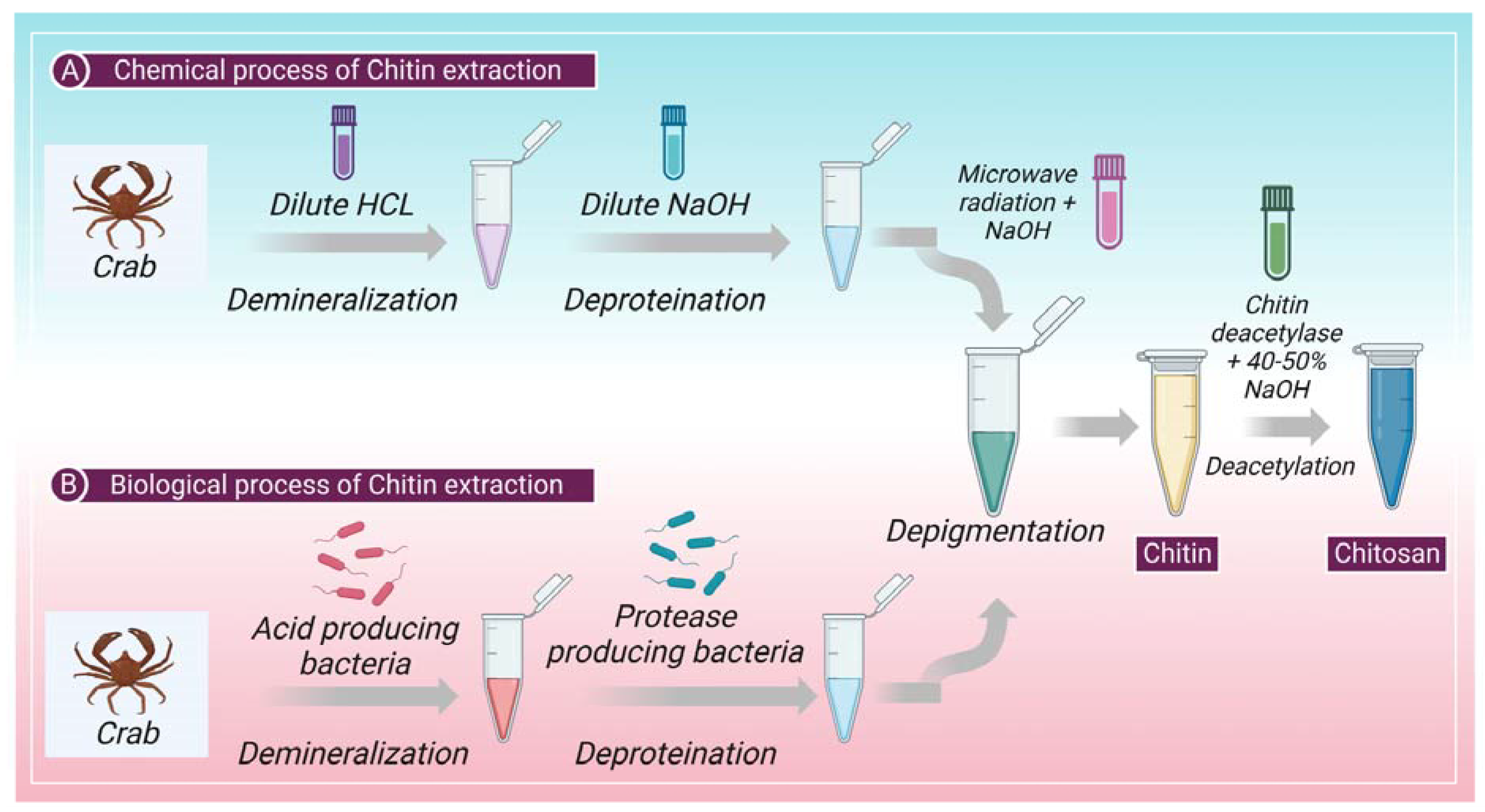
2. Extraction of Chitin: Chemical and Biological Process
2.1. Chemical Process
2.1.1. Chemical Demineralization
2.1.2. Chemical Deproteinization
2.1.3. Depigmentation
2.2. Biological Process
3. Drug Delivery System
3.1. Rotes of Chitosan Administration
3.1.1. Ocular Drug Delivery of CS
3.1.2. Pulmonary Drug Delivery of CS
3.1.3. Mucosal Drug Delivery of CS
3.1.4. Nasal Drug Delivery of CS
3.1.5. Transdermal Drug Delivery of CS
3.1.6. Dermal Delivery of CS
3.1.7. CS Administration for Wound Healing
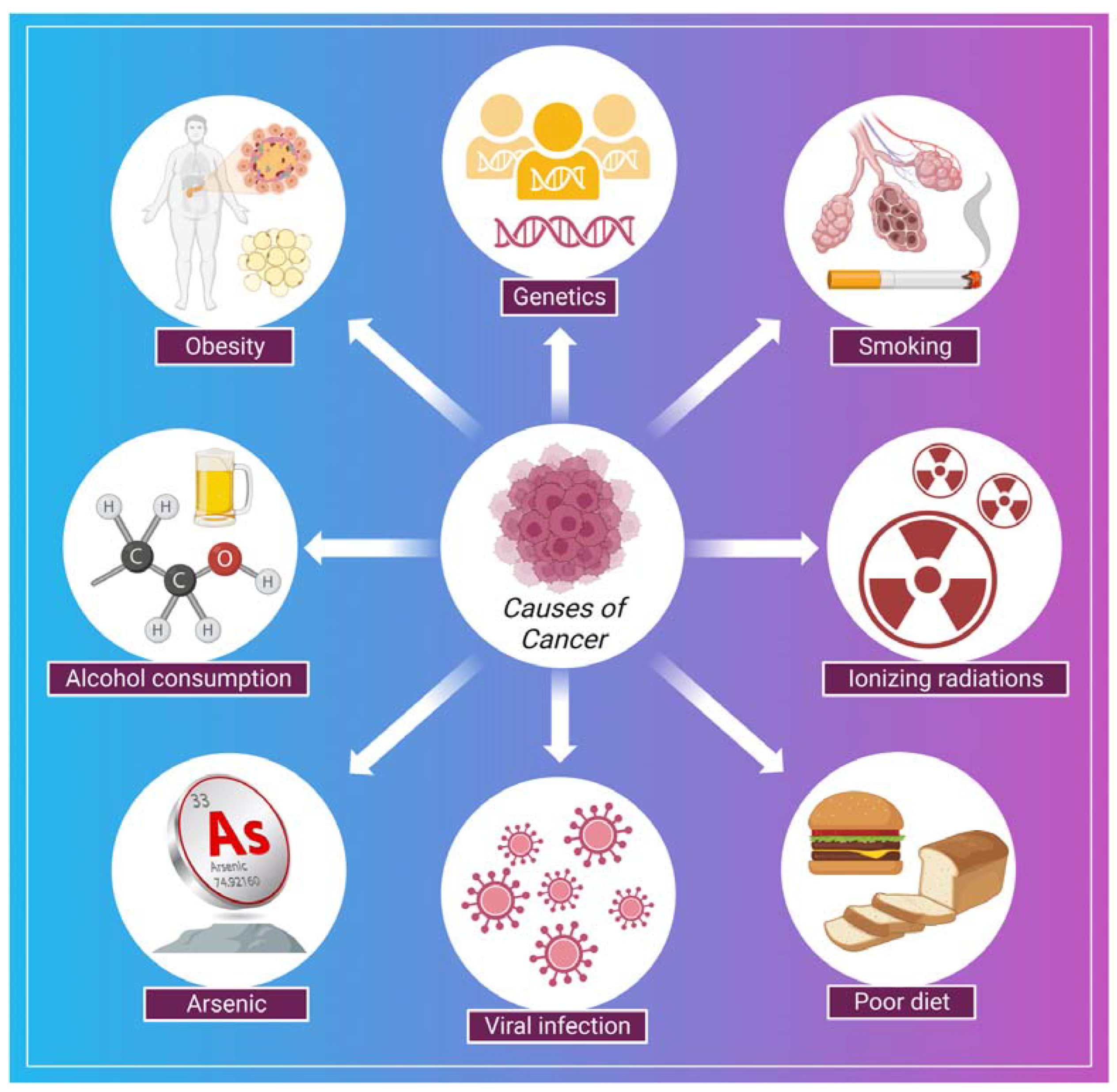
4. Cancer: Symptoms, Causes, Treatment Strategies
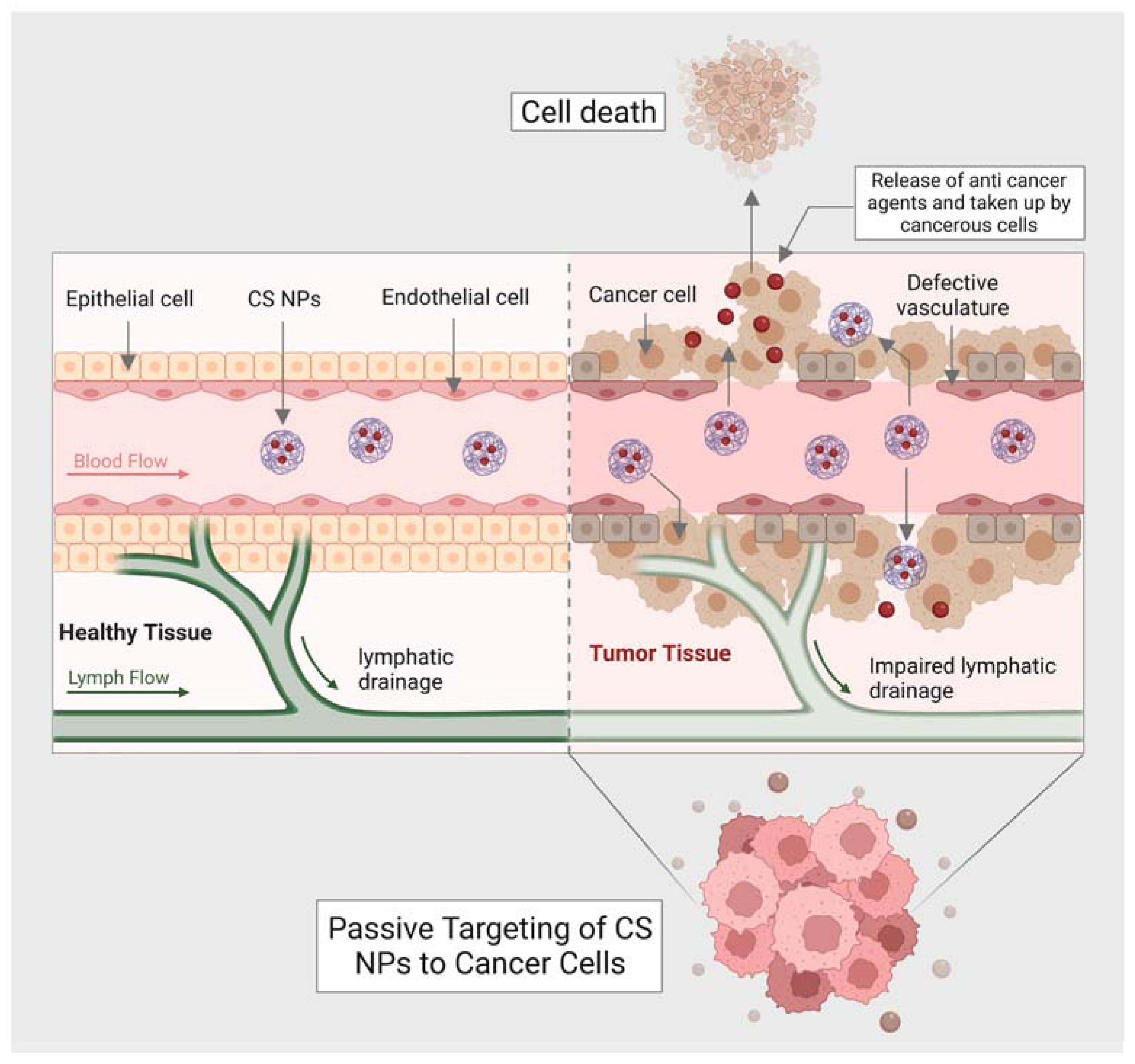
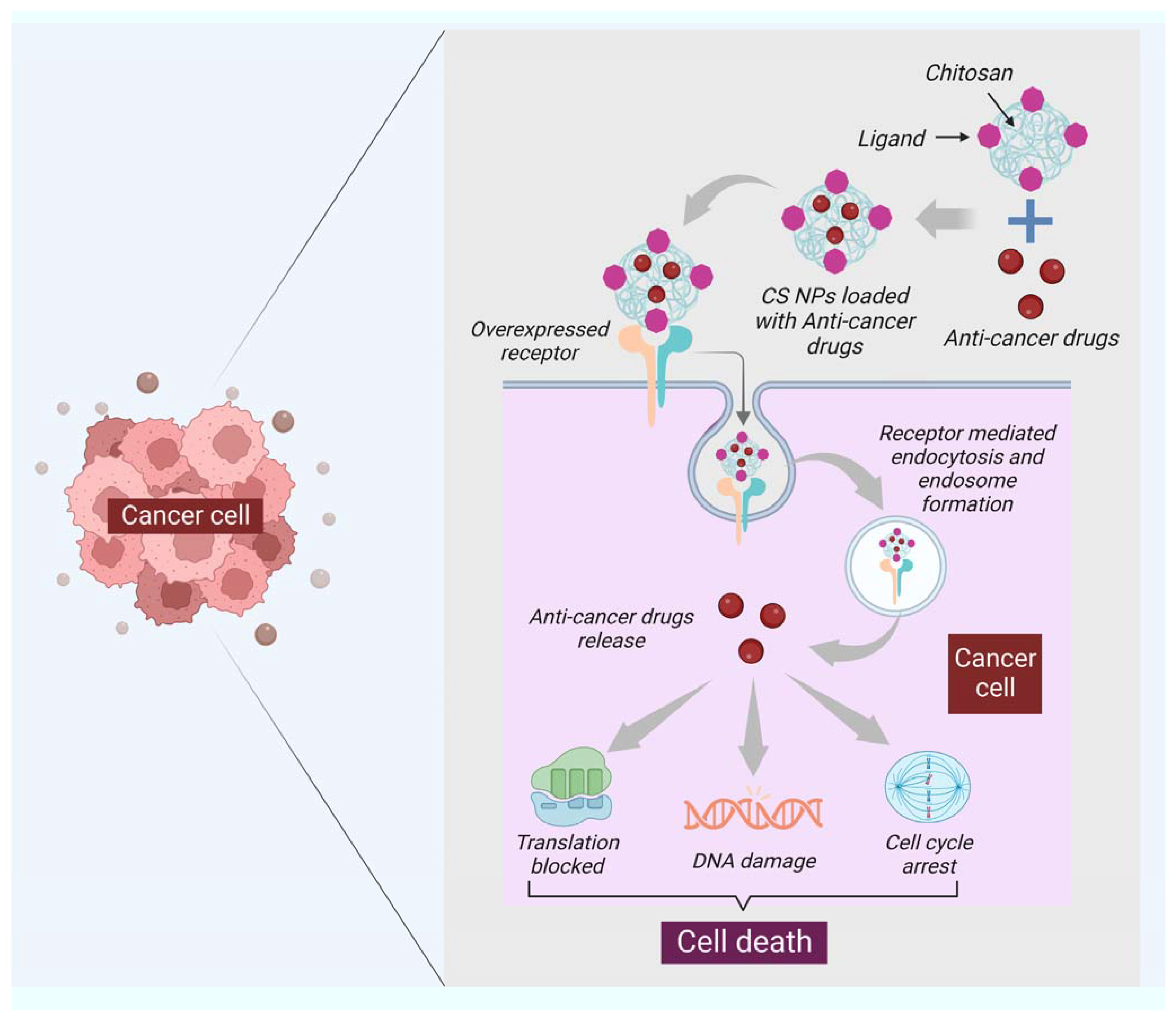
5. Chitin and Chitosan for Drug Delivery and Cancer Treatment
6. Advantages of Using Chitin and Chitosan in Nanomedicine
6.1. Biocompatibility
6.2. Antimicrobial Characteristics
6.3. Mucoadhesive Characteristics
6.4. Biodegradability
7. Problematics of Chitin and Chitosan in Nanomedicine
7.1. Allergenicity
7.2. Limited Solubility
7.3. Variability from Batch to Batch
7.4. Limited Stability
8. Challenges and Future Perspectives
9. Conclusions
Author Contributions
Funding
Institutional Review Board Statement
Informed Consent Statement
Data Availability Statement
Acknowledgments
Conflicts of Interest
Abbreviations
| NPs | Nanoparticles |
| CS | Chitosan |
| MWI | Microwave irradiation |
| BBB | Blood–brain barrier |
| BCB | Blood–cerebrospinal fluid barrier |
| NTB | Nose to the brain |
| HAS | Human serum albumin |
| RH | Ropinirole hydrochloride |
| DOPA | Dopamine |
| PD | Parkinson’s disease |
| AD | Alzheimer’s disease |
| PLGA | poly(lactic-co-glycolic acid |
| SNES | Simulated nasal electrolyte solution |
| CNTs | Carbon nanotubes |
| CaNPs | Calcium Nanoparticles |
| CHI3L 1 | Chitinase-3-like protein-1 |
| VEGF-C | Vascular endothelial growth factor |
| CCNGs | Curcumin loaded chitin nanogels |
| EPR | Enhanced permeability retention |
| PDMSCs | Placental-derived mesenchymal stem cells |
| TNBC | Triple-negative breast cancer |
| TRAIL | Tumor necrosis factor-related apoptosis-inducing ligand |
| HA | Hyaluronic acid |
| Cy3 | Cyanine 3 |
| BCL2 | B-cell lymphoma 2 |
| DOX | Doxorubicin |
| AMP | 2-acrylamide-2-methylpropane sulphonic acid |
| SiRNA | Small interfering RNA |
| OQC | Octadecyl quaternized carboxymethyl chitosan |
| FPNs | Folate-targeted chitosan polymeric nanoparticles |
| CMC | Carboxymethyl chitosan |
| sGNPs | Small gold nanoparticles |
| FITC | Fluorescein isothiocyanate |
| CHC | Chitosan hydrochloride |
| OXPt | Oxaliplatin |
| PEG | Poly(ethylene glycol) |
| Ad | Adenoviral |
| FA | Folic acid |
| TDPA | Thiodipropionic acid |
| PBAP | Phenyl boronic acid pinacol ester |
| PDT | Photodynamic therapy |
| LMW | Low molecular weight |
| siRNA | Small interfering RNA |
| OVCAR | Ovarian adenocarcinoma |
References
- Tsurkan, M.V.; Voronkina, A.; Khrunyk, Y.; Wysokowski, M.; Petrenko, I.; Ehrlich, H. Progress in Chitin Analytics. Carbohydr. Polym. 2021, 252, 117204. [Google Scholar] [CrossRef] [PubMed]
- Wysokowski, M.; Machałowski, T.; Petrenko, I.; Schimpf, C.; Rafaja, D.; Galli, R.; Ziętek, J.; Pantović, S.; Voronkina, A.; Kovalchuk, V.; et al. 3D Chitin Scaffolds of Marine Demosponge Origin for Biomimetic Mollusk Hemolymph-Associated Biomineralization Ex-Vivo. Mar. Drugs 2020, 18, 123. [Google Scholar] [CrossRef] [PubMed] [Green Version]
- Mutsenko, V.V.; Gryshkov, O.; Lauterboeck, L.; Rogulska, O.; Tarusin, D.N.; Bazhenov, V.V.; Schütz, K.; Brüggemeier, S.; Gossla, E.; Akkineni, A.R.; et al. Novel Chitin Scaffolds Derived from Marine Sponge Ianthella Basta for Tissue Engineering Approaches Based on Human Mesenchymal Stromal Cells: Biocompatibility and Cryopreservation. Int. J. Biol. Macromol. 2017, 104, 1955–1965. [Google Scholar] [CrossRef] [PubMed]
- Wysokowski, M.; Petrenko, I.; Stelling, A.; Stawski, D.; Jesionowski, T.; Ehrlich, H. Poriferan Chitin as a Versatile Template for Extreme Biomimetics. Polymers 2015, 7, 235–265. [Google Scholar] [CrossRef] [Green Version]
- Petrenko, I.; Bazhenov, V.V.; Galli, R.; Wysokowski, M.; Fromont, J.; Schupp, P.J.; Stelling, A.L.; Niederschlag, E.; Stöker, H.; Kutsova, V.Z.; et al. Chitin of Poriferan Origin and the Bioelectrometallurgy of Copper/Copper Oxide. Int. J. Biol. Macromol. 2017, 104, 1626–1632. [Google Scholar] [CrossRef] [PubMed]
- Moussian, B. Chitin: Structure, Chemistry and Biology. In Targeting Chitin-Containing Organisms; Yang, Q., Fukamizo, T., Eds.; Advances in Experimental Medicine and Biology; Springer: Singapore, 2019; Volume 1142, pp. 5–18. ISBN 9789811373176. [Google Scholar]
- Kaya, M.; Mujtaba, M.; Ehrlich, H.; Salaberria, A.M.; Baran, T.; Amemiya, C.T.; Galli, R.; Akyuz, L.; Sargin, I.; Labidi, J. On Chemistry of γ-Chitin. Carbohydr. Polym. 2017, 176, 177–186. [Google Scholar] [CrossRef]
- Hou, J.; Aydemir, B.E.; Dumanli, A.G. Understanding the Structural Diversity of Chitins as a Versatile Biomaterial. Philos. Trans. A Math. Phys. Eng. Sci. 2021, 379, 20200331. [Google Scholar] [CrossRef]
- Singh, N.; Chen, J.; Koziol, K.K.; Hallam, K.R.; Janas, D.; Patil, A.J.; Strachan, A.; Hanley, J.G.; Rahatekar, S.S. Chitin and Carbon Nanotube Composites as Biocompatible Scaffolds for Neuron Growth. Nanoscale 2016, 8, 8288–8299. [Google Scholar] [CrossRef] [Green Version]
- Ji, X.; Lei, Z.; Yuan, M.; Zhu, H.; Yuan, X.; Liu, W.; Pu, H.; Jiang, J.; Zhang, Y.; Jiang, X.; et al. Cartilage Repair Mediated by Thermosensitive Photocrosslinkable TGFβ1-Loaded GM-HPCH via Immunomodulating Macrophages, Recruiting MSCs and Promoting Chondrogenesis. Theranostics 2020, 10, 2872–2887. [Google Scholar] [CrossRef]
- Mutsenko, V.V.; Bazhenov, V.V.; Rogulska, O.; Tarusin, D.N.; Schütz, K.; Brüggemeier, S.; Gossla, E.; Akkineni, A.R.; Meißner, H.; Lode, A.; et al. 3D Chitinous Scaffolds Derived from Cultivated Marine Demosponge Aplysina Aerophoba for Tissue Engineering Approaches Based on Human Mesenchymal Stromal Cells. Int. J. Biol. Macromol. 2017, 104, 1966–1974. [Google Scholar] [CrossRef]
- Lomiguen, C.; Vidal, L.; Kozlowski, P.; Prancan, A.; Stern, R. Possible Role of Chitin-Like Proteins in the Etiology of Alzheimer’s Disease. JAD 2018, 66, 439–444. [Google Scholar] [CrossRef] [PubMed]
- Castellani, R.J.; Perry, G.; Smith, M.A. The Role of Novel Chitin-like Polysaccharides in Alzheimer Disease. Neurotox. Res. 2007, 12, 269–274. [Google Scholar] [CrossRef] [PubMed]
- Castellani, R.; Siedlak, S.; Fortino, A.; Perry, G.; Ghetti, B.; Smith, M. Chitin-like Polysaccharides in Alzheimers Disease Brains. CAR 2005, 2, 419–423. [Google Scholar] [CrossRef]
- Pisa, D.; Alonso, R.; Rábano, A.; Horst, M.N.; Carrasco, L. Fungal Enolase, β-Tubulin, and Chitin Are Detected in Brain Tissue from Alzheimer’s Disease Patients. Front. Microbiol. 2016, 7, 1772. [Google Scholar] [CrossRef] [Green Version]
- Sachdeva, B.; Sachdeva, P. MXenes for Neurodegenerative Disorders. Mater. Today Proc. 2022, 73, 294–296. [Google Scholar] [CrossRef]
- Ahmad, F.; Sachdeva, P. A Consolidated Review on Stem Cell Therapy for Treatment and Management of Alzheimer’s Disease. Aging Med. 2022, 5, 182–190. [Google Scholar] [CrossRef]
- Ahmad, F.; Sachdeva, P. Critical Appraisal on Mitochondrial Dysfunction in Alzheimer’s Disease. Aging Med. 2022, 5, 272–280. [Google Scholar] [CrossRef]
- Ghosh, S.; Sachdeva, B.; Sachdeva, P.; Chaudhary, V.; Rani, G.M.; Sinha, J.K. Graphene Quantum Dots as a Potential Diagnostic and Therapeutic Tool for the Management of Alzheimer’s Disease. Carbon. Lett. 2022, 32, 1381–1394. [Google Scholar] [CrossRef]
- Ahmad, F.; Sachdeva, P.; Sarkar, J.; Izhaar, R. Circadian Dysfunction and Alzheimer’s Disease—An Updated Review. Aging Med. 2022, 6, 71–81. [Google Scholar] [CrossRef]
- Ahmad, F.; Sachdeva, P.; Sachdeva, B.; Singh, G.; Soni, H.; Tandon, S.; Rafeeq, M.M.; Alam, M.Z.; Baeissa, H.M.; Khalid, M. Dioxinodehydroeckol: A Potential Neuroprotective Marine Compound Identified by In Silico Screening for the Treatment and Management of Multiple Brain Disorders. Mol. Biotechnol. 2022. [Google Scholar] [CrossRef]
- Mukerjee, N.; Al-Khafaji, K.; Maitra, S.; Suhail Wadi, J.; Sachdeva, P.; Ghosh, A.; Buchade, R.S.; Chaudhari, S.Y.; Jadhav, S.B.; Das, P.; et al. Recognizing Novel Drugs against Keap1 in Alzheimer’s Disease Using Machine Learning Grounded Computational Studies. Front. Mol. Neurosci. 2022, 15, 1036552. [Google Scholar] [CrossRef] [PubMed]
- Madar, I.H.; Sultan, G.; Tayubi, I.A.; Hasan, A.N.; Pahi, B.; Rai, A.; Sivanandan, P.K.; Loganathan, T.; Begum, M.; Rai, S. Identification of Marker Genes in Alzheimer’s Disease Using a Machine-Learning Model. Bioinformation 2021, 17, 348–355. [Google Scholar] [CrossRef] [PubMed]
- Mishra, P.; Mittal, A.K.; Kalonia, H.; Madan, S.; Ghosh, S.; Sinha, J.K.; Rajput, S.K. SIRT1 Promotes Neuronal Fortification in Neurodegenerative Diseases through Attenuation of Pathological Hallmarks and Enhancement of Cellular Lifespan. Curr. Neuropharmacol. 2021, 19, 1019–1037. [Google Scholar] [CrossRef]
- Sachdeva, B.; Sachdeva, P.; Ghosh, S.; Ahmad, F.; Sinha, J.K. Ketamine as a Therapeutic Agent in Major Depressive Disorder and Posttraumatic Stress Disorder: Potential Medicinal and Deleterious Effects. Ibrain 2023, 9, 90–101. [Google Scholar] [CrossRef]
- Sinha, J.K.; Ghosh, S.; Raghunath, M. DNA Damage in Brain May Lead to Cognitive Dysfunctions and Reduced Longevity in WNIN/Ob Obese Rats. Alzheimer’s Dement. 2021, 17, e057963. [Google Scholar] [CrossRef]
- Ghosh, S.; Durgvanshi, S.; Agarwal, S.; Raghunath, M.; Sinha, J.K. Current Status of Drug Targets and Emerging Therapeutic Strategies in the Management of Alzheimer’s Disease. Curr. Neuropharmacol. 2020, 18, 883–903. [Google Scholar] [CrossRef]
- Sotgiu, S.; Musumeci, S.; Marconi, S.; Gini, B.; Bonetti, B. Different Content of Chitin-like Polysaccharides in Multiple Sclerosis and Alzheimer’s Disease Brains. J. Neuroimmunol. 2008, 197, 70–73. [Google Scholar] [CrossRef]
- Li, J.; Cai, C.; Li, J.; Li, J.; Li, J.; Sun, T.; Wang, L.; Wu, H.; Yu, G. Chitosan-Based Nanomaterials for Drug Delivery. Molecules 2018, 23, 2661. [Google Scholar] [CrossRef] [Green Version]
- Sharifi-Rad, J.; Quispe, C.; Butnariu, M.; Rotariu, L.S.; Sytar, O.; Sestito, S.; Rapposelli, S.; Akram, M.; Iqbal, M.; Krishna, A.; et al. Chitosan Nanoparticles as a Promising Tool in Nanomedicine with Particular Emphasis on Oncological Treatment. Cancer Cell. Int. 2021, 21, 318. [Google Scholar] [CrossRef]
- Mohammed, M.; Syeda, J.; Wasan, K.; Wasan, E. An Overview of Chitosan Nanoparticles and Its Application in Non-Parenteral Drug Delivery. Pharmaceutics 2017, 9, 53. [Google Scholar] [CrossRef] [Green Version]
- Muxika, A.; Etxabide, A.; Uranga, J.; Guerrero, P.; de la Caba, K. Chitosan as a Bioactive Polymer: Processing, Properties and Applications. Int. J. Biol. Macromol. 2017, 105, 1358–1368. [Google Scholar] [CrossRef] [PubMed]
- Argüelles-Monal, W.; Lizardi-Mendoza, J.; Fernández-Quiroz, D.; Recillas-Mota, M.; Montiel-Herrera, M. Chitosan Derivatives: Introducing New Functionalities with a Controlled Molecular Architecture for Innovative Materials. Polymers 2018, 10, 342. [Google Scholar] [CrossRef] [Green Version]
- Parhi, R. Drug Delivery Applications of Chitin and Chitosan: A Review. Env. Chem. Lett. 2020, 18, 577–594. [Google Scholar] [CrossRef]
- Ali, A.; Ahmed, S. A Review on Chitosan and Its Nanocomposites in Drug Delivery. Int. J. Biol. Macromol. 2018, 109, 273–286. [Google Scholar] [CrossRef]
- Dubashynskaya, N.V.; Petrova, V.A.; Romanov, D.P.; Skorik, Y.A. PH-Sensitive Drug Delivery System Based on Chitin Nanowhiskers–Sodium Alginate Polyelectrolyte Complex. Materials 2022, 15, 5860. [Google Scholar] [CrossRef]
- El Knidri, H.; Belaabed, R.; Addaou, A.; Laajeb, A.; Lahsini, A. Extraction, Chemical Modification and Characterization of Chitin and Chitosan. Int. J. Biol. Macromol. 2018, 120, 1181–1189. [Google Scholar] [CrossRef] [PubMed]
- Zainol Abidin, N.A.; Kormin, F.; Zainol Abidin, N.A.; Mohamed Anuar, N.A.F.; Abu Bakar, M.F. The Potential of Insects as Alternative Sources of Chitin: An Overview on the Chemical Method of Extraction from Various Sources. Int. J. Mol. Sci. 2020, 21, 4978. [Google Scholar] [CrossRef] [PubMed]
- Negi, A.; Kesari, K.K. Chitosan Nanoparticle Encapsulation of Antibacterial Essential Oils. Micromachines 2022, 13, 1265. [Google Scholar] [CrossRef]
- Pakizeh, M.; Moradi, A.; Ghassemi, T. Chemical Extraction and Modification of Chitin and Chitosan from Shrimp Shells. Eur. Polym. J. 2021, 159, 110709. [Google Scholar] [CrossRef]
- Özel, N.; Elibol, M. A Review on the Potential Uses of Deep Eutectic Solvents in Chitin and Chitosan Related Processes. Carbohydr. Polym. 2021, 262, 117942. [Google Scholar] [CrossRef]
- No, H.K.; Hur, E.Y. Control of Foam Formation by Antifoam during Demineralization of Crustacean Shell in Preparation of Chitin. J. Agric. Food Chem. 1998, 46, 3844–3846. [Google Scholar] [CrossRef]
- Percot, A.; Viton, C.; Domard, A. Characterization of Shrimp Shell Deproteinization. Biomacromolecules 2003, 4, 1380–1385. [Google Scholar] [CrossRef] [PubMed]
- Shahidi, F.; Synowiecki, J. Isolation and Characterization of Nutrients and Value-Added Products from Snow Crab (Chionoecetes opilio) and Shrimp (Pandalus borealis) Processing Discards. J. Agric. Food Chem. 1991, 39, 1527–1532. [Google Scholar] [CrossRef]
- El-Naggar, N.E.-A.; Saber, W.I.A.; Zweil, A.M.; Bashir, S.I. An Innovative Green Synthesis Approach of Chitosan Nanoparticles and Their Inhibitory Activity against Phytopathogenic Botrytis Cinerea on Strawberry Leaves. Sci. Rep. 2022, 12, 3515. [Google Scholar] [CrossRef] [PubMed]
- Synowiecki, J.; Al-Khateeb, N.A. Production, Properties, and Some New Applications of Chitin and Its Derivatives. Crit. Rev. Food Sci. Nutr. 2003, 43, 145–171. [Google Scholar] [CrossRef]
- Liao, J.; Huang, H. Extraction of a Novel Fungal Chitin from Hericium Erinaceus Residue Using Multistep Mild Procedures. Int. J. Biol. Macromol. 2020, 156, 1279–1286. [Google Scholar] [CrossRef]
- Machałowski, T.; Wysokowski, M.; Tsurkan, M.V.; Galli, R.; Schimpf, C.; Rafaja, D.; Brendler, E.; Viehweger, C.; Żółtowska-Aksamitowska, S.; Petrenko, I.; et al. Spider Chitin: An Ultrafast Microwave-Assisted Method for Chitin Isolation from Caribena Versicolor Spider Molt Cuticle. Molecules 2019, 24, 3736. [Google Scholar] [CrossRef] [Green Version]
- Rao, M.B.; Tanksale, A.M.; Ghatge, M.S.; Deshpande, V.V. Molecular and Biotechnological Aspects of Microbial Proteases. Microbiol. Mol. Biol. Rev. 1998, 62, 597–635. [Google Scholar] [CrossRef] [Green Version]
- Smolen, V.F. Bioavailability and Pharmacokinetic Analysis of Drug Responding Systems. Annu. Rev. Pharmacol. Toxicol. 1978, 18, 495–522. [Google Scholar] [CrossRef]
- Coelho, J.F.; Ferreira, P.C.; Alves, P.; Cordeiro, R.; Fonseca, A.C.; Góis, J.R.; Gil, M.H. Drug Delivery Systems: Advanced Technologies Potentially Applicable in Personalized Treatments. EPMA J. 2010, 1, 164–209. [Google Scholar] [CrossRef] [Green Version]
- Liechty, W.B.; Kryscio, D.R.; Slaughter, B.V.; Peppas, N.A. Polymers for Drug Delivery Systems. Annu. Rev. Chem. Biomol. Eng. 2010, 1, 149–173. [Google Scholar] [CrossRef] [PubMed] [Green Version]
- Prabaharan, M. Review Paper: Chitosan Derivatives as Promising Materials for Controlled Drug Delivery. J. Biomater. Appl. 2008, 23, 5–36. [Google Scholar] [CrossRef]
- Sabitha, M.; Sanoj Rejinold, N.; Nair, A.; Lakshmanan, V.-K.; Nair, S.V.; Jayakumar, R. Development and Evaluation of 5-Fluorouracil Loaded Chitin Nanogels for Treatment of Skin Cancer. Carbohydr. Polym. 2013, 91, 48–57. [Google Scholar] [CrossRef] [PubMed]
- Panonnummal, R.; Jayakumar, R.; Sabitha, M. Comparative Anti-Psoriatic Efficacy Studies of Clobetasol Loaded Chitin Nanogel and Marketed Cream. Eur. J. Pharm. Sci. 2017, 96, 193–206. [Google Scholar] [CrossRef]
- Gupta, H.; Velpandian, T.; Jain, S. Ion- and PH-Activated Novel in-Situ Gel System for Sustained Ocular Drug Delivery. J. Drug. Target. 2010, 18, 499–505. [Google Scholar] [CrossRef]
- Zhang, P.; Liu, X.; Hu, W.; Bai, Y.; Zhang, L. Preparation and Evaluation of Naringenin-Loaded Sulfobutylether-β-Cyclodextrin/Chitosan Nanoparticles for Ocular Drug Delivery. Carbohydr. Polym. 2016, 149, 224–230. [Google Scholar] [CrossRef]
- Santhi, K.; Muralidharan, S.; Yee, Y.H.; Min, F.M.; Ting, C.Z.; Devi, D. In-Vitro Characterization of Chitosan Nanoparticles of Fluconazole as a Carrier for Sustained Ocular Delivery. NANOASIA 2017, 7, 41–50. [Google Scholar] [CrossRef]
- Ruge, C.A.; Kirch, J.; Lehr, C.-M. Pulmonary Drug Delivery: From Generating Aerosols to Overcoming Biological Barriers—Therapeutic Possibilities and Technological Challenges. Lancet Respir. Med. 2013, 1, 402–413. [Google Scholar] [CrossRef]
- Rawal, T.; Parmar, R.; Tyagi, R.K.; Butani, S. Rifampicin Loaded Chitosan Nanoparticle Dry Powder Presents an Improved Therapeutic Approach for Alveolar Tuberculosis. Coll. Surf. B Biointerfaces 2017, 154, 321–330. [Google Scholar] [CrossRef]
- Debnath, S.K.; Saisivam, S.; Debanth, M.; Omri, A. Development and Evaluation of Chitosan Nanoparticles Based Dry Powder Inhalation Formulations of Prothionamide. PLoS ONE 2018, 13, e0190976. [Google Scholar] [CrossRef] [Green Version]
- Jafarinejad, S.; Gilani, K.; Moazeni, E.; Ghazi-Khansari, M.; Najafabadi, A.R.; Mohajel, N. Development of Chitosan-Based Nanoparticles for Pulmonary Delivery of Itraconazole as Dry Powder Formulation. Powder Technol. 2012, 222, 65–70. [Google Scholar] [CrossRef]
- Wang, J.; Tauchi, Y.; Deguchi, Y.; Morimoto, K.; Tabata, Y.; Ikada, Y. Positively Charged Gelatin Microspheres as Gastric Mucoadhesive Drug Delivery System for Eradication of H. pylori. Drug. Deliv. 2000, 7, 237–243. [Google Scholar] [CrossRef] [PubMed]
- Dodane, V. Effect of Chitosan on Epithelial Permeability and Structure. Int. J. Pharm. 1999, 182, 21–32. [Google Scholar] [CrossRef] [PubMed]
- Lopez-Moya, F.; Suarez-Fernandez, M.; Lopez-Llorca, L.V. Molecular Mechanisms of Chitosan Interactions with Fungi and Plants. Int. J. Mol. Sci. 2019, 20, 332. [Google Scholar] [CrossRef] [Green Version]
- Garg, U.; Chauhan, S.; Nagaich, U.; Jain, N. Current Advances in Chitosan Nanoparticles Based Drug Delivery and Targeting. Adv. Pharm. Bull. 2019, 9, 195–204. [Google Scholar] [CrossRef] [Green Version]
- Piazzini, V.; Landucci, E.; D’Ambrosio, M.; Tiozzo Fasiolo, L.; Cinci, L.; Colombo, G.; Pellegrini-Giampietro, D.E.; Bilia, A.R.; Luceri, C.; Bergonzi, M.C. Chitosan Coated Human Serum Albumin Nanoparticles: A Promising Strategy for Nose-to-Brain Drug Delivery. Int. J. Biol. Macromol. 2019, 129, 267–280. [Google Scholar] [CrossRef]
- Chatzitaki, A.-T.; Jesus, S.; Karavasili, C.; Andreadis, D.; Fatouros, D.G.; Borges, O. Chitosan-Coated PLGA Nanoparticles for the Nasal Delivery of Ropinirole Hydrochloride: In Vitro and Ex Vivo Evaluation of Efficacy and Safety. Int. J. Pharm. 2020, 589, 119776. [Google Scholar] [CrossRef]
- Prausnitz, M.R.; Langer, R. Transdermal Drug Delivery. Nat. Biotechnol. 2008, 26, 1261–1268. [Google Scholar] [CrossRef]
- Guy, R.H. Transdermal Drug Delivery. In Drug. Delivery; Schäfer-Korting, M., Ed.; Handbook of Experimental Pharmacology; Springer: Berlin/Heidelberg, Germany, 2010; Volume 197, pp. 399–410. ISBN 978-3-642-00476-6. [Google Scholar]
- Nair, S.S. Chitosan-Based Transdermal Drug Delivery Systems to Overcome Skin Barrier Functions. J. Drug. Deliv. Ther. 2019, 9, 266–270. [Google Scholar] [CrossRef] [Green Version]
- Nawaz, A.; Wong, T.W. Microwave as Skin Permeation Enhancer for Transdermal Drug Delivery of Chitosan-5-Fluorouracil Nanoparticles. Carbohydr. Polym. 2017, 157, 906–919. [Google Scholar] [CrossRef]
- Abd-Allah, H.; Abdel-Aziz, R.T.A.; Nasr, M. Chitosan Nanoparticles Making Their Way to Clinical Practice: A Feasibility Study on Their Topical Use for Acne Treatment. Int. J. Biol. Macromol. 2020, 156, 262–270. [Google Scholar] [CrossRef]
- Daeschlein, G. Antimicrobial and Antiseptic Strategies in Wound Management. Int. Wound J. 2013, 10 (Suppl. S1), 9–14. [Google Scholar] [CrossRef] [PubMed]
- Wang, P.-H.; Huang, B.-S.; Horng, H.-C.; Yeh, C.-C.; Chen, Y.-J. Wound Healing. J. Chin. Med. Assoc. 2018, 81, 94–101. [Google Scholar] [CrossRef] [PubMed]
- Patrulea, V.; Ostafe, V.; Borchard, G.; Jordan, O. Chitosan as a Starting Material for Wound Healing Applications. Eur. J. Pharm. Biopharm. 2015, 97, 417–426. [Google Scholar] [CrossRef] [PubMed] [Green Version]
- Fahimirad, S.; Abtahi, H.; Satei, P.; Ghaznavi-Rad, E.; Moslehi, M.; Ganji, A. Wound Healing Performance of PCL/Chitosan Based Electrospun Nanofiber Electrosprayed with Curcumin Loaded Chitosan Nanoparticles. Carbohydr. Polym. 2021, 259, 117640. [Google Scholar] [CrossRef]
- Sachdeva, P.; Ghosh, S.; Ghosh, S.; Han, S.; Banerjee, J.; Bhaskar, R.; Sinha, J.K. Childhood Obesity: A Potential Key Factor in the Development of Glioblastoma Multiforme. Life 2022, 12, 1673. [Google Scholar] [CrossRef]
- Evan, G.I.; Vousden, K.H. Proliferation, Cell Cycle and Apoptosis in Cancer. Nature 2001, 411, 342–348. [Google Scholar] [CrossRef]
- Jolly, R.D.; Thompson, K.G.; Winchester, B.G. Bovine Mannosidosis--a Model Lysosomal Storage Disease. Birth Defects Orig. Artic. Ser. 1975, 11, 273–278. [Google Scholar]
- Koo, M.M.; Swann, R.; McPhail, S.; Abel, G.A.; Elliss-Brookes, L.; Rubin, G.P.; Lyratzopoulos, G. Presenting Symptoms of Cancer and Stage at Diagnosis: Evidence from a Cross-Sectional, Population-Based Study. Lancet Oncol. 2020, 21, 73–79. [Google Scholar] [CrossRef] [Green Version]
- Wu, L.; Qu, X. Cancer Biomarker Detection: Recent Achievements and Challenges. Chem. Soc. Rev. 2015, 44, 2963–2997. [Google Scholar] [CrossRef]
- Wyld, L.; Audisio, R.A.; Poston, G.J. The Evolution of Cancer Surgery and Future Perspectives. Nat. Rev. Clin. Oncol. 2015, 12, 115–124. [Google Scholar] [CrossRef] [PubMed]
- Dolmans, D.E.J.G.J.; Fukumura, D.; Jain, R.K. Photodynamic Therapy for Cancer. Nat. Rev. Cancer 2003, 3, 380–387. [Google Scholar] [CrossRef] [PubMed]
- Baskar, R.; Lee, K.A.; Yeo, R.; Yeoh, K.-W. Cancer and Radiation Therapy: Current Advances and Future Directions. Int. J. Med. Sci. 2012, 9, 193–199. [Google Scholar] [CrossRef] [Green Version]
- Paijens, S.T.; Vledder, A.; de Bruyn, M.; Nijman, H.W. Tumor-Infiltrating Lymphocytes in the Immunotherapy Era. Cell. Mol. Immunol. 2021, 18, 842–859. [Google Scholar] [CrossRef] [PubMed]
- Kubota, T. New Chemotherapy Strategies for Gastric Cancer. In Vivo 2008, 22, 273–278. [Google Scholar]
- Nasir, A.; Khan, A.; Li, J.; Naeem, M.; Khalil, A.A.K.; Khan, K.; Qasim, M. Nanotechnology, A Tool for Diagnostics and Treatment of Cancer. Curr. Top. Med. Chem. 2021, 21, 1360–1376. [Google Scholar] [CrossRef]
- Lee, C.G.; Da Silva, C.A.; Dela Cruz, C.S.; Ahangari, F.; Ma, B.; Kang, M.-J.; He, C.-H.; Takyar, S.; Elias, J.A. Role of Chitin and Chitinase/Chitinase-Like Proteins in Inflammation, Tissue Remodeling, and Injury. Annu. Rev. Physiol. 2011, 73, 479–501. [Google Scholar] [CrossRef] [Green Version]
- Timoshenko, A.V. Chitin Hydrolysate Stimulates VEGF-C Synthesis by MDA-MB-231 Breast Cancer Cells. Cell. Biol. Int. 2011, 35, 281–286. [Google Scholar] [CrossRef]
- Solairaj, D.; Rameshthangam, P.; Arunachalam, G. Anticancer Activity of Silver and Copper Embedded Chitin Nanocomposites against Human Breast Cancer (MCF-7) Cells. Int. J. Biol. Macromol. 2017, 105, 608–619. [Google Scholar] [CrossRef]
- Sachdeva, P.; Mehdi, I.; Kaith, R.; Ahmad, F.; Anwar, M.S. Potential Natural Products for the Management of Autism Spectrum Disorder. Ibrain 2022, 8, 365–376. [Google Scholar] [CrossRef]
- Mangalathillam, S.; Rejinold, N.S.; Nair, A.; Lakshmanan, V.-K.; Nair, S.V.; Jayakumar, R. Curcumin Loaded Chitin Nanogels for Skin Cancer Treatment via the Transdermal Route. Nanoscale 2012, 4, 239–250. [Google Scholar] [CrossRef] [PubMed]
- Li, X.; Min, M.; Du, N.; Gu, Y.; Hode, T.; Naylor, M.; Chen, D.; Nordquist, R.E.; Chen, W.R. Chitin, Chitosan, and Glycated Chitosan Regulate Immune Responses: The Novel Adjuvants for Cancer Vaccine. Clin. Dev. Immunol. 2013, 2013, 387023. [Google Scholar] [CrossRef] [PubMed] [Green Version]
- Ismail, I.A.; Notananda, V.; Schepens, J. Studies on Malaria and Responses of Anopheles Balabacensis Balabacensis and Anopheles Minimus to DDT Residual Spraying in Thailand. Acta Trop. 1975, 32, 206–231. [Google Scholar]
- SUZUKI, K.; OKAWA, Y.; HASHIMOTO, K.; SUZUKI, S.; SUZUKI, M. Protecting Effect of Chitin and Chitosan on Experimentally Induced Murine Candidiasis. Microbiol. Immunol. 1984, 28, 903–912. [Google Scholar] [CrossRef] [PubMed]
- van der Lubben, I.M.; Verhoef, J.C.; Borchard, G.; Junginger, H.E. Chitosan for Mucosal Vaccination. Adv. Drug Deliv. Rev. 2001, 52, 139–144. [Google Scholar] [CrossRef]
- McNeela, E.A.; Jabbal-Gill, I.; Illum, L.; Pizza, M.; Rappuoli, R.; Podda, A.; Lewis, D.J.M.; Mills, K.H.G. Intranasal Immunization with Genetically Detoxified Diphtheria Toxin Induces T Cell Responses in Humans: Enhancement of Th2 Responses and Toxin-Neutralizing Antibodies by Formulation with Chitosan. Vaccine 2004, 22, 909–914. [Google Scholar] [CrossRef]
- Lee, C.G.; Da Silva, C.A.; Lee, J.-Y.; Hartl, D.; Elias, J.A. Chitin Regulation of Immune Responses: An Old Molecule with New Roles. Curr. Opin. Immunol. 2008, 20, 684–689. [Google Scholar] [CrossRef] [Green Version]
- Zaharoff, D.A.; Hance, K.W.; Rogers, C.J.; Schlom, J.; Greiner, J.W. Intratumoral Immunotherapy of Established Solid Tumors with Chitosan/IL-12. J. Immunother. 2010, 33, 697–705. [Google Scholar] [CrossRef] [Green Version]
- Vijayakumar, M.; Priya, K.; Ilavenil, S.; Janani, B.; Vedarethinam, V.; Ramesh, T.; Arasu, M.V.; Al-Dhabi, N.A.; Kim, Y.-O.; Kim, H.-J. Shrimp Shells Extracted Chitin in Silver Nanoparticle Synthesis: Expanding Its Prophecy towards Anticancer Activity in Human Hepatocellular Carcinoma HepG2 Cells. Int. J. Biol. Macromol. 2020, 165, 1402–1409. [Google Scholar] [CrossRef]
- Kamalabadi-Farahani, M.; Vasei, M.; Ahmadbeigi, N.; Ebrahimi-Barough, S.; Soleimani, M.; Roozafzoon, R. Anti-Tumour Effects of TRAIL-Expressing Human Placental Derived Mesenchymal Stem Cells with Curcumin-Loaded Chitosan Nanoparticles in a Mice Model of Triple Negative Breast Cancer. Artif. Cells Nanomed. Biotechnol. 2018, 46, 1011–1021. [Google Scholar] [CrossRef] [Green Version]
- Zhang, W.; Xu, W.; Lan, Y.; He, X.; Liu, K.; Liang, Y. Antitumor Effect of Hyaluronic-Acid-Modified Chitosan Nanoparticles Loaded with SiRNA for Targeted Therapy for Non-Small Cell Lung Cancer. Int. J. Nanomed. 2019, 14, 5287–5301. [Google Scholar] [CrossRef] [PubMed] [Green Version]
- Deng, X.; Cao, M.; Zhang, J.; Hu, K.; Yin, Z.; Zhou, Z.; Xiao, X.; Yang, Y.; Sheng, W.; Wu, Y.; et al. Hyaluronic Acid-Chitosan Nanoparticles for Co-Delivery of MiR-34a and Doxorubicin in Therapy against Triple Negative Breast Cancer. Biomaterials 2014, 35, 4333–4344. [Google Scholar] [CrossRef] [PubMed]
- Kavaz, D.; Kirac, F.; Kirac, M.; Vaseashta, A. Low Releasing Mitomycin C Molecule Encapsulated with Chitosan Nanoparticles for Intravesical Installation. JBNB 2017, 08, 203–219. [Google Scholar] [CrossRef] [Green Version]
- Kumar, S.; Garg, P.; Pandey, S.; Kumari, M.; Hoon, S.; Jang, K.-J.; Kapavarapu, R.; Choung, P.-H.; Sobral, A.J.F.N.; Hoon Chung, J. Enhanced Chitosan-DNA Interaction by 2-Acrylamido-2-Methylpropane Coupling for an Efficient Transfection in Cancer Cells. J. Mater. Chem. B. 2015, 3, 3465–3475. [Google Scholar] [CrossRef]
- Komenek, S.; Luesakul, U.; Ekgasit, S.; Vilaivan, T.; Praphairaksit, N.; Puthong, S.; Muangsin, N. Nanogold-Gallate Chitosan-Targeted Pulmonary Delivery for Treatment of Lung Cancer. AAPS PharmSciTech 2017, 18, 1104–1115. [Google Scholar] [CrossRef]
- Yhee, J.Y.; Song, S.; Lee, S.J.; Park, S.-G.; Kim, K.-S.; Kim, M.G.; Son, S.; Koo, H.; Kwon, I.C.; Jeong, J.H.; et al. Cancer-Targeted MDR-1 SiRNA Delivery Using Self-Cross-Linked Glycol Chitosan Nanoparticles to Overcome Drug Resistance. J. Control. Release 2015, 198, 1–9. [Google Scholar] [CrossRef]
- Sun, P.; Huang, W.; Jin, M.; Wang, Q.; Fan, B.; Kang, L.; Gao, Z. Chitosan-Based Nanoparticles for Survivin Targeted SiRNA Delivery in Breast Tumor Therapy and Preventing Its Metastasis. Int. J. Nanomed. 2016, 11, 4931–4945. [Google Scholar] [CrossRef] [Green Version]
- Darvishi, M.H.; Nomani, A.; Amini, M.; Shokrgozar, M.A.; Dinarvand, R. Novel Biotinylated Chitosan-Graft-Polyethyleneimine Copolymer as a Targeted Non-Viral Vector for Anti-EGF Receptor SiRNA Delivery in Cancer Cells. Int. J. Pharm. 2013, 456, 408–416. [Google Scholar] [CrossRef] [PubMed]
- Xin, L.; Fan, J.-C.; Le, Y.-G.; Zeng, F.; Cheng, H.; Hu, X.; Cao, J.-Q. Construction of METHFR ShRNA/5-Fluorouracil Co-Loaded Folate-Targeted Chitosan Polymeric Nanoparticles and Its Anti-Carcinoma Effect on Gastric Cells Growth. J. Nanopart. Res. 2016, 18, 105. [Google Scholar] [CrossRef]
- Soofiyani, S.R.; Hallaj-Nezhadi, S.; Lotfipour, F.; Hosseini, A.M.; Baradaran, B. Gene Therapy Based on Interleukin-12 Loaded Chitosan Nanoparticles in a Mouse Model of Fibrosarcoma. Iran. J. Basic. Med. Sci. 2016, 19, 1238–1244. [Google Scholar]
- Hemmati, K.; Ahmadi Nasab, N.; Hesaraki, S.; Nezafati, N. In Vitro Evaluation of Curcumin-Loaded Chitosan-Coated Hydroxyapatite Nanocarriers as a Potential System for Effective Treatment of Cancer. J. Biomater. Sci. Polym. Ed. 2021, 32, 1267–1287. [Google Scholar] [CrossRef] [PubMed]
- Kaur, H.; Mishra, N.; Khurana, B.; Kaur, S.; Arora, D. DoE Based Optimization and Development of Spray-Dried Chitosan-Coated Alginate Microparticles Loaded with Cisplatin for the Treatment of Cervical Cancer. Curr. Mol. Pharm. 2021, 14, 381–398. [Google Scholar] [CrossRef]
- Jensen, B.M.; Kühl, C.; Mølsted-Pedersen, L.; Saurbrey, N.; Fog-Pedersen, J. Preconceptional Treatment with Insulin Infusion Pumps in Insulin-Dependent Diabetic Women with Particular Reference to Prevention of Congenital Malformations. Acta Endocrinol. Suppl. 1986, 277, 81–85. [Google Scholar] [CrossRef]
- Chen, G.; Zhao, Y.; Xu, Y.; Zhu, C.; Liu, T.; Wang, K. Chitosan Nanoparticles for Oral Photothermally Enhanced Photodynamic Therapy of Colon Cancer. Int. J. Pharm. 2020, 589, 119763. [Google Scholar] [CrossRef]
- Li, G.; Wang, S.; Deng, D.; Xiao, Z.; Dong, Z.; Wang, Z.; Lei, Q.; Gao, S.; Huang, G.; Zhang, E.; et al. Fluorinated Chitosan to Enhance Transmucosal Delivery of Sonosensitizer-Conjugated Catalase for Sonodynamic Bladder Cancer Treatment Post-Intravesical Instillation. ACS Nano 2020, 14, 1586–1599. [Google Scholar] [CrossRef] [PubMed]
- Castro, F.; Pinto, M.L.; Pereira, C.L.; Serre, K.; Barbosa, M.A.; Vermaelen, K.; Gärtner, F.; Gonçalves, R.M.; De Wever, O.; Oliveira, M.J. Chitosan/γ-PGA Nanoparticles-Based Immunotherapy as Adjuvant to Radiotherapy in Breast Cancer. Biomaterials 2020, 257, 120218. [Google Scholar] [CrossRef]
- Naruphontjirakul, P.; Viravaidya-Pasuwat, K. Development of Anti-HER2-Targeted Doxorubicin-Core-Shell Chitosan Nanoparticles for the Treatment of Human Breast Cancer. Int. J. Nanomed. 2019, 14, 4105–4121. [Google Scholar] [CrossRef] [Green Version]
- Duan, R.; Zhou, Z.; Su, G.; Liu, L.; Guan, M.; Du, B.; Zhang, Q. Chitosan-Coated Gold Nanorods for Cancer Therapy Combining Chemical and Photothermal Effects. Macromol. Biosci. 2014, 14, 1160–1169. [Google Scholar] [CrossRef]
- Yan, L.; Gao, S.; Shui, S.; Liu, S.; Qu, H.; Liu, C.; Zheng, L. Small Interfering RNA-Loaded Chitosan Hydrochloride/Carboxymethyl Chitosan Nanoparticles for Ultrasound-Triggered Release to Hamper Colorectal Cancer Growth in Vitro. Int. J. Biol. Macromol. 2020, 162, 1303–1310. [Google Scholar] [CrossRef] [PubMed]
- Matos, B.N.; Pereira, M.N.; Bravo, M.D.O.; Cunha-Filho, M.; Saldanha-Araújo, F.; Gratieri, T.; Gelfuso, G.M. Chitosan Nanoparticles Loading Oxaliplatin as a Mucoadhesive Topical Treatment of Oral Tumors: Iontophoresis Further Enhances Drug Delivery Ex Vivo. Int. J. Biol. Macromol. 2020, 154, 1265–1275. [Google Scholar] [CrossRef]
- Park, Y.; Kang, E.; Kwon, O.-J.; Hwang, T.; Park, H.; Lee, J.M.; Kim, J.H.; Yun, C.-O. Ionically Crosslinked Ad/Chitosan Nanocomplexes Processed by Electrospinning for Targeted Cancer Gene Therapy. J. Control. Release 2010, 148, 75–82. [Google Scholar] [CrossRef] [PubMed]
- Hwang, H.; Kim, H.-S.; Kwon, J.; Oh, P.-S.; Park, H.S.; Lim, S.T.; Sohn, M.-H.; Jeong, H.-J. Chitosan-Based Hydrogel Microparticles for Treatment of Carcinoma in a Rabbit VX2 Liver Tumor Model. J. Vasc. Interv. Radiol. 2018, 29, 575–583. [Google Scholar] [CrossRef]
- Snima, K.S.; Jayakumar, R.; Unnikrishnan, A.G.; Nair, S.V.; Lakshmanan, V.-K. O-Carboxymethyl Chitosan Nanoparticles for Metformin Delivery to Pancreatic Cancer Cells. Carbohydr. Polym. 2012, 89, 1003–1007. [Google Scholar] [CrossRef] [PubMed]
- Oh, Y.; Lee, N.; Kang, H.W.; Oh, J. In Vitro Study on Apoptotic Cell Death by Effective Magnetic Hyperthermia with Chitosan-Coated MnFe2O4. Nanotechnology 2016, 27, 115101. [Google Scholar] [CrossRef]
- Alkhader, E.; Billa, N.; Roberts, C.J. Mucoadhesive Chitosan-Pectinate Nanoparticles for the Delivery of Curcumin to the Colon. AAPS PharmSciTech 2017, 18, 1009–1018. [Google Scholar] [CrossRef] [PubMed]
- Bharathi, D.; Ranjithkumar, R.; Chandarshekar, B.; Bhuvaneshwari, V. Bio-Inspired Synthesis of Chitosan/Copper Oxide Nanocomposite Using Rutin and Their Anti-Proliferative Activity in Human Lung Cancer Cells. Int. J. Biol. Macromol. 2019, 141, 476–483. [Google Scholar] [CrossRef]
- Jeong, Y.-I.; Kim, T.; Hwang, E.J.; Kim, S.W.; Sonntag, K.-C.; Kim, D.H.; Koh, J.W. Reactive Oxygen Species-Sensitive Nanophotosensitizers of Aminophenyl Boronic Acid Pinacol Ester Conjugated Chitosan-g-Methoxy Poly(Ethylene Glycol) Copolymer for Photodynamic Treatment of Cancer. Biomed. Mater. 2020, 15, 055034. [Google Scholar] [CrossRef]
- Younes, I.; Rinaudo, M. Chitin and Chitosan Preparation from Marine Sources. Structure, Properties and Applications. Mar. Drugs 2015, 13, 1133–1174. [Google Scholar] [CrossRef] [Green Version]
- Yan, D.; Li, Y.; Liu, Y.; Li, N.; Zhang, X.; Yan, C. Antimicrobial Properties of Chitosan and Chitosan Derivatives in the Treatment of Enteric Infections. Molecules 2021, 26, 7136. [Google Scholar] [CrossRef]
- Abd El-Hack, M.E.; El-Saadony, M.T.; Shafi, M.E.; Zabermawi, N.M.; Arif, M.; Batiha, G.E.; Khafaga, A.F.; Abd El-Hakim, Y.M.; Al-Sagheer, A.A. Antimicrobial and Antioxidant Properties of Chitosan and Its Derivatives and Their Applications: A Review. Int. J. Biol. Macromol. 2020, 164, 2726–2744. [Google Scholar] [CrossRef]
- Matica, M.A.; Aachmann, F.L.; Tøndervik, A.; Sletta, H.; Ostafe, V. Chitosan as a Wound Dressing Starting Material: Antimicrobial Properties and Mode of Action. Int. J. Mol. Sci. 2019, 20, 5889. [Google Scholar] [CrossRef] [Green Version]
- Jiang, J.; Chen, X.; Zhang, G.-L.; Hao, H.; Hou, H.-M.; Bi, J. Preparation of Chitosan-Cellulose-Benzyl Isothiocyanate Nanocomposite Film for Food Packaging Applications. Carbohydr. Polym. 2022, 285, 119234. [Google Scholar] [CrossRef]
- Li, Y.; Chi, Y.-Q.; Yu, C.-H.; Xie, Y.; Xia, M.-Y.; Zhang, C.-L.; Han, X.; Peng, Q. Drug-Free and Non-Crosslinked Chitosan Scaffolds with Efficient Antibacterial Activity against Both Gram-Negative and Gram-Positive Bacteria. Carbohydr. Polym. 2020, 241, 116386. [Google Scholar] [CrossRef] [PubMed]
- Liu, X.; Xia, W.; Jiang, Q.; Xu, Y.; Yu, P. Effect of Kojic Acid-Grafted-Chitosan Oligosaccharides as a Novel Antibacterial Agent on Cell Membrane of Gram-Positive and Gram-Negative Bacteria. J. Biosci. Bioeng. 2015, 120, 335–339. [Google Scholar] [CrossRef]
- Philibert, T.; Lee, B.H.; Fabien, N. Current Status and New Perspectives on Chitin and Chitosan as Functional Biopolymers. Appl. Biochem. Biotechnol. 2017, 181, 1314–1337. [Google Scholar] [CrossRef]
- Laffleur, F.; Hintzen, F.; Rahmat, D.; Shahnaz, G.; Millotti, G.; Bernkop-Schnürch, A. Enzymatic Degradation of Thiolated Chitosan. Drug. Dev. Ind. Pharm. 2013, 39, 1531–1539. [Google Scholar] [CrossRef] [PubMed]
- Zhang, X.; Yuan, J.; Li, F.; Xiang, J. Chitin Synthesis and Degradation in Crustaceans: A Genomic View and Application. Mar. Drugs 2021, 19, 153. [Google Scholar] [CrossRef] [PubMed]
- Patel, S.; Goyal, A. Chitin and Chitinase: Role in Pathogenicity, Allergenicity and Health. Int. J. Biol. Macromol. 2017, 97, 331–338. [Google Scholar] [CrossRef]
- Szymańska, E.; Winnicka, K. Stability of Chitosan-a Challenge for Pharmaceutical and Biomedical Applications. Mar. Drugs 2015, 13, 1819–1846. [Google Scholar] [CrossRef]
- Filion, D.; Lavertu, M.; Buschmann, M.D. Ionization and Solubility of Chitosan Solutions Related to Thermosensitive Chitosan/Glycerol-Phosphate Systems. Biomacromolecules 2007, 8, 3224–3234. [Google Scholar] [CrossRef]
- Hajji, S.; Younes, I.; Ghorbel-Bellaaj, O.; Hajji, R.; Rinaudo, M.; Nasri, M.; Jellouli, K. Structural Differences between Chitin and Chitosan Extracted from Three Different Marine Sources. Int. J. Biol. Macromol. 2014, 65, 298–306. [Google Scholar] [CrossRef] [PubMed]
- Riaz Rajoka, M.S.; Zhao, L.; Mehwish, H.M.; Wu, Y.; Mahmood, S. Chitosan and Its Derivatives: Synthesis, Biotechnological Applications, and Future Challenges. Appl. Microbiol. Biotechnol. 2019, 103, 1557–1571. [Google Scholar] [CrossRef] [PubMed]
- Bhattacharjee, R.; Negi, A.; Bhattacharya, B.; Dey, T.; Mitra, P.; Preetam, S.; Kumar, L.; Kar, S.; Das, S.S.; Iqbal, D.; et al. Nanotheranostics to Target Antibiotic-resistant Bacteria: Strategies and Applications. OpenNano 2023, 100138. [Google Scholar] [CrossRef]


| S.No | Cancer Treatment | Method | Ref. |
|---|---|---|---|
| 1. | Biomarker detection | Biomarkers such as proteins, sugars, nucleic acids, cytokinetic and cytogenetic parameters, and entire tumor cells, which are sometimes found in the body’s fluid, is used for the treatment, prognosis, and diagnosis of cancer. | [82] |
| 2. | Surgery | Surgery, the oldest oncological discipline, helps preserve function, quality, and form of life. It is a procedure in which a surgeon removes cancer from the body using invasive surgical tools. | [83] |
| 3. | Photodynamic therapy (PDT) | In PDT treatment, a photosensitizing agent (drugs) and light kill the cancer cells. The photosensitizing agents can be administered into the bloodstream or put directly on the skin. This depends upon which body part for cancer is getting treated. | [84] |
| 4. | Radiation therapy | In this therapy, a high dose of radiation is applied to inhibit cancer cells from its multiplication, and it can also help shrink the tumor size. | [85] |
| 5. | Immunotherapy | The immune system concedes aberrant cells, eradicates them, and most likely ceases or decelerates the growth of numerous malignancies as part of its consistent activity. Immune cells, for example, can frequently be determined around malignancies. These lymphocytes, also called tumor-infiltrating lymphocytes, or TILs, confirm that the immune system recognizes the tumor. People dealing with cancers that have TILs are frequently better than those whose tumors do not have. | [86] |
| 6. | Chemotherapy | Chemotherapy refers to the use of medications to kill cancer cells. This cancer medication prevents cancer cells from growing, dividing, and proliferating. Chemotherapy is a systemic treatment. This implies it circulates throughout the body via the bloodstream. Chemotherapy comes in a variety of forms. Chemotherapy medications are potent chemicals that treat cancer by destroying cells at various stages of the cell cycle. The cell cycle is the process of forming new cells in all cells. Because cancer cells develop faster than normal cells, chemotherapy has a greater impact on these rapidly expanding cells. | [87] |
| S.No. | Chitin/Chitosan Biopolymer | Encapsulated/Loaded/Conjugated Compound | Cell Line/Animal Model | Cancer Type | Result | Ref. |
|---|---|---|---|---|---|---|
| 1. | Chitin | AgNPs | HepG2 cells (HB-8065) | Liver cancer | The HepG2 cell line was significantly affected by the produced AgNPs. Additionally, HepG2 cells treated with AgNPs showed increased expression of apoptosis-related proteins such as Bax, cytochrome-c, caspase-3, caspase-9, and PARP and decreased expression of anti-apoptotic proteins Bcl-2 and Bcl-xL. Therefore, the results of this work indicate that biologically produced AgNPs have anticancer action against HepG2 cells and may be crucial in the future development of novel cancer therapeutics. | [101] |
| 2. | Chitosan | Curcumin | 4T1 cell line and placental-derived mesenchymal stem cells (PDMSCs) | Triple-negative breast cancer (TNBC) | According to findings, TRAIL (tumor necrosis factor-related apoptosis-inducing ligand) expressing PDMSCs and curcumin nanoparticles delivered concurrently effectively causes apoptosis in tumor cells and substantially limits tumor development in vivo. | [102] |
| 3. | Hyaluronic acid (HA)-modified chitosan nanoparticles (CS NPs-HA) | Cyanine 3 (Cy3)-labelled siRNA (sCS NPs-HA) | A546 human cells and female BALB/c mice. | Lung cancer | The tumor growth was inhibited through the downregulation of BCL2 (B-cell lymphoma 2) | [103] |
| 4. | HA-CS-NPs | Co-encapsulation of doxorubicin (DOX) with miR-34a | MDA-MB-231 cells and female BALB/c mice (athymic nude) | Breast cancer | miR-34a can inhibit the migration of breast cancer cells via targeting Notch-1 signaling. | [104] |
| 5. | Mitomycin-C | Chitosan | T24 cell line of bladder cancer | Bladder cancer | Chitosan encapsulates in mitomycin-C showed a decline in the tumor cell activity. | [105] |
| 6. | Low molecular weight (LMW) chitosan | 2-acrylamide-2-methylpropane sulphonic acid (AMP) | A549 (lung adenocarcinoma), HepG2 (hepatocellular carcinoma), HeLa (Cervical Carcinoma) and Balb/c mice model | Lung, cervical and liver cancer | Increased transfection efficiency was seen in cancer cells (A549, HepG2, HeLa), and in the mice model, high luciferase expression was demonstrated. | [106] |
| 7. | Quaternized chitosan-gallic acid-folic acid stabilized gold nanoparticles (Au@QCS-GA-FA) | 3,4,5-tribenzyloxybenzoic acid (GAOBn) | CHAGO cells | Lung cancer | Through active targeting of cancer cells, the combination Au@QCS-GA-FA/GAOBn demonstrated remarkably effective cellular absorption and localization of gold nanoparticles. This showed the potential of Au@QCS-GA-FA as a carrier system for lung cancer treatment that targets the delivery of anticancer agents. | [107] |
| 8. | Thiolated glycol chitosan | Pgp-targeted poly-siRNA (psi-Pgp) | MCF7/adriamycin-resistant breast cancer cell type | Human breast adenocarcinoma | Thereafter intravenous treatment, the psi-Pgp-tGC NPs accumulated in MCF-7/ADR tumors and downregulated P-gp expression to sensitize cancer cells. | [108] |
| 9. | poly(ethylene glycol) -chitosan | small interfering RNA (siRNA) | Murine 4T1 (Mammary tumor cell line of the mouse) | Breast cancer | The siRNA-carrying PEG-chitosan nanoparticles were effectively absorbed by cancer cells, leading to anticancer activity in xenografts. | [109] |
| 10. | biotinylated chitosan-graft-polyethyleneimine (Bio-Chi-g-PEI) | siRNA | Hela and human ovarian adenocarcinoma (OVCAR) cell line | Cervical and ovarian cancer | In cancer cells, epidermal growth factor siRNA could be delivered with efficiency. | [110] |
| 11. | PEGylated and folate-targeted chitosan polymeric nanoparticles (FPNs) | Octadecyl quaternized carboxymethyl chitosan (OQC) | SGC-7901cells | Gastric carcinoma | The outcomes demonstrated that drug-resistant SGC-7901 cells could be reversed by folate-targeted chitosan polymeric nanoparticles (FPNs). | [111] |
| 12. | chitosan | Interleukin-12 | BALB/c mice, WEHI-164 tumor cells | Fibrosarcoma | In a mouse model of fibrosarcoma, IL-12 gene therapy with chitosan nanoparticles had therapeutic benefits on the regression of tumor masses. | [112] |
| 13. | Hydroxyapatite coated with chitosan nanoparticles | Curcumin | U87MG cell line | Brain carcinoma | HA and chitosan have helped in the targeted delivery of curcumin, an anticancer agent. | [113] |
| 14. | Chitosan coated with alginate | Cisplatin | Swiss albino mice. | Cervical cancer | Mucoadhesive spray-dried microparticles may offer a beneficial method for targeted delivery of anticancer treatment via the vaginal route for cervical cancer with increased effectiveness. | [114] |
| 15. | Glycol chitosan | Small gold nanoparticles (sGNPs) | CT26 cancer cells and Balb/C mice | Colorectal carcinoma | Immunogenic and hyperthermal damage was observed in tumor cells resulting in cell death and prevention of cancer. | [115] |
| 16. | Chitosan | 5-Aminolevulinic acid (5-ALA) and photothermal reagent (IR780) | CT-26 cells | Colon cancer | Chitosan has the potential to manage colon cancer via oral administration. | [116] |
| 17. | Fluorinated-chitosan | meso-tetra(4-carboxyphenyl)porphine-conjugated catalase (CAT-TCPP) | MB49 cells | Bladder cancer | Systematic toxicity helped in the treatment of bladder cancer | [117] |
| 18. | Chitosan | Poly(γ-glutamic acid) | 4T1 (orthotopic breast tumor mouse model) | Breast tumor | Chitosan nanoparticles conjugated with poly(γ-glutamic acid) could potentiate radiotherapy and act as an adjuvant in anticancer interventions. | [118] |
| 19. | Pluronic grafted chitosan | Anti-HER2 monoclonal antibody | MCF-7 (human breast cancer cells) and Vero (kidney cell line of African green monkey) | Breast cancer | AntiHER2 conjugated with copolymer chitosan, and DOX can develop as a potential drug carrier for anticancer agents. | [119] |
| 20. | Chitosan | Gold nanorods and DOX | MCF-7 cells, lung cancer A549 cells, Human cervical cancer HeLa cells and fibroblast L929 cells | Lung and cervical cancer | The cytotoxicity was observed against the tumor cells based on a combination of photothermal and chemical therapeutic activity of chitosan, DOX and gold nanorods. | [120] |
| 21. | Carboxymethyl chitosan (CMC) and labelled fluorescein isothiocyanate (FITC)-chitosan hydrochloride (CHC) (FITC-CHC)-CMC | Anti-β-catenin siRNA | HT-29 cells | Colon cancer | The colon cancer cells’ formation of β-catenin protein was decreased to roughly 40.10% after 48 h of anti-β-catenin siRNA transfection, demonstrating a successful reduction in protein which encourages colon cancer proliferation. The findings showed that the siRNA-(FITC-CHC)-CMC delivery system has significant potential for RNAi therapeutical uses in cancer cells. | [121] |
| 22. | Mucoadhesive chitosan | Oxaliplatin (OXPt) | SCC-9 (human tongue cancer cell line) | Oral tumors | The cells entering into apoptosis were increased by the usage of chitosan and resulting in treating oral tumors. | [122] |
| 23. | Chitosan | tumor -targeting adenoviral (Ad), folic acid (FA) and poly(ethylene glycol) (PEG) | Folate receptor-positive human epithelial carcinoma cells from the oral cavity (KB), glioma cells (U343), human embryonic kidney cells (HEK293), and murine macrophage cells (RAW264.7) | Metastatic tumor treatment | Ad/chitosan-PEG-FA nanocomplexes dramatically reduced the inflammatory cytokine, IL-6, production from macrophages, suggesting a potential for systemic delivery. These findings unequivocally show that cancer cell-targeted viral transduction by Ad/chitosan-PEG-FA nanocomplexes could successfully treat metastatic tumors while minimizing immune response to Ad. | [123] |
| 24. | Chitosan | Hydrogel microparticles | VX2 carcinoma model | Liver tumor | While (chitosan hydrogel) CHI embolization did not significantly impair liver function, it did decrease tumor development. | [124] |
| 25. | O-carboxymethyl chitosan (O-CMC) | Metformin | MiaPaCa-2 (Pancreatic cancer cells) | Pancreatic cancer | Research revealed that such a unique strategy would overcome metformin’s present limitations in its therapeutic use against pancreatic cancer. | [125] |
| 26. | Chitosan | MnFe2O4 | MDA-MB 231 cancer cells | Breast cancer | The biocompatibility of chitosan-MnFe2O4 nanoparticles was extremely elevated, and thermal ability is an effectual agent for cancer treatment. | [126] |
| 27. | Chitosan-pectinate | Curcumin | Pectinase (Aspergillus niger) and Mucin type III (Porcine stomach) | Colon cancer | The data strongly suggest that the system may be used as a mucoadhesive curcumin delivery method that is colon-targeted for the potential treatment of colon cancer. | [127] |
| 28. | Chitosan | Copper oxide | A549 cancer cells | Lung cancer | The CS-CuO nanocomposite’s anti-proliferative effectiveness was assessed in the human lung cancer cell line A549. Against A549 cancer cells, the synthesized CS-CuO nanocomposite showed concentration-dependent anti-proliferative action. | [128] |
| 29. | chitosan-g-methoxy poly(ethylene glycol) (ChitoPEG) copolymer | Thiodipropionic acid (TDPA) and phenyl boronic acid pinacol ester (PBAP) | CT26 mouse colorectal carcinoma cells | Colon cancer | ChitoPEG-PBAP nanophotosensitizer is a potential photodynamic candidate for cancer treatment. | [129] |
Disclaimer/Publisher’s Note: The statements, opinions and data contained in all publications are solely those of the individual author(s) and contributor(s) and not of MDPI and/or the editor(s). MDPI and/or the editor(s) disclaim responsibility for any injury to people or property resulting from any ideas, methods, instructions or products referred to in the content. |
© 2023 by the authors. Licensee MDPI, Basel, Switzerland. This article is an open access article distributed under the terms and conditions of the Creative Commons Attribution (CC BY) license (https://creativecommons.org/licenses/by/4.0/).
Share and Cite
Sachdeva, B.; Sachdeva, P.; Negi, A.; Ghosh, S.; Han, S.; Dewanjee, S.; Jha, S.K.; Bhaskar, R.; Sinha, J.K.; Paiva-Santos, A.C.; et al. Chitosan Nanoparticles-Based Cancer Drug Delivery: Application and Challenges. Mar. Drugs 2023, 21, 211. https://doi.org/10.3390/md21040211
Sachdeva B, Sachdeva P, Negi A, Ghosh S, Han S, Dewanjee S, Jha SK, Bhaskar R, Sinha JK, Paiva-Santos AC, et al. Chitosan Nanoparticles-Based Cancer Drug Delivery: Application and Challenges. Marine Drugs. 2023; 21(4):211. https://doi.org/10.3390/md21040211
Chicago/Turabian StyleSachdeva, Bhuvi, Punya Sachdeva, Arvind Negi, Shampa Ghosh, Sungsoo Han, Saikat Dewanjee, Saurabh Kumar Jha, Rakesh Bhaskar, Jitendra Kumar Sinha, Ana Cláudia Paiva-Santos, and et al. 2023. "Chitosan Nanoparticles-Based Cancer Drug Delivery: Application and Challenges" Marine Drugs 21, no. 4: 211. https://doi.org/10.3390/md21040211