Article

Hepialiamides A–C: Aminated Fusaric Acid Derivatives and Related Metabolites with Anti-Inflammatory Activity from the Deep-Sea-Derived Fungus Samsoniella hepiali W7

1
Center for Molecular Metabolism, School of Environmental and Biological Engineering, Nanjing University of Science and Technology, 200 Xiaolingwei Street, Nanjing 210094, China
2
Key Laboratory of Marine Genetic Resources, Third Institute of Oceanography, Ministry of Natural Resources, 184 Daxue Road, Xiamen 361005, China
3
Technical Innovation Center for Utilization of Marine Biological Resources, Third Institute of Oceanography, Ministry of Natural Resources, 184 Daxue Road, Xiamen 361005, China
*
Authors to whom correspondence should be addressed.
These authors contributed equally to this work.
Mar. Drugs 2023, 21(11), 596; https://doi.org/10.3390/md21110596
Submission received: 21 October 2023 / Revised: 7 November 2023 / Accepted: 15 November 2023 / Published: 16 November 2023

Abstract

:
A systematic investigation combined with a Global Natural Products Social (GNPS) molecular networking approach, was conducted on the metabolites of the deep-sea-derived fungus Samsoniella hepiali W7, leading to the isolation of three new fusaric acid derivatives, hepialiamides A–C (13) and one novel hybrid polyketide hepialide (4), together with 18 known miscellaneous compounds (522). The structures of the new compounds were elucidated through detailed spectroscopic analysis. as well as TD-DFT-based ECD calculation. All isolates were tested for anti-inflammatory activity in vitro. Under a concentration of 1 µM, compounds 8, 11, 13, 21, and 22 showed potent inhibitory activity against nitric oxide production in lipopolysaccharide (LPS)-activated BV-2 microglia cells, with inhibition rates of 34.2%, 30.7%, 32.9%, 38.6%, and 58.2%, respectively. Of particularly note is compound 22, which exhibited the most remarkable inhibitory activity, with an IC50 value of 426.2 nM.

Graphical Abstract

1. Introduction

Secondary metabolites produced by marine-derived fungi have been proven to possess a broad spectrum of bioactivities, such as cytotoxicity, antibacterial, antioxidant, antimalarial, anti-inflammatory, and antiviral properties and have been recognized as significant chemical entities for drug discovery [1,2,3,4,5,6,7,8]. However, continuous investigations on secondary metabolites from marine-derived fungi have led to a high frequency of rediscovery of known compounds, for which, dereplication becomes critical in microbial biodiscovery. Mass spectrometry-based GNPS molecular networking (https://gnps.ucsd.edu, accessed on 1 January 2016) is a strategy that has been proven to be a powerful and promising tool for dereplication. GNPS uses an untargeted metabolomics approach that powerfully processes tandem mass spectrometry (MS/MS) fragmentation data. It is a vector-based workflow that calculates cosine scores (between 0 and 1) to determine the degree of similarity between MS2 fragments. These fragment ions (nodes) are then organized into relational networks depending on their similarity [9,10,11]. GNPS has been widely used to identify known compounds and potential new analogs, greatly speeding up the dereplication and structure-based discovery of natural products [12,13,14].
Fusaric acid (FA), featuring a picolinic acid core, is a well-known mycotoxin commonly found in the metabolites of numerous Fusarium species. The biosynthesis of FA and its related analogues has attracted attention since the last century due to the intriguing pyridine moiety, which has been proven to start from an aspartic acid precursor and malonyl-CoA [15,16,17,18]. However, naturally occurring amidated derivatives of FA have not been well-described yet in terms of their structural diversity and biological activity, with only two examples reported so far, including atransfusarin from the endophytic fungus Alternaria atrans MP-7 [19] and 2-(4-butylpicolinamide) acetic acid from Fusarium fujikuroi [20]. Atransfusarin was documented with mild anti-fungi activity while 2-(4-butylpicolinamide) acetic acid was inactive against the tested pathogens.
As part of our ongoing exploration of structurally novel and bioactive secondary metabolites from marine microbes [21,22,23,24], we performed a systematic investigation, aided by a GNPS molecular networking approach, on the crude extract of the culture of fungus Samsoniella epialid W7, isolated from the sulfide at a depth of 3073 m in the South Atlantic. Prior to isolation, the GNPS network of the extract revealed a big molecular cluster within which none of the nodes were identified by the internal compound library (Figure 1), indicating a certain possibility of new compounds to be found among these metabolites. A further scrutiny of their MS/MS fragmentation pattern (Figure S29), followed by HRMS-based molecular formula analysis, strongly suggested that they were new analogues of fusaric amides [20]. The systematic isolation, along with targeted purification, finally yielded three new fusaric acids, hepialiamides A–C (13), and one novel hybrid polyketide, hepialide (4), together with 18 known compounds (522) (Figure 2). The known compounds 8, 11, 13, 21, and 22 exhibited inhibitory activity against LPS-induced NO production. Herein, we report the fermentation, isolation, structure characterization and bioactivity of these isolates.

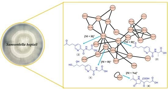
2. Results and Discussion

Compound 1 was isolated as a yellow oil. The HRESI(+)MS of 1 exhibited a protonated molecular ion [M + H]+ at m/z 269.1142, indicating a molecular formula of C12H17N2O5, accounting for seven degrees of unsaturation (DoU). The 1H NMR (Table 1) data of 1 presented the diagnostic signals of three aromatic protons, typically an ABX coupling system at δH 8.51 (1H, d, J = 1.5 Hz, H-6), 7.96 (1H, d, J = 8.0 Hz, H-3), and 7.83 (1H, dd, J = 8.0, 2.0 Hz, H-4), indicating the presence of a 2,5-disubstituted pyridine ring in 1. This was confirmed by the key heteronuclear multiple bond correlation (HMBC) cross-peaks from H-3 to C-5 (δC 141.4), from H-4 to C-2 (δC 147.3) and C-6 (δC 148.5), and from H-6 to C-2 and C-4 (δC 137.4), as well as by the correlation spectroscopy (COSY) correlations of H-3/H-4.
Furthermore, four methylene groups at δH 3.97 (2H, s, H-1′), [2.71 (1H, m, H-8), 2.82 (1H, m, H-8)] and [1.56 (1H, m, H-9), 1.77 (1H, m, H-9)], [3.25 (1H, dd, J = 10.7, 5.7 Hz, H-11), 3.32 (1H, dd, J = 10.7, 5.6 Hz, H-11)] and one oxymethine at δH 3.39 (1H, m, H-10), were observed (Table 1). The 1H-1H COSY correlations of H-8/H-9/H-10/H-11, together with the HMBC correlations from H-11 to C-10 (δC 70.2) and C-9 (δC 34.7), from H-10 to C-9 and C-8 (δC 28.3), from H-9 to C-8 and C-5, and from H-8 to C-4, C-5, and C-6 indicated a 1,2-butanediol moiety at C-5. Further HMBC correlations of H-1′ to C-7 (δC 164.2) and C-2′ (δC 171.1) suggested the presence of a glycine moiety (Figure 3). An amido bond between C-2 of the pyridine ring and C-1′ of the glycine unit was confirmed by key HMBC correlations from H-3 and H-1′ to C-7. Thus, compound 1 was identified as a new fusaric acid derivative and given the trivial name hepialiamide A. The comparison of its optical rotation value ( [ α ] D 25 + 13) with those of similar known compounds, such as (R)-4-phenyl-1,2-butanediol ( [ α ] D 20 + 33) and (S)-4-phenyl-1,2-butanediol ( [ α ] D 20 − 34) [25], and comparative analysis of the experimental and theoretically calculated ECD curves established the absolute configuration of hepialiamide A to be 10R (Figure 4).
Compound 2 was isolated as a yellow oil. The HRESI(+)MS of 2 exhibited a protonated molecular ion [M + H]+ at m/z 251.0950. Analysis of the HRESI(+)MS and 13C NMR data revealed the molecular formula C12H14N2O4 of 2 indicating seven DoUs. The NMR data (Table 1) of 2 were closely related to those of 1, except for the presence of a methyl ketone group at (δC 209.8, C-10) and (δC 29.9, δH 2.13, CH3-11) in 2, which was supported by the HMBC correlations of H3-11 to C-10 and C-9 (δC 44.6). The left of the structure was confirmed by 2D NMR correlations, as shown (Figure 3). Therefore, 2 was identified as a new analogue of 1 and named hepialiamide B.
Compound 3 was obtained as a yellow oil with the molecular formula of C13H16N2O4 implied by its HRESI(+)MS spectrum. The 1D NMR data (Table 1) of 3 were similar to those of 2 except that 3 had an extra methyl group [δC 18.2 (C-4′)] at C-2′, which was confirmed by the COSY correlations of NH (δH 8.74, d)/H-1′ (δH 4.46, d)/H-3′ (δH 1.42, d) and the HMBC correlations from H-1′ to C-2′ (δC 173.8), C-3′ (δC 17.5), and C-7 (δC 163.5). The absolute configuration of the chiral center C-1′ in 3 was established to be S, as indicated by the agreement between the calculated ECD spectrum of 3 and the experimental data (Figure 4). Hence, compound 3 was identified as another new fusaric acid derivative, and named hepialiamide C.
Compound 4 was obtained as a yellow oil. The HRESI(+)MS of 4 exhibited a sodiated molecular ion [M + Na]+ at m/z 292.0800, suggesting a molecular formula (calculated for C12H15NO6Na, 292.0797) that requires six DoUs. The 1H NMR (Table 2) data of 4 presented the diagnostic signals of three methylene groups at δH 2.28 (2H, t, J = 6.3 Hz, H-2′), 2.64 (2H, q, J = 6.9 Hz, H-3′), and 3.10 (2H, q, J = 15.0 Hz, H-3), and two methyl groups at 2.08 (3H, s, H-8) and 1.81 (3H, s, H-9). In the 13C NMR spectrum, 12 carbon signals were observed and classified as two methyl (δC 20.8, 18.5), three methylene (δC 47.4, 36.3, 27.4), and seven non-protonated carbons (δC 207.2, 197.0, 173.5, 171.4, 1129.1, 122.3, 70.3), with the aid of HSQC data. In the 1H-1H COSY spectrum of 4, homonuclear vicinal coupling correlation (Figure 3) between H-7 and H-8, along with the HMBC correlations from H-7 to C-5/6, from H-8 to C-5/6, indicated an isopropenyl group. The HMBC cross-peaks observed from H-3 to C-2/4/9, H-7/8 to C-4, and the diagnostic signals from an exchangeable proton to C-2/4/5/9 permitted the construction of a five-member ring. Finally, based on the rest of the HMBC correlations from H-3 to C-1′, H-2′ to C-1′/3′/4′, from H-3′ to C-1′/4′, and the COSY signals between H-2′ to H-3′, together with a comparable analysis of the chemical shifts of C-2/5 in 4 and those in literature [26,27], the planar structure o f 4 was assigned as shown.
To further assign the absolute configuration of C-2, a theoretical calculation of the ECD spectra of the 4R- and 4S-isomers was conducted. The experimental ECD spectrum of 4 was in good accordance with the calculated result for the 4R-configured 4 (Figure 4). The structure of compound 4 was thus determined and named hepialide.
By comparing the NMR, MS, ECD, and OR data with those reported in the references, 18 previously described components were identified as cordycepin (5) [28], adenosine (6) [29], 3′-deoxyinosine (7) [30], 5′-O-acetyladenosine (8) [31], 5′-acetyl-3′-deoxyadenosine (9) [32], thymidine (10) [33], uridine (11) [34], 5′-O-acetyluridine (12) [35], ergosterol (13) [36], 3β,5α,9α-trihydroxyergosta-7,22-diene-6-one (14) [37], 12-acetoxycycloneran-3,7-diol (15) [38], dibutyl phthalate (16) [39], di-(2-ethylhexyl)phthalate (17) [40], oryzasaccharide A (18) [41], d-glucopyranoside, ethyl, 6-acetate (19) [42], 6-O-acetyl-d-glucose (20) [43], walterolactone A (21) [44], and (4R,5S)-5-hydroxyhexan-4-olide (22) [45]. Among them, 16 and 17 were previously described as plasticizers and common contaminants in natural products [46].
Naturally occurring aminated fusaric acid derivatives are uncommon in fungal metabolites, as mentioned above. Hepialiamides A–C (13) feature an oxidized fusaric acid core structure with a glycine or alanine coupled through an amide bond, representing the third examples of fusaric amides of fungal origin. Hepialide is a novel PKS-NRPS hybrid polyketide with an unusual isopropenylated pyrrolidone moiety that resembles the tetramic acid.
Neuroinflammation, characterized by the activation of microglia cells, is a common denominator in diverse neurological conditions, including neurodegenerative diseases, traumatic brain injury, and neuroinfectious disorders. Microglia cell activation contributes significantly to the progression of these diseases by releasing pro-inflammatory mediators, exacerbating neuronal damage and impairing synaptic plasticity [47]. Hence, inhibiting glial cell activation emerges as a promising therapeutic strategy. The rich diversity and unique chemical structures of marine compounds provide a vast pool of potential drugs. Inflammatory tests were conducted on BV-2 microglia cells. Stimulation of the cells with bacterial products like lipopolysaccharide (LPS) triggers the production of NO and pro-inflammatory cytokines, key mediators in inflammation [48]. Given the significance of high NO levels in inflammation, targeting inducible nitric oxide synthase (iNOS), the enzyme responsible for NO synthesis, has been suggested as an anti-inflammatory therapeutic approach.
In this LPS-induced BV-2 microglia cell model, we evaluated the inhibitory activity of these compounds on NO production at a low concentration (1 μM). The cells were pre-exposed to the compounds for 1 h, followed by LPS stimulation for 24 h, and subsequently, the concentration of nitrite, the primary metabolite derived from NO, was quantified. The results indicated that compounds 8, 11, 13, 21, and 22 reduced NO production by 34.2 ± 1.6%, 30.7 ± 4.8%, 32.9 ± 1.6%, 38.6 ± 2.1%, and 58.2 ± 2.6%, respectively, with respect to vehicle-treated stimulated cells (Figure 5A). Particularly noteworthy, further analysis revealed that compound 22 exhibited a remarkable dose-dependent inhibitory effect, with an IC50 value of 426.2 nM (Figure 5B).
These compounds, especially compound 22, frequently demonstrate anti-inflammatory and immunomodulatory properties, rendering them promising candidates for the development of specific and potent inhibitors that target microglia cell activation pathways. Further research is needed for in-depth evaluation.

3. Materials and Methods

3.1. General Experimental Procedures

Optical rotations were recorded on a Rudolph IV Autopol automatic polarimeter at 25 °C. NMR spectra, including 1H, 13C, DEPT, HSQC, COSY, HMBC, and NOESY were measured on a Bruker Avance 400 MHz spectrometer. Chemical shifts were recorded in δ values using solvent signals (DMSO-d6: δH 2.50/δc 39.5; methanol-d4: δH 3.31/δc 49.0; CDCl3: δH 7.27/δc 77.0) as references. HRESIMS data were measured on a Xevo G2 Q-TOF mass spectrometer (Waters Corporation, Milford, MA, USA). Preparative and semipreparative HPLC were performed on an Agilent Technologies 1260 infinity instrument equipped with the DAD detector. UV spectra were recorded on a UV-8000 UV/Vis spectrometer (Shanghai Yuanxi Instrument Co., Ltd., Shanghai, China). Column chromatography (CC) was performed on ODS (50 μm, Daiso, Hiroshima, Japan), silica gel (Qingdao Marine Chemistry Co., Ltd., Qingdao, China), and Sephadex LH-20 (Amersham Pharmacia Biotech AB, Uppsala, Sweden). The TLC plates were visualized under UV light or by spraying with 10% H2SO4. Solvents for isolation were analytical grade. HRMS and MS/MS data were acquired with ultra-high performance liquid chromatography–quadrupole time-of-flight mass spectrometry (UHPLC-Q-TOF MS, AB SCIEX Triple TOF 5600+, Waters Corporation, Milford, MA, USA), with a Phenomenex Kinetex® C18 (100 × 2.1 mm, 2.6 μm).

3.2. Fungal Identification, Fermentation, and Extract

The strain W7 was isolated from a sulfide sample (W 14.52°, S 13.59°) at a depth of 3073 m from the South Atlantic. It was identified as Samsoniella hepiali (GenBank accession number OR398925), as the ITS DNA gene sequence alignment demonstrated its similarity to Samsoniella hepiali CGMCC 3.17103 (GenBank accession number NR_160318.1). The strain is preserved at the Key Laboratory of Marine Biogenetic Resources, Third Institute of Oceanography, Ministry of Natural Resources (Xiamen, China). The strain Samsoniella hepiali was cultured on a PDA plate at 25 °C for 3 days. Fresh mycelia and spores were inoculated into 250 mL Erlenmeyer flasks (×10) containing 30 mL PDB medium and incubated in a 180 rpm rotary shaker at 28 °C for 5 days. Then, the spore cultures were used to inoculate 25 × 1 L Erlenmeyer flasks containing rice medium (80 g rice and 120 mL distilled H2O for each flask) to perform large-scale fermentation. After 25 days, the fermented culture was extracted with EtOAc three times to provide a crude extract. The extract was redissolved in MeOH and extracted with petroleum ether (PE) three times. The MeOH solution was evaporated under reduced pressure to obtain a defatted extract (25 g).

3.3. UHPLC-Q-TOF and Molecular Networking Analysis

The crude extract was analyzed by LC-MS/MS, equipped with an AtlantisTM Premier BEH C18 AX (2.1 × 100 mm, 1.7 μm) column. Samples were dissolved in MeOH with 1 mg/mL. A 10 μL aliquot of each sample was injected and eluted, using a gradient program with water + 0.1% formic acid (A) and CH3CN + 0.1% formic acid (B). The elution started with a 1% B isocratic phase for 1 min, followed by a gradient from 1% to 99% B in 10 min, maintaining 99% B for 3 min. Then, a gradient from 99% to 1% B in 1 min was applied, and 1% B was maintained for 3 min at a flow rate of 0.4 mL/min, with the temperature maintained at 40 °C. The full mass spectrometry (MS) survey scan was performed in positive electrospray ionization (ESI) mode within the range of 50 to 1000 Da. The MS/MS data were converted to mzXML files using MSConvert software (version 3.01). The converted MS/MS file was submitted to the GNPS platform for molecular networking for dereplication (https://gnps.ucsd.edu, accessed on 1 January 2016). Parameters for molecular network generation were set as follows: both precursor mass and MS/MS fragment ion tolerance were set at 0.02 Da, minimum pairs cosine score 0.6, minimum matched fragment ions 6, minimum cluster size 2, network TopK 10. The spectral networks were imported into Cytoscape 3.9.1 and visualized using the force-directed layout.

3.4. Isolation and Purification

The MeOH extract (25 g) was subjected to CC over silica gel using sequential gradient elution with CH2Cl2-MeOH (100:1, 50:1, 30:1, 20:1, 10:1, 5:1, 2:1, and 0:1) to obtain five fractions (Fr.1–Fr.5), based on TLC properties. Fraction Fr.1 (5 g) was chromatographed over ODS using gradient elution of MeOH-H2O (30 → 80%) to get five subfractions (Fr.1-1–Fr.1-5). Fr.1-1 (625 mg) was subjected to CC on Sephadex LH-20 (MeOH), followed by repeated CC over silica gel, using gradient PE-EtOAc (3:1 → 1:1) to yield 17 (125 mg) and 2 (62 mg). Fr.1-2 (34 mg) was directly separated by Sephadex LH-20 (MeOH) to yield 15 (2 mg). Fr.1-3 (215 mg) was subsequently separated by CC over Sephadex LH-20 (MeOH), silica gel (CH2Cl2-MeOH, 100:1 → 50:1), and preparative TLC (PTLC) (CH2Cl2-MeOH, 20:1) to obtain 22 (14 mg) and 21 (2 mg). Fr.1-4 (137 mg) was subjected to CC over Sephadex LH-20 (MeOH) and silica gel (CH2Cl2-MeOH, 75:1) to yield 3 (40 mg). Fr.1-5 (226 mg) was purified by CC over Sephadex LH-20 (MeOH), silica gel (CH2Cl2-MeOH, 100:1), and preparative TLC (PTLC) (CH2Cl2-MeOH, 100:1) to afford compound 14 (7 mg).
Fraction Fr.2 (1.3 g) was subjected to CC over ODS with MeOH-H2O (2% → 30%), followed by purification using CC on Sephadex LH-20 (MeOH), crystallization (MeOH), and silica gel (EtOAc-MeOH, 20:1 → 5:1) to yield 20 (84 mg), 18 (29 mg), 5 (46 mg), 6 (5 mg), 10 (11 mg), 7 (7 mg), 1 (15 mg), and 11 (26 mg).
Fraction Fr.3 (1.5 g) was subjected to CC over ODS with MeOH-H2O (2% → 50%) and CC on Sephadex LH-20 (MeOH) to yield 19 (4 mg), 4 (11 mg), 12 (11 mg), and 8 (4 mg).
Fraction Fr.4 (9.8 g) was separated by CC over silica gel using sequential gradient elution with PE-EtOAc (100:1, 50:1, 30:1, 20:1, 10:1) and Sephadex LH-20 (CH2Cl2-MeOH, 1:1) to yield 13 (320 mg).
Fraction Fr.5 (5.3 g) was subjected to CC over ODS with MeOH-H2O (10% → 50%), followed by purification using CC on Sephadex LH-20 (MeOH), HPLC (20% → 48%, MeOH-H2O), crystallization (MeOH), and silica gel (EtOAc-MeOH, 20:1) to yield 9 (2 mg) and 16 (4 mg).
Hepialiamide A (1): Yellow oil; [ α ] D 25 + 13 (c 0.10, MeOH), ECD (MeOH) λmax (Δε) 278 (0.07), 245 (−0.09), 217 (0.07); UV (MeOH) λmax (log ε) 229 (2.72), 269 (2.31) nm; HRESIMS m/z 269.1142 [M + H]+ (calcd for C12H17N2O5, 269.1137).
Hepialiamide B (2): Yellow oil; UV (MeOH) λmax (log ε) 230 (2.76), 269 (2.38) nm; 1H and 13C NMR data, Table 1; HRESIMS m/z 251.0950 [M + H]+ (calcd for C12H15N2O4, 251.1032).
Hepialiamide C (3): Yellow oil; [ α ] D 25 + 3 (c 0.10, MeOH); ECD (MeOH) λmax (Δε) 275 (−0.15), 250 (0.13), 213 (−0.17); UV (MeOH) λmax (log ε) 200 (3.06), 269 (2.23) nm; HRESIMS m/z 265.111 [M + H]+ (calcd for C13H17N2O4, 265.1188).
Hepialide (4): Yellow oil; [ α ] D 25 + 5 (c 0.10, MeOH); ECD (MeOH) λmax (Δε) 211 (−0.28); UV (MeOH) λmax (log ε) 228 (2.53), 291 (1.91) nm; HRESIMS m/z 292.0800 [M + Na]+ (calcd for C12H15NO6Na, 292.0797).

3.5. Theoretical ECD Calculation

As reported previously, conformational analysis was first performed via random searching in Sybyl-X 2.0 using the MMFF94S force field with an energy cutoff of 7.0 kcal/mol and an RMSD threshold of 0.2 Å. All conformers were consecutively optimized at PM6 and HF/6-31G(d) levels. Dominant conformers were re-optimized at the B3LYP/6-31G(d) level in the gas phase. The theoretical ECD spectra were calculated using the GIAO method at the MPW1PW91/6-31G (d, p) level in MeOH using Gaussian 09. The ECD spectrum was simulated in SpecDis (version 1.71) by overlapping Gaussian functions for each transition.

3.6. BV-2 Cell Culture and Treatment

BV-2 microglia cells were cultured in DMEM medium containing 10% fetal bovine serum and antibiotics (100 units/mL of penicillin and 100 g/mL of streptomycin) and maintained in a humidified 5% CO2 incubator at 37 °C. For the experiment, cells were seeded overnight into 24-wells with 2 × 10⁴ cells per well. The next day, cells were incubated with fresh culture medium containing the indicated concentration of compounds for an hour, followed by lipopolysaccharides (LPS) treatment (1 μg/mL). Cells treated with the vehicle (DMSO, 0.1%) served as the control.

3.7. Nitrite Quantification

The concentration of nitrite in the culture medium was determined using the Griess Reagent Kit (Therofisher, Shanghai, China). Briefly, 75 μL of cell culture supernatants was reacted with an equal volume of the Griess Reagent Kit for 30 min at room temperature, and the absorbance of the diazonium compound was obtained at a wavelength of 560 nm. Nitrite production by vehicle stimulation was designated as 100% inhibition (Ctrl) compared to LPS stimulation (Veh) for the experiment.

4. Conclusions

In summary, three new aminated fusaric acid derivatives, hepialiamides A–C (13), and one novel hybrid polyketide, hepialide (4), together with 18 known miscellaneous compounds (522), were isolated from the cultures of the deep-sea-derived fungus Samsoniella hepiali W7 with the aid of GNPS molecular networking. Hepialiamides A–C feature a glycine/alanine unit embedded in the fusaric acid backbone, representing the third examples of naturally occurring fusaric amides, while hepialide features an uncommon isopropenylated pyrrolidone moiety. Biologically, compound 8, 11, 13, 21, and 22 showed more than 30% inhibition against NO production induced by LPS at 1 μM, with 22 exhibiting a particularly low nanomole IC50, suggesting potential for developing potent anti-inflammatory drugs.

Supplementary Materials

The following are available online at https://www.mdpi.com/article/10.3390/md21110596/s1. Figures S1–S52: One-dimensional and two-dimensional NMR, UV spectra, ECD calculation details of compounds 14, proton NMR spectra of 522.

Author Contributions

X.-W.Y. and J.-S.W. designed the project; Z.-B.Z. isolated and purified compounds; Z.-B.Z. and W.-Y.L. performed GNPS analysis; L.-H.Y. and X.-W.H. conducted the biological experiments; K.Z. identified the strains; C.-L.X., M.-M.X. and Y.Z. performed the fermentation; X.-W.Y., Z.-B.Z. and T.-Z.W. analyzed the data and wrote the paper, while critical revisions of the publication were performed by all authors. All authors have read and agreed to the published version of the manuscript.

Funding

This work was financially supported by the Xiamen Southern Oceanographic Center (22GYY007HJ07) and the National Natural Science Foundation of China (22177143).

Institutional Review Board Statement

Not applicable.

Informed Consent Statement

Not applicable.

Data Availability Statement

The data presented in this study are available in Supplementary Materials.

Conflicts of Interest

The authors declare no conflict of interest.

References

  1. Newman, D.J.; Cragg, G.M. Natural products as sources of new drugs over the nearly four decades from 01/1981 to 09/2019. J. Nat. Prod. 2020, 83, 770–803. [Google Scholar] [CrossRef] [PubMed]
  2. Haque, N.; Parveen, S.; Tang, T.; Wei, J.; Huang, Z. Marine natural products in clinical use. Mar. Drugs 2022, 20, 528. [Google Scholar] [CrossRef] [PubMed]
  3. Teng, Y.F.; Xu, L.; Wei, M.Y.; Wang, C.Y.; Gu, Y.C.; Shao, C.L. Recent progresses in marine microbial-derived antiviral natural products. Arch. Pharm. Res. 2020, 43, 1215–1229. [Google Scholar] [CrossRef] [PubMed]
  4. Carroll, A.R.; Copp, B.R.; Davis, R.A.; Keyzers, R.A.; Prinsep, M.R. Marine natural products. Nat. Prod. Rep. 2021, 38, 362–413. [Google Scholar] [CrossRef]
  5. Hai, Y.; Cai, Z.M.; Li, P.J.; Wei, M.Y.; Wang, C.Y.; Gu, Y.C.; Shao, C.L. Trends of antimalarial marine natural products: Progresses, challenges and opportunities. Nat. Prod. Rep. 2022, 39, 969–990. [Google Scholar] [CrossRef]
  6. Hafez Ghoran, S.; Taktaz, F.; Ayatollahi, S.A.; Kijjoa, A. Anthraquinones and their analogues from marine-derived fungi: Chemistry and biological activities. Mar. Drugs 2022, 20, 474. [Google Scholar] [CrossRef] [PubMed]
  7. Carroll, A.R.; Copp, B.R.; Davis, R.A.; Keyzers, R.A.; Prinsep, M.R. Marine natural products. Nat. Prod. Rep. 2022, 39, 1122–1171. [Google Scholar] [CrossRef]
  8. Carroll, A.R.; Copp, B.R.; Davis, R.A.; Keyzers, R.A.; Prinsep, M.R. Marine natural products. Nat. Prod. Rep. 2023, 40, 275–325. [Google Scholar] [CrossRef]
  9. Yang, J.Y.; Sanchez, L.M.; Rath, C.M.; Liu, X.; Boudreau, P.D.; Bruns, N.; Glukhov, E.; Wodtke, A.; de Felicio, R.; Fenner, A.; et al. Molecular networking as a dereplication strategy. J. Nat. Prod. 2013, 76, 1686–1699. [Google Scholar] [CrossRef] [PubMed]
  10. Nothias, L.F.; Petras, D.; Schmid, R.; Duhrkop, K.; Rainer, J.; Sarvepalli, A.; Protsyuk, I.; Ernst, M.; Tsugawa, H.; Fleischauer, M.; et al. Feature-based molecular networking in the GNPS analysis environment. Nat. Methods 2020, 17, 905–908. [Google Scholar] [CrossRef] [PubMed]
  11. Aron, A.T.; Gentry, E.C.; McPhail, K.L.; Nothias, L.F.; Nothias-Esposito, M.; Bouslimani, A.; Petras, D.; Gauglitz, J.M.; Sikora, N.; Vargas, F.; et al. Reproducible molecular networking of untargeted mass spectrometry data using GNPS. Nat. Protoc. 2020, 15, 1954–1991. [Google Scholar] [CrossRef]
  12. Wei, X.; Su, J.C.; Hu, J.S.; He, X.X.; Lin, S.J.; Zhang, D.M.; Ye, W.C.; Chen, M.F.; Lin, H.W.; Zhang, C.X. Probing indole diketopiperazine-based hybrids as environmental-induced products from Aspergillus sp. EGF 15-0-3. Org. Lett. 2022, 24, 158–163. [Google Scholar] [CrossRef] [PubMed]
  13. Ding, W.J.; Tian, D.M.; Chen, M.; Xia, Z.X.; Tang, X.Y.; Zhang, S.H.; Wei, J.H.; Li, X.N.; Yao, X.S.; Wu, B.; et al. Molecular networking-guided isolation of cyclopentapeptides from the hydrothermal vent sediment derived fungus Aspergillus pseudoviridinutans TW58-5 and their anti-inflammatory effects. J. Nat. Prod. 2023, 86, 1919–1930. [Google Scholar] [CrossRef] [PubMed]
  14. Wang, Y.; Yang, J.; Hu, L.; Bai, R.; Wang, T.; Xing, X.; Chen, L.; Ding, G. LC-MS/MS-guided molecular networking for targeted discovery of undescribed and bioactive ophiobolins from Bipolaris eleusines. J. Agric. Food Chem. 2023, 71, 11982–11992. [Google Scholar] [CrossRef] [PubMed]
  15. Dobson, T.A.; Desaty, D.; Brewer, D.; Vining, L.C. Biosynthesis of fusaric acid in cultures of Fusarium oxysporum Schlecht. Can. J. Biochem. 1967, 45, 809–823. [Google Scholar] [CrossRef]
  16. Stipanovic, R.D.; Wheeler, M.H.; Puckhaber, L.S.; Liu, J.; Bell, A.A.; Williams, H.J. Nuclear magnetic resonance (NMR) studies on the biosynthesis of fusaric acid from Fusarium oxysporum f. sp. vasinfectum. J. Agric. Food Chem. 2011, 59, 5351–5356. [Google Scholar] [CrossRef] [PubMed]
  17. Studt, L.; Janevska, S.; Niehaus, E.M.; Burkhardt, I.; Arndt, B.; Sieber, C.M.; Humpf, H.U.; Dickschat, J.S.; Tudzynski, B. Two separate key enzymes and two pathway-specific transcription factors are involved in fusaric acid biosynthesis in Fusarium fujikuroi. Environ. Microbiol. 2016, 18, 936–956. [Google Scholar] [CrossRef] [PubMed]
  18. Hai, Y.; Chen, M.; Huang, A.; Tang, Y. Biosynthesis of mycotoxin fusaric acid and application of a PLP-dependent enzyme for chemoenzymatic synthesis of substituted l-pipecolic acids. J. Am. Chem. Soc. 2020, 142, 19668–19677. [Google Scholar] [CrossRef] [PubMed]
  19. Yang, Z.; Dan, W.J.; Li, Y.X.; Peng, G.R.; Zhang, A.L.; Gao, J.M. Antifungal metabolites from Alternaria atrans: An endophytic fungus in Psidium guajava. Nat. Prod. Commun. 2019, 14, 1934578X19844116. [Google Scholar] [CrossRef]
  20. Hilário, F.; Chapla, V.M.; Araujo, A.R.; Sano, P.T.; Bauab, T.M.; dos Santos, L.C. Antimicrobial screening of endophytic fungi isolated from the aerial parts of Paepalanthus chiquitensis (Eriocaulaceae) led to the isolation of secondary metabolites produced by Fusarium fujikuroi. J. Braz. Chem. Soc. 2016, 28, 1389–1395. [Google Scholar] [CrossRef]
  21. Zou, Z.B.; Chen, L.H.; Hu, M.Y.; Xu, L.; Hao, Y.J.; Yan, Q.X.; Wang, C.F.; Xie, C.L.; Yang, X.W. Cladosporioles A and B, two new indole derivatives from the deep-sea-derived fungus Cladosporium cladosporioides 170056. Chem. Biodivers. 2022, 19, e202200538. [Google Scholar] [CrossRef]
  22. Zou, Z.B.; Zhang, G.; Zhou, Y.Q.; Xie, C.L.; Xie, M.M.; Xu, L.; Hao, Y.J.; Luo, L.Z.; Zhang, X.K.; Yang, X.W.; et al. Chemical constituents of the deep-sea-derived Penicillium citreonigrum MCCC 3A00169 and their antiproliferative effects. Mar. Drugs 2022, 20, 736. [Google Scholar] [CrossRef] [PubMed]
  23. He, Z.H.; Xie, C.L.; Wu, T.; Yue, Y.T.; Wang, C.F.; Xu, L.; Xie, M.M.; Zhang, Y.; Hao, Y.J.; Xu, R.; et al. Tetracyclic steroids bearing a bicyclo [4.4.1] ring system as potent antiosteoporosis agents from the deep-sea-derived fungus Rhizopus sp. W23. J. Nat. Prod. 2023, 86, 157–165. [Google Scholar] [CrossRef]
  24. He, Z.H.; Xie, C.L.; Wu, T.; Zhang, Y.; Zou, Z.B.; Xie, M.M.; Xu, L.; Capon, R.J.; Xu, R.; Yang, X.W. Neotricitrinols A-C, unprecedented citrinin trimers with anti-osteoporosis activity from the deep-sea-derived Penicillium citrinum W23. Bioorg. Chem. 2023, 139, 106756. [Google Scholar] [CrossRef] [PubMed]
  25. Wang, F.D.; Yue, J.M. A total synthesis of (+)- and (−)-dihydrokavain with a sonochemical blaise reaction as the key step. Eur. J. Org. Chem. 2005, 2005, 2575–2579. [Google Scholar] [CrossRef]
  26. Roberts, A.; Beaumont, C.; Manzarpour, A.; Mantle, P. Purpurolic acid: A new natural alkaloid from Claviceps purpurea (Fr.) Tul. Fungal Biol. 2016, 120, 104–110. [Google Scholar] [CrossRef] [PubMed]
  27. Zhang, Z.; He, X.; Wu, G.; Liu, C.; Lu, C.; Gu, Q.; Che, Q.; Zhu, T.; Zhang, G.; Li, D. Aniline-tetramic acids from the deep-sea-derived fungus Cladosporium sphaerospermum L3P3 cultured with the HDAC inhibitor SAHA. J. Nat. Prod. 2018, 81, 1651–1657. [Google Scholar] [CrossRef] [PubMed]
  28. Li, Y.Q.; Park, H.J.; Han, E.S.; Park, D.K. Inhibitory effect on B16/F10 mouse melanoma cell and HT-29 human colon cancer cell proliferation and cordycepin content of the butanol extract of Paecilomyces militaris. J. Med. Plant Res. 2011, 5, 1066–1071. [Google Scholar]
  29. Domondon, D.L.; He, W.; De Kimpe, N.; Hofte, M.; Poppe, J. Beta-adenosine, a bioactive compound in grass chaff stimulating mushroom production. Phytochemistry 2004, 65, 181–187. [Google Scholar] [CrossRef]
  30. Qiu, W.; Wu, J.; Choi, J.H.; Hirai, H.; Nishida, H.; Kawagishi, H. Cytotoxic compounds against cancer cells from Bombyx mori inoculated with Cordyceps militaris. Biosci. Biotechnol. Biochem. 2017, 81, 1224–1226. [Google Scholar] [CrossRef]
  31. Tatani, K.; Hiratochi, M.; Nonaka, Y.; Isaji, M.; Shuto, S. Identification of 8-aminoadenosine derivatives as a new class of human concentrative nucleoside transporter 2 inhibitors. ACS. Med. Chem. Lett. 2015, 6, 244–248. [Google Scholar] [CrossRef] [PubMed]
  32. Xue, Y.; Wu, L.; Ding, Y.; Cui, X.; Han, Z.; Xu, H. A new nucleoside and two new pyrrole alkaloid derivatives from Cordyceps militaris. Nat. Prod. Res. 2020, 34, 341–350. [Google Scholar] [CrossRef]
  33. Youssef, D.T.A.; Badr, J.M.; Shaala, L.A.; Mohamed, G.A.; Bamanie, F.H. Ehrenasterol and biemnic acid; new bioactive compounds from the Red Sea sponge Biemna ehrenbergi. Phytochem. Lett. 2015, 12, 296–301. [Google Scholar] [CrossRef]
  34. Ma, Y.T.; Qiao, L.R.; Shi, W.Q.; Zhang, A.L.; Gao, J.M. Metabolites produced by an endophyte Alternaria alternata isolated from Maytenus hookeri. Chem. Nat. Compd. 2010, 46, 504–506. [Google Scholar] [CrossRef]
  35. Ying, Y.M.; Shan, W.G.; Liu, W.H.; Zhan, Z.J. Alkaloids and nucleoside derivatives from a fungal endophyte of Huperzia serrata. Chem. Nat. Comd. 2013, 49, 184–186. [Google Scholar] [CrossRef]
  36. Kaaniche, F.; Hamed, A.; Abdel-Razek, A.S.; Wibberg, D.; Abdissa, N.; El Euch, I.Z.; Allouche, N.; Mellouli, L.; Shaaban, M.; Sewald, N. Bioactive secondary metabolites from new endophytic fungus Curvularia. sp isolated from Rauwolfia macrophylla. PLoS ONE 2019, 14, e0217627. [Google Scholar] [CrossRef]
  37. Sun, Y.; Tian, L.; Huang, J.; Li, W.; Pei, Y.H. Cytotoxic sterols from marine-derived fungus Pennicillium sp. Nat. Prod. Res. 2006, 20, 381–384. [Google Scholar] [CrossRef] [PubMed]
  38. Fang, S.T.; Wang, Y.J.; Ma, X.Y.; Yin, X.L.; Ji, N.Y. Two new sesquiterpenoids from the marine-sediment-derived fungus Trichoderma harzianum P1-4. Nat. Prod. Res. 2019, 33, 3127–3133. [Google Scholar] [CrossRef]
  39. Dearman, R.J.; Cumberbatch, M.; Hilton, J.; Clowes, H.M.; Fielding, I.; Heylings, J.R.; Kimber, I. Influence of dibutyl phthalate on dermal sensitization to fluorescein isothiocyanate. Fundam. Appl. Toxicol. 1996, 33, 24–30. [Google Scholar] [CrossRef] [PubMed]
  40. Zeiger, E.; Haworth, S.; Mortelmans, K.; Speck, W. Mutagenicity testing of di(2-ethylhexyl)phthalate and related chemicals in Salmonella. Environ. Mutagen. 1985, 7, 213–232. [Google Scholar] [CrossRef]
  41. Wang, W.; Guo, J.; Zhang, J.; Liu, T.; Xin, Z. New screw lactam and two new carbohydrate derivatives from the methanol extract of rice bran. J. Agric. Food. Chem. 2014, 62, 10744–10751. [Google Scholar] [CrossRef]
  42. Theil, F.; Schick, H. Enzymes in organic-synthesis. 5. An improved procedure for the regioselective acetylation of monosaccharide derivatives by pancreatin-catalyzed transesterification in organic-solvents. Synthesis 1991, 22, 533–535. [Google Scholar] [CrossRef]
  43. Park, S.; Kazlauskas, R.J. Improved preparation and use of room-temperature ionic liquids in lipase-catalyzed enantio- and regioselective acylations. J. Org. Chem. 2001, 66, 8395–8401. [Google Scholar] [CrossRef] [PubMed]
  44. Li, X.B.; Li, Y.L.; Zhou, J.C.; Yuan, H.Q.; Wang, X.N.; Lou, H.X. A new diketopiperazine heterodimer from an endophytic fungus Aspergillus niger. J. Asian Nat. Prod. Res. 2015, 17, 182–187. [Google Scholar] [CrossRef] [PubMed]
  45. Yan, H.J.; Gao, S.S.; Li, C.S.; Li, X.M.; Wang, B.G. Chemical constituents of a marine-derived endophytic fungus Penicillium commune G2M. Molecules 2010, 15, 3270–3275. [Google Scholar] [CrossRef]
  46. Tao, Y.; Feng, C.; Xu, J.; Shen, L.; Qu, J.; Ju, H.; Yan, L.; Chen, W.; Zhang, Y. Di(2-ethylhexyl) phthalate and dibutyl phthalate have a negative competitive effect on the nitrification of black soil. Chemosphere 2022, 293, 133554. [Google Scholar] [CrossRef]
  47. Silvin, A.; Uderhardt, S.; Piot, C.; Da Mesquita, S.; Yang, K.; Geirsdottir, L.; Mulder, K.; Eyal, D.; Liu, Z.; Bridlance, C.; et al. Dual ontogeny of disease-associated microglia and disease inflammatory macrophages in aging and neurodegeneration. Immunity 2022, 55, 1448–1465. [Google Scholar] [CrossRef]
  48. Orihuela, R.; McPherson, C.A.; Harry, G.J. Microglial M1/M2 polarization and metabolic states. Br. J. Pharmacol. 2016, 173, 649–665. [Google Scholar] [CrossRef]
Figure 1. Molecular network of extract of the fungus Samsoniella hepiali W7. The cluster of new compounds (14) is expanded, and the nodes are marked with the m/z value of the parent ion.
Marinedrugs 21 00596 g001
Figure 2. Compounds 122 from Samsoniella hepiali W7.
Marinedrugs 21 00596 g002
Figure 3. Key HMBC (arrows) and 1H–1H COSY (bold) correlations of compounds 14.
Marinedrugs 21 00596 g003
Figure 4. Experimental and calculated ECD spectra of 1, 3, and 4.
Marinedrugs 21 00596 g004
Figure 5. The impact of compounds on NO release in LPS-induced microglia cells: (A) Nitrite accumulation in the culture media was quantified using the Griess method, and nitrite production inhibition was calculated compared to the only LPS treated group. Results were normalized to the LPS condition and presented as mean ± SEM (n = 4); **** p < 0.0001 vs. Ctrl; ### p < 0.0001, ## p < 0.01 vs. Veh; (B) Inhibitory curve of compound 22 against nitrite production.
Marinedrugs 21 00596 g005
Table 1. 1H (400 Hz) and 13C NMR (100 Hz) spectroscopic data of compounds 13.
No.1 a (δC, Type)1 a (δH, Mult J in Hz)2 b (δC, Type)2 b (δH, Mult J in Hz)3 a (δC, Type)3 a (δH, Mult J in Hz)
2147.3, C 148.4, C 147.4, C
3121.7, CH7.96, d (8.0)122.8, CH7.96, d (8.0)121.6, CH7.93, d (7.6)
4137.4, CH7.83, dd (2.0, 8.0)138.3, CH7.77, dd (2.0, 8.0)137.4, CH7.82, d (8.0)
5141.4, C 141.9, C 141.3, C
6148.5, CH8.51, d (1.5)150.1, CH8.47, d (1.6)148.7, CH8.51, s
7164.2, C 167.0, C 163.5, C
828.3, CH22.71, m; 2.82, m27.5, CH22.90, d (5.2)26.1, CH22.85, m
934.7, CH21.56, m; 1.77, m44.6, CH22.88, d (5.2)43.3, CH22.84, m
1070.2, CH3.39, m209.8, C 207.3, C
1165.8, CH23.25, dd (5.7, 10.7)29.9, CH32.13, s29.7, CH32.09, s
3.32, dd (5.6, 10.7)
1′41.0, CH23.97, s41.9, CH24.15, s47.6, CH4.46, d (7.1)
2′171.1, C 172.8, C 173.8, C
3′ 17.5, CH31.42, d (7.1)
NH 8.92, t (6.0) 9.04, t (5.4) 8.74, d (7.3)
a Measured in DMSO-d6. b Measured in methanol-d4.
Table 2. 1H (400 Hz) and 13C (100 Hz) NMR spectroscopic data of compound 4 in DMSO-d6.
No.δC, TypeδH, Mult (J in Hz)
270.3, C
347.4, CH23.15, d (18.0); 3.06, d (18.0)
4197.0, C
5122.3, C
6129.1, C
718.5, CH32.08, s
820.8, CH31.81, s
9171.4, C
1′207.2, C
2′27.4, CH22.28, t (6.3)
3′36.3, CH22.68, m; 2.60, m
4′173.5, C
NH 10.27, s
Disclaimer/Publisher’s Note: The statements, opinions and data contained in all publications are solely those of the individual author(s) and contributor(s) and not of MDPI and/or the editor(s). MDPI and/or the editor(s) disclaim responsibility for any injury to people or property resulting from any ideas, methods, instructions or products referred to in the content.

