Article

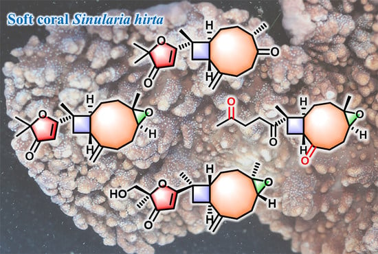
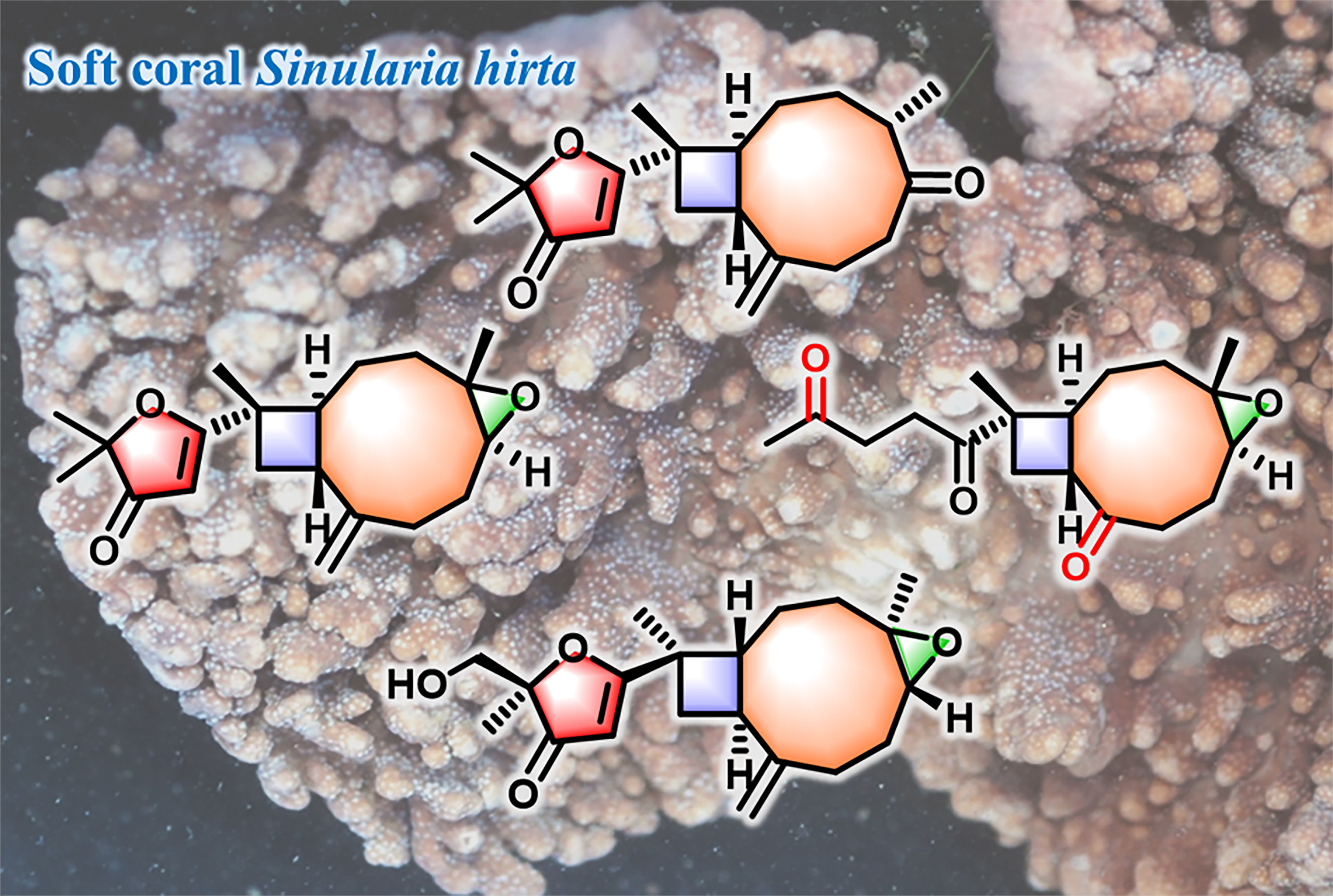
Sinuhirtone A, An Uncommon 17,19-Dinorxeniaphyllanoid, and Nine Related New Terpenoids from the Hainan Soft Coral Sinularia hirta

1
State Key Laboratory of Drug Research, Shanghai Institute of Materia Medica, Chinese Academy of Sciences, 555 Zu Chong Zhi Road, Zhangjiang Hi-Tech Park, Shanghai 201203, China
2
University of Chinese Academy of Sciences, No. 19A Yuquan Road, Beijing 100049, China
3
Jinzhou Medical University, Jinzhou 121001, China
4
Drug Discovery Shandong Laboratory, Bohai Rim Advanced Research Institute for Drug Discovery, Yantai 264117, China
*
Authors to whom correspondence should be addressed.
Mar. Drugs 2022, 20(4), 272; https://doi.org/10.3390/md20040272
Submission received: 1 April 2022 / Revised: 15 April 2022 / Accepted: 16 April 2022 / Published: 18 April 2022

Abstract

:
Chemical investigation of the Hainan soft coral Sinularia hirta resulted in the isolation and identification of a library of sixteen structurally diverse terpenoids, including a dinorditerpenoid with an uncommon 17,19-dinorxeniaphyllane skeleton, namely sinuhirtone A (7), six new xeniaphyllane-type diterpenoids (16), one new norxeniaphyllanoid (8), two new norcaryophyllene-type sesquiterpenoids (9 and 10), together with six known related compounds (1116). Compounds 13 are three new furanone-containing xeniaphyllane-type diterpenoids. The structures of the new compounds, including their absolute configurations, were determined by extensive spectroscopic analysis and a series of quantum chemical calculations, including quantum mechanical-nuclear magnetic resonance (QM–NMR), time-dependent density functional theory–electronic circular dichroism (TDDFT–ECD), and optical rotatory dispersion (ORD) methods. A plausible biosynthetic connection between new compounds 19 was also proposed. New compounds 24, 7, and 8 were evaluated for in vitro cytotoxicity against four cancer cell lines.

Graphical Abstract

1. Introduction

Soft corals of the genus Sinularia (phylum Cnidaria, class Anthozoa, subclass Octocorallia, order Alcyonacea, family Alcyoniidae) are rich sources of various sesquiterpenoids and diterpenoids with diverse structural features [1,2,3]. Many of these metabolites merit further investigation due to their potent biological activities, such as anti-inflammatory [4], cytotoxic [5], immunosuppressive [6], anti-bacterial [7], and antifouling [8] effects. Among all the species of Sinularia, S. hirta (Andaman and Nicobar Islands, India) has only been chemically investigated by Anjaneyulu and co-workers, with two steroids, two cembranoids, and two sesquiterpenoids having been reported [9,10]. Hence, S. hirta deserves a more-in-depth investigation, both chemically and biologically.
During our continuing efforts to explore bioactive secondary metabolites from South China Sea invertebrates [11,12,13,14,15], S. hirta was collected off the coast of Yalong Bay, Hainan, China, and chemically investigated, resulting in the initial isolation of two new norhumulene-type norsesquiterpenoids, sinuhirtins A and B, together with two known norcaryophyllene-type sesquiterpenoids [16]. In order to accumulate more such structurally interesting compounds for biological study, a further systematic chemical investigation of this specimen was carried out this time, resulting in the isolation of a novel dinorditerpenoid, namely sinuhirtone A (7); six new xeniaphyllanoids, namely sinuhirfuranones A–C (13) and sinuhirtins C–E (46); a norxeniaphyllanoid, namely sinuhirtone B (8); two new norcaryophyllenoids, namely sinuhirtins F (9) and G (10); together with six known related compounds (1116). It is worth mentioning that sinuhirtone A features an uncommon 17,19-dinorxeniaphyllane-type skeleton, and sinuhirfuranones A–C represent the first example of marine diterpenoids possessing the 2,2-dimethylfuran-3-one group (Figure 1). Herein, we report the isolation, structural elucidation, and plausible biosynthetic pathway of the new isolates and their bioactivity evaluation.

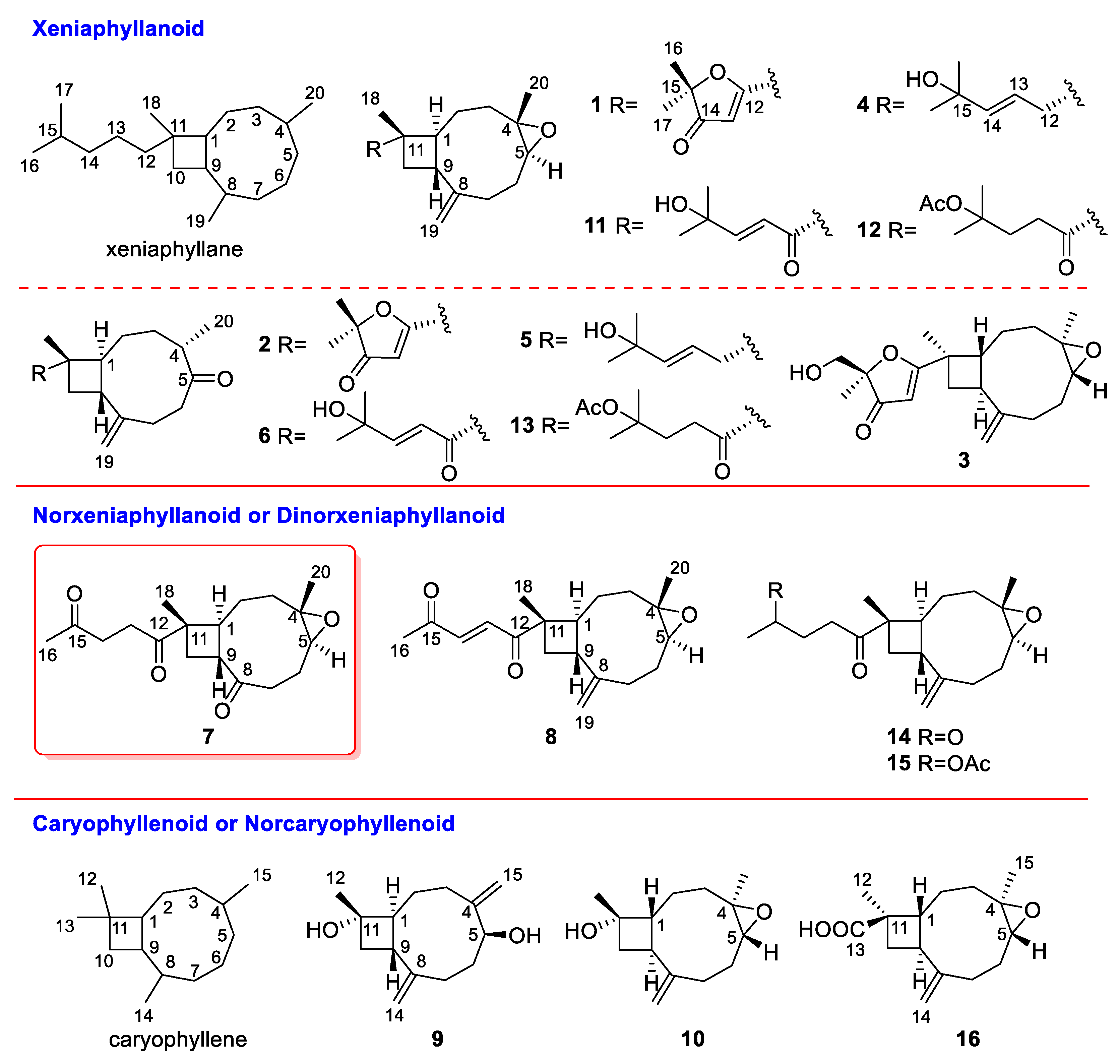
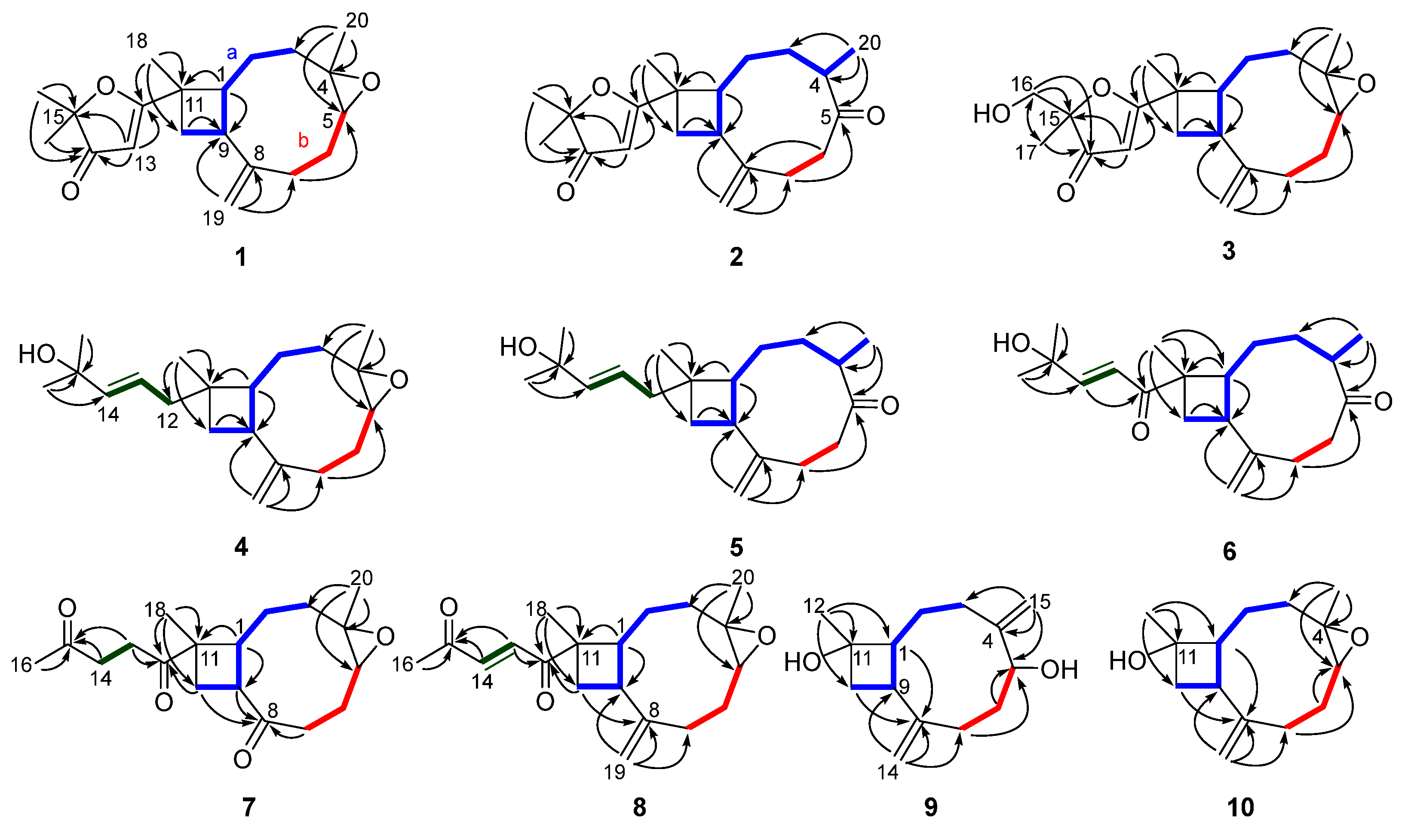
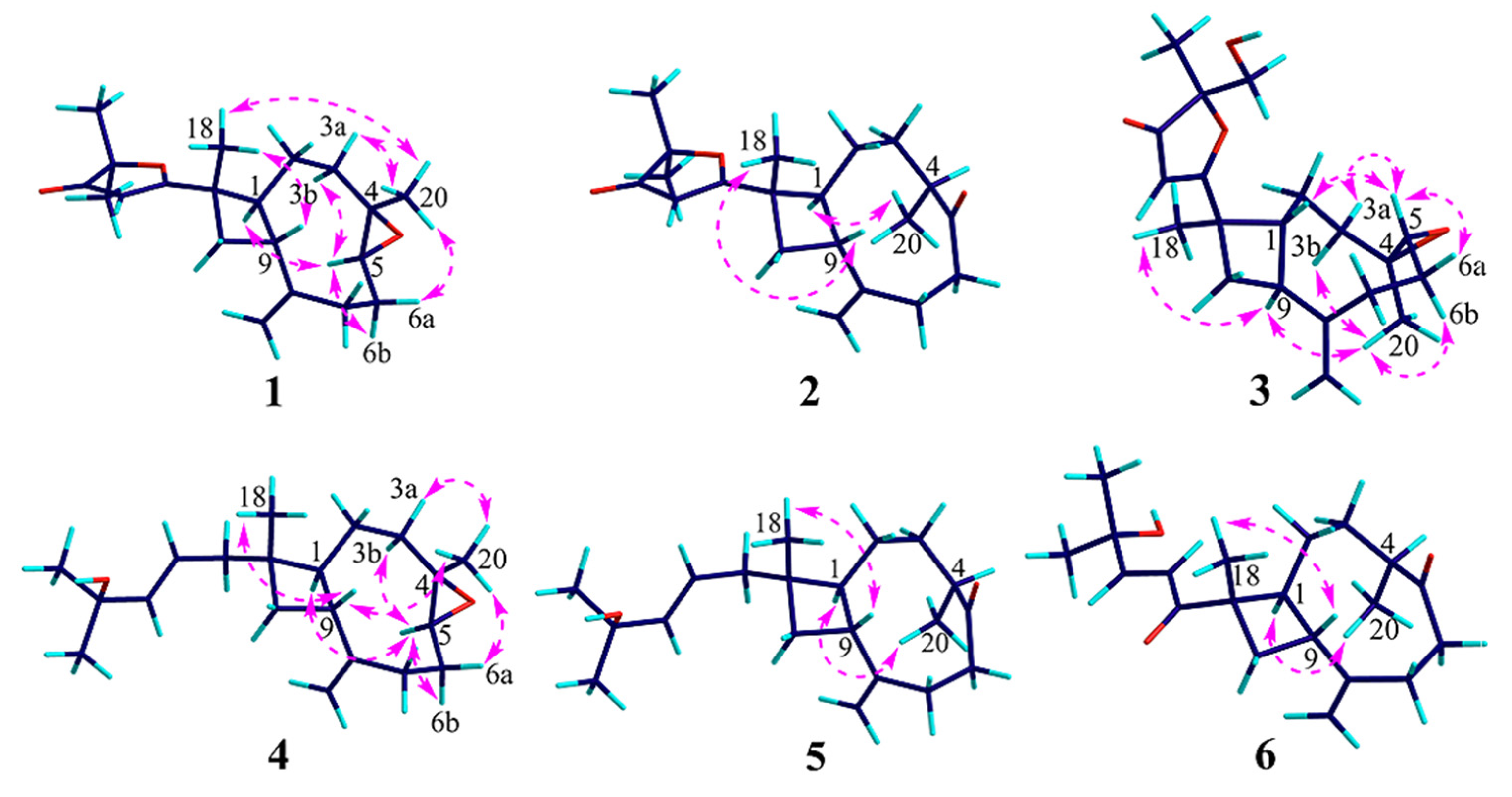
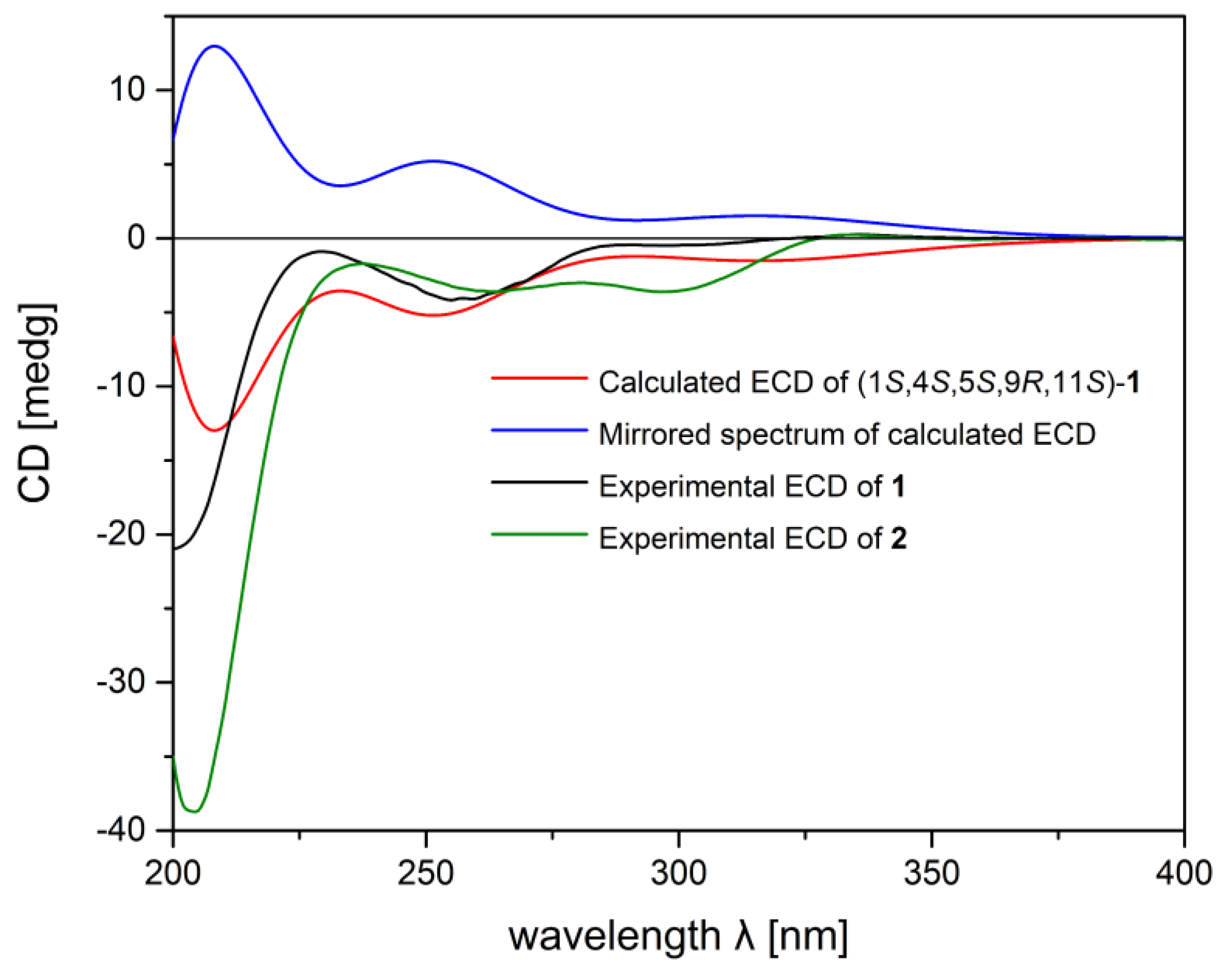
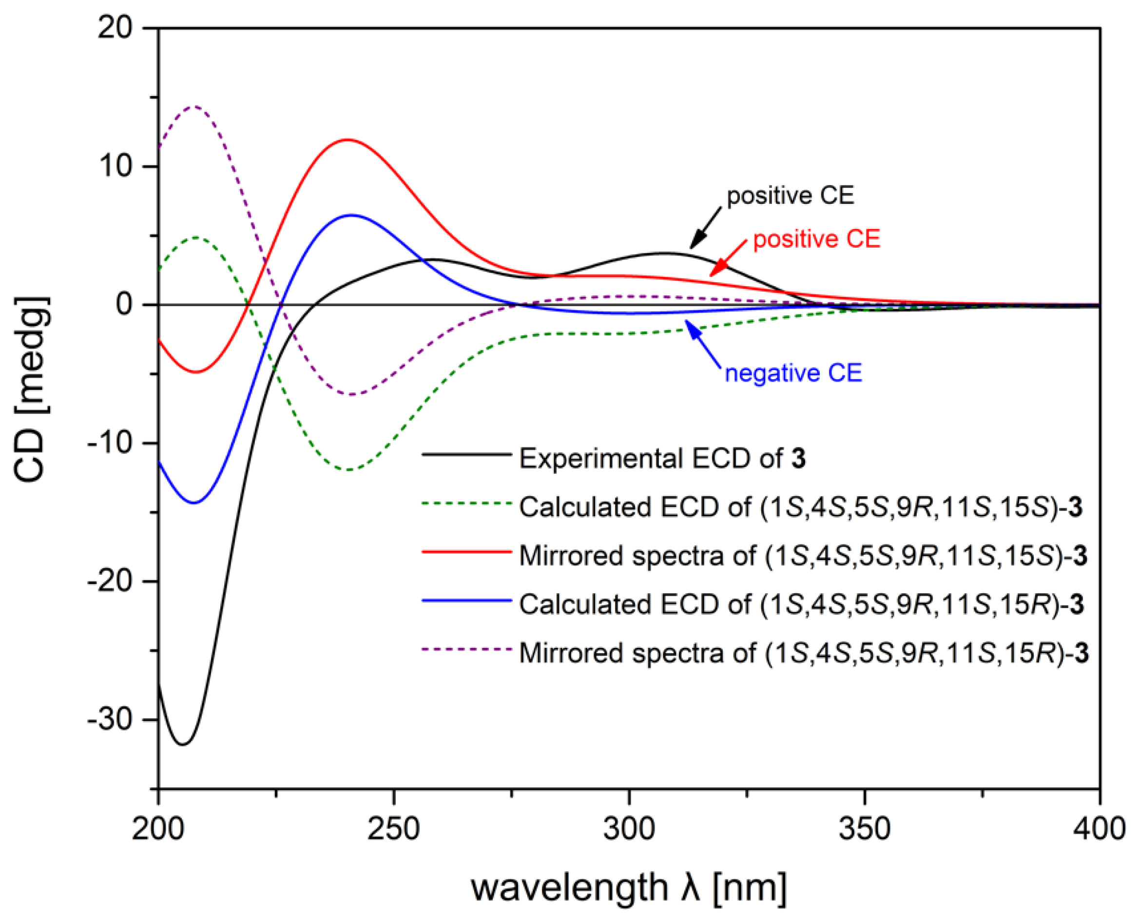
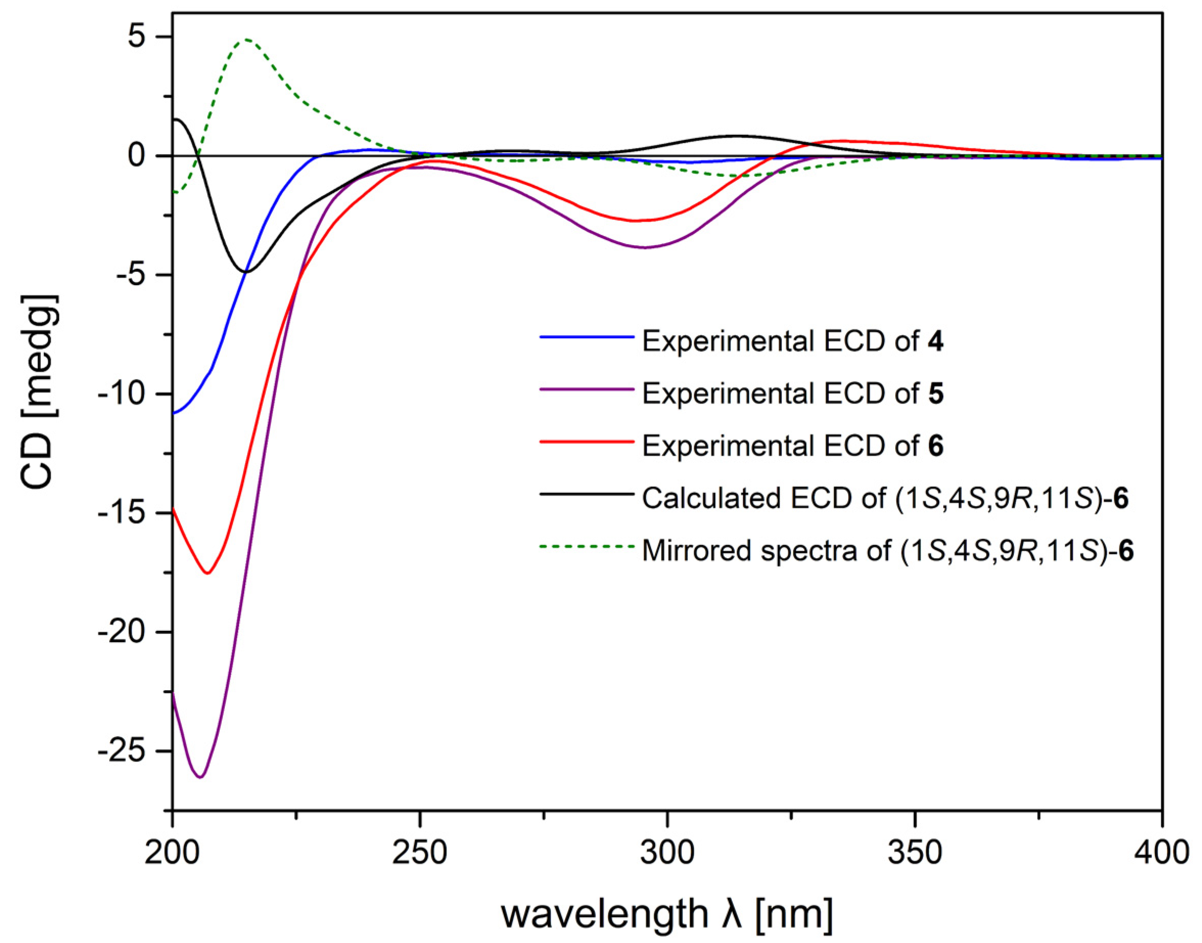
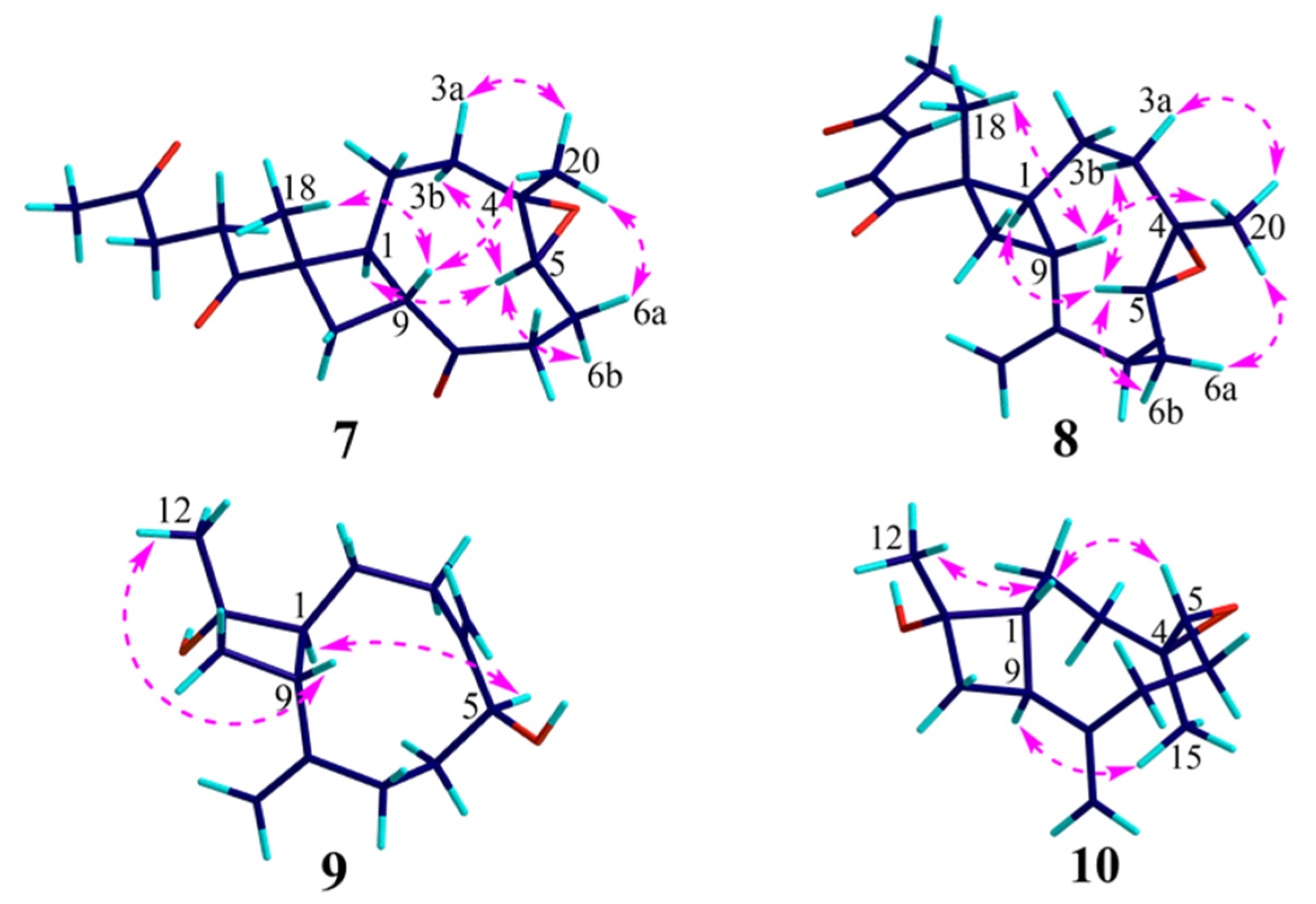
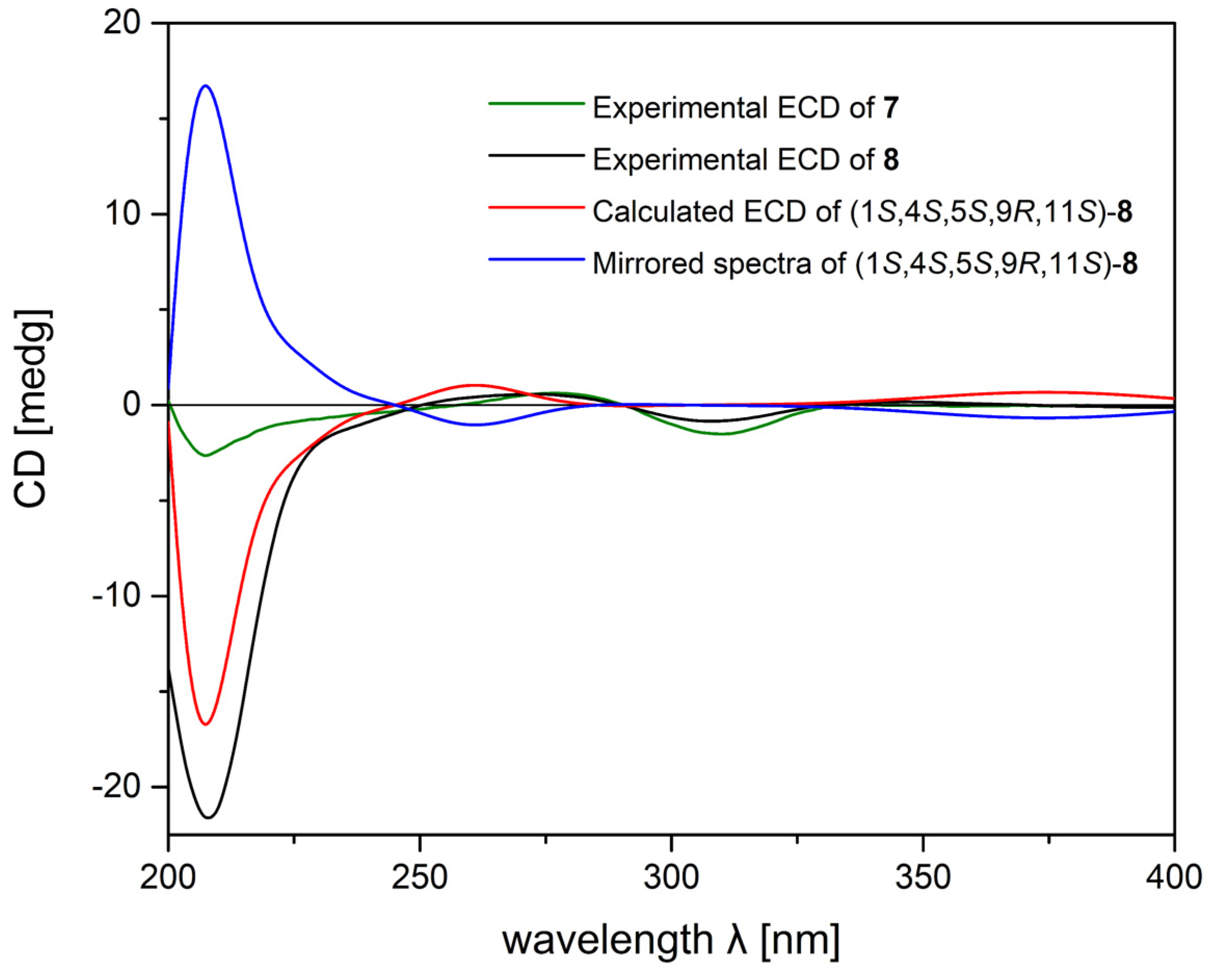
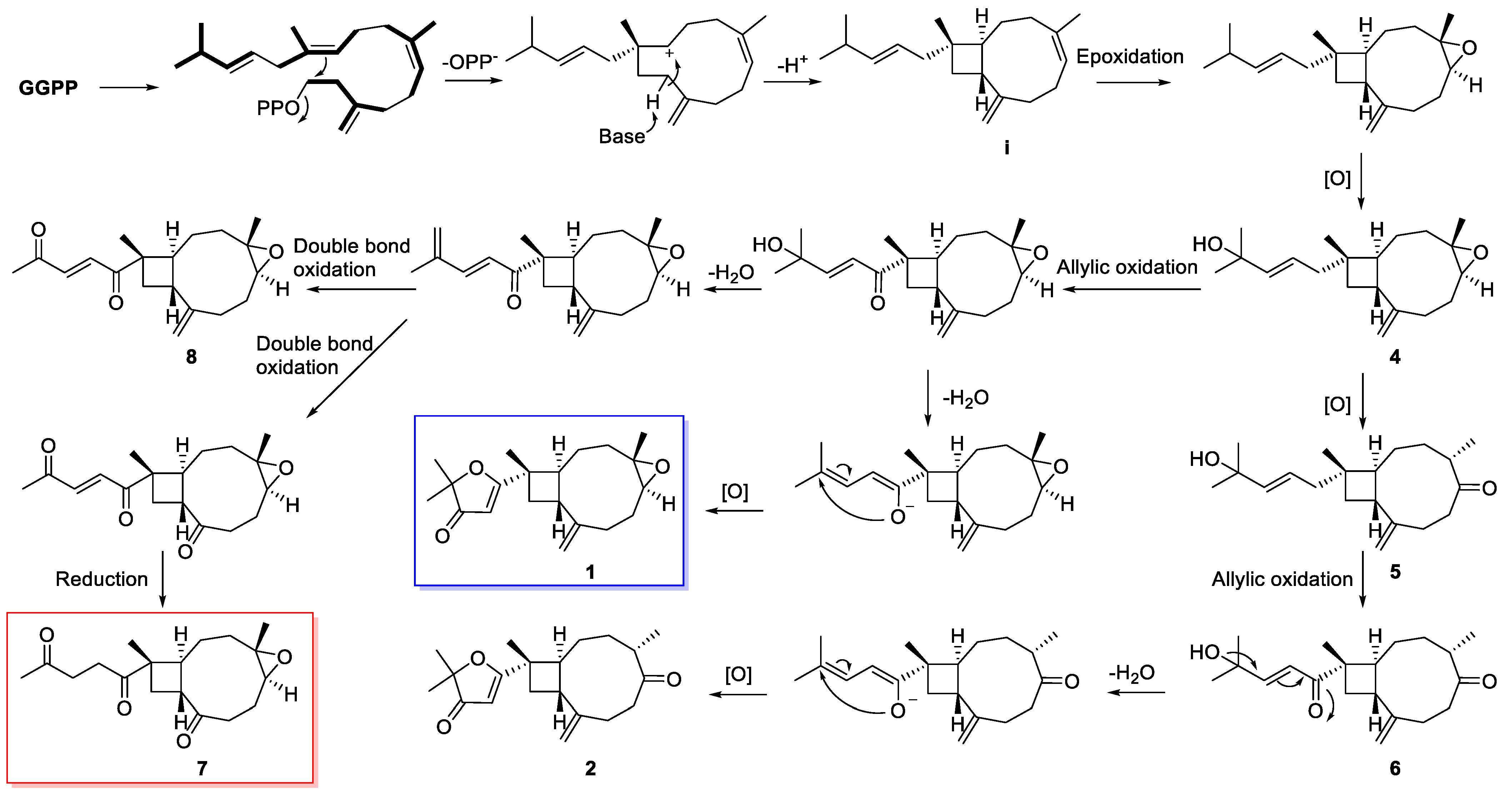
2. Results and Discussion

The frozen body of soft coral S. hirta was cut into pieces and exhaustively extracted with acetone. The Et2O-soluble portion of the acetone extract was purified repeatedly by a combination of silica gel, Sephadex LH-20, and reversed-phase (RP)-HPLC to yield sixteen pure terpenoids: 1 (3.2 mg), 2 (1.1 mg), 3 (0.9 mg), 4 (1.3 mg), 5 (1.6 mg), 6 (1.5 mg), 7 (1.1 mg), 8 (1.5 mg) 9 (4.1 mg), 10 (1.6 mg), 11 (1.7 mg), 12 (1.3 mg), 13 (2.0 mg), 14 (2.2 mg), 15 (1.4 mg), and 16 (1.5 mg). By comparing their NMR spectroscopic data and optical rotation with those reported in the literature, the known compounds were readily identified as nanolobatin B (11) [17], gibberosin D (12) [18], gibberosin Q (13) [19], gibberosin A (14) [18], nanolobatin C (15) [17], and (1R,4R,5R,9S,11R)-4,5-epoxy-caryophyll-8(14)-en-12-oic acid (16) [20], respectively.
Sinuhirfuranone A (1) was obtained as a colorless oil. Its molecular formula, C20H28O3, was deduced by HRESIMS ion peak at m/z 317.2115 ([M + H]+, calcd 317.2111), which suggested seven degrees of unsaturation. The IR spectrum of 1 showed typical absorption indicative of carbonyl group (νmax 1701 cm−1). Its 1H NMR spectrum (Table 1) displayed signals for four singlet methyls at δH 1.22, 1.36, 1.37, and 1.37 and three singlet olefinic protons at δH 4.96, 5.07, and 5.30. The 13C NMR, DEPT, and HSQC spectra indicated 20 carbon signals in 1, including four methyls (δC 17.1, 17.4, 23.0, and 23.1), five sp3 methylenes (δC 27.7, 29.8, 30.1, 35.7, and 38.9), three sp3 methines (δC 48.1, 48.4, and 63.8), three sp3 quaternary carbons (δC 39.0, 59.5, and 88.7), one trisubstituted double bond (δC 98.6 and 196.2), one terminal double bond (δC 114.5 and 150.5), and one ketone group (δC 207.6) (Table 2). One ketone and two double bonds accounted for three degrees of unsaturation, suggesting the presence of a tetracyclic ring system for 1. The 1H–1H COSY spectrum analysis of 1 defined two spin systems a and b (Figure 2) by clear cross peaks of H2-10 (δH 2.26, 1.87)/H-9 (δH 2.79)/H-1 (δH 2.45)/H2-2 (δH 1.76, 1.60)/H2-3 (δH 2.12, 0.97) (a) and H-5 (δH 2.85)/H2-6 (δH 2.16, 1.34)/H2-7 (δH 2.35, 2.17) (b), respectively. Based on HMBC experiment, fragments a and b could be connected by inserting the 8,19-exomethylene double bond and the oxygenated quaternary carbon C-4 (δC 59.5), as indicated by the clear correlations from H2-19 to C-7/C-8/C-9 and H3-20 to C-3/C-4/C-5 (Figure 2). Further HMBC correlations from H3-16 and H3-17 to C-14/C-15, from H3-18 to C-1/C-10/C-11/C-12, and from H-13 to C-12/C-14/C-15 constructed a xeniaphyllane skeleton. The presence of an epoxide group at C-4 and C-5 was deduced by the typical up-fielded 13C NMR signals appearing at δC 59.5 (C-4) and δC 63.8 (C-5). The remaining cycle was deduced to be a furanone ring by the presence of an ether bridge between C-12 and C-15, as evidenced by the typical chemical shifts of an oxygenated carbon at δC 88.7 and a ketone carbonyl at δC 196.2 [12].
The relative configuration (RC) of compound 1 was deduced by NOESY experiment. The crucial NOE correlations (Figure 3) of H3-18 (δH 1.36)/H-9, H3-18/H3-20 (δH 1.22), and H-1/H-5 proved that H3-18, H-9, and H3-20 should be the relative β-orientation, while H-1 and H-5 should be the relative α-orientation. A trans configuration for H-5 and H3-20 in the trisubstituted epoxide was supported by the clear NOE correlations of H-5/H-3b (δH 0.97), H3-20/H-3a (δH 2.12), and H3-20/H-6a (δH 2.16) and the lack of correlation of H-5/H3-20. Thus, the RC of 1 was determined to be 1S*,4S*,5S*,9R*,11S*. Given the conformational flexibility of the core cyclononane ring in 1 that makes the assignment of RC using NOE interactions between remote protons potentially unreliable, a further QM–NMR calculation was performed to further confirm the RC for 1, which could be a showcase for the other cyclononane-containing compounds (210). Two possible configurations, 1a (1S*,4S*,5S*,9R*,11S*) and 1b (1S*,4R*,5R*,9R*,11S*), were calculated, and 1a showed a better result than 1b, verifying the RC of 1 to be 1S*,4S*,5S*,9R*,11S* (Figure S81). The absolute configuration (AC) of compound 1 was further established by TDDFT-ECD calculation due to the presence of an α,β-unsaturated ketone group near the chiral centers. As shown in Figure 4, the Boltzmann-averaged ECD spectrum of (1S,4S,5S,9R,11S)-1 showed a highly matched ECD curve to the experimental curve of 1, allowing the assignment of the AC of 1.
Sinuhirfuranone B (2) was obtained as colorless oil, and its molecular formula C20H28O3, the same as that of 1, was determined by the HRESIMS ion peak at m/z 317.2118 ([M + H]+, calcd 317.2111). In fact, the NMR spectra of 2 (Table 1 and Table 2) showed great similarities to those of 1, with the main differences on the 1H and 13C NMR chemical shifts of C-4, C-5 and its adjacent carbons and protons, suggesting that the epoxide at C-4 (δC 59.5) and C-5 (δC 63.8) in 1 was replaced by a ketone group at C-5 (δC 216.6) in 2. Then, the RC at C-1, C-4, C-9, and C-11 of 2 was elucidated by NOESY experiment. The clear NOE correlations of H-1 (δH 2.09)/H3-20 (δH 1.02) and H-9 (δH 2.56)/H3-18 (δH 1.29) indicated the relative α-orientation of H-1/H3-20 and the relative β-orientation of H-9/H3-18, respectively. As a result, the RC of 2 was elucidated to be 1S*,4S*,9R*,11S*. Finally, the AC of C-11 in 2 was determined to be S by showing the same positive cotton effect (CE) around 220 nm and negative CE around 260 nm in its CD spectrum as that of compound 1 (Figure 4).
Sinuhirfuranone C (3) is a colorless oil, and its molecular formula C20H28O4 was established by the HRESIMS ion peak at m/z 333.2051 ([M + H]+, calcd 333.2060), indicating seven degrees of unsaturation. The 1D and 2D NMR data of 3 (Table 1 and Table 2) were reminiscent of compound 1. A detailed comparison of the NMR data of these two compounds indicated that they differed only by the substitution of a primary hydroxyl group at C-16 (δC 65.9) in 3 rather than a methyl group in 1, which was further confirmed by the observation of HMBC correlations from H3-17 (δH 1.37) to C-14 (δC 206.1)/C-15 (δC 90.5)/C-16 (Figure 2). Based on the extremely similar chemical shifts of the segment C-1–C-11 with C-18–C-20 and compatible NOE correlations with 1 (Figure 3), the RC of C-1, C-4, C-5, C-9, and C-11 in 3 was deduced to be 1S*,4S*,5S*,9R*,11S*, the same as those in compound 1. Given that the remaining chiral center C-15 is independent from other chiral centers, QM-NMR method was applied, and two possible configurations, 3a (1S*,4S*,5S*,9R*,11S*,15R*) and 3b (1S*,4S*,5S*,9R*,11S*,15S*), were calculated (Figure S85). However, it failed since the difference on carbon chemical shifts between the two configurations was too slight. Further, TDDFT-ECD calculation was performed to resolve this problem, and (1S,4S,5S,9R,11S,15R)-3 and (1S,4S,5S,9R,11S,15S)-3 were both calculated. Interestingly, as shown in Figure 5, the mirrored spectrum of (1S,4S,5S,9R,11S,15S)-3 showed a highly matched ECD curve to the experimental curve of 3, allowing the determination of the AC of 3 to be 1R,4R,5R,9S,11R,15R.
Sinuhirtins C–E (46) were isolated as colorless oil. Their molecular formulas were deduced from the HRESIMS data to be C20H32O2 (m/z 305.2482, [M + H]+, calcd 305.2475), C20H32O2 (m/z 327.2297, [M + Na]+, calcd 327.2295), and C20H30O3 (m/z 319.2275, [M + H]+, calcd 319.2268), respectively. They showed NMR data similar to those of co-occurring xeniaphyllane nanolobatin B (11) [17]. In fact, compounds 46 possessed the same skeleton as 11, differing from each other by the oxidative patterns and double-bond positions on the skeleton. Specifically, sinuhirtin C (4) was the decarbonylated product of 11 at C-12 position, as indicated by an aliphatic methylene at δC 46.3 in 4 instead of a carbonyl group at δC 203.7 in 11. The NMR data of sinuhirtin D (5) and 4 only differed on C-4, C-5, and its adjacent carbons and protons. In fact, the epoxide group at C-4 and C-5 in 4 was substituted by the ketone group at C-5 in 5, which was further supported by the observation of HMBC correlations from H3-20 (δH 1.00) to C-3 (δC 29.2)/C-4 (δC 47.2)/C-5 (δC 218.4). Sinuhirtin E (6) was the 12-carbonylation derivative of 5 since the chemical shift at C-12 was shifted downfield from δC 44.3 in 5 to δC 203.8 in 6.
The RC of compound 4 was assigned to be the same as 1 of 1S*,4S*,5S*,9R*,11S* through analysis of their NMR data and interpretation of their NOESY spectra, whereas the RCs of compounds 5 and 6 were determined to be the same as 2 of 1S*,4S*,9R*,11S* on the basis of the careful analysis of their NOESY spectra. To determine their ACs, TDDFT-ECD calculation was applied on compound 6 since the α,β-unsaturated ketone group was near its chiral center C-11 (Figure 6). On the basis of the structural similarities of compounds 46, their ECD curves were compared with each other, resulting in the determination of the same absolute stereochemistry at C-1, C-9, and C-11 as 1S,9R,11S.
Sinuhirtone A (7) was isolated as colorless oil with the chemical formula of C18H26O4 as determined by the HRESIMS (m/z 307.1911 [M + H]+, calcd 307.1904), suggesting six degrees of unsaturation. The 13C NMR and HSQC spectra disclosed 18 carbon resonances, including three methyls (δC 16.4, 17.6, and 30.1), seven sp3 methylenes (δC 25.0, 27.4, 30.5, 31.4, 37.0, 37.6, and 38.5), three sp3 methines (δC 45.8, 51.3, and 61.8), two sp3 quaternary carbons (δC 48.2, and 58.9), and three carbonyls (δC 207.2, 212.5, and 213.3). The NMR data of 7 were strongly reminiscent of those of co-occurring gibberosin A (14). The overall comparison of the 13C NMR data of 7 and 14 revealed that the differences between them mainly happened at C-8 (δC 213.3 for 7, δC 150.8 for 14) and its neighboring carbons C-7 (δC 37.6 for 7, δC 29.4 for 14), C-6 (δ 25.0 for 7, δC 30.2 for 14), and C-9 (δC 51.3 for 7, δC 47.4 for 14), indicating that the terminal double bond at C-8 in 14 was oxidized to be a ketone carbonyl group in 7. It is worth noting that sinuhirtone A, featuring an uncommon 17,19-dinorxeniaphyllane-type skeleton, is the first example of dinorditerpene from marine soft coral.
Sinuhirtone B (8) was also isolated as colorless oil. Its molecular formula, C19H26O3, was established by HREIMS at m/z 302.1883 ([M]+, calcd 302.1876), which suggested seven degrees of unsaturation. The 1H and 13C NMR data of 8 were almost identical to those of 14 except that C-13 and C-14 were both shifted downfield from δC 30.6 and 36.8 in 14 to δC 132.7 and 137.8 in 8, respectively, indicating that C-13 and C-14 were oxidized to be a double bond, which was in agreement with the 2 mass-units loss of molecular weight in 8 (Table 2 and Table 3).
The RCs of 7 and 8 were both assigned to be 1S*,4S*,5S*,9R*,11S*, which was confirmed by the NOESY correlations of H3-18 (δH 1.34)/H-9 (δH 3.08)/H3-20 (δH 1.30) and H-1 (δH 2.64)/H-5 (δH 2.76) in 7, H3-18 (δH 1.34)/H-9 (δH 2.75)/H3-20 (δH 1.22) and H-1 (δH 2.47)/H-5 (δH 2.88) in 8, respectively (Figure 7). Based on the assignment of RCs of compounds 7 and 8, we further decided to perform the TDDFT-ECD calculation to assign the AC of 8 as well as compare the ECD curves of 7 and 8. As shown in Figure 8, both the ECD spectra of 7 and 8 displayed the positive CEs at around 260 nm and negative CEs at 310 nm, which highly matched the calculated ECD of (1S,4S,5S,9R,11S)-8.
Sinuhirtin F (9) was isolated as colorless oil. Its molecular formula was deduced to be C14H22O2 based on the HRESIMS pseudo-molecular ion peak at m/z 223.1689 ([M + H]+, calcd 223.1693), indicating four degrees of unsaturation. Its 1H NMR spectrum (Table 3) displayed signals for one singlet methyl at δH 1.24 and four singlet olefinic protons at δH 4.79, 4.83, 4.96, and 5.05. The 13C NMR, DEPT, and HSQC spectra indicated 14 carbon signals in 9, including one methyl (δC 21.6), five sp3 methylenes (δC 29.9, 32.3, 32.5, 33.2, and 40.3), three sp3 methines (δC 40.0, 58.1, and 75.7), one sp3 quaternary carbon (δC 70.7), and two terminal double bonds (δC 110.0, 114.6, 150.9, and 151.1) (Table 2). Two double bonds accounts for two out of the four degrees of unsaturation, suggesting a bicyclic ring system for 9. The planar structure of 9 was further elucidated by detailed 2D NMR analysis. The 1H–1H COSY correlations of H-10 (δH 2.00)/H-9 (δH 1.88)/H-1 (δH 1.86)/H2-2 (δH 1.97, 1.56)/H2-3 (δH 2.57, 1.95) and H-5 (δH 4.12)/H2-6 (δH 1.95, 1.79)/H2-7 (δH 2.35, 2.09) established two structure fragments in 9 (Figure 2). The connection of these fragments, bearing in mind two terminal double bonds and one methyl, were deduced by the careful analysis of the clear HMBC correlations from H2-14 (δH 4.83, 4.79) to C-7 (δC 33.2)/C-8 (δC 151.1)/C-9 (δC 40.0), from H2-15 (δH 5.05, 4.96) to C-3 (δC 32.5)/C-4 (δC 150.9)/C-5 (δC 75.7), and from H3-12 to C-1 (δC 58.1)/C-10 (δC 40.3)/C-11 (δC 70.7), constructing a norcaryophyllene skeleton. Thus, the planar structure of 9 was established as shown in Figure 1.
To determine the RC of 9 by NOESY correlations is risky since there are two overlapped proton signals at H-1 (δH 1.86) and H-9 (δH 1.87). To tackle this problem, bearing in mind the existence of four chiral centers, eight possible configurations (9a: 1R*,5S*,9S*,11S*; 9b: 1R*,5S*,9R*,11S*; 9c: 1R*,5R*,9S*,11S*; 9d: 1R*,5R*,9R*,11S*; 9e: 1S*,5S*,9S*,11S*; 9f: 1S*,5S*,9R*,11S*; 9g: 1S*,5R*,9S*,11S*; and 9h: 1S*,5R*,9R*,11S*) were calculated by the same QM-NMR method as that for 1 and 3. As a result, 9f showed the highest DP4+ probability (96.73%) among the eight models, indicating the RC of 9 to be 1S*,5S*,9R*,11S*. With the solid establishment of RC of 9, next step was to determine its AC. The initial efforts to resolve the AC of 9 by using TDDFT-ECD approach failed due to the lack of strong chromophore near the chiral centers in 9. In turn, we applied the theoretical specific rotation calculation since it has been proven to be a convincible method for elucidating ACs of natural products [21,22]. The specific rotations of 9 in MeOH solution were measured at the sodium D line and at 589 nm ([α] −40.5). The specific rotations at 589 nm were calculated using TDDFT at the B3LYP/6-311+G* levels with the results for 1S,5S,9R,11S. The calculated conformationally averaged rotation ([α] −50.5) was in good agreement with experimental rotation value, indicating the AC of 9 to be 1S,5S,9R,11S. (Table S11)
Sinuhirtin G (10) was also a colorless oil, the molecular formula of which was determined by the HRESIMS (m/z 223.1688 [M + H]+, calcd 223.1693). The NMR data of 10 (Table 2 and Table 3) showed great similarities to those of nanonorcaryophyllene A, a known norcaryophyllenoid isolated from the Taiwan soft coral Sinularia nanolobata [17]. The overall comparison of the 13C NMR data of 10 and 17 revealed that the differences between them mainly happened at C-9 (δC 48.2 for 10, δC 44.9 for 17), C-10 (δC 40.5 for 10, δC 42.6 for 17), C-11 (δ 73.6 for 10, δC 71.1 for 17), and C-12 (δC 28.3 for 10, δC 21.5 for 17), indicating 10 to be likely the epimer of 17. The RC of 10 was deduced to be 1R*,4R*,5R*,9S*,11S* based on the apparent NOESY correlations of H3-12 (δH 1.27)/H-1 (δH 1.87)/H-5 (δH 2.83) and H-9 (δH 2.88)/H3-15 (δH 1.25) (Figure 7). Then, the AC of 10 was assigned as 1R,4R,5R,9S,11S using the same optical rotation calculation as that of 9.
Sinuhirtone A (7) features the first 17,19-dinorxeniaphyllane skeleton, and sinuhirfuranones A–C (13) possess the rare 2,2-dimethylfuran-3-one group, which are structurally different from other xeniaphyllanoids. However, they do share the same 4/9 bicyclic ring system, indicating that they should be biosynthetic related. Therefore, a plausible biosynthetic connection between 1 and 9 was proposed. As shown in Scheme 1, geranylgeranyl pyrophosphate (GGPP) could be envisaged as an early linear diterpene precursor to form the xeniaphyllane-type skeleton (i) under double-bond migration and ring closure steps, while new compounds were generated after a series of different reactions, including epoxidation, allylic oxidation, elimination, double-bond oxidation, and/or reduction, and so on.
In bioassay, new compounds 24, 7, and 8 were evaluated for in vitro cytotoxicity against four cancer cell lines, including Capan-1, A549, HT-29, and SNU-398, while showing no obvious cytotoxicity towards these cancer cell lines.

3. Materials and Methods

3.1. General Experimental Procedures

Optical rotations were measured on a PerkinElmer 241MC polarimeter. IR spectrum was recorded on a Nicolet iS50 spectrometer (Thermo Fisher Scientific, Madison, WI, USA). 1H and 13C NMR spectra were acquired on a Bruker AVANCE III 500 and 600 spectrometer. Chemical shifts are reported with the residual CHCl3 (δH 7.26 ppm) as the internal standard for 1H NMR spectrometry and CDCl3 (δC 77.2 ppm) for 13C NMR spectrometry. The LREIMS and HREIMS data were recorded on a Finnigan-MAT-95 mass spectrometer (Finnigan-MAT, San Jose, CA, USA). HRESIMS spectra were recorded on an Agilent G6250 Q-TOF (Agilent, Santa Clara, CA, USA). All solvents used for column chromatography and HPLC were of analytical grade (Shanghai Chemical Reagents Co., Ltd., Shanghai, China) and chromatographic grade (Dikma Technologies Inc., Beijing, China), respectively. Sephadex LH-20 (Pharmacia, Peapack, NJ, USA) was also used for column chromatography. Commercial silica gel (Qingdao Haiyang Chemical Co., Ltd., Qingdao, China, 100–200 and 300–400 mesh) was used for column chromatography, and precoated silica gel GF254 plates (Sinopharm Chemical Reagent Co., Shanghai, China) were used for analytical TLC. Reversed-phase (RP) HPLC was performed on an Agilent 1260 series liquid chromatograph equipped with a DAD G1315D detector at 210 nm (Agilent, Santa Clara, CA, USA). An Agilent semipreparative XDB-C18 column (5 μm, 250 × 9.4 mm) was employed for the purification.

3.2. Biological Material

The soft coral S. hirta, a voucher specimen No. YAL-50, was collected by scuba at a depth of −15 m to −20 m off Yalong bay, Sanya, China, in 2006 and identified by Professor Xiu-Bao Li from Hainan University, Hainan, China. The voucher sample S. hirta has been deposited at the Shanghai Institute of Materia Medica, CAS, under registration of No. YAL-50.

3.3. Extraction and Isolation

The frozen animals (210 g, dry weight) were cut into pieces and extracted exhaustively with acetone at room temperature (3 × 3.0 L). The organic extract was evaporated to give a brown residue, which was partitioned between Et2O and H2O. The Et2O solution was concentrated under reduced pressure to give a dark-brown residue (9.8 g), which was fractionated by silica gel column chromatography and eluting with a step gradient (0–100% diethyl ether (EE) in petroleum ether (PE)), yielding twelve fractions (Fr. A–L). Fr. D was further purified by Sephadex LH-20 [PE/CH2Cl2/MeOH (2:1:1)], followed by RP-HPLC [MeCN-H2O (80:20), 3.0 mL/min], to give compounds 2 (1.1 mg), 5 (1.6 mg), 8 (1.5 mg), 10 (1.6 mg), and 12 (1.3 mg). Fr. E was further purified by silica gel CC (300–400 mesh) and eluted with PE-EE (80:20), followed by RP-HPLC [MeCN-H2O (70:30), 3.0 mL/min] to give compounds 1 (3.2 mg), 3 (0.9 mg), 6 (1.5 mg), 7 (1.1 mg), and 9 (4.1 mg). Purification of Fr. H and Fr. J by Sephadex LH-20 [PE/CH2Cl2/MeOH (2:1:1)] and RP-HPL C [MeCN-H2O (60:40), 3.0 mL/min] yielded compounds 4 (1.3 mg), 11 (1.7 mg), 13 (2.0 mg), 14 (2.2 mg), 15 (1.4 mg), and 16 (1.5 mg).

3.4. Spectroscopic Data of Compounds

Sinuhirfuranone A (1): colorless oil; [α ] D 20 +26.2 (c 0.10, MeOH); UV (MeOH) λmax (log ε) 265 (3.14); IR (KBr) νmax: 2973, 2931, 2928, 2861, 1701, 1582, 1195, 1180, 1076 cm−1; 1H and 13C NMR data, see Table 1 and Table 2; HRMS (ESI) m/z: [M + H]+ Calcd. for C20H29O3, 317.2111; Found 317.2115.
Sinuhirfuranone B (2): colorless oil; [α ] D 20 −14.0 (c 0.10, MeOH); UV (MeOH) λmax (log ε) 264 (2.64); IR (KBr) νmax: 2988, 2870, 2928, 1701, 1380, 1142 cm−1; 1H and 13C NMR data, see Table 1 and Table 2; HRMS (ESI) m/z: [M + H]+ Calcd. for C20H29O3, 317.2111; Found 317.2118.
Sinuhirfuranone C (3): colorless oil; [α ] D 20 +42.2 (c 0.10, MeOH); UV (MeOH) λmax (log ε) 265 (2.65); IR (KBr) νmax: 3440, 2978, 2932, 2869, 1698, 1574, 1455, 1381, 1142 cm−1; 1H and 13C NMR data, see Table 1 and Table 2; HRMS (ESI) m/z: [M + H]+ Calcd. for C20H29O4, 333.2060; Found 333.2051.
Sinuhirtin C (4): colorless oil; [α ] D 20 −4.3 (c 0.10, MeOH); IR (KBr) νmax: 3399, 2925, 2854, 1180, 1142, 1132, 1076 cm−1; 1H and 13C NMR data, see Table 1 and Table 2; HRMS (ESI) m/z: [M + H]+ Calcd. for C20H33O2, 305.2475; Found 305.2482.
Sinuhirtin D (5): colorless oil; [α ] D 20 −15.0 (c 0.10, MeOH); IR (KBr) νmax: 3363, 2988, 2870, 1709, 1379, 1142 cm−1; 1H and 13C NMR data, see Table 1 and Table 2; HRMS (ESI) m/z: [M + Na]+ Calcd. for C20H32NaO2, 327.2295; Found 327.2297.
Sinuhirtin E (6): colorless oil; [α ] D 20 −18.6 (c 0.10, MeOH); IR (KBr) νmax: 3446 2989, 2932, 2869, 1688, 1627, 1380, 1196, 1142, 1076 cm−1; 1H and 13C NMR data, see Table 1 and Table 2; HRMS (ESI) m/z: [M + H]+ Calcd. for C20H31O3, 319.2268; Found 319.2275.
Sinuhirtone A (7): colorless oil; [α ] D 20 +63.0 (c 0.10, MeOH); IR (KBr) νmax: 2978, 2932, 2869, 1699, 1381, 1142 cm−1; 1H and 13C NMR data, see Table 2 and Table 3; HRMS (ESI) m/z: [M + H]+ Calcd. for C18H27O4, 307.1904; Found 307.1911.
Sinuhirtone B (8): colorless oil; [α ] D 20 +53.7 (c 0.10, MeOH); UV (MeOH) λmax (log ε) 276 (2.31), 231 (2.67); IR (KBr) νmax: 2977, 2934, 2869, 1703, 1682, 1455, 1384, 1142, 1076 cm−1; 1H and 13C NMR data, see Table 2 and Table 3; HRMS (EI) m/z: [M]+ Calcd. for C19H26O3, 302.1876; Found 302.1883.
Sinuhirtin F (9): colorless oil; [α ] D 20 −40.5 (c 0.10, MeOH); IR (KBr) νmax: 3342, 2959, 2920, 1257, 1180, 1147, 1138, 1078 cm−1; 1H and 13C NMR data, see Table 2 and Table 3; HRMS (ESI) m/z: [M + H]+ Calcd. for C14H23O2, 223.1693; Found 223.1689.
Sinuhirtin G (10): colorless oil; [α ] D 20 −97.3 (c 0.10, MeOH); IR (KBr) νmax: 3439, 2978, 2931, 2869, 1454, 1383, 1227, 1142, 1075 cm−1; 1H and 13C NMR data, see Table 2 and Table 3; HRMS (ESI) m/z: [M + H]+ Calcd. for C14H23O2, 223.1693; Found 223.1688.

3.5. QM-NMR Calculational Section

Conformational search was performed by using the torsional sampling (MCMM) approach and OPLS_2005 force field within an energy window of 21 kJ/mol. Conformers above 1% Boltzmann populations were re-optimized at the B3LYP/6-311G(d,p) level with the IEFPCM solvent model for chloroform. Frequency analysis was also carried out to confirm that the re-optimized geometries were at the energy minima. Subsequently, NMR calculations were performed at the PCM/mPW1PW91/6-31G(d) level, as recommended for DP4+. NMR shielding constants were calculated by using the GIAO method. Finally, shielding constants were averaged over the Boltzmann distribution obtained for each stereoisomer and correlated with the experimental data [12].

3.6. TDDFT-ECD Calculational Section

Conformational search was carried out by using the torsional sampling (MCMM) method and OPLS_2005 force field within an energy window of 21 kJ/mol. Conformers above 1% Boltzmann populations were re-optimized at the MPW1PW91/6-31G(d) level with the IEFPCM solvent model for MeCN. Frequency analysis was also carried out to confirm that the re-optimized geometries were at the energy minima. ECD spectra were obtained by TDDFT calculations showed with the identical functional basis set and solvent model as the energy optimization. At last, the Boltzmann-averaged ECD spectra of the compounds were obtained with SpecDis 1.62 [23].

3.7. Optical Rotation Calculational Section

The specific optical rotation calculations for compounds were carried out by using Information Gaussian 09. The conformers were further optimized at the B3LYP/6-311G(d,p) level with the IEFPCM solvent model for chloroform, all of which were subjected to specific optical rotation calculations at the B3LYP/6-311+G(d) level in chloroform with SMD model. The calculated specific optical rotation values of these conformers were averaged according to the Boltzmann distribution theory and their relative Gibbs free energy. The predicted ORD curves were generated from specific rotations calculated at four different wavelengths [24].

3.8. Bioassay

Cytotoxicity assays were carried out by using Capan-1 (human pancreatic cancer), A549 (human lung cancer), HT-29 (human colon carcinoma), and SNU-398 (human hepatocellular carcinoma), following a previously described procedure for a modification of the MTT colorimetric method [25,26]. To measure the cytotoxicity of tested compounds, five concentrations with three replications were performed on each cell line. Vincristine was used as a positive control for the bioassays using Capan-1, A549, HT-29, and SNU-398 cell lines, respectively.

4. Conclusions

In summary, the systematic chemical investigation of S. hirta of South China Sea yielded a series of terpenoids 110 with different skeletons, which increased the chemical diversity and complexity of marine terpenoids. The stereochemistry of these compounds was assigned by QM-NMR, TDDFT-ECD, and optical rotation calculations, which provided different approaches to deal with the challenges of stereochemistry determination of the complex molecules without single crystals. Structurally, sinuhirtone A (7) represents the first example of 17,19-dinorxeniaphyllane-type skeleton, whereas sinuhirfuranones A–C (13) possess the 2,2-dimethylfuran-3-one group, which is rare among marine diterpenoids. Although some new compounds did not show obvious anti-cancer activities, our proposed biogenetic pathway of these interesting compounds should impel further study on their accumulation for more extensive biological evaluation to understand the role they play on the life cycle of soft coral and to find their possible medicinal application.

Supplementary Materials

The following are available online at https://www.mdpi.com/article/10.3390/md20040272/s1: 1D and 2D NMR, HRMS, and IR spectra of 110; DP4+ calculation of compounds 3 and 9; TDDFT-ECD calculation of compounds 1, 3, 6, and 8; ORD calculation of compounds 9 and 10.

Author Contributions

Conceptualization, Y.-W.G. and X.-W.L.; data curation, Z.-H.C. and S.-Q.L.; formal analysis, Y.-W.G. and Z.-H.C.; funding acquisition, Y.-W.G. and X.-W.L.; investigation, Z.-H.C. and S.-Q.L.; methodology, Y.-W.G. and X.-W.L.; project administration, Y.-W.G., X.-W.L. and G.-Y.H.; resources, Y.-W.G. and X.-W.L.; software, Y.-W.G. and X.-W.L.; supervision, Y.-W.G., X.-W.L. and G.-Y.H.; validation, Y.-W.G., X.-W.L., S.-Q.L. and Z.-H.C.; visualization, Y.-W.G. and S.-Q.L.; writing—original draft preparation, Z.-H.C., S.-Q.L. and G.-Y.H.; writing—review and editing, Y.-W.G., X.-W.L., G.-Y.H. and Z.-H.C. All authors have read and agreed to the published version of the manuscript.

Funding

This research work was financially supported by the National Key Research and Development Program of China (No. 2021YFF0502400), the Natural Science Foundation of China (Nos. 81991521, 82022069, 42076099), the Shanghai Rising-Star Program (No. 20QA1411100), “Youth Innovation Promotion Association” of CAS (No. Y202065), and the SKLDR/SIMM Project (No. SIMM2103ZZ-06).

Institutional Review Board Statement

Not applicable.

Informed Consent Statement

Not applicable.

Data Availability Statement

Data are contained within the article or Supplementary Material.

Acknowledgments

We thank Xiu-Bao Li from Hainan University for the taxonomic identification of the soft coral material.

Conflicts of Interest

The authors declare no conflict of interest.

References

  1. Li, Y.; Pattenden, G. Novel macrocyclic and polycyclic norcembranoid diterpenes from Sinularia species of soft coral. Structural relationships and biosynthetic speculations. Nat. Prod. Rep. 2011, 28, 429–440. [Google Scholar] [CrossRef] [PubMed]
  2. Chen, W.-T.; Li, Y.; Guo, Y.-W. Terpenoids of Sinularia soft corals: Chemistry and bioactivity. Acta Pharm. Sin. B 2012, 2, 227–237. [Google Scholar] [CrossRef] [Green Version]
  3. Yan, X.U.; Liu, J.; Leng, X.; Ouyang, H. Chemical diversity and biological activity of secondary metabolites from soft coral genus Sinularia since 2013. Mar. Drugs 2021, 19, 335. [Google Scholar] [CrossRef] [PubMed]
  4. Wu, Q.; Li, X.-W.; Li, H.; Yao, L.-G.; Tang, W.; Miao, Z.-H.; Wang, H.; Guo, Y.-W. Bioactive polyoxygenated cembranoids from a novel Hainan chemotype of the soft coral Sinularia flexibilis. Bioorg. Med. Chem. Lett. 2019, 29, 185–188. [Google Scholar] [CrossRef]
  5. Jiang, C.-S.; Ru, T.; Yao, L.-G.; Miao, Z.-H.; Guo, Y.-W. Four new cembranoids from the Chinese soft coral Sinularia sp. and their anti-Aβ aggregation activities. Fitoterapia 2019, 136, 104176. [Google Scholar] [CrossRef]
  6. Yang, M.; Li, H.; Zhang, Q.; Wu, Q.; Li, G.; Chen, K.-X.; Guo, Y.-W.; Tang, W.; Li, X.-W. Highly diverse cembranoids from the South China Sea soft coral Sinularia scabra as a new class of potential immunosuppressive agents. Bioorg. Med. Chem. 2019, 27, 3469–3476. [Google Scholar] [CrossRef]
  7. Liang, L.-F.; Wang, X.-J.; Zhang, H.-Y.; Liu, H.-L.; Li, J.; Lan, L.-F.; Zhang, W.; Guo, Y.-W. Bioactive polyhydroxylated steroids from the Hainan soft coral Sinularia depressa Tixier-Durivault. Bioorg. Med. Chem. Lett. 2013, 23, 1334–1337. [Google Scholar] [CrossRef]
  8. Chen, D.-W.; Chen, W.; Liu, D.; van Ofwegen, L.; Proksch, P.; Lin, W.-H. Asteriscane-type sesquiterpenoids from the soft coral Sinularia capillosa. J. Nat. Prod. 2013, 76, 1753–1763. [Google Scholar] [CrossRef]
  9. Anjaneyulu, A.S.R.; Rao, G.V.; Rao, N.S.K. Sesqui- and diterpenoids of the soft coral Sinularia hirta of the Andaman & Nicobar Islands. Indian J. Chem. B 1996, 35B, 815–818. [Google Scholar]
  10. Anjaneyulu, V.; Rao, K.N.; Babu, J.S.; Kobayashi, M. Isolation of 24-methylenecholesterol-3-O-α-L-fucopyranoside and 24-methylenecholest-7-ene-3β,6α,9α,11α-tetrol from a soft coral of the Andaman and Nicobar Islands. Indian J. Chem. B 1994, 33B, 144–147. [Google Scholar]
  11. Wu, Q.; Li, S.-W.; Xu, H.; Wang, H.; Hu, P.; Zhang, H.; Luo, C.; Chen, K.-X.; Nay, B.; Guo, Y.-W.; et al. Complex polypropionates from a South China Sea photosynthetic mollusk: Isolation and biomimetic synthesis highlighting novel rearrangements. Angew. Chem. Int. Ed. 2020, 59, 12105–12112. [Google Scholar] [CrossRef] [PubMed]
  12. Chen, Z.-H.; Li, W.-S.; Zhang, Z.-Y.; Luo, H.; Wang, J.-R.; Zhang, H.-Y.; Zeng, Z.-R.; Chen, B.; Li, X.-W.; Guo, Y.-W. Sinusiaetone A, an anti-inflammatory norditerpenoid with a bicyclo [11.3.0]hexadecane nucleus from the Hainan soft coral Sinularia siaesensis. Org. Lett. 2021, 23, 5621–5625. [Google Scholar] [CrossRef] [PubMed]
  13. Li, G.; Li, H.; Tang, W.; Guo, Y.-W.; Li, X.-W. Klyflaccilides A and B, diterpenoids with 6/5/8/3 fused tetracyclic carbon skeleton from the Hainan soft coral Klyxum flaccidum. Org. Lett. 2019, 21, 5660–5664. [Google Scholar] [CrossRef]
  14. Ye, F.; Li, J.; Wu, Y.; Zhu, Z.-D.; Mollo, E.; Gavagnin, M.; Gu, Y.-C.; Zhu, W.-L.; Li, X.-W.; Guo, Y.-W. Sarinfacetamides A and B, nitrogenous diterpenoids with tricyclo[6.3.1.01,5]dodecane scaffold from the South China Sea soft coral Sarcophyton infundibuliforme. Org. Lett. 2018, 20, 2637–2640. [Google Scholar] [CrossRef]
  15. Ye, F.; Zhu, Z.-D.; Chen, J.-S.; Li, J.; Gu, Y.-C.; Zhu, W.-L.; Li, X.-W.; Guo, Y.-W. Xishacorenes A–C, diterpenes with bicyclo[3.3.1]nonane nucleus from the Xisha soft coral Sinularia polydactyla. Org. Lett. 2017, 19, 4183–4186. [Google Scholar] [CrossRef] [PubMed]
  16. Lu, S.-Q.; Li, X.-W.; Li, S.-W.; Cui, Z.; Guo, Y.-W.; Han, G.-Y. Sinuhirtins A and B, two uncommon norhumulene-type terpenoids from the South China Sea soft coral Sinularia hirta. Tetrahedron Lett. 2019, 60, 151308. [Google Scholar] [CrossRef]
  17. Ahmed, A.F.; Su, J.-H.; Shiue, R.-T.; Pan, X.-J.; Dai, C.-F.; Kuo, Y.-H.; Sheu, J.-H. New β-caryophyllene-derived terpenoids from the soft coral Sinularia nanolobata. J. Nat. Prod. 2004, 67, 592–597. [Google Scholar] [CrossRef]
  18. Chen, S.-P.; Chao, C.-H.; Huang, H.-C.; Wu, Y.-C.; Lu, C.-K.; Dai, C.-F.; Sheu, J.-H. New β-caryophyllene-derived terpenoids from the Formosan soft coral Sinularia gibberosa. Bull. Chem. Soc. Jpn. 2006, 79, 1547–1551. [Google Scholar] [CrossRef]
  19. Chen, S.-P.; Su, J.-H.; Yeh, H.-C.; Ahmed, A.F.; Dai, C.-F.; Wu, Y.-C.; Sheu, J.-H. Novel norhumulene and xeniaphyllane-derived terpenoids from a Formosan soft coral Sinularia gibberosa. Chem. Pharm. Bull. 2009, 57, 162–166. [Google Scholar] [CrossRef] [Green Version]
  20. Abraham, W.R.; Ernst, L.; Stumpf, B. Biotransformation of caryophyllene by Diplodia gossypina. Phytochemistry 1990, 29, 115–120. [Google Scholar] [CrossRef]
  21. Mennucci, B.; Claps, M.; Evidente, A.; Rosini, C. Absolute configuration of natural cyclohexene oxides by time dependent density functional theory calculation of the optical rotation: The absolute configuration of (−)-sphaeropsidone and (−)-episphaeropsidone revised. J. Org. Chem. 2007, 72, 6680–6691. [Google Scholar] [CrossRef] [PubMed]
  22. Niu, S.; Xie, C.-L.; Xia, J.-M.; Liu, Q.-M.; Peng, G.-Z.; Liu, G.-M.; Yang, X.-W.; Botryotins, A.-H. Tetracyclic diterpenoids representing three carbon skeletons from a deep-sea-derived Botryotinia fuckeliana. Org. Lett. 2020, 22, 580–583. [Google Scholar] [CrossRef] [PubMed]
  23. Chen, Z.-H.; Yao, L.-G.; Wu, Q.; Guo, Y.-W. Uncommon polycyclic merosesquiterpenoids and asteriscanoids from the Hainan soft coral Sinularia humesi. Chin. J. Chem. 2021, 39, 2377–2385. [Google Scholar] [CrossRef]
  24. Sun, L.-L.; Li, W.-S.; Li, J.; Zhang, H.-Y.; Yao, L.-G.; Luo, H.; Guo, Y.-W.; Li, X.-W. Uncommon diterpenoids from the South China Sea soft coral Sinularia humilis and their stereochemistry. J. Org. Chem. 2021, 86, 3367–3376. [Google Scholar] [CrossRef]
  25. Mosmann, T. Rapid colorimetric assay for cellular growth and survival: Application to proliferation and cytotoxicity assays. J. Immunol. Methods 1983, 65, 55–63. [Google Scholar] [CrossRef]
  26. Hansen, M.B.; Nielsen, S.E.; Berg, K. Re-examination and further development of a precise and rapid dye method for measuring cell growth/cell kill. J. Immunol. Methods 1989, 119, 203–210. [Google Scholar] [CrossRef]
Figure 1. Structures of compounds 116, xeniaphyllane, and caryophyllene.
Marinedrugs 20 00272 g001
Figure 2. Selected 1H–1H COSY (bold lines) and key HMBC (single arrows) correlations of 110. Fragments a (blue) and b (red) represent two different spin systems, respectively.
Marinedrugs 20 00272 g002
Figure 3. Key NOESY correlations of compounds 16.
Marinedrugs 20 00272 g003
Figure 4. Experimental ECD spectrum of 1 (black) and 2 (green); calculated ECD spectrum of (1S,4S,5S,9R,11S)-1 (red) and its enantiomer (blue).
Marinedrugs 20 00272 g004
Figure 5. Experimental ECD spectra of 3 (black), calculated ECD spectrum of (1S,4S,5S,9R,11S,15S)-3 (dashed green), mirrored spectra of (1S,4S,5S,9R,11S,15S)-3 (red), calculated ECD spectrum of (1S,4S,5S,9R,11S,15R)-3 (blue), and mirrored spectra of (1S,4S,5S,9R,11S,15R)-3 (dashed purple).
Marinedrugs 20 00272 g005
Figure 6. Experimental ECD spectra of 4 (blue), 5 (purple), 6 (red), calculated ECD spectrum of (1S,4S,9R,11S)-6 (black), and mirrored spectra of (1S,4S,9R,11S)-6 (dashed green).
Marinedrugs 20 00272 g006
Figure 7. Key NOESY correlations of compounds 710.
Marinedrugs 20 00272 g007
Figure 8. Experimental ECD spectra of 7 (green), 8 (black), calculated ECD spectrum of (1S,4S,5S,9R,11S)-8 (red), and mirrored spectra of (1S,4S,5S,9R,11S)-8 (blue).
Marinedrugs 20 00272 g008
Scheme 1. Plausible biosynthetic connection between compounds 19 (OPP: diphosphate).
Marinedrugs 20 00272 sch001
Table 1. 1H NMR data (δ in ppm, J in Hz) for compounds 16 in CDCl3.
No.1 a2 a3 a4 a5 a6 a
δH Mult (J Hz)δH Mult (J Hz)δH Mult (J Hz)δH Mult (J Hz)δH Mult (J Hz)δH Mult (J Hz)
12.45 t (9.6)2.09 m2.48 t (9.6)1.89 t (9.6)1.90 t (7.8)2.12 m
2a1.76 dt (14.4, 3.6)1.66 m1.77 m1.64 m1.51 m1.73 m
2b1.60 m1.37 m1.60 m1.43 m1.24 m1.37 m
3a2.12 dt (13.2, 3.6)1.84 m2.10 m2.06 dt (13.2, 3.6)1.88 m1.92 m
3b0.97 td (13.2, 5.4)1.62 m0.98 td (13.2, 4.8)0.96 td (13.2, 4.8)1.68 m1.67 m
4 2.52 m 2.52 m2.54 m
52.85 dd (10.8, 4.2) 2.86 dd (10.2, 4.2)2.88 dd (10.2, 4.2)
6a2.16 ddd (13.2, 7.8, 4.8)2.51 m2.27 m2.13 ddd (15.0, 7.8, 4.2)2.53 m2.53 m
6b1.34 overlap 1.32 m1.32 m
7a2.35 m2.49 m2.37 ddd (13.2, 8.4, 4.2)2.33 ddd (13.2, 8.4, 4.2)2.45 m2.51 m
7b2.17 m 2.17 m2.25 ddt (12.0, 7.8, 4.2) 2.48 m
92.79 q (9.6)2.56 m2.81 q (9.6)2.64 q (9.6)2.50 m2.53 m
10a2.26 m2.16 t (10.8)2.29 m1.65 m1.61 m2.06 t (10.2)
10b1.87 dd (10.8, 8.4)1.80 dd (10.8, 8.4)1.89 dd (10.8,8.4) 1.52 m1.77 dd (11.4, 8.4)
12 2.00 d (6.6)1.91 m
135.30 s5.26 s5.37 s5.59 overlap5.55 ddd (15.6, 7.8, 6.6)6.47 d (15.0)
14 5.61 overlap5.61 d (15.6)6.92 d (15.0)
16a1.37 s1.38 s3.81 d (12.0)1.30 s1.33 s1.40 s
16b 3.69 d (12.0)
171.37 s1.37 s1.37 s1.30 s1.39 s1.40 s
181.36 s1.29 s1.39 s1.03 s0.98 s1.21 s
19a5.07 s4.96 s5.07 s4.97 s4.86 s4.91 s
19b4.96 s4.95 s4.96 s4.86 s4.82 s4.91 s
201.22 s1.02 s1.21 s1.19 s1.00 d (6.6)1.03 d (6.6)
a Recorded at 600 MHz. Chemical shifts (ppm) refer to CHCl3 (δH 7.26). Assignments were deduced by analysis of 1D and 2D NMR spectra.
Table 2. 13C NMR and DEPT spectroscopic data for compounds 110 in CDCl3.
No.1 a2 a3 a4 a5 a6 a7 a8 a9 a10 a
δC Mult.δC Mult.δC Mult.δC Mult.δC Mult.δC Mult.δC Mult.δC Mult.δC Mult.δC Mult.
148.4 CH49.0 CH48.7 CH48.8 CH44.6 CH45.7 CH45.8 CH45.5 CH58.1 CH52.7 CH
227.7 CH227.1 CH227.7 CH227.9 CH226.2 CH227.0 CH227.4 CH228.3 CH229.8 CH226.1 CH2
338.9 CH230.2 CH238.8 CH239.0 CH229.2 CH229.5 CH238.5 CH238.7 CH232.5 CH239.0 CH2
459.5 qC48.2 CH59.5 qC59.9 qC47.2 CH48.1 CH58.9 qC59.5 qC150.9 qC59.8 qC
563.8 CH216.6 qC63.7 CH64.0 CH218.4 qC217.5 qC61.8 CH63.6 CH75.7 CH63.4 CH
630.1 CH241.8 CH230.1 CH230.3 CH243.4 CH241.9 CH225.0 CH230.1 CH232.3 CH229.9 CH2
729.8 CH232.3 CH229.8 CH229.9 CH231.4 CH232.1 CH237.6 CH229.9 CH233.2 CH230.8 CH2
8150.5 qC151.9 qC150.4 qC151.9 qC153.3 qC152.2 qC213.3 qC150.5 qC151.1 qC151.4 qC
948.1 CH42.7 CH48.1 CH48.8 CH44.0 CH42.2 CH51.3 CH47.4 CH40.0 CH48.2 CH
1035.7 CH235.0 CH235.6 CH238.1 CH235.7 CH234.4 CH231.4 CH235.2 CH240.3 CH240.5 CH2
1139.0 qC38.9 qC39.2 qC37.3 qC38.1 qC47.7 qC48.2 qC48.2 qC70.7 qC73.6 qC
12196.2 qC196.2 qC198.1 qC46.3 CH244.3 CH2203.8 qC212.5 qC203.2 qC21.7 CH328.3 CH3
1398.6 CH98.5 CH100.4 CH123.1 CH123.4 CH120.7 CH30.5 CH2132.7 CH
14207.6 qC207.8 qC206.1 qC140.7 CH141.5 CH153.3 CH37.0 CH2137.8 CH110.0 CH2113.0 CH2
1588.7 qC88.7 qC90.5 qC70.9 qC70.4 qC71.1 qC207.2 qC197.9 qC114.6 CH216.9 CH3
1623.0 CH323.0 CH365.9 CH230.2 CH333.0 CH329.3 CH330.1 CH329.3 CH3
1723.1 CH323.1 CH318.4 CH330.1 CH329.4 CH329.4 CH3
1817.4 CH317.4 CH317.4 CH319.8 CH321.0 CH316.6 CH317.6 CH316.7 CH3
19114.5 CH2112.9 CH2114.6 CH2113.1 CH2112.0 CH2112.7 CH2 114.4 CH2
2017.1 CH316.2 CH317.1 CH317.2 CH315.2 CH315.6 CH316.4 CH317.2 CH3
a Recorded at 125 MHz in CDCl3. Chemical shifts (ppm) refer to CHCl3 (δC 77.2). Assignments were deduced by analysis of 1D and 2D NMR spectra.
Table 3. 1H NMR data (δ in ppm, J in Hz) for compounds 710 in CDCl3.
No.7 a8 b9 a10 a
δH Mult (J Hz)δH Mult (J Hz)δH Mult (J Hz)δH Mult (J Hz)
12.64 m2.47 t (9.5)1.86 m1.87 t (9.6)
2a1.84 dt (15.0, 3.6)1.83 m1.97 m1.74 dt (14.4, 4.2)
2b1.64 m1.59 m1.56 m1.65 m
3a2.15 dt (13.2, 3.6)2.12 m2.57 dt (13.2, 4.2)2.16 dt (13.2, 3.6)
3b1.09 td (13.2, 4.8)1.10 td (13.0, 5.0)1.95 m1.00 td (13.2, 4.8)
52.76 dd (10.2, 4.2)2.88 dd (10.5, 4.5)4.12 dd (9.6, 3.6)2.83 dd (10.2, 4.2)
6a2.40 m2.25 m1.95 m2.24 m
6b1.46 m1.33 m1.79 m1.35 m
7a2.57 m2.30 m2.35 ddd (14.4, 10.2, 4.2)2.35 m
7b 2.13 m2.09 m2.09 m
93.08 q (9.0)2.75 q (9.5)1.88 m2.88 q (9.6)
10a2.63 m2.15 m2.00 m2.02 m
10b1.79 dd (11.4, 7.8)1.90 dd (11.0, 8.0) 1.93 m
12 1.24 s1.27 s
132.65 m6.98 s
14a2.73 m6.98 s4.83 s5.02 s
14b 4.79 s4.93 s
15a 5.05 s1.25 s
15b 4.96 s
162.19 s2.36 s
181.34 s1.34 s
19a 5.00 s
19b 4.93 s
201.30 s1.22 s
a Recorded at 600 MHz. b Recorded at 500 MHz. Chemical shifts (ppm) refer to CHCl3 (δH 7.26). Assignments were deduced by analysis of 1D and 2D NMR spectra.
Publisher’s Note: MDPI stays neutral with regard to jurisdictional claims in published maps and institutional affiliations.

