Article

Six New Antimicrobial Metabolites from the Deep-Sea Sediment-Derived Fungus Aspergillus fumigatus SD-406

1
CAS and Shandong Province Key Laboratory of Experimental Marine Biology, Institute of Oceanology, Chinese Academy of Sciences, Nanhai Road 7, Qingdao 266071, China
2
Laboratory of Marine Biology and Biotechnology, Qingdao National Laboratory for Marine Science and Technology, Wenhai Road 1, Qingdao 266237, China
3
College of Marine Sciences, University of Chinese Academy of Sciences, Yuquan Road 19A, Beijing 100049, China
4
Center for Ocean Mega-Science, Chinese Academy of Sciences, Nanhai Road 7, Qingdao 266071, China
*
Authors to whom correspondence should be addressed.
Mar. Drugs 2022, 20(1), 4; https://doi.org/10.3390/md20010004
Submission received: 30 November 2021 / Revised: 18 December 2021 / Accepted: 19 December 2021 / Published: 21 December 2021
(This article belongs to the Special Issue Bioactive Compounds from Marine Sediment Derived Fungi)

Abstract

:
Six new metabolites, including a pair of inseparable mixtures of secofumitremorgins A (1a) and B (1b), which differed in the configuration of the nitrogen atom, 29-hydroxyfumiquinazoline C (6), 10R-15-methylpseurotin A (7), 1,4,23-trihydroxy-hopane-22,30-diol (10), and sphingofungin I (11), together with six known compounds (25 and 89), were isolated and identified from the deep-sea sediment-derived fungus Aspergillus fumigatus SD-406. Their structures were determined by detailed spectroscopic analysis of NMR and MS data, chiral HPLC analysis of the acidic hydrolysate, X-ray crystallographic analysis, J-based configuration analysis, and quantum chemical calculations of ECD, OR, and NMR (with DP4+ probability analysis). Among the compounds, 1a/1b represent a pair of novel scaffolds derived from indole diketopiperazine by cleavage of the amide bond following aromatization to give a pyridine ring. Compounds 1, 4, 6, 7, 10 and 11 showed inhibitory activities against pathogenic bacteria and plant pathogenic fungus, with MIC values ranging from 4 to 64 μg/mL.

Graphical Abstract

1. Introduction

Deep-sea sediment has proven to be a treasure trove for structurally unique and biologically active secondary metabolites [1]. In the extreme environment of the deep-sea, microorganisms have gradually developed unique metabolic mechanisms out of adaption, thus possessing the great potential to produce natural products with significant biological properties, such as antimicrobial [2,3], cytotoxic [4], and antiviral [5] activities.
In the course of discovering bioactive metabolites from deep-sea-derived fungi [2,3,4], the fungal strain Aspergillus fumigatus SD-406, which was obtained from sediments collected from the deep-sea in the East China Sea, displayed abundant metabolites and antimicrobial activity in the preliminary screening, and thus attracted us to perform intensive chemical investigations on it. As a result, six new metabolites, including a mixture of a pair of inseparable isomers, secofumitremorgins A (1a) and B (1b), which differed in the configuration of the nitrogen atom, 29-hydroxyfumiquinazoline C (6), 10R-15-methylpseurotin A (7), 1,4,23-trihydroxy-hopane-22,30-diol (10), and sphingofungin I (11), together with six known compounds, fumitremorgin C (2) [6], 12,13-dihydroxyfumitremorgin C (3) [7], cyclotryprostatin B (4) [8], fumiquinazoline C (5) [9], 14-norpseurotin A (8) [10], and pseurotin A (9) [11] have been isolated and identified (Figure 1). Among these compounds, 1a/1b represent a pair of novel scaffolds derived from indole diketopiperazine by cleavage of the amide bond following aromatization to give a pyridine ring. Details of the isolation and purification, structure elucidation, and biological evaluation of compounds 111 are described herein.

2. Results and Discussion

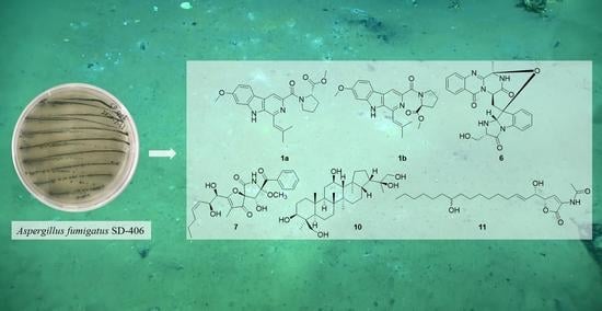
2.1. Structure Elucidation

The culture broth of fungus Aspergillus fumigatus SD-406 was extracted with EtOAc, and the crude extract was subjected to multiple chromatographic methods (a combination of column chromatography on Silica gel, Sephadex LH-20, and Lobar LiChroprep RP-18) to yield subfractions, which were further purified by preparative TLC and semipreparative HPLC to give compounds 111.
Compounds 1a/1b were isolated as a mixture present in a ratio of 1:0.7 (major:minor). Attempts to separate two isomers by various types of chiral columns along with different elution ratios, unfortunately failed. The mixture (1a/1b) was found to have the same molecular formula as C23H25N3O4 on the basis of the HRESIMS m/z 406.1769 [M−H] (calculated for C23H24O4N3, 406.1772), requiring 13 degrees of unsaturation (Figure S8 in the supplementary material). The 1H- and 13C-NMR spectra (Figures S2 and S3 in the supplementary material) of the mixture presented two sets of similar data (Table 1), revealing that their planar structures both possessed 4 methyls (including 2 methoxyls), 3 aliphatic methylenes, 6 methines (including 5 sp2 hybridized and 1 connected to heteroatoms), and 10 quaternary carbons (including 2 carbonyls). Detailed analysis and comparison of the 1D and 2D NMR data (Figures S4–S7 in the supplementary material) indicated that the planar structures of 1a/1b were similar to that of the known compound fumitremorgin C (2) [6]. However, signals for the methine groups at C-3 (δH/C 5.98/51.1) and C-12 (δH/C 4.16/56.9), methylene group at C-13 (δH/C 3.51/22.0), and amino carbonyl at C-5 (δC 165.9) in 2 were absent. Instead, signals for sp2 hybridized quaternary carbons at δC, major 138.5/δC, minor 138.2 and δC, major 141.9/δC, minor 141.8, for sp2 hybridized methines at δH/C, major 8.33/113.3/δH/C, minor 8.43/113.4, and for methyl ester groups at δC, major 172.6/δC, minor 172.9 and δH/C, major 3.67/51.6/δH/C, minor 3.46/51.4 were observed in the NMR spectra of 1a/1b (Table 1), respectively. The above observation suggested that compounds 1a and 1b might be the derivatives of cleavage at the amide bond between N-4 and C-5 of fumitremorgin C (2). Meanwhile, aromatization occurred to generate a pyridine ring. This deduction was further verified by the key HMBC correlations from H-13 to C-2, C-11 and C-15, from H-26 and H-7 to C-5, and from H-21 to C-3 (Figure 2).
The main differences between the two sets of NMR data were the chemical shifts around proline and isopentenyl moieties (Figure 3). Compared to the minor set, obvious higher chemical shifts for C-7, C-8, C-9, C-11, C-22, and C-24, and lower chemical shifts for C-6 and C-21 in the major set were observed (Figure 3). Based on the above deviation, a distinction in configuration of nitrogen atom N-10 between 1a and 1b was considered, and two candidate structures isomers 1 and 2 were proposed (Figure 4). The C-N bond in the amino carbonyl cannot freely rotate due to the delocalization of the nitrogen atom’s lone electron pairs, thus resulting in the different orientation of the methyl ester group in isomers 1/2, which explained well the differences in chemical shifts of proline and isopentenyl moieties. Besides, in the major set, the influence of π-systems of the aromatic rings led to obvious lower chemical shifts of H-6 and H-7α/β, whereas H-8α/β and H-9α/β remained unaffected (Figure 3). In the minor set, on the other hand, the chemical shifts for H-8α/β and H-9α/β were pushed to lower values by the aromatic rings, while H-6 and H-7α/β were unaffected (Figure 3). Thus, the major NMR data were assigned to isomer 1 (1a) and the minor NMR data were assigned to isomer 2 (1b).
To further confirm the assignment, a comparison of the observed NMR data with those of computed values for two possible isomers using DFT-NMR calculations with DP4+ probability analysis (see excel files in the supplementary material) was carried out [12]. As a result, the experimental NMR data of the major 1H and 13C NMR resonances corresponded to the computed NMR data for isomer 1 (100% probability, Table S5 in the supplementary material), while the calculated chemical shifts for isomer 2 were consistent with the minor 1H and 13C NMR resonances (100% probability, Table S6 in the supplementary material).
To determine the configuration of proline, a chiral HPLC analysis of the mixture’s acidic hydrolysate was carried out. The result of the HPLC analysis showed that the retention time of the acidic hydrolysate of mixture 1 was identical with that of L-Pro (Figure S1 in the supplementary material), indicating an L-proline, accordingly the 6S configuration of 1a/1b. Thus, the structures of 1a and 1b were assigned as shown in Figure 1 and named secofumitremorgin A and secofumitremorgin B, respectively.
Compound 6 was obtained as a white solid. Its molecular formula was deduced as C24H21N5O5 on the basis of HRESIMS m/z 460.1603 [M+H]+ (calculated for C24H22N5O5, 460.1615), indicating 17 degrees of unsaturation (Figure S17 in the supplementary material). The 1H- and 13C-NMR spectra (Table 2, and Figures S11 and S12 in the supplementary material) displayed 1 methyl, 2 sp3 hybrid methylenes with 1 oxygenated, 8 aromatic methines and 3 sp3 hybrid methines connected to heteroatoms, and 10 quaternary carbons with 3 amino carbonyls. Detailed analysis of the 1D and 2D NMR data (Figures S13–S15 in the supplementary material) revealed that 6 showed close similarity to fumiquinazoline C (5) [9], except that the doublet methyl CH3-29 (δH 1.07/δC 18.8) of 5 was replaced by an oxygenated methylene (δH 3.41 Ha, 3.21 Hb/δC 61.8) in 6. This was further supported by 1H-1H COSY correlations for the spin system of 19-NH/H-20/H-21/21-OH, to propose the structure of 6 (Figure 2).
The relative configuration of 6 was deduced from analysis of NOESY data (Figure 5, and Figure S16 in the supplementary material). The NOESY correlations from H3-16 to H-14 indicated the cofacial orientation of them. Moreover, a NOE cross-peak from H-18 to H-20 suggested the same spatial orientation of them, while NOESY correlation from H-29 to H-15 placed these groups on the opposite face. The above observation also revealed the β-orientation of the ether bridge between C-3 and C-17 [13]. Thus, the relative configuration of 6 was identical to that of 5. The absolute configuration of 6 was subsequently assigned as 3R,14R,17S,18S,20S, based on the same sign of the optical rotation to that of 5 ([α ] 25 D D −193.7 (c = 0.17, CHCl3)) [13].
To further verify the absolute configuration of 6, time-dependent, density functional (TDDFT)-ECD calculations at the BH&HLYP/TZVP level were performed. The calculated ECD spectrum for the (3R,14R,17S,18S,20S)-6 matched well with that of the experimental curve (Figure S18 in the supplementary material), allowing the establishment of the absolute configuration of 6 as 3R,14R,17S,18S,20S (Figure 6). The trivial name 29-hydroxyfumiquinazoline C was assigned to 6.
Compound 7 was isolated as a yellowish solid. The molecular formula was determined as C23H27NO8 by HRESIMS m/z 446.1799 [M+H]+ (calculated for C23H28NO8, 446.1809), which was 14 amu more than that of the previously-reported pseurotin A (9) [11], and accounted for 11 degrees of unsaturation (Figure S25 in the supplementary material). The 1H-, 13C-, and HSQC NMR spectra (Table 2, and Figures S19, S20, S22 and S23 in the supplementary material) revealed signals for three methyls (including one methoxyl), two aliphatic methylenes, ten methines (including three oxygenated and seven sp2 hybridized), two oxygenated, sp3 hybridized quaternary carbons, three sp2 hybridized quaternary carbons, two keto carbonyls (δC 196.7 and δC 196.3), and one amino carbonyl (δC 166.5). Detailed comparison of its NMR data with those of pseurotin A (9) showed close similarity in the planar structure, except for the additional appearance of an aliphatic methylene C-15 (δH 1.30/δC 22.2). Moreover, the significantly higher chemical shift for C-14 (δC 29.2) was also observed in 7. The above observation indicated that 7 was the 15-methylation derivative of 9. 1H-1H COSY correlations (Figure S21 in the supplementary material) for the spin system of H-10/H-11/H-12/H-13/H-14/H-15/H-16 further confirmed the planar structure of 7 (Figure 2).
To clarify the relative configurations of the chiral centers C-10 and C-11, J-based configuration analysis [14] was performed using 3JH-H, 3JH-C, and 2JH-C coupling constants obtained from the 1H NMR and J-HMBC spectra (Figure S24 in the supplementary material). The medium 3JH10-H11 (5.6 Hz), the small 3JH10-C12 (1.5 Hz), the small 3JH11-C3 (2.0 Hz), and the medium 2JH10-C11 (4.6 Hz) matched with a pair of gauche/anti equilibrating rotamers in Figure 7, indicating the syn/syn orientation of H-10 and H-11. The absolute configurations of C-5 and C-8 were assigned based on electronic circular dichroism (ECD) spectrum of 7 (Figure S26 in the supplementary material), according to the previously reported rule [15]. As described in the previous reference, a negative Cotton effect at around 280 nm in the ECD spectrum (Figure S26 in the supplementary material) revealed S configuration of C-8, while the negative Cotton effect at around 230 nm and positive Cotton effect at 250 nm suggested the 5S configuration. The cis configurations of 8-OCH3 and 9-OH were deduced from the large coupling constant (J = 9.0 Hz) between H-9 and 9-OH [16,17]. Therefore, the absolute configuration of C-9 was assigned as 9R.
Subsequently, NMR calculations with DP4+ probability analysis (see the excel files in the supplementary material) were carried out to correlate the stereochemical relationship between C-5/C-8/C-9 and C-10/C-11. The experimental 1H and 13C NMR data of 7 were compared with the calculated 1H and 13C NMR data of 7a and 7b (two possible isomers of 7, Figure 8) and matched well with those calculated for the isomer 7a (5S,8S,9R,10R,11S) with a DP4+ probability of 100% (Table S7 in the supplementary material). It could be seen that compound 7 possessed different absolute configurations with pseurotin A (9, 5S,8S,9R,10S,11S), which might lead to the opposite rotation of 7 [ α ] D 25 +6.0 (c 0.17, MeOH) to that of pseurotin A [ α ] D 20 −5 ± 1 (c 0.5, MeOH) [18]. Therefore, the chiral centers of compound 7 were tentatively assigned as 5S,8S,9R,10R,11S. Compound 7 was given the trivial name 10R-15-methylpseurotin A.
Compound 10, initially obtained as a white amorphous powder, was found to have the molecular formula C30H52O5 on the basis of HRESIMS data m/z 527.3521 [M+35Cl] (calculated for C30H52O5Cl, 527.3509), suggesting five degrees of unsaturation (Figure S34 in the supplementary material). The 1D NMR spectra (Table 3, and Figures S28 and S29 in the supplementary material) suggested 10 was a pentacyclic triterpenoid containing six singlet methyls, eleven methylenes with two oxygenated, seven methines with two oxygenated, and six quaternary carbons with one oxygenated. Combined with 2D NMR (Figures S30–S32 in the supplementary material), 10 was suggested to be similar to the previously reported hopane-22,30-diol [19]. However, the signals for two methylenes at C-3 (δC 42.2) and C-12 (δC 24.2), and one methyl at C-23 (δC 33.4) present in the NMR spectra of hopane-22,30-diol were not detected in those of 10, while resonances for two oxygenated methines at C-3 (δH 3.16/δC 78.6) and C-12 (δH 3.67/δC 67.9), and one oxygenated methylene at C-23 (δH 3.25, 3.80/δC 62.8) were observed in the NMR of 10. These data indicated that C-3, C-12, and C-23 in hopane-22,30-diol were all substituted by hydroxy in 10. This deduction was supported by key 1H-1H COSY correlations from 3-OH to H-3, from 12-OH to H-12, and from 23-OH to H-23 (Figure 2).
The relative configuration of 10 was partially assigned by NOESY spectrum (Figure 5, and Figure S33 in the supplementary material). NOE cross-peaks from H-5 to H-9 and from H-9 to H-27 indicated the cofacial orientation of these groups. Further, NOE correlations from H-23 to H-25, from H-13 to H-26, and from H-11α to H-25 and H-26 placed them on the same side. However, relative configurations of the rest of the chiral centers could not be assigned through analysis of NMR data.
After many attempts, the single crystal of 10 suitable for X-ray diffraction was obtained by slowly crystallizing in solvent MeOH at −4 °C (Figure 9), which not only confirmed the planar structure but also determined its relative configurations. Since the Flack parameter [−0.04(5)] was negative, calculation of optical rotation (OR) was performed at three different levels including BH&HLYP/TZVP, CAM-B3LYP/TZVP, and PBE0/TZVP. The calculated OR values (Table S1 in the supplementary material) for the (3S,4S,5R,8R,9R,10R,12R,13R,14R,17S,18S,21S,22S)-isomer of compound 10 matched well with that of the experimental OR [α ] D 25 +30.0 (c 0.20, MeOH), allowing the establishment of the absolute configuration of 10 as 3S,4S,5R,8R,9R,10R,12R,13R,14R,17S,18S,21S,22S, which was identical with the result of X-ray diffraction. Thus, the trivial name 1,4,23-trihydroxy-hopane-22,30-diol was assigned to 10.
Compound 11 was isolated as a white amorphous powder. The HRESIMS data m/z 394.2579 [M−H] (calculated for C22H36NO5, 394.2599) demonstrated its molecular formula to be C22H37NO5, accounting for five degrees of unsaturation (Figure S40 in the supplementary material. The 1D NMR (Table 3, and Figures S35 and S36 in the supplementary material) and HSQC spectra (Figure S38 in the supplementary material) displayed signals for a triplet methyl, 11 aliphatic methylenes, 3 oxygenated methines, 3 olefinic methines, 3 quaternary carbons with 2 carbonyls, and an amino group. The lipid side chain was deduced from the highly overlapped peak at δH 1.22–1.27, with an integral of 20 protons in the 1H-NMR spectrum. These NMR features were similar to those of sphingofungin H, which was isolated from Aspergillus penicilliodes Speg [20], except that the signals of an oxygenated methine C-3 (δH 4.57/δC 70.1) and a nitrogen-bearing methine C-2 (δH 4.70/δC 55.5) in sphingofungin H were replaced by an olefinic methine C-3 (δH 7.32/δC 127.4) and an olefinic quaternary carbon C-2 (δC 126.7) in 11. These observations indicated that 11 was a dehydrated derivative of sphingofungin H at C-3 and C-2 with an additional double bond. This deduction was supported by key 1H-1H COSY correlation from H-3 to H-4 (Figure S37 in the supplementary material) and HMBC correlation from H-3 to C-1 (Figure S39 in the supplementary material).
The absolute configurations of C-4 and C-5 in 11 were established as 4S,5S based on the ECD spectrum (Figure S41 in the supplementary material), which showed a negative Cotton effect at ~210 nm and a positive Cotton effect at ~240 nm, which is similar to those of the previously reported acetyl derivative of malondungin [21]. The trivial name sphingofungin I was assigned to 11.
The other known compounds, fumitremorgin C (2) [6], 12,13-dihydroxyfumitremorgin C (3) [7], cyclotryprostatin B (4) [8], fumiquinazoline C (5) [9], 14-norpseurotin A (8) [10], and pseurotin A (9) [11] were also isolated and identified. The structures of these compounds were determined by comparing their spectroscopic data with that reported in the literature.

2.2. Antimicrobial Activity

The isolated compounds (111) were evaluated for antimicrobial activity against several human-, aquatic-, and plant-pathogenic microbes (Table 4). Compound 4 showed activity against Fusarium graminearum Schw. with an MIC value of 64 μg/mL. Compound 1 exhibited activity against aquatic pathogenic bacterium Vibrio alginolyticus and Edwardsiella tarda, and the plant pathogenic fungi F. graminearum Schw. with MIC values of 32, 64, and 4 μg/mL, respectively. However, 2 and 3 showed no obvious activity against tested strains. These data indicated that the cleavage of diketopiperazine allowed for stronger activity against aquatic-, and plant-pathogens. Compared with compound 5, the hydroxylation at C-29 of compound 6 afforded its weak inhibitory effect against the plant pathogenic fungi F. oxysporum with an MIC value of 64 μg/mL. Compared with compounds 8 and 9, compound 7 with the extension of side chain exhibited moderate activity against the plant-pathogenic fungi F. graminearum Schw., with an MIC value of 16 μg/mL. The triterpenoid 10 displayed moderate activity against V. alginolyticus and F. graminearum Schw., with MIC values of 16 and 32 μg/mL, respectively. In addition, the five-membered lactone 11 showed excellent antibacterial activity against Pseudomonas aeruginosa, V. alginolyticus, and E. tarda with MIC values of 8, 8, and 8 μg/mL, respectively.

3. Experimental Section

3.1. General Experimental Procedures

Detailed information for apparatus, reagents, solvents, and materials are same as described in our previous publication [3].

3.2. Fungal Material

The fungus Aspergillus fumigatus SD-406 was isolated from the deep-sea sediment of the East China Sea (121°20.2′ E, 26°45.5′ N), collected in September 2017. The fungal strain was identified as Aspergillus fumigatus according to the ITS (internal transcript spacer) region sequence, which is the same (100%) as that of Aspergillus fumigatus (accession No. MT635279). The sequence data of SD-406 have been deposited in GenBank with the accession No. OL662987. The strain is preserved at the Key Laboratory of Experimental Marine Biology, Institute of Oceanology, Chinese Academy of Sciences (IOCAS).

3.3. Fermentation, Extraction and Isolation

The fungal strain Aspergillus fumigatus SD-406 was cultivated on potato dextrose agar medium at 28 °C for 7 days, which was then transferred into 100 × 1 L Erlenmeyer flasks with rice solid medium (each flask containing 70 g rice, peptone from animal 0.3 g, yeast extract 0.5 g, corn steep liquor 0.2 g, monosodium glutamate 0.1 g and naturally sourced seawater) and incubated at room temperature for 30 days. Then, the solid fermented substrate was extracted three times with EtOAc and the combined extracts were concentrated under reduced pressure to give a dark brown crude extract (76 g).
The EtOAc extract was subjected to Silica gel VLC (vacuum liquid chromatography) and fractionated using solvent mixtures of increasing polarity consisting of petroleum ether (PE) and EtOAc (50:1 to 1:1), and subsequently with CH2Cl2-MeOH (50:1 to 1:1) to yield 11 fractions (Frs. 1–11). Purification of fraction 6 (15.6 g), performed with CC (Column Chromatography) over Lobar LiChroprep RP-18 with a MeOH-H2O gradient (from 1:9 to 10:0), yielded 10 subfractions (fractions 6.1–6.10). Fraction 6.2 (565.0 mg) was subjected to CC on Si gel (CH2Cl2-MeOH, 70:1 to 10:1) and then purified by semipreparative HPLC (50% MeOH-H2O, 12 mL/min) to give 8 (25.3 mg) and 9 (23.8 mg). Fraction 6.4 (1.0 g) was further purified by CC on Si gel eluting with a CH2Cl2/MeOH gradient (from 150:1 to 50:1) to yield seven subfractions (Frs. 6.4.1–6.4.7). Fraction 6.4.3 was further purified by semipreparative HPLC (65% MeOH-H2O, 12 mL/min) to give 5 (27.4 mg). Fraction 6.4.4 was further purified by preparative TLC, as well as Sephadex LH-20 (MeOH), to yield 4 (4.2 mg). Fraction 6.4.7 was purified by semipreparative HPLC (60% MeOH−H2O, 10 mL/min) to give 7 (13.1 mg). Fraction 6.5 (1.2 g) was subjected to CC on Si gel (CH2Cl2-MeOH, 200:1 to 50:1) and subsequent recrystallization afforded 3 (25.7 mg). Fraction 7 (5.4 g) was further fractionated by CC over Lobar LiChroprep RP-18 with a MeOH-H2O gradient (from 1:9 to 10:0) to yield 10 subfractions (Frs. 7.1–7.10). Fraction 7.4 was separated by CC on Si gel and Sephadex LH-20 (MeOH) to obtain 6 (9.8 mg). Fraction 7.5 was purified by CC on Si gel eluting with CH2Cl2-MeOH 100/1 to give 2 (26.6 mg). Fraction 8 (7.2 g) was fractionated by CC over Lobar LiChroprep RP-18 with a MeOH-H2O gradient (from 1:9 to 10:0) to yield 10 subfractions (Frs. 8.1−8.10). Fraction 8.7 was further separated by Sephadex LH-20 (MeOH) and preparative TLC to give 11 (3.5 mg). Fraction 9 (4.0 g) was also purified by CC over Lobar LiChroprep RP-18 with a MeOH-H2O gradient (from 1:9 to 10:0) to afford 10 subfractions (Frs. 9.1–9.10). Recrystallization of fraction 9.5 yielded 10 (5.6 mg). Fraction 9.6 (80.0 mg) was further purified by CC on Si gel eluting with CH2Cl2-MeOH 80-1 and then by semipreparative HPLC (80% MeOH−H2O, 3 mL/min) to give 1 (6.0 mg).
Secofumitremorgin A/B (1a/1b): colorless amorphous powder; [α ] D 25 −30.0 (c = 0.3, MeOH); UV (MeOH) λmax (log ε) 215 (4.28) nm, 281 (4.48) nm, 305 (4.08) nm, 340 (3.77) nm; 1H and 13C NMR data, Table 1; HRESIMS m/z 406.1769 [M-H] (calculated for C23H24O4N3, 406.1772).
29-Hydroxyfumiquinazoline C (6): white solid; [α ] D 25 −276.0 (c = 0.17, MeOH); UV (MeOH) λmax (log ε) 224 (3.73) nm, 280 (3.19) nm, 304 (2.87) nm, 316 (2.79) nm; ECD (0.44 mm, MeOH) λmaxε) 226 (−39.00), 254 (+6.86), 304 (−12.10) nm; 1H and 13C NMR data, Table 2; HRESIMS m/z 460.1603 [M+H]+ (calculated for C24H22N5O5, 460.1615).
10R-15-Methylpseurotin A (7): yellowish solid; [α ] D 25 +6.0 (c = 0.17, MeOH); UV (MeOH) λmax (log ε) 252 (3.25) nm, 280 (3.07) nm; ECD (0.45 mm, MeOH) λmaxε) 233 (−3.83), 252 (+6.24), 277 (−14.66), 312 (+4.24), nm; 1H and 13C NMR data, Table 2; HRESIMS m/z 446.1799 [M+H]+ (calculated for C23H28NO8, 446.1809).
1,4,23-Trihydroxy-hopane-22,30-diol (10): white amorphous powder; mp 323–326 °C; [α ] D 25 +30.0 (c = 0.20, MeOH); 1H and 13C NMR data, Table 3; HRESIMS m/z 527.3521 [M+35Cl] (calculated for C30H52O5Cl, 527.3509).
Sphingofungin I (11): white amorphous powder; [α ] D 25 +30.4 (c = 0.23, MeOH); UV (MeOH) λmax (log ε) 246 (3.07) nm, 331 (2.18) nm; ECD (0.51 mm, MeOH) λmaxε) 210 (−3.98), 246 (+4.52) nm; 1H and 13C NMR data, Table 3; HRESIMS m/z 394.2579 [M−H] (calculated for C22H36NO5, 394.2599).

3.4. Acidic Hydrolysis of Compound 1

Compound 1 (1 mg) was dissolved in 10 mL of 6 N HCl and heated in a sealed tube at 110 °C for 24 h. The solutions were then evaporated to dryness under reduced pressure. Each sample, including the standard amino acids l-Pro and d-Pro, were dissolved in 1 mL of eluting solvent (2 mM CuSO4·5H2O in 100 mL of H2O). Chiral HPLC analysis, both alone and by co-injection with standards, was carried out using a Phenomenex-Chirex-3126 column (150 mm × 4.60 mm, 5 μm; flow rate 1.0 mL/min at 25 °C; detection at 254 nm).

3.5. X-ray Crystallographic Analysis of Compound 10

Crystallographic data were collected on an Agilent Xcalibur Eos Gemini CCD plate diffractometer utilizing graphite–monochromatic Cu-Kα radiation (λ = 1.54178 Å) at 293 (2) K [22]. The data were corrected for absorption using the program SADABS [23]. The structures were solved by direct methods with the SHELXTL software package [24]. All nonhydrogen atoms were refined anisotropically. The H atoms connected to C atoms were calculated theoretically, and those to O atoms were assigned by difference Fourier maps [25]. The structures were optimized by full-matrix, least-squares techniques.
Crystal data for compound 10: C30H52O5·H2O, F.W. = 510.73, monoclinic space group P2(1), unit cell dimensions a = 6.5814 (7) Å, b = 29.791 (3) Å, c = 7.1418 (7) Å, V = 1331.1 (2) Å, α = γ = 90°, β = 108.085 (8)°, Z = 2, dcalcd = 1.274 mg/m3, crystal dimensions 0.60 × 0.15× 0.12 mm, μ = 0.685 mm−1, F(000) = 564. The 4871 measurements yielded 4742 independent reflections after equivalent data were averaged. The final refinement gave R1 = 0.0292 and wR2 = 0.0771 [I > 2σ(I)]. The Flack parameter was −0.04(5).

3.6. ECD Calculation of Compound 6 and OR Calculation of Compound 10

Conformational searches were carried out via molecular mechanics with the MM+ method in HyperChem 8.0 software, and the geometries were optimized at the gas-phase B3LYP/6-31G(d) level in Gaussian09 software (Version D.01; Gaussian, Inc.: Wallingford, CT, USA) [26] to afford the energy-minimized conformers. Then, the optimized conformers were subjected to the calculations of ECD spectra using the TD-DFT at BH&HLYP/TZVP levels, and solvent effects of the MeOH solution were evaluated at the same DFT level using the SCRF/PCM method.
Optical rotations of the optimized conformers were calculated using the TDDFT method at BH&HLYP/TZVP, CAM-B3LYP/TZVP and PBE0/TZVP levels in methanol (λ = 589.4 nm). The calculated optical rotations were later obtained according to the Boltzmann weighting of each conformer.

3.7. Computational NMR Chemical Shift and DP4+ Analyses

All the theoretical calculations were conducted in Gaussian09 program package [26]. Conformational searches for possible isomers based on molecular mechanics were conducted with the MM+ method in HyperChem 8.0 software. The corresponding stable conformers, whose Boltzmann distributions were higher than 2%, were further optimized at the B3LYP/6-31G(d) PCM level in DMSO (Tables S2–S4, and Figures S9, S10 and S27 in the supplementary material). Then, all optimized conformers were subjected to the DFT method at the mPW1PW91/6-31+G(d) PCM level in DMSO to acquire calculated shielding tensors. Then, the calculated shielding tensors were averaged based on Boltzmann distribution theory. Finally, the DP4+ analysis of the calculated shielding tensors and experimental chemical shifts was applied, using the Excel template provided by the original authors [12].

3.8. Bioassay

The antimicrobial activities against human pathogenic bacteria (Pseudomonas aeruginosa), aquatic pathogens (Edwardsiella tarda and Vibrio alginolyticus), and plant pathogens (Fusarium oxysporum and Fusarim graminearum Schw.) were determined by a serial dilution technique, using 96-well microtiter plates with minor modifications as per our previous report [27]. The human, aquatic, and plant pathogenic strains were offered by the Institute of Oceanology, Chinese Academy of Sciences. Chloramphenicol was used as a positive control for the bacteria, and amphotericin B was used as a positive control for the fungi.

4. Conclusions

In summary, 11 compounds, including 6 new compounds (1a, 1b, 6, 7, 10 and 11), were obtained from the deep-sea, sediment-derived fungus Aspergillus fumigatus SD-406. Among them, secofumitremorgins A/B featured an unusual seco-diketopiperazine scaffold and the formation of a pyridine moiety. The stereoconfigurations of isolated compounds were determined by chiral HPLC analysis of the acidic hydrolysate, X-ray crystallographic analysis, J-based configuration analysis, and quantum chemical calculations of ECD, OR, and NMR (with DP4+ probability analysis). Compounds 1, 4, 6, 7, 10 and 11 showed inhibitory activities against pathogenic bacteria and plant-pathogenic fungus, with MIC values ranging from 4 to 64 μg/mL, possessing the potential to be developed as antibiotic lead compounds.

Supplementary Materials

The following supporting information can be downloaded at https://www.mdpi.com/article/10.3390/md20010004/s1, Table S1: OR calculations of compound 10; Tables S2–S6: DP4+ probability analysis data of compounds 1a, 1b, and 7; Figure S1: Chiral HPLC analysis of the acidic hydrolysate of compounds 1a/1b; Figures S2–S8, S11–26 and S28–S41: The HRESIMS, 1D and 2D NMR spectra, ECDs of compounds 1, 6, 7, 10 and 11; Figures S9, S10 and S27: Optimized geometries of predominant conformers for compounds 1a, 1b and 7, respectively. The excel files for DP4+ probability analysis for compounds 1a, 1b and 7 were also provided.

Author Contributions

L.-H.Y. performed the experiments for the isolation, structure elucidation, bioactivity evaluation, and prepared the manuscript; X.-M.L. performed the 1D and 2D NMR experiments; L.-P.C. participated in the identification and small-scale screening of the fungus SD-406. X.L. contributed to the isolation of the fungus Aspergillus fumigatus SD-406 and revised the manuscript; B.-G.W. supervised the research work and revised the manuscript. All authors have read and agreed to the published version of the manuscript.

Funding

This research work was financially supported by the Strategic Priority Research Program of Chinese Academy of Sciences, Grant No. XDA22050401, by the National Natural Science Foundation of China (42076090 and U2006203), by the Senior User Project of RV KEXUE (KEXUE2020GZ02), and by the Shandong Provincial Natural Science Foundation (ZR2019ZD18 and ZR2021ZD28).

Acknowledgments

B.-G.W. acknowledges the support of the RV KEXUE of the National Major Science and Technology Infrastructure from Chinese Academy of Sciences (for sampling). The computational work was supported by Oceanographic Data Center, IOCAS.

Conflicts of Interest

The authors declare no conflict of interest.

References

  1. Yurchenko, A.N.; Girich, E.V.; Yurchenko, E.A. Metabolites of marine sediment-derived Fungi: Actual trends of biological activity studies. Mar. Drugs 2021, 19, 88. [Google Scholar] [CrossRef]
  2. Chi, L.-P.; Li, X.-M.; Wan, Y.-P.; Li, X.; Wang, B.-G. Ophiobolin sesterterpenoids and farnesylated phthalide derivatives from the deep sea cold-seep-derived fungus Aspergillus insuetus SD-512. J. Nat. Prod. 2020, 83, 3652–3660. [Google Scholar] [CrossRef] [PubMed]
  3. Li, Y.-H.; Li, X.-M.; Li, X.; Yang, S.-Q.; Shi, X.-S.; Li, H.-L.; Wang, B.-G. Antibacterial alkaloids and polyketide derivatives from the deep sea-derived fungus Penicillium cyclopium SD-413. Mar. Drugs 2020, 18, 553. [Google Scholar] [CrossRef]
  4. Chi, L.-P.; Li, X.-M.; Li, L.; Li, X.; Wang, B.-G. Cytotoxic thiodiketopiperazine derivatives from the deep sea-derived fungus Epicoccum nigrum SD-388. Mar. Drugs 2020, 18, 160. [Google Scholar] [CrossRef] [Green Version]
  5. Niu, S.; Si, L.; Liu, D.; Zhou, A.; Zhang, Z.; Shao, Z.; Wang, S.; Zhang, L.; Zhou, D.; Lin, W. Spiromastilactones: A new class of influenza virus inhibitors from deep-sea fungus. Eur. J. Med. Chem. 2016, 108, 229–244. [Google Scholar] [CrossRef] [PubMed]
  6. Cui, C.-B.; Kakeya, H.; Osada, H. Novel mammalian cell cycle inhibitors, tryprostatins A, B and other diketopiperazines produced by Aspergillus fumigatus. J. Antibiot. 1996, 49, 534–540. [Google Scholar] [CrossRef] [Green Version]
  7. Abraham, W.R.; Arfmann, H.A. 12,13-Dihydroxy-fumitremorgin C from Aspergillus fumigatus. Phytochemistry 1990, 29, 1025–1026. [Google Scholar] [CrossRef]
  8. Cui, C.-B.; Kakeya, H.; Osada, H. Novel mammalian cell cycle inhibitors, cyclotroprostatins A–D, produced by Aspergillus fumigatus, which inhibit mammalian cell cycle at G2/M phase. Tetrahedron 1997, 53, 59–72. [Google Scholar] [CrossRef]
  9. Numata, A.; Takahashi, C.; Matsushita, T.; Miyamoto, T.; Kawai, K.; Usami, Y.; Matsumura, E.; Inoue, M.; Ohishi, H.; Shingu, T. Fumiquinazolines, novel metabolites of a fungus isolated from a saltfish. Tetrahedron Lett. 1992, 33, 1621–1624. [Google Scholar] [CrossRef]
  10. Zhang, M.; Wang, W.-L.; Fang, Y.-C.; Zhu, T.-J.; Gu, Q.-Q.; Zhu, W.-M. Cytotoxic alkaloids and antibiotic nordammarane triterpenoids from the marine-derived fungus Aspergillus sydowi. J. Nat. Prod. 2008, 71, 985–989. [Google Scholar] [CrossRef]
  11. Wang, F.-Z.; Li, D.-H.; Zhu, T.-J.; Zhang, M.; Gu, Q.-Q. Pseurotin A1 and A2, two new 1-oxa-7-azaspiro[4.4]non-2-ene-4,6-diones from the holothurian-derived fungus Aspergillus fumigatus WFZ-25. Can. J. Chem. 2011, 89, 72–76. [Google Scholar] [CrossRef]
  12. Grimblat, N.; Zanardi, M.M.; Sarotti, A.M. Beyond DP4: An improved probability for the stereochemical assignment of isomeric compounds using quantum chemical calculations of NMR shifts. J. Org. Chem. 2015, 80, 12526–12534. [Google Scholar] [CrossRef] [PubMed]
  13. Takahashi, C.; Matsushita, T.; Doi, M.; Minoura, K.; Shingu, T.; Kumeda, Y.; Numata, A. Fumiquinazolines A–G, novel metabolites of a fungus separated from a Pseudolabrus marine fish. J. Chem. Soc. Perkin Trans. 1 1995, 18, 2345–2353. [Google Scholar] [CrossRef]
  14. Bifulco, P.; Dambruoso, P.; Gomez-Paloma, L.; Riccio, R. Determination of relative configuration in organic compounds by NMR spectroscopy and computational methods. Chem. Rev. 2007, 107, 3744–3779. [Google Scholar] [CrossRef] [PubMed]
  15. Yamada, T.; Ohshima, M.; Yuasa, K.; Kikuchi, T.; Tanaka, R. Assignment of the CD Cotton effect to the chiral center in pseurotins, and the stereochemical Revision of pseurotin A2. Mar. Drugs 2016, 14, 74. [Google Scholar] [CrossRef] [Green Version]
  16. Yamada, T.; Kitada, H.; Kajimoto, T.; Numata, A.; Tanaka, R. The relationship between the CD Cotton effect and the absolute configuration of FD-838 and its seven stereoisomers. J. Org. Chem. 2010, 75, 4146–4153. [Google Scholar] [CrossRef]
  17. Xu, X.; Han, J.; Wang, Y.; Lin, R.; Yang, H.; Li, J.; Wei, S.; Polyak, S.W.; Song, F. Two new spiro-heterocyclic γ-lactams from a marine-derived Aspergillus fumigatus strain CUGBMF170049. Mar. Drugs 2019, 17, 289. [Google Scholar] [CrossRef] [Green Version]
  18. Bloch, P.; Tamm, C. Isolation and structure of pseurotin A, a microbial metabolite of Pseudeurotium ovalis STOLK with an unusual heterospirocyclic system. Helv. Chim. Acta 1981, 64, 304–315. [Google Scholar] [CrossRef]
  19. Masuda, K.; Yamashita, H.; Shiojima, K.; Itoh, T.; Ageta, H. Fern constituents: Triterpenoids isolated from rhizomes of pyrrosia lingua. I. Chem. Pharm. Bull. 1997, 45, 590–594. [Google Scholar] [CrossRef]
  20. Zhang, H.; Zhu, H.-T.; Wang, D.; Yang, C.-R.; Zhang, Y.-J. Sphingofungins G and H: New five-membered lactones from Aspergillus penicilliodes Speg. Nat. Prod. Res. 2019, 33, 1284–1291. [Google Scholar] [CrossRef] [PubMed]
  21. Berova, N.; Breinholt, J.; Jensen, G.W.; Kjaer, A.; Lo, L.-C.; Nakanishi, K.; Nielsen, R.I.; Olsen, C.E.; Pedersen, C.; Stidsen, C.E. Malonofungin: An antifungal aminomalonic acid from Phaeoramularia fusimaculans. Acta Chem. Scand. 1994, 48, 240–251. [Google Scholar] [CrossRef] [PubMed] [Green Version]
  22. Crystallographic Data of Compound 10 Have Been Deposited in the Cambridge Crystallographic Data Centre as CCDC 2124932. Available online: https://www.ccdc.cam.ac.uk/ (accessed on 30 November 2021).
  23. Sheldrick, G.M. SADABS, Software for Empirical Absorption Correction; University of Göttingen: Göttingen, Germany, 1996. [Google Scholar]
  24. Sheldrick, G.M. SHELXTL, Structure Determination Software Programs; Bruker Analytical X-ray System Inc.: Madison, WI, USA, 1997. [Google Scholar]
  25. Sheldrick, G.M. SHELXL-97 and SHELXS-97, Program for X-ray Crystal Structure Solution and Refinement; University of Göttingen: Göttingen, Germany, 1997. [Google Scholar]
  26. Frisch, M.J.; Trucks, G.W.; Schlegel, H.B.; Scuseria, G.E.; Robb, M.A.; Cheeseman, J.R.; Scalmani, G.; Barone, V.; Mennucci, B.; Petersson, G.A.; et al. Gaussian 09; Revision D.01; Gaussian, Inc.: Wallingford, CT, USA, 2013. [Google Scholar]
  27. Meng, L.-H.; Li, X.-M.; Zhang, F.-Z.; Wang, Y.-N.; Wang, B.-G. Talascortenes A–G, highly oxygenated diterpenoid acids from the sea-anemone-derived endozoic fungus Talaromyces scorteus AS-242. J. Nat. Prod. 2020, 83, 2528–2536. [Google Scholar] [CrossRef] [PubMed]
Figure 1. Structures of the isolated compounds 111.
Marinedrugs 20 00004 g001
Figure 2. Key 1H-1H COSY (bold lines) and HMBC (red arrows) correlations of compounds 1a, 1b, 6, 7, 10 and 11.
Marinedrugs 20 00004 g002
Figure 3. The main differences in chemical shifts between two groups of 1H NMR and 13C NMR data for compound 1.
Marinedrugs 20 00004 g003
Figure 4. Structures of two isomers for DP4+ probability analysis of compound 1a and 1b.
Marinedrugs 20 00004 g004
Figure 5. Key NOE correlations of compounds 6 and 10 (black solid lines: β-orientation; red dashed lines: α-orientation).
Marinedrugs 20 00004 g005
Figure 6. Experimental and calculated ECD spectra of compound 6.
Marinedrugs 20 00004 g006
Figure 7. J-based configuration analysis of the chiral centers C-10 and C-11 of compound 7.
Marinedrugs 20 00004 g007
Figure 8. Structures of two possible isomers for DP4+ probability analysis of compound 7.
Marinedrugs 20 00004 g008
Figure 9. X-ray crystallographic structures of compound 10 [Flack parameter −0.04(5)].
Marinedrugs 20 00004 g009
Table 1. 1H and 13C NMR data of compounds 1a and 1b (measured in DMSO-d6).
No.1a (Major)1b (Minor)
δC, Type aδH (J in Hz) bδC, Type aδH (J in Hz) b
1-NH 11.75, s 11.55, s
2134.6, C 134.4, C
3138.5, C 138.2, C
5172.6, C 172.9, C
659.6, CH4.55, dd, (8.5, 4.6)60.7, CH5.21, dd, (8.6, 3.5)
728.3, CH2β 1.87, m; α 2.25, m31.3, CH2β 1.97, m; α 2.28, m
825.2, CH21.89, m21.6, CH21.83, m
949.6, CH2β 4.01, m; α 3.89, m47.8, CH23.69, m
11166.7, C 166.2, C
12141.9, C 141.8, C
13113.3, CH8.33, s113.4, CH8.43, s
14128.1, C 128.3, C
15114.9, C 114.9, C
16122.7, CH8.17, d, (8.7)122.7, CH8.17, d, (8.6)
17109.6, CH6.88, dd, (8.7, 2.2)109.6, CH6.87, dd, (8.6, 2.2)
18160.4, C 160.4, C
1994.8, CH7.04, d, (2.2)94.8, CH7.04, d, (2.2)
20142.3, C 142.5, C
21119.5, CH6.79, s120.0, CH6.59, s
22141.1, C 140.4, C
2320.2, CH32.19, s20.0, CH31.91, s
2427.1, CH32.06, s26.5, CH32.03, s
2555.3, CH33.88, s55.3, CH33.87, s
2651.6, CH33.67, s51.4, CH33.46, s
a Measured at 125 MHz; b Measured at 500 MHz.
Table 2. 1H and 13C NMR data for compounds 6 and 7 (measured in DMSO-d6).
No.6No.7
δC, Type aδH (J in Hz) bδC, Type aδH (J in Hz) b
1169.9, C 2186.9, C
2-NH 9.97, s3111.5, C
383.7, C 4196.7, C
4150.3, C 591.1, C
6146.4, C 6166.5, C
7128.2, CH7.82, dd, (8.3, 1.2)7-NH 9.94, s
8134.7, CH7.91, td, (8.3, 1.6)892.4, C
9128.0, CH7.65, td, (8.3, 1.2)8-OCH351.6, CH33.25, s
10126.2, CH8.21, dd, (8.3, 1.6)974.9, CH4.40, d, (9.4)
11120.9, C 9-OH 6.22, s, (9.4)
12160.0, C 1071.9, CH4,34, t, (5.3)
1451.3, CH5.34, dd, (6.7, 1.2)1168.3, CH4.45, m
1530.7, CH2β 3.31, m ; α 1.90, m12129.8, CH5.42, dd, (8.7, 11.0)
1623.9, CH31.90, s13131.9, CH5.43, dd, (6.9, 11.0)
1786.4, C 1429.2, CH21.99, m
1887.2, CH5.18, d, (8.6)1522.2, CH21.30, m
19-NH 2.32, t, (8.6)1613.5, CH30.83, t, (7.4)
2064.3, CH3.64, m175.6, CH31.64, s
21169.6, C 18196.3, C
23135.7, C 19133.4, C
24114.7, CH7.32, dd, (7.3, 1.2)20130.2, CH8.25, d, (7.8)
25129.6, CH7.35, td, (7.3, 1.2)21128.3, CH7.53, t, (7.8)
26125.8, CH7.24, td, (7.3, 1.2)22133.7, CH7.67, t, (7.8)
27124.8, CH7.31, dd. (7.3, 1.2)23128.3, CH7.53, t, (7.8)
28138.8, C 24130.2, CH8.25, d, (7.8)
2961.8, CH2β 3.21, m; α 3.41, m
29-OH 4.16, t, (5.5)
a Measured at 125 MHz; b measured at 500 MHz.
Table 3. 1H and 13C NMR data for compounds 10 and 11 (measured in DMSO-d6).
No.10No.11
δC, Type aδH (J in Hz) bδC, Type aδH (J in Hz) b
138.1, CH2α 0.89, m; β 1.61, mNH 10.03, s
227.3, CH2α 1.58, m; β 1.58, m1168.9, C
378.6, CH3.16, m2126.7, C
3-OH 4.96, d, (4.8)3127.4, CH7.32, d, (1.9)
442.1, C 483.5, CH4.98, m
555.3, CH0.69, m571.5, CH4.15, d, (5.2)
618.6, CH2α 1.56, m; β 1.45, m6128.5, CH5.32, m
733.0, CH2α 1.28, m; β 1.15, m7132.9, CH5.65, m
840.8, C 831.3, CH21.98, m
948.5, CH1.20, m929.0, CH21.20–1.29, m
1036.2, C 1028.8, CH21.20–1.29, m
1132.2, CH2α 1.67, m; β 1.21, m1128.4, CH21.20–1.29, m
1267.9, CH3.67, m1225.1, CH21.20–1.29, m
12-OH 3.82, overlap1337.1, CH21.20–1.29, m
1355.0, CH1.20, m1469.5, CH3.34, m
1442.2, C 1537.1, CH21.20–1.29, m
1534.7, CH2α 1.26, m; β 1.08, m1625.1, CH21.20–1.29, m
1620.9, CH2α 1.53, m; β 1.94, m1728.4, CH21.20–1.29, m
1754.1, CH1.29, m1831.5, CH21.20–1.29, m
1842.9, C 1922.0, CH21.20–1.29, m
1943.9, CH2α 1.93, m; β 1.03, m2013.8, CH31.20–1.29, m
2025.6, CH2α 1.38, m; β 1.52, m21169.5, C
2143.2, CH2.13, m2222.9, CH32.06, s
2274.4, C
22-OH 3.59, s
2362.8, CH2α 3.80, overlap; β 3.25, dd, (10.9, 7.7)
23-OH 4.06, dd, (7.7, 3.1)
2422.8, CH31.05, s
2515.8, CH30.76, s
2616.3, CH30.88, s
2717.6, CH30.86, s
2816.0, CH30.81, s
2923.1, CH30.95, s
3069.2, CH23.17, m
30-OH 4.33, t, (5.7)
a Measured at 125 MHz; b measured at 500 MHz.
Table 4. Antimicrobial activities of compounds a 1, 4, 6, 7, 10 and 11 (MIC, μg/mL).
StrainCompound
14671011Positive Control
Pseudomonas aeruginosab- d----84
Vibrio alginolyticusb32---1681
Edwardsiella tardab64----81
Fusarium oxysporumc--64---2
Fusarium graminearum Schw. c464-1632-0.5
a Compounds 2, 3, 5, 8, and 9 showed no activity against tested strains. b Chloramphenicol as positive control. c Amphotericin B as positive control. d (-) = MIC > 64 μg/mL.
Publisher’s Note: MDPI stays neutral with regard to jurisdictional claims in published maps and institutional affiliations.

