
Sponges and Their Symbionts as a Source of Valuable Compounds in Cosmeceutical Field

 and
and 
Abstract
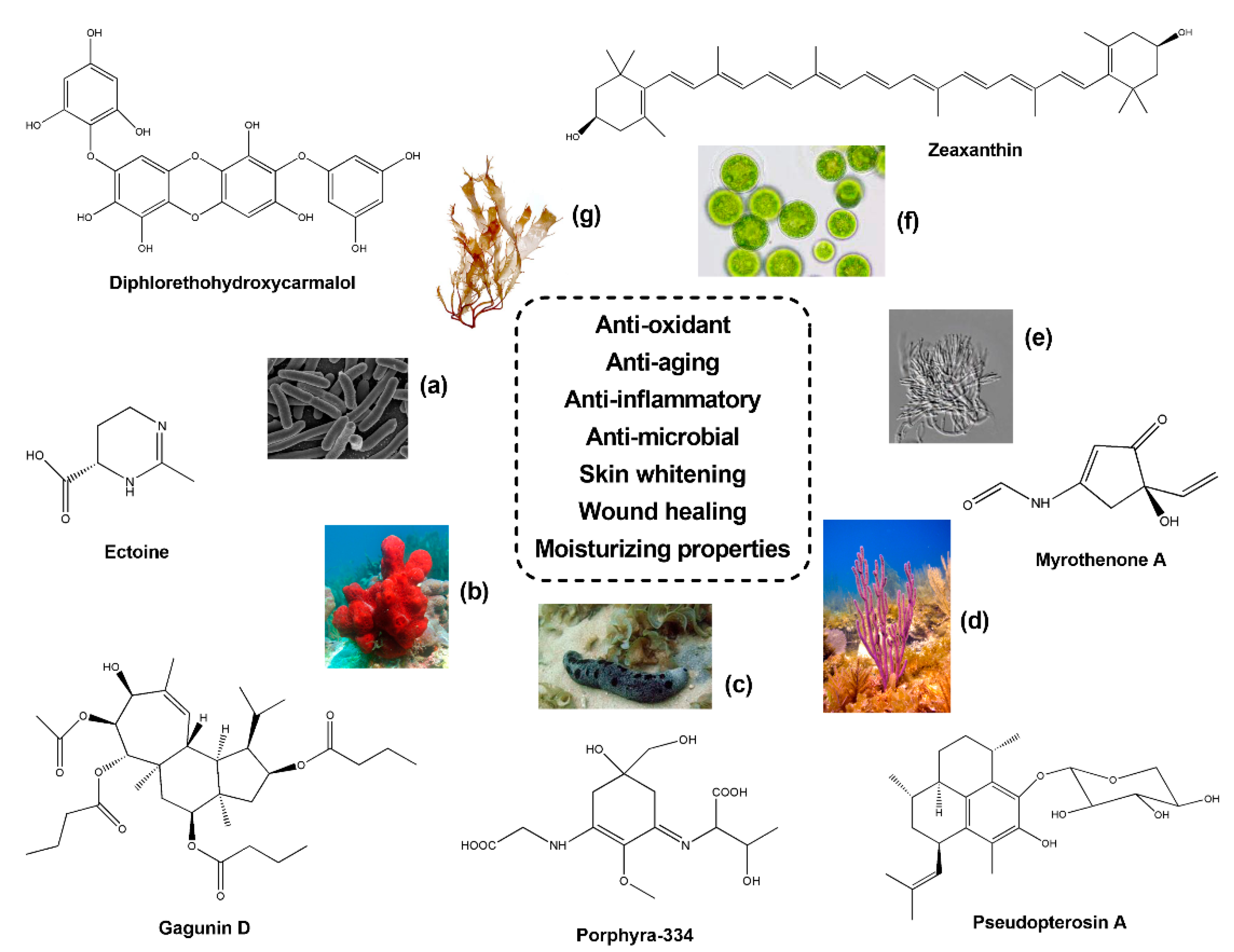
:1. Introduction
2. Sponge Symbionts in Cosmeceutical Field
2.1. Bacteria
2.2. Fungi
3. Sponges
4. Conclusions
Author Contributions
Funding
Institutional Review Board Statement
Data Availability Statement
Acknowledgments
Conflicts of Interest
References
- Thomas, T.R.A.; Kavlekar, D.P.; LokaBharathi, P.A. Marine drugs from sponge-microbe association—A review. Mar. Drugs 2010, 8, 1417–1468. [Google Scholar] [CrossRef] [Green Version]
- Thacker, R.W.; Freeman, C.J. Sponge-microbe symbioses: Recent advances and new directions. Adv. Mar. Biol. 2012, 62, 57–111. [Google Scholar]
- Webster, N.S.; Taylor, M.W. Marine sponges and their microbial symbionts: Love and other relationships. Environ. Microbiol. 2012, 14, 335–346. [Google Scholar] [CrossRef] [PubMed]
- Mehbub, M.F.; Lei, J.; Franco, C.; Zhang, W. Marine sponge derived natural products between 2001 and 2010: Trends and opportunities for discovery of bioactives. Mar. Drugs 2014, 12, 4539–4577. [Google Scholar] [CrossRef] [PubMed] [Green Version]
- Kumar, M.S.; Pal, A.K. A review of bioactive compounds from marine organisms with special mention on the potential of marine sponges in pharmacological applications. J. Mar. Biol. Assoc. India 2016, 58, 87–96. [Google Scholar] [CrossRef] [Green Version]
- Bibi, F.; Faheem, M.; Azhar, E.; Yasir, M.; Alvi, S.; Kamal, M.; Ullah, I.; Naseer, M. Bacteria from marine sponges: A source of new drugs. Curr. Drug Metab. 2016, 17. [Google Scholar] [CrossRef]
- Perdicaris, S.; Vlachogianni, T.; Valavanidis, A. Bioactive natural substances from marine sponges: New developments and prospects for future pharmaceuticals. Nat. Prod. Chem. Res. 2013, 1, 3. [Google Scholar] [CrossRef]
- Simister, R.L.; Deines, P.; Botté, E.S.; Webster, N.S.; Taylor, M.W. Sponge-specific clusters revisited: A comprehensive phylogeny of sponge-associated microorganisms. Environ. Microbiol. 2012, 14, 517–524. [Google Scholar] [CrossRef] [PubMed]
- Moitinho-Silva, L.; Nielsen, S.; Amir, A.; Gonzalez, A.; Ackermann, G.L.; Cerrano, C.; Astudillo-Garcia, C.; Easson, C.; Sipkema, D.; Liu, F.; et al. The sponge microbiome project. Gigascience 2017, 6, 1–13. [Google Scholar] [CrossRef] [PubMed]
- Tianero, M.D.; Balaich, J.N.; Donia, M.S. Localized production of defence chemicals by intracellular symbionts of Haliclona sponges. Nat. Microbiol. 2019, 4, 1149–1159. [Google Scholar] [CrossRef] [PubMed]
- Yang, Q.; Zhang, W.; Franco, C.M.M. Response of sponge microbiomes to environmental variations. In Symbiotic Microbiomes of Coral Reefs Sponges and Corals; Li, Z., Ed.; Springer Nature: Basingstoke, UK, 2019; pp. 181–247. ISBN 9789402416121. [Google Scholar]
- Freeman, C.J.; Thacker, R.W. Complex interactions between marine sponges and their symbiotic microbial communities. Limnol. Oceanogr. 2011, 56, 1577–1586. [Google Scholar] [CrossRef]
- Guzman, C.; Conaco, C. Gene expression dynamics accompanying the sponge thermal stress response. PLoS ONE 2016, 11, 1–15. [Google Scholar] [CrossRef] [Green Version]
- Marty, M.J.; Vicente, J.; Oyler, B.L.; Place, A.; Hill, R.T. Sponge symbioses between Xestospongia deweerdtae and Plakortis spp. are not motivated by shared chemical defense against predators. PLoS ONE 2017, 12, 1–20. [Google Scholar] [CrossRef] [PubMed]
- Shinde, P.; Banerjee, P.; Mandhare, A. Marine natural products as source of new drugs: A patent review (2015–2018). Expert Opin. Ther. Pat. 2019, 29, 283–309. [Google Scholar] [CrossRef] [PubMed]
- Draelos, Z.D. Cosmeceuticals: What’s real, what’s not. Dermatol. Clin. 2019, 37, 107–115. [Google Scholar] [CrossRef] [PubMed]
- Ahsan, H. The biomolecules of beauty: Biochemical pharmacology and immunotoxicology of cosmeceuticals. J. Immunoass. Immunochem. 2019, 40, 91–108. [Google Scholar] [CrossRef]
- Alves, T.F.R.; Morsink, M.; Batain, F.; Chaud, M.V.; Almeida, T.; Fernandes, D.A.; Silva, C.F.; Souto, E.B. Synthetic polymers in cosmetic formulations. Cosmetics 2020, 7, 75. [Google Scholar] [CrossRef]
- Sharad, J. Cosmeceuticals. In Advances in Integrative Dermatology; França, K., Lotti, T., Eds.; John Wiley & Sons Ltd.: Hoboken, NJ, USA, 2019; pp. 393–411. [Google Scholar]
- Khezri, K.; Saeedi, M.; Maleki Dizaj, S. Application of nanoparticles in percutaneous delivery of active ingredients in cosmetic preparations. Biomed. Pharmacother. 2018, 106, 1499–1505. [Google Scholar] [CrossRef]
- Bilal, M.; Iqbal, H.M.N. New insights on unique features and role of nanostructured materials in cosmetics. Cosmetics 2020, 7, 24. [Google Scholar] [CrossRef] [Green Version]
- Ben Haddada, M.; Gerometta, E.; Chawech, R.; Sorres, J.; Bialecki, A.; Pesnel, S.; Spadavecchia, J.; Morel, A.L. Assessment of antioxidant and dermoprotective activities of gold nanoparticles as safe cosmetic ingredient. Colloids Surf. B Biointerfaces 2020, 189, 110855. [Google Scholar] [CrossRef]
- Kesavan Pillai, S.; Kleyi, P.; de Beer, M.; Mudaly, P. Layered double hydroxides: An advanced encapsulation and delivery system for cosmetic ingredients-an overview. Appl. Clay Sci. 2020, 199, 105868. [Google Scholar] [CrossRef]
- Poulose, N.; Sajayan, A.; Ravindran, A.; Sreechithra, T.V.; Vardhan, V.; Selvin, J.; Kiran, G.S. Photoprotective effect of nanomelanin-seaweed concentrate in formulated cosmetic cream: With improved antioxidant and wound healing properties. J. Photochem. Photobiol. B Biol. 2020, 205, 111816. [Google Scholar] [CrossRef]
- Genç, Y.; Bardakci, H.; Yücel, Ç.; Karatoprak, G.Ş.; Akkol, E.K.; Barak, T.H.; Sobarzo-Sánchez, E. Oxidative stress and marine carotenoids: Application by using nanoformulations. Mar. Drugs 2020, 18, 423. [Google Scholar] [CrossRef]
- Gupta, R.K.; Soni, P.; Shrivastava, J.; Rajput, P.; Parashar, S.; Kalpana, B.; Nidana, R.; Vikriti, E. Cosmeceutical role of medicinal plants/herbs: A review on commercially available cosmetic ingredients. Int. J. Innov. Sci. Technol. 2018, 3, 70–73. [Google Scholar] [CrossRef]
- Joshi, L.S.; Pawar, H.A. Herbal cosmetics and cosmeceuticals: An overview. Nat. Prod. Chem. Res. 2015, 3, 170. [Google Scholar] [CrossRef]
- Charles Dorni, A.I.; Amalraj, A.; Gopi, S.; Varma, K.; Anjana, S.N. Novel cosmeceuticals from plants—An industry guided review. J. Appl. Res. Med. Aromat. Plants 2017, 7, 1–26. [Google Scholar] [CrossRef]
- Alves, A.; Sousa, E.; Kijjoa, A.; Pinto, M. Marine-derived compounds with potential use as cosmeceuticals and nutricosmetics. Molecules 2020, 25, 2536. [Google Scholar] [CrossRef] [PubMed]
- Pallela, R.; Na-Young, Y.; Kim, S.K. Anti-photoaging and photoprotective compounds derived from marine organisms. Mar. Drugs 2010, 8, 1189–1202. [Google Scholar] [CrossRef] [PubMed] [Green Version]
- Felician, F.F.; Xia, C.; Qi, W.; Xu, H. Collagen from marine biological sources and medical applications. Chem. Biodivers. 2018, 15, e1700557. [Google Scholar] [CrossRef] [PubMed]
- Manandhar, B.; Wagle, A.; Seong, S.H.; Paudel, P.; Kim, H.R.; Jung, H.A.; Choi, J.S. Phlorotannins with potential anti-tyrosinase and antioxidant activity isolated from the marine seaweed Ecklonia Stolonifera. Antioxid. 2019, 8, 240. [Google Scholar] [CrossRef] [PubMed] [Green Version]
- Couteau, C.; Coiffard, L. Phycocosmetics and other marine cosmetics, specific cosmetics formulated using marine resources. Mar. Drugs 2020, 18, 322. [Google Scholar] [CrossRef] [PubMed]
- Pilevneli, A.D.; Konuklugil, B. Marine derived tyrosinase inhibitors. Ege J. Fish. Aquat. Sci. 2020, 37, 427–436. [Google Scholar] [CrossRef]
- Solano, F. Photoprotection and skin pigmentation: Melanin-related molecules and some other new agents obtained from natural sources. Molecules 2020, 25, 1537. [Google Scholar] [CrossRef] [Green Version]
- Chang, T.S. Natural melanogenesis inhibitors acting through the down-regulation of tyrosinase activity. Materials 2012, 5, 1661–1685. [Google Scholar] [CrossRef] [Green Version]
- Balakrishnan, D.; Kandasamy, D.; Nithyanand, P. A review on antioxidant activity of marine organisms. Int. J. Chem. Tech. Res. 2014, 6, 3431–3436. [Google Scholar]
- Balboa, E.M.; Conde, E.; Soto, M.L.; Pérez-Armada, L.; Domínguez, H. Cosmetics from marine sources. In Springer Handbook of Marine Biotechnology; Kim, S.K., Ed.; Elsevier: Amsterdam, The Netherlands, 2015; Volume 44, pp. 1015–1042. [Google Scholar]
- Taofiq, O.; Heleno, S.A.; Calhelha, R.C.; Alves, M.J.; Barros, L.; Barreiro, M.F.; González-Paramás, A.M.; Ferreira, I.C.F.R. Development of mushroom-based cosmeceutical formulations with anti-inflammatory, anti-tyrosinase, antioxidant, and antibacterial properties. Molecules 2016, 21, 1372. [Google Scholar] [CrossRef] [Green Version]
- Brunt, E.G.; Burgess, J.G. The promise of marine molecules as cosmetic active ingredients. Int. J. Cosmet. Sci. 2016, 40, 1–15. [Google Scholar] [CrossRef] [Green Version]
- Chrapusta, E.; Kaminski, A.; Duchnik, K.; Bober, B.; Adamski, M.; Bialczyk, J. Mycosporine-like amino acids: Potential health and beauty ingredients. Mar. Drugs 2017, 15, 326. [Google Scholar] [CrossRef] [PubMed] [Green Version]
- Galasso, C.; Corinaldesi, C.; Sansone, C. Carotenoids from marine organisms: Biological functions and industrial applications. Antioxidants 2017, 6, 96. [Google Scholar] [CrossRef] [Green Version]
- Núñez-Pons, L.; Avila, C.; Romano, G.; Verde, C.; Giordano, D. UV-protective compounds in marine organisms from the southern ocean. Mar. Drugs 2018, 16, 336. [Google Scholar] [CrossRef] [Green Version]
- Vílchez, C.; Forján, E.; Cuaresma, M.; Bédmar, F.; Garbayo, I.; Vega, J.M. Marine carotenoids: Biological functions and commercial applications. Mar. Drugs 2011, 9, 319–333. [Google Scholar] [CrossRef] [Green Version]
- Wada, N.; Sakamoto, T.; Matsugo, S. Mycosporine-like amino acids and their derivatives as natural antioxidants. Antioxidants 2015, 4, 603–646. [Google Scholar] [CrossRef]
- Birben, E.; Sahiner, U.; Sackesen, C.; Erzurum, S.; Kalayci, O. Oxidative stress and antioxidant defense. Science 1997, 22, 161–168. [Google Scholar] [CrossRef] [PubMed] [Green Version]
- Younes, I.; Rinaudo, M. Chitin and chitosan preparation from marine sources. Structure, properties and applications. Mar. Drugs 2015, 13, 1133–1174. [Google Scholar] [CrossRef] [PubMed] [Green Version]
- Sansone, C.; Brunet, C. Marine algal antioxidants. Antioxidants 2020, 9, 206. [Google Scholar] [CrossRef] [PubMed] [Green Version]
- Tripathi, V.C.; Horam, S.; Singh, A.; Lata, M.; Reddy, T.J.; Arockiaraj, J.; Pasupuleti, M. The discovery of antioxidants in marine microorganisms and their protective effects on the hepatic cells from chemical-induced oxidative stress. Free Radic. Res. 2020, 54, 150–161. [Google Scholar] [CrossRef]
- Siavash, H.C.; Fadzilah, A.A.M. Cosmeceutical values, antimicrobial activities and antioxidant properties of cashew leaves extract. Afr. J. Biotechnol. 2011, 10, 4573–14582. [Google Scholar] [CrossRef] [Green Version]
- Baldisserotto, A.; Malisardi, G.; Scalambra, E.; Andreotti, E.; Romagnoli, C.; Vicentini, C.B.; Manfredini, S.; Vertuani, S. Synthesis, antioxidant and antimicrobial activity of a new phloridzin derivative for dermo-cosmetic applications. Molecules 2012, 17, 13275–13289. [Google Scholar] [CrossRef] [Green Version]
- Herman, A.; Herman, A.P.; Domagalska, B.W.; Młynarczyk, A. Essential oils and herbal extracts as antimicrobial agents in cosmetic emulsion. Indian J. Microbiol. 2013, 53, 232–237. [Google Scholar] [CrossRef] [PubMed] [Green Version]
- Adegoke, T.; Arotupin, D.; Ekundayo, T. Antimicrobial activities of some commercial cosmetics on selected cutaneous microflora. J. Adv. Microbiol. 2017, 4, 1–9. [Google Scholar] [CrossRef] [Green Version]
- Kim, J.W.; Yu, H.; Park, K.M.; Chang, P.S. Antimicrobial characterization of erythorbyl laurate for practical applications in food and cosmetics. J. Chem. 2020, 2020, 1–8. [Google Scholar] [CrossRef]
- Pillaiyar, T.; Manickam, M.; Namasivayam, V. Skin whitening agents: Medicinal chemistry perspective of tyrosinase inhibitors. J. Enzym. Inhib. Med. Chem. 2017, 32, 403–425. [Google Scholar] [CrossRef] [PubMed] [Green Version]
- McClements, D.J.; Gumus, C.E. Natural emulsifiers—Biosurfactants, phospholipids, biopolymers, and colloidal particles: Molecular and physicochemical basis of functional performance. Adv. Colloid Interface Sci. 2016, 234, 3–26. [Google Scholar] [CrossRef] [PubMed] [Green Version]
- Adu, S.A.; Naughton, P.J.; Marchant, R.; Banat, I.M. Microbial biosurfactants in cosmetic and personal skincare pharmaceutical formulations. Pharmaceutics 2020, 12, 1099. [Google Scholar] [CrossRef]
- Corinaldesi, C.; Barone, G.; Marcellini, F.; Dell’Anno, A.; Danovaro, R. Marine microbial-derived molecules and their potential use in cosmeceutical and cosmetic products. Mar. Drugs 2017, 15, 118. [Google Scholar] [CrossRef]
- Kong, S.Z.; Li, J.C.; Li, S.D.; Liao, M.N.; Li, C.P.; Zheng, P.J.; Guo, M.H.; Tan, W.X.; Zheng, Z.H.; Hu, Z. Anti-aging effect of chitosan oligosaccharide on d-galactose-induced subacute aging in mice. Mar. Drugs 2018, 16, 181. [Google Scholar] [CrossRef] [Green Version]
- Jahan, A.; Ahmad, I.Z.; Fatima, N.; Ansari, V.A.; Akhtar, J. Algal bioactive compounds in the cosmeceutical industry: A review. Phycologia 2017, 56, 410–422. [Google Scholar] [CrossRef]
- Pereira, L. Seaweeds as source of bioactive substances and skin care therapy—Cosmeceuticals, algotheraphy, and thalassotherapy. Cosmetics 2018, 5, 68. [Google Scholar] [CrossRef] [Green Version]
- Hamidi, M.; Safarzadeh Kozani, P.; Safarzadeh Kozani, P.; Pierre, G.; Michaud, P.; Delattre, C. Marine bacteria versus microalgae: Who is the best for biotechnological production of bioactive compounds with antioxidant properties and other biological applications? Mar. Drugs 2020, 18, 28. [Google Scholar] [CrossRef] [Green Version]
- Thiyagarasaiyar, K.; Goh, B.H.; Jeon, Y.J.; Yow, Y.Y. Algae metabolites in cosmeceutical: An overview of current applications and challenges. Mar. Drugs 2020, 18, 323. [Google Scholar] [CrossRef]
- Martins, A.; Vieira, H.; Gaspar, H.; Santos, S. Marketed marine natural products in the pharmaceutical and cosmeceutical industries: Tips for success. Mar. Drugs 2014, 12, 1066–1101. [Google Scholar] [CrossRef] [Green Version]
- Calado, R.; Leal, M.C.; Gaspar, H.; Santos, S.; Marques, A.; Nunes, M.L.; Vieira, H. How to succeed in marketing marine natural products for nutraceutical, pharmaceutical and cosmeceutical markets. In Grand Challenges in Biology and Biotechnology; Rampelotto, P.H., Trincone, A., Eds.; Springer International Publishing: Manhattan, NY, USA, 2018; pp. 317–403. ISBN 9783319690759. [Google Scholar]
- Hassane, C.S.; Fouillaud, M.; Le Goff, G.; Sklirou, A.D.; Boyer, J.B.; Trougakos, I.P.; Jerabek, M.; Bignon, J.; de Voogd, N.J.; Ouazzani, J.; et al. Microorganisms associated with the marine sponge Scopalina hapalia: A reservoir of bioactive molecules to slow down the aging process. Microorganisms 2020, 8, 1262. [Google Scholar] [CrossRef]
- Imhoff, J.F.; Labes, A.; Wiese, J. Bio-mining the microbial treasures of the ocean: New natural products. Biotechnol. Adv. 2011, 29, 468–482. [Google Scholar] [CrossRef] [PubMed]
- Gao, Q.; Garcia-Pichel, F. Microbial ultraviolet sunscreens. Nat. Rev. Microbiol. 2011, 9, 791–802. [Google Scholar] [CrossRef] [PubMed]
- Morabito, K.; Shapley, N.C.; Steeley, K.G.; Tripathi, A. Review of sunscreen and the emergence of non-conventional absorbers and their applications in ultraviolet protection. Int. J. Cosmet. Sci. 2011, 33, 385–390. [Google Scholar] [CrossRef]
- Young, A.J.; Lowe, G.L. Carotenoids—antioxidant properties. Antioxidants 2018, 7, 28. [Google Scholar] [CrossRef] [PubMed] [Green Version]
- Dharmaraj, S.; Ashokkumar, B.; Dhevendaran, K. Food-grade pigments from Streptomyces sp. isolated from the marine sponge Callyspongia diffusa. Food Res. Int. 2009, 42, 487–492. [Google Scholar] [CrossRef]
- Shindo, K.; Asagi, E.; Sano, A.; Hotta, E.; Minemura, N.; Mikami, K.; Tamesada, E.; Misawa, N.; Maoka, T. Diapolycopenedioic acid xylosyl esters A, B, and C, novel antioxidative glyco-C30-carotenoic acids produced by a new marine bacterium Rubritalea Squalenifaciens. J. Antibiot. (Tokyo) 2008, 61, 185–191. [Google Scholar] [CrossRef] [Green Version]
- Abdelmohsen, U.R.; Szesny, M.; Othman, E.M.; Schirmeister, T.; Grond, S.; Stopper, H.; Hentschel, U. Antioxidant and anti-protease activities of diazepinomicin from the sponge-associated Micromonospora strain RV115. Mar. Drugs 2012, 10, 2208–2221. [Google Scholar] [CrossRef]
- Arunachalam, K.; Amirtham Jacob Appadorai, R. Antioxidant potential and biochemical evaluation of metabolites from the marine bacteria Virgibacillus sp. associated with the sponge Callyspongia diffusa. Free Radic. Antioxid. 2013, 3, 47–51. [Google Scholar] [CrossRef] [Green Version]
- Dupont, S.; Carré-Mlouka, A.; Descarrega, F.; Ereskovsky, A.; Longeon, A.; Mouray, E.; Florent, I.; Bourguet-Kondracki, M.L. Diversity and biological activities of the bacterial community associated with the marine sponge Phorbas tenacior (Porifera, Demospongiae). Lett. Appl. Microbiol. 2014, 58, 42–52. [Google Scholar] [CrossRef]
- Balakrishnan, D.; Bibiana, A.S.; Vijayakumar, A.; Santhosh, R.S.; Dhevendaran, K.; Nithyanand, P. Antioxidant activity of bacteria associated with the marine sponge Tedania anhelans. Indian J. Microbiol. 2015, 55, 13–18. [Google Scholar] [CrossRef]
- Mohan, G.; Centre, N.; Sustainable, F.; Management, C.; Kumar, T.T.A. Marine natural product, Pyrrolo[1,2-a]pyrazine-1,4-dione, hexahydro- (C7H10N2O2) of antioxidant properties from Bacillus species at Lakshadweep archipelago. J. Coast. Life Med. 2014, 2, 636–641. [Google Scholar] [CrossRef]
- Velho-Pereira, S.; Parvatkar, P.; Furtado, I.J. Evaluation of antioxidant producing potential of halophilic bacterial bionts from marine invertebrates. Indian J. Pharm. Sci. 2015, 77, 183–189. [Google Scholar]
- El-Moneam, N.M.A.; El-Assar, S.A.; Shreadah, M.A.; Nabil-Adam, A. Isolation, identification and molecular screening of Pseudomonas sp. metabolic pathways NRPs and PKS associated with the Red Sea Sponge, Hyrtios aff. erectus, Egypt. J. Pure Appl. Microbiol. 2017, 11, 1299–1311. [Google Scholar] [CrossRef]
- Vijayan, V.; Jasmin, C.; Anas, A.; Parakkaparambil Kuttan, S.; Vinothkumar, S.; Perunninakulath Subrayan, P.; Nair, S. Sponge-associated bacteria produce non-cytotoxic melanin which protects animal cells from photo-toxicity. Appl. Biochem. Biotechnol. 2017, 183, 396–411. [Google Scholar] [CrossRef] [PubMed]
- Prastya, M.E.; Astuti, R.I.; Batubara, I.; Wahyudi, A.T. Antioxidant, antiglycation and in vivo antiaging effects of metabolite extracts from marine sponge-associated bacteria. Indian J. Pharm. Sci. 2019, 81, 344–354. [Google Scholar] [CrossRef] [Green Version]
- Prastya, M.E.; Astuti, R.I.; Batubara, I.; Takagi, H.; Wahyudi, A.T. Chemical screening identifies an extract from marine Pseudomonas sp.-PTR-08 as an anti-aging agent that promotes fission yeast longevity by modulating the Pap1–ctt1+ pathway and the cell cycle. Mol. Biol. Rep. 2020, 47, 33–43. [Google Scholar] [CrossRef]
- Prastya, M.E.; Astuti, R.I.; Batubara, I.; Takagi, H.; Wahyudi, A.T. Natural extract and its fractions isolated from the marine bacterium Pseudoalteromonas flavipulchra STILL-33 have antioxidant and antiaging activities in Schizosaccharomyces pombe. FEMS Yeast Res. 2020, 20, 1–14. [Google Scholar] [CrossRef]
- Cheng, C.; Othman, E.M.; Reimer, A.; Grüne, M.; Kozjak-Pavlovic, V.; Stopper, H.; Hentschel, U.; Abdelmohsen, U.R. Ageloline A, new antioxidant and antichlamydial quinolone from the marine sponge-derived bacterium Streptomyces sp. SBT345. Tetrahedron Lett. 2016, 57, 2786–2789. [Google Scholar] [CrossRef]
- Phadale, R.; Kumar, M.S. Characterization of an antimicrobial and antioxidant compound from a marine bacterium GSA10 associated with the sponge Halichondria glabrata. J. Microbiol. Biotechnol. Food Sci. 2018, 7, 651–658. [Google Scholar] [CrossRef]
- Odekina, P.A.; Agbo, M.O.; Omeje, E.O. Antimicrobial and antioxidant activities of novel marine bacteria (Bacillus 2011SOCCUF3) isolated from marine sponge (Spongia officinalis). Pharm. Sci. 2020, 26, 82–87. [Google Scholar] [CrossRef] [Green Version]
- Dhasayan, A.; Selvin, J.; Kiran, S. Biosurfactant production from marine bacteria associated with sponge Callyspongia diffusa. 3 Biotech 2015, 5, 443–454. [Google Scholar] [CrossRef] [Green Version]
- Alemán-Vega, M.; Sánchez-Lozano, I.; Hernández-Guerrero, C.J.; Hellio, C.; Quintana, E.T. Exploring antifouling activity of biosurfactants producing marine bacteria isolated from gulf of California. Int. J. Mol. Sci. 2020, 21, 6068. [Google Scholar] [CrossRef]
- Agrawal, S.; Adholeya, A.; Barrow, C.J.; Deshmukh, S.K. Marine fungi: An untapped bioresource for future cosmeceuticals. Phytochem. Lett. 2018, 23, 15–20. [Google Scholar] [CrossRef]
- Vitale, G.A.; Coppola, D.; Esposito, F.P.; Buonocore, C.; Ausuri, J.; Tortorella, E.; de Pascale, D. Antioxidant molecules from marine fungi: Methodologies and perspectives. Antioxidants 2020, 9, 1183. [Google Scholar] [CrossRef]
- Jeewon, R.; Luckhun, A.B.; Bhoyroo, V.; Sadeer, N.B.; Mahomoodally, M.F.; Rampadarath, S.; Puchooa, D.; Venkateswara Sarma, V.; Sundara, S.; Durairajan, K.; et al. Pharmaceutical potential of marine fungal endophytes. In Endophytes and Secondary Metabolites; Jha, S., Ed.; Springer Nature: Basingstoke, UK, 2019; pp. 1–23. [Google Scholar]
- Devi, R.; Kaur, T.; Guleria, G.; Rana, K.L.; Kour, D.; Yadav, N.; Yadav, A.N.; Saxena, A.K. Fungal secondary metabolites and their biotechnological applications for human health. In New and Future Developments in Microbial Biotechnology and Bioengineering; Elsevier: Amsterdam, The Netherlands, 2020; pp. 147–161. [Google Scholar]
- Tanod, W.A.; Muliadin, M.; Adel, Y.S.; Dewanto, D.K. Potential marine-derived fungi isolated from sponges produce new and beneficial compounds. J. Fish. Mar. Aquat. Sci. 2020, 2, 52–66. [Google Scholar]
- Mohapatra, B.R.; Bapuji, M. Characterization of acetylcholinesterase from Arthrobacter ilicis associated with the marine sponge (Spirastrella sp.). J. Appl. Microbiol. 1998, 84, 393–398. [Google Scholar] [CrossRef]
- Zhang, D.; Yang, X.; Kang, J.S.; Choi, H.D.; Son, B.W. Circumdatin I, a new ultraviolet-A protecting benzodiazepine alkaloid from a marine isolate of the fungus Exophiala. J. Antibiot. (Tokyo) 2008, 61, 40–42. [Google Scholar] [CrossRef]
- Zhang, J.; Yuan, B.; Liu, D.; Gao, S.; Proksch, P.; Lin, W. Brasilianoids A-F, new meroterpenoids from the sponge-associated fungus Penicillium brasilianum. Front. Chem. 2018, 6, 1–13. [Google Scholar] [CrossRef] [PubMed] [Green Version]
- Sun, H.H.; Mao, W.J.; Jiao, J.Y.; Xu, J.C.; Li, H.Y.; Chen, Y.; Qi, X.H.; Chen, Y.L.; Xu, J.; Zhao, C.Q.; et al. Structural characterization of extracellular polysaccharides produced by the marine fungus Epicoccum nigrum JJY-40 and their antioxidant activities. Mar. Biotechnol. 2011, 13, 1048–1055. [Google Scholar] [CrossRef] [PubMed]
- Li, J.L.; Lee, Y.M.; Hong, J.; Bae, K.S.; Choi, J.S.; Jung, J.H. A new antioxidant from the marine sponge-derived fungus Aspergillus versicolor. Nat. Prod. Sci. 2011, 17, 14–18. [Google Scholar]
- Kawahara, T.; Takagi, M.; Shin-Ya, K. JBIR-124: A novel antioxidative agent from a marine sponge-derived fungus Penicillium citrinum SpI080624G1f01. J. Antibiot. (Tokyo) 2012, 65, 45–47. [Google Scholar] [CrossRef] [PubMed] [Green Version]
- Abdel-Monem, N.; Abdel-Azeem, A.M.; El Ashry, E.S.H.; Ghareeb, D.A.; Nabil-Adam, A. Assessment of secondary metabolites from marine-derived fungi as antioxidant. Open J. Med. Chem. 2013, 3, 60–73. [Google Scholar] [CrossRef] [Green Version]
- Pang, X.; Lin, X.; Yang, J.; Zhou, X.; Yang, B.; Wang, J.; Liu, Y. Spiro-phthalides and Isocoumarins isolated from the marine-sponge-derived fungus Setosphaeria sp. SCSIO41009. J. Nat. Prod. 2018, 81, 1860–1868. [Google Scholar] [CrossRef]
- Du, X.; Liu, D.; Huang, J.; Zhang, C.; Proksch, P.; Lin, W. Polyketide derivatives from the sponge associated fungus Aspergillus europaeus with antioxidant and NO inhibitory activities. Fitoterapia 2018, 130, 190–197. [Google Scholar] [CrossRef]
- El-Hady, F.K.A.; Abdel-aziz, M.S.; Shaker, K.H.; El-shahid, Z.A.; Ibrahim, L.S. Antioxidant, acetylcholinesterase and α-glucosidase potentials of metabolites from the marine fungus. Int. J. Pharm. Sci. Rev. Res. 2015, 30, 272–278. [Google Scholar]
- Sun, Y.; Liu, J.; Li, L.; Gong, C.; Wang, S.; Yang, F.; Hua, H.; Lin, H. New butenolide derivatives from the marine sponge-derived fungus Aspergillus terreus. Bioorganic Med. Chem. Lett. 2018, 28, 315–318. [Google Scholar] [CrossRef]
- Henríquez, M.; Vergara, K.; Norambuena, J.; Beiza, A.; Maza, F.; Ubilla, P.; Araya, I.; Chávez, R.; San-Martín, A.; Darias, J.; et al. Diversity of cultivable fungi associated with Antarctic marine sponges and screening for their antimicrobial, antitumoral and antioxidant potential. World J. Microbiol. Biotechnol. 2014, 30, 65–76. [Google Scholar] [CrossRef] [PubMed]
- Julianti, E.; Oh, H.; Jang, K.H.; Lee, J.K.; Lee, S.K.; Oh, D.C.; Oh, K.B.; Shin, J. Acremostrictin, a highly oxygenated metabolite from the marine fungus Acremonium strictum. J. Nat. Prod. 2011, 74, 2592–2594. [Google Scholar] [CrossRef]
- Ding, L.J.; Gu, B.B.; Jiao, W.H.; Yuan, W.; Li, Y.X.; Tang, W.Z.; Yu, H.B.; Liao, X.J.; Han, B.N.; Li, Z.Y.; et al. New furan and cyclopentenone derivatives from the sponge-associated fungus Hypocrea koningii PF04. Mar. Drugs 2015, 13, 5579–5592. [Google Scholar] [CrossRef] [PubMed] [Green Version]
- Ding, L.J.; Yuan, W.; Li, Y.X.; Liao, X.J.; Sun, H.; Peng, Q.; Han, B.N.; Lin, H.W.; Li, Z.Y.; Yang, F.; et al. Hypocrol A, a new tyrosol derivative from a sponge-derived strain of the fungus Hypocrea koningii. Nat. Prod. Res. 2016, 30, 1633–1638. [Google Scholar] [CrossRef] [PubMed]
- Sayed, M.A.; El-rahman, T.M.A.A.; El-diwany, A.I.; Product, M.; Sayed, S.M. Biodiversity and bioactivity of red sea sponge associated endophytic fungi. Int. J. Adv. Res. Eng. Appl. Sci. 2016, 5, 1–15. [Google Scholar]
- Zhao, Y.; Liu, D.; Proksch, P.; Yu, S.; Lin, W. Isocoumarin derivatives from the sponge-associated fungus Peyronellaea glomerata with antioxidant activities. Chem. Biodivers. 2016, 13, 1186–1193. [Google Scholar] [CrossRef] [PubMed]
- El-Hady, F.K.A.; Abdel-Aziz, M.S.; Shaker, K.H.; El-Shahid, Z.A. Tyrosinase, acetylcholinesterase inhibitory potential, antioxidant and antimicrobial activities of sponge derived fungi with correlation to their GC/MS analysis. Int. J. Pharm. Sci. Rev. Res. 2014, 26, 338–345. [Google Scholar]
- Trianto, A.; Widyaningsih, S.; Radjasa, O.; Pribadi, R. Symbiotic fungus of marine sponge Axinella sp. producing antibacterial agent. Conf. Ser. Earth Environ. Sci. 2017, 55, 012005. [Google Scholar] [CrossRef]
- Handayani, D.; Andalas, U.; Sandrawati, N.; Andalas, U.; Syafni, N.; Putra, D.P. Tyrosinase inhibitory activity of ethyl acetate extracts from marine sponge-derived fungi Haliclona fascigera. Biosci. Res. 2019, 16, 2369–2373. [Google Scholar]
- Scopel, M.; Abraham, W.R.; Henriques, A.T.; MacEdo, A.J. Dipeptide cis-cyclo(Leucyl-Tyrosyl) produced by sponge associated Penicillium sp. F37 inhibits biofilm formation of the pathogenic Staphylococcus epidermidis. Bioorganic Med. Chem. Lett. 2013, 23, 624–626. [Google Scholar] [CrossRef] [PubMed]
- Letsiou, S.; Bakea, A.; Le Goff, G.; Lopes, P.; Gardikis, K.; Weis, M.; Benayahu, Y.; Ouazzani, J. Marine fungus Aspergillus chevalieri TM2-S6 extract protects skin fibroblasts from oxidative stress. Mar. Drugs 2020, 18, 460. [Google Scholar] [CrossRef]
- Laport, M.S. Isolating bacteria from sponges: Why and how? Curr. Pharm. Biotechnol. 2018, 18, 1224–1236. [Google Scholar] [CrossRef]
- Amigó, M.; Terencio, M.C.; Mitova, M.; Iodice, C.; Payá, M.; De Rosa, S. Potential antipsoriatic avarol derivatives as antioxidants and inhibitors of PGE2 generation and proliferation in the HaCaT cell line. J. Nat. Prod. 2004, 67, 1459–1463. [Google Scholar] [CrossRef]
- Amigó, M.; Payá, M.; De Rosa, S.; Terencio, M.C. Antipsoriatic effects of avarol-3′-thiosalicylate are mediated by inhibition of TNF-α generation and NF-kB activation in mouse skin. Br. J. Pharmacol. 2007, 152, 353–365. [Google Scholar] [CrossRef] [Green Version]
- Chairman, K.; Singh, A.J.A.R.; Alagumuthu, G. Cytotoxic and antioxidant activity of selected marine sponges. Asian Pac. J. Trop. Dis. 2012, 2, 234–238. [Google Scholar] [CrossRef]
- Seradj, H.; Moein, M.; Eskandari, M.; Maaref, F. Antioxidant activity of six marine sponges collected from the Persian gulf. Iran. J. Pharm. Sci. 2012, 8, 249–255. [Google Scholar]
- Abdillah, S.; Nurhayati, A.P.D.; Nurhatika, S.; Setiawan, E.; Heffen, W.L. Cytotoxic and antioxidant activities of marine sponge diversity at Pecaron Bay Pasir Putih Situbondo East Java, Indonesia. J. Pharm. Res. 2013, 6, 685–689. [Google Scholar] [CrossRef]
- Aktas, N.; Genc, Y.; Gozcelioglu, B.; Konuklugil, B.; Sebnem Harput, U. Radical scavenging effect of different marine sponges from mediterranean coasts. Rec. Nat. Prod. 2013, 7, 96–104. [Google Scholar]
- Botić, T.; Cör, D.; Anesi, A.; Guella, G.; Sepčić, K.; Janussen, D.; Kersken, D.; Knez, Ž. Fatty acid composition and antioxidant activity of Antarctic marine sponges of the genus Latrunculia. Polar Biol. 2015, 38, 1605–1612. [Google Scholar] [CrossRef]
- Hagiwara, K.; Garcia Hernandez, J.E.; Harper, M.K.; Carroll, A.; Motti, C.A.; Awaya, J.; Nguyen, H.Y.; Wright, A.D. Puupehenol, a potent antioxidant antimicrobial meroterpenoid from a Hawaiian deep-water Dactylospongia sp. sponge. J. Nat. Prod. 2015, 78, 325–329. [Google Scholar] [CrossRef]
- Rhandour, Z.; Tarbaoui, M.; Oumam, M.; Elamraoui, B.; Bennamara, A.; Abourriche, A. Determination of polyphenols, tannins, flavonoids and antioxidant activity in extracts of two genus Ircinia marine sponges of Atlantic Morrocan Coast. Front. Mar. Sci. 2016, 3. [Google Scholar] [CrossRef]
- Rivera, A.P.; Uy, M.M. In vitro antioxidant and cytotoxic activities of some marine sponges collected off misamis oriental coast, Philippines. E-J. Chem. 2012, 9, 354–358. [Google Scholar] [CrossRef]
- Nabil-Adam, A.; Shreadah, M.A.; El Moneam, N.M.A.; El-Assar, S.A. Various in vitro bioactivities of secondary metabolites isolated from the sponge Hyrtios aff. erectus from the red sea coast of Egypt. Turk. J. Pharm. Sci. 2020, 17, 127–135. [Google Scholar] [CrossRef] [PubMed]
- Oogarah, P.N.; Ramanjooloo, A.; Rovisham, J.; Doorga, S.; Meyepa, C.; Wilhelmus, R.; Van Soest, M.; Edgard, D.; Marie, P. Assessing antioxidant activity and phenolic content of marine sponges from mauritius waters. Int. J. Pharmacogn. Phytochem. Res. 2020, 12, 123–131. [Google Scholar] [CrossRef]
- Warsidah, M.; Sofiana, M.S.J.; Safitri, I.; Sapar, A.; Aritonang, A.B.; Muttalib, Y.S.; Fadly, D. Protein isolation from sponge Niphates sp. as an antibacterial and antioxidant. Syst. Rev. Pharm. 2020, 11, 518–521. [Google Scholar] [CrossRef]
- Hwang, J.; Kim, D.; Park, J.S.; Park, H.J.; Shin, J.; Lee, S.K. Photoprotective activity of Topsentin, a bis(indole) alkaloid from the marine sponge Spongosorites genitrix, by regulation of COX-2 and Mir-4485 expression in UVB-irradiated human keratinocyte cells. Mar. Drugs 2020, 18, 87. [Google Scholar] [CrossRef] [Green Version]
- Cheung, F.W.K.; Guo, J.; Ling, Y.H.; Che, C.T.; Liu, W.K. Anti-melanogenic property of geoditin A in murine B16 melanoma cells. Mar. Drugs 2012, 10, 465–476. [Google Scholar] [CrossRef] [Green Version]
- Lee, H.Y.; Jang, E.J.; Bae, S.Y.; Jeon, J.E.; Park, H.J.; Shin, J.; Lee, S.K. Anti-melanogenic activity of Gagunin D, a highly oxygenated diterpenoid from the marine sponge Phorbas sp., via modulating tyrosinase expression and degradation. Mar. Drugs 2016, 14, 212. [Google Scholar] [CrossRef] [Green Version]
- Yanti, C.; Vendy, V.; Hwang, J.K. In vitro anti-acne activity of marine sponge Acanthella cavernosa extracts. Int. J. Biol. Pharm. Res. 2015, 6, 388–392. [Google Scholar]
- Chander, M.P.; Vijayachari, P. Antimicrobial and anti-oxidant potentials of marine sponges of South Andaman, India. Bangladesh J. Pharmacol. 2018, 13, 13–15. [Google Scholar] [CrossRef] [Green Version]
- Campos, P.E.; Herbette, G.; Chendo, C.; Clerc, P.; Tintillier, F.; Voogd, N.J.D.; Papanagnou, E.D.; Trougakos, I.P.; Jerabek, M.; Bignon, J.; et al. Osirisynes G-I, new long-chain highly oxygenated polyacetylenes from the Mayotte marine sponge Haliclona sp. Mar. Drugs 2020, 18, 350. [Google Scholar] [CrossRef]
- Ghatak, S.; Maytin, E.V.; MacK, J.A.; Hascall, V.C.; Atanelishvili, I.; Moreno Rodriguez, R.; Markwald, R.R.; Misra, S. Roles of proteoglycans and glycosaminoglycans in wound healing and fibrosis. Int. J. Cell Biol. 2015, 2015, 1–20. [Google Scholar] [CrossRef] [Green Version]
- Pallela, R.; Ehrlich, H.; Bhatnagar, I. Marine sponges: Chemico-biological and biomedical applications. In Marine Sponges: Chemicobiological and Biomedical Applications; Pallela, R., Ehrlich, H., Eds.; Springer: New Delhi, India, 2016; pp. 1–381. ISBN 9788132227946. [Google Scholar]
- Swatschek, D.; Schatton, W.; Kellermann, J.; Müller, W.E.G.; Kreuter, J. Marine sponge collagen: Isolation, characterization and effects on the skin parameters surface-pH, moisture and sebum. Eur. J. Pharm. Biopharm. 2002, 53, 107–113. [Google Scholar] [CrossRef]
- Langasco, R.; Cadeddu, B.; Formato, M.; Lepedda, A.J.; Cossu, M.; Giunchedi, P.; Pronzato, R.; Rassu, G.; Manconi, R.; Gavini, E. Natural collagenic skeleton of marine sponges in pharmaceutics: Innovative biomaterial for topical drug delivery. Mater. Sci. Eng. C 2017, 70, 710–720. [Google Scholar] [CrossRef] [PubMed]
- Pozzolini, M.; Millo, E.; Oliveri, C.; Mirata, S.; Salis, A.; Damonte, G.; Arkel, M.; Scarfì, S. Elicited ROS scavenging activity, photoprotective, and wound-healing properties of collagen-derived peptides from the marine sponge Chondrosia reniformis. Mar. Drugs 2018, 16, 465. [Google Scholar] [CrossRef] [PubMed] [Green Version]


| Source | Sponge Host | Extract/Compound | Biological Activity | Reference |
|---|---|---|---|---|
| R. squalenifaciens | Halichondria okadai | Diapolycopenedioic acid xylosyl ester A | Anti-oxidant | [72] |
| Streptomyces | C. diffusa | Carotenoid extracts | Anti-aging | [71] |
| Micromonospora sp. RV115 | A. aerophoba | Diazepinomicin | Anti-oxidant | [73] |
| Virgibacillus sp. | C. diffusa | Ethyl acetate extracts | Anti-oxidant | [74] |
| Vibrio (P1Ma8 and P1Ma5) | P. tenacior | CH2Cl2/MeOH (1:1) extracts | Anti-oxidant | [75] |
| Bacillus sp. | T. anhelans | Ethyl acetate extracts | Anti-oxidant | [76] |
| Bacillus sp. | Sponges from Lakshadweep archipelago | Pyrrolo[1,2-a]pyrazine-1,4-dione, hexahydro-C7H10N2O2 | Anti-oxidant | [77] |
| Halomonas sp. MB-30 and Alcaligenes sp. MB-I9 | C. diffusa | Isolates of bacteria | Biosurfactants | [87] |
| C. israelensis | Callyspongia fibrosa | Ethyl acetate extracts | Anti-oxidant | [78] |
| Streptomyces sp. SBT345 | A. oroides | Ageloline A | Anti-oxidant | [84] |
| Pseudomonas sp. | H. aff. Erectus | Ethyl acetate extracts | Anti-oxidant | [79] |
| V. alginolyticus | H. pigmentifera, S. pumila and S. officinalis | Melanin extracts | Anti-oxidant | [80] |
| GSA10 | H. glabrata | Ethyl acetate extracts | Anti-oxidant | [85] |
| HAL-08, HAL-13, HAL-74 and PTR-21 | Haliclona sp. and Petrosia sp. | Ethyl acetate extracts | Anti-oxidant | [81] |
| PTR-08, PTR-40, PTR-41, and PTR-47 | Petrosia sp. | Ethyl acetate extracts | Anti-oxidant | [82] |
| P. flavipulchra STILL-33 | Stylotella sp. | Ethyl acetate extracts | Anti-oxidant and anti-aging | [83] |
| B. niabensis (My-30) | M. ramulosa | Isolates of bacteria | Biosurfactants | [88] |
| Bacillus 2011SOCCUF3 | S. officinalis | Methanol crude extracts | Anti-oxidant and anti-microbial | [86] |
| SH-82 (M. fluostatini) | S. hapalia | Ethyl acetate and methanol extracts | Anti-oxidant | [66] |
| Source | Sponge Host | Extract/Compound | Biological Activity | Reference |
|---|---|---|---|---|
| Exophiala | H. panicea | Circumdatin | Anti-UV | [95] |
| A. strictum | Unidentified marine sponge of the class Choristida | Acremostrictin | Anti-microbial and anti-oxidant | [106] |
| E. nigrum JJY-40 | Unidentified marine sponge | ENP1, ENP2 | Anti-oxidant | [97] |
| A. versicolor | Petrosia sp. | Aromatic polyketide | Anti-oxidant | [98] |
| P. citrinum SpI080624G1f01 | Unidentified marine sponge | JBIR-124 | Anti-oxidant | [99] |
| C. globosum, G. dankaliensis and N. oryzae | H. communis | Ethyl acetate extract | Anti-oxidant and anti-inflammatory | [100] |
| Penicillium sp. F37 | A. corrugata | Cis-cyclo(Leucyl-Tyrosyl) | Anti-biofilm | [114] |
| A. sydowii strain W4-2 and unidentified fungus FS1 | Agelas sp. and Amphimedon viridis | Crude extract of static cultures | Anti-oxidant, anti-tyrosinase and anti-microbial | [111] |
| F09T15-3, F09-T15-6, F09-T18-16 | Tedania sp., Hymeniacidon sp., Dendrilla sp. and three Poecilosclerida | Ethyl acetate extract of culture medium | Anti-oxidant | [105] |
| H. koningii PF04 | P. fusca | Hypofurans A/B and Hypocrenones A/B/C | Anti-microbial | [107] |
| A. unguis RSPG_204 | Agelas sp. | Several metabolites from mycelia and culture supernatant extracts | Anti-oxidant and anti-tyrosinase | [103] |
| H. koningii PF04 | P. fusca | Hypocrol A and Trichodenol B | Anti-microbial and anti-oxidant | [108] |
| A. oryzae, C. cladosporioides, A. fumigatus | A. citrina, S. rigida, O. lobularis, C. girardae, M. miriabilis, C. celata and S. difficilis | Mycelia and culture filtrate extracts | Anti-microbial and antioxidant | [109] |
| P. glomerata | Amphimedon sp. | Alternariol and Peyroisocumarins A and B | Anti-microbial and anti-oxidant | [110] |
| A. sydowii | KN-15-3 | Culture extract | Anti-microbial | [112] |
| A. europaeus WZXY-SX-4-1 | X. testudinaria | Eurobenzophenone C, 3-de-O-methylsulochrin and 14-de-Omethyl-5-methoxysulochrin | Anti-oxidant | [102] |
| Setosphaeria sp. SCSIO41009 | Callyspongia sp. | 7-O-demethylmonocerin | Anti-oxidant | [101] |
| A. terreus | P. fusca | Butyrolactone I, Butyrolactone II, 5-[(3,4-dihydro-2,2-dimethyl-2H-1-benzopyran-6-yl)-methyl]-3-hydroxy-4-(4-hydroxyphenyl)-2(5H)-furanone and Aspernolide A | Anti-oxidant | [104] |
| P. brasilianum WZXY-m122-9 | Unidentified marine sponge | Brasilianoid A | Anti-UV | [96] |
| Penicilium sp., A. niger and T. megninii | H. fascigera | Ethyl acetate extract | Anti-tyrosinase | [113] |
| A. chavalieri TM2-S6 | Axinella sp. | Tetrahydroauroglaucin and Flavoglaucin | Anti-aging and anti-oxidant | [115] |
| Sponge Species and Genera | Extract/Compound | Biological Activity | Reference |
|---|---|---|---|
| C. reniformis | Collagen | Wound healing | [138] |
| D. avara | Avarol-3′-thiosalicylate | Anti-oxidant and anti-inflammatory | [117] |
| D. avara | Avarol-3′-thiosalicylate | Anti-oxidant and anti-inflammatory | [118] |
| R. globostellata and S. inconstans | Ethyl acetate extracts | Anti-oxidant | [119] |
| G. japonica | Geoditin A | Skin whitening | [131] |
| A. suberitoides, D. elegans, S. massa and Haliclona sp. | Hexane and ethyl acetate extracts | Anti-oxidant | [126] |
| F. reticulata, C. siphonella, N. furcata, Callyspongia sp., C. clavata and P. clavatus | Dichloromethane and methanol extracts | Anti-oxidant | [120] |
| F. reticulata, Acanthella sp., P. contignata, X. exigua and A. suberitoides | Total extracts | Anti-oxidant | [121] |
| D. avara and C. carbolloi | Methanol extracts | Anti-oxidant | [122] |
| Latrunculia bocagei and Latrunculia biformis | Fatty acids extracts | Anti-oxidant | [123] |
| Dactylospongia sp. | Puupehenol | Anti-oxidant and anti-microbial | [124] |
| A. cavernosa | Methanol, ethanol and hexane extracts | Anti-acne | [133] |
| Phorbas sp. | Gagunin D | Skin whitening | [132] |
| I. spinulosa | Crude extract | Anti-oxidant | [125] |
| S. lamella, S. officinalis, H. communis and S. spinosulus | Glycosaminoglycans | Wound healing | [139] |
| N. exigu, H. erecta and X. testudinaria | Methanol extracts | Anti-oxidant and anti-fungal | [134] |
| C. reniformis | Collagen hydrolysate fractions | Wound healing and anti-oxidant | [140] |
| Haliclona sp. | Osirisynes A, B, E, G, H and I | Anti-aging | [135] |
| S. genitrix | Topsentin | Anti-inflammatory | [130] |
| H. aff. erectus | Crude extract | Anti-oxidant | [127] |
| A. donnani and Pseudosuberites sp. | Crude extract | Anti-oxidant | [128] |
| Niphates sp. | Protein extract and the ammonium sulfate fractions | Anti-oxidant | [129] |
Publisher’s Note: MDPI stays neutral with regard to jurisdictional claims in published maps and institutional affiliations. |
© 2021 by the authors. Licensee MDPI, Basel, Switzerland. This article is an open access article distributed under the terms and conditions of the Creative Commons Attribution (CC BY) license (https://creativecommons.org/licenses/by/4.0/).
Share and Cite
Esposito, R.; Ruocco, N.; Viel, T.; Federico, S.; Zupo, V.; Costantini, M. Sponges and Their Symbionts as a Source of Valuable Compounds in Cosmeceutical Field. Mar. Drugs 2021, 19, 444. https://doi.org/10.3390/md19080444
Esposito R, Ruocco N, Viel T, Federico S, Zupo V, Costantini M. Sponges and Their Symbionts as a Source of Valuable Compounds in Cosmeceutical Field. Marine Drugs. 2021; 19(8):444. https://doi.org/10.3390/md19080444
Chicago/Turabian StyleEsposito, Roberta, Nadia Ruocco, Thomas Viel, Serena Federico, Valerio Zupo, and Maria Costantini. 2021. "Sponges and Their Symbionts as a Source of Valuable Compounds in Cosmeceutical Field" Marine Drugs 19, no. 8: 444. https://doi.org/10.3390/md19080444