Article

Estrogenic Effects of Extracts and Isolated Compounds from Belowground and Aerial Parts of Spartina anglica

1
Department of Life Science, College of Bio-Nano Technology, Gachon University, Seongnam 13120, Korea
2
College of Pharmacy, Yeungnam University, Gyeongsan 38541, Korea
3
Research Institute of Cell Culture, Yeungnam University, Gyeongsan 38541, Korea
4
College of Korean Medicine, Gachon University, Seongnam 13120, Korea
5
Department of Food Science, Gyeongnam National University of Science and Technology, Jinju 52725, Korea
6
Natural Products Research Institute, College of Pharmacy, Seoul National University, 1 Gwanak-ro, Gwanak-gu, Seoul 08826, Korea
*
Authors to whom correspondence should be addressed.
These authors contributed equally to this work.
Mar. Drugs 2021, 19(4), 210; https://doi.org/10.3390/md19040210
Submission received: 23 February 2021 / Revised: 1 April 2021 / Accepted: 8 April 2021 / Published: 11 April 2021
(This article belongs to the Special Issue Marine Natural Products Modulating the Immune System)

Abstract

:
Menopause, caused by decreases in estrogen production, results in symptoms such as facial flushing, vaginal atrophy, and osteoporosis. Although hormone replacement therapy is utilized to treat menopausal symptoms, it is associated with a risk of breast cancer development. We aimed to evaluate the estrogenic activities of Spartina anglica (SA) and its compounds and identify potential candidates for the treatment of estrogen reduction without the risk of breast cancer. We evaluated the estrogenic and anti-proliferative effects of extracts of SA and its compounds in MCF-7 breast cancer cells. We performed an uterotrophic assay using an immature female rat model. Among extracts of SA, belowground part (SA-bg-E50) had potent estrogenic activity. In the immature female rat model, the administration of SA-bg-E50 increased uterine weight compared with that in the normal group. Among the compounds isolated from SA, 1,3-di-O-trans-feruloyl-(-)-quinic acid (1) had significant estrogenic activity and induced phosphorylation at serine residues of estrogen receptor (ER)α. All extracts and compounds from SA did not increase MCF-7 cell proliferation. Compound 1 is expected to act as an ERα ligand and have estrogenic effects, without side effects, such as breast cancer development.

Graphical Abstract

1. Introduction

Female hormones are produced by mature follicles in the ovary, promote the development of secondary sexual characteristics in women, and play a role in thickening the membrane for the implantation of the fertilized egg in the uterine wall [1,2]. Owing to the action of female hormones, the uterine wall collapses and thickens periodically, which is called menstruation [3]. The ovaries genetically regulate the fertility of women by the generation of female hormones; however, in menopause, this function is lost, which leads to various physiological changes [4,5]. Recently, the number of women affected by menopause (the period after menstruation ends) is increasing in line with an increase in lifespan. Reportedly, women now spend one-third of their lives in menopause [6]. Menopause presents with symptoms such as facial flushing, vaginal atrophy, vaginal dryness, insomnia, and osteoporosis owing to decreased estrogen production from the ovaries. It can lead to a lower quality of life and depression [7,8,9,10].
Among the female hormones, estrogen, which plays an important role in the regulation of the female menstrual cycle, regulates the transcription of target genes by binding to the estrogen receptor (ER) [11,12]. Menopause is caused by decreases in estrogen production [13]. Hormone replacement therapy (HRT), which is the administration of female hormones to counter the lack of estrogen in the body in menopausal women, is used to treat menopausal symptoms [14]. However, HRT has been reported to have side effects that increase the risk of breast cancer development [15,16]. Therefore, research on treatments that can reduce the risk of breast cancer [17] and alleviate menopausal symptoms is required.
Phytochemicals that can act as potential ER agonists or antagonists are called phytoestrogens [18]. Phytoestrogens often serve as selective estrogen receptor modulators and, therefore, have organ-specific estrogen activity [19]. They exhibit antagonistic activity in the uterus and breast and are active in the bones, cardiovascular system, and brain [20]. Additionally, plant-derived chemicals containing these compounds have potential for HRT because of their ER-mediated molecular mechanisms of action [21]. Phytoestrogens are secondary metabolites biosynthesized in plants that contain at least one hydroxyl group. The interactions of these hydroxyl groups with the ligand-binding domains of ERα and ERβ lead to receptor activation [22]. Isoflavones, stilbenes, and lignans have been reported to be phytoestrogens, and they have been demonstrated to relieve various symptoms of menopause [23,24,25].
Spartina anglica C. E. Hubb (Poaceae) is a perennial halophyte in the coastal areas of England and France [26]. This herbaceous plant, called cord-grass, has dense roots and has been used for protection against coastal erosion [27]. However, it was recently reported to disturb the mudflat ecosystem [28]. Since the first report on the occurrence of S. anglica in the southern parts of Ganghwa Island, South Korea, in 2012, its range has expanded rapidly and the Korean government has designated this invasive halophyte as an “unintroduced species” [29]. Owing to the rapidly expanding range of this plant, the cost of waste biomass treatment is increasing. To decrease the management cost, we attempted to determine the bioactivity of this plant and its constituents. Recently, we reported the antioxidant, tyrosinase inhibitory, and pancreatic lipase inhibitory activities of the extract and phytochemicals of S. anglica [30]. However, their estrogenic activities have not been studied.
Thus, we investigated the estrogenic effects of the extracts and fractions of S. anglica in the MCF-7 human breast cancer cell line. Isolation steps were conducted to identify potential candidates for the treatment of estrogen reduction and obtain five secondary metabolites from S. anglica (Figure 1). We also evaluated the effects of these active compounds against uterine hypertrophy in the immature female rat model. In the present study, we describe the estrogenic effects of compounds from S. anglica in MCF-7 cells and an immature female rat model.

2. Results and Discussion

2.1. Estrogenic Effects of Different Extracts from Belowground and Aerial Parts of S. anglica on MCF-7 Cells

To evaluate the potential estrogenic activity of the extracts of S. anglica, Soto’s E-screen assay using an ER-positive MCF-7 human breast cancer cell line was performed [31]. Queens One tablet (tab.), which was used as a positive control, increased MCF-7 cell proliferation in a concentration-dependent manner (25 μg/mL: 115.2 ± 6.3%, 50 μg/mL: 129.0 ± 4.6%, 100 μg/mL: 132.5 ± 5.9%; Figure 2). Moreover, because proliferation was significantly suppressed in the presence of ICI 182,780, which is an ER antagonist, the proliferation caused by Queens One Table was regarded as estrogen-responsive proliferation.
Among the tested extracts, the 50% ethanol extract from the belowground part of S. anglica (SA-bg-E50) induced cell proliferation at 25–100 μg/mL (25 μg/mL: 118.4 ± 3.4%, 50 μg/mL: 110.0 ± 2.8%, 100 μg/mL: 108.4 ± 4.5%); however, the effect was not concentration-dependent. It seems that SA-bg-E50 contains the estrogenic phytochemicals along with their competitive inhibitors. These two opposite actions can cause concentration independent estrogenic activities of SA-bg-E50. The proliferation in the groups treated with 25 and 50 μg/mL was significantly suppressed to 95.5 ± 4.1% and 93.2 ± 3.9%, respectively, by ICI 182,780, whereas the 100 μg/mL-treated group did not show a significance. These results indicate that SA-bg-E50 has significant estrogenic activity at concentrations of 25–100 μg/mL. The 30% ethanol extract from the aerial part of S. anglica (SA-a-E30) increased cell proliferation in a concentration-dependent manner (25 μg/mL: 111.4 ± 4.6%, 50 μg/mL: 112.2 ± 8.3%, 100 μg/mL: 115.7 ± 8.5%). Moreover, proliferation was partially suppressed in the presence of ICI 182,780 (25 μg/mL: 94.3 ± 5.5%, 50 μg/mL: 96.9 ± 5.5%, 100 μg/mL: 107.7 ± 10.0%). These results suggest that SA-a-E30 does not have meaningful estrogenic activity, and it is considered a partial agonist of the ER. Taken together, SA-bg-E50 has potent estrogenic activity compared with the other extracts tested in this study; thus, we focused on SA-bg-E50 in subsequent experiments.

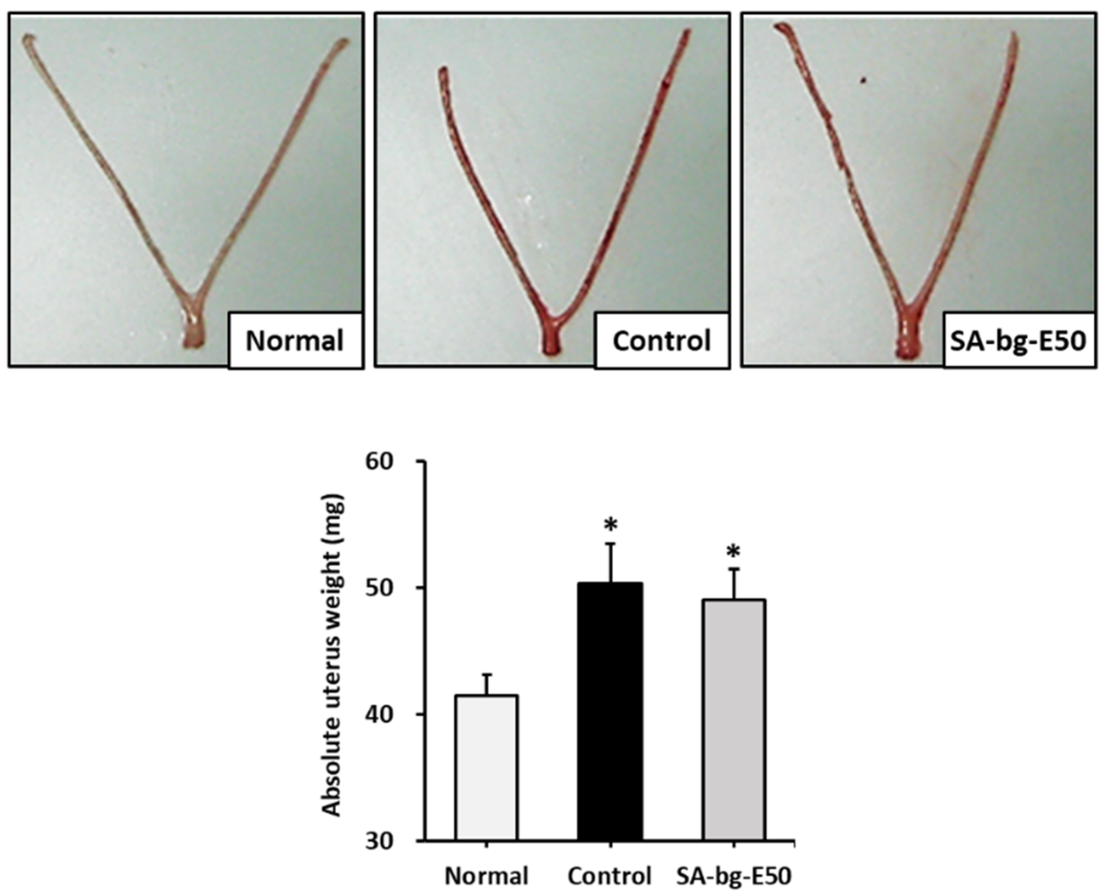
2.2. Uterotrophic Activity of 50% Ethanol Extract from Belowground Part of S. anglica in the Immature Rat

To determine the potential of SA-bg-E50 to have estrogenic effects, we evaluated the estrogenic effect of SA-bg-E50 in an immature female rat model. 17β-Estradiol (10 μg/kg) was subcutaneously injected for 3 days in the control group, and SA-bg-E50 (200 mg/kg) was orally administered for 3 days in the SA-bg-E50 group.
The uterine weight of the positive control group was significantly increased, to 50.3 ± 3.1 mg, compared with that of the normal group (41.5 ± 1.6 mg). After the administration of SA-bg-E50, the uterine weight was 49.0 ± 2.5 mg. The results indicate that SA-bg-E50 hypertrophies the uterus owing to its estrogenic activity (Figure 3).

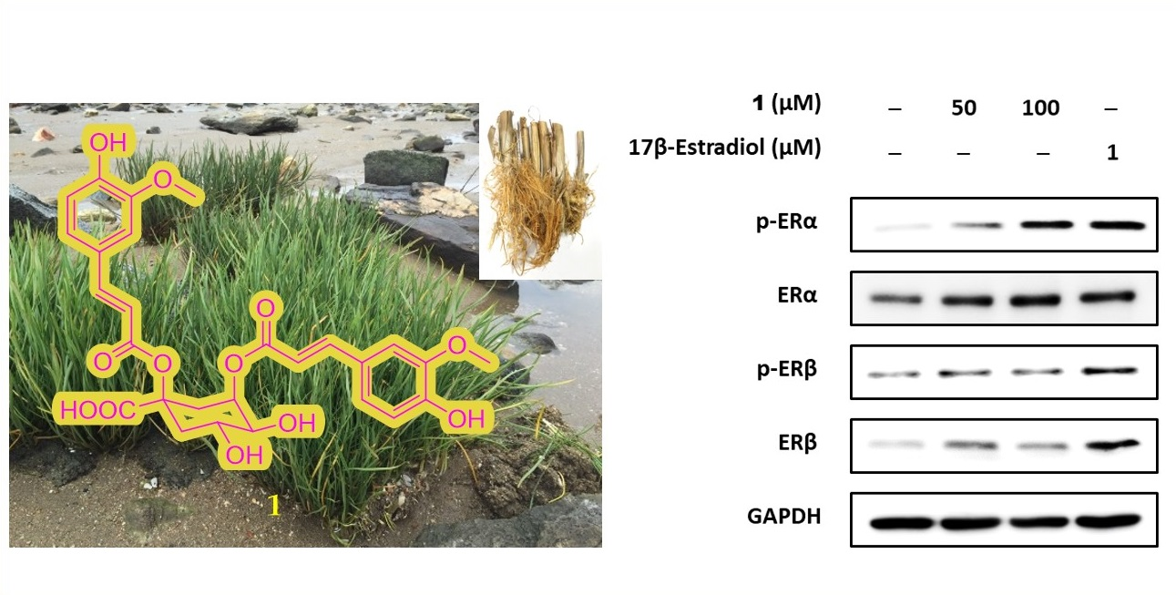
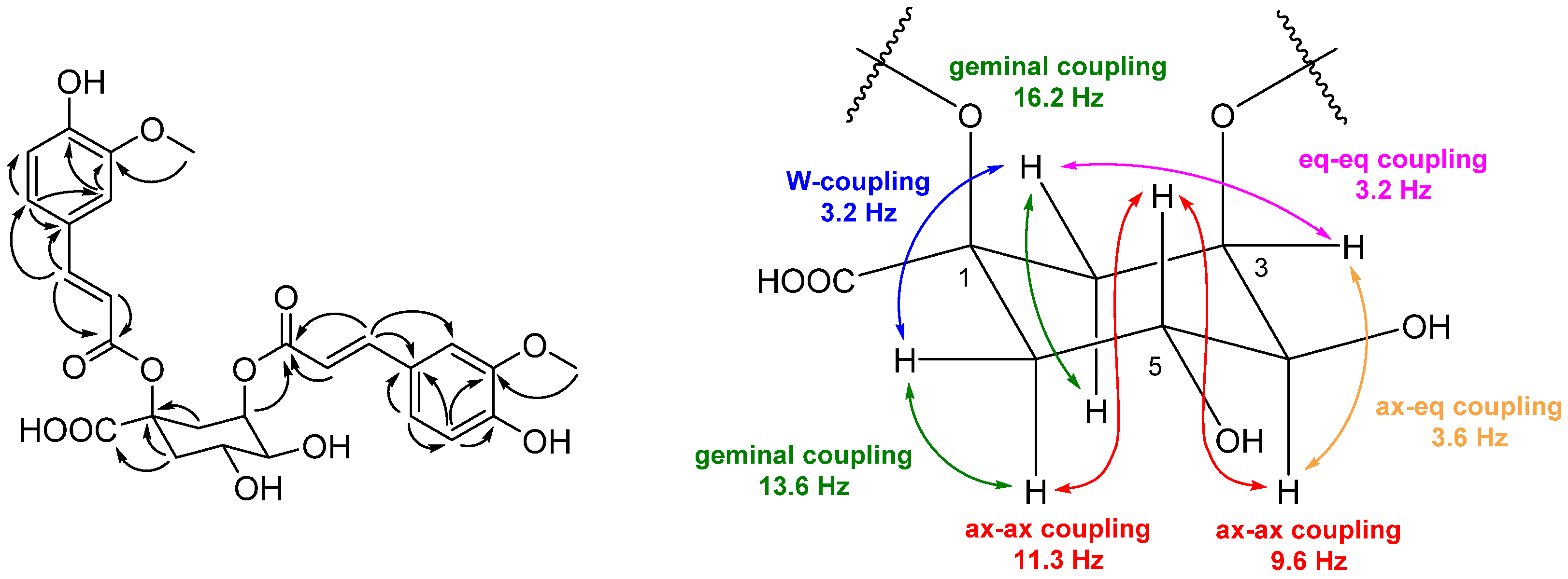
2.3. Structure Elucidation of Compounds 1–5 from S. anglica

Compound 1 exhibited a deprotonated [M − H] ion at m/z 543.2 in low-resolution electron spray ionization mass spectrometry (LR-ESI-MS). Based on the interpretation of the 1D and 2D nuclear magnetic resonance (NMR) data of 1 (Supplementary Materials, Figures S1 and S2), two sets of vinyl groups, two sets of 1,3,4-trisubstituted benzene rings, and a quinic acid moiety were identified. The 1H-1H coupling constants of H-7′/H-8′ (7.53 (1H, d, J = 15.9 Hz, H-7′)/6.26 (1H, d, J = 15.9 Hz, H-8′)) and H-7″/H-8″ (7.54 (1H, d, J = 15.9 Hz, H-7″)/6.16 (1H, d, J = 15.9 Hz, H-8″)) were the trans configurations of two sets of olefinic protons. The proton signals for the two aromatic systems with 1,3,4-trisubstitution were observed at δH 6.93 (1H, d, J = 1.9, H-2′), 6.88 (1H, dd, J = 8.2, 1.9, H-6″), 6.83 (1H, d, J = 1.9, H-2″), 6.75 (1H, dd, J = 8.2, 1.9, H-6′), 6.67 (1H, d, J = 8.2, H-5″), and 6.57 (1H, d, J = 8.1, H-5′). The proton and carbon resonances at δH/δC 3.67 (3H, s)/56.18 ppm and 3.60 (3H, s)/56.08 ppm indicated the presence of two methoxy groups. Through the analysis of heteronuclear multiple bond correlation (HMBC), two ferulate units were identified. The 1H and 13C NMR spectra of 1 showed the characteristic resonances of a quinic acid at δH/δC 5.36/73.21, 4.28/67.80, 3.64/75.31, 2.97/2.31/32.74, and 2.54/1.84/41.46 ppm. One feruloyl residue was connected to the C-1 of quinic acid, as shown by the downfield shift in 13C NMR at δC 81.12 (C-1) ppm. The position of the other ferulate unit was determined by HMBC from H-3 to C-9″ (Figure 4). The numbering system of acyl-quinic acid was critical because of the confusing nomenclature caused by mixing IUPAC with non-IUPAC numbering [32]. A number of reported publications related to acyl-quinic acid were identified with inconsistencies in numbering and/or structure with IUPAC numbering [33,34]. The planar structure of 1 was determined to be 1,3-di-O-trans-feruloyl quinic acid by the IUPAC nomenclature of the quinic acid moiety. The two of vicinal coupling constants of H-5 were observed to be large (3JH-4,H-5 = 9.6 Hz, 3JH-5,H-6ax = 11.3 Hz) which supported the three protons of H-4, H-5 and H-6ax as axial position (Figure 4). In addition, other coupling constants, including long-range coupling through a 1,3-diequatorial interaction (W-coupling) between H-2eq and H-6eq, were well-matched with those of quinic acid, rather than with those of epi-quinic acid (Figure 4) [35]. The comparison of the specific rotation of 1 with that of similar chemical compounds (cynarine; 1,3-dicaffeoylquinic acid) supported the absolute configuration of quinic acid in 1 as (−)-quinic acid (experimental; [α] 25 D = −9.7 (c = 0.1, MeOH), literature; [α] 25 D = −59.0 (c = 4, MeOH) [36]. Compound 1 was identified as 1,3-di-O-trans-feruloyl-(−)-quinic acid with the absolute configuration of 1R, 3R, 4S, 5R (1α, 3α, 4α, 5β).
Compound 2 exhibited a protonated [M + H]+ ion at m/z 433.4 in the LR-ESI-MS spectrum. In the 1H NMR spectrum, compound 2 showed the characteristic features of flavonoid glycoside; a singlet resonance at δH 6.42 ppm (H-8, ring A), two ortho coupled signals [δH 7.90 (d, J = 8.8 Hz, H-2′/6′) and 6.91 ppm (d, J = 8.8 Hz, H-3′/5′)] of a 1,4-substituted phenyl group (ring B), a singlet proton signal at δH 6.71 ppm (H-3, ring C), an anomeric proton signal [4.58 ppm (d, J = 9.8 Hz, H-1″)], and protons attached to oxygenated carbons (δH 4.10–3.00 ppm) of sugar units. The sugar unit was identified to be C-β-D-glucopyranose based on the oxygenated carbons at δC 81.51 (C-5″), 79.05 (C-3″), 73.27 (C-1″), 70.66 (C-2″), 70.26 (C-4″), and 61.48 (C-6″) ppm and the large coupling constant value between H-1″ and H-2″. The carbon resonances at δC 109.10 (C-6) and 94.00 (C-8) ppm supported 6-C-glycoside rather than 8-C-glycoside. The comparison of experimental and reported [37] spectroscopic data, including MS and 1D NMR spectra, led to the identification of 2 as isovitexin (Supplementary Materials, Figure S3).
Compounds 35 were identified as p-hydroxybenzaldehyde (3), N-trans-feruloyltyramine (4), and p-coumaric acid (5) by comparing 1D NMR and LR-ESI-MS data with those reported in the literature (Supplementary Materials, Figures S4–S6) [38,39,40].

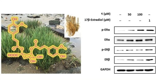
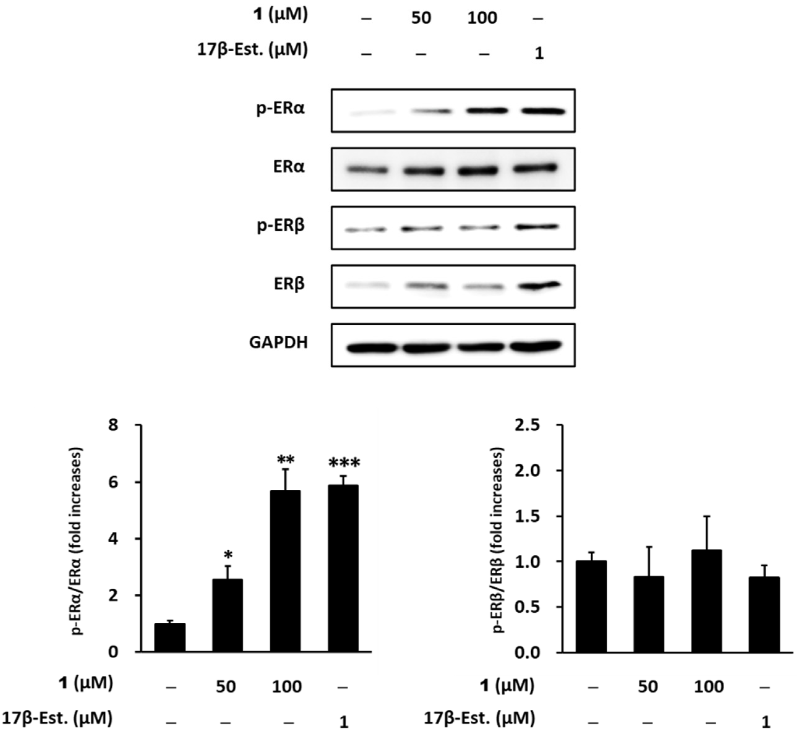
2.4. Estrogenic Effects of Compounds Isolated from Belowground Part of S. anglica on MCF-7 Cells

We evaluated the estrogenic potential of the compounds isolated from belowground part of S. anglica in MCF-7 cells. 1,3-Di-O-trans-feruloyl-(-)-quinic acid (1) induced significant cell proliferation in a concentration-dependent manner (50 μM: 109.0 ± 4.7%, 100 μM: 115.4 ± 2.1%). Cell proliferation was significantly suppressed to 94.1 ± 2.6% (50 μM) and 95.9 ± 6.8% (100 μM) by ICI 182,780. These results indicate that compound 1 has significant estrogenic activity at concentrations of 50 and 100 μM. Isovitexin (2) also increased cell proliferation in a concentration-dependent manner (50 μM: 106.9 ± 4.1%, 100 μM: 111.2 ± 1.9%). Cell proliferation was suppressed to 89.8 ± 9.9% (50 μM) and 93.4 ± 7.1% (100 μM) by ICI 182,780, however it was not significant. (Figure 5). Taken together, compound 1 has potent significant estrogenic activity compared with other compounds; therefore, we focused on the bioactivity of compound 1 in subsequent investigations.
ERα, an estrogen receptor 1, is a ligand-dependent nuclear hormone receptor transcription factor. The binding of a ligand (17β-estradiol) to ERα recruits estrogen response element to ERα [41]. ERα ligands, such as 17β-estradiol, induce the phosphorylation of Serine 118 of ERα [42]. To identify the potential of compound 1 as ERα and ERβ ligands, we carried out western blotting to evaluate ERα and ERβ activation by compound 1. 17β-Estradiol (1 μM), which was a positive control, increased the phosphorylation of ERα to 5.88 ± 0.3-fold (p < 0.001) compared with untreated cells. Similarly, compound 1 increased ERα phosphorylation (50 μM: 2.55 ± 0.4, p < 0.05, 100 μM: 5.68 ± 0.7, p < 0.001; Figure 6). However, compound 1 and 17β-Estradiol (1 μM) not increased the phosphorylation of ERβ compared with untreated cells (Figure 6).
The results indicate that compound 1 induces phosphorylation of serine residues of ERα; thus, compound 1 may be considered an ERα ligand. Compound 1, which contains phenolic hydroxyl groups similar to 17β-estradiol, seems to result in the activation of ERα via interactions with the hydroxyl groups and ligand-binding domains of ERα.

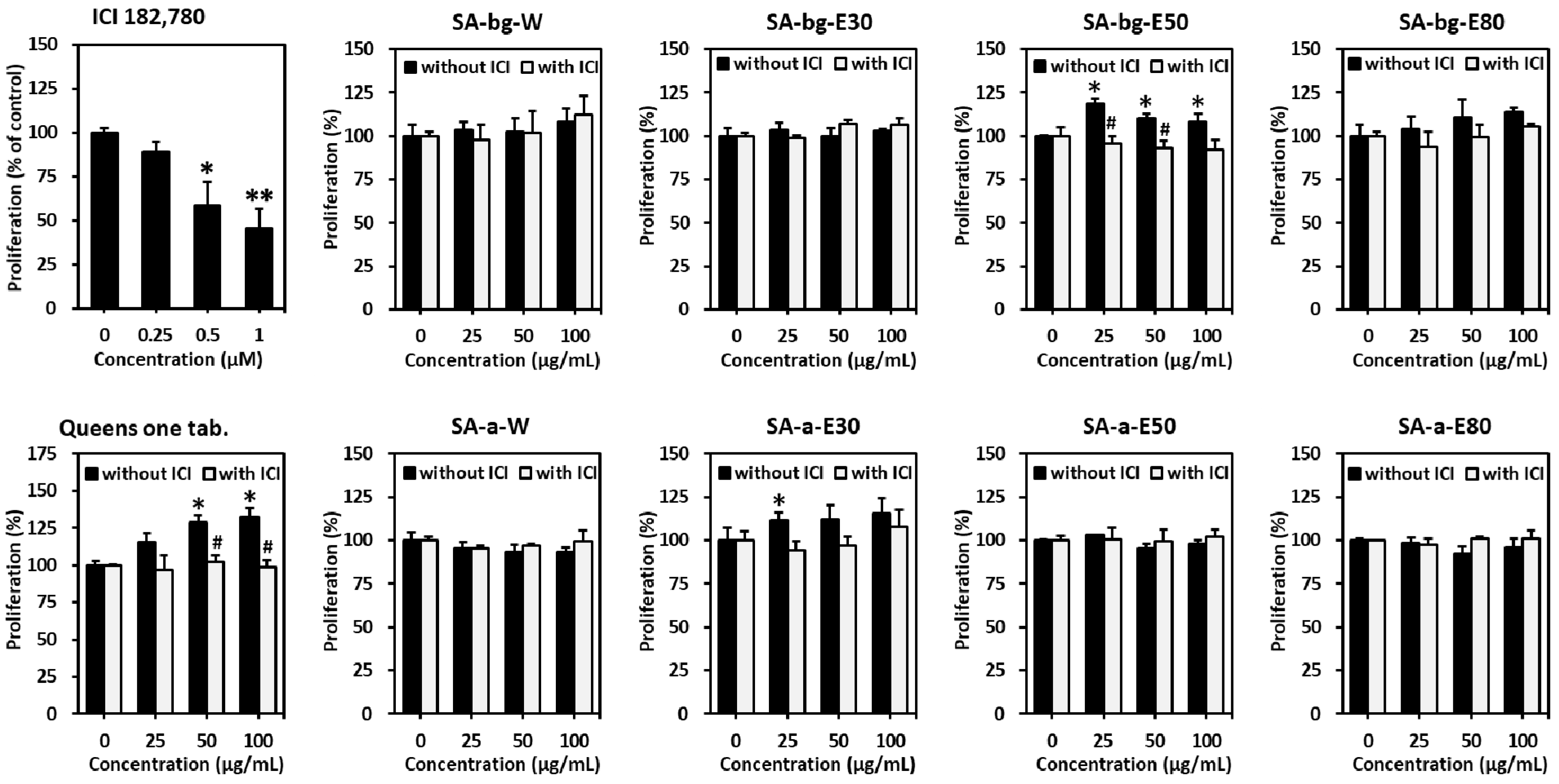
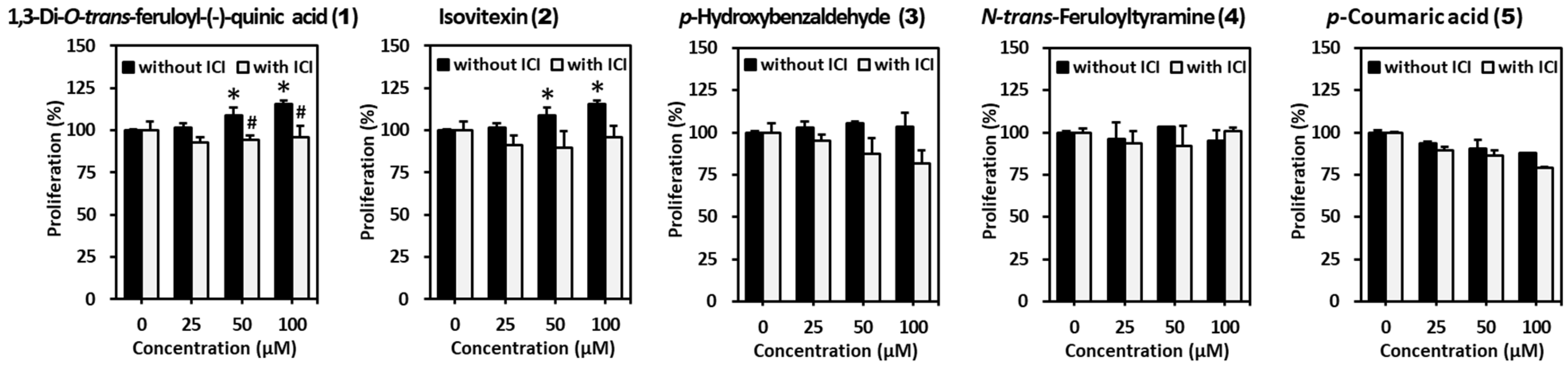
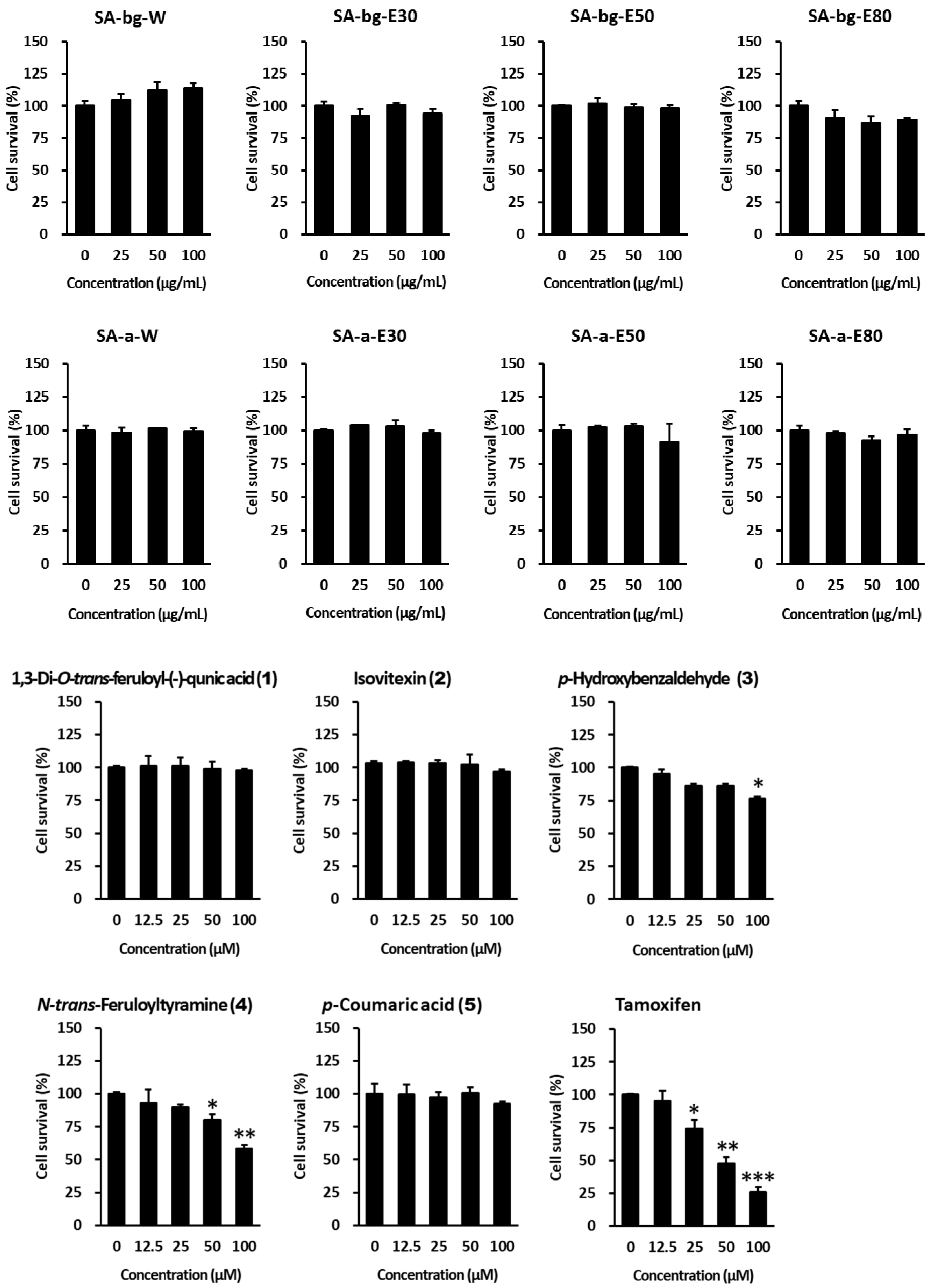
2.5. Anti-Proliferative Effect of Extracts and Compounds from S. anglica on MCF-7 Cells

Breast cancer is divided into ER-positive and ER-negative, depending on the presence or absence of ER expression. More than 70% of breast cancers are ER-positive, and high estrogen levels have a close impact. High levels of estrogen in the blood increase the risk of breast cancer development. Moreover, HRT, which involves the administration of female hormones to compensate for the lack of estrogen in menopausal women, increases the risk of breast cancer development [43,44]. HRT acts as a ligand in ER-positive breast cancer cells and increases cell proliferation.
Several previous studies on the safety of estrogen-like active compounds regarding ER-positive MCF-7 breast cancer cell proliferation under normal conditions have been reported [45,46,47]. Therefore, to evaluate the safety regarding the adverse effects associated with HRT treatment, the cytotoxic effects of extracts and compounds from S. anglica were measured in MCF-7 breast cancer cells using a cell viability assay.
Therefore, we investigated the proliferation of ER-positive MCF-7 human breast cancer cells under normal conditions. None of the extracts and compounds from the belowground and aerial parts of S. anglica resulted in an increase in cell proliferation. Furthermore, p-hydroxybenzaldehyde (3) and N-trans-feruloyltyramine (4) reduced MCF-7 cell proliferation (Figure 7). The results indicate that extracts of S. anglica and its compounds have estrogenic effects, without increasing the risk of breast cancer development.

3. Materials and Methods

3.1. General Experimental Procedures

Optical rotation was measured using a Jasco DIP-1000 polarimeter (JASCO Inc., Tokyo, Japan) coupled with a sodium lamp (589 nm). Vacuum liquid chromatography (VLC) was performed using Silica Gel 60 (230–400 mesh, Merck KGaA, Darmstadt, Germany). LR-ESI-MS was performed on an Agilent 1260 series HPLC system coupled with a 6120 series single quadrupole mass spectrometer (Agilent, Santa Clara, CA, USA). HPLC was carried out on a Gilson HPLC system (321 pump, and UV/Vis-155, Gilson Inc., Middleton, WI, USA) and a Waters HPLC system (PDA 996, and 600 controllers, Waters Corporation, Milford, MA, USA). The 1D and 2D NMR spectra were obtained using a Bruker Avance DPX250 spectrometer (Bruker, Billerica, USA) and 600 MHz Fourier transform nuclear magnetic resonance spectrometer (VNS600, Agilent) at the Core Research Support Center for Natural Products and Medical Materials.

3.2. Plant Material

S. anglica was collected from a mudflat on Ganghwa Island in South Korea in July 2016. The plant was identified by Dr. Hyukjae Choi, College of Pharmacy, Yeungnam University. The collected S. anglica was washed with tap water and dried at 24 °C in the shade. The dried plant specimen was deposited at the College of Pharmacy in Yeungnam University in South Korea.

3.3. Extraction and Isolation of Compounds from S. anglica

The dried plant specimen was separated into two parts (aerial and belowground). A portion of the aerial (SA-a, 10 g) and belowground (SA-bg, 10 g) parts was extracted three times with four extraction solvents (100% deionized water (H2O), 30% ethanol in water, 50% ethanol in water, and 80% ethanol in water) at 20 ± 5 °C for 12 h to obtain the crude extracts of SA-bg-W (0.12 g), SA-bg-E30 (0.35 g), SA-bg-E50 (0.58 g), SA-bg-E80 (0.55 g), SA-a-W (0.92 g), SA-a-E30 (2.57 g), SA-a-E50 (1.34 g), and SA-a-E80 (3.30 g).
The belowground (dry wt. 10 kg) parts of S. anglica was extracted with 50% ethanol in deionized water to obtain crude extracts of belowground parts (590 g) for compound isolation.
A portion (150 g) of the 50% ethanolic extracts of the belowground parts was suspended in 1.0 L of deionized water and partitioned with hexanes, methylene chloride (MC), and ethyl acetate (EA) three times. Each layer was dried under reduced pressure to yield Hex-bg (472.5 mg), MC-bg (461.3 mg), and EA-bg (315.0 mg) fractions and a residual aqueous fraction (H2O-bg, 148 g).
The EA-bg fraction was subjected to preparative HPLC using a reversed-phase C18 column (Phenomenex Luna C18, 100 Å, 22.5 × 250 mm) under the acidified solvent conditions (solvent A, 95% deionized water, 5% CH3CN, and 0.05% formic acid; solvent B 100% CH3CN and 0.05% formic acid; A:B = 80:20 for isocratic elution; flow rate = 6.0 mL/min; detection at 210 and 254 nm) to give a pure compound 1 (42.2 mg, tR = 38.8 min). The other broad peak collection at the retention time range between 30.5 and 32.5 min was further purified by HPLC using a reversed-phase C18 column (Phenomenex Luna C18, 4.6 × 250 mm) under the following solvent conditions (solvent A, 95% deionized water, 5% CH3CN, and 0.05% formic acid; solvent B, 100% CH3CN and 0.05% formic acid; A:B = 85:15 for isocratic elution; flow rate = 1.0 mL/min) to obtain compound 2 (1.7 mg, tR = 26.4 min).
The MC-bg (461.3 mg) fraction was subjected to NP VLC and eluted with MC/MeOH to obtain seven fractions (MC-bg-A–MC-bg-G). The separation of MC-bg-D (96.5 mg) using preparative HPLC (Phenomenex Luna C18, 100 Å, 22.5 × 250 mm) under the following conditions: solvent A (100% deionized water), solvent B (100% CH3CN); A:B = 74:26; flow rate = 6.0 mL/min; detection at 210 and 254 nm afforded compound 3 (0.6 mg, tR = 13.8 min) and compound 4 (5.5 mg, tR = 35.6 min). MC-bg-F (85.2 mg) was separated by HPLC using a semi-preparative column (10.0 × 250 mm, 5 μm) under a B15% isocratic condition (solvent A, 95% deionized water, 5% CH3CN, and 0.05% formic acid; solvent B, 100% CH3CN and 0.05% formic acid) at a flow rate of 2.0 mL/min for 60 min to yield compound 5 (1.4 mg, tR = 32.5 min).

3.4. MCF-7 Culture

The MCF-7 human breast cancer cell line was obtained from the American Type Culture Collection (Bethesda, MD, USA). Cells were maintained at 37 °C in a humidified, 5% CO2 atmosphere in Roswell Park Memorial Institute 1640 medium (RPMI 1640; Corning, Manassas, VA, USA) supplemented with 10% fetal bovine serum (Atlas, Fort Collins, CO, USA) and penicillin/streptomycin (Gibco, Grand Island, NY, USA).

3.5. Determination of Estrogenic Effect in MCF-7 Cells

Experiments were performed using an E-screen assay as previously described [31]. Briefly, MCF-7 cells were plated at a density of 1 × 10⁴ cells/well in 48-well plates, allowed to adhere overnight, and challenged with the indicated concentrations of samples in phenol red-free RPMI (Gibco, Carlsbad, CA, USA) supplemented with 10% charcoal–dextran stripped serum (Innovative Research, Peary CourtNovi, MI, USA) for 144 h. Queens One Tablet was used as positive control in this study, it is constituted 200 mg of Red clover extract in tablet (360 mg), and its active compounds are isoflavones. It is commonly used for improvement of the symptoms about menopause such as hot flush, night sweats, emotional lability, agitation and insomnia. To confirm the estrogenic effect, the cells were maintained in the presence or absence of the estrogen receptor antagonist ICI 182,780 (Tocris Bioscience, Bristol, UK). Thereafter, Ez-Cytox (Daeil Lab Service Co., Seoul, Korea) was added into each well and the cells were incubated for 1 h. Subsequently, the absorbance was measured using a microplate reader (SPARK 10M; Tecan, Männedorf, Switzerland) at a wavelength of 450 nm. Data are presented as the mean ± SEM of three independent experiments performed in triplicate. Cell viability is expressed as a percentage of that in the untreated group (100%).

3.6. Determination of Anti-Proliferative Effect in MCF-7 Cells

Experiments were conducted as previously described [48]. Briefly, MCF-7 cells were plated at a density of 1 × 10⁴ cells/well in 96-well plates, allowed to adhere overnight, and challenged with the indicated concentrations of samples for 24 h. Thereafter, Ez-Cytox was added into each well and the cells were incubated for 1 h. Subsequently, the absorbance was measured using a microplate reader (PowerWave XS; Bio-Tek Instruments, Winooski, VT, USA) at a wavelength of 450 nm. Data are presented as the mean ± SEM of three independent experiments performed in triplicate. Cell viability is expressed as a percentage of that in the untreated group (100%).

3.7. Detection of Protein Expression in MCF-7 Cells

Experiments were conducted as previously described [48]. Briefly, MCFs were plated at a density of 2 × 10⁵ cells/well in 6-well plates, allowed to adhere overnight, and challenged with the indicated concentrations of samples for 24 h. The levels of phospho-ERα (p-ERα), ERα, phosphor-ERβ (p-ERβ), ERβ and glyceraldehyde 3-phosphate dehydrogenase (GAPDH) in the cells were determined by western blotting. Cell lysates were extracted using radioimmunoprecipitation assay buffer (Tech & Innovation, Gangwon, Korea). Protein concentration was determined using the Pierce™ BCA Protein Assay Kit (Pierce, Rockford, IL, USA). Protein separation was conducted using sodium dodecyl sulfate-polyacrylamide gel electrophoresis. Thereafter, proteins were transferred to a polyvinylidene difluoride membrane (Merck Millipore, Darmstadt, Germany) and blocked with 5% skim milk. Afterward, the membrane was incubated with diluted primary antibodies against p-ERα, ERα, p-ERβ, ERβ, and GAPDH (Cell Signaling Technology, Danvers, MA, USA) overnight at 4 °C. After washing the membrane, it was incubated at room temperature with a secondary rabbit IgG antibody for 1 h. The protein signal was measured using SuperSignal® West Femto Maximum Sensitivity Chemiluminescent Substrate (Pierce) and the Fusion Solo Chemiluminescence System (PEQLAB Biotechnologie GmbH, Erlangen, Germany). Protein expression was normalized to that of GAPDH (reference protein). The analysis was performed using a Fusion Solo Chemiluminescence System. Relative protein expression was calculated and compared with that in an untreated group using ImageJ software (Version 1.51J; National Institutes of Health, Bethesda, MD, USA).

3.8. Animals

Crj:CD (SD) rats (female, 14-day-old pups) were obtained from DBL (Chungbuk, Korea). After adaptation for 1 week, the 21-day-old immature rats were individually housed in cages. The immature rats were weighed and randomly assigned to each group. Body weight was recorded daily throughout the study. The rats were provided free access to water and a commercial diet. The room was maintained at a temperature of 22 ± 2 °C and relative humidity of 50 ± 10% and artificially illuminated with a fluorescent lamp in a 12-h light/dark cycle. All animals were managed based on the animal experimental guidelines suggested by the Institutional Animal Care and Use Committee (GIACUC-R2019035, approval on 15 November 2019).

3.9. Uterotrophic Assay

The rat uterotrophic assay proposed by the OECD is a screening method for the determination of the estrogenic properties of endocrine-disrupting chemicals [49]. In the present study, to test the estrogenic properties of the sample, we performed an uterotrophic assay with immature rats. The rats were divided into three groups of three rats. Corn oil and water were administered to the normal group and estradiol in corn oil (0.01 mg estradiol/kg body weight in 4 mL corn oil/kg body weight) was injected subcutaneously in the back of the rats daily in the control group. In the SA-bg-E50 group, corn oil (4 mL/kg body weight) was injected subcutaneously and SA-bg-E50 was orally administered at a dosage of 200 mg/kg body weight per day for 3 days. The normal and control groups were orally administered water for 3 days. Rats were euthanized approximately 24 h after the final administration. During necropsy, the uterus was carefully removed and weighed without any attached fat or mesentery.

3.10. Statistical Analyses

All data are expressed as the mean ± standard error of mean (SEM). Statistical analyses were conducted using one-way analysis of variance (ANOVA) and Tukey’s post-test to evaluate differences among the various experimental groups (GraphPad Prism 7.0, GraphPad Software Inc., San Diego, CA, USA). p < 0.05, p < 0.01, and p < 0.001 were considered significant.

4. Conclusions

Menopause presents with symptoms including facial flushing, vaginal atrophy, and osteoporosis, which are caused by decreased estrogen production. HRT, which is the administration of female hormones, is utilized to treat estrogen reduction, but it has been reported to increase the risk of breast cancer development. Therefore, we aimed to evaluate the estrogenic activities of S. anglica and its constituents and identify potential candidates for the treatment of estrogen reduction without the risk of breast cancer. We evaluated the estrogenic effects of extracts of S. anglica and its compounds in MCF-7 breast cancer cells. SA-bg-E50 and 1,3-di-O-trans-feruloyl-(−)-quinic acid (1) had significant estrogenic activity. Furthermore, the administration of SA-bg-E50 increased the uterine weight compared with that in the normal group in an immature female rat model. Compound 1 was regarded as an ERα ligand that induced the phosphorylation of serine residues of ERα. Additionally, compound 1 did not affect MCF-7 cell proliferation. Consequently, compound 1, as an ERα ligand, could be an active compound of S. anglica and have estrogenic effect without side effects, such as the developing risk of breast cancer.

Supplementary Materials

The following data are available online at https://www.mdpi.com/article/10.3390/md19040210/s1, Figure S1: 1D NMR spectra of compound 1 in CD3OD; Figure S2: 2D NMR spectra of compound 1 in CD3OD; Figure S3: 1D NMR spectra of compound 2 in DMSO-d6; Figure S4: 1D NMR spectra of compound 3 in CD3OD; Figure S5: 1D NMR spectra of compound 4 in CD3OD; Figure S6: 1D NMR spectra of compound 5 in CD3OD.

Author Contributions

Conceptualization, H.C. and K.S.K.; extract preparation, H.K.; formal analysis, S.L., J.-W.N., and J.Y.B.; data curation, S.L., J.-W.N., and J.Y.B.; writing—original draft preparation, S.L. and G.J.K.; writing—review and editing, S.L., G.J.K., S.H.S., H.C., and K.S.K.; project administration, H.C. and K.S.K. All authors have read and agreed to the published version of the manuscript.

Funding

This research was funded by the Marine Biotechnology Program funded by the Ministry of Oceans and Fisheries and the Gachon University research fund of 2020 (GCU-202002360001).

Institutional Review Board Statement

All animals were managed based on the animal experimental guidelines suggested by the Institutional Animal Care and Use Committee (GIACUC-R2019035, approval on 15 November 2019).

Informed Consent Statement

Not applicable.

Conflicts of Interest

The authors declare no conflict of interest.

References

  1. Li, S.; O’Neill, S.R.; Zhang, Y.; Holtzman, M.J.; Takemaru, K.I.; Korach, K.S.; Winuthayanon, W. Estrogen receptor α is required for oviductal transport of embryos. FASEB J. 2017, 31, 1595–1607. [Google Scholar] [CrossRef] [PubMed] [Green Version]
  2. Sever, D.M.; Staub, N.L. Hormones, sex accessory structures, and secondary sexual characteristics in amphibians. In Hormones and Reproduction of Vertebrates; Elsevier: Burlington, MA, USA, 2011; pp. 83–98. [Google Scholar]
  3. Hausmann, M.; Slabbekoorn, D.; Van Goozen, S.H.; Cohen-Kettenis, P.T.; Güntürkün, O. Sex hormones affect spatial abilities during the menstrual cycle. Behav. Neurosci. 2000, 114, 1245–1250. [Google Scholar] [CrossRef]
  4. Messier, V.; Rabasa-Lhoret, R.; Barbat-Artigas, S.; Elisha, B.; Karelis, A.D.; Aubertin-Leheudre, M. Menopause and sarcopenia: A potential role for sex hormones. Maturitas 2011, 68, 331–336. [Google Scholar] [CrossRef] [PubMed]
  5. Greendale, G.A.; Lee, N.P.; Arriola, E.R. The menopause. Lancet 1999, 353, 571–580. [Google Scholar] [CrossRef]
  6. Leidy, L. Biological aspects of menopause: Across the lifespan. Annu. Rev. Anthropol. 1994, 23, 231–253. [Google Scholar] [CrossRef] [PubMed]
  7. Velasco-Téllez, C.; Cortés-Bonilla, M.; Ortiz-Luna, G.; Sánchez-Zelayeta, L.; Méndez-Serrano, H.; Salazar-Jiménez, C.; Zavala-García, A.; Sánchez-Cevallos, A. Quality of life and menopause. In Quality of Life-Biopsychosocial Perspectives; IntechOpen: London, UK, 2020. [Google Scholar]
  8. Bruce, D.; Rymer, J. Symptoms of the menopause. Best Pract. Res. Clin. Obstet. Gynaecol. 2009, 23, 25–32. [Google Scholar] [CrossRef]
  9. Dalal, P.K.; Agarwal, M. Postmenopausal syndrome. Indian J. Psychiatry 2015, 57, S222. [Google Scholar] [CrossRef] [PubMed]
  10. Al-Safi, Z.A.; Santoro, N. Menopausal hormone therapy and menopausal symptoms. Fertil. Steril. 2014, 101, 905–915. [Google Scholar] [CrossRef] [PubMed]
  11. Carroll, J.S.; Brown, M. Estrogen receptor target gene: An evolving concept. Mol. Endocrinol. 2006, 20, 1707–1714. [Google Scholar] [CrossRef] [PubMed]
  12. Bjornstrom, L.; Sjoberg, M. Mechanisms of estrogen receptor signaling: Convergence of genomic and nongenomic actions on target genes. Mol. Endocrinol. 2005, 19, 833–842. [Google Scholar] [CrossRef] [PubMed] [Green Version]
  13. Grodin, J.; Siiteri, P.; MacDonald, P. Source of estrogen production in postmenopausal women. J. Clin. Endocrinol. Metab. 1973, 36, 207–214. [Google Scholar] [CrossRef]
  14. Nelson, H.D.; Humphrey, L.L.; Nygren, P.; Teutsch, S.M.; Allan, J.D. Postmenopausal hormone replacement therapy: Scientific review. JAMA 2002, 288, 872–881. [Google Scholar] [CrossRef] [PubMed]
  15. Chen, C.-L.; Weiss, N.S.; Newcomb, P.; Barlow, W.; White, E. Hormone replacement therapy in relation to breast cancer. JAMA 2002, 287, 734–741. [Google Scholar] [CrossRef]
  16. Dietel, M. Hormone replacement therapy (HRT), breast cancer and tumor pathology. Maturitas 2010, 65, 183–189. [Google Scholar] [CrossRef] [PubMed]
  17. Cuzick, J. Hormone replacement therapy and the risk of breast cancer. Eur. J. Cancer 2008, 44, 2344–2349. [Google Scholar] [CrossRef]
  18. Cos, P.; De Bruyne, T.; Apers, S.; Berghe, D.V.; Pieters, L.; Vlietinck, A.J. Phytoestrogens: Recent developments. Planta Med. 2003, 69, 589–599. [Google Scholar] [PubMed] [Green Version]
  19. Adlercreutz, H.; Mazur, W. Phyto-oestrogens and western diseases. Ann. Med. 1997, 29, 95–120. [Google Scholar] [CrossRef]
  20. Ibrahim, N.K.; Hortobagyi, G.N. The evolving role of specific estrogen receptor modulators (SERMs). Surg. Oncol. 1999, 8, 103–123. [Google Scholar] [CrossRef]
  21. An, B.H.; Jeong, H.; Zhou, W.; Liu, X.; Kim, S.; Jang, C.Y.; Kim, H.S.; Sohn, J.; Park, H.J.; Sung, N.H. Evaluation of the biological activity of Opuntia ficus indica as a tissue-and estrogen receptor subtype-selective modulator. Phytother. Res. 2016, 30, 971–980. [Google Scholar] [CrossRef]
  22. Wang, C.-C.; Prasain, J.K.; Barnes, S. Review of the methods used in the determination of phytoestrogens. J. Chromatogr. B 2002, 777, 3–28. [Google Scholar] [CrossRef]
  23. Nikolić, I.L.; Savić-Gajić, I.M.; Tačić, A.D.; Savić, I.M. Classification and biological activity of phytoestrogens: A review. Adv. Technol. 2017, 6, 96–106. [Google Scholar] [CrossRef] [Green Version]
  24. Dixon, R.A. Phytoestrogens. Annu. Rev. Plant. Biol. 2004, 55, 225–261. [Google Scholar] [CrossRef] [PubMed] [Green Version]
  25. Basu, P.; Maier, C. Phytoestrogens and breast cancer: In vitro anticancer activities of isoflavones, lignans, coumestans, stilbenes and their analogs and derivatives. Biomed. Pharmacother. 2018, 107, 1648–1666. [Google Scholar] [CrossRef] [PubMed]
  26. Hubbard, J. Spartina marshes in southern England: VI. Pattern of invasion in Poole Harbour. J. Ecol. 1965, 799–813. [Google Scholar] [CrossRef]
  27. Baumel, A.; Ainouche, M.; Misset, M.; Gourret, J.; Bayer, R. Genetic evidence for hybridization between the native and the introduced Spartina alterniflora (Poaceae) in South-West France: Spartina x neyrautii re-examined. Plant Syst. Evol. 2003, 237, 87–97. [Google Scholar] [CrossRef]
  28. Millard, A.V.; Evans, P.R. Spartina anglica in great Britain. In Focus on Nature Conservation, Liverpool University, United Kingdom, 10th November 1982; Doody, P., Ed.; Nature Conservancy Council: Cambridgeshire, UK, 1984; Volume 5, pp. 41–48. [Google Scholar]
  29. Kim, E.-K.; Kil, J.; Joo, Y.-K.; Jung, Y.-S. Distribution and botanical characteristics of unrecorded alien weed Spartina anglica in Korea. Weed Turfgrass Sci. 2015, 4, 65–70. [Google Scholar] [CrossRef]
  30. Kim, G.J.; Park, S.; Kim, E.; Kwon, H.; Park, H.-J.; Nam, J.-W.; Roh, S.-S.; Choi, H. Antioxidant, pancreatic lipase inhibitory and tyrosinase inhibitory activities of extracts of the invasive plant Spartina anglica (cord-grass). Antioxidants 2021, 10, 242. [Google Scholar] [CrossRef]
  31. Trinh, T.A.; Park, E.J.; Lee, D.; Song, J.H.; Lee, H.L.; Kim, K.H.; Kim, Y.; Jung, K.; Kang, K.S.; Yoo, J.E. Estrogenic activity of sanguiin H-6 through activation of estrogen receptor α coactivator-binding site. Nat. Prod. Sci. 2019, 25, 28–33. [Google Scholar] [CrossRef]
  32. Abrankό, L.; Clifford, M.N. An unambiguous nomenclature for the acyl-quinic acids commonly known as chlorogenic acids. J. Agric. Food Chem. 2017, 65, 3602–3608. [Google Scholar] [CrossRef]
  33. Olennikov, D.N.; Kashchenko, N.I. 1,5-Di-O-isoferuloylquinic acid and other phenolic compounds from pollen of Calendula officinalis. Chem. Nat. Comp. 2014, 50, 589–593. [Google Scholar] [CrossRef]
  34. Wenzl, P.; Chaves, A.L.; Mayer, J.E.; Rao, I.M.; Nair, M.G. Roots of nutrient-deprived Brachiaria species accumulate 1,3-di-O-trans-feruloylquinic acid. Phytochemistry 2000, 55, 389–395. [Google Scholar] [CrossRef]
  35. Scholz-Böttcher, B.M.; Ernst, L.; Maier, H.G. New stereoisomers of quinic acid and their lactones. Liebigs Ann. Chem. 1991, 1991, 1029–1036. [Google Scholar] [CrossRef]
  36. Panizzi, L.; Scarpati, M.L. Constitution of cynarine, the active principle of the artichoke. Nature 1954, 174, 1062. [Google Scholar] [CrossRef]
  37. Yao, Y.; Cheng, X.; Wang, L.; Wang, S.; Ren, G. A determination of potential α-glucosidase inhibitors from azuki beans (Vigna angularis). Int. J. Mol. Sci. 2011, 12, 6445–6451. [Google Scholar] [CrossRef] [Green Version]
  38. Rho, T.; Yoon, K.D. Chemical constituents of Nelumbo nucifera seeds. Nat. Prod. Sci. 2017, 23, 253–257. [Google Scholar] [CrossRef] [Green Version]
  39. Hwang, J.T.; Kim, Y.; Jang, H.-J.; Oh, H.-M.; Lim, C.-H.; Lee, S.W.; Rho, M.-C. Study of the UV light conversion of feruloyl amides from Portulaca oleracea and their inhibitory effect on IL-6-induced STAT3 activation. Molecules 2016, 21, 865. [Google Scholar] [CrossRef] [PubMed]
  40. Li, H.; Matsunaga, S.; Fusetani, N. Simple antifungal metabolites from a marine sponge, Halichondria sp. Comp. Biochem. Physiol. B Comp. Biochem. 1994, 107, 261–264. [Google Scholar] [CrossRef]
  41. Paech, K.; Webb, P.; Kuiper, G.G.; Nilsson, S.; Gustafsson, J.-Å.; Kushner, P.J.; Scanlan, T.S. Differential ligand activation of estrogen receptors ERα and ERβ at AP1 sites. Science 1997, 277, 1508–1510. [Google Scholar] [CrossRef]
  42. Chen, D.; Washbrook, E.; Sarwar, N.; Bates, G.J.; Pace, P.E.; Thirunuvakkarasu, V.; Taylor, J.; Epstein, R.J.; Fuller-Pace, F.V.; Egly, J.-M. Phosphorylation of human estrogen receptor α at serine 118 by two distinct signal transduction pathways revealed by phosphorylation-specific antisera. Oncogene 2002, 21, 4921–4931. [Google Scholar] [CrossRef] [Green Version]
  43. Hulka, B.S.; Moorman, P.G. Reprint of Breast cancer: Hormones and other risk factors. Maturitas 2008, 61, 203–213. [Google Scholar] [CrossRef]
  44. Ross, R.K.; Paganini-Hill, A.; Wan, P.C.; Pike, M.C. Effect of hormone replacement therapy on breast cancer risk: Estrogen versus estrogen plus progestin. J. Natl. Cancer Inst. 2000, 92, 328–332. [Google Scholar]
  45. Parrella, A.; Lavorgna, M.; Criscuolo, E.; Russo, C.; Isidori, M. Estrogenic activity and cytotoxicity of six anticancer drugs detected in water systems. Sci. Total Environ. 2014, 485, 216–222. [Google Scholar] [CrossRef]
  46. Hsieh, C.J.; Hsu, Y.L.; Huang, Y.F.; Tsai, E.M. Molecular mechanisms of anticancer effects of phytoestrogens in breast cancer. Curr. Protein Pept. Sci. 2018, 19, 323–332. [Google Scholar] [CrossRef]
  47. Ahn, S.Y.; Jo, M.S.; Lee, D.; Baek, S.E.; Baek, J.; Yu, J.S.; Kim, K.H. Dual effects of isoflavonoids from Pueraria lobata roots on estrogenic activity and anti-proliferation of MCF-7 human breast carcinoma cells. Bioorg. Chem. 2019, 83, 135–144. [Google Scholar] [CrossRef] [PubMed]
  48. Jung, M.-Y.; Seo, C.-S.; Baek, S.-E.; Lee, J.; Shin, M.-S.; Kang, K.S.; Lee, S.; Yoo, J.-E. Analysis and identification of active compounds from Gami-Soyosan toxic to MCF-7 human breast adenocarcinoma cells. Biomolecules 2019, 9, 272. [Google Scholar] [CrossRef] [PubMed] [Green Version]
  49. Higashihara, N.; Shiraishi, K.; Miyata, K.; Oshima, Y.; Minobe, Y.; Yamasaki, K. Subacute oral toxicity study of bisphenol F based on the draft protocol for the “Enhanced OECD Test Guideline no. 407”. Arch. Toxicol. 2007, 81, 825–832. [Google Scholar] [CrossRef] [PubMed]
Figure 1. The structures of isolated compounds 15.
Marinedrugs 19 00210 g001
Figure 2. Estrogenic effects of the different extracts from belowground and aerial parts of S. anglica on MCF-7 cells. MCF-7 cells were challenged with 25, 50, and 100 μg/mL extracts in the presence or absence of 0.5 μM ICI 182,780 for 144 h in a charcoal–dextran stripped media condition. Proliferation was determined using an EZ-Cytox kit. Data are presented as mean ± standard error of mean (SEM) of at least three independent experiments. * p < 0.05 versus untreated cells. ** p < 0.01 versus untreated cells. # p < 0.05 versus each group without ICI 182,780. (SA-bg-W: water extract from belowground part of S. anglica, SA-bg-E30: 30% ethanol extract from belowground part, SA-bg-E50: 50% ethanol extract from belowground part, SA-bg-E80: 80% ethanol extract from belowground part, SA-a-W: water extract from aerial part, SA-a-E30: 30% ethanol extract from aerial part, SA-a-E50: 50% ethanol extract from aerial part, SA-a-E80: 80% ethanol extract from aerial part).
Marinedrugs 19 00210 g002
Figure 3. Uterotrophic activity of 50% ethanol extract from belowground part of S. anglica (SA-bg-E50) in the immature rat. 17β-Estradiol (10 μg/kg) was subcutaneously injected for 3 days. SA-bg-E50 (200 mg/kg) was orally administered for 3 days. Rats were euthanized 24 h after the final administration and the uterus was removed and weighed. Data are presented as mean ± SEM of at least three independent experiments. * p < 0.05 versus normal group. (Normal: corn oil and water treatment group, Positive control: 10 μg/kg 17β-estradiol injected group, SA-bg-E50: 50% ethanol extract from belowground part of S. anglica administered group).
Marinedrugs 19 00210 g003
Figure 4. Key HMBC correlations and coupling constant analysis of the quinic acid unit of 1.
Marinedrugs 19 00210 g004
Figure 5. Estrogenic effects of compounds from belowground part of S. anglica on MCF-7 cells. MCF-7 cells were challenged with 25, 50, and 100 μM of compounds in the presence or absence of 500 nM ICI 182,780 for 144 h in a charcoal–dextran stripped media condition. Proliferation was determined using the EZ-Cytox kit. Data are presented as mean ± SEM of at least three independent experiments. * p < 0.05 versus untreated cells. # p < 0.05 versus each group without ICI 182,780.
Marinedrugs 19 00210 g005
Figure 6. Protein levels of phospho-estrogen receptor-α (Ser 118), estrogen receptor-α, phospho-estrogen receptor-β (Ser 105), estrogen receptor-β and GAPDH against 1,3-di-O-trans-feruloyl-(−)-quinic acid (1)-treated MCF-7 cells. MCF-7 cells were challenged with 50 and 100 μM compound 1 for 24 h in a charcoal–dextran stripped media condition. 17β-Estradiol (17β-Est.) was used as a positive control. Subsequently, the protein levels were determined by western blotting. Data are presented as mean ± SEM of at least three independent experiments. * p < 0.05, ** p < 0.01, and *** p < 0.001 versus untreated cells.
Marinedrugs 19 00210 g006
Figure 7. Anti-proliferative effects of the different extracts and compounds from belowground and aerial parts of S. anglica on MCF-7 cells. MCF-7 cells were challenged with 25, 50, and 100 μg/mL extracts and 12.5, 25, 50, and 100 μM of compounds for 24 h in a normal media condition. Tamoxifen used as positive control. Proliferation was determined using the EZ-Cytox kit. Data are presented as mean ± SEM of at least three independent experiments. * p < 0.05, ** p < 0.01 and *** p < 0.001 versus untreated cells. (SA-bg-W: water extract from belowground parts of S. anglica, SA-bg-E30: 30% ethanol extract from belowground parts of S. anglica, SA-bg-E50: 50% ethanol extract from belowground parts of S. anglica, SA-bg-E80: 80% ethanol extract from belowground parts of S. anglica, SA-a-W: water extract from aerial parts of S. anglica, SA-a-E30: 30% ethanol extract from aerial parts of S. anglica, SA-a-E50: 50% ethanol extract from aerial parts of S. anglica, SA-a-E80: 80% ethanol extract from aerial parts of S. anglica).
Marinedrugs 19 00210 g007
Publisher’s Note: MDPI stays neutral with regard to jurisdictional claims in published maps and institutional affiliations.

