Article

Characterization of ACE Inhibitory Peptides Prepared from Pyropia pseudolinearis Protein

1
Laboratory of Marine Chemical Resource Development, Faculty of Fisheries Sciences, Hokkaido University, Hakodate, Hokkaido 041-8611, Japan
2
Chair of Marine Chemical Resource Development, Graduate School of Fisheries Sciences, Hokkaido University, Hakodate, Hokkaido 041-8611, Japan
3
Laboratory of Aquaculture Genetics and Genomics, Faculty of Fisheries Sciences, Hokkaido University, Hakodate, Hokkaido 041-8611, Japan
4
Laboratory of Humans and the Ocean, Faculty of Fisheries Sciences, Hokkaido University, Hakodate, Hokkaido 041-8611, Japan
5
Hokkaido Industrial Technology Center, Department of Research and Development, Hakodate, Hokkaido 041-0801, Japan
*
Author to whom correspondence should be addressed.
Mar. Drugs 2021, 19(4), 200; https://doi.org/10.3390/md19040200
Submission received: 10 March 2021 / Revised: 25 March 2021 / Accepted: 30 March 2021 / Published: 1 April 2021

Abstract

:
More than 7000 red algae species have been classified. Although most of them are underused, they are a protein-rich marine resource. The hydrolysates of red algal proteins are good candidates for the inhibition of the angiotensin-I-converting enzyme (ACE). The ACE is one of the key factors for cardiovascular disease, and the inhibition of ACE activity is related to the prevention of high blood pressure. To better understand the relationship between the hydrolysates of red algal proteins and the inhibition of ACE activity, we attempted to identify novel ACE inhibitory peptides from Pyropia pseudolinearis. We prepared water soluble proteins (WSP) containing phycoerythrin, phycocyanin, allophycocyanin, and ribulose 1,5-bisphosphate carboxylase/oxygenase. In vitro analysis showed that the thermolysin hydrolysate of the WSP had high ACE inhibitory activity compared to that of WSP. We then identified 42 peptides in the hydrolysate by high-performance liquid chromatography and mass spectrometry. Among 42 peptides, 23 peptides were found in chloroplast proteins. We then synthesized the uncharacterized peptides ARY, YLR, and LRM and measured the ACE inhibitory activity. LRM showed a low IC50 value (0.15 μmol) compared to ARY and YLR (1.3 and 5.8 μmol). In silico analysis revealed that the LRM sequence was conserved in cpcA from Bangiales and Florideophyceae, indicating that the novel ACE inhibitory peptide LRM was highly conserved in red algae.

1. Introduction

Japan’s Ministry of Agriculture, Forestry and Fisheries promotes “Shokuiku” (Food and Nutrition Education). The guideline recommends eating well-balanced meals combined with vegetables, fruits, milk products, beans, and fish. In addition, information of the nutritional and bioactive functions of foods based on scientific evidence is required. The importance of such food-derived components has been recognized not only in Japan but also worldwide. One of the examples of functional components is bioactive peptide. Until now, there have been various reports for the functions of bioactive peptides, such as decreases in blood pressure, glucose, cholesterol, and neutral lipids and increases in immune response [1]. Among these peptides, several antihypertensive peptides from natural sources, such as sesame, milk, sardine, and seaweed have been used as food for specified health uses (FOSHU) in Japan [2,3].
Since high blood pressure (hypertension) has no obvious indicating symptoms, it is called a “Silent Killer”. Hypertension is a major risk for cardiovascular diseases and causes serious health problems regarding heart failure, atrial fibrillation, chronic kidney disease, heart valve diseases, aortic syndromes, and dementia [4,5]. The renin-angiotensin system (RAS) is one of the blood pressure regulators. The angiotensin-I-converting enzyme (ACE: EC 3.4.15.1), which is a dipeptidyl carboxypeptidase, is a key enzyme in RAS. The enzyme converts the inactive angiotensin I to an active angiotensin II, resulting in an increase in blood pressure [6]. Several ACE inhibitory drugs including captopril, enalapril, lisinopril, and alacepril are used, but these drugs are normally associated with side effects such as dry cough, angioedema, taste disturbance, and skin rash [4,6,7]. Hence, ACE inhibitory peptides derived from natural sources are desired for the preventive therapy of high blood pressure.
On the other hand, Japanese people have been using seaweed as a food source and industrial material since ancient times. Recently, the health functions of seaweed’s nutritional components have been reported [8,9,10,11,12,13,14,15,16,17,18,19,20]. In particular, the idea of using seaweed proteins as bioactive peptides has attracted much attention [21]. Among seaweeds, the protein content of red algae tends to be higher than that of brown and green algae [22]. Red algae are currently classified into seven classes—Bangiophyceae, Compsopogonophyceae, Cyanidiophyceae, Florideophyceae, Porphyridiophyceae, Rhodellophyceae, and Stylonematophyceae [23], which are composed of more than 7000 species in the world [24] and 800 species on the coast of Japan. However, only a limited number of species such as nori (Neopyropia sp.) and agar (Gelidium sp. and Gracilaria sp.) are used as industrial foodstuffs, and some species such as Grateloupia asiatica, Gloiopeltis furcata, Chondria crassicaulis, and Nemalion vermiculare are consumed as local food. Therefore, other red algae are also expected to be used effectively as food material and a source of bioactive components. The main proteins in red algae are phycobiliproteins that form phycobilisome in a chloroplast and play a light-harvesting role in photosynthesis [25]. Phycobiliproteins primarily consist of phycoerythrin (PE), phycocyanin (PC), and allophycocyanin (APC), and these proteins commonly possess α- and β-subunits [26,27]. APC constitutes the core domain of phycobilisomes, while PE and PC are components of the rods. Since PE is at the tip region of the rods, it is easily released in water layers accompanied by ribulose 1,5-bisphosphate carboxylase/oxygenase (Rubisco) [28]. Regarding the peptides in the hydrolysates of phycobiliproteins and of Rubisco from red algae, many reports on their health benefit have been published, i.e., their ACE inhibitory activity [29,30,31,32,33,34,35,36], renin inhibitory activity [37], DPP-IV inhibitory activity [38], antioxidant activity [33,39,40,41], anti-inflammatory activity [42], and antiplatelet aggregation activity [43].
Since red algal phycobiliproteins are commonly composed of many hydrophobic and aromatic amino acid residues (>50%), thermolysin is suitable for the preparation of ACE inhibitory peptides [26,35,44,45]. In our previous study, we attempted to understand the correlation between in vitro and in silico analyses using IC50 values of ACE inhibitory peptides prepared by the thermolysin hydrolysis of phycobiliproteins and Rubisco [46,47]. The in vitro ACE inhibitory activity was well correlated with the IC50 value from in silico analysis. Namely, the in silico approach has the potential to predict the biological activity of peptides derived from red algal protein. However, there were slight gaps in the correlation due to the lack of information on ACE inhibitory peptides from red algae. This indicates that the hydrolysates of red algal phycobiliproteins and Rubisco still contain uncovered ACE inhibitory peptides. Therefore, we used Pyropia pseudolinearis, which is called “Uppurui Nori” in Japan and is eaten in certain local areas, as a sample in this study. We first extracted phycobiliproteins and Rubisco as water-soluble proteins (WSP) and prepared their hydrolysates. We then identified the ACE inhibitory peptides in the hydrolysate and found three novel peptides. Further, we determined IC50 values using synthetic peptides and performed docking simulation between the ACE and three peptides. We also determined the nucleotide sequence of the chloroplast proteins of PE, PC, and APC to reveal the source of the peptides. This study is useful for the evaluation of proteins from red algae as a source of ACE inhibitory peptides.

2. Results and Discussion

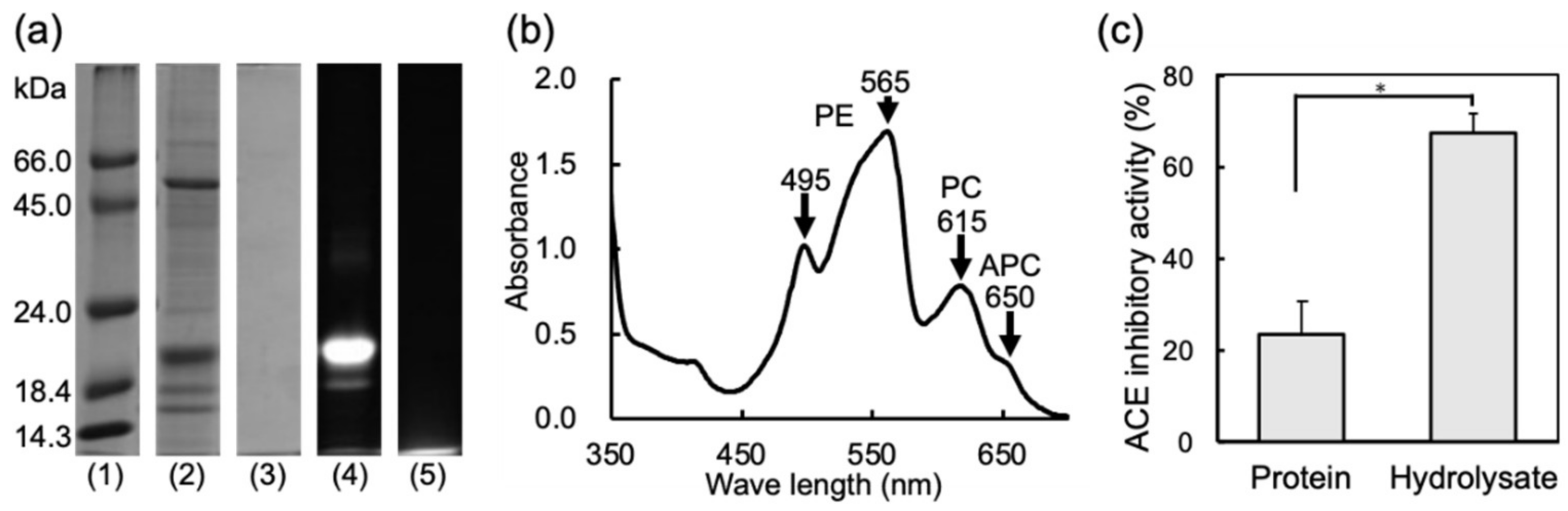
2.1. Properties and ACE Inhibitory Activities of Water Soluble Protein (WSP) and the Thermolysin Hydrolysate of the WSP

To evaluate the potential of ACE inhibitory activity from P. pseudolinearis, we first prepared WSP and its thermolysin hydrolysate. The composition of WSP was confirmed by SDS-PAGE and spectral analysis. The components of WSP were mainly found to be 20,000 and 55,000 MW (Figure 1a). The former band was fluoresced by the irradiation of excitation light (490–560 nm), suggesting phycobiliproteins. The maximum absorption peaks of WSP were found at around 495, 565, 615, and 650 nm (Figure 1b). These peaks corresponded to PE (495 and 565 nm), PC (615 nm), and APC (650 nm). The absorbance of PE was high compared with PC and APC. This result indicated that the main component of 20,000 MW was PE. A previous study revealed that 55,000 MW of the protein in the water-extract of red algae was the Rubisco large subunit [47], suggesting that the WSP of P. pseudolinearis also contained PE and the Rubisco large subunit as main components. The digestion of proteins was confirmed by SDS-PAGE, showing that protein bands were completely hydrolyzed (Figure 1a). The hydrolysate of the WSP was centrifuged at 4 °C, 15,000× g, for 10 min, and the supernatant was lyophilized and used for the detection of ACE inhibitory peptides. We compared the ACE inhibitory activities of WSP and the hydrolysate (Figure 1c). WSP inhibited 23.6% of the ACE activity, while the hydrolysate of the WSP inhibited 67.7% activity, showing that the hydrolysate contained ACE inhibitory peptides. It is thought that the ACE inhibitory activity of the peptide is affected by its amino acid sequence and molecular weight. In the present study, to evaluate proteins from P. pseudolinearis as a source of ACE inhibitory peptides, we prepared its hydrolysate in the same method used in our previous study [35]. We identified 15 tripeptides, 10 tetrapeptides, 11 pentapeptides, and 6 hexapeptides, and these peptides’ compositions were similar to that of our previous result [35]. In the future, we would like to study the optimal condition under which peptides are prepared with the highest inhibitory activity by adjusting the hydrolysis time. We afterwards attempted to identify ACE inhibitory peptides by HPLC.

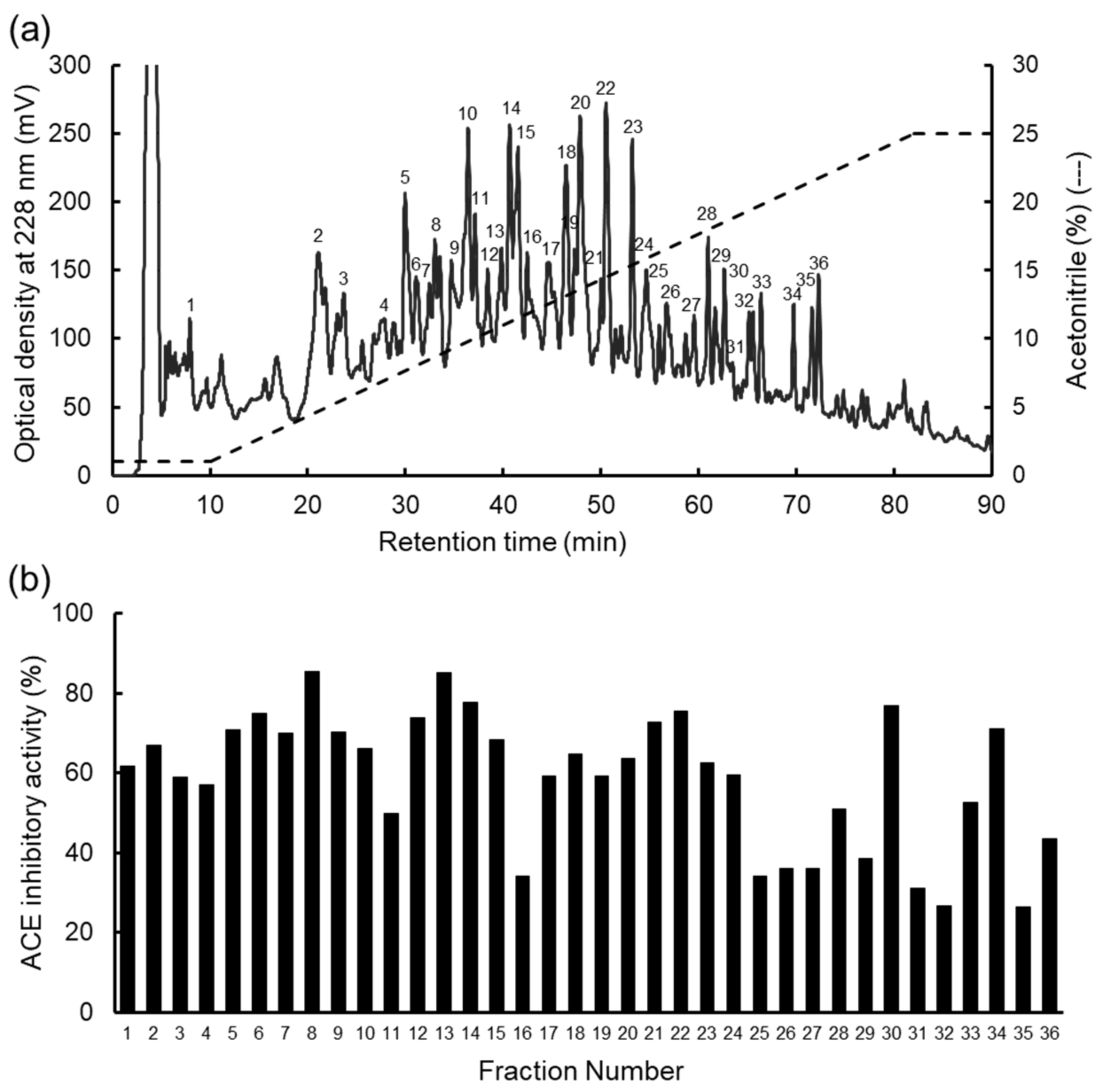
2.2. Fractionation of the Hydrolysate of the WSP

To determine the ACE inhibitory peptides, fractionation was performed by RP-HPLC (Figure 2a). Thirty-six fractions were obtained by HPLC. The ACE inhibitory activity was measured in each fraction (Figure 2b), so the activity was detected in all fractions. We have previously prepared the thermolysin hydrolysates of WSP from dulse [35], Mazzaella japonica [26,36], and G. asiatica [47], which showed that the patterns of the chromatogram differed, even though the main WSP components of these red algae were phycobiliproteins and Rubisco. The hydrolysate of the WSP from P. pseudolinearis contained many peptides. Peptide peaks, which were eluted after 22% acetonitrile (FN 34–36), were not found in the other hydrolysates. The peaks of hydrolysates from dulse and G. asiatica were detected with up to 15% acetonitrile, and the peak from M. japonica was detected with up to 18% acetonitrile. The difference might be due to minor proteins detected on SDS-PAGE. Therefore, we attempted to determine the peptide structures in all fractions.

2.3. Identification of Peptides from the WSP

Since we detected the ACE inhibitory activity in 36 fractions, peptide sequences were determined by MALDI-TOF/MS/MS. Among 36 fractions, 42 peptides were determined (Table 1). The complete chloroplast genome of Pyropia pulchra was determined [48,49], which is the related species in P. pseudolinearis [50]. We employed the P. pulchra chloroplast genome and performed in silico thermolysin digestion in it (Table 1). Among the 42 peptides, 23 kinds of peptides were found in chloroplast proteins, which were constituted of 101 peptide sequences from 66 chloroplast proteins. Among them, it was found that 31 peptides were obtained by in silico digestion. Although the peptides VRFK, FFR, FAR, LRM, FRV, and FGRPF in Table 1 were degraded by in silico thermolysin digestion, the peptides were detected in in vitro digestion. Peptide LDY, which was identified as an ACE inhibitory peptide, was coded on 17 chloroplast proteins, including phycobiliproteins apcA, apcB, cpcA, and cpeA. Peptide YLR, which was coded on 12 chloroplast proteins, was not identified as an ACE inhibitory peptide and produced by in silico digestion. Peptide LRY, which was identified as an ACE inhibitory peptide, was coded on 8 chloroplast proteins, including apcB, cpcB, and cpeB, and produced by in silico digestion. Contradictions, i.e., where some peptides were detected in vitro but not produced by in silico analysis, were due to an insufficient understanding of the protease reaction in the complex protein sources. To confirm the sequences in Uppurui Nori, we then determined the nucleotide sequences of phycobiliproteins from P. pseudolinearis.

2.4. Sequencing and Comparison of Phycobiliproteins

We employed P. pulchra chloroplast proteins to find the protein sources of the produced peptides. Phycobiliproteins are the major proteins in red algae. Therefore, we determined the protein sequencing of the phycobiliproteins. Phycobiliproteins (PE, PC, and ACP) were composed of α- and β-chains, and the gene coding regions of these chains were continually coded with a short AT-rich spacer. The sequences were deposited in the DNA Data Bank of Japan (DDBJ) as LC599086-LC599088. The phycobiliprotein sequences of P. pseudolinearis were compared to those of other red algae. High identities were obtained in the Bangiophyceae species (>98.8%) and in Florideophyceae species (>88.1%) (Table 2). We employed two sequences from Florideophyceae (Palmaria palmata in Japan and G. asiatica), which were thought to be a related species (Nemaliophycidae) and a distant species (Rhodymeniophycidae) [51]. The lowest identity of the sequence was obtained from the cpeB of the distant species P. palmata in Japan, indicating that Florideophyceae identities were constant. The data in Table 1 were obtained from P. pulchra, and the differences in phycobiliproteins between P. pseudolinearis and P. pulchra were cpcB and apcA. The differences were not involved in the ACE inhibitory peptide sequences, indicating that ACE inhibitory peptides from phycobiliproteins were also conserved in P. pseudolinearis.

2.5. Identification of Novel ACE Inhibitory Peptides from Red Algae

Among the predicted 42 peptides, we focused on the peptides from phycobiliproteins since phycobiliproteins were the major components of the WSP. We previously determined several ACE inhibitory peptides from Japanese dulse and presumed that the peptides LDY, LRY, VYRT, AGGEY, VDHY, LKNPG, and YRD were from phycobiliproteins [35]. To find novel ACE inhibitory peptides, we focused on the rest of the peptides from phycobiliproteins. Among 42 peptides, 9 were from phycobiliproteins. Three were already identified as ACE inhibitory peptides using synthetic peptides (Table 3). Wu et al. reported that the tripeptides having an amino acid sequence of aromatic, basic, and hydrophobic in order from the N-terminus possessed high ACE inhibition activity [52]. Following the tripeptide rule in ACE inhibition, three candidate peptides were selected: ARY, YLR and LRM. We then synthesized the three peptides and measured the activity. ARY and YLR, which follows one tripeptide rule (ARY in arginine and YLR in tyrosine), showed 1.3 and 5.8 μmol IC50 values, respectively (Table 3). LRM, which follows two tripeptide rules (LRM in arginine and methionine), showed a low IC50 value (0.15 μmol) compared to ARY and YLR. LRM in cpcA and YLR in cpcA, apcA, and apcB were conserved in Bangiales and Florideophyceae. From these results, we identified three novel ACE inhibitory peptides (ARY, YLR, and LRM), which were highly conserved in red algae. Several ACE inhibitory peptides derived from nori (AKYSY) and sardines (VY) have been used as functional ingredients in Japan (FOSHU). In the present study, we found that LRM possessed relatively high ACE inhibitory activity. In the future, we would like to investigate the digestibility and absorption of ACE inhibitory peptides derived from red algae.

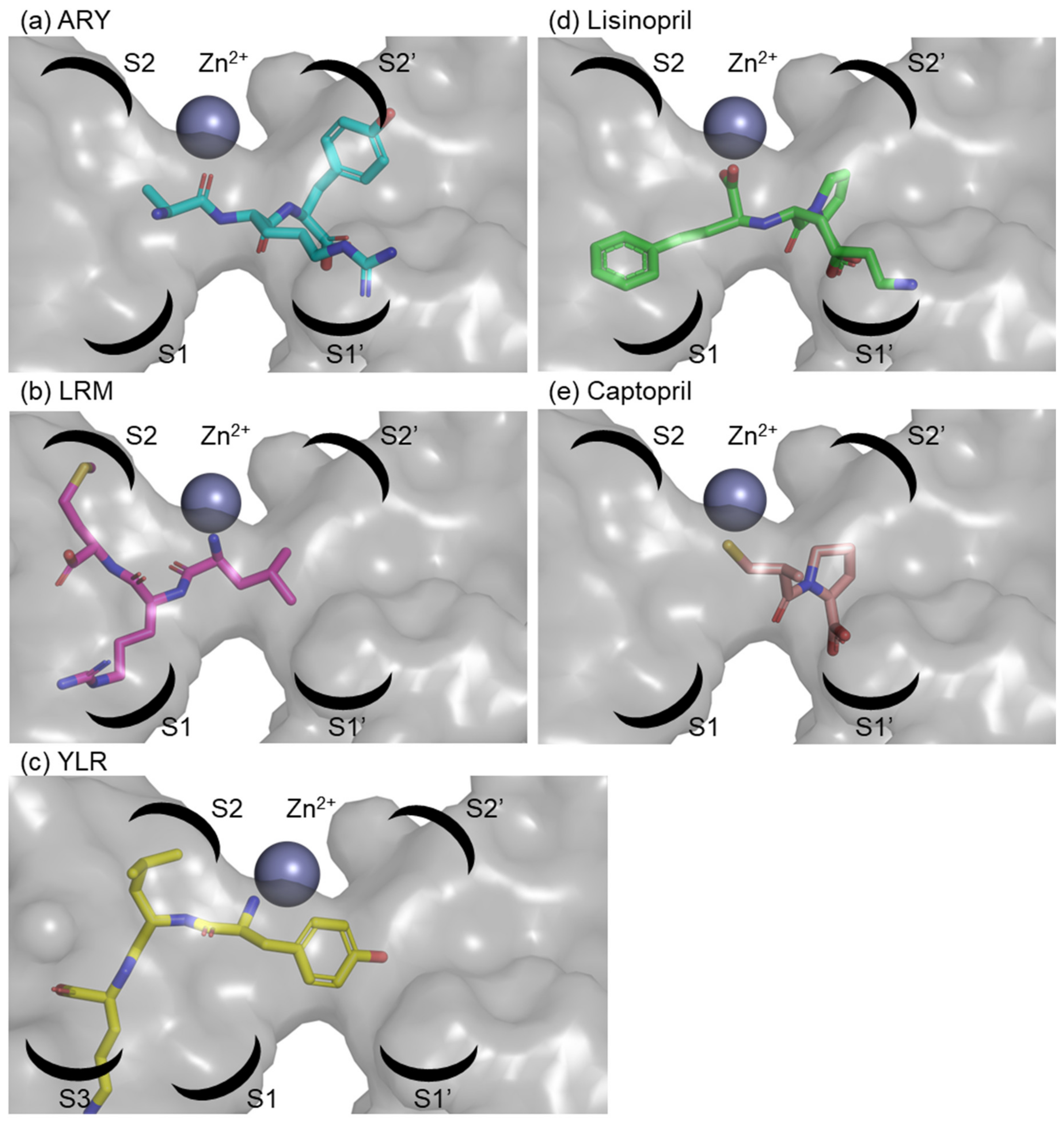
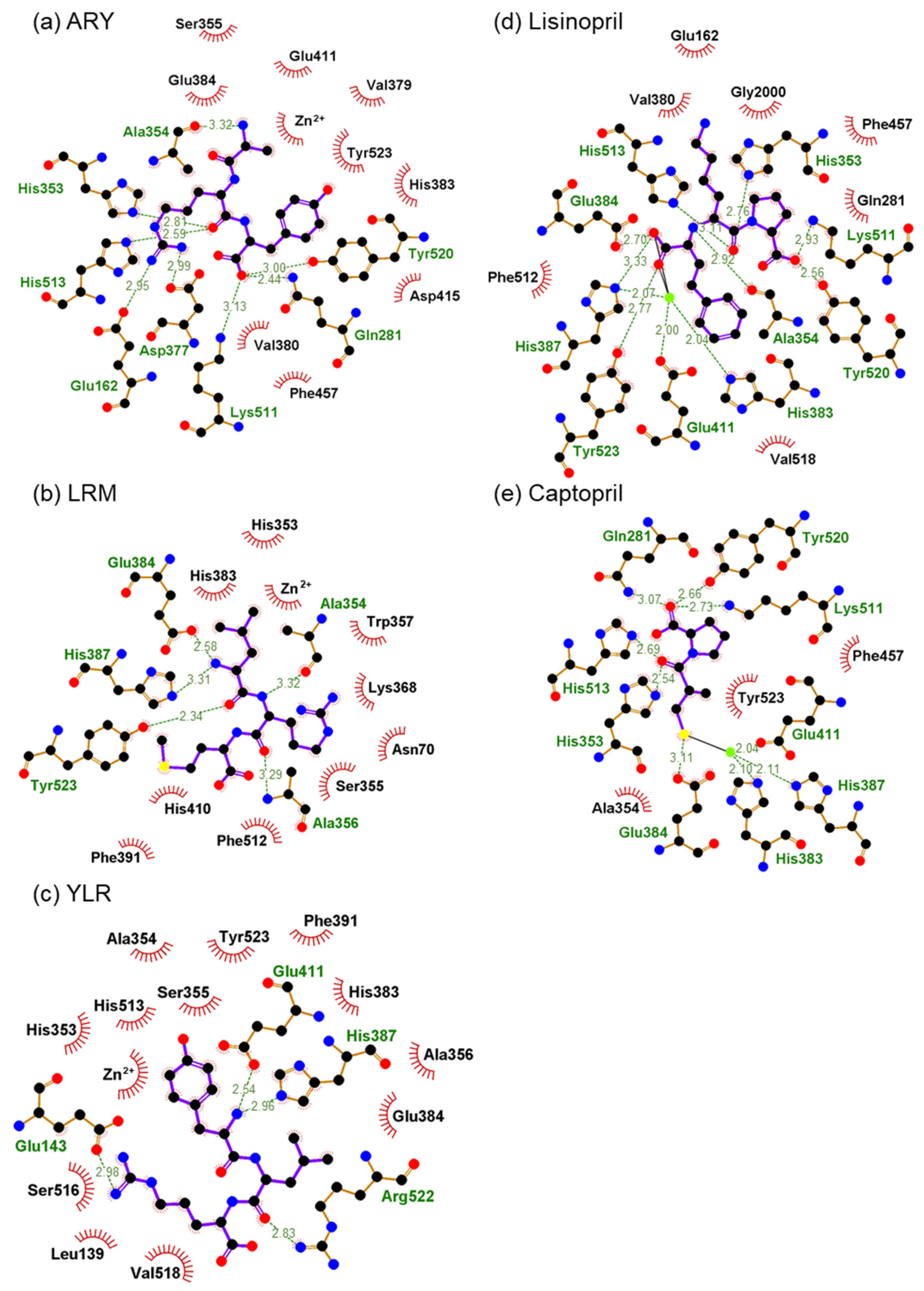
2.6. In Silico Analysis of Binding of Peptides and ACE

We identified three novel ACE inhibitory peptides (ARY, YLR, and LRM). Among them, LRM showed strong ACE inhibitory activity compared to the other two peptides. To know the inhibitory mechanism, the docking simulation between ACE and three peptides was performed by using the HPEPDOCK server. The docking energy scores with ACE were as follows: ARY: –155.685 kcal/mol; YLR: –166.290 kcal/mol; LRM: –140.683 kcal/mol. The results contradicted the in vitro synthetic peptide analysis. The binding of the three peptides in ACE was as follows: ARY was coordinated in the S1′ and S2′ pockets of the ACE, LRM was in the S2 and S1 pockets, and YLR was in the S3 and S2 pockets (Figure 3). Although they were coordinated near a zinc ion, their binding and zinc ions were not found. On the other hand, the ACE inhibitor Lisinopril was coordinated in the S1, S1′, and S2′ pockets (PDB, 1O86) containing the interaction with the zinc ion. The ACE inhibitor Captopril was coordinated in the S1′ and S2′ pockets (PDB, 1UZF) containing the interaction with the zinc ion. Bindings with peptides or inhibitors are shown as 2-D image in Figure 4. The edges of the two inhibitors were bound to the ACE. Namely, one side of Lisinopril was bound to the zinc ion, Glu384, and His387, and the other side was bound to Lys511 and Tyr520. This binding was also found in Captopril. One side of Captopril was bound to the zinc ion and Glu384, and the other side was bound to Gln281, Lys511, and Tyr520. Among the three peptides, one side of the main chain of YLR was bound to His387 and Glu411, one side of the main chain of LRM was bound to Glu384 and His387, and one side of the main chain of ARY was bound to Gln281, Lys511, and Tyr520. Although LRM showed strong ACE inhibition by in vitro synthetic peptide analysis, the inhibition was weak compared to that of the inhibitors (the IC50 value of Captopril was approximately 17–21 nM [53,54]). The difference might be due to the loss of interaction with the zinc ion.

3. Materials and Methods

3.1. Materials

Uppurui Nori, P. pseudolinearis, was collected from the coast of Hakodate, Japan. The sample was stored at –30 °C until use. ACE from rabbit lung was purchased from Sigma-Aldrich Co. (St. Louis, MO, USA). Millex-GV (pore size: 0.22 µm) and Millex-LG (pore size: 0.20 µm) were purchased from MERCK MILLIPORE Ltd. (Billerica, MA, USA). Synthetic peptides (purity: ≥95%) were purchased from Medical Biological Laboratories Co. (Nagoya, Japan). Hyppuryl-L-histidyl-L-leucine (Hip-His-Leu), thermolysin (EC 3.4.24.27) from Bacillus thermoproteolyticus, trifluoroacetic acid (TFA), and all other regents were purchased from Fujifilm Wako Pure Chemical (Osaka, Japan).

3.2. Preparation of the WSP and Its Hydrolysate

Preparation of the WSP and the hydrolysate from P. pseudolinearis was as per our previous studies [35,47]. Namely, the frozen P. pseudolinearis samples were lyophilized and ground into a fine powder by Wonder Blender WB-1 (OSAKA CHEMICAL Co., Osaka, Japan). The powder was dissolved in 20 v/w distilled water, and protein extraction was performed at 4 °C for 12 h. The WSP was recovered by centrifugation at 4 °C, 15,000× g, for 10 min, and the supernatant was then used as the WSP. The protein hydrolysate of the WSP was prepared by the digestion of protein extract with 1.0 wt. % thermolysin at 70 °C for 3 h. The reaction was stopped by boiling for 10 min. The visible ray absorption spectrum of the WSP was analyzed by a spectrophotometer (UV-1800, SHIMADZU, Kyoto, Japan). Sodium dodecyl sulfate-polyacrylamide gel electrophoresis (SDS-PAGE) was performed by Laemmli’s method with a 0.1% SDS-13.75% polyacrylamide slab gel [55]. The gel was stained with 0.1% Coomassie Brilliant Blue (CBB) R-250 in 50% methanol–7% acetic acid, and the background of the gel was decolorized with 7% acetic acid. Fluorescence of phycobiliproteins on the slab gel was detected by gel documentation LED illuminator (VISIRAYS AE-6935GN: ATTO, Tokyo, Japan).

3.3. Separation of WSP Hydrolysate and Determination of Peptide Structures

The hydrolysate of the WSP was separated by RP-HPLC. The hydrolysate was dissolved (20 mg/mL) in ultra-pure water containing 0.1% TFA and applied to sequential filtration by Millex-GV and Millex-LG. An aliquot of the sample (100 μL) was applied to a Mightysil RP-18GP column (4.6 × 150 mm) (Kanto Kagaku, Tokyo, Japan) and eluted with a linear gradient of acetonitrile from 1–25% containing 0.1% TFA at flow rate of 1.0 mL/min. Absorbance of the eluent was monitored at 228 nm. The eluted peptide fractions were collected and labeled 1–36. The fractions were used for the ACE inhibitory assay. The amino acid sequences of peptides were determined by Matrix Assisted Laser Desorption / Ionization Time of Flight Tandem Mass Spectrometry (MALDI-TOF/MS/MS) using a 4700 Proteomics Analyzer with DeNovo Explorer software version 3.6 (Applied Biosystems, Carlsbad, CA, USA). α-Cyano-4-hydroxycinnamic acid was used as matrix.

3.4. Assay of ACE Inhibitory Activity

The assay was performed using the same method as described previously [35]. The ACE inhibitory activities of the WSP, WSP hydrolysate, and synthetic peptides were measured in triplicate, and the mean ± standard error for each was calculated. Statistical analyses were carried out using the Student’s t test. In this study, the IC50 was defined as an absolute quantity of the peptide to inhibit 50% of 1.0 U ACE.

3.5. Extraction and Sequencing of Chloroplast DNA

Chloroplast DNA was extracted from the thalli of P. pseudolinearis according to the CTAB method with some modifications [56,57,58]. The nucleotide sequences of chloroplast DNA were analyzed by using a next generation sequencer, the Ion PGM System (Thermo Fisher SCIENTIFIC, Waltham, MA, USA). The data were assembled with CLC Genomics Workbench 9.5.4 (QIAGEN, Hilden, Germany). Nucleotide and deduced amino acid sequences of P. pseudolinearis phycobiliproteins were detected using BLAST (Basic Local Alignment Search Tool). The sequences of phycobiliproteins were deposited as DDBJ Accession No. LC599086-LC599088.

3.6. Bioinformatics Analysis of ACE Inhibitory Peptides

The information on the reported ACE inhibitory peptide was obtained from the BIOPEP-UWM database (http://www.uwm.edu.pl/biochemia/index.php/pl/biopep) on 11 November 2020. From the database, 1019 ACE inhibitory peptides were extracted. The peptide sequences from Uppurui Nori were manually annotated. The thermolysin-digestion products of chloroplast proteins were predicted using PeptideCutter (https://web.expasy.org/peptide_cutter/). Docking simulation of the ACE inhibitory peptide with the ACE was performed using the HPEPDOCK server [59] using default parameters. The ACE from human testis complex with lisinopril (PDB code 1O86) was employed for docking simulation. Before docking, the inhibitor and water molecules were removed, and hydrogens were added.

4. Conclusions

In this study, we prepared a water-soluble protein (WSP) hydrolysate from the red alga P. pseudolinearis, which mainly contained PE, PC, APC, and Rubisco. We then determined the ACE inhibitory activity in the thermolysin hydrolysate of the WSP. We identified 42 peptides by HPLC and MALDI-TOF/MS/MS and confirmed that 23 peptides were from chloroplast proteins. The synthetic peptide analyses showed that ARY, YLR, and LRM were the ACE inhibitory peptides. Among them, LRM showed a low IC50 value (0.15 μM) compared with ARY and YLR (1.3 and 5.8 μM). Docking simulation analyses showed that three peptides were coordinated in the active site of the ACE. However, these peptides do not bind to the zinc ion, which may be the reason for their relatively low ACE inhibitory activity. Among these three peptides, LRM was conserved in the primary structure of cpcA from Bangiales and Florideophyceae, indicating that the novel ACE inhibitory peptide LRM can be obtained from many red algae. Therefore, red algae have the potential not only as a protein source but also as an ingredient for supplements and functional foods for human. In a following study, we will seek to investigate how effective any practical applications of the WSP hydrolysate are.

Author Contributions

H.K. and H.Y. conceived and designed the research; S.K. and T.U. contributed to sample collection; K.T., S.K., R.M., and T.S. performed the experiments and analyzed the data; Y.K. and H.K. contributed to writing and editing the manuscript. All authors have read and agreed to the published version of the manuscript.

Funding

This study was supported in part by Science and Technology Research Promotion Program for Agriculture, Forestry, Fisheries and Food Industry (27004B).

Data Availability Statement

Not applicable.

Conflicts of Interest

The authors declare no conflict of interest.

References

  1. Tu, M.; Cheng, S.; Lu, W.; Du, M. Advancement and prospects of bioinformatics analysis for studying bioactive peptides from food-derived protein: Sequence, structure, and functions. Trac. Trend. Anal. Chem. 2018, 105, 7–17. [Google Scholar] [CrossRef]
  2. Matsufuji, H.; Matsui, T.; Seki, E.; Osajima, K.; Nakashima, M.; Osajima, Y. Angiotensin I-converting enzyme inhibitory peptides in an alkaline protease hydrolyzate derived from sardine muscle. Biosci. Biotechnol. Biochem. 1994, 58, 2244–2245. [Google Scholar] [CrossRef] [PubMed]
  3. Nakano, D.; Ogura, K.; Miyakoshi, M.; Ishii, F.; Kawanishi, H.; Kurumazuka, D.; Kwak, C.-J.; Ikemura, K.; Takaoka, M.; Moriguchi, S.; et al. Antihypertensive effect of angiotensin I-converting enzyme inhibitory peptides from a sesame protein hydrolysate in spontaneously hypertensive rats. Biosci. Biotechnol. Biochem. 2006, 70, 1118–1126. [Google Scholar] [CrossRef] [PubMed] [Green Version]
  4. Daskaya-Dikmen, C.; Yucetepe, A.; Karbancioglu-Guler, F.; Daskaya, H.; Ozcelik, B. Angiotensin-I-converting enzyme (ACE)-inhibitory peptides from plants. Nutrients 2017, 9, 316. [Google Scholar] [CrossRef] [PubMed]
  5. Fuchs, F.D.; Whelton, P.K. High Blood Pressure and Cardiovascular Disease. Hypertension 2020, 75, 285–292. [Google Scholar] [CrossRef] [PubMed]
  6. Schmieder, R.E.; Hilgers, K.F.; Schlaich, M.P.; Schmidt, B.M.W. Reninangiotensin system and cardiovascular risk. Lancet 2007, 369, 1208–1219. [Google Scholar] [CrossRef]
  7. Umemura, S.; Arima, H.; Arima, S.; Asayama, K.; Dohi, Y.; Hirooka, Y.; Horio, T.; Hoshide, S.; Ikeda, S.; Ishimitsu, T.; et al. The Japanese Society of Hypertension Guidelines for the Management of Hypertension (JSH 2019). Hypertens. Res. 2019, 42, 1235–1481. [Google Scholar]
  8. Wijesekara, I.; Pangestuti, R.; Kim, S.-K. Biological activities and potential health benefits of sulfated polysaccharides derived from marine algae. Carbohydr. Polym. 2011, 84, 14–21. [Google Scholar] [CrossRef]
  9. Yamamoto, Y.; Kishimura, H.; Kinoshita, Y.; Saburi, W.; Kumagai, Y.; Yasui, H.; Ojima, T. Enzymatic production of xylooligosaccharides from red alga dulse (Palmaria sp.) wasted in Japan. Process. Biochem. 2019, 82, 117–122. [Google Scholar] [CrossRef]
  10. Chen, H.; Yan, X.; Zhu, P.; Lin, J. Antioxidant activity and hepatoprotective potential of agaro-oligosaccharides in vitro and in vivo. Nutr. J. 2006, 5, 31. [Google Scholar] [CrossRef] [Green Version]
  11. Kobayashi, M.; Kumagai, Y.; Yamamoto, Y.; Yasui, H.; Kishimura, H. Identification of a key enzyme for the hydrolysis of β-(1→3)-xylosyl linkage in red alga dulse xylooligosaccharide from Bifidobacterium adolescentis. Mar. Drugs 2020, 18, 174. [Google Scholar] [CrossRef] [PubMed] [Green Version]
  12. De Ruiter, G.A.; Rudolph, B. Carrageenan biotechnology. Trends Food Sci. Technol. 1997, 8, 389–395. [Google Scholar] [CrossRef]
  13. Cherry, P.; Yadav, S.; Strain, C.R.; Allsopp, P.J.; McSorley, E.M.; Ross, R.P.; Stanton, C. Prebiotics from seaweeds: An ocean of opportunity? Mar. Drugs 2019, 17, 327. [Google Scholar] [CrossRef] [PubMed] [Green Version]
  14. Gurpilhares, D.D.B.; Cinelli, L.P.; Simas, N.K.; Pessoa, A., Jr.; Sette, L.D. Marine prebiotics: Polysaccharides and oligosaccharides obtained by using microbial enzymes. Food Chem. 2019, 280, 175–186. [Google Scholar] [CrossRef] [PubMed]
  15. Holdt, S.L.; Kraan, S. Bioactive compounds in seaweed: Functional food applications and legislation. J. Appl. Phycol. 2011, 23, 543–597. [Google Scholar] [CrossRef]
  16. Roleda, M.Y.; Marfaing, H.; Desnica, N.; Jónsdóttir, R.; Skjermo, J.; Rebours, C.; Nitschke, U. Variations in polyphenol and heavy metal contents of wild-harvested and cultivated seaweed bulk biomass: Health risk assessment and implication for food applications. Food Control 2019, 95, 121–134. [Google Scholar] [CrossRef]
  17. Rosa, G.P.; Tavares, W.R.; Sousa, P.M.C.; Pagès, A.K.; Seca, A.M.L.; Pinto, D.C.G.A. Seaweed secondary metabolites with beneficial health effects: An overview of successes in in vivo studies and clinical trials. Mar. Drugs 2020, 18, 8. [Google Scholar] [CrossRef] [Green Version]
  18. Kendel, M.; Couzinet-Mossion, A.; Viau, M.; Fleurence, J.; Barnathan, G.; Wielgosz-Collin, G. Seasonal composition of lipids, fatty acids, and sterols in the edible red alga Grateloupia turuturu. J. Appl. Phycol. 2013, 25, 425–432. [Google Scholar] [CrossRef]
  19. Schubert, N.; García-Mendoza, E.; Pacheco-Ruiz, I. Carotenoid composition of marine red algae 1. J. Phycol. 2006, 42, 1208–1216. [Google Scholar] [CrossRef]
  20. Aryee, A.N.A.; Agyei, D.; Akanbi, T.O. Recovery and utilization of seaweed pigments in food processing. Curr. Opin. Food Sci. 2018, 19, 113–119. [Google Scholar] [CrossRef]
  21. Harnedy, P.A.; FitzGerald, R.J. Bioactive proteins, peptides, and amino acid from macroalgae. J. Appl. Phycol. 2020, 47, 218–232. [Google Scholar] [CrossRef] [PubMed]
  22. Mai, K.; Mercer, J.P.; Donlon, J. Comparative studies on the nutrition of two species of abalone, Haliotis tuberculata L. and Haliotis discus hannai Ino: II. Amino acid composition of abalone and six species of macroalgae with an assessment of their nutritional value. Aquaculture 1994, 128, 115–130. [Google Scholar] [CrossRef]
  23. Paiano, M.O.; Cortona, A.D.; Costa, J.F.; Liu, S.-L.; Verbruggen, H.; Clerck, O.D.; Necchi, O., Jr. Organization of plastid genomes in the freshwater red algal order Batrachospermales (Rhodphyta). J. Phycol. 2018, 54, 25–33. [Google Scholar] [CrossRef] [Green Version]
  24. Yang, E.C.; Boo, S.M.; Bhattacharya, D.; Saunders, G.W.; Knoll, A.H.; Fredericq, S.; Graf, L.; Yoon, H.S. Divergence time estimates and the evolution of major lineages in the florideophyte red algae. Sci. Rep. 2016, 6, 21361. [Google Scholar] [CrossRef] [PubMed] [Green Version]
  25. Glazer, A.N. Light harvesting by phycobilisomes. Ann. Rev. Biophys. Chem. 1985, 14, 47–77. [Google Scholar] [CrossRef] [PubMed]
  26. Miyabe, Y.; Furuta, T.; Takeda, T.; Kanno, G.; Shimizu, T.; Tanaka, Y.; Gai, Z.; Yasui, H.; Kishimura, H. Structural properties of phycoerythrin from dulse Palmaria palmata. J. Food Biochem. 2017, 41, e12301. [Google Scholar] [CrossRef] [Green Version]
  27. Kitade, Y.; Miyabe, Y.; Yamamoto, Y.; Takeda, H.; Shimizu, T.; Yasui, H.; Kishimura, H. Structural characteristics of phycobiliproteins from red alga Mazzaella japonica. J. Food Biochem. 2018, 42, e12436. [Google Scholar] [CrossRef] [Green Version]
  28. Sfriso, A.A.; Gallo, M.; Baldi, F. Phycoerythrin productivity and diversity from five red macroalgae. J. Appl. Phycol. 2018, 30, 2523–2531. [Google Scholar] [CrossRef]
  29. Wijesekara, I.; Kim, S.-K. Angiotensin-I-converting enzyme (ACE) inhibitors from marine resources: Prospects in the pharmaceutical industry. Mar. Drugs 2010, 8, 1080–1093. [Google Scholar] [CrossRef] [Green Version]
  30. Sun, S.; Xu, X.; Sun, X.; Zhang, X.; Chen, X.; Xu, N. Preparation and identification of ACE inhibitory peptides from the marine macroalga Ulva intestinalis. Mar. Drugs 2019, 17, 179. [Google Scholar] [CrossRef] [Green Version]
  31. Noda, H. Health benefits and nutritional properties of nori. J. Appl. Phycol. 1993, 5, 255–258. [Google Scholar] [CrossRef]
  32. He, H.-L.; Chen, X.-L.; Wu, H.; Sun, C.-Y.; Zhang, Y.-Z.; Zhou, B.-C. High throughput and rapid screening of marine protein hydrolysates enriched in peptides with angiotensin-I-converting enzyme inhibitory activity by capillary electrophoresis. Bioresour. Technol. 2007, 98, 3499–3505. [Google Scholar] [CrossRef]
  33. Cian, R.E.; Alaiz, M.; Vioque, J.; Drago, S.R. Enzyme proteolysis enhanced extraction of ACE inhibitory and antioxidant compounds (peptides and polyphenols) from Porphyra columbina residual cake. J. Appl. Phycol. 2013, 25, 1197–1206. [Google Scholar] [CrossRef]
  34. Suetsuna, K. Purification and identification of angiotensin I converting enzyme inhibitors from the red alga Porphyra yezoensis. J. Mar. Biotechnol. 1998, 6, 163–167. [Google Scholar]
  35. Furuta, T.; Miyabe, Y.; Yasui, H.; Kinoshita, Y.; Kishimura, H. Angiotensin I converting enzyme inhibitory peptides derived from phycobiliproteins of dulse Palmaria palmata. Mar. Drugs 2016, 14, 32. [Google Scholar] [CrossRef] [PubMed] [Green Version]
  36. Kumagai, Y.; Kitade, Y.; Kobayashi, M.; Watanabe, K.; Kurita, H.; Takeda, H.; Yasui, H.; Kishimura, H. Identification of ACE inhibitory peptides from red alga Mazzaella japonica. Eur. Food Res. Technol. 2020, 246, 2225–2231. [Google Scholar] [CrossRef]
  37. Fitzgerald, C.; Aluko, R.E.; Hossain, M.; Rai, D.K.; Hayes, M. Potential of a renin inhibitory peptide from the red seaweed Palmaria palmata as a functional food ingredient following confirmation and characterization of a hypotensive effect in spontaneously hypertensive rats. J. Agric. Food Chem. 2014, 62, 8352–8356. [Google Scholar] [CrossRef]
  38. Harnedy, P.A.; O’Keeffe, M.B.; FitzGerald, R.J. Purification and identification of dipeptidyl peptidase (DPP) IV inhibitory peptides from the macroalga Palmaria palmata. Food Chem. 2015, 172, 400–406. [Google Scholar] [CrossRef]
  39. Fitzgerald, C.; Gallagher, E.; Tasdemir, D.; Hayes, M. Heart health peptides from macroalgae and their potential use in functional foods. J. Agric. Food Chem. 2011, 59, 6829–6836. [Google Scholar] [CrossRef]
  40. Sonani, R.R.; Singh, N.K.; Kumar, J.; Thakar, D.; Madamwar, D. Concurrent purification and antioxidant activity of phycobiliproteins from Lyngbya sp. A09DM: An antioxidant and anti-aging potential of phycoerythrin in Caenorhabditis elegans. Process. Biochem. 2014, 49, 1757–1766. [Google Scholar] [CrossRef]
  41. Sato, N.; Furuta, T.; Takeda, T.; Miyabe, Y.; Ura, K.; Takagi, Y.; Yasui, H.; Kumagai, Y.; Kishimura, H. Antioxidant activity of proteins extracted from red alga dulse harvested in Japan. J. Food Biochem. 2019, 43, e12709. [Google Scholar] [CrossRef]
  42. Sakai, S.; Komura, Y.; Nishimura, Y.; Sugawara, T.; Hirata, T. Inhibition of mast cell degranulation by phycoerythrin and its pigment moiety phycoerythrobilin, prepared from Porphyra yezoensis. Food Sci. Technol. Res. 2011, 17, 171–177. [Google Scholar] [CrossRef] [Green Version]
  43. Cian, R.E.; Garzon, A.G.; Ancona, D.B.; Guerrero, L.C.; Drago, S.R. Hydrolyzates from Pyropia columbina seaweed have antiplatelet aggregation, antioxidant and ACE I inhibitory peptides which maintain bioactivity after simulated gastrointestinal digestion. LWT Food Sci. Technol. 2015, 64, 881–888. [Google Scholar] [CrossRef]
  44. Sato, M.; Hosokawa, T.; Yamaguchi, T.; Nakano, T.; Muramoto, K.; Kahara, T.; Funayama, K.; Kobayashi, A.; Nakano, T. Angiotensin I-converting enzyme inhibitory peptides derived from Wakame (Undaria pinnatifida) and their antihypertensive effect in spontaneously hypertensive rats. J. Agric. Food Chem. 2002, 50, 6245–6252. [Google Scholar] [CrossRef]
  45. Suetsuna, K.; Maekawa, K.; Chen, J.-R. Antihypertensive effects of Undaria pinnatifida (wakame) peptide on blood pressure in spontaneously hypertensive rats. J. Nutr. Biochem. 2004, 15, 267–272. [Google Scholar] [CrossRef]
  46. Kumagai, Y.; Miyabe, Y.; Takeda, T.; Adachi, K.; Yasui, H.; Kishimura, H. In silico analysis of relationship between proteins from plastid genome of red alga Palmaria sp. (Japan) and angiotensin I converting enzyme inhibitory peptides. Mar. Drugs 2019, 17, 190. [Google Scholar] [CrossRef] [Green Version]
  47. Sumikawa, K.; Takei, K.; Kumagai, Y.; Shimizu, T.; Yasui, H.; Kishimura, H. In silico analysis of ACE inhibitory peptides from chloroplast proteins of red alga Grateloupia asiatica. Mar. Biotechnol. 2020, 22, 391–402. [Google Scholar] [CrossRef] [PubMed]
  48. Ng, P.K.; Lin, S.M.; Lim, P.E.; Liu, L.C.; Chen, C.M.; Pai, T.W. Complete chloroplast genome of Gracilaria firma (Gracilariaceae, Rhodophyta), with discussion on the use of chloroplast phylogenomics in the subclass Rhodymeniophycidae. BMC Genom. 2017, 18, 40. [Google Scholar] [CrossRef] [PubMed] [Green Version]
  49. Lee, J.; Kim, K.M.; Yang, E.C.; Miller, K.A.; Boo, S.M.; Bhattacharya, D.; Yoon, H.S. Reconstructing the complex evolutionary history of mobile plasmids in red algal genomes. Sci. Rep. 2016, 6, 23744. [Google Scholar] [CrossRef] [PubMed]
  50. Yang, L.-E.; Deng, Y.-Y.; Xu, G.-P.; Russell, S.; Lu, Q.-Q.; Brodie, J. Redefining Pyropia (Bangiales, Rhodophyta): Four new genera, resurrection of Porphyrella and description of Calidia pseudolobata sp. nov. from China. J. Phycol. 2020, 56, 862–879. [Google Scholar] [CrossRef] [PubMed]
  51. Saunders, G.W.; Hommersand, M.H. Assessing red algal supraordinal diversity and taxonomy in the context of contemporary systematic data. Am. J. Bot. 2004, 91, 1494–1507. [Google Scholar] [CrossRef] [PubMed] [Green Version]
  52. Wu, J.; Aluko, R.E.; Nakai, S. Structural requirements of angiotensin I-converting enzyme inhibitory peptides: Quantitative structure−activity relationship study of di- and tripeptides. J. Agric. Food Chem. 2006, 54, 732–738. [Google Scholar] [CrossRef]
  53. Baldwin, J.E.; Adlington, R.M.; Russell, A.T.; Smith, M.L. Carbon based nucleophilic ring opening of activated monocyclic β-lactams; Synthesis and stereochemical assignment of the ACE inhibitor WF-10129. Tetrahedron 1995, 51, 4733–4762. [Google Scholar] [CrossRef]
  54. Hooper, N.M. Angiotensin converting enzyme: Implications from molecular biology for its physiological functions. Int. J. Biochem. 1991, 23, 641–647. [Google Scholar] [CrossRef]
  55. Laemmli, U.K. Cleavage of structural proteins during the assembly of the head of bacteriophage T4. Nature 1970, 227, 680–685. [Google Scholar] [CrossRef] [PubMed]
  56. Cota-Sánchez, J.H.; Remarchuk, K.; Ubayasena, K. Ready-to-use DNA extracted with a CTAB method adapted for herbarium specimens and mucilaginous plant tissue. Plant Mol. Biol. Rep. 2006, 24, 161. [Google Scholar] [CrossRef]
  57. Kumagai, Y.; Tsubouchi, R.; Miyabe, Y.; Takeda, T.; Adachi, K.; Yasui, H.; Kishimura, H. Complete sequence of mitochondrial DNA of red alga dulse Palmaria palmata (Linnaeus) Weber & Mohr in Japan. Mitochondrial DNA Part B 2019, 4, 3177–3178. [Google Scholar] [PubMed]
  58. Watanabe, K.; Kishimoto, T.; Kumagai, Y.; Shimizu, T.; Uji, T.; Yasui, H.; Kishimura, H. Complete sequence of mitochondrial DNA of Gloiopeltis furcata (Postels and Ruprecht) J. Agardh. Mitochondrial DNA Part B 2019, 4, 2543–2544. [Google Scholar] [CrossRef] [Green Version]
  59. Zhou, P.; Jin, B.; Li, H.; Huang, S.-Y. HPEPDOCK: A web server for blind peptide–protein docking based on a hierarchical algorithm. Nucleic Acids Res. 2018, 46, W443–W450. [Google Scholar] [CrossRef]
Figure 1. Properties and ACE inhibitory activities of P. pseudolinearis WSP and its hydrolysate. (a) SDS-PAGE. Lane 1, Maker; Lane 2, WSP; Lane 3, the thermolysin hydrolysate of the WSP; Lane 4, WSP (Fluorescence); Lane 5, the thermolysin hydrolysate of the WSP (Fluorescence). (b) Visual ray absorption spectra of WSP. (c) ACE inhibitory activities of WSP and the hydrolysate. Bars represent standard errors. * p < 0.05.
Marinedrugs 19 00200 g001
Figure 2. HPLC chromatogram of the WSP hydrolysate and ACE inhibitory activity. (a) Chromatogram of the WSP hydrolysate by HPLC. Peptides were separated by Mightysil RP-18GP column. The numbers on the peaks (1–36) were pooled. (b) ACE inhibitory activity of each fraction (1–36).
Marinedrugs 19 00200 g002
Figure 3. Binding motifs of ARY, LRM, and YLR in the active site of ACE. (a) ARY and ACE interaction; (b) LRM and ACE interaction; (c) YLR and ACE interaction; (d) Lisinopril and ACE interaction (PDB 1O86); (e) Captopril and ACE interaction (PDB 1UZF). Zinc ion is shown as a dark gray sphere. The binding pockets of the enzymes are labeled within the surface-rendered catalytic channel of the C-domain of ACE.
Marinedrugs 19 00200 g003
Figure 4. Two-dimensional molecular docking results for ARY, LRM, and YLR with ACE (PDB: 1O86). (a) ARY and ACE interaction; (b) LRM and ACE interaction; (c) YLR and ACE interaction; (d) Lisinopril and ACE interaction (PDB 1O86); (e) Captopril and ACE interaction (PDB 1UZF).
Marinedrugs 19 00200 g004
Table 1. ACE inhibitory peptide sequences in the WSP from P. pseudolinearis and identification of the peptide sources from in silico digestion of P. pulchra plastid proteins.
FNPeptideNumber of Peptides in Chloroplast ProteinsIn Silico Thermolysin Digestion
a× b
3AYR6rbcLapcE, atpA, cpcG, psaL, rps7
4MTFF0- c-
5VRFK d1gltB-
6KFR4-accA, cemA, chlI, rpl16
6WHKPA0--
6FFKWEF0--
7FGGR1petJ-
7LVER0--
8YRD7-cpeA, moeB, odpA, orf148, rps14, ycf3, ycf21
8FFR d4psaB, ycf22psbT, ycf63
9ARY6atpI, chlB, gltBapcA, orf263, ycf46
9RFR3-odpA, rpoC1 (2) e
10FAR d7clpC, orf174, ycf26atpI, chlB, cpeA, ilvH
10YLR12-accA, apcA, apcB, apcD, apcE, apcF, carA, cpcA, orf114, rpl3, rpl19, rps9
10VYRT1-cpeA
10FVCG0--
11FFLREF0--
12SRGL1-rpl23
13ACWR0--
13RFAPR0--
14ACPSGT0--
16WER1-psbA
17LDY17chlN, cpcA, cpeA, infB, rbcL, ycf16accA, apcA, apcB, atpA, infC, orf111, orf565, rpoA, rps1, syh, ycf24
17LLEER0--
18PGCRR0--
18FLWWLR0--
20AAGRFP1-cpeA
23LVFFGH0--
24PVAFN0--
25LRM d2cpcA psbB
26LRY8apcB, apcE, apcF, cpcB, cpeBapcD, apcE, rbcR
27CPSNN0--
27AWRRP0--
27LWT4trpGpsaB, psbD, psbZ
28YRF3-cpcG, odpA, syh
28FRV d8petB, rbcL, rps5, rpoB (2), ycf26pgmA, rpoB
29VNLF1-ycf38
29PGDTY0--
30EWYPH0--
32KTFPY0--
34FGRPF d1rbcL-
36VESR2-rbcL, rpoA
Total101 peptides in 66 kinds of proteins (31 peptides were produced by in silico digestion)
Data from Pyropia pulchra plastid (NC_029861.1). a The peptide is produced by thermolysin digestion from the protein. b The peptide is confirmed in the protein sequence, but it is not produced by thermolysin digestion. c “-” not detected in the plastid genome. d The peptides are hydrolyzed by in silico thermolysin digestion. Hydrolysis position of peptides show “-”: VR-FK; LR-M; FR-V; FGRP-F; F-FR; F-AR. e The parentheses indicate number of in silico thermolysin digestion site in proteins.
Table 2. Identity of phycobiliproteins from P. pseudolinearis with other red algae.
SpeciesAccession No.Identity (%)
cpeA acpeB acpcA bcpcB bapcA capcB c
Pyropia pulchraNC_029861.110010010098.899.4100
Porphyra purpureaNC_00092510099.499.499.4100100
Neopyropia yezoensisKC5107210010010098.3100100
Palmaria palmata in JapanAB80766289.088.188.990.195.098.1
Grateloupia asiaticaAP01812990.291.592.690.196.393.8
a, DDBJ accession No. LC599086. b, DDBJ accession No. LC599087. c, DDBJ accession No. LC599088.
Table 3. ACE inhibitory activity of the synthetic peptide.
PeptideIC50 (μmol)Reference
ARY1.3This study
YLR5.8
LRM0.15
VYRT0.14[35]
LDY6.1
FEQWAS>2.8
LRY0.044
Publisher’s Note: MDPI stays neutral with regard to jurisdictional claims in published maps and institutional affiliations.

