4. Experimental
4.1. General
Infrared (IR) spectra were recorded on a 1720X Fourier transformation-infrared (FT-IR) spectrometer (Perkin Elmer, MA, USA) or an IRAffinity-1S FT-IR spectrometer (Shimadzu, Kyoto, Japan). High-resolution mass spectra (HRMS) were recorded on a JMS-700 (2) mass spectrometer (JEOL, Tokyo, Japan). Nuclear magnetic resonance (NMR) spectra were recorded at 27 °C on 400-MR-DD2 (Agilent Technologies, CA, USA) and 600-DD2 (Agilent Technologies, CA, USA) instruments in CDCl
3 or acetone-
d6 using tetramethylsilane as an internal standard. Specific rotations were measured with a JASCO P-2300 spectrometer (JASCO Co., Tokyo, Japan). Liquid column chromatography was conducted on silica gel (Fuji Silysia FL-60D). Analytical thin layer chromatography (TLC) was performed on Silicagel 70 F
254 plates (Wako Pure Chemical Industries, Tokyo, Japan), and compounds were detected by dipping the plates into an ethanolic solution of phosphomolybdic acid followed by heating. Microwave (MW)-aided reactions were carried out in a Biotage Initiator
® reactor (PartnerTech Atvidaberg AB for Biotage Sweden AB, Uppsala, Sweden). NaBH
4 and trifluoroacetic acid (TFA) were purchased from Wako Pure Chemical Industries (Wako Pure Chemical Industries, Tokyo, Japan). BF
3·Et
2O was purchased from Sigma-Aldrich Co., LLC (St. Lewis, MO, USA). DMP was purchased from TCI (Tokyo Chemical Industry Co., Ltd., Tokyo, Japan). (−)-Shikimic acid and (−)-quinic acid were purchased from Carbosynth, Ltd. (UK) and Merck (Merck & Co., Inc., Darmstadt, Germany), respectively. α-Glucosidase (yeast, lot 26010), β-glucosidase (sweet almond, lot 81241), α-mannosidase (Jack Bean, lot 055K7047), α-galactosidase (green coffee bean, lot SMBP1296V), β-galactosidase (bovine liver, lot SMBL0488V), and deoxymannojirimycin were purchased from Sigma-Aldrich Co., LLC (St. Lewis, MO, USA). Deoxygalactonojirimycin was purchased from Funakoshi Co., Ltd., Tokyo, Japan). 1-Deoxynojirimycin (DNJ) was isolated from the leaves of
Morus alba L. (Procedure for isolation of DNJ is described in
Section 4.5.)
4.2. Condensation of 10 or 15 with 11
General procedure (synthesis of (−)-
12,
Scheme 1): To a solution of (+)-
10 (68.4 mg, 0.23 mmol) and anti-epoxide (−)-
11 (50.9 mg, 0.15 mmol) in CH
2Cl
2 (0.15 mL) was added BF
3·Et
2O (3 μL, 0.025 mmol) at 0 °C. After 1 h of stirring, the reaction mixture was treated with Et
3N (20 µL, 0.14 mmol) and concentrated under vacuum to afford a crude residue that was purified by silica gel column chromatography (eluent = hexane:EtOAc, 1:1 v/v) to afford (−)-
12 (68.8 mg, 65 %) as a colorless oil.
(−)-12: oil; −7.2 (c 1.175, CHCl3); IR (film) νmax 3438 (OH), 1726 (C=O), 1658 (C=C) cm−1; 1H-NMR (acetone-d6, 400 MHz, ppm) δ 1.30−1.70 (20H, m), 3.41 (3H, s, 6’-OMe), 3.73 (3H, s, COOMe), 3.75 (3H, s, COOMe), 4.01–4.04 (1H, m, H-5’), 4.21 (1H, overlapped, H-5), 4.23 (1H, overlapped, H-6), 4.25 (1H, overlapped, H-4’), 4.37 (1H, br d, J = 5.4 Hz, H-6’), 4.58 (1H, d, J = 3.3 Hz, 5’-OH), 4.70 (1H, ddd, J = 6.1, 3.9, 0.9 Hz, H-3’), 4.74 (1H, dd, J = 5.6, 2.3 Hz, H-4), 4.78 (1H, ddd, J = 5.7, 2.7, 1.2 Hz, H-3), 6.50 (1H, dd, J = 2.5, 1.0 Hz, H-2), 6.55 (1H, dd, J = 3.7, 1.2 Hz, H-2’); 13C-NMR (acetone-d6, 100 MHz, ppm) δ 24.47 (CH2), 24.51 (CH2), 24.69 (CH2), 24.72 (CH2), 25.7 (CH2), 25.8 (CH2), 35.8 (CH2), 36.3 (CH2), 37.6 (CH2), 38.7 (CH2), 52.0 (CH3, C-8), 52.1 (CH3, C-8’), 60.3 (CH3, 6’-OMe), 71.6 (CH, C-3’), 72.5 (CH, C-3), 72.8 (CH, C-5’), 74.0 (CH, C-4), 75.9 (CH, C-6), 76.2 (CH, C-4’), 77.0 (CH, C-6’), 80.5 (C, C-5), 111.1 (Cq), 111.3 (Cq), 133.38 (Cq, C-1), 133.42 (CH, C-2), 134.8 (Cq, C-1’), 136.1 (CH, C-2’), 167.39 (Cq, C-7), 167.43 (Cq, C-7’); high-resolution electron impact mass spectrum (HREIMS) m/z calcd for C29H40O11 (M)+ 564.2571, found 564.2570.
(+)-12 (51.2 mg, 90 %) was prepared from (−)-10 (30 mg, 0.10 mmol) and (+)-11 (40.0 mg, 0.15 mmol): oil; +7.7 (c 0.43, CHCl3); IR (film) νmax 3431 (OH), 1727 (C=O), 1658 (C=C) cm−1; 1H-NMR (acetone-d6, 600 MHz, ppm) δ 1.50−1.70 (20H, m), 3.41 (3H, s, 6’-OMe), 3.73 (3H, s, COOMe), 3.75 (3H, s, COOMe), 4.07 (1H, ddd, J = 6.8, 5.6, 3.5 Hz, H-5’), 4.21 (1H, dd, J = 6.8, 2.9 Hz, H-5), 4.23 (1H, dt, J = 6.8, 1.2 Hz, H-6), 4.25 (1H, t, J = 6.5 Hz, H-4’), 4.37 (1H, ddd, J = 5.6, 1.2, 1.1 Hz, H-6’), 4.60 (1H, d, J = 3.5 Hz, OH), 4.70 (1H, ddd, J = 6.1, 3.8, 1.1 Hz, H-3’), 4.74 (1H, ddd, J = 5.9, 2.9, 0.8 Hz, H-4), 4.78 (1H, ddd, J = 5.9, 3.0, 1.5 Hz, H-3), 6.50 (1H, ddd, J = 3.0, 1.2, 0.9 Hz, H-2), 6.55 (1H, dd, J = 3.8, 1.2 Hz, H-2’); 13C-NMR (acetone-d6, 150 MHz, ppm) δ 24.47 (CH2), 24.51 (CH2), 24.68 (CH2), 24.70 (CH2), 25.7 (CH2), 25.8 (CH2), 35.7 (CH2), 36.3 (CH2), 37.6 (CH2), 38.7 (CH2), 52.0 (CH3, C-8 or C-8’), 52.1 (CH3, C-8 or C-8’), 60.3 (CH3, 6’-OMe), 71.6 (CH, C-3’), 72.5 (CH, C-3), 72.7 (CH, C-5’), 74.0 (CH, C-4), 75.9 (CH, C-6), 76.2 (CH, C-4’), 77.0 (CH, C-6’), 80.5 (C, C-5), 111.1 (Cq), 111.3 (Cq), 133.4 (Cq, C-1), 133.5 (CH, C-2), 134.7 (Cq, C-1’), 136.1 (CH, C-2’), 167.40 (Cq, C-7), 167.44 (Cq, C-7’); HRMS m/z calcd for C29H40O11 (M)+ 564.2571, found 564.2574.
(−)-16 (150.3 mg, 78 %) was prepared from (+)-15 (101.4 mg, 0.34 mmol) and (−)-11 (94.6 mg, 0.36 mmol): oil; −75.6 (c 0.60, CHCl3); IR (film) νmax 3477 (OH), 1722 (C=O), 1657 (C=C) cm−1; 1H-NMR (C6D6, 300 MHz, ppm) δ 1.00−1.90 (20 H, m), 3.33 (3H, s, H-8), 3.35 (3H, s, H-8’), 3.67 (3H, s, OMe), 4.02 (1H, d, J = 4.4 Hz, H-6’), 4.04 (1H, dd, J = 21.1, 8.2 Hz, H-5’), 4.16 (1H, dd, J = 4.4, 1.2 Hz, H-5), 4.21 (1H, overlapped, H-4’), 4.27 (1H, dd, J = 6.5, 3.6 Hz, H-3), 4.35 (1H, ddd, J = 6.4, 3.9, 0.5 Hz, H-3’), 4.50 (1H, s, 5’-OH), 4.64 (1H, dd, J = 4.4, 1.2 Hz, H-6), 4.66 (1H, dd, J = 3.6, 1.5 Hz, H-4), 6.57 (1H, dd, J = 3.8, 1.5 Hz, H-2’), 6.79 (1H, d, J = 2.9 Hz, H-2); 13C-NMR (acetone-d6, 75 MHz, ppm) δ 24.7 (CH2), 24.8 (2 × CH2), 25.0 (CH2), 25.7 (CH2), 26.0 (CH2), 36.4 (CH2), 36.6 (CH2), 38.4 (CH2), 38.9 (CH2), 52.1 (2 × CH3, C-8, 8’), 62.3 (CH3, 6’-OMe), 72.0 (CH, C-3’), 72.1 (CH, C-4), 73.3 (CH, C-3), 73.6 (CH, C-6), 74.9 (CH, C-5’),77.1 (CH, C-4’), 78.1 (CH, C-6’), 79.4 (CH, C-5), 112.0 (Cq), 112.9 (Cq), 131.6 (Cq, C-1), 132.9 (CH, C-2’), 136.9 (Cq, C-1’), 136.9 (CH, C-2) 166.8 (Cq, C-7), 166.9 (Cq, C-7’); HREIMS m/z calcd for C29H40O11 (M)+ 564.2571, found 564.2567.
(+)-16 (76.2 mg, 90%) was prepared from (−)-15 (45.0 mg, 0.15 mmol) and (+)-11 (40.0 mg, 0.15 mmol): oil; +75.4 (c 0.82, CHCl3); IR (film) νmax 3488 (OH), 1723 (C=O), 1653 (C=C) cm−1; 1H-NMR (C6D6, 600 MHz, ppm) δ 1.50−1.90 (20H, m), 3.36 (3H, s, COOMe), 3.39 (3H, s, COOMe), 3.66 (3H, s, 6’-OMe), 4.02–4.08 (2H, m, H-5’ and H-6’), 4.15 (1H, dd, J = 4.7, 3.3 Hz, H-5), 4.21 (1H, ddd, J =6.8, 6.7, 2.1 Hz, H-4’), 4.31 (1H, ddd, J = 5.8, 3.2, 0.6 Hz, H-3), 4.39 (1H, ddd, J = 6.4, 3.8, 0.8 Hz, H-3’), 4.47 (1H, s, 5’-OH), 4.62 (1H, d, J = 4.7 Hz, H-6), 4.66 (1H, dd, J = 5.9, 2.7 Hz, H-4), 6.59 (1H, dd, J = 3.6, 1.4 Hz, H-2’), 6.79 (1H, dd, J = 3.2, 0.5 Hz, H-2); 13C-NMR (C6D6, 150 MHz, ppm) δ 24.1 (2 × CH2), 24.3 (CH2), 24.4 (CH2), 25.2 (CH2), 25.4 (CH2), 35.8 (CH2), 36.1 (CH2), 37.8 (CH2), 38.3 (CH2), 51.56 (CH3, C-8), 51.60 (CH3, C-8’), 61.7 (CH3, 6’-OMe), 71.4 (CH, C-3’), 71.5 (CH, C-4), 72.8 (CH, C-3), 73.1 (CH, C-6), 74.2 (CH, C-5’), 76.5 (CH, C-4’), 77.5 (CH, C-6’), 78.9 (CH, C-5), 111.4 (Cq), 112.4 (Cq), 131.1 (Cq, C-1), 132.5 (CH, C-2’), 136.2 (Cq, C-1’), 136.4 (CH, C-2) 166.2 (Cq, C-7), 166.4.(Cq, C-7’); HREIMS m/z calcd for C29H40O11 (M)+ 564.2571, found 564.2570.
(−)-19 (139 mg, 46%) was prepared from (−)-10 (187 mg, 0.70 mmol) and (−)-11 (113 mg, 0.54 mmol): white powder; mp 165–170 °C: −48.4 (c 0.1, CHCl3); IR (KBr) νmax 3422 (OH), 1726 (C=O), 1438 (C=C) cm−1; 1H-NMR (CDCl3, 600 MHz, ppm) δ 1.30−1.80 (20H, m), 3.71 (3H, s, 6’-OMe), 3.795 (3H, s, COOMe), 3.796 (3H, s, COOMe), 3.91 (1H, ddd, J = 9.4, 7.3, 2.6 Hz, H-5’), 4.05 (1H, dd, J = 8.5, 2.3 Hz, H-5), 4.13 (1H, dd, J = 9.1, 6.5 Hz, H-4’), 4.25 (1H, ddd, J = 7.7, 1.8, 1.4 Hz, H-5’), 4.41 (1H, ddd, J = 8.5, 1.8, 1.7 Hz, H-6), 4.61 (1H, overlapped, H-3’), 4.62 (1H, overlapped, H-3), 4.84 (1H, ddd, J = 5.6, 2.1, 1.2 Hz, H-4), 4.97 (1H, d, J = 2.6 Hz, OH), 6.42 (1H, ddd, J = 2.9, 1.8, 1.3 Hz, H-2), 6.60 (1H, dd, J = 4.4, 2.0 Hz, H-2’); 13C-NMR (CDCl3, 150 MHz, ppm) δ 23.6 (CH2), 23.8 (CH2), 23.92 (CH2), 23.95 (CH2), 24.9 (CH2), 25.0 (CH2), 35.1 (CH2), 35.8 (CH2), 37.2 (CH2), 38.1 (CH2), 52.0 (CH3, C-8 or C-8’), 52.1 (CH3, C-8 or C8’), 61.9 (CH3, 6’-OMe), 70.7 (CH, C-3), 72.4 (CH, C-3’), 73.4 (CH, C-4), 75.4 (CH, C-5’), 75.7 (CH, C-4’), 76.6 (CH, C-6), 80.0 (CH, C-6’), 82.0 (CH, C-5), 110.8 (Cq), 111.7 (Cq), 130.5 (CH, C-2’), 132.7 (Cq, C-1), 135.4 (CH, C-2), 135.9 (Cq, C-1’), 166.3 (Cq, C-7’), 166.7 (Cq, C-7); HRMS m/z calcd for C29H40O11 (M)+ 564.2571, found 564.2570.
(+)-19 (80 mg, 44%) was prepared from (+)-10 (86.0 mg, 0.29 mmol) and (+)-11 (86.2 mg, 0.29 mmol): white powder; mp 165–169 °C: +69.0 (c 0.75, CHCl3); IR (KBr) νmax 3408 (OH), 1725 (C=O), 1440 (C=C) cm−1; 1H-NMR (CDCl3, 400 MHz, ppm) δ 1.30−1.80 (20H, m), 3.72 (3H, s, OMe), 3.79 (6H, s, 2 × COOMe), 3.91 (1H, dd, J = 8.9, 6.2 Hz), 4.05 (1H, br d, J = 8.1 Hz), 4.13 (1H, dd, J = 8.4, 5.9 Hz), 4.25 (1H, br d, J = 6.5 Hz), 4.41 (1H, br d, J = 7.8 Hz), 4.59–4.65 (2H, m), 4.82–4.86 (1H, m), 5.00–5.03 (1H, m), 6.41 (1H, m), 6.60 (1H, m); 13C-NMR (CDCl3, 100 MHz, ppm) δ 23.6, 23.7, 23.89, 23.92, 24.9, 24.9, 35.1, 35.8, 37.2, 38.1, 52.0, 52.1, 61.9, 70.6, 72.3, 73.4, 75.4, 75.6, 76.6, 80.0, 82.0, 110.8, 111.6, 130.5, 132.6, 135.4, 135.8, 166.2, 166.6; HREIMS m/z calcd for C29H40O11 (M)+ 564.2571, found 564.2568.
(−)-22 (42.8 mg, 59%) was prepared from (−)-15 (38.6 mg, 0.13 mmol) and (−)-11 (45.1 mg, 0.17 mmol): oil; [α]D22.5 −44.9 (c 0.73, CHCl3); IR (film) νmax 3445 (OH), 1717 (C=O), 1653 (C=C) cm−1; 1H-NMR (CDCl3, 600 MHz, ppm) δ 1.21−1.74 (20H, m), 3.31 (3H, s, OMe), 3.76 (3H, s, COOMe), 3.82 (3H, s, COOMe), 3.87 (1H, br t, J = 8.5 Hz, H-5’), 4.07 (1H, dd, J = 4.1, 3.0 Hz, H-5), 4.20 (1H, d, J = 7.1 Hz, H-6’), 4.21 (1H, dd, J = 9.1, 6.8 Hz, H-4’), 4.51–4.53 (1H, m, H-4), 4.65 (1H, ddd, J = 6.7, 3.8, 1.1 Hz, H-3’), 4.73 (1H, dd, J = 5.9, 3.8 Hz, H-3), 4.82 (1H, s, -OH), 4.83 (1H, d, J = 4.1 Hz, H-6), 6.71 (1H, dd, J = 3.8, 2.0 Hz, H-2’), 6.97 (1H, dd, J = 3.8, 0.9 Hz, H-2); 13C-NMR (CDCl3, 150 MHz, ppm) δ 23.6 (CH2), 23.7 (CH2), 23.9 (CH2), 25.0 (CH2), 35.2 (CH2), 35.7 (CH2), 37.1 (CH2), 37.8 (CH2), 52.0 (CH3, COOMe), 52.4 (CH3, COOMe), 56.1 (CH3, 6-OMe), 68.5 (CH, C-6), 70.8 (CH, C-3’), 72.7 (CH, C-3), 73.1 (CH, C-5’), 73.5 (CH, C-4), 76.0 (CH, C-4’), 78.4 (CH, C-5), 78.5 (CH, C-6’), 111.6 (Cq), 112.0 (Cq), 129.1 (Cq, C-1), 132.4 (CH, C-2’), 135.7 (Cq, C-1’), 140.5 (CH, C-2), 166.3 (Cq, C-7’), 167.0 (Cq, C-7); HREIMS m/z calcd for C29H40O11 (M)+ 564.2571, found 564.2573.
(+)-22 (41.5 mg, 63%) was prepared from (+)-15 (34.8 mg, 0.12 mmol) and (+)-11 (34.3 mg, 0.13 mmol): oil; [α]D21.5 +51.7 (c 0.73, CHCl3); IR (film) νmax 3474 (OH), 1699 (C=O), 1652 (C=C) cm−1; 1H-NMR (CDCl3, 400 MHz, ppm) δ 1.21−1.71 (20H, m), 3.30 (3H, s, OMe), 3.76 (3H, s, COOMe), 3.82 (3H, s, COOMe), 3.87 (1H, br t, J = 8.8 Hz, H-5’), 4.07 (1H, dd, J = 4.1, 3.1 Hz, H-5), 4.20 (1H, d, J = 6.9 Hz, H-6’), 4.21 (1H, dd, J = 9.2, 6.9 Hz, H-4’), 4.52 (1H, dd, J =5.7, 2.5 Hz, H-4), 4.66 (1H, br dd, J = 6.3, 3.7 Hz, H-3’), 4.74 (1H, dd, J = 5.9, 3.7 Hz, H-3), 4.83 (1H, d, J = 3.9 Hz, H-6), 4.85 (1H, br s, -OH), 6.72 (1H, dd, J = 3.8, 2.0 Hz, H-2’), 6.98 (1H, br d, J = 3.5 Hz, H-2); 13C-NMR (CDCl3, 100 MHz, ppm) δ 23.55, 23.62, 23.89, 23.91, 25.0, 35.2, 35.7, 37.0, 37.8, 52.0, 52.4, 56.1, 68.4, 70.8, 72.7, 73.0, 73.4, 75.9, 78.3, 78.5, 110.6, 112.0, 129.0, 132.4, 135.7, 140.5, 166.2, 166.9; HREIMS m/z calcd for C29H40O11 (M)+ 564.2571, found 564.2571.
4.3. Inversion of Configuration at C5’
General procedure (synthesis of (+)-
13,
Scheme 1): A microwave vial containing a CH
2Cl
2 solution (2 mL) of (−)-
12 (47.6 mg, 0.084 mmol) was treated with DMP (77.0 mg, 0.23 mmol) at 0 °C, sealed, and irradiated in the MW reactor at 80 °C for 2.5 h. After cooling, the reaction mixture was treated with aqueous saturated (sat.) Na
2S
2O
3 (20 mL) and saturated aqueous NaHCO
3 (10 mL) and extracted with
tert-butyl methyl ether (3 × 20 mL). The combined organic layers were washed with brine (30 mL) and water (30 mL), dried over MgSO
4, filtered, and concentrated in a vacuum to afford a crude residue containing the desired ketone. To a solution of NaBH
4 (1.0 mg, 0.026 mmol) in MeOH (0.6 mL), a solution of the crude residue in MeOH (2.0 mL) at 0 °C was added dropwise. After 30 min of stirring, the reaction mixture was treated with saturated aqueous NH
4Cl (30 mL) and extracted with CH
2Cl
2 (3 × 30 mL). The combined organic layers were washed with brine (30 mL), dried over MgSO
4, filtered, and concentrated in a vacuum to afford the second crude residue, which was purified by silica gel column chromatography (eluent = hexane:EtOAc, 2:1 v/v) to afford
(+)-13 (22.9 mg, 48% from (−)-
12) as a colorless oil.
(+)-13: oil; +26.8 (c 1.03, CHCl3); IR (film) νmax 3414 (OH), 1721 (C=O), 1658 (C=C) cm−1; 1H-NMR (CDCl3, 300 MHz, ppm) δ 1.20−1.90 (20H, m), 3.39 (3H, s, 6’-OMe), 3.70–3.80 (1H, overlapped, H-3), 3.75 (3H, s, COOMe), 3.85 (3H, s, COOMe), 4.18 (1H, dd, J = 5.9, 3.2 Hz, H-5’), 4.31(1H, d, J = 5.9 Hz, H-5), 4.44–4.48 (1H, m, H-6), 4.55 (1H, d, J = 4.9 Hz, H-4’), 4.63 (1H, dd, J = 5.5, 3.5 Hz, H-6’), 4.68–4.74 (2H, m, H-3’, 4), 4.81 (1H, d, J = 11.7 Hz, 5’-OH), 6.72 (1H, br s, H-2), 6.75 (1H, dd, J = 3.2, 0.6 Hz, H-2’); 13C-NMR (CDCl3, 75 MHz, ppm) δ 23.6 (CH2), 23.7 (CH2), 23.8 (CH2), 23.9 (CH2), 24.9 (CH2), 25.0 (CH2), 34.0 (CH2), 35.7 (CH2), 36.0 (CH2), 37.6 (CH2), 51.9 (CH3, COOMe), 52.3 (CH3, COOMe), 59.3 (CH3, 6’-OMe), 68.0 (CH, C-5’), 71.5 (CH, C-3), 72.4 (CH, C-3’), 73.2 (2 × CH, C4, C-6’), 74.6 (CH, C-6 or C-4’), 74.7 (CH, C-6 or C-4’), 79.7 (CH, C-5), 110.8 (Cq), 111.7 (Cq), 129.7 (Cq, C-1’), 131.3 (Cq, C-1), 137.5 (CH, C-2 or C-2’), 137.7 (CH, C-2 or C-2’), 166.6 (Cq, C-7 or C-7’), 166.7 (Cq, C-7 or C-7’); HREIMS m/z calcd for C29H40O11 (M)+ 564.2571, found 564.2570.
(−)-13 (46.3 mg) was prepared in 54% yield from (+)-12 (85.6 mg, 0.15 mmol): oil; −28.3 (c 0.745, CHCl3); IR (film) νmax 3363 (OH), 1720 (C=O), 1655 (C=C) cm−1; 1H-NMR (CDCl3, 600 MHz, ppm) δ 1.36 −1.80 (20 H, m), 3.39 (3H, s, OMe), 3.73–3.75 (1H, m, H-5’), 3.74 (3H, s, COOMe), 3.84 (3H, s, COOMe), 4.18 (1H, dd, J = 5.9, 3.5 Hz, H-5’), 4.31(1H, d, J = 6.1 Hz, H-5), 4.46 (1H, ddd, J = 6.4, 5.6, 1.1 Hz, H-6), 4.55 (1H, d, J = 5.0 Hz, H-4’), 4.62 (1H, ddd, J = 5.6, 3.2, 0.9 Hz, H-3’), 4.70 (1H, ddd, J = 6.4, 3.2, 0.9 Hz, H-3), 4.72 (1H, dd, J = 6.4, 3.5 Hz, H-4), 6.73 (1H, d, J = 2.3 Hz, H-2), 6.75 (1H, dd, J = 3.2, 1.2 Hz, H-2’); 13C-NMR (CDCl3, 150 MHz, ppm) δ 23.67 (CH2), 23.73 (CH2), 23.85 (CH2), 23.92 (CH2), 24.97 (CH2), 25.06 (CH2), 34.0 (CH2), 35.8 (CH2), 36.0 (CH2), 37.6 (CH2), 51.8 (CH3, COOMe), 52.3 (CH3, COOMe), 59.3 (OMe), 68.1 (CH, C-5’), 71.5 (CH, C-3), 72.4 (CH, C-3’), 73.2 (CH, C-4 or C-6’), 73.3 (CH, C-4 or C-6’), 74.7 (CH, C-6 or C-4’), 74.7 (CH, C-6 or C-4’), 79.6 (CH, C-5), 110.8 (Cq), 111.8 (Cq), 129.8 (Cq, C-1’), 131.3 (Cq, C-1), 137.65 (CH, C-2 or C-2’), 137.70 (CH, C-2 or C-2’), 166.6 (Cq, C-7 or C-7’), 166.7 (Cq, C-7 or C-7’); HRMS m/z calcd for C29H40O11 (M)+ 564.2571, found 564.2565.
(+)-17 (34.8 mg, 33%) was prepared from (−)-16 (106.6 mg, 0.19 mmol): oil; +11.7 (c 0.34, CHCl3); IR (film) νmax 3309 (OH), 1718 (C=O), 1655 (C=C) cm−1; 1H-NMR (acetone-d6, 300 MHz, ppm) δ 1.20−1.80 (20 H, m), 3.45 (3H, s, 6’-OMe), 3.75 (3H, s, H-8), 3.79 (3H, s, H-8’), 3.91–3.60 (1H, m, H-5’), 4.16 (1H, dd, J = 4.1, 2.9 Hz, H-5), 4.20 (1H, br d, J = 3.8 Hz, H-6), 4.52 (1H, br d, J = 5.3 Hz, H-4’), 4.58 (1H, d, J = 3.9 Hz, H-6’), 4.75 (1H, dd, J = 5.3, 2.9 Hz, H-3’), 4.82 (1H, ddd, J = 6.1, 3.5, 0.6 Hz, H-3), 4.92–4.98 (1H, m, H-4), 6.68 (1H, d, J = 3.5 Hz, H-2), 6.77 (1H, d, J = 2.7 Hz, H-2’); 13C-NMR (acetone-d6, 75 MHz, ppm) δ 23.5 (CH2), 23.6 (CH2), 23.7 (CH2), 23.9 (CH2), 24.8 (2C, CH2), 35.2 (CH2), 35.4 (CH2), 37.2 (CH2), 37.5 (CH2), 51.3 (CH3, C-8), 51.5 (CH3, C-8’), 60.5 (CH3, 6’-OMe), 71.2 (CH, C-5’), 71.6 (CH, C-6’), 72.3 (CH, C-4), 72.5 (CH, C-3’), 72.8 (CH, C-3), 74.9 (2 × CH, C-6, 4’), 79.2 (CH, C-5), 110.8 (Cq, C-1’), 111.0 (Cq, C-1), 129.4 (Cq), 130.5 (Cq), 136.5 (CH, C-2), 138.2 (CH, C-2’), 165.9 (Cq, C-7), 166.6 (Cq, C-7’); HREIMS m/z calcd for C29H40O11 (M)+ 564.2571, found 564.2573.
(−)-17 (24.6 mg, 37%) was prepared from (+)-16: oil; −14.5 (c 0.36, CHCl3); IR (film) νmax 3518 (OH), 1717 (C=O), 1652 (C=C) cm−1; 1H-NMR (acetone-d6, 600 MHz, ppm) δ 1.25−1.70 (20H, m), 3.45 (3H, s, 6’-OMe), 3.75 (3H, s, COOMe), 3.79 (3H, s, COOMe), 3.94 (2H, ddd, J = 10.9, 5.0, 2.4 Hz, H-5’), 4.15 (1H, dd, J = 4.1, 2.9 Hz, H-5), 4.20 (1H, ddd, J = 4.1, 1.2, 0.9 Hz, H-6), 4.52 (1H, dd, J = 5.3, 2.4 Hz, H-4’), 4.59 (1H, d, J = 5.0 Hz, H-6’), 4.74 (1H, ddd, J = 5.3, 2.9, 0.9 Hz, H-3’), 4.81 (1H, ddd, J = 6.2, 3.5, 0.6 Hz, H-3), 4.95 (1H, ddd, J = 6.1, 2.9, 1.5 Hz, H-4), 6.68 (1H, dd, J = 3.5, 0.9 Hz, H-2), 6.77 (1H, dd, J = 2.9, 1.2 Hz, H-2’); 13C-NMR (acetone-d6, 150 MHz, ppm) δ 24.5 (CH2), 24.58 (CH2), 24.65 (CH2), 24.8 (CH2), 25.75 (CH2), 25.78 (CH2), 36.1 (CH2), 36.3 (CH2), 38.2 (CH2), 38.4 (CH2), 52.2 (CH3, C-8), 52.4 (CH3, C-8’), 61.4 (CH3, 6’-OMe), 72.1 (CH, C-5’), 72.2 (CH, C-6’), 72.5 (CH, C-4), 73.2 (CH, C-3’), 73.4 (CH, C3), 73.7 (CH, C-6), 75.8 (CH, C-4’), 80.2 (CH, C-5), 111.7 (Cq), 111.9 (Cq), 130.4 (Cq, C-1’), 131.6 (Cq, C-1), 137.3 (C, C-2), 139.1 (C, C-2’), 166.8 (Cq, C-7), 167.5 (Cq, C-7’); HREIMS m/z calcd for C29H40O11 (M)+ 564.2571, found 564.2571.
(−)-20 (41.8 mg, 48%) was prepared from (−)-19 (87.2 mg, 0.15 mmol): white powder; −23.3 (c 0.44, CHCl3); IR (KBr) νmax 3430 (OH), 1725 (C=O), 1653 (C=C) cm−1; 1H-NMR (acetone-d6, 600 MHz, ppm) δ 1.32−1.66 (20H, m), 3.73 (3H, s, 6’-OMe), 3.76 (3H, s, H-8’), 3.78 (3H, s, H-8), 3.88 (1H, ddd, J = 12.6, 5.0, 2.9 Hz, H-5’), 4.08 (1H, dd, J = 8.5, 2.3 Hz, H-5), 4.22 (1H, dt, J = 8.5, 1.7 Hz, H-6), 4.49–4.51 (1H, m, H-4’), 4.63 (1H, d, J = 5.0 Hz, H-6’), 4.66 (1H, d, J = 12.6 Hz, 5’-OH), 4.72 (1H, ddd, J = 5.6, 3.2, 1.2 Hz, H-3’), 4.74 (1H, ddd, J = 5.0.2.9,1.7 Hz, H-3), 4.98 (1H, br ddd, J = 5.0,2.0,1.5 Hz, H-4), 6.31 (1H, dt, J = 3.0, 1.4 Hz, H-2), 6.70 (1H, dd, J = 2.9,1.8 Hz, H-2’); 13C-NMR (acetone-d6, 150 MHz, ppm) δ 23.60 (CH2), 23.61 (CH2), 23.8 (CH2), 24.78 (CH2), 24.81 (CH2), 35.4 (CH2), 35.5 (CH2), 37.2 (CH2), 37.5 (CH2), 51.2 (CH3, C-8 or C-8’), 51.5 (CH3, C-8 or C-8’), 61.8 (CH3, 6’-OMe), 68.5 (CH, C-5’), 71.2 (CH, C-6’), 72.30 (CH, C-3), 72.34 (CH, C-3’), 72.9 (CH, C-6), 75.2 (CH, C-4), 77.0 (CH, C-4’), 81.4 (CH, C-5), 109.7 (Cq), 110.8 (Cq), 129.4 (Cq, C-1’), 132.9 (CH, C-1), 135.3 (Cq, C-2), 137.8 (CH, C-2’), 166.1 (Cq, C-7’), 166.7 (Cq, C-7); HRMS m/z calcd for C29H40O11 (M)+ 564.2570, found 564.2570.
(+)-20 (44.6 mg, 51%) was prepared from (+)-19 (79.3 mg, 0.14 mmol): oil; +15.6 (c 0.27, CHCl3); IR (film) νmax 3419 (OH), 1725 (C=O), 1652 (C=C) cm−1; 1H-NMR (acetone-d6, 400 MHz, ppm) δ 1.32−1.66 (20H, m), 3.74 (3H, s, OMe), 3.76 (3H, s, C7OOMe), 3.78 (3H, s, C7’OOMe), 3.88 (1H, ddd, J = 12.3, 4.9, 2.9 Hz), 4.10 (1H, dd, J = 8.6, 2.4 Hz), 4.22 (1H, dt, J = 8.4, 1.8 Hz), 4.48–4.52 (1H, m), 4.63 (1H, d, J = 4.7 Hz), 4.67 (1H, d, J = 12.5 Hz), 4.71–4.76 (2H, m), 4.98–5.00 (1H, m), 6.30 (1H, br s), 6.70 (1H, dd, J = 3.1, 1.0 Hz); 13C-NMR (acetone-d6, 100 MHz, ppm) δ 23.58, 23.59, 23.8, 24.75, 24.78, 35.4, 35.5, 37.2, 37.4, 51.2, 51.4, 61.8, 68.5, 71.1, 72.29, 72.33, 72.8, 75.2, 77.0, 81.4, 109.7, 110.8, 129.3, 132.9, 135.3, 137.8, 166.0, 166.6; HREIMS m/z calcd for C29H40O11 (M)+ 564.2570, found 564.2573.
(+)-23 (7.5 mg, 53%) was prepared from (−)-22 (14.1 mg, 0.025 mmol): oil: +24.6 (c 0.26, CHCl3); IR (film) νmax 3445 (OH), 1722 (C=O), 1437 (C=C) cm−1; 1H-NMR (CDCl3, 600 MHz, ppm) δ 1.29−1.71 (20H, m), 3.44 (3H, s, OMe), 3.87 (1H, br t, J = 3.5 Hz, H-5’), 3.78 (3H, s, COOMe), 3.82 (3H, s, COOMe), 3.99 (1H, dd, J = 4.7, 3.0 Hz, H-5), 4.35 (1H, dd, J = 5.9, 3.0 Hz, H-4), 4.57 (1H, dd, J = 5.2, 3.2 Hz, H-4’), 4.62 (1H, dd, J = 5.3, 3.5 Hz, H-3), 4.64 (1H, d, J = 5.3 Hz, H-6’), 4.72 (1H, dd, J = 5.9, 3.5 Hz, H-3’), 4.76 (1H, d, J = 4.4 Hz, H-6), 6.86 (1H, dd, J = 3.5, 0.9 Hz, H-2), 6.93 (1H, dd, J = 3.8, 0.9 Hz, H-2’); 13C-NMR (CDCl3, 150 MHz, ppm) δ 23.66 (CH2), 23.73 (CH2), 23.8 (CH2), 23.9(CH2), 25.0 (CH2), 35.4 (CH2), 35.5 (CH2), 37.2 (CH2), 52.25 (CH3, COOMe), 52.31 (CH3, COOMe), 57.9 (CH3, 6-OMe), 68.4 (CH, C-5’), 69.3 (CH, C-6), 70.9 (CH, C-6’), 72.2 (CH, C-3’), 72.6 (CH, C-3), 73.8 (CH, C-4), 74.6 (CH, C-4’), 76.2 (CH, C-5), 110.85 (Cq), 111.88 (Cq), 129.8 (Cq, C-1), 130.4 (Cq, C-1’), 139.0 (CH, C-2’), 139.0 (CH, C-2), 166.9 (Cq, C-7’), 167.1 (Cq, C-7); HREIMS m/z calcd for C29H40O11 (M)+ 564.2571, found 564.2567.
(−)-23 (5.5 mg, 25%) was prepared from (+)-22 (22.1 mg, 0.039 mmol): oil: −29.7 (c 0.26, CHCl3); IR (film) νmax 3495 (OH), 1717 (C=O), 1436 (C=C) cm−1; 1H-NMR (CDCl3, 400 MHz, ppm) δ 1.25−1.72 (20H, m), 3.44 (3H, s, OMe), 3.75–3.79 (1H, m, H-5’), 3.78 (3H, s, COOMe), 3.82 (3H, s, COOMe), 3.99 (1H, dd, J = 4.3, 2.9 Hz, H-5), 4.35 (1H, dd, J = 5.6, 2.7 Hz, H-4), 4.58 (1H, dd, J = 5.6, 3.2 Hz, H-4’), 4.62–4.65 (2H, overlapped, H-3, H-6’), 4.72 (1H, dd, J = 5.9, 3.6 Hz, H-3’), 4.76 (1H, d, J = 3.4 Hz, H-6), 6.86 (1H, br d, J = 3.0 Hz, H-2), 6.94 (1H, br d, J = 3.5 Hz, H-2’); 13C-NMR (CDCl3, 100 MHz, ppm) δ 23.66, 23.74, 23.8, 23.9, 25.0, 35.4, 35.5, 37.2, 52.29, 52.35, 57.9, 68.4, 69.3, 70.8, 72.2, 72.7, 73.8, 74.6, 76.1,110.9, 111.9, 111.9, 129.8, 130.4, 139.0, 139.1, 166.9, 167.1; HREIMS m/z calcd for C29H40O11 (M)+ 564.2571, found 564.2573.
4.4. Deprotection to Carbadisaccharides
General procedure (synthesis of (+)-
6 from (+)-
13,
Scheme 1): A microwave vial containing a methanolic solution (0.5 mL) of (+)-
13 (36.0 mg, 0.064 mmol) was charged with TFA (4.5 mL) at 0 °C, sealed, and irradiated in the MW reactor at 80 °C for 5.5 h. After cooling, the reaction mixture was concentrated in a vacuum to afford a crude residue that was purified by silica gel column chromatography (eluent = MeOH:CH
2Cl
2, 2:8 v/v) to afford (+)-
6 (6.6 mg, 26%).
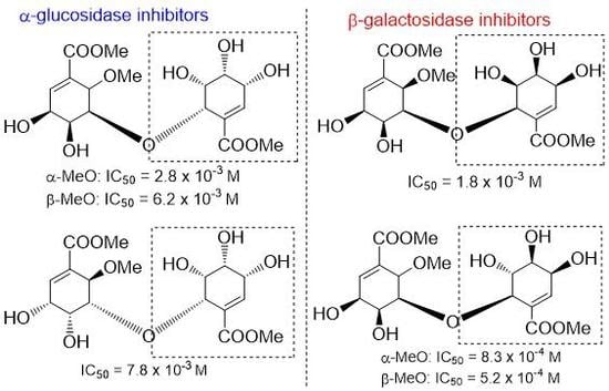
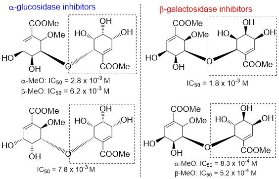
(+)-6:oil; +57.8 (c 0.37, EtOH); IR (film) νmax 3384 (OH), 1718 (C=O), 1653 (C=C) cm−1; 1H-NMR (acetone-d6, 400 MHz, ppm) δ 3.48 (3H, s, 6’-OMe), 3.75 (1H, overlapped, H-5’) 3.75 (3H, s, COOMe), 3.80 (3H, s, COOMe), 3.95(1H, br d, J = 3.1 Hz, H-4’), 4.03 (1H, br s, H-4’), 4.13 (1H, br d, J = 4.7 Hz, H-3), 4.22 (1H, overlapped, H-3’), 4.24 (1H, br d, J = 2.4 Hz, H-5), 4.35 (1H, d, J = 3.1 Hz, H-6), 4.52 (1H, d, J = 3.9 Hz, H-6’), 6.69 (1H, br s, H-2’), 6.88 (1H, d, J = 4.5 Hz, H-2); 13C-NMR (acetone-d6, 100 MHz, ppm) δ 52.1 (CH3, C-8), 52.4 (CH3, C-8’), 58.6 (CH3, 6’-OMe), 65.9 (CH, C-3), 68.0 (CH, C-4), 69.2 (CH, C-3’), 70.5 (CH, C-5’), 72.4 (CH, C-4’), 76.2 (CH, C-6), 76.8 (CH, C-6’), 82.7 (CH, C-5), 130.0 (Cq, C-1), 130.4 (Cq, C-1’), 141.7 (CH, C-2), 142.7 (CH, C-2’), 167.0 (Cq, C-7’), 167.5 (Cq, C-7); HRFABMS m/z calcd for C17H25O11Na (M + Na)+ 427.1216, found 427.1216.
(+)-14 (4.2 mg, 22%) was prepared from (−)-12 (27.0 mg, 0.048 mmol): white powder; +27.5 (c 0.095, EtOH); IR (film) νmax 3361 (OH), 1724 (C=O), 1657 (C=C) cm−1; 1H-NMR (acetone-d6, 600 MHz, ppm) δ 3.48 (3H, s, 6’-OMe), 3.73 (3H, s, COOMe), 3.76 (3H, s, COOMe), 3.88 (1H, ddd, J = 9.7, 5.0, 2.4 Hz, H-4), 3.94 (1H, br dd, J = 10.0, 4.7 Hz, H-4’), 4.04 (1H, ddd, J = 10.5, 5.3, 5.0 Hz, H-3), 4.18 (1H, d, J = 4.7 Hz, H-5), 4.22 (1H, br d, J = 2.9 Hz, H-6’), 4.25 (1H, d, J = 7.3 Hz, -OH), 4.35 (1H, d, J = 10.7 Hz, -OH), 4.40 (1H, d, J = 3.2 Hz, H-6), 4.42 (1H, br ddd, J = 7.3, 3.5, 3.2 Hz, H-3’), 4.47 (1H, ddd, J = 5.6, 3.8, 3.5 Hz, H-5’), 4.67 (1H, d, J = 3.8 Hz, -OH) 6.73 (1H, br d, J = 2.9 Hz, H-2’), 6.87 (1H, d, J = 4.7 Hz, H-2); 13C-NMR (acetone-d6, 150 MHz, ppm) δ 52.1 (2 × CH3, C8, C8’), 58.8 (CH3, 6’-OMe), 66.3 (CH, C-3), 66.7 (CH, C-3’), 67.2 (CH, C-4), 69.6 (CH, C-5’), 70.3 (CH, C-4’), 76.0 (CH, C-6), 76.8 (CH, C-6’),81.2 (CH, C-5), 130.4 (Cq, C-1’), 130.5 (Cq, C-1), 141.3 (CH, C-2), 141.5 (CH, C-2’), 167.0 (Cq, C-7’), 167.5 (Cq, C-7); HRFABMS m/z calcd for C17H25O11Na (M + Na)+ 427.1216, found 427.1216.
(−)-6 (8.4 mg, 58%) was prepared from (−)-13 (20.2 mg, 0.036 mmol): white powder; −61.5 (c 0.22, EtOH); IR (film) νmax 3392 (OH), 1718 (C=O), 1653 (C=C) cm−1; 1H-NMR (acetone-d6, 600 MHz, ppm) δ 3.47 (3H, s, 6’-OMe), 3.73 (1H, overlapped, H-5’) 3.74 (3H, s, COOMe), 3.80 (3H, s, COOMe), 3.94(1H, dd, J = 5.0, 1.8 Hz, H-4’), 4.03 (1H, br s, H-4’), 4.12 (1H, br dd, J = 5.0, 4.7 Hz, H-3), 4.21 (1H, overlapped, H-3’), 4.22 (1H, dd, J = 3.0, 2.3 Hz, H-5), 4.33 (1H, d, J = 3.2 Hz, H-6), 4.51 (1H, d, J = 4.1 Hz, H-6’), 6.69 (1H, d, J = 1.2 Hz, H-2’), 6.87 (1H, d, J = 4.7 Hz, H-2); 13C-NMR (acetone-d6, 150 MHz, ppm) δ 52.1 (CH3, C-8), 52.4 (CH3, C-8’), 58.6 (CH3, 6’-OMe), 66.0 (CH, C-3), 68.0 (CH, C-4), 69.2 (CH, C-3’), 70.5 (CH, C-5’), 72.4 (CH, C-4’), 76.2 (CH, C-6), 76.8 (CH, C-6’), 82.7 (CH, C-5), 130.1 (Cq, C-1), 130.5 (Cq, C1’), 141.7 (CH, C-2), 142.6 (CH, C-2’), 167.0 (Cq, C-7’), 167.5 (Cq, C-7); HRFABMS m/z calcd for C17H25O11 (M + H)+ 405.1397, found 405.1399.
(−)-14 (11.1 mg, 40%) was prepared from (+)-12 (38.4 mg, 0.068 mmol): white powder; −27.6 (c 0.13, EtOH); IR (film) νmax 3415 (OH), 1720 (C=O), 1655 (C=C) cm−1; 1H-NMR (acetone-d6, 600 MHz, ppm) δ 3.50 (3H, s, 6’-OMe), 3.74 (3H, s, COOMe), 3.78 (3H, s, COOMe), 3.92 (1H, dd, J = 5.0, 2.4 Hz, H-4), 3.96 (1H, br dd, J = 5.0, 4.7 Hz, H-4’), 4.08 (1H, dd, J = 5.0, 4.7 Hz, H-3), 4.15 (1H, dd, J = 2.9, 2.7 Hz, H-5), 4.22 (1H, d, J = 2.9 Hz, H-6’), 4.41 (1H, d, J = 3.2 Hz, H-6), 4.43 (1H, br dd, J = 3.3, 2.9 Hz, H-3’), 4.47 (1H, dd, J = 5.6, 3.2 Hz, H-5’), 6.75 (1H, d, J = 2.6 Hz, H-2’), 6.88 (1H, d, J = 4.7 Hz, H-2); 13C-NMR (acetone-d6, 150 MHz, ppm) δ 52.2 (CH3, C-8), 52.3 (CH3, C-8’), 58.8 (CH3, 6’-OMe), 66.1 (CH, C-3), 66.5 (CH, C-3’), 67.1 (CH, C-4), 69.2 (CH, C-5’), 70.0 (CH, C-4’), 76.0 (CH, C-6), 76.6 (CH, C-6’), 80.9 (CH, C-5), 130.3 (Cq, C-1’), 130.4 (Cq, C-1), 141.4 (CH, C-2), 141.7 (CH-C-2’), 167.2 (Cq, C-7’), 167.6 (Cq, C-7); HRFABMS m/z calcd for C17H25O11 (M + H)+ 405.1397, found 405.1391.
(−)-7 (6.2 mg, 54%) was prepared from (+)-17 (16.1 mg, 0.029 mmol): oil; −10.5 (c 0.10, EtOH); IR (film) νmax 3355 (OH), 1717 (C=O), 1652 (C=C) cm−1; 1H-NMR (acetone-d6, 400 MHz, ppm) δ 3.47 (3H, s, 6’-OMe), 3.77 (3H, s, COOMe), 3.79 (3H, s, COOMe), 3.82–3.86 (1H, m, H-5’), 3.88–4.04 (2H, m, OH, OH), 3.98–4.04 (1H, m, H-4’), 4.09 (1H, br d, J = 2.2 Hz, H-5), 4.18–4.28 (3H, m, OH, H-3’, H-3), 4.32 (1H, d, J = 3.7 Hz, H-6), 4.43–4.49 (1H, m, H-4), 4.67 (1H, d, J = 3.7 Hz, H-6’), 4.66–4.74 (1H, br, OH), 6.73 (1H, d, J = 2.2 Hz, H-2), 6.82 (1H, d, J = 2.0 Hz, H-2’); 13C-NMR (acetone-d6, 100 MHz, ppm) δ 51.6 (CH3, C-8), 51.7 (CH3, C-8’), 60.9 (CH3, 6’-OMe), 68.6 (CH, C-3), 68.9 (CH, C-3’), 69.1 (CH, C-5’), 70.2 (CH, C-4), 72.2 (CH, C-4’), 75.0 (CH, C-6’), 75.4 (CH, C-6), 79.6 (CH, C-5), 129.5 (CH, C-1),129.7 (CH, C-1’),141.4 (CH, C-2), 142.4 (CH, C-2’), 165.9 (Cq, C-7), 166.4 (Cq, C-7’); HREIMS m/z calcd for C17H25O11 (M + H)+ 405.1397, found 405.1395.
(−)-18 (5.0 mg, 41%) was prepared from (−)-16 (17.0 mg, 0.030 mmol): oil; Rf = 0.16 (MeOH:CH2Cl2 = 1:9 v/v); −21.6 (c 0.275, EtOH); IR (film) νmax 3357 (OH), 1718 (C=O), 1657 (C=C) cm−1; 1H-NMR (acetone-d6, 400 MHz, ppm) δ 3.31 (1H, d, J = 5.3 Hz, -OH), 3.49 (3H, s, 6’-OMe), 3.70–3.78 (1H, m, H-4’), 3.777 (3H, s, COOMe), 3.779 (3H, s, COOMe), 3.82–3.88 (2H, m, -OH, -OH), 3.99 (1H, dd, J = 3.7, 2.2 Hz, H-5), 4.07 (1H, d, J = 7.0 Hz, -OH), 4.15 (1H, d, J = 8.0 Hz, -OH), 4.20–4.28 (2H, overlapped, H-5’, H-3), 4.32–4.40 (3H, overlapped, H-6, H-6’, H-3’), 4.42–4.48 (1H, m, H-4), 4.56 (1H, d, J = 3.3 Hz, OH) 6.74 (1H, dd, J = 2.4, 1.4 Hz, H-2), 6.77 (1H, d, J = 3.3 Hz, H-2’); 13C-NMR (acetone-d6, 100 MHz, ppm) δ 51.56 (CH3, C-8), 51.61 (CH3, C-8’), 61.1 (CH3, 6’-OMe), 65.9 (CH2, C-3’), 69.1 (CH2, C-3), 70.3 (CH2, C-4), 70.4 (CH2, C-4’), 71.0 (CH2, C-5’), 75.5 (CH2, C-6), 77.7 (CH, C-6’), 79.4 (CH, C-5), 129.2 (CH, C-1), 131.8 (CH, C-1’), 139.1 (CH, C-2’), 141.8 (CH, C-2), 165.8 (Cq, C-7), 166.8 (Cq, C-7’); HREIMS m/z calcd for C17H25O11 (M + H)+ 405.1397, found 405.1399.
(+)-7 (6.8 mg, 52%) was prepared from (−)-17 (20.1 mg, 0.036 mmol): oil; +11.4 (c 0.11, EtOH); IR (film) νmax 3359 (OH), 1717 (C=O), 1652 (C=C) cm−1; 1H-NMR (acetone-d6, 400 MHz, ppm) δ 3.47 (3H, s, 6’-OMe), 3.77 (3H, s, COOMe), 3.79 (3H, s, COOMe), 3.84 (1H, ddd, J = 8.2, 4.1, 2.2 Hz, H-5’), 3.89 (1H, d, J = 7.4 Hz, -OH), 3.94 (1H, d, J = 9.7 Hz, OH), 4.00 (1H, d, J = 9.8 Hz, -OH), 3.98–4.04 (1H, m, H-4’), 4.09 (1H, dd, J = 3.8, 2.3 Hz, H-5), 4.19 (1H, d, J = 8.8 Hz, -OH), 4.22–4.28 (2H, m, H-3’ and H-3), 4.32 (1H, d, J = 4.0 Hz, H-6), 4.43–4.49 (1H, m, H-4), 4.68 (1H, d, J = 8.3 Hz, -OH), 4.67 (1H, d, J = 4.1 Hz, H-6’), 6.73 (1H, dd, J = 2.6, 4.3 Hz, H-2), 6.83 (1H, dd, J = 2.5, 1.3 Hz, H-2’); 13C-NMR (acetone-d6, 100 MHz, ppm) δ 51.6 (CH3, C-8), 51.7 (CH3, C-8’), 60.9 (CH3, 6’-OMe), 68.6 (CH, C-3), 68.9 (CH, C-3’), 69.1 (CH, C-5’), 70.3 (CH, C-4), 72.2 (CH, C-4’), 75.0 (CH, C-6’), 75.4 (CH, C-6), 79.6 (CH, C-5), 129.5 (Cq, C-1), 129.7 (Cq, C-1’), 141.3 (CH, C-2), 142.4 (CH, C-2’), 165.9 (Cq, C-7), 166.4 (Cq, C-7’); HREIMS m/z calcd for C17H24O11 (M)+ 404.1319, found 404.1315.
(+)-18 (7.1 mg, 62%) was prepared from (+)-16 (16.1 mg, 0.029 mmol): oil; +24.3 (c 0.36, EtOH); IR (liquid film) νmax 3361 (OH), 1718 (C=O), 1655 (C=C) cm−1; 1H-NMR (acetone-d6, 400 MHz, ppm) δ 3.31 (1H, d, J = 5.1 Hz, -OH), 3.49 (3H, s, 6’-OMe), 3.70–3.78 (1H, m, H-4’), 3.776 (3H, s, COOMe), 3.778 (3H, s, COOMe), 3.86 (1H, br d, J = 9.5 Hz, -OH), 3.99 (1H, dd, J = 3.7, 2.0 Hz, H-5), 4.07 (1H, br s, -OH), 4.14 (1H, br d, J = 8.2 Hz, -OH), 4.20–4.28 (2H, overlapped, H-5’, H-3), 4.32–4.40 (3H, overlapped, H-6, H-6’, H-3’), 4.42–4.48 (1H, m, H-4), 4.55 (1H, br s, -OH) 6.74 (1H, dd, J = 2.2, 1.4 Hz, H-2), 6.77 (1H, br d, J = 33 Hz, H-2’); 13C-NMR (acetone-d6, 100 MHz, ppm) δ 51.56 (CH3, C-8), 51.61 (CH3, C-8’), 61.1 (CH3, 6’-OMe), 65.9 (CH2, C-3’), 69.1 (CH2, C-3), 70.4 (2 × CH, C-4, C-4’), 71.0 (CH, C-5’), 75.5 (CH, C-6), 77.7 (CH, C-6’), 79.4 (CH, C-5), 129.2 (Cq, C-1), 131.8 (Cq, C-1’), 139.1 (CH, C-2’), 141.8 (CH, C-2), 165.8 (Cq, C-7), 166.8 (Cq, C-7’); HREIMS m/z calcd for C17H24O11 (M)+ 404.1319, found 404.1317.
(−)-8 (10.3 mg, 45%) was prepared from (−)-20 (31.8 mg, 0.056 mmol): oil; −96.7 (c 0.48, EtOH); IR (film) νmax 3419 (OH), 1728 (C=O), 1653 (C=C) cm−1; 1H-NMR (acetone-d6, 600 MHz, ppm) δ 3.32 (3H, s, 6-OMe), 3.59–3.78 (1H, m, H-5’), 3.76 (3H, s, COOMe), 3.81 (3H, s, COOMe), 3.96–3.99 (2H, m, H-4, H-4’), 4.13 (1H, br t, J = 4.1 Hz, H-3), 4.33 (1H, dd, J = 4.1, 1.8 Hz, H-5), 4.54 (1H, d, J = 4.1 Hz, H-6), 4.58 (1H, J = 4.2 Hz, H-6’), 6.83 (1H, dd, J = 2.3, 1.5 Hz, H-2’), 6.85 (1H, d, J = 4.1 Hz, H-2); 13C-NMR (acetone-d6, 150 MHz, ppm) δ 52.1 (CH3, COOMe), 52.5 (CH3, COOMe), 59.0 (CH3, -OMe), 66.7 (CH, C-3), 67.8 (CH, C-4), 69.1 (CH, C-3’), 69.7 (CH, C-5’), 72.3 (CH, C-4’), 73.4 (CH, C-6’), 76.5 (CH, C-6), 80.6 (CH, C-5), 130.1 (Cq, C-1’), 130.9 (Cq, C-1), 141.7 (CH, C-2), 143.3 (CH, C-2’), 167.4 (Cq, C-7), 167.5 (Cq, C-7); HRMS m/z calcd for C17H25O11 (M + H)+ 405.1397, found 405.1390.
(−)-21 (10.3 mg, 45%) was prepared from (−)-20 (31.8 mg, 0.056 mmol): oil; −215.4 (c 0.35, EtOH); IR (film) νmax 3393 (OH), 1716 (C=O), 1653 (C=C) cm−1; 1H-NMR (acetone-d6, 600 MHz, ppm) δ 3.35 (3H, s, 6-OMe), 3.75 (1H, dd, J = 6.7, 3.5 Hz, H-4’), 3.77 (3H, s, COOMe), 3.780 (3H, s, COOMe), 4.00 (2H, dd, J = 4.7, 2.0 Hz, H-4), 4.08 (1H, dd, J = 6.7, 4.1 Hz, H-5’), 4.14 (1H, dd, J = 4.7, 4.1 Hz, H-3), 4.28 (1H, dd, J = 4.1, 2,1 Hz, H-5), 4.35 (1H, d, J = 3.8 Hz, H-6’), 4.37 (1H, t, J = 3.8 Hz, H-3’), 4.42 (1H, br d, J = 4.1 Hz, H-6’), 4.35 (1H, J = 3.8 Hz, H-6’), 4.42 (1H, d, J = 3.8 Hz, H-6), 4.35 (1H, J = 3.8 Hz, H-6’), 4.64 (1H, d, J = 7.1 Hz, -OH), 6.81 (1H, d, J = 3.5 Hz, H-2’), 6.83 (1H, d, J = 4.1 Hz, H-2); 13C-NMR (acetone-d6, 150 MHz, ppm) δ 52.2 (CH3, COOMe), 52.4 (CH3, COOMe), 59.2 (CH3, -OMe), 66.4 (CH, C-3’), 66.9 (CH, C-3), 67.9 (CH, C-4), 70.7 (CH, C-4’), 71.8 (CH, C-5’), 76.66 (CH, C-6’), 76.70 (CH, C-6), 81.1 (CH, C-5), 130.8 (Cq, C-1), 131.1 (Cq, C-1’), 141.1 (CH, C-2’), 141.5 (CH, C-2), 167.6 (Cq, C-7), 167.9 (Cq, C-7’); HRMS m/z calcd for C17H25O11 (M + H)+ 405.1397, found 405.1393.
(+)-8 (10.5 mg, 33%) was prepared from (+)-20 (44.6 mg, 0.079 mmol): oil; +86.9 (c 0.43, EtOH); IR (film) νmax 3383 (OH), 1716 (C=O), 1652 (C=C) cm−1; 1H-NMR (acetone-d6, 400 MHz, ppm) δ 3.51 (3H, s, -OMe), 3.76 (3H, s, COOMe), 3.80 (3H, s, COOMe), 3.95–4.00 (2H, m, H-4, H-4’), 4.10–4.14 (2H, m), 4.24–4.28 (1H, m), 4.32 (1H, dd, J = 4.3, 2.0 Hz, H-5), 4.52 (1H, d, J = 4.3 Hz, H-6), 4.59 (1H, d, J = 4.3 Hz, H-6’), 6.81 (1H, d, J = 1.6 Hz, H-2’), 6.84 (1H, d, J = 4.3 Hz, H-2); 13C-NMR (acetone-d6, 100 MHz, ppm) δ 50.7, 51.1, 57.6, 65.3, 66.4, 67.7, 68.3, 70.9, 72.0, 75.1, 79.3, 128.7, 129.4, 140.3, 142.0, 165.9, 166.1; HREIMS m/z calcd for C17H25O11 (M + H)+ 405.1397, found 405.1393.
(+)-21 (9.0 mg, 32%) was prepared from (+)-19 (40.0 mg, 0.07 mmol): oil; +211 (c 0.095, EtOH); IR (film) νmax 3419 (OH), 1716 (C=O), 1653 (C=C) cm−1; 1H-NMR (acetone-d6, 400 MHz, ppm) δ 3.49 (3H, s, 6-OMe), 3.76 (3H, s, COOMe), 3.79 (3H, s, COOMe), 3.83 (1H, br d, J = 7.8 Hz) 3.90–3.39 (2H, m), 4.04–4.14 (2H, m), 4.19–4.24 (1H, m), 4.28 (1H, dd, J = 3.9, 2.9 Hz), 4.36 (1H, d, J = 4.1 Hz), 4.36–4.39 (1H, m), 4.41 (1H, d, J = 3.9 Hz, H-6’), 4.53–4.57 (1H, m), 6.80 (1H, d, J = 3.3 Hz), 6.82 (1H, d, J = 4.3 Hz); 13C-NMR (acetone-d6, 100 MHz, ppm) 52.2, 52.4, 59.2, 66.4, 66.9, 67.8, 70.7, 71.8, 76.6, 76.7, 81.1, 130.7, 131.1, 141.0, 141.1, 167.5, 167.9 HREIMS m/z calcd for C17H25O11 (M)+ 405.1396, found 405.1390.
(−)-24 (1.9 mg, 26%) was prepared from (−)-22 (10.2 mg, 0.018 mmol): oil; −20.0 (c 0.05, EtOH); IR (film) νmax 3403 (OH), 1700 (C=O), 1653 (C=C) cm−1; 1H-NMR (acetone-d6, 600 MHz, ppm) δ 3.56 (3H, s, OMe), 3.70 (1H, dt, J = 7.0, 4.4 Hz, H-4’), 3.72 (1H, d, J = 10.2 Hz, -OH), 3.77 (3H, s, COOMe), 3.78 (3H, s, COOMe), 3.92 (1H, d, J = 6.5 Hz, -OH), 3.94 (1H, d, J = 7.9 Hz, -OH), 4.04 (1H, dd, J = 3.8, 2.1 Hz, H-6), 4.10–4.14 (1H, m, H-3), 4.14 (1H, d, J = 7.1 Hz, OH), 4.23 (1H, br dt, J = 7.9, 4.4 Hz, H-5’), 4.35 (1H, br dt, J = 7.0, 4.1 Hz, H-3’), 4.38 (1H, d, J = 3.8 Hz, 3’-OH), 4.40 (1H, d, J = 4.7 Hz, H-6’), 4.53 (1H, d, J = 3.8 Hz, H-6), 6.70 (1H, dd, J = 2.6, 1.1 Hz, H-2), 6.72 (1H, dd, J = 4.1, 0.9 Hz, H-2’); 13C-NMR (acetone-d6, 150 MHz, ppm) 52.1 (CH3, -COOMe), 52.2 (CH3, -COOMe), 61.1(CH3, -OMe), 66.6 (CH, C-3’), 69.5 (CH, C-3), 70.2 (CH, C-4), 71.1 (CH, C-4’), 71.9 (CH, C-5’), 76.7 (CH, C-6), 78.0 (CH, C-6’), 79.0 (CH, C-5), 130.6 (Cq, C-1), 132.7 (Cq, C-1’), 139.3 (CH, C-2’), 141.8 (CH, C-2), 166.8 (Cq, C-7), 167.6 (Cq, C-7’); HREIMS m/z calcd for C17H24O11 M+ 404.1318, found 404.1311.
(−)-9 (2.8 mg, 43%) was prepared from (+)-23 (9.1 mg, 0.016 mmol): oil; −49.0 (c 0.085, CHCl3); IR (film) νmax 3420 (OH), 1719 (C=O), 1655 (C=C), 1507 (C=C), 1458 (C=C) cm−1; 1H-NMR (CDCl3, 600 MHz, ppm) δ 3.56 (3H, s, OMe), 3.81–3.85 (2H, m, overlapped), 3.819 (3H, s, COOMe), 3.824 (3H, s, COOMe), 3.93 (1H, br s, -OH), 3.94 (1H, d, J = 2.6 Hz, -OH), 4.02–4.04 (1H, m), 4.14–4.17 (1H, m), 4.22–4.23 (1H, m), 4.25 (1H, dd, J = 3.5, 2.1 Hz), 4.35 (1H, m, OH), 4.51 (1H, d, J = 3.5 Hz), 4.63 (1H, d, J = 4.4 Hz), 6.88 (1H, d, J = 4.1 Hz), 6.94 (1H, dd, J = 3.2, 0.9 Hz); 13C-NMR (CDCl3, 150 MHz, ppm) 52.3, 52.5, 59.2, 67.3, 67.8, 69.0, 69.2, 70.5, 73.4, 74.9, 78.7, 129.4, 129.9, 140.2, 141.3, 166.4, 166.7; HREIMS m/z calcd for C17H22O10 (M − H2O)+ 386.1213, found 386.1210.
(+)-24 (5.3 mg, 38%) was prepared from (+)-22 (14.1 mg, 0.023 mmol): oil; +20.7 (c 0.29, EtOH); IR (film) νmax 3400 (OH), 1702 (C=O), 1652 (C=C) cm−1; 1H-NMR (acetone-d6, 400 MHz, ppm) δ 3.56 (3H, s, OMe), 3.71 (1H, dd, J = 7.4, 4.1 Hz, H-4’), 3.77 (3H, s, COOMe), 3.78 (3H, s, COOMe), 4.07 (1H, dd, J = 3.7, 1.8 Hz, H-6), 4.14 (1H, br s, -OH), 4.18 (1H, br s, -OH), 4.23 (1H, dd, J = 7.4, 4.8 Hz, H-5’), 4.35 (1H, br t, J = 4.1 Hz, H-3’), 4.40 (1H, d, J = 4.8 Hz, H-6’), 4.53 (1H, d, J = 3.7 Hz, H-6), 6.70 (1H, dd, J = 2.6, 1.0 Hz, H-2), 6.72 (1H, br d, J = 3.9 Hz, H-2’); 13C-NMR (acetone-d6, 100 MHz, ppm) 52.07, 52.13, 61.1, 66.5, 69.5, 70.1, 71.0, 71.8, 76.5, 78.0, 79.0, 130.5, 132.6, 139.3, 141.8, 166.7, 167.6; HREIMS m/z calcd for C17H22O10 (M − H2O)+ 386.1213, found 386.1209.
(+)-9 (5.5 mg, 73%) was prepared from (−)-23 (10.7 mg, 0.019 mmol): oil; +49.5 (c 0.17, CHCl3); IR (film) νmax 3410 (OH), 1719 (C=O), 1655 (C=C), 1506 (C=C), 1456 (C=C) cm−1; 1H-NMR (CDCl3, 400 MHz, ppm) δ 3.58 (3H, s, OMe), 3.78–3.84 (2H, m, overlapped), 3.82 (3H, s, COOMe), 3.83 (3H, s, COOMe), 3.90–3.96 (1H, m), 4.40–4.18 (1H, m), 4.12–4.18 (1H, m), 4.20–4.26 (1H, m), 4.29 (1H, dd, J = 3.3, 1.8 Hz), 4.44 (1H, br s, OH), 4.47 (1H, d, J = 3.7 Hz), 4.63 (1H, d, J = 4.1 Hz), 6.87 (1H, d, J = 3.9 Hz), 6.93 (1H, br d, J = 2.9 Hz,); 13C-NMR (CDCl3, 100 MHz, ppm) 52.3, 52.5, 59.2, 67.1, 67.9, 68.8, 69.2, 70.5, 74.6, 75.1, 78.5, 129.4, 130.1, 139.7, 141.4, 166.6, 166.7; HREIMS m/z calcd for C17H25O11 (M + H)+ 405.1396, found 405.1395.
4.5. Assays of Glycosidase Inhibitory Activity
Assay of α-Glucosidase Inhibitory Activity
The assay reaction mixture comprised 0.1 M acetate buffer (pH 5.0, 45 μL), 20 mM
p-nitrophenyl-α-
d-glucopyranoside solution (25 μL), and α-glucosidase solution (25 μL, stock solution of 1.0 mg/mL in 50 mM Tris-HCl-buffer at pH 7.8 diluted 20-fold with 10 mM phosphate buffer at pH 7.0), with the test samples or DNJ (5 μL solution, concentration range 0.1–20 mg/mL). After 20 min incubation at 37 °C, the reaction was quenched by addition of Na
2CO
3 solution (0.5 M, 100 μL). The amount of liberated
p-nitrophenol was measured colorimetrically at 400 nm (optical density at 400 nm: ODtest). Inhibition efficiencies (%) were calculated as 100 − 100 × (ODtest − ODblank)/(control ODtest − control ODblank), and IC
50 values (
Table 1) were obtained from inhibition curves.
Assays of β-glucosidase, α-mannosidase, β-mannosidase, and β-galactosidase inhibitory activities were carried out as above using
p-nitrophenyl-β-
d-glucopyranoside,
p-nitrophenyl-α-
d-mannopyranoside,
p-nitrophenyl-β-
d-mannopyranoside, and
p-nitrophenyl-β-
d-galactopyranoside as substrates. The corresponding IC
50 values are listed in
Table 1.
*Isolation of positive control deoxynojirimycin: Dried leaves of Morus alba L. (0.5 kg) were cut finely and then extracted with hot water (10 L) for 2 h. The extracted solution was chromatographed on an Amberlite CG-50 (H+-form) column (6.5 mm inside diameter (i.d.) × 30 cm length). After washing the column with water and then 50 % MeOH, the adsorbed material was eluted with 50 % MeOH-28 % ammonia solution (9:1). The eluted fraction was concentrated in vacuo to give a basic fraction (5.0 g). This fraction was chromatographed on a Dowex 50W-X4 column (200–400 mesh, 5.0 mm i.d. × 20 cm ) pretreated with formic acid-ammonium formate buffer (0.2 M ammonia formate, adjusted to pH 5.7 with 1 M formic acid), with stepwise elution (H2O, H2O-28% ammonia solution (99:1, 9:1)). The fraction (H2O-28 % ammonia solution (99:1)) was re-chromatographed on semi-preparative HPLC (column:Shodex NH2P (4.6 mm i.d. × 250 mm), solvent:CH3CN-H2O (80:20), flow rate: 1.0 mL/min, column temperature: ambient). 1-Deoxynojirimycin (40 mg) was finally obtained.
4.6. Docking Simulation
The docking analysis was carried out using α-glucosidase protein (PDB code 3A4A) using the Dock induced-fit function in Molecular Operating Environment (MOE) version 2018.0101 (Chemical Computing Group Inc., Quebec, Canada) to better understand the inhibitory mechanisms. [
18,
19] The calculation of the binding affinity scoring function was performed with the amber 10:eht force field, triangle matcher as placement, and GBVI/WSA dG as the binding affinity scoring function [
26]. In the protein preparation with respect to charged residues in the binding site, the Protonate three dimensional (3D) option in MOE was used to determine the ionization states and add hydrogens to the system [
27]. The function of the Protonate 3D allows to assign ionization states and position hydrogens in a macromolecular structure given its 3D coordinates from the crystal structure. Hydrogen atoms are required for all atom molecular mechanics, dynamics, or electrostatic calculations. The addition of hydrogen atoms to a macromolecule is a non-trivial task; generally, one must determine the rotamers of -SH -OH -CH
3 and -NH
3 groups in cysteine (CYS), serine (SER), tyrosine (TYR), threonine(THR), methionine (MET), and lysine (LYS), the ionization states of acids and bases in arginine (ARG), aspartic acid (ASP), glutamic acid (GLU), LYS, histidine (HIS), the tautomers of imidazoles (HIS) and carboxylic acids (ASP, GLU), the protonation state of metal-ligand atoms CYS, HIS, ASP, GLU, etc., and the ionization state of metals.

 ,
, 


