Review

Occurrence and Properties of Thiosilvatins

by
Maria Michela Salvatore
1,
Rosario Nicoletti
2,3,
Marina DellaGreca
1 and
Anna Andolfi
1,*
1
Department of Chemical Sciences, University of Naples ‘Federico II’, 80126 Naples, Italy
2
Council for Agricultural Research and Economics, Research Centre for Olive, Citrus and Tree Fruit, 81100 Caserta, Italy
3
Department of Agriculture, University of Naples ‘Federico II’, 80055 Portici, Italy
*
Author to whom correspondence should be addressed.
Mar. Drugs 2019, 17(12), 664; https://doi.org/10.3390/md17120664
Submission received: 27 September 2019 / Revised: 19 November 2019 / Accepted: 21 November 2019 / Published: 26 November 2019
(This article belongs to the Special Issue Metabolomic Approach to Investigate Marine Fungi for Drug Discovery)

Abstract

:
The spread of studies on biodiversity in different environmental contexts is particularly fruitful for natural product discovery, with the finding of novel secondary metabolites and structural models, which are sometimes specific to certain organisms. Within the large class of the epipolythiodioxopiperazines, which are typical of fungi, thiosilvatins represent a homogeneous family that, so far, has been reported in low frequency in both marine and terrestrial contexts. However, recent observations indicate that these compounds have been possibly neglected in the metabolomic characterization of fungi, particularly from marine sources. Aspects concerning occurrence, bioactivities, structural, and biosynthetic properties of thiosilvatins are reviewed in this paper.

1. Introduction

A huge chapter on research on biodiversity is represented by studies concerning the biochemical properties of the manifold organisms which are part of natural ecosystems. Novel secondary metabolites are continuously discovered, disclosing a surprising chemodiversity in terms of both structural and biosynthetic aspects. Although most classes of compounds are spread throughout the several kingdoms of nature, a certain specificity results in some cases.
An example in this respect is represented by the epipolythiodioxopiperazines (ETPs), so far only reported from fungi [1]. ETPs are a large and structurally diverse class of bioactive secondary metabolites originating from diketopiperazines and characterized by the presence of a disulfide bridge or a polysulfide dioxopiperazine six-membered ring. Due to their bioactivities, ETPs are receiving attention in recent years [2].
This review is focused on thiosilvatins, a specific family of ETPs resulting from the enzymatic assemblage of two amino acids (i.e., l-tyrosine/l-phenylalanine and glycine), generally integrated with two methylated sulfur atoms. Unlike a related family including hyalodendrin, gliovictin, and their analogues, in this homogeneous group of compounds, the pivotal nitrogen deriving from the aromatic amino acid is not engaged in structural modifications other than methylation (Figure 1).
The finding of natural products displaying this kind of molecular structure only started in the 1980s [3], and even nowadays reports are quite infrequent. The present paper provides a review of the current knowledge concerning occurrence, bioactivities, structural, and biosynthetic aspects of these compounds. To ensure a comparative examination of the several structures reviewed herewith, the name of some compounds was adapted to conform to previously characterized analogs.

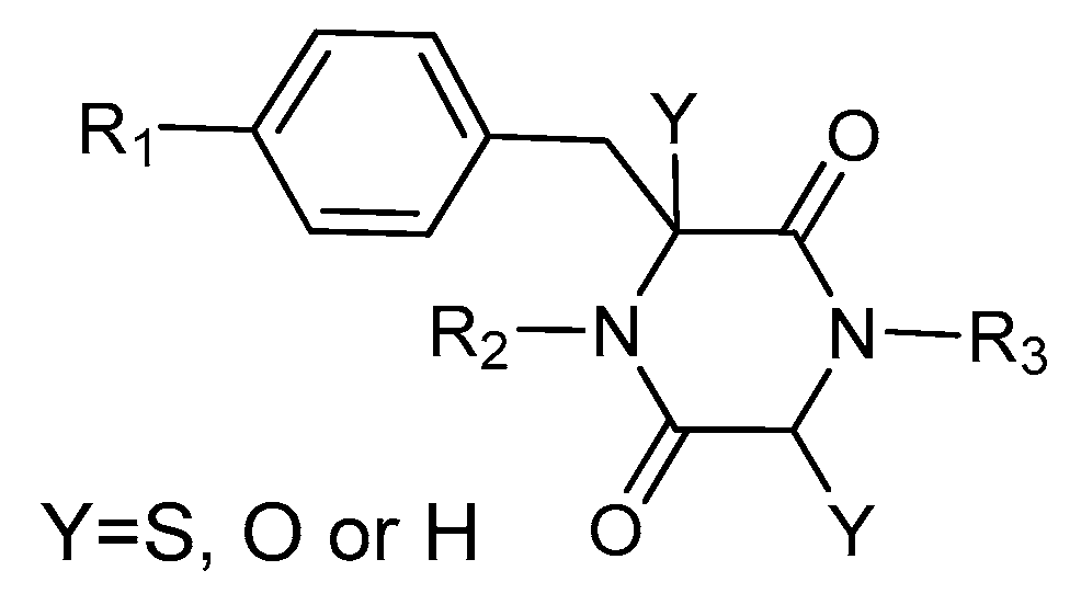
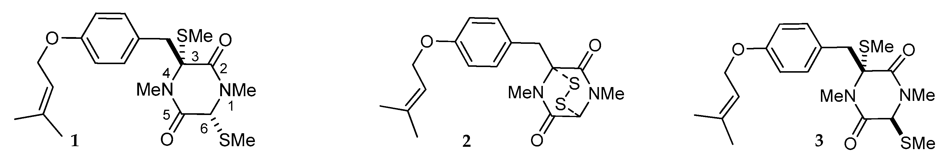
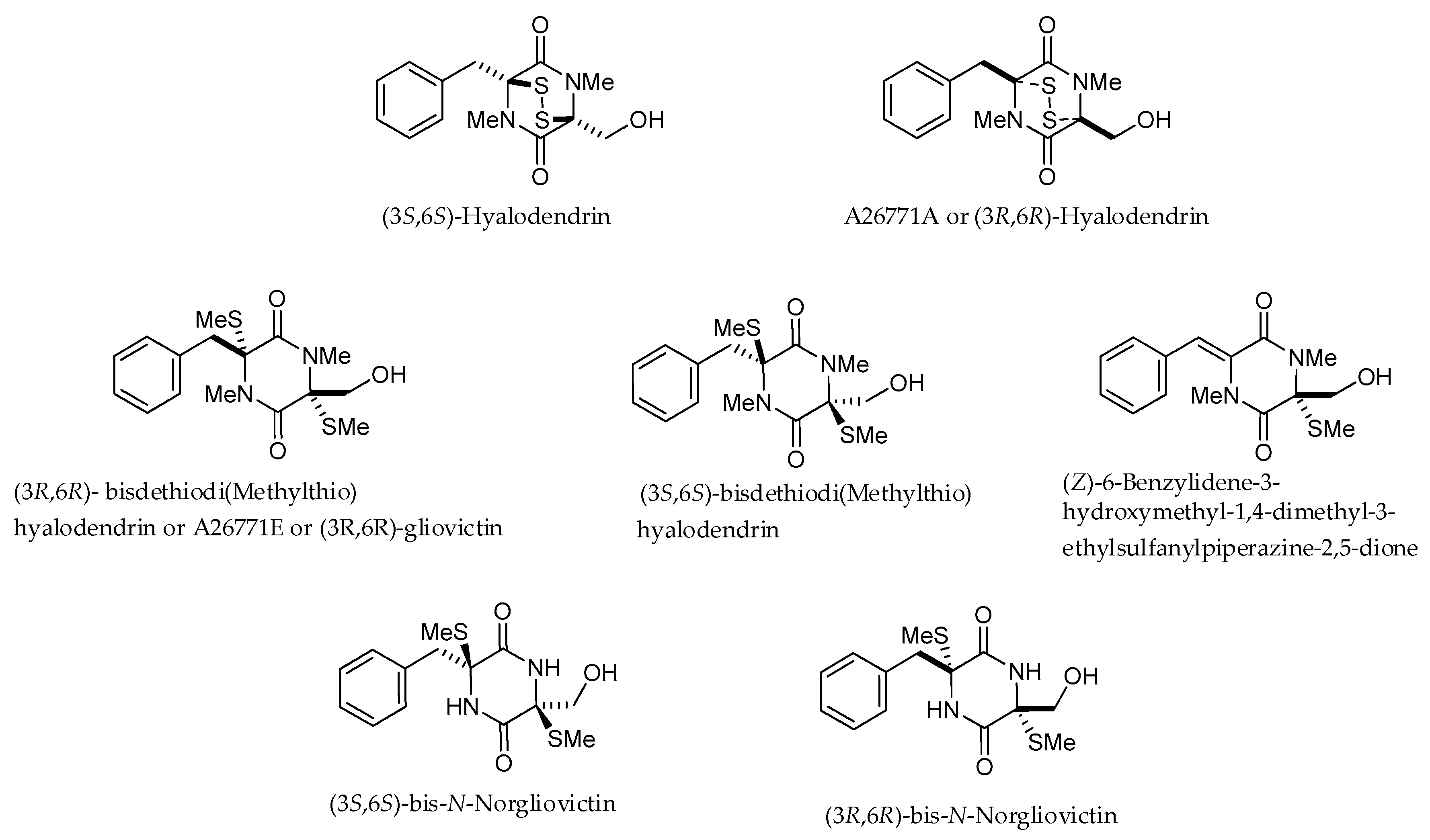
2. Structures and Chemical Properties
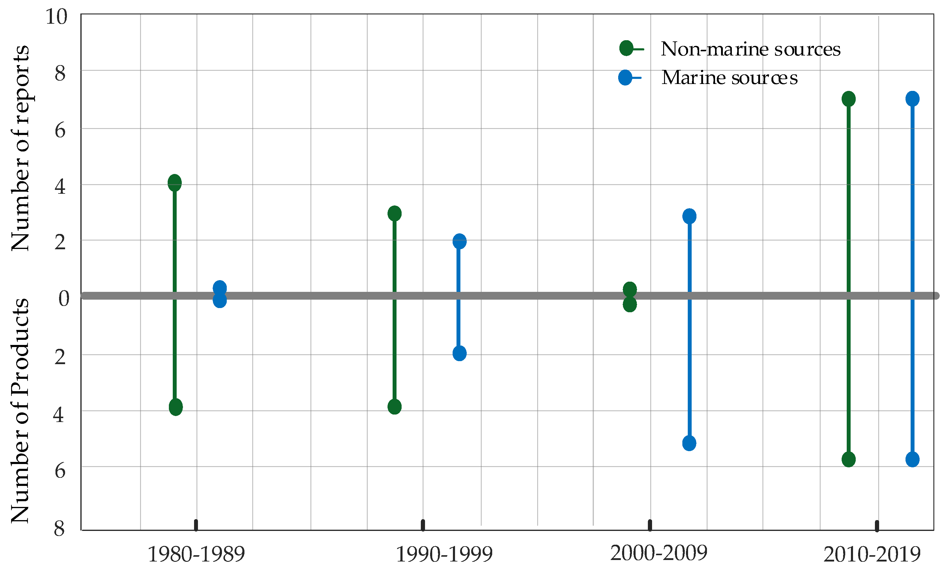
(3R,6R)-1,4-Dimethyl-3-(4-(3-methyl-2-butenyloxy)benzyl)-3,6-bis(methylthio)piperazine-2,5-dione (1), the founder product of this compound series, was isolated for the first time in 1981 along with its deprenyl analogue (16) [3]. Subsequently, 1 was named cis-bis(methylthio)silvatin when the only sulfur bridged thiosilvatin, dithiosilvatin (2), was characterized and submitted to a reductive methylation giving 1 and its epimer in C-6 (3) [4] (Figure 2).
The most relevant structural modifications observed in this class of compounds are in the number of sulfurs, the degree of methylation of heteroatoms, and the presence of a dimethyl allylic chain (Table 1). A controversial issue concerning thiosilvatins could be represented by nomenclature. In fact, several compounds were inconsistently designated with trivial names, abbreviations, or according to the IUPAC standards, which generally are not linked to the name of the founder compound. For instance, Sch 54794 (4) and Sch 54796 (5) have the same structures and stereostructures of 1 and 3, but their amino functions are not methylated; consequently, they could be respectively named cis- and trans-dinor-bis(methylthio)silvatin [5]. Likewise, cis-3-(4-hydroxybenzyl)-1,4-dimethyl-3,6-bis(methylthio)-2,5-piperazinedione (16) and trans-6-(4-hydroxybenzyl)-1,4-dimethyl-3,6-bis(methylthio)piperazine-2,5-dione (17) were named in the original manuscripts according to IUPAC standards [3,6], but they could be easily named respectively cis- and trans-deprenyl-bis(methylthio)silvatin.
Many difficulties in nomenclature derive from the stereostructural aspects. Concerning compounds 2527, they were not named in the original manuscript [7]; however, a trivial name can be assigned according to their structurally related compound bilain B. However, the absence of a complete stereostructure determination prevents clarifying if compound 25 is actually its epimer or rather its diasteroisomer.
In recent years, names of some new compounds, such as saroclazines, fusaperazines, and bilains, were assigned considering their sources, rather than referring to their structural relationships. For the reasons explained above, in Table 1 some compounds have also been renamed according to the founder compound.
In general, the biosynthesis of secondary metabolites is stereospecific. In fact, the stereochemistry of chiral carbons in the dioxopiperazinic ring of thiosilvatins is essentially 3R,6R, even if these compounds display some structural differences. This is observed in strains belonging to unrelated species, such as Fusarium chlamydosporum [8], Penicillium waksmanii [9], Penicillium brevicompactum [10], Trichoderma virens [3,11], all producing thiosilvatins with the same stereostructure [i.e., cis-bis(methylthio)silvatin (1) and its deprenyl analogue (16)] (Table 1, Table 2 and Table 3).
On the other hand, this is not a common trend in compounds belonging to the ETPs class [2]. In fact, both stereoisomers (i.e., 3R,6R and 3S,6R) were reported for compounds in the hyalodendrin/gliovictin family, deriving from the amino acids l-phenylalanine and l-serine [12,13,14] (Figure 3). Interestingly, this family also includes a compound named vertihemiptellide A, representing the first dimer resulting from the formation of disulfide bridges between two hyalodendrin units [15].

3. Fungal Sources

As introduced above, so far ETPs have been only reported from fungi. More specifically, thiosilvatins have been detected as secondary metabolites of 22 strains belonging to 17 taxa that occupy different geographic and climatic zones, terrestrial and marine habitats, and are associated with different substrates/hosts (Table 2 and Table 3). With the exception of a single taxon in the Basidiomycota, that is Coriolus (=Irpex) consors, all the other strains are representative of taxa in the Ascomycota. Particularly, they belong to the Sordariomycetes (order Hypocreales, 7 strains/6 taxa; order Xylariales, 1 strain/taxon), and to the Eurotiomycetes (14 strains/9 taxa, all of them in the Eurotiales). Twelve strains, that is more than half of the total number, belong to the genus Penicillium, well known for its widespread occurrence in every ecological context including the sea [16]. The species Penicillium crustosum and T. virens include strains from both kind of sources.
With reference to the specific compounds, 1 undoubtedly represents the most common product of this family, having been reported as a secondary metabolite of about 2/3 of the strains, while its trans stereoisomer has been detected in just four of these strains, both marine and terrestrial. Compounds 4, 5, 12, 14 and 16 were also obtained from strains from both environments. Among the rest, compounds 6, 7, 11, 15 and 1724 have been reported from just a single strain of marine origin, while compounds 2, 810, 13, and 2527 have been only found in terrestrial strains. These data could be indicative of a relatively higher chemodiversity characterizing marine strains, also considering that reports from marine sources only started in 1998 when there were already four strains and eight products known from terrestrial sources (Figure 4). Since 1998, the new products discovered from marine fungi more than doubled those obtained from non-marine strains. Moreover, in the last two years there were five reports concerning new thiosilvatins-producing strains from marine sources compared to two from terrestrial sources, which might imply that a more widespread occurrence at sea is likely to be disclosed as investigations concerning marine fungal strains progress. Finally, no comparison can be made between strains of the same species (P. crustosum and T. virens) obtained from both marine and terrestrial sources, whose secondary metabolite profiles do not match, or share single compounds. This could be interpreted not only in terms of intraspecific variation, but also as a consequence of the different culturing and extraction procedures. Moreover, it must also be considered that detection of some compounds is often impaired by their presence in low quantities, or by inherent difficulties in the identification depending on their infrequent occurrence. However, the finding of two species from both marine and terrestrial sources within such a limited strain sample supports a recently-consolidated inference that most fungal species are able to thrive in different environmental conditions, obliterating the old misconception that the occurrence of specialized taxa occurs in either marine or non-marine contexts [16,31,32].

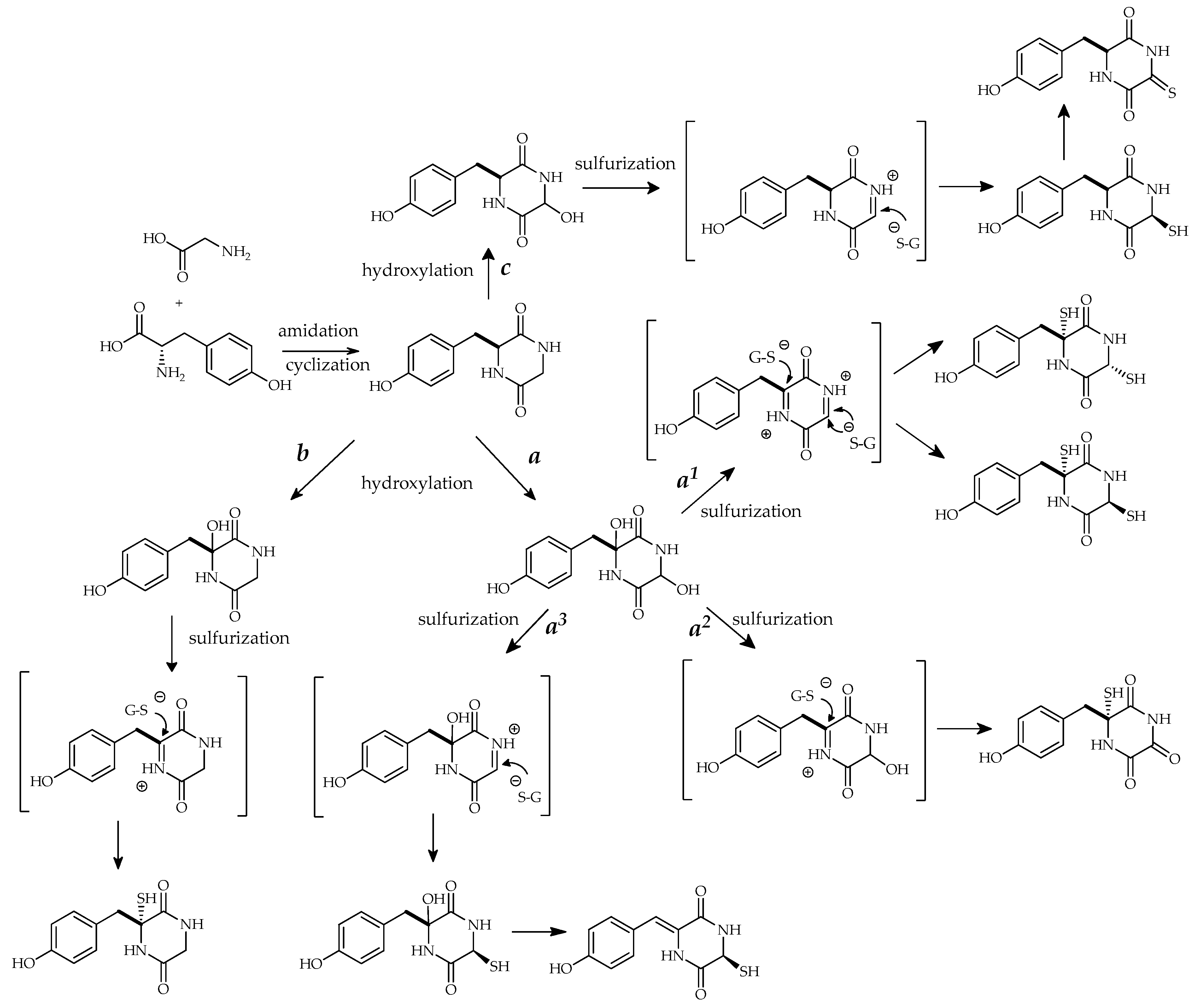
4. Proposed Biosynthetic Pathways for Thiosilvatins

The biosynthesis of ETPs involves non-ribosomal peptide synthetases (NRPSs), multi-domain enzymes controlling all activities required to incorporate constituents into their products, and a range of associated enzymes [33,34]. In fact, the non-ribosomal pathway is frequently used by microorganisms to produce a wide range of structurally diverse secondary metabolites [35].
In general, the genes that encode enzymes for secondary metabolite biosynthesis are clustered in the fungal genome [36]. Some ETP gene clusters, such as the ones involved in sirodesmin and gliotoxin biosynthesis, were identified by generating mutations in these genes and analyzing secondary metabolite profiles of the resultant mutants. In fact, the gene deletions may result in abrogation of the biosynthetic pathway. The comparative analysis of many fungal genome sequences has displayed similarities between the gliotoxin and sirodesmin clusters, proving the conservation of the main biosynthetic genes in the ETP clusters. It is thus likely that similar core enzymes are responsible for the biosynthesis of the ETP backbone, but the structural diversity depends on other genes that appear only in some clusters, many of them remaining to be fully identified [37,38,39,40,41].
Based on these pieces of evidence, the thiosilvatin biosynthesis was predicted according to the one reported for gliotoxin [1,42,43]. In fact, similar to other ETPs, thiosilvatins derive from the condensation of two amino acids which can be further altered by epimerization, methylation, or cyclization. The origin and mechanism of incorporation of the sulfur atoms into the dipeptide are unclear, and according to different hypotheses they could be derived from methionine, cysteine, sodium sulfate, or glutathione. Particularly, the formation of a diiminium intermediate followed by nucleophilic attack of the cysteine thiolate residue of glutathione is possibly involved (Figure 5) [2,39]. In order to justify the presence of C-6 epimers on the piperazine ring, two possible mechanisms of nucleophilic attack have been proposed (Figure 5, reaction mechanism a1).
An alternative biosynthetic pathway, even if less credited, has been proposed for the sulfurization of diketopiperazines. Due to the slow rate of dipeptide cyclization, the sulfur insertions and further chemical transformations might occur while the linear dipeptide is still covalently bound to the NRPS [44].
A different biosynthetic pathway could be possible for monosulfurate compounds, which represent an extensive group in the thiosilvatin compounds series, as exemplified in the reaction mechanisms a2, a3, and b in Figure 5. Sulfur insertion could happen on C-3 or C-6 of the hydroxypiperazine ring in the iminium intermediate followed by hydroxyl oxidation or water elimination to obtain precursors of 12, 13 and 14, 15.
Silvathione (8) might have a different biosynthetic pathway with monoimmyl intermediate which involves C-6 and 1N (Figure 5, reaction mechanism c).
Further chemical transformations (e.g., methylation, oxidation) possibly occur on the backbone of thiosilvatins in order to obtain an ample variety of natural products. In fact, nitrogen and oxygen atoms can be methylated, while the phenolic hydroxyls are frequently prenylated (i.e., 115, 2124).

5. Biological Activities

Although no conclusive demonstration has been obtained yet, the opinion is prevalent that ETPs are important for the producing strains in the interaction with other organisms. These compounds have been reported for a wide array of bioactive properties, including antibiotic, antiviral, cytotoxic and anti-inflammatory effects. Bioactivities basically depend on thiol-disulphide exchange reactions, and the relative effects of the single compounds are considered to be somehow related to the oxidation/reduction status of the sulfurs [1].
Unlike the homologue hyalodendrin/gliovictin family, a few members of which have been more extensively investigated with reference to their antibiotic and antiproliferative properties, and mechanisms of action [15,45,46], for thiosilvatins the available data are still quite preliminary for drawing a clear judgment concerning their biological activity and opportunities for pharmaceutical exploitation.
No antifungal properties could be evidenced in assays carried out with cis-bis(methylthio)silvatin on Parastagonospora (Septoria) nodorum [19], and yeast strains of Candida albicans [30] and Saccharomyces cerevisiae [29]. Also, this compound and bilain A did not display antihelmintic activity against the barber’s pole worm (Haemonchus contortus), a common parasitic nematode of ruminants [19], while cis-deprenyl-bis(methylthio)silvatin (16) and trans-deprenyl-bis(methylthio)silvatin (17) did not show toxic effects on Artemia salina and four marine phytoplankton species (Chattonella marina, Heterosigma akashiwo, Karlodinium veneficum, and Prorocentrum donghaiense) at a concentration of 100 μg mL−1 [6].
Assays concerning antibacterial activity mostly provided negative results, too. In fact, 16 and 17 were inactive against five marine-derived pathogenic Gram-negative bacteria (Vibrio parahaemolyticus, V. anguillarum, V. harveyi, V. splendidus, and Pseudoalteromonas citrea) in an agar disk-diffusion assay at a dose of 20 μg/disk [6]. No effects were observed for 1 against Escherichia coli and Bacillus subtilis [19] and, together with its trans stereoisomer (3), fusaperazine E (14) and trans-dinor-bis(methylthio)silvatin (5), against Enterococcus faecalis [27]. However, more recently, some extent of antibacterial properties by cis-bis(methylthio)silvatin have been reported against Staphylococcus aureus (MIC 43.4 µg mL−1) [30], E. coli and B. subtilis (IC50 30.0 μg mL−1) [29].
In line with the recent trend to screen natural products in the aim of finding new anticancer compounds, more circumstantial data are available with reference to the antiproliferative activity against tumor cell lines. In this respect, fusaperazine A (18) and 1 exhibited weak cytotoxic activities against P388 murine lymphocytic leukaemia cells (ED50 22.8 and 7.7 µg mL−1, respectively) [8], confirming previous findings concerning the latter compound [9]. In another study cis-bis(methylthio)silvatin was cytotoxic (0.15 µM) on NS-1 mouse myeloma cells, while bilain A (22) was inactive [19]. Again on P388 cells, cis-dinor-bis(methylthio)silvatin (4) exhibited weak cytotoxic activity (ED50 21.5 µg mL−1), whereas its analogue 5 was inactive along with fusaperazine B (11) and 16 [8]. Afterwards, 4 and 5 were found to remarkably inhibit the growth of two human cell lines HEp2 (larynx carcinoma) and HepG2 (liver carcinoma) [22]. Citriperazines A and B (19, 20) did not exhibit cytotoxic activity against three human prostate cancer cells (22Rv1, PC-3 and LNCaP) at concentrations up to 100 µM, also without any significant effect on cell cycle progression [22]. Cytotoxic effects have been also reported for saroclazine B (7) against HeLa (cervyx uteri carcinoma) cells (IC50 4.2 µM) [24], and fusaperazine F (15) against the K562 (chronic myelogenous leukemia) cell line (IC50 12.7 μM) [21].
In a quite peculiar assay carried out on zebrafish larvae, 1 and 6-oxo-methylthiosilvatin (12) promoted gastrointestinal motility via acting on the cholinergic nervous system, while bilains D–F (2527) lacking the double bond in the lateral chain were inactive [7].
Finally, on account of the platelet-activating factor (PAF) inhibitory effects also known for other diketopiperazines [47], a weak activity was displayed by compound 5 in the PAF assay (IC50 50 µM), while the related 4 was inactive [5].

6. Conclusions

As introduced above, literature concerning the occurrence and properties of thiosilvatins is not extensive. Although half of the reports refer to strains of Penicillium, the available data show that biosynthetic aptitude for these compounds can be found in distantly related fungal species, in line with what is known for the homologue hyalodendrin/gliovictin family, other ETPs, and many mycotoxins. Actually, this biological phenomenon, known under the name of synapomorphy, is quite difficult to explain in phylogenetic terms, since it would imply that the genetic base encoding for biosynthesis of these secondary metabolites was acquired or lost many times along with the separation of lineages during the evolution of fungi. However, as the work on genome sequencing of fungi progresses, the evidence is accumulating that biosynthesis of many classes of mycotoxins is controlled by clustered genes. And the discovery that fungi may exchange gene clusters through the so-called horizontal gene transfer (HGT) has disclosed a more reasonable biological explanation, according to which fungal species thriving in the same ecological niche or sharing the same substrate may somehow establish a successful interaction at the genetic level resulting in modification of their metabolome [48,49].
In this respect, the occurrence in clusters of genes involved in the biosynthesis of ETPs has been demonstrated in the case of some major members of this class, such as gliotoxin [50], sirodesmin [37], and verticillin [51]. Moreover, gene clusters with all eight genes encoding for the common ETP moiety have been found in several unrelated ascomycetes species [1]. Following assumptions in comparative genomics, more recent evidence indicates that such a cluster may be present even in fungal species which so far have not been reported for production of these compounds [52].
The accumulation of data concerning metabolomics of fungal strains/species is fundamental in order to provide more circumstantial support to this theory and to shed light on the circumstances which make HGT possible. In this regard, thiosilvatins appear to represent a meaningful group of compounds, characterized by a uniform molecular model, possibly reflecting a definite biosynthetic scheme.
More prompts for developing investigations on the biological properties of thiosilvatins derive from the availability of more refined bioassays able to elucidate the effects of compounds displaying low levels of cytotoxicity. An interesting example in this respect is provided by the finding of a diketopiperazine derivative inhibiting prion replication in the micromolar range, which introduces these compounds as a promising lead scaffold in the search of products against these problematic disease determinants [53]. Finally, the very recent finding from a strain of Penicillium roqueforti from blue cheese [29] introduces the opportunity to better investigate the effects deriving from a dietary intake of cis-bis(methylthio)silvatin, also in association with the roquefortines and other bioactive products reported from this species of biotechnological relevance [54,55].

Author Contributions

Conceptualization, R.N. and A.A.; writing—review and editing, M.M.S., R.N., M.D. and A.A.

Funding

Financial support was provided by Ministero Italiano dell’Istruzione, dell’Università e della Ricerca (MIUR) through Finanziamento delle Attività Base della Ricerca (FFABR) 2017.

Conflicts of Interest

The authors declare no conflict of interest.

References

  1. Fox, E.M.; Howlett, B.J. Biosynthetic gene clusters for epipolythiodioxopiperazines in filamentous fungi. Mycol. Res. 2008, 112, 162–169. [Google Scholar] [CrossRef]
  2. Welch, T.R.; Williams, R.M. Epidithiodioxopiperazines. occurrence, synthesis and biogenesis. Nat. Prod. Rep. 2014, 31, 1376–1404. [Google Scholar] [CrossRef]
  3. Hanson, J.R.; O’Leary, M.A. New piperazinedione metabolites of Gliocladium deliquescens. J. Chem. Soc. Perkin Trans. 1 1981, 0, 218–220. [Google Scholar] [CrossRef]
  4. Kawahara, N.; Nozawa, K.; Nakajima, S.; Kawai, K.I. Studies on fungal products. Part 13. Studies on fungal products. Part 13. Isolation and structures of dithiosilvatin and silvathione, novel dioxopiperazine derivatives from Aspergillus silvaticus. J. Chem. Soc. Perkin Trans. 1 1987, 0, 2099–2101. [Google Scholar] [CrossRef]
  5. Chu, M.; Mierzwa, R.; Truumees, I.; Gentile, F.; Patel, M.; Gullo, V.; Chan, T.-M.; Puar, M.S. Two novel diketopiperazines isolated from the fungus Tolypocladium sp. Tetrahedron Lett. 1993, 34, 7537–7540. [Google Scholar] [CrossRef]
  6. Shi, Z.Z.; Miao, F.P.; Fang, S.T.; Yin, X.L.; Ji, N.Y. Sulfurated diketopiperazines from an algicolous isolate of Trichoderma virens. Phytochem. Let. 2018, 27, 101–104. [Google Scholar] [CrossRef]
  7. He, X.; Yang, J.; Qiu, L.; Feng, D.; Ju, F.; Tan, L.; Li, Y.-Z.; Gu, Y.-C.; Zhang, Z.; Guo, D.-L.; et al. Thiodiketopiperazines produced by Penicillium crustosum and their activities to promote gastrointestinal motility. Molecules 2019, 24, 299. [Google Scholar] [CrossRef] [PubMed]
  8. Usami, Y.; Aoki, S.; Hara, T.; Numata, A. New dioxopiperazine metabolites from a Fusarium species separated from a marine alga. J. Antibiot. 2002, 55, 655–659. [Google Scholar] [CrossRef] [PubMed]
  9. Amagata, T.; Minoura, K.; Numata, A. Cytotoxic metabolites produced by a fungal strain from a Sargassum alga. J. Antibiot. 1998, 51, 432–434. [Google Scholar] [CrossRef] [PubMed]
  10. Ayer, W.A.; Altena, I.V.; Browne, L.M. Three piperazinediones and a drimane diterpenoid from Penicillium brevi-compactum. Phytochemistry 1990, 29, 1661–1665. [Google Scholar] [CrossRef]
  11. Kirby, G.W.; Rao, G.V.; Robins, D.J. New co-metabolites of gliotoxin in Gliocladium virens. J. Chem. Soc. Perkin Trans. 1 1988, 2, 301–304. [Google Scholar] [CrossRef]
  12. DeVault, R.L.; Rosenbrook, W. A novel class of diketopiperazines. J. Antibiot. 1973, 26, 532–534. [Google Scholar] [CrossRef] [PubMed]
  13. Michel, K.H.; Chaney, M.O.; Jones, N.D.; Hoehn, M.M.; Nagarajan, R. Epipolythiopiperazinedione antibiotics from Penicillium turbatum. J. Antibiot. 1974, 27, 57–64. [Google Scholar] [CrossRef] [PubMed]
  14. Strunz, G.M.; Kakushima, M.; Stillwell, M.A.; Heissner, C.J. Hyalodendrin: A new fungitoxic epidithiodioxopiperazine produced by a Hyalodendron species. J. Chem. Soc. Perkin Trans. 1 1973, 0, 2600–2602. [Google Scholar] [CrossRef]
  15. Isaka, M.; Palasarn, S.; Rachtawee, P.; Vimuttipong, S.; Kongsaeree, P. Unique diketopiperazine dimers from the insect pathogenic fungus Verticillium hemipterigenum BCC 1449. Org. Lett. 2005, 7, 2257–2260. [Google Scholar] [CrossRef] [PubMed]
  16. Nicoletti, R.; Trincone, A. Bioactive compounds produced by strains of Penicillium and Talaromyces of marine origin. Mar. Drugs 2016, 14, 37. [Google Scholar] [CrossRef] [PubMed]
  17. Rahbæk, L.; Sperry, S.; Piper, J.E.; Crews, P. Deoxynortrichoharzin, a new polyketide from the saltwater culture of a sponge-derived Paecilomyces fungus. J. Nat. Prod. 1998, 61, 1571–1573. [Google Scholar] [CrossRef]
  18. Rukachaisirikul, V.; Khamthong, N.; Sukpondma, Y.; Phongpaichit, S.; Hutadilok-Towatana, N.; Graidist, P.; Sakayaroj, J.; Kirtikara, K. Cyclohexene, diketopiperazine, lactone and phenol derivatives from the sea fan-derived fungi Nigrospora sp. PSU-F11 and PSU-F12. Arch. Pharm. Res. 2010, 33, 375–380. [Google Scholar] [CrossRef]
  19. Capon, R.J.; Stewart, M.; Ratnayake, R.; Lacey, E.; Gill, J.H. Citromycetins and bilains A–C: New aromatic polyketides and diketopiperazines from Australian marine-derived and terrestrial Penicillium spp. J. Nat. Prod. 2007, 70, 1746–1752. [Google Scholar] [CrossRef]
  20. Wang, J.; Liu, P.; Wang, Y.; Wang, H.; Li, J.; Zhuang, Y.; Zhu, W. Antimicrobial aromatic polyketides from gorgonian-associated fungus, Penicillium commune 518. Chin. J. Chem. 2012, 30, 1236–1242. [Google Scholar] [CrossRef]
  21. Liu, C.C.; Zhang, Z.Z.; Feng, Y.Y.; Gu, Q.Q.; Li, D.H.; Zhu, T.J. Secondary metabolites from Antarctic marine-derived fungus Penicillium crustosum HDN153086. Nat. Prod. Res. 2019, 33, 414–419. [Google Scholar] [CrossRef] [PubMed]
  22. Yurchenko, A.N.; Berdyshev, D.V.; Smetanina, O.F.; Ivanets, E.V.; Zhuravleva, O.I.; Rasin, A.B.; Khudyakova, Y.V.; Popov, R.S.; Dyshlovoy, S.A.; Amsberg, G.V.; et al. Citriperazines AD produced by a marine algae-derived fungus Penicillium sp. KMM 4672. Nat. Prod. Res. 2019, 1–6. [Google Scholar]
  23. Li, C.Y.; Ding, W.J.; Shao, C.L.; She, Z.G.; Lin, Y.C. Secondary metabolites of a marine mangrove fungus (Penicillium sp. No. 2556) from South China sea. J. Chin. Med. Mater. 2008, 31, 960–962. [Google Scholar]
  24. Li, F.; Guo, W.; Wu, L.; Zhu, T.; Gu, Q.; Li, D.; Che, Q. Saroclazines A–C, thio-diketopiperazines from mangrove-derived fungi Sarocladium kiliense HDN11-84. Arch. Pharm. Res. 2018, 41, 30–34. [Google Scholar] [CrossRef]
  25. Wang, G.-Y.-S.; Abrell, L.M.; Avelar, A.; Borgeson, B.M.; Crews, P. New hirsutane based sesquiterpenes from salt water cultures of a marine sponge-derived fungus and the terrestrial fungus Coriolus consors. Tetrahedron 1998, 54, 7335–7342. [Google Scholar] [CrossRef]
  26. Visagie, C.M.; Renaud, J.B.; Burgess, K.M.N.; Malloch, D.W.; Clark, D.; Ketch, L.; Urb, M.; Luis-Seize, G.; Assabguie, R.; Sumarah, M.W.; et al. Fifteen new species of Penicillium. Persoonia 2016, 36, 247–280. [Google Scholar] [CrossRef] [PubMed]
  27. Guimarães, D.O.; Borges, W.S.; Vieira, N.J.; De Oliveira, L.F.; Da Silva, C.H.; Lopes, N.P.; Dias, L.G.; Durán-Patrón, R.; Collado, I.G.; Pupo, M.T. Diketopiperazines produced by endophytic fungi found in association with two Asteraceae species. Phytochemistry 2010, 71, 1423–1429. [Google Scholar] [CrossRef]
  28. Wang, H.; Chen, G.; Liu, Y.; Hua, H.; Bai, J.; Pei, Y.H. 1H and 13C NMR assignments of two new piperazine-trione from the fungus Penicillium crustosum YN-HT-15. Magn. Reson. Chem. 2015, 53, 620–623. [Google Scholar] [CrossRef]
  29. Hammerl, R.; Frank, O.; Schmittnägel, T.; Ehrmann, M.A.; Hofmann, T. Functional metabolome analysis of Penicillium roqueforti by means of differential Off-Line LC–NMR. J. Agric. Food Chem. 2019, 67, 5135–5146. [Google Scholar] [CrossRef]
  30. Yang, M.H.; Li, T.X.; Wang, Y.; Liu, R.H.; Luo, J.; Kong, L.Y. Antimicrobial metabolites from the plant endophytic fungus Penicillium sp. Fitoterapia 2017, 116, 72–76. [Google Scholar] [CrossRef]
  31. Pang, K.L.; Overy, D.P.; Jones, E.G.; da Luz Calado, M.; Burgaud, G.; Walker, A.K.; Johnson, J.A.; Kerr, R.G.; Cha, H.-J.; Bills, G.F. ‘Marine fungi’ and ‘marine-derived fungi’ in natural product chemistry research: Toward a new consensual definition. Fungal Biol. Rev. 2016, 30, 163–175. [Google Scholar] [CrossRef]
  32. Nicoletti, R.; Andolfi, A. The Marine-Derived Filamentous Fungi in Biotechnology. In Grand Challenges in Marine Biotechnology; Springer: Cham, Switzerland, 2018; pp. 157–189. [Google Scholar]
  33. Davis, C.; Carberry, S.; Schrettl, M.; Singh, I.; Stephens, J.C.; Barry, S.M.; Kavanagh, K.; Challis, G.L.; Brougham, D.; Doyle, S. The role of glutathione S-transferase GliG in gliotoxin biosynthesis in Aspergillus fumigatus. Chem. Biol. 2011, 18, 542–552. [Google Scholar] [CrossRef] [PubMed]
  34. Stachelhaus, T.; Mootz, H.D.; Bergendahl, V.; Marahiel, M.A. Peptide bond formation in nonribosomal peptide biosynthesis catalytic role of the condensation domain. J. Biol. Chem. 1998, 273, 22773–22781. [Google Scholar] [CrossRef] [PubMed] [Green Version]
  35. Brakhage, A.A. Regulation of fungal secondary metabolism. Nat. Rev. Microbiol. 2013, 11, 21–32. [Google Scholar] [CrossRef] [PubMed]
  36. Khaldi, N.; Seifuddin, F.T.; Turner, G.; Haft, D.; Nierman, W.C.; Wolfe, K.H.; Fedorova, N.D. SMURF: Genomic mapping of fungal secondary metabolite clusters. Fungal Genet. Biol. 2010, 47, 736–741. [Google Scholar] [CrossRef] [PubMed] [Green Version]
  37. Gardiner, D.M.; Cozijnsen, A.J.; Wilson, L.M.; Pedras, M.S.C.; Howlett, B.J. The sirodesmin biosynthetic gene cluster of the plant pathogenic fungus Leptosphaeria maculans. Mol. Microbiol. 2004, 53, 1307–1318. [Google Scholar] [CrossRef]
  38. Gardiner, D.M.; Waring, P.; Howlett, B.J. The epipolythiodioxopiperazine (ETP) class of fungal toxins: Distribution, mode of action, functions and biosynthesis. Microbiology 2005, 151, 1021–1032. [Google Scholar] [CrossRef] [Green Version]
  39. Jiang, C.S.; Guo, Y.W. Epipolythiodioxopiperazines from fungi: Chemistry and bioactivities. Mini Rev. Med. Chem. 2011, 11, 728–745. [Google Scholar] [CrossRef]
  40. Gu, B.; He, S.; Yan, X.; Zhang, L. Tentative biosynthetic pathways of some microbial diketopiperazines. Appl. Microbiol. Biotechnol. 2013, 97, 8439–8453. [Google Scholar] [CrossRef]
  41. Scharf, D.H.; Brakhage, A.A.; Mukherjee, P.K. Gliotoxin–bane or boon? Environ. Microbiol. 2016, 18, 1096–1109. [Google Scholar] [CrossRef]
  42. Suhadolnik, R.J.; Chenoweth, R.G. Biosynthesis of Gliotoxin. I. 1 Incorporation of Phenylalanine-1-and-2-C14. J. ACS 1958, 80, 4391–4392. [Google Scholar]
  43. Winstead, J.A.; Suhadolnik, R.J. Biosnthesis of gliotoxin. II. 1, 2 Further studies on the incorporation of carbon-14 and tritium-labeled precursors. J. ACS 1960, 82, 1644–1647. [Google Scholar]
  44. Balibar, C.J.; Walsh, C.T. GliP, a multimodular nonribosomal peptide synthetase in Aspergillus fumigatus, makes the diketopiperazine scaffold of gliotoxin. Biochemistry 2006, 45, 15029–15038. [Google Scholar] [CrossRef] [PubMed]
  45. Stillwell, M.A.; Magasi, L.P.; Strunz, G.M. Production, isolation, and antimicrobial activity of hyalodendrin, a new antibiotic produced by a species of Hyalodendron. Can. J. Chem. 1974, 20, 759–764. [Google Scholar] [CrossRef] [PubMed] [Green Version]
  46. Song, Y.; Dou, H.; Gong, W.; Liu, X.; Yu, Z.; Li, E.; Tan, R.; Hou, Y. Bis-N-norgliovictin, a small-molecule compound from marine fungus, inhibits LPS-induced inflammation in macrophages and improves survival in sepsis. Eur. J. Pharmacol. 2013, 705, 49–60. [Google Scholar] [CrossRef]
  47. Hemmi, K.; Shimazaki, N.; Shima, I.; Okamoto, M.; Keizo, Y.; Hashimoto, M. Chapter 2: PAF antagonists from microbial origin: Structure-activity relationship of diketopiperazine derivatives. In Handbook of PAF and PAF Antagonists; CRC Press: Boca Raton, FL, USA, 2019; p. 51689. [Google Scholar]
  48. Fitzpatrick, D.A. Horizontal gene transfer in fungi. FEMS Microbiol. Lett. 2012, 329, 1–8. [Google Scholar] [CrossRef]
  49. Manganiello, G.; Marra, R.; Staropoli, A.; Lombardi, N.; Vinale, F.; Nicoletti, R. The shifting mycotoxin profiles of endophytic Fusarium strains: A case study. Agriculture 2019, 9, 143. [Google Scholar] [CrossRef] [Green Version]
  50. Gardiner, D.; Howlett, B. Bioinformatic and expression analysis of the putative gliotoxin gene cluster of Aspergillus fumigatus. FEMS Microbiol. Lett. 2005, 248, 241–248. [Google Scholar] [CrossRef] [Green Version]
  51. Wang, Y.; Hu, P.; Pan, Y.; Zhu, Y.; Liu, X.; Che, Y.; Liu, G. Identification and characterization of the verticillin biosynthetic gene cluster in Clonostachys rogersoniana. Fungal Genet. Biol. 2017, 103, 25–33. [Google Scholar] [CrossRef]
  52. Cairns, T.; Meyer, V. In silico prediction and characterization of secondary metabolite biosynthetic gene clusters in the wheat pathogen Zymoseptoria tritici. BMC Genomics 2017, 18, 631. [Google Scholar] [CrossRef] [Green Version]
  53. Bolognesi, M.L.; Ai Tran, H.N.; Staderini, M.; Monaco, A.; López-Cobeñas, A.; Bongarzone, S.; Biarnés, X.; López-Alvarado, P.; Cabezas, N.; Caramelli, M.; et al. Discovery of a class of diketopiperazines as antiprion compounds. Chem. Med. Chem. 2010, 5, 1324–1334. [Google Scholar] [CrossRef] [PubMed]
  54. Fontaine, K.; Mounier, J.; Coton, E.; Hymery, N. Individual and combined effects of roquefortine C and mycophenolic acid on human monocytic and intestinal cells. World Mycot. J. 2016, 9, 51–62. [Google Scholar] [CrossRef]
  55. Hymery, N.; Mounier, J.; Coton, E. Effect of Penicillium roqueforti mycotoxins on Caco-2 cells: Acute and chronic exposure. Toxicol. Vitr. 2018, 48, 188–194. [Google Scholar] [CrossRef] [PubMed]
Figure 1. Basic structure of thiosilvatins.
Marinedrugs 17 00664 g001
Figure 2. Structures of cis-bis(methylthio)silvatin (1, NM = 408 U), dithiosilvatin (2, NM = 378 U), trans-bis(methylthio)silvatin (3, NM = 408 U).
Marinedrugs 17 00664 g002
Figure 3. Hyalodendrins and gliovictins.
Marinedrugs 17 00664 g003
Figure 4. Head to tail comparison of number of reports dealing with thiosilvatins and number of new products obtained from marine and non-marine sources.
Marinedrugs 17 00664 g004
Figure 5. Proposed biosynthetic pathways for thiosilvatins. S-G represents glutathione.
Marinedrugs 17 00664 g005
Table 1. Thiosilvatins reported as natural products. Compound names proposed in this review are underlined.
CodeCompoundStructureFormula, Nominal Mass (U)
N-Demethyl analogues
4Sch 54794;
cis-dinor-bis(methylthio)silvatin
Marinedrugs 17 00664 i001C18H24N2O3S2
380
5Sch 54796;
trans-dinor-bis(methylthio)silvatin
Marinedrugs 17 00664 i002C18H24N2O3S2
380
6Saroclazine A Marinedrugs 17 00664 i003C19H26N2O3S2
394
7Saroclazine B Marinedrugs 17 00664 i004C19H26N2O3S2
394
Dethio analogues
8Silvathione Marinedrugs 17 00664 i005C18H22N2O3S
346
93-(4-(3-Methyl-2-butenyloxy)benzyl)-3-(methylthio)-2,5-piperazinedione;
dinor-methylthiosilvatin
Marinedrugs 17 00664 i006C17H22N2O3S
334
106-Hydroxy-3-(4-(3-methyl-2-butenyloxy)benzyl)-3-(methylthio)piperazine-2,5-dione;
dinor-hydroxy-methylthiosilvatin
Marinedrugs 17 00664 i007C17H22N2O4S
350
11Fusaperazine B (relative stereochemistry) Marinedrugs 17 00664 i008C18H24N2O4S
364
121,4-Dimethyl-6-(4-(3-methyl-2-butenyloxy)benzyl)-6-methylsulfanyl-piperazine-2,3,5-trione;
6-oxo-methylthiosilvatin
Marinedrugs 17 00664 i009C19H24N2O4S
376
136-(4-(3-Methyl-2-butenyloxy)benzyl)-6-methylsulfanyl-piperazine-2,3,5-trione;
dinor-6-oxo-methylthiosilvatin
Marinedrugs 17 00664 i010C17H20N2O4S
348
14Fusaperazine E Marinedrugs 17 00664 i011C19H24N2O3S
360
15Fusaperazine F Marinedrugs 17 00664 i012C19H24N2O3S
360
Deprenyl analogues
16cis-3-(4-Hydroxybenzyl)-1,4-dimethyl-3,6-bis(methylthio)-2,5-piperazinedione;
cis-deprenyl-bis(methylthio)silvatin
Marinedrugs 17 00664 i013C15H20N2O3S2
340
17trans-6-(4-Hydroxybenzyl)-1,4-dimethyl-3,6-bis(methylthio)piperazine-2,5-dione;
trans-deprenyl-bis(methylthio)silvatin
Marinedrugs 17 00664 i014C15H20N2O3S2
340
18Fusaperazine A Marinedrugs 17 00664 i015C13H16N2O3S2
312
19Citriperazine A Marinedrugs 17 00664 i016C13H16N2O2S2
296
20Citriperazine B Marinedrugs 17 00664 i017C13H16N2O2S2
296
Prenyl chain modified analogues
21cis-3-(4-(4-Hydroxy-3-methyl-2-butenyl)oxy)benzyl)-1,4-dimethyl-3,6-bis(methylthio)piperazine-2,5-dione;
bis-(methylthio)silvatinol
Marinedrugs 17 00664 i018C20H28N2O4S2
424
22Bilain A Marinedrugs 17 00664 i019C20H26N2O5S2
438
23Bilain B Marinedrugs 17 00664 i020C20H30N2O5S2
442
24Bilain C Marinedrugs 17 00664 i021C23H31N3O6S2
509
25Bilain D Marinedrugs 17 00664 i022C20H30N2O5S2
442
26Bilain E Marinedrugs 17 00664 i023C21H32N2O5S2
456
27Bilain F Marinedrugs 17 00664 i024C21H32N2O5S2
456
Table 2. Marine-derived fungal strains producing thiosilvatins.
Species (Strain)SourceGeographic OriginCompound CodeRef.
Cordyceps javanicus1 (961331)Jaspis cf. coriacea (sponge)Fiji1, 3[17]
Fusarium chlamydosporum (OUPS-N124)Carpopeltis affinis (red alga)Japan1, 4, 5, 11, 16, 18[8]
Nigrospora sp. (PSU-F12)Annella sp. (gorgonian)Similan Islands (Thailand)5[18]
Penicillium bilaiae (MST-MF667)Boat rampHuon estuary, Tasmania (Australia)1, 22, 23, 24[19]
Penicillium commune (518)Muricella abnormalis (gorgonian)Danzhou, Hainan (China)1[20]
Penicillium crustosum (HDN153086)SedimentPrydz Bay (Antarctica)1, 3, 15[21]
Penicillium sp. (KMM 4672)Padina sp. (brown alga)Vietnam19, 20[22]
Penicillium sp. (2556)Mangrove plantChina4, 5[23]
Penicillium waksmanii (OUPS-N133)Sargassum ringgoldianum (brown alga)Japan1, 16, 21[9]
Sarocladium kiliense (HDN11-84)Rhizosphere soil of Thespesia populnea (mangrove)Guangxi (China)1, 6, 7, 12[24]
Trichoderma virens (Y13-3)Gracilaria vermiculophylla
(red alga)
Yangma Island (China)16, 17[6]
1 This strain identified with the older species name of Paecilomyces cf. javanica in the original report.
Table 3. Fungal strains from non-marine sources producing of thiosilvatins.
Species (Strain)SourceGeographic OriginCompound CodeRef.
Aspergillus silvaticus (IFO8173)SoilTafo (Ghana)8, 2[4]
Coriolus (=Irpex) consors (ATCC11574)ATCC collection 1, 3[25]
Penicillium amphipolaria (DAOM695760)SoilQuartermain Mountains (Antarctica)14[26]
Penicillium brevicompactumContaminant in culture of Ceratocystis ulmi (plant pathogenic fungus)Edmonton (Canada)1, 9, 10, 16[10]
Penicillium crustosum (VR4)Viguiera robusta (plant)Brazil1, 3, 14[27]
Penicillium crustosum (MK285663)Fruiting body of Isaria cicadae (entomopathogenic fungus)Sichuan province (China)1, 13, 25, 26, 27[7]
Penicillium crustosum (YN-HT-15)Red soilYunnan (China)12, 13[28]
Penicillium roqueforti (ATCC10110)Blue cheeseUSA1[29]
Penicillium sp.Endophytic in Pinellia ternata (plant)Nanjing (China)1[30]
Tolypocladium sp.Quercus virginiana (plant)Tamalupas (Mexico)1, 4, 5, 9[5]
Trichoderma virens1 (CMI101525)SoilCalifornia, USA1, 16[3,11]
1 This strain identified with the older species name of Gliocladium deliquescens in the original report.

