Article

Bacillamidins A–G from a Marine-Derived Bacillus pumilus

1
Key Laboratory of Luminescent and Real-Time Analytical Chemistry (Ministry of Education), College of Pharmaceutical Sciences, Southwest University, Chongqing 400715, China
2
Department of Chemistry, University of British Columbia, Vancouver, BC V6T1Z1, Canada
3
School of Life Sciences, Southwest University, Chongqing 400715, China
*
Author to whom correspondence should be addressed.
Mar. Drugs 2018, 16(9), 326; https://doi.org/10.3390/md16090326
Submission received: 20 August 2018 / Revised: 7 September 2018 / Accepted: 7 September 2018 / Published: 11 September 2018

Abstract

:
Seven long-chain amides, including five previously undescribed bacillamidins A–E (15) and two previously reported synthetic analogs, bacillamidins F (6) and G (7), were isolated from extracts of the marine-derived Bacillus pumilus strain RJA1515. The structures of the new compounds were established by extensive analysis of 1D and 2D nuclear magnetic resonance (NMR) data as well as high resolution mass spectrometry (HRMS), and the absolute configurations of the stereogenic carbons of 14 were established by comparison of the calculated and the experimental electronic circular dichroism (ECD) spectra. The cytotoxic and antimicrobial activities of 17 were evaluated.

1. Introduction

Marine microorganisms have become a promising source of structurally diverse and bioactive compounds [1,2]. Marine Bacillus species, which are ubiquitous in the marine ecosystem [3], can produce versatile secondary metabolites that exhibit a wide range of biological activities, such as antimicrobial, anticancer, and antialgal activities [4,5,6,7]. The emergence of microbial resistance to currently available antibiotics makes the treatment of infectious diseases more challenging, and the development of new antimicrobial agents is an urgent need [8].
In a previous study by our group, the ethyl acetate (EtOAc) extract of the culture broth of the strain RJA1515, identified as Bacillus pumilus, showed potent antimicrobial activity against Gram-negative bacteria through the inhibition of the enzyme citrate synthase type II [9]. Our ongoing chemical investigation on the same strain led to the isolation of seven long-chain amides including five new compounds, bacillamidins A–E (15), and two previously reported synthetic analogs, bacillamidins F (6) and G (7) (Figure 1). This paper describes the isolation, the structural elucidation, and the in vitro cytotoxicity and antibacterial activity assays of the isolated compounds.

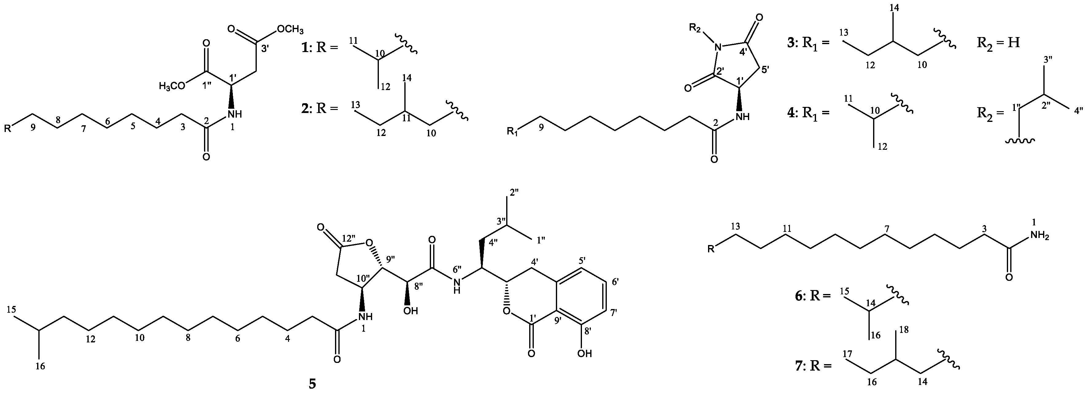
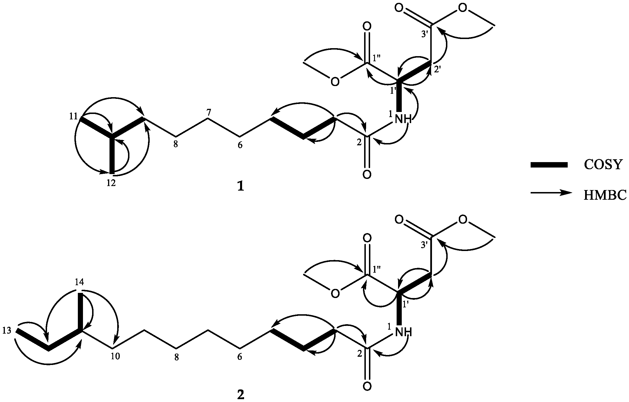
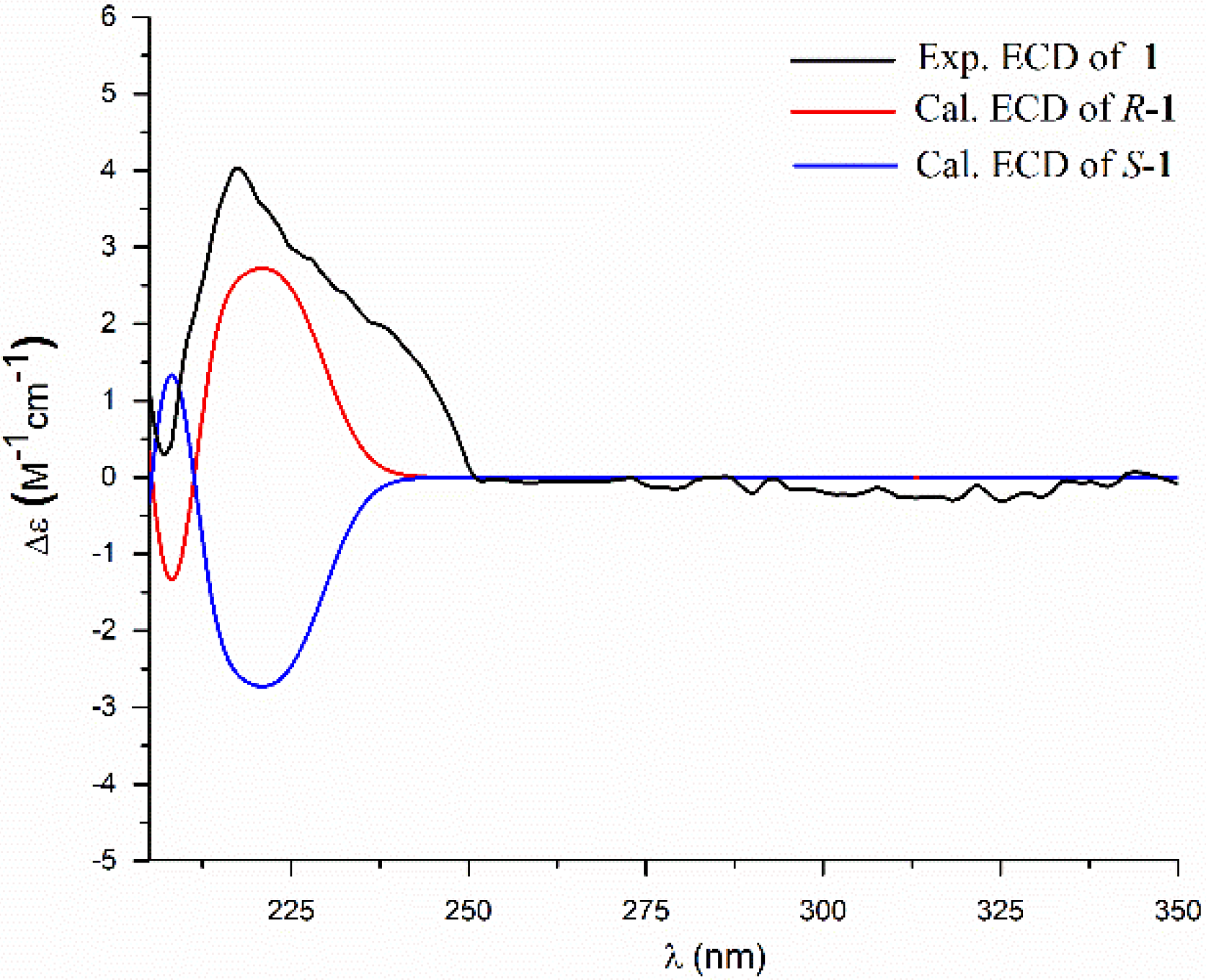
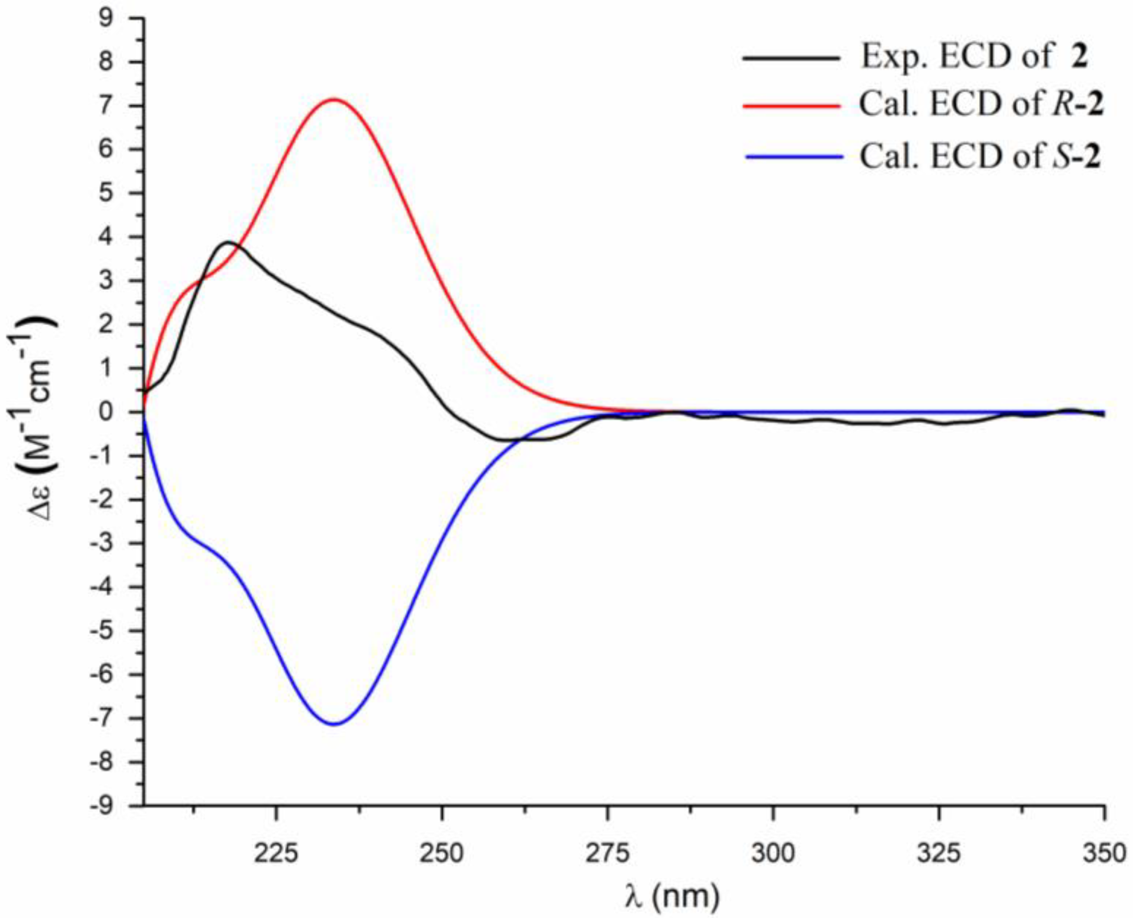
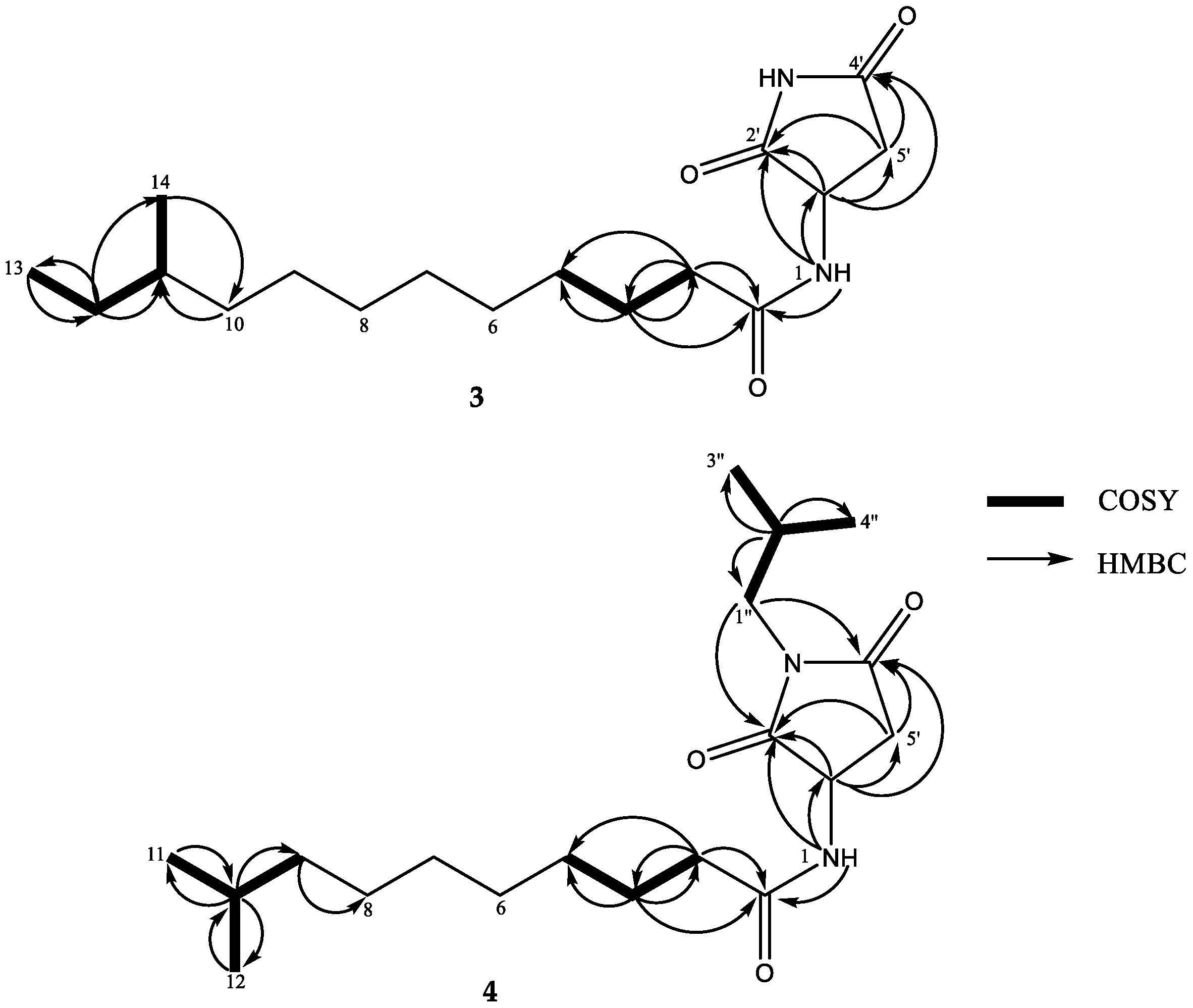
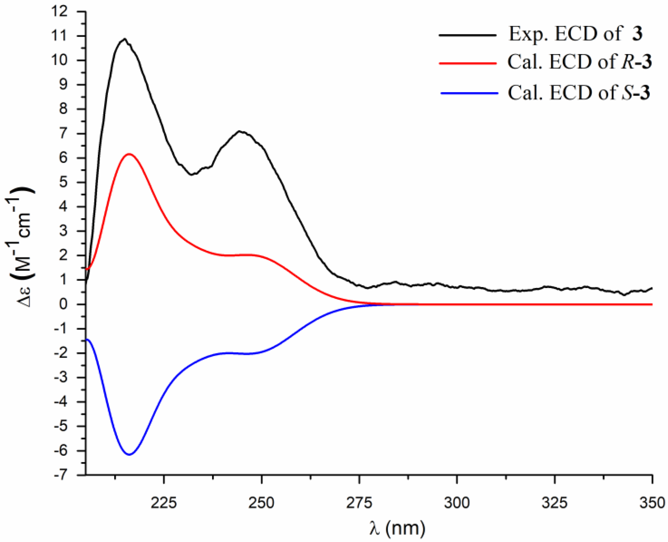
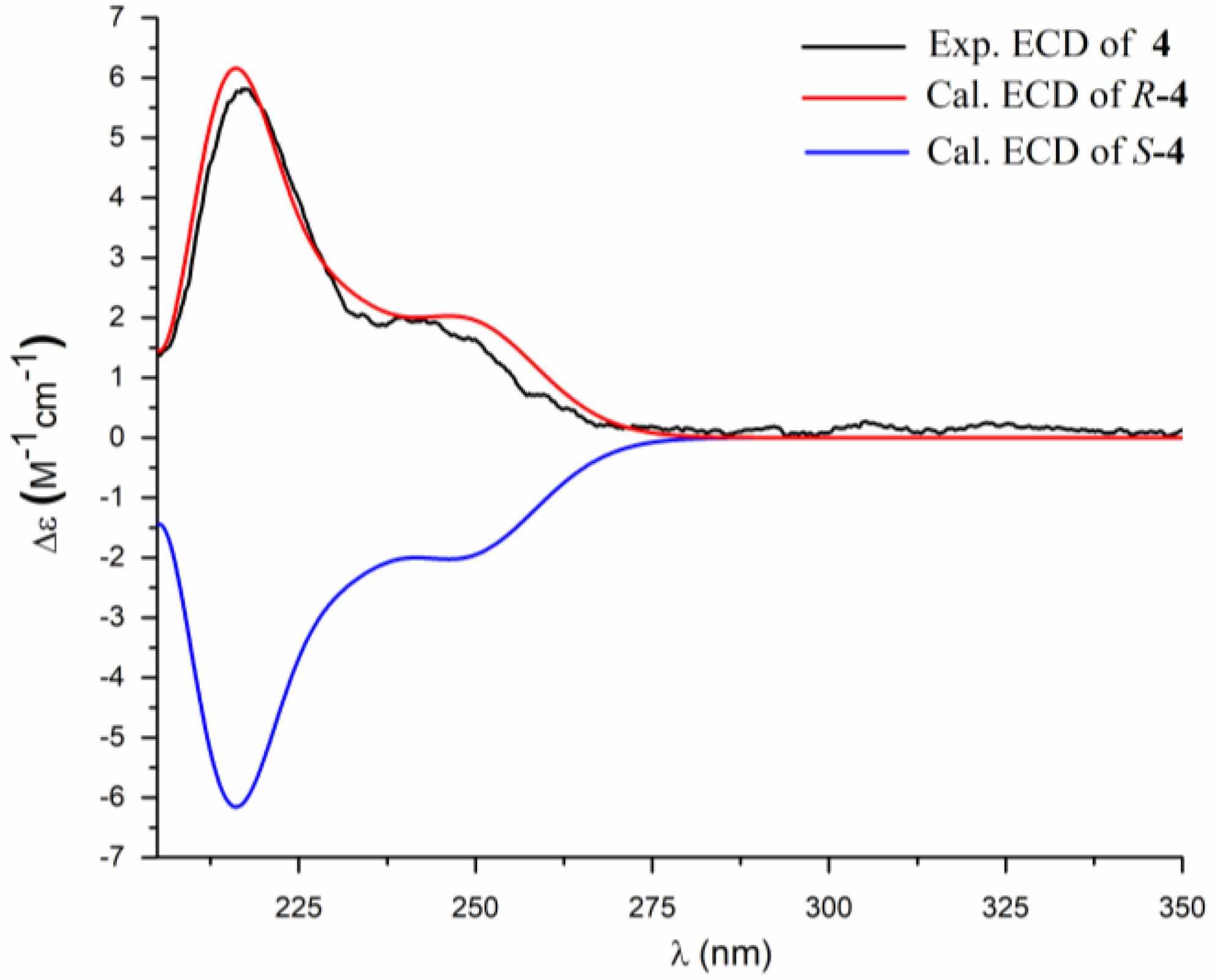
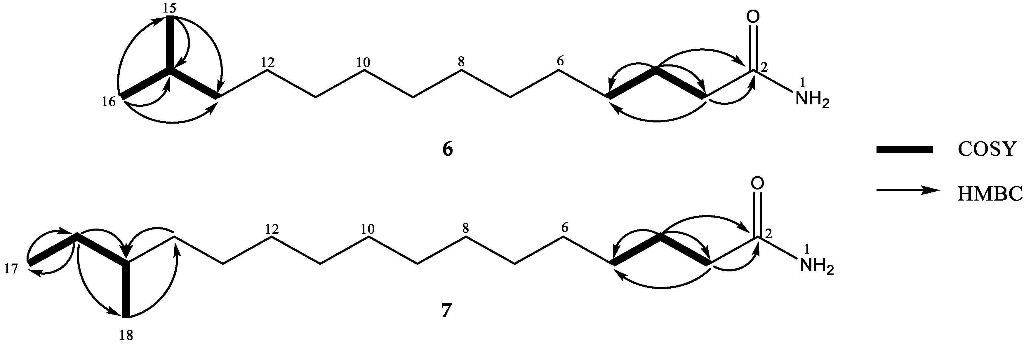
2. Results and Discussion

The strain B. pumilus RJA1515 was cultured in solid agar for 14 days, then the bacterial cultures and the agar were extracted with EtOAc to afford a crude ethyl acetate extract. The extract was fractionated and purified by repeated column chromatography to give bacillamidins A–G (17).
Compound 1 was obtained as a white powder. The molecular formula was determined as C17H31NO5 by high resolution electrospray ionization mass spectrometry (HRESIMS) (m/z 352.2096 [M + Na]+, calcd. 352.2100), which indicated three degrees of unsaturation. The 1H NMR spectrum (Table 1, Figure S1) displayed an ABX spin system at δH 4.60 (1H, ddd, J = 9.6, 7.6, 6.0 Hz), 2.79 (1H, dd, J = 16.4, 9.6 Hz), 2.67 (1H, dd, J = 16.4, 6.0 Hz), a proton doublet at δH 8.31 (1H, d, J = 7.2 Hz), two methyl doublets at δH 0.84 (6H, d, J = 6.4 Hz) and two methoxyl groups at δH 3.61 (3H, s) and 3.60 (3H, s), as well as numerous methylene protons from δH 1.13 to 2.08. The 13C NMR and heteronuclear single quantum correlation (HSQC) data of 1 (Figures S2 and S3) indicated the presence of two methoxyl carbons (δC 51.6, 52.0), three ester or amide carbonyls (δC 170.4, 171.2, 172.2), two methine sp3 (δC 27.3, 48.4), eight methylene sp3 (δC 25.1, 26.7, 28.4, 28.7, 29.2, 34.9, 35.6, 38.4) and two methyl (δC 22.5) carbons.
The presence of an alkanone moiety was corroborated by hydrogen-hydrogen correlation spectroscopy (1H-1H COSY) and heteronuclear multiple bond correlation (HMBC) correlations (Figure 2, Figures S4 and S5). The 1H-1H COSY spectrum showed correlations from the methyl doublet at δH 0.84 (CH3-11 and CH3-12) to H-10 (δH 1.48), thus, a gem-dimethyl terminus was established (Table 1). An alkyl chain moiety with eleven carbon atoms was established by HMBC correlations from H-3 (δH 2.08, t, J = 7.6 Hz) to C-4 (δC 25.1) and C-5 (δC 28.4) and from the terminal gem-dimethyl protons (CH3-11 and CH3-12) to C-9 (δC 38.4), as well as the presence of a broad singlet, integrated for sixteen methylene protons (H2-3 to H2-10). The HMBC correlation from H-3 to C-2 (δC 172.2) suggested the connectivity of an alkyl chain to the C-2 amide carbonyl carbon. An amide group was confirmed by the HMBC correlation from the proton doublet of H-1 at δH 8.31 to C-2. The HMBC correlations from H-1 to C-1′ (δC 48.4), from H-1′ (δH 4.60) to C-1″ (δC 170.4) and C-2′ (δC 35.6), and from H-2′a (δH 2.79)/H-2′b (δH 2.67) to C-1′ and C-3′ (δC 171.2) suggested the presence of the aspartic acid moiety [10]. In addition, the HMBC correlations from the two methoxyl groups at δH 3.61 and δH 3.60 to C-1″ and C-3′, respectively, indicated the locations of two methyl ester groups.
The absolute configuration of C-1′ was defined as R by comparison of the experimental optical rotation (OR) of 1 ([α] D 25 −16.6) with the calculated OR of R-1 ([α] D 25 −15.1), and by comparison of the calculated and the experimental ECD spectra (Figure 3). Thus, the structure of 1 was determined as (1′R)-9-methyldecanoyl dimethylaspartate, named bacillamidin A.
Compound 2 was obtained as a white powder. Its molecular formula was established as C19H35NO5 by HRESIMS (m/z 380.2409 [M + Na]+, calcd. 380.2413), implying three degrees of unsaturation. The 1H and 13C NMR spectra of 2 (Table 1, Figures S9 and S10) resemble those of 1 except for the presence of two additional methylene groups (δH 1.23; δC 29.9, 29.0), one methyl triplet at δH 0.83 (3H, t, J = 9.0 Hz), and one methyl doublet δH 0.78 (3H, d, J = 9.0 Hz). The 1H-1H COSY cross-peaks from H-11 (δH 1.23, m) to H-12 (δH 1.10, m) and H-14 (δH 0.78, d, J = 9.0 Hz) and from H-12 to H-13 (δH 0.83, t, J = 9.0 Hz) and the HMBC correlations (Figure 2, Figures S12 and S13) from H-13 to C-12 (δC 29.0) and C-11 (δC 33.7) and from H-14 to C-10 (δC 36.0), C-11, and C-12 indicated that 2 had a terminal sec-butyl group in the alkyl chain. All the aforementioned information indicated the presence of a 10-methyldodecanoyl motif in 2, instead of the 9-methyldecanoyl moiety in 1. The absolute configuration of C-1′ was established by comparison of the calculated and the experimental ECD spectra (Figure 4). However, the absolute configuration of C-11 was not determined. Thus, the structure of 2 was determined as (1′R)-10-methyldodecanoyl dimethylaspartate, named bacillamidin B.
Compound 3 was obtained as a white powder. Its molecular formula was determined as C17H30N2O3 by HRESIMS (m/z 333.2145 [M + Na]+, calcd. 333.2154), indicating four degrees of unsaturation. The 1H NMR spectrum (Table 2, Figure S17) displayed an ABX spin system at δH 4.35 (1H, ddd, J = 9.6, 7.6, 6.0 Hz), 2.84 (1H, dd, J = 16.4, 9.6 Hz), 2.53 (1H, dd, J = 16.4, 6.0 Hz), a proton doublet at δH 8.42 (1H, d, J = 7.6 Hz), one methyl triplet at δH 0.83 (3H, t, J = 9.0 Hz), one methyl doublet δH 0.78 (3H, d, J = 9.0 Hz), and a singlet of one proton (δH 11.19, s), as well as numerous methylene protons from δH 1.13 to 1.23. The 13C NMR and HSQC data of 3 (Figures S18 and S19) indicated the presence of three ester or amide carbonyls (δC 172.5, 176.4, 177.7), as well as two methine sp3 (δC 33.7, 49.5), ten methylene sp3 (δC 25.0, 26.5, 28.5, 28.8, 29.0, 29.0, 29.3, 34.9, 36.0, 36.2), and two methyl carbons (δC 11.2, 19.1). The HMBC correlations from H-1′ (δH 4.35) to C-2′ (δC 177.7), C-4′ (δC 176.4), and C-5′ (δC 36.0) and from H-5′a (δH 2.84)/H-5′b (δH 2.53) to C-4′ and C-2′, in addition to a NH singlet (δH 11.19), suggested the presence of a succimide substructure [11]. The HMBC correlations from NH-1 (δH 8.42, d, 7.6 Hz) to C-1′ (δC 49.5) and C-2′ indicated the linkage between the amide and succimide portion. The 1H-1H COSY and HMBC spectra (Figure 5, Figures S20 and S21) of 3 indicated the same alkyl side chain as a terminal sec-butyl group in 2. The absolute configuration of C-1′ was established by comparison of the calculated and the experimental ECD spectra (Figure 6). The absolute configuration of C-11 was not determined at a current state. Thus, the structure of 3 was determined as (3′R)-N-(2, 5-dioxopyrrolidin-3-yl)-10-methyldodecamide, named bacillamidin C.
Compound 4 was obtained as a white powder. The molecular formula was determined as C19H34N2O3 according to its HRESIMS data (m/z 361.2461 [M + Na]+, calcd. 361.2467). The 1H and 13C NMR, 1H-1H COSY and HMBC spectra (Table 2, Figures S25, S26, S28 and S29) showed that the alkyl side chain of 4 possessed a gem-dimethyl terminus, similar to that of 1. The rest 1H and 13C NMR data of 4 was very similar to those of 3 except for the absence of the NH singlet at δH 11.19 and the presence of a sec-butyl moiety (two methyl doublets at δH 0.84, J = 6.6 Hz/δC 20.0, a multiplet at δH 1.90, /δC 26.7, and a doublet at δH 3.19, J = 7.2 Hz/δC 45.4). Therefore, the structure of 4 was established as shown in Figure 3. The absolute configuration of C-1′ was established by comparison of the calculated and the experimental ECD spectra (Figure 7). Therefore, the structure of 4 was determined as (3′R)-N-(1-isobutyl-2, 5-dioxopyrrolidin-3-yl)-9-methyldecanamide, named bacillamidin D.
Compound 5 was obtained as a white powder and its molecular formula was established as C35H54N2O5 by HRESIMS (m/z 653.3775 [M + Na]+, calcd. 653.3778), implying ten degrees of unsaturation. The UV spectrum of 5 showed absorption maxima at 248 and 300 nm which are characteristic of the amicoumacins [12,13]. The 1H NMR spectrum (Table 3, Figure S33) exhibited the signals of the protons of the 1,2,3-trisubstituted benzene ring at δH 7.48 (1H, dd, J = 8.4, 7.6 Hz), 6.84 (1H, d, J = 8.4 Hz), and 6.82 (1H, d, J = 7.6 Hz), three sp3 oxymethines at δH 4.69 (1H, ddd, J = 10.4, 8.0, 2.8 Hz, H-3′), 4.61 (1H, dd, J = 2.4, 2.0 Hz, H-9″), and 4.27 (1H, dd, J = 6.0, 2.4 Hz, H-8″), two nitrogen-bearing sp3 methines at δH 4.32 (1H, m, H-10″) and 4.19 (1H, dddd, 3.6, 7.6, 10.4, 13.6 Hz, H-5″), a broad peak at δH 1.19 (12H, H-5~H-10), and three doublets of four methyl groups at δH 0.88 (H-2″), 0.83 (H-15, H-16), and 0.80 (H-1″). The 13C NMR spectrum (Figure S34) exhibited four ester or amide carbonyls (δC 168.9, 169.8, 171.9, 175.6), three quaternary sp2 (δC 108.3, 140.4, 160.8), three methine sp2 (δC 115.2, 118.5, 136.2), three oxymethine sp3 (δC 71.6, 80.8, 85.7), and two nitrogen-bearing methine sp3 (δC 46.1, 48.3) carbons. These NMR data were similar to those of amicoumacin C [14]. The gross structure of 5 was therefore established based on the HSQC and HMBC correlations (Figure 8), indicating that an amicoumacin unit was linked to an alkanone side chain with a gem-dimethyl terminus.
The negative sign of the Cotton effect for the n-π* transition of the carbonyl group at 260 nm in the ECD spectrum (Figure S42) was in agreement with the 3′S configuration [13]. Moreover, the JH-3′/H-5″ (10.4 Hz) value was indicative of the trans configuration between H-3′ and H-5″ [13]. In addition, the ECD spectrum of 5 showed a positive Cotton effect at 220 and 242 nm and a negative Cotton effect at 260 and 314 nm, which were in good agreement with those for amicoumacin B and amicoumacin C [15]. Thus, the structure of 5 was determined as (3′S,5″S,8″S,9″S,10″S)-8′,8″-dihydroxy-9″-oxotetrahydrofuran-10″-yl-13-methyltetradecanamide, named bacillamidin E.
Compounds 6 and 7 were obtained as white powders. Their molecular formulas were determined as C15H31NO and C17H35NO according to sodium-adduct ion peaks at m/z 264.2301 for 6 and 292.2616 for 7. The structures of 6 and 7 were further established by analyses of their 1H, 13C, HSQC and HMBC NMR spectral data (Table S1, Figure 9, Figures S43–S46 and S51–S54) as 13-methyltetradecanamide (bacillamidin F) [16], and 14-methylhexadecanamide (bacillamidin G) [17], respectively. The absolute configuration of C-15 of 7 was not determined at current state. Although 6 and 7 have been already synthesized [16,17], this is the first report of their isolation from a natural source.
The cytotoxic activity against human hepatoma cell line (HepG2), human lung cancer cell line (A549), human breast cancer cell line (MDA-MB-231), and human gastric cancer cell line (SGC7901) and the antibacterial activity against Pseudomonas aeruginosa PA-01, Acinetobacter baumannii ATCC19606 and Escherichia coli BW25113 of 17 were investigated (Table 4). None of the compounds showed significant cytotoxicity in all the cell lines tested. Compounds 14 showed antibacterial activity against P. aeruginosa PA-01 and A. baumannii ATCC19606 with minimum inhibitory concentration (MIC) values ranging from 58 to 64 μg/mL.

3. Materials and Methods

3.1. General Experimental Procedures

The melting points were measured on a SGWX-4A microscope (Shanghai Physical-Optics Instruments Co., Shanghai, China). UV spectra were recorded with a Shimadzu UV-260 spectrophotometer (Shimadzu Corp., Kyoto, Japan) in MeOH; λmax (log ε) in nm. IR spectra were recorded with KBr pellets on an Avatar 360 E.S.P spectrophotometer (Thermo Nicolet Co., Boston, MA, USA). NMR spectra were recorded on a Bruker DRX500 (400 MHz for 1H, 100 MHz for 13C) spectrometer (Bruker, Bremen, Germany) in DMSO-d6; δ in ppm rel. with Me4Si as internal references, and J in Hz. HR-ESI-MS was measured with a Xevo G2-S Q-Tof MS (Waters MS Technologies, Manchester, UK) in positive-ion mode; in m/z (rel. %). ECD measurements were performed using a JASCO J-1500 spectropolarimeter (Jasco Co., Ltd., Tokyo, Japan). Column chromatography (CC) was performed with silica gel (200–300 mesh, Qingdao Marine Chemical Factory, Qingdao, China), Sephadex LH-20 gel (GE Healthcare Bio-Sciences AB, Uppsala, Sweden), and RP silica gel (ODS-A-HG, YMC, Kyoto, Japan). TLC was performed on silica gel plates GF254 (Yan-tai Institute of Chemical Technology, Yantai, China). The spots on TLC were visualized by UV light (254 nm) and sprayed with 10% H2SO4, followed by heating. Semi-preparative high performance liquid chromatography (HPLC) was carried out on a Shimadzu LC-6AD chromatograph (Shimadzu Corp., Kyoto, Japan) with an ODS column (RP-18, 250 × 10 mm, YMC, 5 μm) with a flow rate of 2.0 mL/min.

3.2. Strain and Cultivation

Bacillus pumilus strain RJA1515 was isolated from marine sediments collected in Bamfield, British Columbia at a depth of 84 m by Raymond J. Andersen’s group, in January 2010. The strain was cultured on solid agar at room temperature in 160 pans measuring 50 cm × 30 cm, equivalent to 6.8 L volume of the marine medium 1 (MM1) [18]. After 14 days, the bacterial cultures and the agar were extracted twice with EtOAc (2 × 80 L). Then the extracts were re-dissolved in 2.5 L of EtOAc and back extracted three times with 600 mL of water after being combined and dried in vacuo. The organic fraction was then dried in vacuo to give an extract (32.0 g).

3.3. Extraction and Isolation

The EtOAc extract (32.0 g) was subjected to an open silica gel column (900.0 g) and eluted with a step gradient of petroleum ether (PE) and EtOAc to yield fourteen fractions (Fr. A–N). The PE/EtOAc (7:3) fraction Fr. H (250.1 mg) was purified on a Sephadex LH-20 column and eluted with CHCl3/MeOH (1:3) to give fractions H1 and H2. Fraction H1 (141.3 mg) was fractionated by RP-18 CC (MeOH-H2O, from 20:80 to 100:0) to afford fractions H1/1–H1/5. Fraction H1/4 (43.8 mg) was further separated on semi-preparative HPLC with MeOH/H2O (0–35 min: 87:13) as eluent to yield 1 (2.2 mg, tR 15.1 min) and 2 (2.6 mg, tR 9.0 min). Fraction M (PE-EtOAc, 1:1, 410.8 mg) was chromatographed on RP silica gel CC (25.0 g) with a step gradient of MeOH-H2O (20:80–100:0) to obtain fractions M1-5. Fraction M4 was purified by preparative HPLC with MeOH/H2O (0–60 min: 78:22) to give 3 (2.1 mg, tR 42.1 min) and 4 (5.1 mg, tR 34.9 min). Fraction N (PE-EtOAc, 3:7, 458.9 mg) was chromatographed on RP silica gel CC (25.0 g) with a step gradient of MeOH-H2O (20:80–100:0) to give fractions N1-5. Fraction N5 was further purified by semi-preparative HPLC with MeOH/H2O (74:26) to obtain 5 (2.8 mg, tR 25.1 min). Fraction J (PE-EtOAc, 6:4, 699.3 mg) was chromatographed on RP silica gel CC (25.0 g) with a step gradient of MeOH-H2O (20:80–100:0) to give five fractions. Fraction J5 (46.4 mg) was further purified by preparative HPLC with MeOH/H2O (0–60 min: 87:13) to yield 6 (9.9 mg, tR 18.3 min) and 7 (3.6 mg, tR 21.3 min).
Bacillamidin A (1): amorphous, white powder; m.p.: 86.8–88.2 °C; [α] D 25 = −16.6 (c 0.20, MeOH); ECD (c = 1.08 mM, MeOH) λmaxε): 218 (+4.12); UV (MeOH) λmax (log ε) 225 (2.85) nm; IR (KBr) νmax 3314, 2918, 2851, 1749, 1732, 1649, 1564, 1545, 1435, 1302, 1242, 1163 cm−1; 1H and 13C NMR data, Table 1; HRESIMS m/z 352.2096 [M + Na]+ (calcd. for C17H31NO5Na, 352.2100).
Bacillamidin B (2): amorphous, white powder; m.p.: 85.7–87.1 °C; [α] D 25 = −16.0 (c 0.20, MeOH); ECD (c = 1.02 mM, MeOH) λmaxε): 215 (+5.90); UV (MeOH) λmax (log ε) 225 (2.47) nm; IR (KBr) νmax 3314, 2961, 2918, 2851, 1751, 1732, 1647, 1560, 1543, 1435, 1302, 1242, 1211, 1163 cm−1; 1H and 13C NMR data, Table 1; HRESIMS m/z 380.2409 [M + Na]+ (calcd. for C19H35NO5Na, 380.2313).
Bacillamidin C (3): amorphous, white powder; m.p.: 87.8–89.2 °C; [α] D 25 = −26.1 (c 0.20, MeOH); ECD (c = 2.24 mM, MeOH) λmaxε): 215 (+10.88), 245 (+7.06); UV (MeOH) λmax (log ε) 226 (2.62) nm; IR (KBr) νmax 3238, 2961, 2922, 2851, 1722, 1703, 1655, 1524, 1416, 1373, 1196, 1171 cm−1; 1H and 13C NMR data, Table 2; HRESIMS m/z 333.2145 [M + Na]+ (calcd. for C17H30N2O3Na, 333.2154).
Bacillamidin D (4): amorphous, white powder; m.p.: 91.4–92.6 °C; [α] D 25 = −24.3 (c 0.20, MeOH); ECD (c = 2.00 mM, MeOH) λmaxε): 218 (+5.60), +245 (+1.90); UV (MeOH) λmax (log ε) 226 (2.49) nm; IR (KBr) νmax 3238, 2961, 2922, 2851, 1722, 1659, 1520, 1196, 1171 cm−1; 1H and 13C NMR data, Table 2; HRESIMS m/z 361.2461 [M + Na]+ (calcd. for C19H34N2O3Na, 361.2467).
Bacillamidin E (5): amorphous, white powder; m.p.: 92.3–93.9 °C; [α] D 25 = −42.0 (c 0.20, MeOH); ECD (c = 0.59 mM, MeOH) λmaxε): 220 (+1.49), 226 (−0.47), 242 (+4.48), 260 (−3.92), 314 (−0.74), +341 (0.15); UV (MeOH) λmax (log ε) 248 (3.47), 300 (2.95) nm; IR (KBr) νmax 3452, 2922, 2853, 1686, 1649, 1566, 1555, 1468, 1412 cm−1; 1H and 13C NMR data, Table 3; HRESIMS m/z 653.3775 [M + Na]+ (calcd. for C35H54N2O8Na, 653.3778).

3.4. ECD Calculation Methods

In general, conformational analyses were carried out via random searching in the Sybyl-X 2.0 using the MMFF94S force field with an energy cutoff of 2.5 kcal/mol. The results showed five lowest energy conformers for 13, and six for 4. Subsequently, the conformers were re-optimized using density functional theory (DFT) at the b3lyp/6-31+g(d) level in gas phase by the GAUSSIAN 09 program. The energies, oscillator strengths, and rotational strengths (velocity) of the first 60 electronic excitations were calculated using the time-dependent DFT methodology at the b3lyp/6-311++g(d,p) level in vacuo. The ECD spectra were simulated by the overlapping Gaussian function (half the bandwidth at 1/e peak height, σ = 0.30 for 1 and 2, σ = 0.16 for 3, σ = 0.25 for 4).
To get the final spectra, the simulated spectra of the conformers were averaged according to the Boltzmann distribution theory and their relative Gibbs free energy (ΔG). By comparing the experiment spectrum with the calculated ECD model molecules, the absolute configuration of the only chiral center of all the four compounds was determined to be R.

3.5. Bioassay

Cytotoxicity assays were evaluated on the human tumor cell lines HepG2 (human hepatocellular carcinoma cell line), A549 (human lung carcinoma cell line), MDA231 (human breast carcinoma cells), and SGC7901 (human gastric carcinoma cells) by MTT method according to our previous study [19].
Antimicrobial activity was measured by the liquid broth microdilution method [20] against Pseudomonas aeruginosa PA-01, Acinetobacter baumannii ATCC19606, and Escherichia coli BW25113, with ofloxacin as the positive control.

4. Conclusions

Microorganisms from marine sediments are promising sources of bioactive natural products. Chemical investigation of the marine-derived Bacillus pumilus RJA1515 led to the isolation and identification of five new long-chain amides (15) and two previously reported long-chain alkylamides (6, 7). The absolute configuration of 1 was determined by comparison of the calculated and the experimental OR and ECD spectra, while those of 2–5 were determined by comparison of their calculated and the experimental ECD spectra. Compounds 14 showed antibacterial activity against P. aeruginosa PA-01 and A. baumannii ATCC19606 with MIC values ranging from 58 to 64 μg/mL.

Supplementary Materials

The following are available online at https://www.mdpi.com/1660-3397/16/9/326/s1. This section includes 1D, 2D NMR spectra and other spectroscopic data for compounds 17.

Author Contributions

S.-Y.Z. performed the isolation, purification, and characterization of all the compounds and prepared the manuscript; Y.-J.H. contributed to the isolation and purification of the isolates; F.-C.M. performed the structure elucidation and revised the manuscript; S.-Y.Q. and R.W. contributed to the determination of cytotoxic and antimicrobial activities; R.J.A. provided the strain RJA1515 and contributed to the identification of the strain; Z.-H.L. performed the 1D and 2D NMR experiments; M.C. designed and supervised the research and revised the manuscript.

Funding

This work was financially supported by the Chongqing Social Undertaking and Livelihood Security Project (No. cstc2017shmsA130079) and the National Natural Science Foundation of China (No. 81774005).

Conflicts of Interest

The authors declare no conflict of interest.

References

  1. Joint, I.; Muhling, M.; Querellou, J. Culturing marine bacteria—An essential prerequisite for biodiscovery. Microb. Biotechnol. 2010, 3, 564–575. [Google Scholar] [CrossRef] [PubMed] [Green Version]
  2. Blunt, J.W.; Copp, B.R.; Keyzers, R.A.; Munro, M.H.G.; Prinsep, M.R. Marine natural products. Nat. Prod. Rep. 2017, 34, 235–294. [Google Scholar] [CrossRef] [PubMed] [Green Version]
  3. Rampelotto, P.H. Resistance of microorganisms to extreme environmental conditions and its contribution to astrobiology. Sustainability 2010, 2, 1602–1623. [Google Scholar] [CrossRef]
  4. Schinke, C.; Martins, T.; Queiroz, S.C.N.; Melo, I.S.; Reyes, F.G.R. Antibacterial compounds from marine bacteria, 2010–2015. J. Nat. Prod. 2017, 80, 1215–1228. [Google Scholar] [CrossRef] [PubMed]
  5. Mondol, M.A.M.; Shin, H.J.; Islam, M.T. Diversity of secondary metabolites from marine Bacillus species: Chemistry and biological activity. Mar. Drugs 2013, 11, 2846–2872. [Google Scholar] [CrossRef] [PubMed]
  6. Devi, P.; Wahidullah, S.; Rodrigues, C.; Souza, L.D. The sponge-associated bacterium Bacillus licheniformis SAB1: A source of antimicrobial compounds. Mar. Drugs 2010, 8, 1203–1212. [Google Scholar] [CrossRef] [PubMed]
  7. Choudhary, A.; Naughton, L.M.; Montánchez, I.; Dobson, A.D.W.; Rai, D.K. Current status and future prospects of marine natural products (MNPs) as antimicrobials. Mar. Drugs 2017, 15, 272. [Google Scholar] [CrossRef] [PubMed]
  8. Hogberg, L.D.; Heddini, A.; Cars, O. The global need for effective antibiotics: Challenges and recent advances. Trends Pharmacol. Sci. 2010, 31, 509–515. [Google Scholar] [CrossRef] [PubMed]
  9. Daoust, J. Isolation and Structure Elucidation of Bioactive Marine Natural Products. Ph.D. Thesis, The University of British Columbia, Vancouver, BC, Canada, 2011; doi:10.14288/1.0062104. [Google Scholar]
  10. Nord, L.I.; Vaag, P.; Duus, J. Quantification of organic and amino acids in beer by 1H NMR spectroscopy. Anal. Chem. 2004, 76, 4790–4798. [Google Scholar] [CrossRef]
  11. Jin, T.Y.; Shen, T.; Zhou, M.X.; Li, A.L.; Feng, D.; Zheng, B.; Gong, J.; Sun, J.W.; Li, L.Y.; Xiang, L. Chemical constituents from Portulaca oleracea and their bioactivities. J Chin. Pharm. Sci. 2016, 25, 898–905. [Google Scholar] [CrossRef]
  12. Iton, J.; Omoto, S.; Nishizawa, N.; Kodama, Y.; Lnouye, S. Chemical structures of amicoumacins produced by Bacillus pumilus. Agric. Biol. Chem. 1982, 46, 2659–2665. [Google Scholar] [CrossRef]
  13. Bai, J.; Liu, D.; Yu, S.; Proksch, P.; Lin, W. Amicoumacins from the marine-derived bacterium Bacillus sp. with the inhibition of NO production. Tetrahedron Lett. 2014, 55, 6286–6291. [Google Scholar] [CrossRef]
  14. Shimojima, Y.; Hayashi, H.; Ooka, T.; Shibukawa, W.; Iitaka, Y. Studies on AI-77s, microbial products with pharmacological activity structures and the chemical nature of AI-77s. Tetrahedron Lett. 1982, 23, 5435–5438. [Google Scholar] [CrossRef]
  15. Azumi, M.; Ogawa, K.; Fujita, T.; Takeshita, M.; Yoshida, R.; Furumai, T.; Igarashi, Y. Bacilosarcins A and B, novel bioactive isocoumarins with unusual heterocyclic cores from the marine-derived bacterium Bacillus subtilis. Tetrahedron 2008, 64, 6420–6425. [Google Scholar] [CrossRef]
  16. Takahashi, M.; Osawa, K.; Ueda, J.; Tsuai, C.T. On the chemical structure of a new acid amide, N-(13-methyltetradecyl)-acetamide, from the fruits of Cpsicum annuum L (author’s transl). Yakugaku Zasshi J. Pharm. Soc. Jpn. 1977, 97, 758–761. [Google Scholar] [CrossRef]
  17. Milbum, A.H.; Truter, E.V. The components of wool wax. Part II. Synthesis of the acids and alcohols of the iso- and the (+)-anteiso-series. J. Chem. Soc. 1954, 3344–3351. [Google Scholar] [CrossRef]
  18. Williams, D.E.; Dalisay, D.; Chen, J.; Polishchuck, E.A.; Patrick, G.N.; Ko, M.; Av-Gay, Y.; Li, H.; Magarvey, N.; Andersen, R.J. Aminorifamycins and sporalactams produced in culture by a Micromonospora sp. isolated from a northeastern-Pacific marine sediment are potent antibiotics. Org. Lett. 2017, 19, 766–769. [Google Scholar] [CrossRef] [PubMed]
  19. Zhou, S.Y.; Wang, R.; Deng, L.Q.; Zhang, X.L.; Min, C. A new isoflavanone from Ficus tikoua Bur. J. Nat. Res. 2018, 1–7. [Google Scholar] [CrossRef] [PubMed]
  20. Wiegand, I.; Hilpert, K.; Hancock, R.E. Agar and broth dilution methods to determine the minimal inhibitory concentration (MIC) of antimicrobial substances. Nat. Protocol. 2008, 3, 163–175. [Google Scholar] [CrossRef] [PubMed]
Figure 1. Structures of 17.
Marinedrugs 16 00326 g001
Figure 2. Key 1H-1H COSY and HMBC correlations for 1 and 2.
Marinedrugs 16 00326 g002
Figure 3. Comparison of the experimental and the calculated ECD spectra of 1.
Marinedrugs 16 00326 g003
Figure 4. Comparison of the experimental and the calculated ECD spectra of 2.
Marinedrugs 16 00326 g004
Figure 5. Key 1H-1H COSY and HMBC correlations for 3 and 4.
Marinedrugs 16 00326 g005
Figure 6. Comparison of the experimental and the calculated ECD spectra of 3.
Marinedrugs 16 00326 g006
Figure 7. Comparison of the experimental and the calculated ECD spectra of 4.
Marinedrugs 16 00326 g007
Figure 8. Key 1H-1H COSY and HMBC correlations for 5.
Marinedrugs 16 00326 g008
Figure 9. Key 1H-1H COSY and HMBC correlations for 6 and 7.
Marinedrugs 16 00326 g009
Table 1. 1H and 13C NMR data (400 and 100 MHz, in DMSO-d6) of 1 and 2.
Position12
δH (J, Hz)δC, TypeδH (J, Hz)δC, Type
1-NH8.31, d (7.6) 8.42, d (7.6)
2 172.2, CO 172.2, CO
32.08, t (7.6)34.9, CH22.08, t (7.6)34.9, CH2
41.48, m25.1, CH21.47, m25.1, CH2
51.23, m28.4, CH21.23, m28.4, CH2
61.23, m28.7, CH21.23, m28.7, CH2
71.23, m29.2, CH21.23, m28.9, CH2
81.23, m26.7, CH21.23, m29.3, CH2
91.13, m38.4, CH21.23, m26.4, CH2
101.48, m27.4, CH1.09, m; 1.27, m36.0, CH2
110.84, d (6.4)22.5, CH31.30, m33.7, CH
120.84, d (6.4)22.5, CH31.10, m29.0, CH2
13 0.83, t (9.0)11.2, CH3
14 0.78, d (9.0)19.1, CH3
1′4.60, ddd (9.6, 7.6, 6.0)48.4, CH4.60, ddd (9.6, 7.6, 6.0)48.4, CH
2′2.79, dd (16.4, 9.6)35.6, CH22.79, dd (16.4, 9.6)35.6, CH2
2.67, dd (16.4, 6.0) 2.67, dd (16.4, 6.0)
3′ 171.2, CO 171.3, CO
3′-OCH33.61, s51.6, OCH33.61, s51.6, OCH3
1″ 170.4, CO 170.4, CO
1″-OCH33.60, s52.0, OCH33.60, s52.1, OCH3
Table 2. 1H and 13C-NMR data (400 and 100 MHz, in DMSO-d6) of 3 and 4.
Position34
δH (J, Hz)δC, TypeδH (J, Hz)δC, Type
1-NH8.42, d (7.6) 8.51, d (7.6)
2 172.5, CO 172.6, CO
32.08, t (7.6)34.9, CH22.08, t (7.6)34.8, CH2
41.23, m25.0, CH21.48, m25.0, CH2
51.23, m28.5, CH21.23, m28.4, CH2
61.23, m28.8, CH21.23, m28.8, CH2
71.23, m29.0, CH21.23, m29.2, CH2
81.23, m29.3, CH21.23, m26.7, CH2
91.23, m26.5, CH21.13, m38.4, CH2
101.09, m; 1.27, m36.2, CH21.48, m27.4, CH
111.30, m33.7, CH0.83, d (6.6)22.5, CH3
121.10, m29.0, CH20.83, d (6.6)22.5, CH3
130.83, t (9.0)11.2, CH3
140.78, d (9.0)19.1, CH3
1′4.35, ddd (9.6, 7.6, 6.0)49.5, CH4.35, ddd (9.6, 7.6, 6.0)48.4, CH
2′ 177.7, CO 176.5, CO
3′-NH11.19, s
4′ 176.4, CO 175.2, CO
5′2.84, dd (16.4, 9.6)36.0, CH22.91, dd (16.4, 9.6)34.8, CH2
2.53, dd (16.4, 6.0) 2.53, dd (16.4, 6.0)
1″ 3.19, d (7.2)45.4, CH2
2″ 1.90, m26.7, CH
3″ 0.84, d (6.6)20.0, CH3
4″ 0.84, d (6.6)20.0, CH3
Table 3. 1H and 13C-NMR data (400 and 100 MHz, in DMSO-d6) of 5.
Position5
δH (J, Hz)δC, TypePositionδH (J, Hz)δC, Type
1-NH8.34, d (6.8) 1″0.80, d (6.4)21.3, CH3
2 171.9, CO2″0.88, d (6.4)23.3, CH3
31.90, t (7.6)35.0, CH23″1.56, m24.0, CH
41.48, m24.9, CH24″1.32, m38.4, CH2
5-101.25, m28.5–29.2 1.68, m
111.25, m29.3, CH25″4.19, dddd (3.6, 7.6, 10.4, 13.6)48.3, CH
121.25, m26.7, CH26″-NH7.94, d (9.6)
131.13, m38.4, CH27″ 169.8, CO
141.49, m27.4, CH8″4.27, dd (6.0, 2.4)71.6, CH
150.83, d, (6.6)22.5, CH38″-OH6.18, d (6.0)
160.83, d, (6.6)22.5, CH39″4.61, dd (2.4, 2.0)85.7, CH
1′ 168.9, CO10″4.32, m46.1, CH
3′4.69, ddd (10.4, 8.0, 2.8)80.8, CH11″2.16, dd (18.0, 2.2)36.0, CH2
4′2.87, m29.0, CH2 2.85, m
5′6.82, d (7.6)118.5, CH12″ 175.6, CO
6′7.48, dd (8.4, 7.6)136.2, CH
7′6.84, d (8.4)115.2, CH
8′ 160.8, C
8″-OH10.79, s
9′ 108.3, C
10′ 140.4, C
Table 4. Antimicrobial activity of 17 (MIC, μg/mL).
CompoundsP. aeruginosa PA-01A. baumannii ATCC19606E. coli BW25113
16458>128
26464>128
36464>128
46458>128
5>128>128>128
6>128>128>128
7>128>128>128
ofloxacin8161

