Review

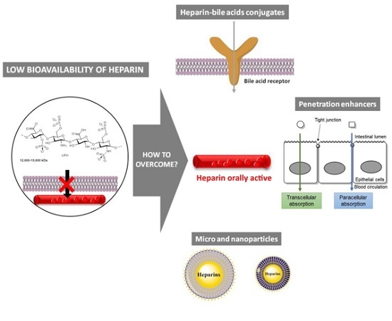
Strategies to Overcome Heparins’ Low Oral Bioavailability

by
Ana Rita Neves
1,†,
Marta Correia-da-Silva
1,2,†,
Emília Sousa
1,2,* and
Madalena Pinto
1,2
1
Organic Chemistry and Pharmaceutical Laboratory, Department of Chemical Sciences, Faculty of Pharmacy, University of Porto, Rua Jorge Viterbo Ferreira 228, 4050-313 Porto, Portugal
2
Interdisciplinary Centre of Marine and Environmental Research (CIIMAR/CIMAR), University of Porto, Rua dos Bragas 289, 4050-123 Porto, Portugal
*
Author to whom correspondence should be addressed.
These authors contributed equally to this work.
Pharmaceuticals 2016, 9(3), 37; https://doi.org/10.3390/ph9030037
Submission received: 22 April 2016 / Revised: 15 June 2016 / Accepted: 23 June 2016 / Published: 29 June 2016
(This article belongs to the Special Issue Grand Celebration: 100th Anniversary of the Discovery of Heparin)

Abstract

:
Even after a century, heparin is still the most effective anticoagulant available with few side effects. The poor oral absorption of heparins triggered the search for strategies to achieve oral bioavailability since this route has evident advantages over parenteral administration. Several approaches emerged, such as conjugation of heparins with bile acids and lipids, formulation with penetration enhancers, and encapsulation of heparins in micro and nanoparticles. Some of these strategies appear to have potential as good delivery systems to overcome heparin’s low oral bioavailability. Nevertheless, none have reached the market yet. Overall, this review aims to provide insights regarding the oral bioavailability of heparin.

Graphical Abstract

1. Introduction

Heparin has been one of the most effective and widely used drugs of the past century. Discovery of heparin dates back to 1916, when isolated blood fractions that were able to clot blood were identified by Jay McLean, a second-year medical student working under the direction of William Howell [1].
Chemically, heparin is a mixture of highly sulfated glycosaminoglycans with molecular weight around 15 kDa. The presence of sulfate and carboxylic acid groups in the structure of heparin makes it one of the most negatively charged biological macromolecules in nature [2,3,4]. In physiological conditions, ionization of sulfate and carboxylic acid groups takes place, which attracts positively charged counter ions, more commonly sodium, to form a heparin salt.
Low molecular weight heparins (LMWHs), such as unfractioned heparin (UFH), are also mixtures of glycosaminoglycans but with an average molecular weight of about 5 kDa [5]. LMWHs derive from UFH by chemical or enzymatic depolymerization and have a more predictable pharmacokinetic profile [6] and a slightly different mechanism of action.
A unique pentasaccharide sequence present in both UFH and LMHWs is essential for their anticoagulant activity. UFH binds to antithrombin III (ATIII) that undergoes a conformational change and becomes activated as an inhibitor of thrombin and factor Xa (FXa) while LMWHs are shorter and show limited inhibition of thrombin [5]. Because the bioavailability of subcutaneous UFH is lower than that of intravenous UFH, UFH is preferably administered intravenously to avoid the administration of large doses of subcutaneous UFH [7]. Bioavailability of subcutaneous LMWHs is higher than that of subcutaneous UFH, so LMWHs are administered subcutaneously [5]. However, none exhibit oral bioavailability due to their highly negative charge, large molecular weight [8], and rapid metabolism in gastrointestinal (GI) tract (Figure 1) [9]. The rapid metabolism in the GI tract is mainly due to low stability at low pH [10] and enzymatic degradation by intestinal microflora [11,12]. Additionally, after GI absorption heparins undergo high first pass metabolism by heparinase in the liver [13].
Parenteral administration of heparins is a costly and invasive method that leads to low patient adherence. On the other hand, vitamin K antagonists (VKA), orally active anticoagulants, suffer from a number of limitations, namely high food and drug interactions, long onset/offset of action, and need of regular monitoring by blood tests. New oral anticoagulants were recently introduced in the market that overcome some of those VKA limitations, namely dabigatran (direct thrombin inhibitor), rivaroxaban, apixaban, and edoxaban (FXa inhibitors), but there are some hesitations about their wide use in the treatment of thromboembolic diseases, namely the absence of an antidote and extensive drug interaction with P-glycoprotein (P-gp) substrates [14]. In general, heparins have several advantages over VKA and new oral anticoagulants (Figure 2) which makes them the most effective anticoagulants available. Furthermore, all these orally active anticoagulants lack the polypharmacological actions of heparins which are thought to be involved beyond the coagulation cascade [15], like antimetastatic [16] and anti-inflammatory activities [17].
The development of a non-invasive delivery of heparin is undoubtedly an unmet clinical need. Therefore, several strategies that promote the absorption of heparins in GI tract have been investigated in the last decades.
Although the oral route is by far the most desirable, it is important to emphasize that there also have been attempts to achieve bioavailability of heparins through other routes, such as nasal [18], pulmonary [19], and transdermal [20].
This review focus on (physico-)chemical-biological and technological strategies that are expected to improve oral bioavailability of heparins, namely the increase of lipophilicity, target receptor-mediated endocytosis, modifications of the tight junctions, enhancement of cell permeabilization, and protection against acidic pH of the stomach and GI metabolism.

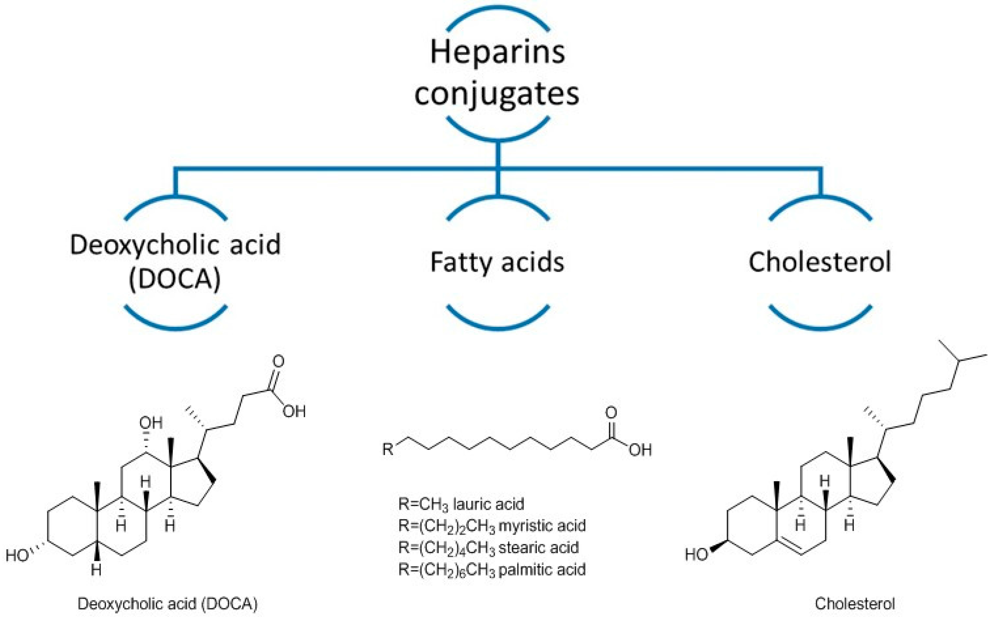
2. Heparin Conjugates

Following a drug conjugate strategy, heparins have been covalently bond to other molecules in order to achieve oral bioavailability in one of two ways: increasing lipophilicity and permeability or enabling absorption via transporter proteins [21].
Lipids and deoxycholic acid (DOCA) (Figure 3) have been proven to be suitable choices for conjugation with heparins [22,23,24,25,26,27,28,29,30,31,32,33,34]. As they are naturally occurring substances, the administration of these conjugates may involve few toxic effects [32]. Conjugates of heparin with palmitic acid, lauric acid, and cholesterol (Figure 3) were reported [32]. Absorption of these heparin-lipids conjugates increased in rats, probably due to their higher lipophilicity. Nevertheless, heparin-cholesterol and heparin-fatty acids conjugates showed lower effect in clotting times than heparin-DOCA conjugates [32]. DOCA is a secondary bile acid produced from cholesterol and one of its functions is to stimulate intestinal lipids absorption [35].
DOCA is absorbed in the intestinal membrane through apical sodium bile acid transporters and this mechanism of absorption was also hypothesized to increase the transcellular absorption of heparins [22]. Numerous studies have proven that heparins-DOCA conjugates increased oral bioavailability of heparins by increasing their intestinal absorption (Figure 4A–C) [23,24,26,27,32]. DOCA was conjugated with heparins using different synthetic approaches, all of them involving the formation of one amide bond (Figure 4A–D). Some conjugates involved the carboxylic groups of heparin to conjugate with DOCA (Figure 4A,B) and others the amine groups (Figure 4C). Lee et al. carried out histological examination of GI tissue membrane after oral administration of heparin-DOCA and showed no damage on microvilli and cell layer [27,31,32]. These results proved that the increased absorption of heparins conjugates was not caused by the disruption of the GI epithelium.
One problem associated with the use of DOCA is that the conjugate with heparins forms micelles in water and DOCA is located within the core of micelles. As a consequence, lower interaction of DOCA with apical sodium bile acid transporters occurs, limiting the absorption of the conjugate. Thus, the bioavailability of heparin-DOCA conjugates was improved through the formulation with dimethylsulfoxide (DMSO) as a solubilizer [22,28,30,31]. DMSO is known to interfere with lipid membranes [36]. The absorption of heparin-DOCA with DMSO was three times higher compared with heparin-DOCA conjugate alone. Later, Kim et al. developed a tricaprylin microemulsion to dissolve heparin-DOCA conjugate [34]. Microemulsions are thermodynamically stable, liquid mixtures of oil, water, surfactant and co-surfactant that are used in pharmaceutical science to improve the solubility of drugs (amphiphilic or lipophilic). Tricaprylin is a medium-chain triglyceride marketed as a medical food for the clinical dietary management of Alzheimer’s disease. Heparin-DOCA conjugate in water was mixed with tricaprylin, polyoxyethylene 20 sorbitan monooleate (Tween 80) and sorbitan monolaurate (Span 20). Heparin-DOCA conjugate was dissolved in a microemulsion with tricaprylic and the absorption efficiency of the heparin conjugate was increased indicating that solubilization of heparin-DOCA conjugate is important for the absorption in the intestine.
A recent study reported the absorption of a heparin-tetrameric DOCA conjugate (Figure 4D) with a remarkable systemic anticoagulant activity and high oral bioavailability of 33.5% and 19.9% in rats and monkeys, respectively [37]. This study showed a functional transformation of apical sodium bile acid transporters upon interaction with tetrameric DOCA derivative, which led to receptor-mediated endocytosis [38].
Conventional anticancer drugs are toxic, and research is focus in the development of more specific drugs that target only molecular abnormalities specific to cancer cells. The benefits of heparin in cancer are both dependent and independent from its anticoagulant activities [39]. A LMWH-DOCA conjugate (Figure 4A) was also tested for antimetastatic [40] and antiangiogenic [41,42] activities. These studies reported for the first time the antitumor effect of an orally active heparin and confirmed that it inhibits tumor angiogenesis. The co-administration of LMWH-DOCA conjugate and docetaxel, a drug widely used in the treatment of several human cancers, showed a higher tumor growth inhibition compared to LMWH-DOCA conjugate or docetaxel alone [42]. Considering that DOCA has low solubility in water, LMWHs were conjugated with taurocholic acid and docetaxel [43,44]. Oral bioavailability and synergistic effects of heparin and docetaxel were observed in this conjugate. Recently, a conjugate of LMWH with taurocholic acid and tetrameric DOCA was prepared and an increased oral bioavailability and promising angiogenesis inhibition were observed [45]. The same conjugate was co-administered with deoxycholyl-l-lysyl-methylester and an increased oral bioavailability was also observed [46].
However, there are a number of bile acids available, with different affinities to apical sodium bile acid transporters. In a study considering the effect of different bile acids on the uptake of taurocholate assay [47], chenodeoxycholic acid was the most efficient, followed by DOCA, cholic acid, ursodeoxycholic acid, and litocholic acid. Therefore, it would be interesting to study the conjugation of heparin with other bile acids, namely with chenodeoxycholic acid.
Conjugation with several bile acids, including DOCA, was successfully performed to improve oral bioavailability of other drugs, namely acyclovir [47] and insulin [48] but none have yet entered clinical trials. Consequently, conjugation of heparins with bile acids seems a valuable strategy to improve oral bioavailability of heparins. However, this strategy will not protect heparins from degradation in the stomach, intestine, and liver. On the other hand, there are some studies indicating that an increased colonic exposure to bile acids could play a role in the development of cancer, since higher fecal concentrations of bile acids were found in populations with a high incidence of colorectal cancer [49,50,51], and the levels of exposure to bile acids must be considered in the development of heparin-bile acids conjugates.

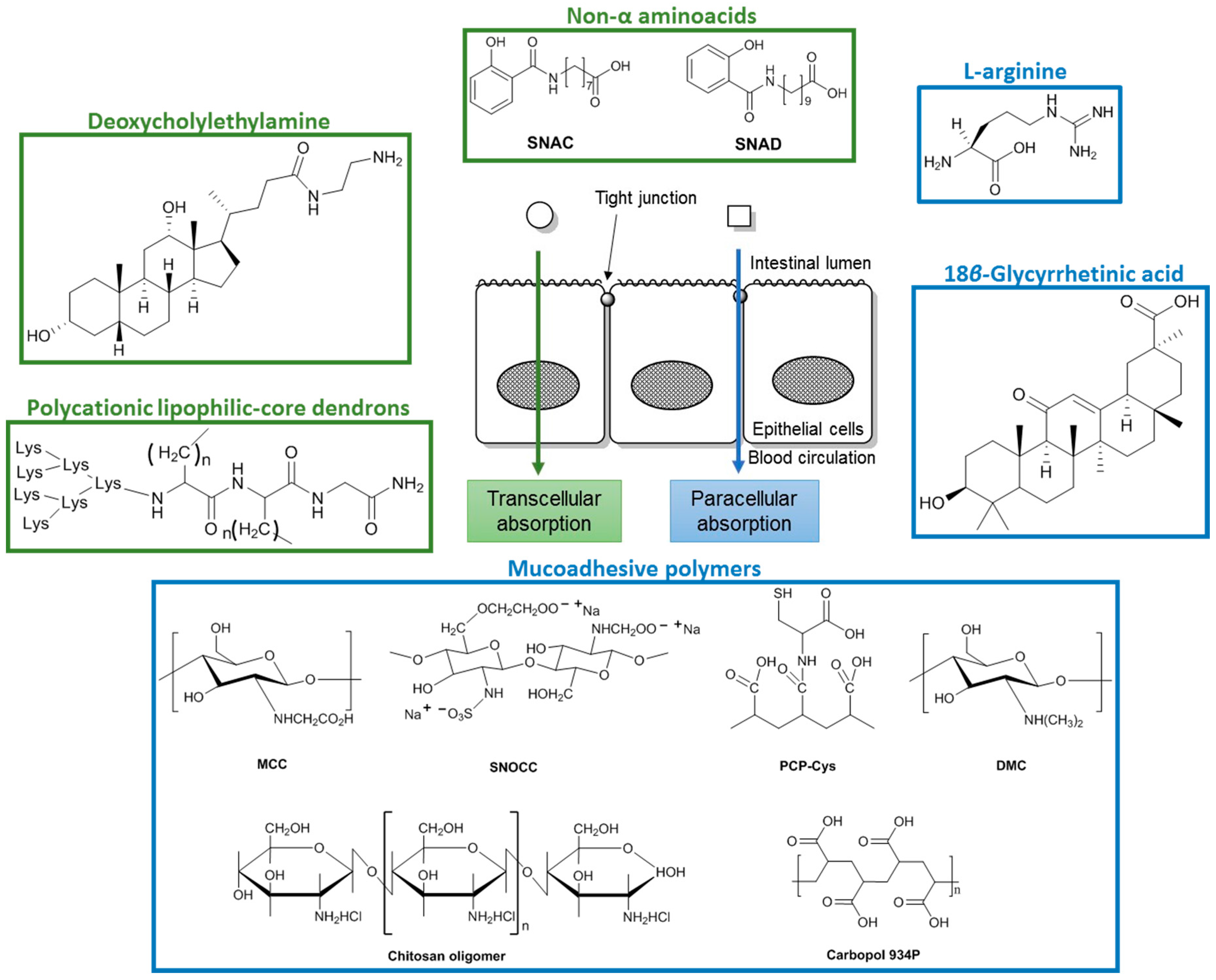
3. Co-Administration with Penetration Enhancers

In this section, penetration enhancers that have already been used to improve UFH and LMWHs absorption is revised. The weak association between penetration enhancers and drugs allows the spontaneous release of the drug into the circulation [52]. However, in contrast to drug conjugate strategy, some dissociation before absorption cannot be excluded.
Penetration enhancers improve absorption of drugs in the GI tract either through paracellular absorption, by opening the tight junctions between adjacent cells, or through transcellular absorption, by increasing the lipophilic properties of the drug (Figure 5) [21,53]. Many of the penetration enhancers that act through paracellular absorption are described as causing cytotoxicity or membrane damage [54].

3.1. Paracellular Absorption

l-Arginine (Figure 5), a nonessential amino acid, is the principal physiological precursor of nitric oxide and plays a versatile role in the GI tract physiology. Intestinal absorption of a LMWH (ardeparin) was increased in the presence of l-arginine in Caco-2 cells [55]. In vivo studies in rats showed that absorption of ardeparin in the presence of l-arginine is higher in the colon and jejunum, and for 250 mg of l-arginine it was observed an increased anti-FXa activity compared with ardeparin alone. A sodium bicarbonate solution was administered with the LMWH/l-arginine formulation to increase the acidic pH of the stomach and to avoid degradation. Anti-FXa activity was used for estimating plasma levels of LMWH and a significant increase in plasma anti-FXa levels within 90 min to 0.26 IU/mL was observed after administration of ardeparin/l-arginine formulation (~2 fold compared with the control, 0.13 IU/mL). The effect of several concentrations of l-arginine on cell viability was investigated through mitochondrial dehydrogenase activity and there was no significant variation compared to the negative control [55].
18β-Glycyrrhetinic acid (GA, Figure 5) is a pentacyclic triterpenoid amyrin derivative obtained from the hydrolysis of glycyrrhizic acid, naturally present in the roots of the plant Glycyrrhiza glabra. GA was tested as a penetration enhancer in order to increase the intestinal absorption of LMWHs [56]. A sodium bicarbonate solution was also administered with the LMWH/GA formulation. Absorption of LMWHs was increased both in vitro and in vivo after co-administration with GA. Anti-FXa activity was used for estimating plasma levels of LMWH and a significant increase in plasma anti-FXa levels within 4 h to 0.28 IU/mL was observed after administration of GA (50 μg/kg) with ardeparin (1200 IU/kg). After exposure to GA, no significant toxicity was found in Caco-2 cells monolayers, at relatively low concentrations. The authors hypothesised that GA acts by increasing paracellular absorption of LMWHs [57]. This study was conducted by Hisamitsu Pharmaceutical Co., Inc. (Saga Prefecture, Japan); however, at the moment, GA seems to be applied only on transdermal formulations with pain relief drugs [58]. Recently, anticoagulant properties through uncompetitive inhibition of FXa were described for GA [59]. Therefore, co-administration of heparin with GA seems an interesting strategy that could increase both intestinal absorption and FXa inhibition. However, higher bleeding risk should be considered.
Mucoadhesive polymers increase the residence and the contact times of the drug with mucous membranes leading to a longer-lasting therapeutic effect [60]. These polymers are not absorbed in the GI tract due to their high molecular weight. Three mucoadhesive polymers, chitosan derivatives, poly(acrylates) and thiolated polymers, were described to increase the absorption of heparins in GI tract [61,62,63,64,65,66].
Chitosan is a polysaccharide with mucoadhesive properties comprising glucosamine and N-acetylglucosamine subunits [54]. In vitro studies have shown that chitosan opens epithelial tight junctions in a concentration- and pH-dependent way [54]. However, chitosan was incompatible with LMWHs [67] and chitosan derivatives were prepared [61,62]. Mono-N-carboxymethyl chitosan (MCC, Figure 5), a polyampholyte chitosan derivative was shown to increase in vitro paracellular absorption of LMWHs in Caco-2 cell monolayers [61]. The in vivo intestinal absorption of LMWHs was also increased with MCC in rats. Anti-FXa activity was used for estimating plasma levels of LMWH and therapeutic LMWH levels (0.3–0.7 anti-FXa U/mL) were reached 3 h after administration with MCC. A sulfonate derivative of N,O-carboxymethyl chitosan (SNOCC, Figure 5) increased the permeation and absorption of LMWHs both in vitro (Caco-2 cell monolayers) and in vivo (rats) after intraduodenal administration [62]. The absorption of reviparin was assessed by measuring the anti-FXa plasma levels and an increase in plasma anti-FXa levels to 0.4 anti-Xa U/mL was observed after administration of SNOCC at 3% (w/v) with reviparin. Recently, another chitosan derivative, N,N-dimethyl chitosan (DMC, Figure 5), was used to form a polyelectrolyte complex with UFH [63]. In vitro release studies showed that heparin is released from the complex which indicates that DMC could be used to increase the oral bioavailability of heparin. Moreover, DMC was able to efficiently protect heparin in simulated gastric fluid in which heparin is degraded. Chitosan oligomers (Figure 5) are obtained through complete deacetylation of chitosan and according to the number of sugar units in their structure they are dimer, tetramer, or hexamer. In contrast to chitosan, which has poor solubility in water at physiological pH, these chitosan oligomers are water-soluble and have relative low molecular weight. Absorption of LMWHs from the small intestine of rats was greatly increased in the presence of an optimal concentration of 0.5% (w/v) of chitosan hexamer, and the absorption of LMWH was dependent on the concentration of hexamer [64]. Biological markers such as protein and lactate dehydrogenase released from intestinal epithelial cells were measured to assess the toxicity of chitosan hexamers after intestinal administration and revealed that chitosan oligomers did not induced remarkable membrane damage to the intestine over the concentration range tested and therefore these compounds could be used as penetration enhancers with possibly few toxic effects [64].
The co-administration of LMWHs with poly(acrylate) derivative Carbopol 934P, which interferes with the intercellular junctions, was investigated. LMWH intestinal absorption was achieved with enhanced anti-FXa levels after administration of 10,800 anti-Xa U/kg LMWH with 1% (w/v) Carbopol 934P and the effect was sustained for 6 h [66].
Thiolated polymers or thiomers improve mucoadhesion properties and permeation enhancing properties due to the thiol groups [65]. The presence of thiol groups offers the advantage of forming disulfide bonds between these novel polymers and the mucus gel layer, mimicking the natural mechanism of secreted mucus glycoproteins, which are also covalently anchored in the mucus layer by the formation of disulfide bonds [60]. Polycarbophil-cystein (PCP-Cys, Figure 5) is a thiomer that was found to increase the absorption of LMWHs achieving anticoagulant levels when formulated with glutathione (GSH) [65,68]. The oral administration of a LMWH with PCP-Cys/GSH resulted in a significantly increase of LMWH absorption compared with control tablets comprising unmodified PCP or with an orally given aqueous LMWH solution, with an absolute bioavailability of ~20% [65].
Labrasol is a well-defined mixture of glycerides and fatty acids esters which was described to enhance the intestinal absorption of LMWHs [69,70]. The anti-FXa plasma levels obtained after administration of a formulation containing 200 IU/kg of LMWH and 50.0 mg/kg of labrasol was 0.50 IU/mL, which was considered adequate to exert anticoagulant activity in plasma. Significant absorption of LMWHs occurred in jejunum compared with duodenum and ileum. Intestinal membrane permeability changes induced by labrasol were transient and reversible. Later, LMWHs were dispersed with labrasol and the mixture was solidified with three different adsorbents (microporous calcium silicate, magnesium aluminometa silicate, and silicon dioxide) [71]. In a dissolution study developed in vitro, silicon dioxide system showed the fastest release rate of LMWHs among the used emulsifiers but in in vivo studies, microporous calcium silicate system showed the highest plasma anti-FXa activity (Cmax = 0.42 ± 0.01 IU/mL) [71]. However, the mechanism by which labrasol increases LMWH intestinal absorption has not been elucidated, though it is believed to be a combination of increased solubility and enhanced permeability.

3.2. Transcellular Absorption

Sodium N-(8-[2-hydroxybenzoyl] amino) caprylate (SNAC) and sodium N-[10-(2-hydroxybenzoyl) amino] decanoate) (SNAD) (Figure 5) are non α-aminoacids that interact non-covalently with heparin, neutralizing its negative ionic charge to render it more lipophilic, and allowing transcellular absorption [72]. Once the complex crosses the membrane, SNAC dissociates from the therapeutic agent [52,72]. SNAC increased the absorption of heparin through the GI tract in therapeutic doses [73]. Oral heparin/SNAC entered clinical trials and showed good results in healthy volunteers and in patients undergoing elective total hip arthroplasty [74]. Oral LMWHs/SNAD also prevented deep venous thrombosis [75]. These studies demonstrated for the first time that heparins can be effectively orally delivered into the bloodstream in patients [76].
A randomized, double-blind, controlled study performed in humans to study the safety and the effect of heparin/SNAC in anticoagulation showed that heparin/SNAC increased the coagulation parameters (APTT, anti-FIIa and FXa, and tissue factor pathway inhibitor) and, that neither heparin or SNAC alone changed these parameters [77]. A major limitation seems to be heparin high first pass metabolism by heparinase in the liver after GI absorption. In a phase I clinical trial to characterize the pharmacokinetic/pharmacodynamic profile of heparin administered orally with SNAC to healthy volunteers, a higher dose of oral heparin/SNAC (75,000 IU) was necessary to achieve an anticoagulant effect similar to parenteral administration (10,000 IU) [78]. For both anti-FXa and IIa activity, oral heparin/SNAC formulation showed higher half-life time compared to intravenous injection of heparin sodium (0.666 U/mL vs. 0.0841 U/mL). Anti-FXa achieved at peak with oral heparin was 0.547 U/mL, lower than with parenteral administration for both anti-FXa and IIa activities. Area under the curve was also lower for oral heparin/SNAC than parenteral administration, for both anti-FXa and IIa activities. However, in Phase III clinical trial oral formulation of heparin/SNAC did not show superior efficacy compared to enoxaparin in patients that undergone total hip replacement [79]. Additionally, unexpected low compliance was observed due to the bad taste.
Polycationic lipophilic-core dendrons (Figure 5) have been shown to enhance absorption of LMWHs in rats [80]. After administration of LMWH (7500 IU/kg) with a polycationic lipophilic-core dendron (7 mol equivalents) a significantly increase in Cmax was observed. These structures establish lipophilic ion-pairs with the polyanionic LMWHs making them more hydrophobic. This ion-pair model of absorption assumes that the dendrons are absorbed as a complex with LMWHs. However, poor aqueous solubility of the complex dendron-LMWHs limited their absorption. Through lipophilic ion pairing, Lee et al. designed a DOCA derivative, deoxycholylethylamine (DOCA-NH2) (Figure 5), to complex with LMWHs [81]. The complex was dissolved in propylene glycol and administered in rats by oral gavage. Physical association of DOCA-NH2 with LMWHs turned the later more lipophilic and in vivo experiments indicated that DOCA-NH2 significantly affected the oral absorption of the LMWHs, and at a molar ratio of 1:5 the oral absorption of LMWHs was high. An anti-FXa value of 0.83 ± 0.11 IU/mL was observed after oral administration of a 50 mg/kg dose of LMWH/DOCA-NH2. Nevertheless, the complex LMWHs/DOCA-NH2 showed reduced solubility due to its size and propylene glycol had to be used as solubilizer. The oral absorption of LMWHs/DOCA-NH2 complex was higher than of LMWHs/DOCA complex. The mechanism by which LMWHs absorption is improved is not clear and may be related to bile acids transporters or passive absorption. However, toxicological studies were not performed.
A number of penetration enhancers were shown to improve oral availability of heparins. In this strategy, chemical structure of heparins is preserved [82] and once the complex crosses the membrane, penetration enhancers dissociate spontaneously from the therapeutic agent [52]. However, systemic toxic side effects of these compounds cannot be excluded [83]. Consequently, mucoadhesive polymers, which are not absorbed in the GI tract due to its high molecular weight, seem more valuable compared to other penetration enhancers. In general, poor solubility of complexes formed with heparins, local and systemic toxic effects, and the fact that heparins’ degradation is not avoided, are the main drawbacks of this strategy.

4. Micro and Nanoparticles

Micro and nanoparticles have received considerable interest in recent years due to their ability to control the pattern of drugs’ release and due to their good pharmacokinetics.
Both polymeric micro and nanoparticles have been used to improve oral absorption of heparins [84,85,86,87,88,89,90]. Additionally, due to their unique physical and biological properties [91], nanoparticles protect heparins from pH and enzymatic degradation and are able to completely avoid the first pass metabolism by the liver.
Biodegradable polymers (poly-ε-caprolactone and poly-d,l-lactic-co-glycolic acid) and non-biodegradable polymers (Euparin dragit® RS and RL) were used to prepare heparin-loaded nanoparticles [84,86,88,90]. Anticoagulant activity was maintained, but only in vitro studies were performed. These polymers were also used to prepare microparticles to encapsulate heparin [85]. Initial in vitro tests showed satisfactory encapsulation efficiency and controlled drug release with retention of the anticoagulant activity [90]. Further in vivo studies in rabbits showed absorption of heparin after oral administration [88]. The mechanism involved in the enhancement of oral bioavailability of heparin loaded polymeric nanoparticles was hypothesized to be through mucoadhesion: first there is an electrostatic interaction between nanoparticles and mucus, and after that heparin on the particle surface is released and replaced by mucin [92].
Microparticles obtained through complex coacervation of an optimized LMWH/acacia gum mixture with either gelatin A or B led to an increase of the oral bioavailability of tinzaparin [93]. Gelatin B microparticles showed higher oral bioavailability compared with gelatin A which was negligible. In vitro drug release occurred at physiological pH (pH 7.4).
Later, LMWH-loaded nanoparticles were prepared using polyester and polycationic polymethacrylate [84]. Oral absorption of this LMWH was improved and its anticoagulant effect was prolonged for up to 8 h.
Chen et al. prepared a nanoparticle system shelled with low molecular weight chitosan for oral delivery of heparin [94] that releases heparin at physiological pH. Chitosan and heparin were able to form polyelectrolyte complexes via electrostatic interaction at low pH, resulting in a matrix structure with a spherical shape with average diameters in the nano scale. A significant anticoagulant activity in plasma was observed after oral administration in rats. Intestinal absorption of heparin was significantly enhanced with a total bioavailability of 20.5%. A biodistribution study was performed and it suggested that the absorption of chitosan into the systemic circulation is minimal. Chitosan nanoparticles have been coated with an anionic polysaccharide alginate and tested as an efficient drug delivery system to achieve intestinal absorption of a LMWH (enoxaparin) [95]. Anionic polysaccharide alginate was used to overcome the problems of low solubility of chitosan at physiological pH. Alginate-coated chitosan nanoparticles loaded with enoxaparin released only 2% of the LMWH in simulated gastric fluid (pH 1.2) with faster release rate in simulated intestinal fluid conditions leading to the release of more than 60% of the LMWH. In vitro permeation studies showed that more than 75% of enoxaparin was transferred across the intestinal epithelium after 90 min. The in vivo pharmacokinetic studies showed that the alginate-coated chitosan nanoparticles significantly enhanced the oral bioavailability of enoxaparin compared with enoxaparin solution. LMWHs loaded N-trimethyl chitosan (TMC) nanoparticles were developed by Paliwal et al. to achieve oral bioavailability of LMWHs through mucoadhesive properties of chitosan derivatives [96]. These nanoparticles increased 2.4 times the bioavailability of LMWHs in comparison to LMWH solution.
In the literature, there is a combination of strategies through the encapsulation of heparin-lipid and heparin-DOCA conjugates in solid lipid nanoparticles [25]. Solid lipid nanoparticles consist of biodegradable physiological lipids or lipidic substances and stabilizers which are generally recognized as safe [97]. Encapsulation of this conjugate into solid lipid nanoparticles of phosphatidylcholine significantly improved the bioavailability of LMWHs after oral route administration with insignificant toxicity to different GI tissues. Histological examination of GI tract tissues after one hour of oral administration of heparin-lipid conjugate loaded solid lipid nanoparticles was performed and toxicity was not found in any part of the GI epithelium [25].
More recently, LMWHs were encapsulated in particles with polyaminomethacrylates (Eudragit® RL or RS) using polyethylene glycol derivatives as solvents [98]. The use of short chain polyethylene glycol derivatives is a possible advantage due to their low toxicity when compared with other solvents and achieved nearly a complete drug release in vitro.
One advantage of micro and nanoparticles is that they protect heparins from degradation and at the same time promote heparin absorption in the GI tract. Nevertheless, this approach has drawbacks related to the delay created in drug absorption and the lack of control retained over absorption time as a result of the variability in intestinal motility and gastric emptying. Toxicity also has to be considered, since polymeric materials modify tight junctions and could lead to the absorption of endotoxins and other potentially toxic compounds [99].

5. Conclusions

Heparin has unique properties that are not achieved with new oral anticoagulants. The development of an orally active heparin will avoid the inconvenience of parenteral administration. In the last few years, several strategies have been attempted in order to overcome heparin’s poor bioavailability: conjugation with lipophilic molecules, co-administration with penetration enhancers, and micro and nanoparticles. While the chemical structure of heparins is preserved with penetration enhancers and the release from the therapeutic agent occurs spontaneously, dissociation before absorption is avoided by chemical conjugation. With both strategies, heparin’s degradation is not circumvented. In contrast, micro and nanoparticles protect heparins from pH and enzymatic degradation and are able to avoid the first pass metabolism. Efficient absorption with minimal toxicity has been the major concern. In fact, some of these strategies may modified tight junctions and led to systemic toxicity. Considering the use of naturally occurring substances as carriers, conjugation can correspond to a more suitable strategy. Nevertheless, none have reached the market yet. With the gathered information, we hope to assist in the rational planning of strategies to overcome heparin’s bioavailability and expect this century to be fruitful in rendering an oral active heparin or heparin-like to reach the market.

Acknowledgments

This work was supported by ERDF, COMPETE, and FCT under the Strategic Funding UID/Multi/04423/2013, projects PTDC/MAR-BIO/4694/2014 and PTDC/AAG-TEC/0739/2014, and INNOVMAR—Innovation and Sustainability in the Management and Exploitation of Marine Resources, reference NORTE-01-0145-FEDER-000035, Research Line NOVELMAR.

Author Contributions

The manuscript was conceived by M.C.S., E.S., and M.P.; A.R.N. and M.C.S. performed literature review and wrote draft manuscripts. M.P. and E.S. supervised development of the manuscript, and assisted in data interpretation, manuscript evaluation, and editing.

Conflicts of Interest

The authors declare no conflict of interest.

Abbreviations

The following abbreviations are used in this manuscript:
ATIIIAntithrombin III
DMCN,N-Dimethyl chitosan
DMSODimethylsulfoxide
DOCADeoxycholic acid
DOCA-NH2N-Deoxycholylethylamine
FXaFactor Xa
GA18β-Glycyrrhetinic acid
GIGastrointestinal
GSHGlutathione
LMWHsLow molecular weight heparins
MCCMono-N-carboxymethyl chitosan
PCP-CysPolycarbophil-cystein
SNACSodium N-(8-[2-hydroxybenzoyl] amino) caprylate
SNADSodium N-[10-(2-hydroxybenzoyl) amino] decanoate
SNOCCN,O-carboxymethyl chitosan
UFHUnfractioned heparin
VKAVitamin K antagonists

References

  1. Wardrop, D.; Keeling, D. The story of the discovery of heparin and warfarin. Br. J. Haematol. 2008, 141, 757–763. [Google Scholar] [CrossRef] [PubMed]
  2. Page, C. Heparin and related drugs: Beyond anticoagulant activity. ISRN Pharmacol. 2013, 2013, 13. [Google Scholar] [CrossRef] [PubMed]
  3. Lugemwa, F.; Shaikh, K.; Hochstedt, E. Facile and efficient acetylation of primary alcohols and phenols with acetic anhydride catalyzed by dried sodium bicarbonate. Catalysts 2013, 3, 954–965. [Google Scholar] [CrossRef]
  4. Leone-Bay, A.; Paton, D.R.; Weidner, J.J. The development of delivery agents that facilitate the oral absorption of macromolecular drugs. Med. Res. Rev. 2000, 20, 169–186. [Google Scholar] [CrossRef]
  5. Weitz, J.I. Low-molecular-weight heparins. N. Engl. J. Med. 1997, 337, 688–699. [Google Scholar] [PubMed]
  6. Hirsh, J.; Levine, M. Low molecular weight heparin. Blood 1992, 79, 1–17. [Google Scholar] [PubMed]
  7. McRae, S.J.; Ginsberg, J.S. Initial treatment of venous thromboembolism. Circulation 2004, 110. [Google Scholar] [CrossRef] [PubMed]
  8. Jaques, L.B. Heparins-anionic polyelectrolyte drugs. Pharmacol. Rev. 1979, 31, 99–166. [Google Scholar] [PubMed]
  9. Qi, Y.; Zhao, G.; Liu, D.; Shriver, Z.; Sundaram, M.; Sengupta, S.; Venkataraman, G.; Langer, R.; Sasisekharan, R. Delivery of therapeutic levels of heparin and low-molecular-weight heparin through a pulmonary route. Proc. Natl. Acad. Sci. USA 2004, 101, 9867–9872. [Google Scholar] [CrossRef] [PubMed]
  10. Jandik, K.A.; Kruep, D.; Cartier, M.; Linhardt, R.J. Accelerated stability studies of heparin. J. Pharm. Sci. 1996, 85, 45–51. [Google Scholar] [CrossRef] [PubMed]
  11. Ahn, M.Y.; Shin, K.H.; Kim, D.-H.; Jung, E.-A.; Toida, T.; Linhardt, R.J.; Kim, Y.S. Characterization of a bacteroides species from human intestine that degrades glycosaminoglycans. Can. J. Microbiol. 1998, 44, 423–429. [Google Scholar] [CrossRef] [PubMed]
  12. Kim, D.H.; Kim, B.T.; Park, S.Y.; Kim, N.Y.; Han, M.J.; Shin, K.H.; Kim, W.S.; Kim, Y.S. Degradation of acharan sulfate and heparin by Bacteroides stercoris HJ-15, a human intestinal bacterium. Arch. Pharmacal Res. 1998, 21, 576–580. [Google Scholar] [CrossRef]
  13. Linker, A.; Hovingh, P. Isolation and characterization of oligosaccharides obtained from heparin by the action of heparinase. Biochemistry 1972, 11, 563–568. [Google Scholar] [CrossRef] [PubMed]
  14. Bauer, K.A. Pros and cons of new oral anticoagulants. Hematol. Am. Soc. Hematol. Educ. Progr. 2013, 2013, 464–470. [Google Scholar] [CrossRef] [PubMed]
  15. Ludwig, R.J. Therapeutic use of heparin beyond anticoagulation. Curr. Drug Discov. Technol. 2009, 6, 281–289. [Google Scholar] [CrossRef] [PubMed]
  16. Varki, N.M.; Varki, A. Heparin inhibition of selectin-mediated interactions during the hematogenous phase of carcinoma metastasis: Rationale for clinical studies in humans. Semin. Thromb. Hemost. 2002, 28, 53–66. [Google Scholar] [CrossRef] [PubMed]
  17. Wang, L.; Brown, J.R.; Varki, A.; Esko, J.D. Heparin’s anti-inflammatory effects require glucosamine 6-O-sulfation and are mediated by blockade of l- and P-selectins. J. Clin. Investig. 2002, 110, 127–136. [Google Scholar] [CrossRef] [PubMed]
  18. Arnold, J.; Ahsan, F.; Meezan, E.; Pillion, D.J. Nasal administration of low molecular weight heparin. J. Pharm. Sci. 2002, 91, 1707–1714. [Google Scholar] [CrossRef] [PubMed]
  19. Yang, T.; Mustafa, F.; Bai, S.; Ahsan, F. Pulmonary delivery of low molecular weight heparins. Pharm. Res. 2004, 21, 2009–2016. [Google Scholar] [CrossRef] [PubMed]
  20. Betz, G.; Nowbakht, P.; Imboden, R.; Imanidis, G. Heparin penetration into and permeation through human skin from aqueous and liposomal formulations in vitro. Int. J. Pharm. 2001, 228, 147–159. [Google Scholar] [CrossRef]
  21. Gomez-Orellana, I. Strategies to improve oral drug bioavailability. Expert Opin. Drug Deliv. 2005, 2, 419–433. [Google Scholar] [CrossRef] [PubMed]
  22. Kim, S.K.; Lee, D.Y.; Lee, E.; Lee, Y.-K.; Kim, C.Y.; Moon, H.T.; Byun, Y. Absorption study of deoxycholic acid-heparin conjugate as a new form of oral anti-coagulant. J. Control. Release 2007, 120, 4–10. [Google Scholar] [CrossRef] [PubMed]
  23. Park, J.W.; Jeon, O.C.; Kim, S.K.; Al-Hilal, T.A.; Moon, H.T.; Kim, C.Y.; Byun, Y. Anticoagulant efficacy of solid oral formulations containing a new heparin derivative. Mol. Pharm. 2010, 7, 836–843. [Google Scholar] [CrossRef] [PubMed]
  24. Eom, J.S.; Koh, K.S.; Al-Hilal, T.A.; Park, J.W.; Jeon, O.C.; Moon, H.T.; Byun, Y. Antithrombotic efficacy of an oral low molecular weight heparin conjugated with deoxycholic asset on microsurgical anastomosis in rats. Thromb. Res. 2010, 126, 220–224. [Google Scholar] [CrossRef] [PubMed]
  25. Paliwal, R.; Paliwal, S.R.; Agrawal, G.P.; Vyas, S.P. Biomimetic solid lipid nanoparticles for oral bioavailability enhancement of low molecular weight heparin and its lipid conjugates: In vitro and in vivo evaluation. Mol. Pharm. 2011, 8, 1314–1321. [Google Scholar] [CrossRef] [PubMed]
  26. Lee, Y.; Nam, J.H.; Shin, H.C.; Byun, Y. Conjugation of low-molecular-weight heparin and deoxycholic acid for the development of a new oral anticoagulant agent. Circulation 2001, 104, 3116–3120. [Google Scholar] [CrossRef] [PubMed]
  27. Lee, Y.-K.; Kim, S.K.; Lee, D.Y.; Lee, S.; Kim, C.-Y.; Shin, H.-C.; Moon, H.T.; Byun, Y. Efficacy of orally active chemical conjugate of low molecular weight heparin and deoxycholic acid in rats, mice and monkeys. J. Control. Release 2006, 111, 290–298. [Google Scholar] [CrossRef] [PubMed]
  28. Kim, S.K.; Kim, K.; Lee, S.; Park, K.; Park, J.H.; Kwon, I.C.; Choi, K.; Kim, C.-Y.; Byun, Y. Evaluation of absorption of heparin-doca conjugates on the intestinal wall using a surface plasmon resonance. J. Pharm. Biomed. Anal. 2005, 39, 861–870. [Google Scholar] [CrossRef] [PubMed]
  29. Khatun, Z.; Nurunnabi, M.; Cho, K.J.; Lee, Y.-K. Imaging of the GI tract by QDs loaded heparin–deoxycholic acid (DOCA) nanoparticles. Carbohydr. Polym. 2012, 90, 1461–1468. [Google Scholar] [CrossRef] [PubMed]
  30. Kim, S.K.; Lee, D.Y.; Kim, C.Y.; Nam, J.H.; Moon, H.T.; Byun, Y. A newly developed oral heparin derivative for deep vein thrombosis: Non-human primate study. J. Control. Release 2007, 123, 155–163. [Google Scholar] [CrossRef] [PubMed]
  31. Kim, S.K.; Vaishali, B.; Lee, E.; Lee, S.; Lee, Y.-K.; Kumar, T.S.; Moon, H.T.; Byun, Y. Oral delivery of chemical conjugates of heparin and deoxycholic acid in aqueous formulation. Thromb. Res. 2006, 117, 419–427. [Google Scholar] [CrossRef] [PubMed]
  32. Lee, Y.-K.; Kim, S.; Byun, Y. Oral delivery of new heparin derivatives in rats. Pharm. Res. 2000, 17, 1259–1264. [Google Scholar] [CrossRef] [PubMed]
  33. Park, K.; Kim, K.; Kwon, I.C.; Kim, S.K.; Lee, S.; Lee, D.Y.; Byun, Y. Preparation and characterization of self-assembled nanoparticles of heparin-deoxycholic acid conjugates. Langmuir 2004, 20, 11726–11731. [Google Scholar] [CrossRef] [PubMed]
  34. Kim, S.K.; Lee, E.H.; Vaishali, B.; Lee, S.; Lee, Y.-K.; Kim, C.-Y.; Moon, H.T.; Byun, Y. Tricaprylin microemulsion for oral delivery of low molecular weight heparin conjugates. J. Control. Release 2005, 105, 32–42. [Google Scholar] [CrossRef] [PubMed]
  35. Hylemon, P.B.; Zhou, H.; Pandak, W.M.; Ren, S.; Gil, G.; Dent, P. Bile acids as regulatory molecules. J. Lipid Res. 2009, 50, 1509–1520. [Google Scholar] [CrossRef] [PubMed]
  36. Notman, R.; Noro, M.; O’Malley, B.; Anwar, J. Molecular basis for dimethylsulfoxide (DMSO) action on lipid membranes. J. Am. Chem. Soc. 2006, 128, 13982–13983. [Google Scholar] [CrossRef] [PubMed]
  37. Al-Hilal, T.A.; Park, J.; Alam, F.; Chung, S.W.; Park, J.W.; Kim, K.; Kwon, I.C.; Kim, I.-S.; Kim, S.Y.; Byun, Y. Oligomeric bile acid-mediated oral delivery of low molecular weight heparin. J. Control. Release 2014, 175, 17–24. [Google Scholar] [CrossRef] [PubMed]
  38. Al-Hilal, T.A.; Chung, S.W.; Alam, F.; Park, J.; Lee, K.E.; Jeon, H.; Kim, K.; Kwon, I.C.; Kim, I.-S.; Kim, S.Y.; et al. Functional transformations of bile acid transporters induced by high-affinity macromolecules. Sci. Rep. 2014, 4, 4163. [Google Scholar] [CrossRef] [PubMed]
  39. Mousa, S.A.; Petersen, L.J. Anti-cancer properties of low-molecular-weight heparin: Preclinical evidence. Thromb. Haemost. 2009, 102, 258–267. [Google Scholar] [CrossRef] [PubMed]
  40. Lee, D.Y.; Park, K.; Kim, S.K.; Park, R.W.; Kwon, I.C.; Kim, S.Y.; Byun, Y. Antimetastatic effect of an orally active heparin derivative on experimentally induced metastasis. Clin. Cancer Res. 2008, 14, 2841–2849. [Google Scholar] [CrossRef] [PubMed]
  41. Lee, D.Y.; Lee, S.W.; Kim, S.K.; Lee, M.; Chang, H.W.; Moon, H.T.; Byun, Y.; Kim, S.Y. Antiangiogenic activity of orally absorbable heparin derivative in different types of cancer cells. Pharm. Res. 2009, 26, 2667–2676. [Google Scholar] [CrossRef] [PubMed]
  42. Lee, D.Y.; Kim, S.K.; Kim, Y.S.; Son, D.H.; Nam, J.H.; Kim, I.S.; Park, R.W.; Kim, S.Y.; Byun, Y. Suppression of angiogenesis and tumor growth by orally active deoxycholic acid-heparin conjugate. J. Control. Release 2007, 118, 310–317. [Google Scholar] [CrossRef] [PubMed]
  43. Khatun, Z.; Nurunnabi, M.; Reeck, G.R.; Cho, K.J.; Lee, Y.-K. Oral delivery of taurocholic acid linked heparin-docetaxel conjugates for cancer therapy. J. Control. Release 2013, 170, 74–82. [Google Scholar] [CrossRef] [PubMed]
  44. Khatun, Z.; Nurunnabi, M.; Cho, K.J.; Byun, Y.; Bae, Y.H.; Lee, Y.-K. Oral absorption mechanism and anti-angiogenesis effect of taurocholic acid-linked heparin-docetaxel conjugates. J. Control. Release 2014, 177, 64–73. [Google Scholar] [CrossRef] [PubMed]
  45. Kim, J.-Y.; Al-Hilal, T.A.; Chung, S.W.; Kim, S.Y.; Ryu, G.H.; Son, W.C.; Byun, Y. Antiangiogenic and anticancer effect of an orally active low molecular weight heparin conjugates and its application to lung cancer chemoprevention. J. Control. Release 2015, 199, 122–131. [Google Scholar] [CrossRef] [PubMed]
  46. Alam, F.; Al-Hilal, T.A.; Chung, S.W.; Seo, D.; Mahmud, F.; Kim, H.S.; Kim, S.Y.; Byun, Y. Oral delivery of a potent anti-angiogenic heparin conjugate by chemical conjugation and physical complexation using deoxycholic acid. Biomaterials 2014, 35, 6543–6552. [Google Scholar] [CrossRef] [PubMed]
  47. Tolle-Sander, S.; Lentz, K.A.; Maeda, D.Y.; Coop, A.; Polli, J.E. Increased acyclovir oral bioavailability via a bile acid conjugate. Mol. Pharm. 2004, 1, 40–48. [Google Scholar] [CrossRef] [PubMed]
  48. Lee, S.; Kim, K.; Kumar, T.S.; Lee, J.; Kim, S.K.; Lee, D.Y.; Lee, Y.-K.; Byun, Y. Synthesis and biological properties of insulin-deoxycholic acid chemical conjugates. Bioconj. Chem. 2005, 16, 615–620. [Google Scholar] [CrossRef] [PubMed]
  49. Reddy, B.S.; Wynder, E.L. Large-bowel carcinogenesis: Fecal constituents of populations with diverse incidence rates of colon cancer. J. Natl. Cancer Inst. 1973, 50, 1437–1442. [Google Scholar] [PubMed]
  50. Reddy, B.S.; Wynder, E.L. Metabolic epidemiology of colon cancer: Fecal bile acids and neutral sterols in colon cancer patients and patients with adenomatous polyps. Cancer 1977, 39, 2533–2539. [Google Scholar] [CrossRef]
  51. Ajouz, H.; Mukherji, D.; Shamseddine, A. Secondary bile acids: An underrecognized cause of colon cancer. World J. Surg. Oncol. 2014, 12, 1–5. [Google Scholar] [CrossRef] [PubMed]
  52. Al-Hilal, T.A.; Alam, F.; Byun, Y. Oral drug delivery systems using chemical conjugates or physical complexes. Adv. Drug Deliv. Rev. 2013, 65, 845–864. [Google Scholar] [CrossRef] [PubMed]
  53. Park, J.W.; Kim, S.K.; Al-Hilal, T.A.; Jeon, O.C.; Moon, H.T.; Byun, Y. Strategies for oral delivery of macromolecule drugs. Biotechnol. Bioprocess Eng. 2010, 15, 66–75. [Google Scholar] [CrossRef]
  54. Aungst, B.J. Intestinal permeation enhancers. J. Pharm. Sci. 2000, 89, 429–442. [Google Scholar] [CrossRef]
  55. Motlekar, N.A.; Srivenugopal, K.S.; Wachtel, M.S.; Youan, B.B. Modulation of gastrointestinal permeability of low-molecular-weight heparin by l-arginine: In vivo and in vitro evaluation. J. Pharm. Pharmacol. 2006, 58, 591–598. [Google Scholar] [CrossRef] [PubMed]
  56. Motlekar, N.A.; Srivenugopal, K.S.; Wachtel, M.S.; Youan, B.B.C. Evaluation of the oral bioavailability of low molecular weight heparin formulated with glycyrrhetinic acid as permeation enhancer. Drug Dev. Res. 2006, 67, 166–174. [Google Scholar] [CrossRef] [PubMed]
  57. Imai, T.; Sakai, M.; Ohtake, H.; Azuma, H.; Otagiri, M. In vitro and in vivo evaluation of the enhancing activity of glycyrrhizin on the intestinal absorption of drugs. Pharm. Res. 1999, 16, 80–86. [Google Scholar] [CrossRef] [PubMed]
  58. Hisamitsu Pharmaceutical. Products. Available online: http://www.hisamitsu.co.jp/english/products/ (accessed on 24 February 2016).
  59. Jiang, L.; Wang, Q.; Shen, S.; Xiao, T.; Li, Y. Discovery of glycyrrhetinic acid as an orally active, direct inhibitor of blood coagulation factor xa. Thromb. Res. 2014, 133, 501–506. [Google Scholar] [CrossRef] [PubMed]
  60. Bernkop-Schnürch, A.; Greimel, A. Thiomers. Am. J. Drug Deliv. 2005, 3, 141–154. [Google Scholar] [CrossRef]
  61. Thanou, M.; Nihot, M.; Jansen, M.; Verhoef, J.C.; Junginger, H. Mono-N-carboxymethyl chitosan (MCC), a polyampholytic chitosan derivative, enhances the intestinal absorption of low molecular weight heparin across intestinal epithelia in vitro and in vivo. J. Pharm. Sci. 2001, 90, 38–46. [Google Scholar] [CrossRef]
  62. Thanou, M.; Henderson, S.; Kydonieus, A.; Elson, C. N-sulfonato-N,O-carboxymethylchitosan: A novel polymeric absorption enhancer for the oral delivery of macromolecules. J. Control. Release 2007, 117, 171–178. [Google Scholar] [CrossRef] [PubMed]
  63. Bueno, P.V.A.; Souza, P.R.; Follmann, H.D.M.; Pereira, A.G.B.; Martins, A.F.; Rubira, A.F.; Muniz, E.C. N,N-dimethyl chitosan/heparin polyelectrolyte complex vehicle for efficient heparin delivery. Int. J. Biol. Macromol. 2015, 75, 186–191. [Google Scholar] [CrossRef] [PubMed]
  64. Zhang, H.; Mi, J.; Huo, Y.; Huang, X.; Xing, J.; Yamamoto, A.; Gao, Y. Absorption enhancing effects of chitosan oligomers on the intestinal absorption of low molecular weight heparin in rats. Int. J. Pharm. 2014, 466, 156–162. [Google Scholar] [CrossRef] [PubMed]
  65. Kast, C.E.; Guggi, D.; Langoth, N.; Bernkop-Schnurch, A. Development and in vivo evaluation of an oral delivery system for low molecular weight heparin based on thiolated polycarbophil. Pharm. Res. 2003, 20, 931–936. [Google Scholar] [CrossRef] [PubMed]
  66. Thanou, M.; Verhoef, J.C.; Nihot, M.T.; Verheijden, J.H.; Junginger, H.E. Enhancement of the intestinal absorption of low molecular weight heparin (LMWH) in rats and pigs using carbopol 934P. Pharm. Res. 2001, 18, 1638–1641. [Google Scholar] [CrossRef] [PubMed]
  67. Thanou, M.; Verhoef, J.; Junginger, H. Oral drug absorption enhancement by chitosan and its derivatives. Adv. Drug Deliv. Rev. 2001, 52, 117–126. [Google Scholar] [CrossRef]
  68. Schmitz, T.; Leitner, V.M.; Bernkop-Schnurch, A. Oral heparin delivery: Design and in vivo evaluation of a stomach-targeted mucoadhesive delivery system. J. Pharm. Sci. 2005, 94, 966–973. [Google Scholar] [CrossRef] [PubMed]
  69. Mori, S.; Matsuura, A.; Rama Prasad, Y.V.; Takada, K. Studies on the intestinal absorption of low molecular weight heparin using saturated fatty acids and their derivatives as an absorption enhancer in rats. Biol. Pharm. Bull. 2004, 27, 418–421. [Google Scholar] [CrossRef] [PubMed]
  70. Rama Prasad, Y.V.; Minamimoto, T.; Yoshikawa, Y.; Shibata, N.; Mori, S.; Matsuura, A.; Takada, K. In situ intestinal absorption studies on low molecular weight heparin in rats using labrasol as absorption enhancer. Int. J. Pharm. 2004, 271, 225–232. [Google Scholar] [CrossRef] [PubMed]
  71. Ito, Y.; Kusawake, T.; Prasad, Y.V.R.; Sugioka, N.; Shibata, N.; Takada, K. Preparation and evaluation of oral solid heparin using emulsifier and adsorbent for in vitro and in vivo studies. Int. J. Pharm. 2006, 317, 114–119. [Google Scholar] [CrossRef] [PubMed]
  72. Malkov, D.; Wang, H.Z.; Dinh, S.; Gomez-Orellana, I. Pathway of oral absorption of heparin with sodium N-[8-(2-hydroxybenzoyl)amino] caprylate. Pharm. Res. 2002, 19, 1180–1184. [Google Scholar] [CrossRef] [PubMed]
  73. Pineo, G.; Hull, R.; Marder, V. Oral delivery of heparin: SNAC and related formulations. Best Pract. Res. Clin. Haematol. 2004, 17, 153–160. [Google Scholar] [CrossRef] [PubMed]
  74. Berkowitz, S.D.; Marder, V.J.; Kosutic, G.; Baughman, R.A. Oral heparin administration with a novel drug delivery agent (SNAC) in healthy volunteers and patients undergoing elective total hip arthroplasty. J. Thromb. Haemost. 2003, 1, 1914–1919. [Google Scholar] [CrossRef] [PubMed]
  75. Salartash, K.; Gonze, M.D.; Leone-Bay, A.; Baughman, R.; Sternbergh, W.C., III; Money, S.R. Oral low-molecular weight heparin and delivery agent prevents jugular venous thrombosis in the rat. J. Vasc. Surg. 1999, 30, 526–532. [Google Scholar] [CrossRef]
  76. Emisphere Technologies. Emisphere Technologies Announces Clinical Data on Solid Oral Heparin Formulations. Available online: http://ir.emisphere.com/releasedetail.cfm?ReleaseID=356222 (accessed on 22 February 2016).
  77. Baughman, R.A.; Kapoor, S.C.; Agarwal, R.K.; Kisicki, J.; Catella-Lawson, F.; FitzGerald, G.A. Oral delivery of anticoagulant doses of heparin: A randomized, double-blind, controlled study in humans. Circulation 1998, 98, 1610–1615. [Google Scholar] [CrossRef] [PubMed]
  78. Mousa, S.A.; Zhang, F.; Aljada, A.; Chaturvedi, S.; Takieddin, M.; Zhang, H.; Chi, L.; Castelli, M.C.; Friedman, K.; Goldberg, M.M.; et al. Pharmacokinetics and pharmacodynamics of oral heparin solid dosage form in healthy human subjects. J. Clin. Pharmacol. 2007, 47, 1508–1520. [Google Scholar] [CrossRef] [PubMed]
  79. Emisphere Technologies. Emisphere Technologies Presents First Analysis of Protect Trial at the Annual Meeting of the American Society of Hematology. Available online: http://ir.emisphere.com/releasedetail.cfm?ReleaseID=356230 (accessed on 3 April 2016).
  80. Hayes, P.Y.; Ross, B.P.; Thomas, B.G.; Toth, I. Polycationic lipophilic-core dendrons as penetration enhancers for the oral administration of low molecular weight heparin. Biorg. Med. Chem. 2006, 14, 143–152. [Google Scholar] [CrossRef] [PubMed]
  81. Lee, D.Y.; Lee, J.; Lee, S.; Kim, S.K.; Byun, Y. Lipophilic complexation of heparin based on bile acid for oral delivery. J. Control. Release 2007, 123, 39–45. [Google Scholar] [CrossRef] [PubMed]
  82. Abdelrahim, A.S.; Ziora, Z.M.; Bergeon, J.A.; Moss, A.R.; Toth, I. Design and synthesis of a series of novel, cationic liposaccharide derivatives as potential penetration enhancers for oral drug delivery. Tetrahedron 2009, 65, 9436–9442. [Google Scholar] [CrossRef]
  83. Bernkop-Schnürch, A.; Kast, C.; Guggi, D. Permeation enhancing polymers in oral delivery of hydrophilic macromolecules: Thiomer/GSH systems. J. Control. Release 2003, 93, 95–103. [Google Scholar] [CrossRef] [PubMed]
  84. Hoffart, V.; Lamprecht, A.; Maincent, P.; Lecompte, T.; Vigneron, C.; Ubrich, N. Oral bioavailability of a low molecular weight heparin using a polymeric delivery system. J. Control. Release 2006, 113, 38–42. [Google Scholar] [CrossRef] [PubMed]
  85. Hoffart, V.; Ubrich, N.; Lamprecht, A.; Bachelier, K.; Vigneron, C.; Lecompte, T.; Hoffman, M.; Maincent, P. Microencapsulation of low molecular weight heparin into polymeric particles designed with biodegradable and nonbiodegradable polycationic polymers. Drug Deliv. 2003, 10, 1–7. [Google Scholar] [CrossRef] [PubMed]
  86. Hoffart, V.; Ubrich, N.; Simonin, C.; Babak, V.; Vigneron, C.; Hoffman, M.; Lecompte, T.; Maincent, P. Low molecular weight heparin-loaded polymeric nanoparticles: Formulation, characterization, and release characteristics. Drug Dev. Ind. Pharm. 2002, 28, 1091–1099. [Google Scholar] [CrossRef] [PubMed]
  87. Jiao, Y.; Ubrich, N.; Hoffart, V.; Marchand-Arvier, M.; Vigneron, C.; Hoffman, M.; Maincent, P. Anticoagulant activity of heparin following oral administration of heparin-loaded microparticles in rabbits. J. Pharm. Sci. 2002, 91, 760–768. [Google Scholar] [CrossRef] [PubMed]
  88. Jiao, Y.; Ubrich, N.; Marchand-Arvier, M.; Vigneron, C.; Hoffman, M.; Lecompte, T.; Maincent, P. In vitro and in vivo evaluation of oral heparin-loaded polymeric nanoparticles in rabbits. Circulation 2002, 105, 230–235. [Google Scholar] [CrossRef] [PubMed]
  89. Jiao, Y.Y.; Ubrich, N.; Hoffart, V.; Marchand-Arvier, M.; Vigneron, C.; Hoffman, M.; Maincent, P. Preparation and characterization of heparin-loaded polymeric microparticles. Drug Dev. Ind. Pharm. 2002, 28, 1033–1041. [Google Scholar] [CrossRef] [PubMed]
  90. Jiao, Y.Y.; Ubrich, N.; Marchand-Arvier, M.; Vigneron, C.; Hoffman, M.; Maincent, P. Preparation and in vitro evaluation of heparin-loaded polymeric nanoparticles. Drug Deliv. 2001, 8, 135–141. [Google Scholar] [CrossRef] [PubMed]
  91. Kemp, M.M.; Linhardt, R.J. Heparin-based nanoparticles. Wiley Interdiscip. Rev. Nanomed. Nanobiotechnol. 2010, 2, 77–87. [Google Scholar] [CrossRef] [PubMed]
  92. Alf, L.; Petra, K.; Nathalie, U.; Philippe, M.; Dirk, N. Low molecular weight heparin nanoparticles: Mucoadhesion and behaviour in Caco-2 cells. Nanotechnology 2006, 17, 3673. [Google Scholar]
  93. Lamprecht, A.; Ubrich, N.; Maincent, P. Oral low molecular weight heparin delivery by microparticles from complex coacervation. Eur. J. Pharm. Biopharm. 2007, 67, 632–638. [Google Scholar] [CrossRef] [PubMed]
  94. Chen, M.C.; Wong, H.S.; Lin, K.J.; Chen, H.L.; Wey, S.P.; Sonaje, K.; Lin, Y.H.; Chu, C.Y.; Sung, H.W. The characteristics, biodistribution and bioavailability of a chitosan-based nanoparticulate system for the oral delivery of heparin. Biomaterials 2009, 30, 6629–6637. [Google Scholar] [CrossRef] [PubMed]
  95. Bagre, A.P.; Jain, K.; Jain, N.K. Alginate coated chitosan core shell nanoparticles for oral delivery of enoxaparin: In vitro and in vivo assessment. Int. J. Pharm. 2013, 456, 31–40. [Google Scholar] [CrossRef] [PubMed]
  96. Paliwal, R.; Paliwal, S.R.; Agrawal, G.P.; Vyas, S.P. Chitosan nanoconstructs for improved oral delivery of low molecular weight heparin: In vitro and in vivo evaluation. Int. J. Pharm. 2012, 422, 179–184. [Google Scholar] [CrossRef] [PubMed]
  97. Saupe, A.; Rades, T. Solid lipid nanoparticles. In Nanocarrier Technologies; Mozafari, M.R., Ed.; Springer: Dordrecht, The Netherlands, 2006; pp. 41–50. [Google Scholar]
  98. Viehof, A.; Lamprecht, A. Oral delivery of low molecular weight heparin by polyaminomethacrylate coacervates. Pharm. Res. 2013, 30, 1990–1998. [Google Scholar] [CrossRef] [PubMed]
  99. Goldberg, M.; Gomez-Orellana, I. Challenges for the oral delivery of macromolecules. Nat. Rev. Drug Discov. 2003, 2, 289–295. [Google Scholar] [CrossRef] [PubMed]
Figure 1. Structural features of heparins that limit their oral bioavailability. UFH—Unfractioned heparin.
Pharmaceuticals 09 00037 g001
Figure 2. Advantages of heparins over vitamin K antagonists (VKA) and new oral anticoagulants (NOACs) drugs.
Pharmaceuticals 09 00037 g002
Figure 3. Chemical structure of deoxycholic acid (DOCA), fatty acids, and cholesterol.
Pharmaceuticals 09 00037 g003
Figure 4. Structure of different heparins-DOCA conjugates. (A) Heparin-DOCA conjugate in which both carboxylic groups of heparin and DOCA were conjugated through a linker [22,23,24,30,31,33]; (B) Heparin-DOCA conjugate in which a carboxylic group of heparin and a hydroxyl group of DOCA were conjugated through a linker [29]; (C) Heparin-DOCA conjugate in which an amine group of heparin and a carboxylic group of DOCA were conjugated without a linker [25,26,27,28,32]; (D) Heparin-tetrameric DOCA conjugate [37].
Pharmaceuticals 09 00037 g004
Figure 5. Chemical structure of penetration enhancers that increase oral bioavailability of heparins. MCC—Mono-N-carboxymethyl chitosan; SNOCC: N,O-Carboxymethyl chitosan; PCP-Cys: Polycarbophil-cystein; DMC—N,N-Dimethyl chitosan; SNAD—Sodium N-[10-(2-hydroxybenzoyl) amino decanoate; SNAC: Sodium N-(8-[2-hydroxybenzoyl] amino) caprylate.
Pharmaceuticals 09 00037 g005

