Article

Effect of Matricaria aurea Essential Oils on Biofilm Development, Virulence Factors and Quorum Sensing-Dependent Genes of Pseudomonas aeruginosa

by
Haitham Qaralleh
1,*,
Sultan Ayesh Mohammed Saghir
2,
Muhamad O. Al-limoun
3,
Saif M. Dmor
2,
Khaled Khleifat
1,
Basma Ezzat Mustafa Al-Ahmad
4,
Laila Al-Omari
5,
Yasser Tabana
6,7,
Ramzi A. Mothana
8,
Hanan M. Al-Yousef
8 and
Abdulaziz M. Alqahtani
8
1
Department of Medical Laboratory Sciences, Mutah University, Mutah 61710, Jordan
2
Department of Medical Analysis, Princess Aisha Bint Al-Hussein College of Nursing and Medical Sciences, Al-Hussein Bin Talal University, Ma’an 71111, Jordan
3
Department of Biological Sciences, Faculty of Science, Mutah University, Mutah 61710, Jordan
4
Department of Fundamental Dental Medical Sciences, Kulliyyah of Dentistry, International Islamic University Malaysia, Kuantan 25200, Pahang, Malaysia
5
Faculty of Allied Medical Sciences, Al-Ahliyya Amman University, Amman 19111, Jordan
6
Faculty of Pharmacy and Pharmaceutical Sciences, University of Alberta, Edmonton, AB T6G 2E1, Canada
7
Department of Biochemistry, University of Alberta, Edmonton, AB T6G 2R3, Canada
8
Department of Pharmacognosy, College of Pharmacy, King Saud University, Riyadh 11451, Saudi Arabia
*
Author to whom correspondence should be addressed.
Pharmaceuticals 2024, 17(3), 386; https://doi.org/10.3390/ph17030386
Submission received: 9 February 2024 / Revised: 9 March 2024 / Accepted: 13 March 2024 / Published: 18 March 2024
(This article belongs to the Special Issue Natural Anti-Biofilm Agents)

Abstract

:
The emergence of drug-resistant microorganisms presents a substantial global public health threat. The increase in pathogens resistant to commonly prescribed antibiotics underscores the urgent requirement to explore alternative treatment strategies. This study adopts a novel approach by harnessing natural resources, specifically essential oils (EO), to combat bacterial pathogenicity. The primary aim of this research was to analyze the chemical composition of the aerial part of the Matricaria aurea (M. aureas) EO and evaluate its potential for inhibiting quorum sensing (QS) and disrupting biofilm formation in Pseudomonas aeruginosa (P. aeruginosa). The gas chromatography-mass spectrometry (GCMS) analysis unveiled that α-bisabolol oxide A constituted the predominant portion, comprising 64.8% of the total, with β-bisabolene at 6.3% and α-farnesene at 4.8% following closely behind. The antibiofilm efficacy was observed at concentrations of 0.3, 0.15, and 0.08 mg/mL, demonstrating negligible effects on cell viability. Furthermore, the EO from M. aurea effectively inhibited the formation of P. aeruginosa biofilms by diminishing aggregation, hydrophobicity, and swarming motility. Significantly, the EO treatment resulted in a conspicuous decrease in the production of pyocyanin, rhamnolipid, and extracellular polymeric substances (EPS), along with a reduction in the enzymatic activity of protease and chitinase. The EO effectively hindered QS by disrupting QS mechanisms, resulting in a marked decline in the secretion of N-Acyl homoserine lactone (AHL) molecules and the expression of phazA1 and aprA genes. This investigation offers compelling evidence supporting the potential of M. aurea EO as a promising therapeutic candidate for addressing infectious diseases induced by biofilm formation.

1. Introduction

Pseudomonas aeruginosa (P. aeruginosa) stands as the principal causative agent behind nosocomial pneumonia and respiratory failure [1]. Individuals with compromised immune systems, such as those living with Acquired Immunodeficiency Syndrome (AIDS), are experiencing a notable increase in life-threatening infections linked to P. aeruginosa [1]. This bacterium plays a pivotal role in promoting bacteremia and sepsis among patients undergoing chemotherapy [2]. Infections caused by P. aeruginosa often affect individuals with diabetic ulcers, burn injuries, corneal ulcers, and surgical wounds [3]. Additionally, individuals with cystic fibrosis (CF) often face persistent P. aeruginosa infections, which play a pivotal role in the progression of pulmonary failure and, tragically, can result in fatalities for these patients [4]. Moreover, the ability of P. aeruginosa to produce extracellular enzymes frequently categorizes it as a prevalent spoilage microorganism, especially in food products recognized for their high water content and rich nutrients [3,4].
In the 1960s, when P. aeruginosa initially emerged as a significant cause of septicemia, the mortality rate was approximately 90% [5]. The advent of antibiotics marked a significant advancement in the treatment of P. aeruginosa bacteremia, yet it continues to pose a significant infection risk, associated with considerable morbidity ranging from 18% to 61% and a persistent mortality rate [6,7,8]. Adding to the challenge of combating infections caused by this bacterium is its robust inherent resistance as well as acquired resistance to a wide array of currently available antibiotics [9,10]. Given the formidable nature of this challenge and the severity of P. aeruginosa infections, it has been designated as one of the priority pathogens within the group known as ESKAPE, underscoring the urgent demand for potent antibacterial agents to address this threat [9,11]. Researchers are continually investigating novel agents that can overcome antibiotic resistance mechanisms while minimizing harm to the host. The quest for novel anti-quorum sensing (QS) agents is of considerable interest, given the diverse avenues available [12]. Proficient QS inhibitors diminish the pathogenicity of P. aeruginosa and curtail its capacity to induce disease, thereby enabling the immune system to efficiently combat the infection [13].
P. aeruginosa employs a QS system, a regulatory mechanism, to synchronize the production of multiple virulence factors via intercellular communication. The QS process is triggered by an auto-inducer (AI), a diffusible molecule initially generated at a low concentration [14]. As cell density escalates, the AI concentration steadily elevates until it attains a critical threshold level. Subsequently, the AI re-enters the bacteria and selectively attaches to its designated target protein to form an AI–protein complex [15]. The AI–protein complex stimulates the activation of genes responsible for regulating several virulence factors and the formation of biofilm [16]. Hence, an effective approach to control biofilm formation is to disrupt this communication system using specific natural substances, with plant-based compounds emerging as the most promising alternatives. Because of their wide variety and intricate biochemistry, natural substances exhibit a broad array of mechanisms against QS mediators, effectively impeding the formation of biofilms.
A recent study has unveiled that numerous natural substances exert significant effects on QS mediators [17,18]. Medicinal plants have shown a strong propensity for hindering the development of biofilms [19]. EOs, present in various medicinal plants, have garnered substantial attention in the field of medical chemistry [20]. The intricate composition of EOs enables them to modulate various biological processes through multiple mechanisms of action. It plays a crucial role in inhibiting bacterial communication by demonstrating its anti-quorum sensing (anti-QS) capabilities [21].
Matricaria aurea (M. aurea), often known as Golden Chamomile, is a widely used medicinal herb in Jordan. It is a member of the Asteraceae family. It is dispersed over Europe and the Mediterranean region. M. aurea and Matricaria chamomilla are distinct species, both referred to as babunij, and are utilized in traditional medicine as unified herbal remedies. M. aurea is often grown in the southern region of Jordan, while Matricaria chamomilla grows in the northern region of Jordan. The flowers of Matricaria sp. are commonly employed in traditional medicine to alleviate the symptoms of gastroenteritis, including stomach ache, abdominal pain, and diarrhea. Additionally, they are utilized to cure and alleviate respiratory tract symptoms such as sore throat, bronchitis, cough, and cold [22]. Nevertheless, M. aurea has received limited scientific attention. Some research has demonstrated the analgesic, anti-inflammatory, antibacterial, and antioxidant properties of extracts derived from this plant [23,24]. In their study, Kheder et al. [24] discovered that the EO derived from Tunisian M. aurea exhibits noteworthy antibacterial properties against Staphylococcus aureus, Micrococcus luteus, and Escherichia coli. This was observed through the low values of the minimum inhibitory concentration (MIC), which ranged from 50 to 100 µg/mL. However, when tested against P. aeruginosa, the EO did not show any inhibitory effect even at the highest concentration tested (1 mg/mL).
As a result, M. aurea was chosen for this study based on its historical traditional use and its modest antibacterial activity against P. aeruginosa. The primary aim of this study was to assess the influence of the M. aurea EO on the QS system and biofilm formation in P. aeruginosa. An examination was carried out to evaluate the effect of QS-related genes, specifically phaZa1, aprA, pelA, and pslA, on their gene expression. Furthermore, the chemical composition of the M. aurea EOs was scrutinized using gas chromatography-mass spectrometry (GCMS).

2. Results

2.1. Chemical Composition of M. aurea EO

A pale-yellow EO was extracted from the aerial parts of M. aurea. The average yield of the oil was 0.21% (v/w) based on the dry weight of the plant. The GCMS analysis showed the presence of 35 components, which accounted for approximately 95.8% of the EO from M. aurea. The primary constituents of this EO, as indicated in Table 1, consisted of oxygenated sesquiterpenes (66.8%) and hydrocarbon sesquiterpenes (17.5%). The chemical α-bisabolol oxide A accounted for the highest proportion at 64.8%, followed by β-bisabolene at 6.3% and α-farnesene at 4.8%.
The components are recorded based on their elution order from a DB5-SMS column. KIcal refers to the computed KI concerning a combination of n-alkanes (C8–C31) on a DB5-SMS column. KIlet: KI values of the detected compounds as documented in the NIST library.

2.2. Antibacterial Activity

The inhibition zone for the positive control, erythromycin, was 10.33 mm, while there was no inhibition zone observed for the negative control dimethyl sulfoxide (DMSO) (Table 2). According to the inhibition zone (12.8 mm) and MIC value (1.25 mg/mL), the M. aurea EO showed moderate antibacterial activity against P. aeruginosa.

2.3. Cell Viability

The assay was performed to confirm that the EO’s antibiofilm activity was not due to a reduction in viable cells (Figure 1). A significant reduction (p < 0.001) in the viable cell count was observed at concentrations equal to 1.25 mg/mL (MIC) and 0.6 mg/mL (½ MIC). Cell viability was not significantly reduced at lower concentrations. DMSO, the negative control, exhibited no effect on P. aeruginosa viability.

2.4. Antibiofilm

The effect of the M. aurea EO on the capability of P. aeruginosa to develop a biofilm was evaluated using various concentrations (0.04–1.25 mg/mL) (Figure 2). A complete inhibition of the P. aeruginosa biofilm (percent of inhibition greater than 90%) was achieved at treatment concentrations equal to MIC (1.25 mg/mL), ½ MIC (0.6 mg/mL), and ¼ MIC (0.3 mg/mL). However, the MBIC50 appears to be within the range between 0.15 and 0.08 mg/mL. Hence, the suppression of biofilm development at doses of 1.25 and 0.6 mg/mL was due to the decrease in viable cells. Consequently, the concentrations of 0.3, 0.15, and 0.08 mg/mL have been selected for further analysis since they have no significant effect on cell viability. DMSO, the negative control, exhibited no antibiofilm activity.

2.5. Biofilm Visualization

The effect of the M. aurea EO on the biofilm of P. aeruginosa was observed using a light microscope and SEM. The light microscope image revealed the presence of densely aggregated cells in the untreated P. aeruginosa biofilm (Figure 3A). After being treated with the M. aurea EO, the density of the aggregated cells dropped and became more dispersed, reaching its peak at a concentration of 0.3 mg/mL (Figure 3B–D).
The effect was also studied using SEM. The picture illustrates the typical structure of a P. aeruginosa biofilm, which consists of multiple layers of clustered cells (Figure 4A). This biofilm can be characterized as an architectural arrangement with interconnected channels. Conversely, the P. aeruginosa biofilm that was treated with the M. aurea EO exhibited dispersed and non-adherent cells, with these indications becoming more pronounced at a higher concentration (0.3 mg/mL) (Figure 4B–D). These findings suggest that the treatment with the M. aurea EO may lead to a decrease in the capacity of P. aeruginosa to produce a biofilm.

2.6. Violacein Inhibition of C. violaceum

The impact of the M. aurea EO on the QS system of C. violaceum was initially assessed using the well diffusion method. The findings indicated that the well containing 1 mg of the EO resulted in a 13 mm zone of inhibition in violacein production. A quantitative investigation was conducted to determine the production of violacein, and the percentage of violacein inhibition was obtained (Figure 5). There was a significant decrease in violacein production when the treatment concentrations were 0.3 (p < 0.001) and 0.15 (p < 0.01) mg/mL, resulting in a respective inhibition of 41.68 and 29.47%. The results suggest that the M. aurea EO exhibits significant anti-QS activity.

2.7. Effect on Biofilm Development

2.7.1. Swarming Motility

Figure 6A demonstrates that the untreated P. aeruginosa had a swarming motility of 41.5 mm. When P. aeruginosa was treated with the M. aurea EO, a decrease in its motility was found, and this decrease was dependent on the dosage. The swarming motility diameter was significantly decreased to 26.5 (p < 0.001) mm and 39.5 (p < 0.05) mm at dosages of 0.3 mg/mL and 0.15 mg/mL, respectively. The swarming motility was not significantly reduced by the treatment with 0.08 mg/mL.

2.7.2. Aggregation

When the untreated cells were compared to the cells treated with 0.3 mg/mL of the M. aurea EO, a substantial decrease in the aggregation ability of P. aeruginosa was seen (Figure 6B). The untreated cells had an aggregation percentage of 19.84%, while the treated cells had a reduced aggregation percentage of 9.48% (p ˂ 0.01) (Figure 6B). The treatment with a concentration of 0.15 mg/mL resulted in a statistically significant reduction (p ˂ 0.05) in the aggregation ability, reducing it to 13.43%. There was no notable decrease in the ability to form aggregates at a concentration of 0.08 mg/mL.

2.7.3. Hydrophobicity

The data demonstrated that the application of the M. aurea EO resulted in a significant (p ˂ 0.001) decrease in the hydrophobicity of P. aeruginosa when treated with concentrations of 0.3 and 0.15 mg/mL (Figure 6C). The hydrophobicity exhibited a substantial decrease from 80.52% (untreated) to 46.98 and 63.64%, respectively. The hydrophobicity did not significantly decrease after treatment with 0.08 mg/mL of the M. aurea EO.

2.7.4. EPS Production

When the P. aeruginosa cells were treated with 0.3 mg/mL of the M. aurea EO, there was a significant reduction (p ˂ 0.05) in EPS generation to 78.73% compared to the untreated cells (Figure 6D). Nevertheless, the administration of 0.15 and 0.08 mg/mL leads to a decrease in the EPS, although this decrease is not statistically significant.

2.8. Virulence Factors

2.8.1. Pyocyanin

The effect of the M. aurea EO on the production of pyocyanin in P. aeruginosa was evaluated (Figure 7A). A dose-dependent inhibition of pyocyanin production was observed. A significant (p ˂ 0.001) reduction in pyocyanin production at the treatment concentrations of 0.3, 0.15, and 0.08 mg/mL resulted in a percentage of pyocyanin inhibition of 69.44, 39.69, and 16.7%, respectively.

2.8.2. Rhamnolipids

As depicted in Figure 7B, the evaluation was conducted to determine the impact of the M. aurea EO on the synthesis of rhamnolipids in P. aeruginosa. A suppression of rhamnolipids synthesis was detected, which varied depending on the dose. There was a statistically significant decrease (p ˂ 0.001) in the formation of rhamnolipids at treatment concentrations of 0.3, 0.15, and 0.08 mg/mL. This led to a percentage inhibition of rhamnolipids production of 55.69, 44.68, and 30.29%, respectively.

2.8.3. LasA Protease

The impact of the M. aurea EO on the activity of LasA protease in P. aeruginosa was assessed (Figure 7C). A decrease in LasA protease activity was observed, with the extent of the decrease varying depending on the concentration. Protease activity exhibited a statistically significant reduction (p ˂ 0.001) at treatment concentrations of 0.3, 0.15, and 0.08 mg/mL, resulting in corresponding inhibition percentages of 75.08, 62.39, and 36.52%.

2.8.4. Chitinase Activity

An evaluation was conducted to investigate the influence of the M. aurea EO on the activity of chitinase in P. aeruginosa, as shown in Figure 7D. An observed reduction in chitinase activity was noted, with the percentage of the reduction varying according to the concentration. The chitinase activity showed a significant decrease (p ˂ 0.001) when treated with doses of 0.3 and 0.15 mg/mL, resulting in inhibition percentages of 58.12 and 36.05%, respectively. There was no substantial decrease observed at the treatment concentration of 0.08 mg/mL.

2.9. Mechanism of QS

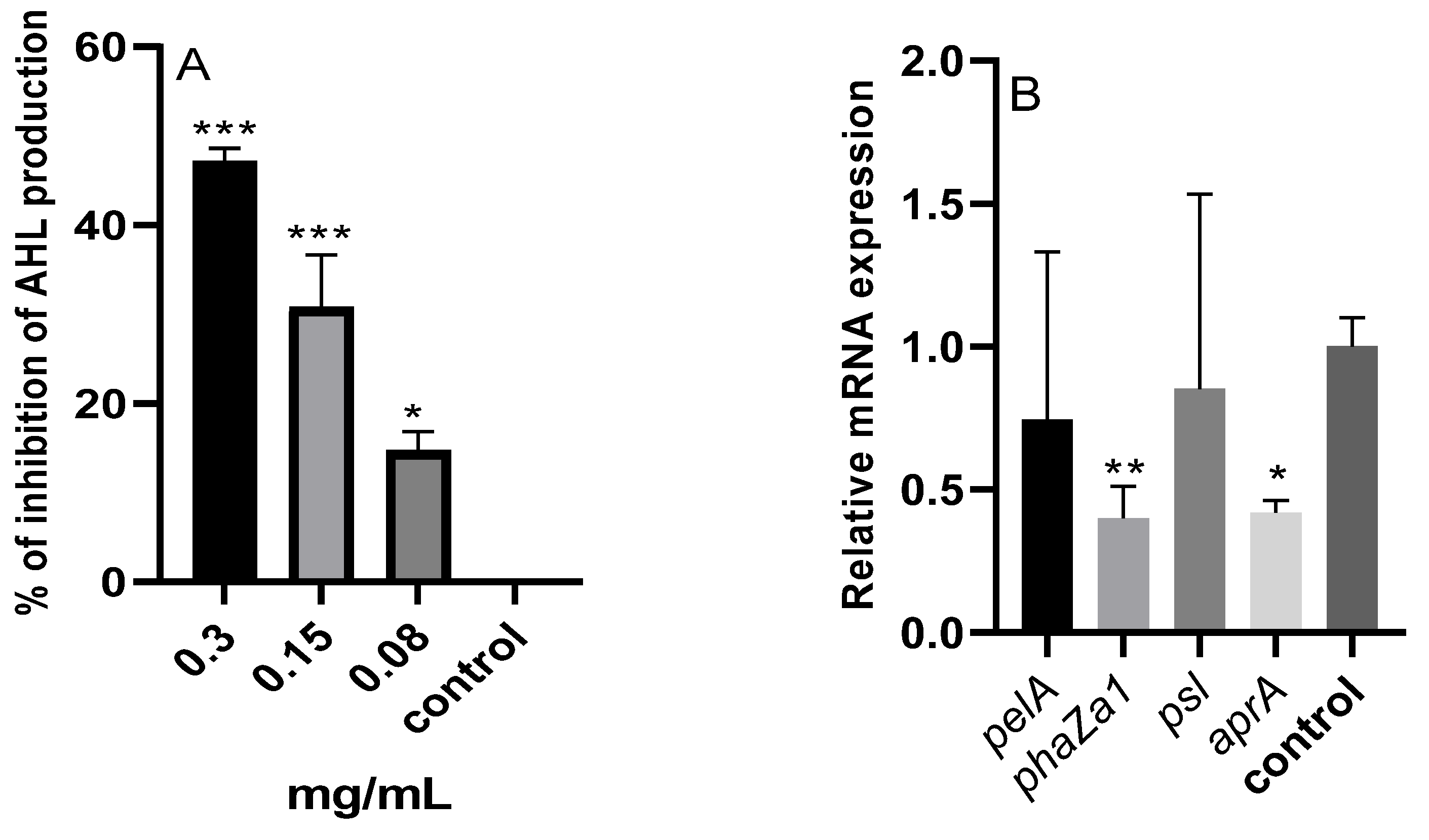
2.9.1. N-Acyl Homoserine Lactones (AHL)

As seen in Figure 8A, the assessment was carried out to ascertain the impact of the M. aurea EO on P. aeruginosa’s production of AHL. There was a discernible drop in AHL, with the percentage of the inhibition changing according to the concentration. At doses of 0.3 and 0.15 mg/mL, the generation of AHL decreased significantly (p < 0.001), with corresponding inhibition percentages of 47.14 and 30.76%. Significant (p < 0.05) was also the notable drop of 14.74% at the treatment concentration of 0.08 mg/mL.

2.9.2. qRT-PCR

The mRNA expression levels of four genes were studied using RT-PCR (Figure 8B). Treating P. aeruginosa with a concentration of 0.3 mg/mL of the M. aurea EO resulted in a significant reduction in the expression of phaZa1 (p < 0.01) and aprA (p < 0.05) genes. There was no significant reduction in the expression of the pelA and psl genes.

3. Discussion

Biofilms present a substantial and enduring threat, especially within the realm of various diseases, as they are linked to elevated rates of both illness and mortality [25,26]. Biofilms frequently exhibit limited susceptibility to antibiotics, prompting the need for the exploration and development of alternative methods to address infectious diseases associated with biofilms [27]. QS is a crucial mechanism that regulates the expression of genes associated with virulence factors. This regulatory system functions by assessing cell density and relies on extracellular signals [27,28]. Consequently, the substances that specifically target the QS system will ultimately hinder intercellular communication by exerting an influence on the genes that are engaged in QS. Medicinal herbs are rich in natural compounds that possess the ability to interfere with QS signals, consequently exerting a direct impact on the control of virulence factors linked to a wide range of diseases [18,29,30].
The current work utilized the EO of M. aurea to target P. aeruginosa and analyzed its efficacy against different virulence factors expressed by this bacterium. It showed that α-bisabolol oxide A, β-bisabolene, and α-farnesene were the main ingredients in the M. aurea EO. Few investigations have documented the chemical composition of M. aurea, revealing the existence of various significant chemicals that may be attributed to both abiotic and biotic causes. Khan et al. [31] found that sesquiterpene hydrocarbons were the main chemical class present in the aerial parts of Jordanian M. aurea oil. Among these, (E, E)-α-farnesene accounted for 50.2% of the composition, followed by γ-gurjunenepoxide at 8.5%, (E)-β-farnesene at 8.1%, and (Z, E)-α-farnesene at 4.4%. On the other hand, the most dominant chemical group in the Saudi M. aurea EO was oxygenated sesquiterpenes. The dominant compositions in this class were α-bisabolol at 27.8%, γ-gurjunenepoxide at 21.7%, (E, E)-α-farnesene at 16.3%, and cis-spiroether at 7.5%. The primary components of the M. aurea EO, as described in another study from Saudi Arabia, are bisabolol oxide A (64.8%), n-Nonadecane (6.7%), and (2R,3R, ALL-E)-2,3-epoxy-2,6,10,14-tetramethyl-16-(phenylthio) hexadeca-6,10,14-triene (5.8%) [32]. A prior study conducted in Iran indicated that α-bisabolene oxide A (59%), α-bisabolol oxide A (22.3%), and chamazolene (8.8%) were the primary constituents of the M. aurea EO [33]. When comparing the current study with existing research, it became evident that the EO from M. aurea contained similar chemical components, but their respective proportions varied significantly. The differences in the EO composition can be credited to many factors, such as weather conditions, nutrient availability, soil type, and the timing of harvesting.
The results of the antibacterial activity of the M. aurea EO against P. aeruginosa are in agreement with other reports [24,34]. Studies also showed that the antibacterial activity of the methanol, ethanol, acetone, ethyl acetate, and chloroform extracts of M. aurea was mild against P. aeruginosa [35]. However, the M. aurea EO and extracts possess broad-spectrum antibacterial activity against both gram-positive and gram-negative bacteria [24,34,35]. In addition, plant species belonging to the same family, Asteraceae, have been found to exhibit broad-spectrum antimicrobial activity against Gram-positive and Gram-negative as well as P. aeruginosa, such as Ageratum conyzoides, Tagetes erecta, Eucalyptus occidentalis, E. striaticalyx, and E. stricklandii [36,37,38].
The results of this study also underscored that the EO from M. aurea possesses the capability to inhibit biofilm formation without impacting the viability of the cells. Prior research has yielded similar outcomes, demonstrating that EOs derived from different plants effectively hinder the production of biofilms by various microbes. For instance, reports on the EOs from Tagetes minuta, cinnamon oil, Thymus daenensis, and Satureja hortensis found that they effectively prevented the production of biofilms, as well as other virulence factors mediated by QS [31,39,40]. Plants belonging to the same family as M. aurea have been reported to have antibiofilm activity, such as Launaea capitata [41], Helichrysum caespitium [42], and Vernonia adoensis [43].
The biofilm develops in a series of stages, starting with the creation of the initial biofilm layer, then followed by the movement of microorganisms to the surface, attachment, reproduction, maturation, and finally detachment [44]. The initial stage of biofilm growth was determined to be primary attachment and surface adherence. Bacterial adherence is primarily facilitated by the extracellular polymeric matrix composition, cell surface hydrophobicity, and flagellar motility [45]. This study found that the EO of M. aurea effectively reduced the hydrophobicity of bacteria. The decrease in hydrophobicity can be attributed to the presence of EO components that bind to the site of adhesion, thereby diminishing the hydrophobicity of the bacteria. The hydrophobicity of a surface affects the early attachment phase in the formation of a biofilm. The reduction in hydrophobicity may account for the decrease in biofilm growth by inhibiting adhesion. Furthermore, the results indicated that the M. aurea EO hindered bacterial aggregation, hence impeding the formation of biofilms. Furthermore, bacterial motility is a highly significant component that can affect the onset of biofilm development. Swarming motion substantially facilitates the first adhesion [46]. A crucial element necessary for P. aeruginosa swarming movement is the synthesis of rhamnolipid as a biosurfactant, which functions to lower surface tension and enable motility across the surface [47]. This study found that the sub-MIC M. aurea EO had a dose-dependent inhibitory effect on swarming motility and rhamnolipids production. This may suggest that one of the mechanisms preventing swarming motility is the inhibition of rhamnolipid production. O’May and Tufenkji [48] reported that a rhamnolipid supplement could partially restore the inhibition of swarming motility caused by the extracts.
The production of EPS is necessary for the establishment of the biofilm structure and the formation of the microcolony. The presence of EPS in bacteria provides a protective barrier that hinders the entry of antibiotics into the bacterial cell, hence contributing to antibiotic resistance [49]. Furthermore, the formation of EPS leads to alterations in the structure of biofilms, which are linked to heightened resistance against antibacterial medications. Therefore, decreasing the formation of EPS will enhance the elimination of biofilms by intensifying their contact with antimicrobial substances. This study demonstrates that the M. aurea EO significantly reduces the production of EPS.
A preliminary investigation showed that M. aurea EO possesses anti-QS activity, as indicated by the reduction in violacein production in C. violaceum. Violacein is produced via C. violaceum as an acyl homoserin lactone purple pigment. The inhibition of this pigment production indicates an inhibition of the QS pathway and an agent that inhibits the production of violacein was considered a possible anti quorum-sensing candidate [50].
QS is a crucial regulatory mechanism employed by bacteria, including the opportunistic pathogen P. aeruginosa, to coordinate gene expression in a population-dependent manner [51]. P. aeruginosa utilizes QS-dependent genes to sense and respond to changes in cell density through the production and detection of signaling molecules, such as N-acyl homoserine lactones [51]. The activation of QS-dependent genes allows P. aeruginosa to synchronize behaviors, such as biofilm formation, virulence factor secretion, and antibiotic resistance, in a concerted manner [52]. This sophisticated communication system enhances the adaptability and survival of the bacterial community in diverse environments. Understanding the role of QS-dependent genes is crucial for devising strategies to disrupt bacterial communication and control pathogenicity [52].
The use of M. aurea resulted in a substantial decrease in the production of protease, chitinase, pyocyanin, and rhamnolipid by P. aeruginosa, as seen in this investigation. The production of these molecules occurs outside of cells, and they serve as markers for assessing the QS system [53]. These variables have a crucial role in the pathogenicity of P. aeruginosa. The secretion of elastase and protease plays a crucial role in adhesion and colonization. Pyocyanin oxidizes glutathione and reduces its concentration in airway epithelial cells, hence diminishing an essential cellular antioxidant that plays a crucial role in regulating redox-sensitive signaling pathways [54,55]. Rhamnolipids function as a surfactant and facilitate the initiation of biofilm formation [56]. The substantial decrease in the generation of these harmful substances suggests that the M. aurea EO exhibits unique anti-QS activity that may disrupt the ability of P. aeruginosa to cause disease and impede the progression of the infection.
Administering M. aurea to P. aeruginosa led to a notable decrease in the release of AHL. This finding suggests that the EO hindered QS by interfering with the QS mechanisms, such as the las, rhl, and pqs systems [57]. Inhibiting these QS systems decreases the production of autoinducers (AIs) such as AHL. P. aeruginosa has a minimum of four operational QS systems, with two of them controlled by an AHL called Las and Rhl. The other two systems are regulated by quinolones and carbaldehydes, known as the Pqs and Iqs QS systems, respectively. The LAS system regulates the Rhl system, while the PQS system up-regulates the Rhl circuit. The Iqs system governs the Pqs system and RhlA system [58].
AIs control the activation of virulence pathways, enhance their signaling route through positive feedback, and have the ability to modify other systems through interconnected QS circuits [59]. The activation of these QS circuits controls the gene expression of different virulence factors. The Las circuit induces the activation of genes such as lasB and lasA (elastases A and B), aprA (AprA alkaline protease), as well as psl and pel (Psl and Pel exopolysaccharides) [60]. Furthermore, the Rhl circuit not only controls these virulence pathways but also stimulates the release of rhamnolipids [61,62]. The Pqs circuit primarily induces the release of pyocyanin, as well as the synthesis of elastases A and B, hydrogen cyanide, lectins A and B, two types of exopolysaccharides (Pel and Psl), and rhamnolipids [63,64]. This study also assessed the impact of M. aurea on the mRNA expression of the las-dependent genes aprA, psl, and pel, as well as the rhl-dependent gene phaZa1. The activation of the phaZa1 gene regulates the release of pyocyanin at a high level [65]. P. aeruginosa releases the virulence factor alkaline protease (AprA) to improve its ability to survive. AprA enzymatically cleaves monomeric flagellin, resulting in reduced activation of Toll-like receptor 5. Furthermore, AprA enzymatically breaks down host proteins, including complement proteins and cytokines. According to reports, QS has a considerable impact on the expression of the Pel and Psl operons. These operons play a crucial role in the production of two important matrix polysaccharides, Pel and Psl [66]. The secretion of these chemicals is essential for the processes of biofilm adherence, structure, and protection. Treating P. aeruginosa with M. aurea resulted in a significant decrease in the expression of aprA and phaZa1 genes. However, there was no significant difference in the expression of the pelA and pls genes. P. aeruginosa, in reality, generates a minimum of three distinct polysaccharides outside of its cells, which contribute to the formation of biofilms. These polysaccharides are known as alginate, PEL, and PSL [67]. P. aeruginosa organizes the production of its biofilm matrix exopolysaccharides. P. aeruginosa decreases the production of other polysaccharides when the production of one specific polysaccharide is raised to preserve equilibrium in the general quantity of exopolysaccharides. The excessive production of Psl significantly reduced the synthesis of alginate. The activation of Pel also inhibited the production of Psl polysaccharides. Additionally, the excessive production of alginate had a significant impact on the synthesis of other lipopolysaccharides [68].
More than 5000 sesquiterpenes derivatives have been identified, and the Asteraceae family appears to be the major supplier of these derivatives. They have been reported to have broad biological activity, including antimicrobial, anti-tumor, anti-ulcer, and anti-diabetic activities [69]. The biofilm and QS inhibitory effect of the M. aurea EO can be related to the presence of its primary components of sesquiterpenes, including α-bisabolol oxide A, β-bisabolene, and α-farnesene. However, the combined effect of both main and minor compounds should not be ignored. Significantly, extracts containing a high concentration of bisabolol and bisabolol derivatives have been found to possess antibiofilm and anti-QS properties. The concentration of 0.5 µg/mL of Matricaria chamomilla’s EO, which consists of α-bisabolol oxide B (7.3%) and α-bisabolol (7.3%) as its main constituents, leads to a notable decrease in both P. aeruginosa biofilm and alginate formation [70]. The extract from the brown macroalga Padina gymnospora, which contains 69% α-bisabolol, reduces the formation of biofilms and the expression of virulence factors controlled by QS in Serratia marcescens [71].
Certain limitations need to be emphasized in this study. The investigation was carried out using a single clinically isolated bacterium, potentially impeding the broad applicability of this study’s outcomes. P. aeruginosa serves as the model organism, and to enhance result generalization, the inclusion of other bacteria, such as S. aureus and E. coli, is necessary. To ensure the trial’s quality, positive controls are essential for both biofilm and virulence factors. Additional parameters are requisite to validate the inhibitory impact on biofilm and virulence factors and substantiate the findings. The examination of only a few quorum-sensing genes has taken place, and incorporating other genes or employing more sophisticated protocols could contribute to elucidating the precise mechanism of action.

4. Materials and Methods

4.1. Chemicals

Tetrazolium salt 2,3,5-triphenyl-tetrazolium chloride (TTC), oricinaol, and Carbazole were from Santa Cruz Biotechnology, Dallas, TX, USA. Azocasein and n-hexadecane were purchased from Sigma Aldrich, St. Louis, MO, USA. Trichloroacetic acid (TCA) was from Thermo Fischer Scientific, Shanghai, China. Chitin azure was from Bioscientific Carbosynth, Compton, UK. Muller Hinton broth (MHB), Muller Hinton Agar (MHA), and Luria Bertani broth (LB) were from Eco biolab, Budapest, Hungary. DMSO was from Alpha Chemika, Mumbai, India. Erythromycin disc was from Oxoid, UK. Diethyl ether was from SDFCL, Mumbai, India. Ethanol and ethyl acetate were from AZ chem, EU, Phoenix, AZ, USA.

4.2. Plant Materials and EO Extraction

M. aurea was collected in March 2021 from the Al-Karak region in Jordan. Dr. Feryal Al-Khresat from the Department of Biology at Mu’tah University in Al-Karak, Jordan successfully determined the taxonomic classification of the plant. The voucher specimens (NO: MU2023-08) were stored in the Department of Biology, Faculty of Science, Mutah University, Mutah, Jordan.
A 100 g sample of the plant material’s fresh aerial parts was subjected to hydro-distillation for 3 h utilizing simple Clevenger equipment. The aforementioned technique was repeated more than 10 times. The oil was separated from the water phase (1 L) using diethyl ether (100 mL). The diethyl ether was volatilized, and the oil was dehydrated using anhydrous sodium sulfate. Ultimately, the obtained oil was preserved at a temperature of 4 °C until it could be further examined.
Stock solutions of the M. aurea EO were made using 10% DMSO. For treated cultures, the required final concentrations of the oil were prepared using sterile broth, and the adjusted bacterial culture was added in a proportion of 1:20 (v:v). The untreated culture was prepared using broth media and the adjusted bacterial culture in a proportion of 1:20 (v:v). In all experiments, a solution containing broth media, the extract solvent (DMSO), and the adjusted bacterial culture in a proportion of 1:20 (v:v) was used as a negative control.

4.3. Gas Chromatography Mass Spectrometry

The GCMS investigation was performed using a Shimadzu qp2010 Plus (Kyoto, Japan). A column of DB5-SMS (30 m × 0.25 mm ID × 0.25 m of film thickness) was utilized. The volume of injection was 1.0 in a ratio of 1:100. Helium was used as a mobile phase, and it was operated at a flow rate of 1 mL/min. The heating rate was programmed at 4 °C/min starting from 50 °C (initial temperature) to 290 °C (final temperature) and held at 50 °C for five minutes with a total run time of 68 min. Both the injector and transfer-line temperatures were set at 250 °C. Electron ionization was the mode used in the MS with a 70 electron volt (eV) electron energy and a 250° C ion source temperature. The identification of the eluted peaks was made based on the retention time and the linear index of standard alkane compounds (C8–C20). In addition, the MS spectra of the candidate compounds were compared with their analogs in the NIST library and published data [24,31,72]. Indeed, co-chromatography was performed for certain compounds (α-terpinene, limonene, β-caryophyllene, α-humulene, and myristicin) under similar circumstances. The percentage of each identified compound was calculated based on the peak area of the identified compound as a proportion of the total area of all detected peaks.

4.4. Bacterial Strains

P. aeruginosa was obtained from a urine sample of a patient diagnosed with a urinary tract infection at the AlKarak Government Hospital in Karak, Jordan. The identification of the bacteria was performed using the Biomérieux VITEK® 2 system, Marcy-l’Étoile, France. The organism was identified as P. aeruginosa, which produces β-Lactamase. The strain Chromobacterium violaceum ATCC 12472 was acquired from the American Type Culture Collection (ATCC, Manassas, VA, USA).
The preparation of the bacterial culture was made by inoculating one single colony into a sterile 5 mL nutrient broth (NB) and incubating for 24 h at 37 °C. The prepared culture was adjusted to 0.5 McFarland Standard (1.5 × 10⁸ CFU/mL) using sterile broth.

4.5. Antibacterial Activity

4.5.1. Disc Diffusion Assay

The experiment was conducted using a cell density of 1.5 × 10⁸ of P. aeruginosa with MHA as the medium. A volume of 100 µL from a 24 h old bacterial culture was applied onto the agar surface using a swab. Subsequently, an azsterile disc was loaded with 1 mg of the prepared EO. The disc was allowed to dry and thereafter put onto the surface of the agar plate. Erythromycin (10 µg) was also tested as a positive control. The inhibitory zone that formed after the 24 h incubation at 37 °C was measured in millimeters [73].

4.5.2. MIC

The minimum inhibitory concentration (MIC) of the M. aurea EO against P. aeruginosa was assessed by utilizing a stock solution of 10 mg/mL of 10% DMSO. A 96-well plate was utilized, along with MHB (Mueller Hinton Broth). The EO was diluted by a factor of two to achieve final concentrations of 5, 2.5, 1.25, 0.6, 0.3, 0.15, 0.08, 0.04, 0.02, and 0.01 mg/mL. Then, a 10 µL bacterial inoculum containing a 1.5 × 10⁸ CFU/mL was added. The MIC is determined when the concentration being tested completely inhibits the observable growth of the bacteria after 24 h incubation at 37 °C using an ELIZA reader (MCL-2100C, Guangzhou, China) [74].

4.6. Cell Viability

A 96-well plate was prepared in a manner identical to the one prepared in the MIC methodology. After being incubated for 24 h at a temperature of 37 °C, the contents of each well were removed and cleaned extensively. Following the drying process, 200 µL of broth media containing 0.2% glucose and 50 µL of TTC (tetrazolium salt 2,3,5-triphenyl-tetrazolium chloride) solution were introduced into each well. The samples were then incubated in darkness at a temperature of 37 °C and a shaking speed of 150 rpm. The absorbance was measured at a wavelength of 405 nm after 6 h [27].

4.7. Antibiofilm

The antibiofilm efficacy was assessed using a crystal violet assay. A polystyrene 96-well plate was prepared in a manner identical to the one prepared in the MIC methodology. Following a 24 h incubation period at 37 °C, the contents of each well were discarded and thoroughly washed. Subsequently, crystal violet was introduced into each well. After 15 min of incubation at room temperature, the contents were removed and subsequently cleaned using tap water. Subsequently, an ethanol (98%) decolorizer was introduced into each well and left for 15 min. Next, the contents of each well were transferred to a new 96-well plate, and the optical density (OD590) was recorded. The biofilm inhibition percentage was determined relative to the optical density (OD590) of the standard P. aeruginosa biofilm [26].

4.8. Biofilm Visualization

Untreated and treated P. aeruginosa were cultivated on a 6-well plate with glass coverslips for 24 h at a temperature of 37 °C. Then, the coverslips were removed. A batch of these prepared coverslips was treated for inspection under a light microscope (NIKON, Tokyo, Japan). The coverslips were subjected to crystal violet staining for 1 min, followed by thorough rinsing and subsequent drying. The second set was utilized for scanning electron microscope (SEM) observation. The coverslips were fixed in 2.5% glutaraldehyde for 1 h. Furthermore, the coverslips were cleaned using a sodium acetate buffer with a concentration of 0.1 M and a pH of 7.3. Then, they were dehydrated using a sequence of ethanol solutions with different concentrations (10, 30, 50, 70, and 100%). Afterward, the samples underwent analysis using scanning electron microscopy (Thermo Scientific Phenom Desktop SEM, JU-24112022, Waltham, MA, USA).

4.9. Violacein Assay

The impact of the M. aurea EO on the synthesis of violacein by C. violaceum was assessed qualitatively by the well diffusion method and quantitatively using a spectrophotometric methodology. For the well diffusion approach, a volume of 100 µL of C. violaceum containing 1 × 10⁶ CFU/mL was evenly spread on LB agar. Subsequently, a well was created at the center of the cultivated plate, and 100 µL of DMSO containing 1 mg of the M. aurea EO was added to it. The incubation was conducted at a temperature of 37 °C for 24 h. The diameter of the inhibitory zone was determined in mm.
To conduct the quantitative study, untreated and treated cultures of C. violaceum were prepared and incubated at 37 °C for 24 h. Then, a 1 mL aliquot of each culture was subjected to centrifugation, and the resulting pellet was collected and suspended in DMSO. The mixture was agitated vigorously and the DMSO extract was subsequently separated from the cells using centrifugation. The concentration of crude violacein in the collected supernatant was quantified by measuring the absorbance at 575 nm with a comparison to the untreated control culture [75].

4.10. Effect on Biofilm Development

4.10.1. Swarming Motility

The test was conducted using semisolid swarming agar plates [76]. Following autoclaving, varying amounts of the M. aurea EO were introduced into the empty Petri dishes, and subsequently, the autoclaved media was poured to achieve the desired final concentrations. The agar plates were allowed to solidify, and a single dot of P. aeruginosa was cultured. The motility zone was evaluated in millimeters after a 48 h incubation period at 37 °C.

4.10.2. Aggregation

After the untreated and treated P. aeruginosa cultures were incubated for 24 h at 37 °C, the OD at 600 nm was measured using a spectrophotometer (Shimadzu UV-1601, Kyoto, Japan). Then, portions of these cultures were taken and vortexed for 60 s. Then, the OD was measured again. Accordingly, the percentage of aggregation was determined [77].

4.10.3. Hydrophobicity

An initial absorbance at 600 nm (OD1) for untreated and treated cultures of P. aeruginosa incubated for 24 h at 37 °C was reported. Then, a 1:1 mixture of these cultures and n-hexadecane was made. The prepared mixture was vortexed using a vortex (Witeg, Wertheim, Germany) for 120 s. The aqueous phase was collected, and the OD at 600 nm was measured (OD2). Then, the percent of hydrophobicity was determined [78].

4.10.4. EPS Production

The untreated and treated cultures of P. aeruginosa were incubated for 24 h at 37 °C. Subsequently, the culture underwent centrifugation, and a portion (1 mL) of the resultant supernatant was obtained and combined with cold ethanol (3 mL). Following a 24 h incubation at 4 °C, the combination underwent centrifugation, and the collected pellet was suspended in water (3 mL). To determine the total carbohydrates, the water containing EPS was treated with a 1:5 solution of phenol (5%) and H2SO4 (98%). The OD at 490 nm was reported, and the percentage of the EPS was determined about the untreated culture [79].

4.11. Effect on Virulence Factors

4.11.1. Pyocyanin

The untreated and treated culture of P. aeruginosa was incubated for 24 h at 37 °C. Next, the culture was subjected to centrifugation (Bibby Sterilin Ltd., Bargoed, UK), and a part of the resulting supernatant was combined with chloroform to isolate the pyocyanin pigment. The extraction solution was vigorously agitated using a vortex mixer, and the resulting green-blue layer was isolated and combined with hydrochloric acid (HCl). The pink solution that was created was collected and the amount of pyocyanin present was quantified using a spectrophotometer at a wavelength of 520 nm [80].

4.11.2. Rhamnolipids

The untreated and treated culture of P. aeruginosa was incubated for 24 h at 37 °C. Next, the culture was subjected to centrifugation, and a part of the resulting supernatant was combined with diethyl ether to isolate the rhamnolipids. Next, a fraction of the diethyl ether layer was collected, and the solvent was evaporated. The crude material was mixed with 200 µL of water. Subsequently, a solution of orcinol-sulfuric acid was introduced. The solution was placed in an incubator and maintained at a temperature of 24 °C for 30 min. The optical density at 421 nm was determined and used to compute the proportion of rhamnolipid formation [81].

4.11.3. LasA Protease

The untreated and treated culture of P. aeruginosa was incubated for 24 h at 37 °C. Next, the culture was subjected to centrifugation, and a part (100 µL) of the resulting supernatant was collected. Then, 100 µL of azocasein and 3 mL of phosphate buffer (50 mM, pH 7) were introduced. The solution was incubated for 1 h at a temperature of 37 °C, with a shaking speed of 150 rpm. Subsequently, 500 microliters of trichloroacetic acid were introduced. Following a 10 min period, the solution underwent centrifugation, and the OD of the liquid portion was measured at 366 nm [82].

4.11.4. Chitinase

An exact quantity of 1.3 mg/mL of chitin azure was introduced into 130 mL of a sodium phosphate buffer with a concentration of 50 mM (pH 7.0). The mixture was then incubated for 7 days at a temperature of 37 °C, with a shaking speed of 150 rpm. Simultaneously, both the untreated and treated culture of P. aeruginosa was incubated for 24 h at a temperature of 37 °C. Subsequently, the culture underwent centrifugation, and a portion (500 µL) of the resultant supernatant was obtained and combined with the chitin azure solution (4.5 mL). Following 24 h, the combination underwent centrifugation, and the OD of the resulting supernatant was reported at 570 nm [80].

4.12. Mechanism of QS

4.12.1. N-Acyl Homoserine Lactones (AHL)

The untreated and treated cultures of P. aeruginosa were incubated for 24 h at 37 °C. Subsequently, the culture underwent centrifugation, and a portion (2 mL) of the supernatant was obtained and combined with ethyl acetate (3 mL). The solution was vortexed for 10 min and incubated at 40 °C. After 24 h, a portion of the ethyl acetate phase was mixed with 50 μL of a (1:1) mixture of (2M) hydroxyl amine and NaOH (3.5 M). Then, 90 μL of a (1:1) mixture of 10% ferric chloride in 4 M HCl was added to 95% ethanol. Then, the OD at 520 nm was measured, and the percentage of AHL inhibition was determined in the untreated culture [83].

4.12.2. qRT-PCR

Total RNA extraction from the P. aeruginosa cultures was processed using the Direct-zol™ RNA Miniprep kit (Zymo Research Company, Tustin, CA, USA) following the manufacturer’s instructions. Isolated RNA was then used for complementary DNA synthesis using SensiFAST™ cDNA Synthesis Kit (Bioline Reagents, London, UK) according to the manufacturer’s steps.
The Bioline SensiFAST™ SYBR® No-ROX Kit (Bioline Reagents, UK) workflow was used to quantify genes of interest in a technical triplicate manner for each sample, followed by a melting curve run and quality analysis. Genes of interest expression were normalized to the expression of 16S rRNA as a housekeeping gene using the 2ΔCT formula. The primers (Forward and Reverse) used in qPCR are listed in Table 3.

4.13. Statistical Analysis

All experiments were performed in triplicate. Results were reported in the form of standard deviation, which was calculated using the Microsoft Excel 2009 software. The figures were prepared using GraphPad Prism (8.0). A one-way ANOVA was used to determine the significant difference between groups based on P-values as follows: * p < 0.05, ** p < 0.01, *** p < 0.001 compared to control untreated cells.

5. Conclusions

In conclusion, this study provides compelling evidence supporting the potential of the EO derived from M. aurea as a promising candidate for the treatment of infectious diseases associated with biofilm formation. The EO has demonstrated effective modulation of key virulence factors in P. aeruginosa, notably through disruption of the QS system. The observed efficacy could result from the presence of dominant active compounds or minor ingredients, possibly operating synergistically. This research not only underscores the therapeutic promise of the M. aurea derived EO but also suggests avenues for further exploration of its complex molecular mechanisms and potential applications in combating biofilm-associated infections.

Author Contributions

Conceptualization, H.Q., S.A.M.S., M.O.A.-l. and R.A.M.; methodology, H.Q., S.M.D., S.A.M.S., Y.T., H.M.A.-Y. and A.M.A.; software, H.Q., B.E.M.A.-A. and L.A.-O.; validation, H.Q., K.K., B.E.M.A.-A. and L.A.-O.; formal analysis, H.Q., S.A.M.S., Y.T. and R.A.M.; investigation, H.Q., S.A.M.S., M.O.A.-l., S.M.D., K.K., B.E.M.A.-A., Y.T., H.M.A.-Y. and A.M.A.; resources, H.Q., R.A.M., H.M.A.-Y. and A.M.A.; data curation, S.M.D., K.K. and B.E.M.A.-A.; writing—original draft preparation, H.Q., R.A.M., S.A.M.S., M.O.A.-l., S.M.D. and R.A.M.; writing—review and editing, K.K., B.E.M.A.-A., L.A.-O., Y.T., H.M.A.-Y. and A.M.A.; visualization, H.Q., R.A.M. and Y.T.; supervision, R.A.M. and H.Q.; project administration, R.A.M.; funding acquisition, R.A.M., H.M.A.-Y. and A.M.A. All authors have read and agreed to the published version of the manuscript.

Funding

The authors extend their appreciation to Researchers Supporting Project number (RSP2024R119), King Saud University, Riyadh, Saudi Arabia, for funding this work.

Institutional Review Board Statement

Not applicable.

Informed Consent Statement

Not applicable.

Data Availability Statement

Data is contained within the article.

Conflicts of Interest

The authors declare no conflicts of interest.

References

  1. Qin, S.; Xiao, W.; Zhou, C.; Pu, Q.; Deng, X.; Lan, L.; Liang, H.; Song, X.; Wu, M. Pseudomonas aeruginosa: Pathogenesis, Virulence Factors, Antibiotic Resistance, Interaction with Host, Technology Advances and Emerging Therapeutics. Signal Transduct. Target. Ther. 2022, 7, 199. [Google Scholar] [CrossRef]
  2. Paprocka, P.; Durnaś, B.; Mańkowska, A.; Król, G.; Wollny, T.; Bucki, R. Pseudomonas aeruginosa Infections in Cancer Patients. Pathogens 2022, 11, 679. [Google Scholar] [CrossRef]
  3. Wood, S.J.; Kuzel, T.M.; Shafikhani, S.H. Pseudomonas aeruginosa: Infections, Animal Modeling, and Therapeutics. Cells 2023, 12, 199. [Google Scholar] [CrossRef] [PubMed]
  4. Karruli, A.; Catalini, C.; D’Amore, C.; Foglia, F.; Mari, F.; Harxhi, A.; Galdiero, M.; Durante-Mangoni, E. Evidence-Based Treatment of Pseudomonas aeruginosa Infections: A Critical Reappraisal. Antibiotics 2023, 12, 399. [Google Scholar] [CrossRef] [PubMed]
  5. Tapper, M.L.; Armstrong, D. Bacteremia Due to Pseudomonas aeruginosa Complicating Neoplastic Disease: A Progress Report. J. Infect. Dis. 1974, 130, S14–S23. [Google Scholar] [CrossRef] [PubMed]
  6. Chatzinikolaou, I.; Abi-Said, D.; Bodey, G.P.; Rolston, K.V.I.; Tarrand, J.J.; Samonis, G. Recent Experience with Pseudomonas aeruginosa Bacteremia in Patients with Cancer: Retrospective Analysis of 245 Episodes. Arch. Intern. Med. 2000, 160, 501–509. [Google Scholar] [CrossRef] [PubMed]
  7. Kang, C.-I.; Kim, S.-H.; Kim, H.-B.; Park, S.-W.; Choe, Y.-J.; Oh, M.; Kim, E.-C.; Choe, K.-W. Pseudomonas aeruginosa Bacteremia: Risk Factors for Mortality and Influence of Delayed Receipt of Effective Antimicrobial Therapy on Clinical Outcome. Clin. Infect. Dis. 2003, 37, 745–751. [Google Scholar] [CrossRef] [PubMed]
  8. Elmassry, M.M.; Colmer-Hamood, J.A.; Kopel, J.; San Francisco, M.J.; Hamood, A.N. Anti-Pseudomonas aeruginosa Vaccines and Therapies: An Assessment of Clinical Trials. Microorganisms 2023, 11, 916. [Google Scholar] [CrossRef] [PubMed]
  9. Pang, Z.; Raudonis, R.; Glick, B.R.; Lin, T.-J.; Cheng, Z. Antibiotic Resistance in Pseudomonas aeruginosa: Mechanisms and Alternative Therapeutic Strategies. Biotechnol. Adv. 2019, 37, 177–192. [Google Scholar] [CrossRef] [PubMed]
  10. Centers for Disease Control and Prevention. Antibiotic Resistance Threats in the United States, 2019; Centers for Disease Control and Prevention: Atlanta, GA, USA, 2019. [Google Scholar]
  11. Shrivastava, S.R.; Shrivastava, P.S.; Ramasamy, J. World Health Organization Releases Global Priority List of Antibiotic-Resistant Bacteria to Guide Research, Discovery, and Development of New Antibiotics. J. Med. Soc. 2018, 32, 76–77. [Google Scholar] [CrossRef]
  12. Zloh, M. AR Mechanism-Based Drug Design. In Antibiotic Drug Resistance; John Wiley & Sons, Inc.: Hoboken, NJ, USA, 2019; pp. 275–309. [Google Scholar]
  13. Ciofu, O.; Tolker-Nielsen, T. Tolerance and Resistance of Pseudomonas aeruginosa Biofilms to Antimicrobial Agents—How P. aeruginosa Can Escape Antibiotics. Front. Microbiol. 2019, 10, 913. [Google Scholar] [CrossRef]
  14. Li, Z.; Nair, S.K. Quorum Sensing: How Bacteria Can Coordinate Activity and Synchronize Their Response to External Signals? Protein Sci. 2012, 21, 1403–1417. [Google Scholar] [CrossRef]
  15. Naoun, A.A.; Raphael, I.; Forsthuber, T.G. Immunoregulation via Cell Density and Quorum Sensing-like Mechanisms: An Underexplored Emerging Field with Potential Translational Implications. Cells 2022, 11, 2442. [Google Scholar] [CrossRef] [PubMed]
  16. Duplantier, M.; Lohou, E.; Sonnet, P. Quorum Sensing Inhibitors to Quench P. aeruginosa Pathogenicity. Pharmaceuticals 2021, 14, 1262. [Google Scholar] [CrossRef]
  17. Bouyahya, A.; Chamkhi, I.; Balahbib, A.; Rebezov, M.; Shariati, M.A.; Wilairatana, P.; Mubarak, M.S.; Benali, T.; El Omari, N. Mechanisms, Anti-Quorum-Sensing Actions, and Clinical Trials of Medicinal Plant Bioactive Compounds against Bacteria: A Comprehensive Review. Molecules 2022, 27, 1484. [Google Scholar] [CrossRef]
  18. Qaralleh, H. Chemical Composition and Quorum Sensing Inhibitory Effect of Nepeta curviflora Methanolic Extract against ESBL Pseudomonas aeruginosa. J. Pharmacopunct. 2023, 26, 307–318. [Google Scholar] [CrossRef] [PubMed]
  19. Bouyahya, A.; Dakka, N.; Et-Touys, A.; Abrini, J.; Bakri, Y. Medicinal Plant Products Targeting Quorum Sensing for Combating Bacterial Infections. Asian Pac. J. Trop. Med. 2017, 10, 729–743. [Google Scholar] [CrossRef]
  20. Qaralleh, H.; Khleifat, K.M.; Khlaifat, A.M.; Al-limoun, M.; Al-Tawarah, N.M.; Alhroob, A.M.; Alsaudi, A.B. Chemical Composition, Antioxidant and Inhibitory Effect of Cupressus sempervirens Essential Oils and Methanolic Extract on Beta-Lactamase Producing Isolates. Res. J. Pharm. Technol. 2021, 14, 4673–4679. [Google Scholar] [CrossRef]
  21. Camele, I.; Elshafie, H.S.; Caputo, L.; De Feo, V. Anti-Quorum Sensing and Antimicrobial Effect of Mediterranean Plant Essential Oils against Phytopathogenic Bacteria. Front. Microbiol. 2019, 10, 2619. [Google Scholar] [CrossRef] [PubMed]
  22. Oran, S.A.; Al-Eisawi, D.M. Check-List of Medicinal Plants in Jordan. Dirasat 1998, 25, 84–112. [Google Scholar]
  23. Qnais, E. The Analgesic Effect of the Ethanolic Extract of Matricaria aurea. Turk. J. Biol. 2011, 35, 347–352. [Google Scholar] [CrossRef]
  24. Kheder, F.B.H.; Mahjoub, M.A.; Zaghrouni, F.; Kwaja, S.; Helal, A.N.; Mighri, Z. Chemical Composition Antioxidant and Antimicrobial Activities of the Essential Oils of Matricaria aurea Loefl. Growing in Tunisia. J. Essent. Oil Bear. Plants 2014, 17, 493–505. [Google Scholar] [CrossRef]
  25. Høiby, N.; Ciofu, O.; Johansen, H.K.; Song, Z.; Moser, C.; Jensen, P.Ø.; Molin, S.; Givskov, M.; Tolker-Nielsen, T.; Bjarnsholt, T. The Clinical Impact of Bacterial Biofilms. Int. J. Oral Sci. 2011, 3, 55–65. [Google Scholar] [CrossRef] [PubMed]
  26. Qaralleh, H.; Al-Limoun, M.; Khlaifat, A.; Khleifat, K.; Al-Tawarah, N.; Hashem, A.-H.; Alsharafa, K. Antibacterial and Antibiofilm Activities of a Traditional Herbal Formula against Respiratory Infection Causing Bacteria. Trop. J. Nat. Prod. Res. 2020, 4, 527–534. [Google Scholar]
  27. Gordya, N.; Yakovlev, A.; Kruglikova, A.; Tulin, D.; Potolitsina, E.; Suborova, T.; Bordo, D.; Rosano, C.; Chernysh, S. Natural Antimicrobial Peptide Complexes in the Fighting of Antibiotic Resistant Biofilms: Calliphora vicina Medicinal Maggots. PLoS ONE 2017, 12, e0173559. [Google Scholar] [CrossRef]
  28. Qaralleh, H. Thymol Rich Thymbra capitata Essential Oil Inhibits Quorum Sensing, Virulence and Biofilm Formation of Beta Lactamase Producing Pseudomonas aeruginosa. Nat. Prod. Sci. 2019, 25, 172–180. [Google Scholar] [CrossRef]
  29. Zhao, X.; Yu, Z.; Ding, T. Quorum-Sensing Regulation of Antimicrobial Resistance in Bacteria. Microorganisms 2020, 8, 425. [Google Scholar] [CrossRef] [PubMed]
  30. Tonkin, M.; Khan, S.; Wani, M.Y.; Ahmad, A. Quorum Sensing-a Stratagem for Conquering Multi-Drug Resistant Pathogens. Curr. Pharm. Des. 2021, 27, 2835–2847. [Google Scholar] [CrossRef]
  31. Khan, M.; Khan, M.; Alshareef, E.; Alaqeel, S.I.; Alkhathlan, H.Z. Chemical Characterization and Chemotaxonomic Significance of Essential Oil Constituents of Matricaria aurea Grown in Two Different Agro-Climatic Conditions. Plants 2023, 12, 3553. [Google Scholar] [CrossRef]
  32. Siddiqui, N.A. Chemical Constituents of Essential Oil from Flowers of Matricaria aurea Grown in Saudi Arabia. Indian J. Drugs 2014, 2, 164–168. [Google Scholar]
  33. Jalali, Z.; Sefidkon, F.; Assareh, M.H.; Attar, F. Comparison of Sesquiterpens in the Essential Oils of Anthemis Hyalina DC., Matricaria recutita L. and Matricaria aurea (Loefl.) Schultz-Bip. Iran. J. Med. Aromat. Plants Res. 2008, 24, 31–37. [Google Scholar]
  34. Yaz, H.H.; Uysal, I.; Polat, A.O.; Mohammed, F.S.; Sevindik, M. Usage Areas, Biological Activities and Volatile Oil Compounds of Matricaria aurea and Matricaria chamomilla. Lek. Sirovine 2023, 43, 1. [Google Scholar] [CrossRef]
  35. Rizwana, H.; Alwhibi, M.S.; Soliman, D.A. Research Article Antimicrobial Activity and Chemical Composition of Flowers of Matricaria aurea a Native Herb of Saudi Arabia. Int. J. Pharmacol. 2016, 12, 576–586. [Google Scholar] [CrossRef]
  36. Khammassi, M.; Polito, F.; Amri, I.; Khedhri, S.; Hamrouni, L.; Nazzaro, F.; Fratianni, F.; De Feo, V. Chemical Composition and Phytotoxic, Antibacterial and Antibiofilm Activity of the Essential Oils of Eucalyptus occidentalis, E. striaticalyx and E. stricklandii. Molecules 2022, 27, 5820. [Google Scholar] [CrossRef]
  37. Trinh, P.-C.; Thao, L.-T.-T.; Ha, H.-T.-V.; Nguyen, T. DPPH-Scavenging and Antimicrobial Activities of Asteraceae Medicinal Plants on Uropathogenic Bacteria. Evid.-Based Complement. Altern. Med. 2020, 2020, 7807026. [Google Scholar] [CrossRef] [PubMed]
  38. Perović, J.; Šaponjac, V.T.; Kojić, J.; Krulj, J.; Moreno, D.A.; García-Viguera, C.; Bodroža-Solarov, M.; Ilić, N. Chicory (Cichorium Intybus L.) as a Food Ingredient–Nutritional Composition, Bioactivity, Safety, and Health Claims: A Review. Food Chem. 2021, 336, 127676. [Google Scholar] [CrossRef]
  39. Sharifi, A.; Nayeri Fasaei, B. Selected Plant Essential Oils Inhibit Biofilm Formation and LuxS-and Pfs-mediated Quorum Sensing by Escherichia coli O157:H7. Lett. Appl. Microbiol. 2022, 74, 916–923. [Google Scholar] [CrossRef]
  40. Kalia, M.; Yadav, V.K.; Singh, P.K.; Sharma, D.; Pandey, H.; Narvi, S.S.; Agarwal, V. Effect of Cinnamon Oil on Quorum Sensing-Controlled Virulence Factors and Biofilm Formation in Pseudomonas aeruginosa. PLoS ONE 2015, 10, e0135495. [Google Scholar] [CrossRef]
  41. Emad, F.; Khalafalah, A.K.; El Sayed, M.A.; Mohamed, A.-E.H.; Stadler, M.; Helaly, S.E. Three New Polyacetylene Glycosides (PAGs) from the Aerial Part of Launaea capitata (Asteraceae) with Anti-Biofilm Activity against Staphylococcus aureus. Fitoterapia 2020, 143, 104548. [Google Scholar] [CrossRef]
  42. Bassey, K.; Mamabolo, P.; Cosa, S. An Andrographolide from Helichrysum caespitium (Dc.) Sond. Ex Harv., (Asteraceae) and Its Antimicrobial, Antiquorum Sensing, and Antibiofilm Potentials. Biology 2021, 10, 1224. [Google Scholar] [CrossRef]
  43. Masuku, M.; Mozirandi, W.; Mukanganyama, S. Evaluation of the Antibacterial and Antibiofilm Effects of Ethyl Acetate Root Extracts from Vernonia adoensis (Asteraceae) against Pseudomonas aeruginosa. Sci. World J. 2023, 2023, 5782656. [Google Scholar] [CrossRef] [PubMed]
  44. Sauer, K.; Stoodley, P.; Goeres, D.M.; Hall-Stoodley, L.; Burmølle, M.; Stewart, P.S.; Bjarnsholt, T. The Biofilm Life Cycle: Expanding the Conceptual Model of Biofilm Formation. Nat. Rev. Microbiol. 2022, 20, 608–620. [Google Scholar] [CrossRef]
  45. Brindhadevi, K.; LewisOscar, F.; Mylonakis, E.; Shanmugam, S.; Verma, T.N.; Pugazhendhi, A. Biofilm and Quorum Sensing Mediated Pathogenicity in Pseudomonas aeruginosa. Process Biochem. 2020, 96, 49–57. [Google Scholar] [CrossRef]
  46. Shrout, J.D.; Chopp, D.L.; Just, C.L.; Hentzer, M.; Givskov, M.; Parsek, M.R. The Impact of Quorum Sensing and Swarming Motility on Pseudomonas aeruginosa Biofilm Formation Is Nutritionally Conditional. Mol. Microbiol. 2006, 62, 1264–1277. [Google Scholar] [CrossRef]
  47. Copeland, M.F.; Weibel, D.B. Bacterial Swarming: A Model System for Studying Dynamic Self-Assembly. Soft Matter 2009, 5, 1174–1187. [Google Scholar] [CrossRef]
  48. O’May, C.; Tufenkji, N. The Swarming Motility of Pseudomonas aeruginosa Is Blocked by Cranberry Proanthocyanidins and Other Tannin-Containing Materials. Appl. Environ. Microbiol. 2011, 77, 3061–3067. [Google Scholar] [CrossRef]
  49. Kassinger, S.J.; van Hoek, M.L. Biofilm Architecture: An Emerging Synthetic Biology Target. Synth. Syst. Biotechnol. 2020, 5, 1–10. [Google Scholar] [CrossRef]
  50. McLean, R.J.C.; Pierson, L.S., III; Fuqua, C. A Simple Screening Protocol for the Identification of Quorum Signal Antagonists. J. Microbiol. Methods 2004, 58, 351–360. [Google Scholar] [CrossRef]
  51. López-Jácome, L.E.; Garza-Ramos, G.; Hernández-Durán, M.; Franco-Cendejas, R.; Loarca, D.; Romero-Martínez, D.; Nguyen, P.T.D.; Maeda, T.; González-Pedrajo, B.; Díaz-Guerrero, M. AiiM Lactonase Strongly Reduces Quorum Sensing Controlled Virulence Factors in Clinical Strains of Pseudomonas aeruginosa Isolated from Burned Patients. Front. Microbiol. 2019, 10, 2657. [Google Scholar] [CrossRef] [PubMed]
  52. Shravani, V.; Selvi, G.A.S.; Mantravadi, H. Detection of Quorum Sensing Virulence Factor Genes and Its Consanguinity to Antibiotic Sensitivity Profile in the Clinical Isolates of Pseudomonas aeruginosa. Iran. J. Basic Med. Sci. 2023, 26, 899. [Google Scholar] [PubMed]
  53. Danaraj, J.; Mariasingarayan, Y.; Ayyappan, S.; Karuppiah, V. Seagrass Halodule Pinifolia Active Constituent 4-Methoxybenzioic Acid (4-MBA) Inhibits Quorum Sensing Mediated Virulence Production of Pseudomonas aeruginosa. Microb. Pathog. 2020, 147, 104392. [Google Scholar] [CrossRef]
  54. Karatuna, O.; Yagci, A. Analysis of Quorum Sensing-Dependent Virulence Factor Production and Its Relationship with Antimicrobial Susceptibility in Pseudomonas aeruginosa Respiratory Isolates. Clin. Microbiol. Infect. 2010, 16, 1770–1775. [Google Scholar] [CrossRef]
  55. O’Malley, Y.Q.; Reszka, K.J.; Spitz, D.R.; Denning, G.M.; Britigan, B.E. Pseudomonas aeruginosa Pyocyanin Directly Oxidizes Glutathione and Decreases Its Levels in Airway Epithelial Cells. Am. J. Physiol. Cell. Mol. Physiol. 2004, 287, L94–L103. [Google Scholar] [CrossRef]
  56. Rather, M.A.; Hassan, T.; Dar, B.A.; Shawl, A.S.; Qurishi, M.A.; Ganai, B.A. Essential Oil Composition of Nepeta Raphanorhiza Benth Growing in Kashmir Valley. Rec. Nat. Prod. 2012, 6, 67. [Google Scholar]
  57. Ashraf, M.I.; Ong, S.-K.; Mujawar, S.; Pawar, S.; More, P.; Paul, S.; Lahiri, C. A Side-Effect Free Method for Identifying Cancer Drug Targets. Sci. Rep. 2018, 8, 6669. [Google Scholar] [CrossRef]
  58. Pesci, E.C.; Pearson, J.P.; Seed, P.C.; Iglewski, B.H. Regulation of Las and Rhl Quorum Sensing in Pseudomonas aeruginosa. J. Bacteriol. 1997, 179, 3127–3132. [Google Scholar] [CrossRef]
  59. Soukarieh, F.; Williams, P.; Stocks, M.J.; Camara, M. Pseudomonas aeruginosa Quorum Sensing Systems as Drug Discovery Targets: Current Position and Future Perspectives. J. Med. Chem. 2018, 61, 10385–10402. [Google Scholar] [CrossRef]
  60. Ueda, A.; Wood, T.K. Connecting Quorum Sensing, c-Di-GMP, Pel Polysaccharide, and Biofilm Formation in Pseudomonas aeruginosa through Tyrosine Phosphatase TpbA (PA3885). PLoS Pathog. 2009, 5, e1000483. [Google Scholar] [CrossRef]
  61. Muimhneacháin, E.Ó.; Reen, F.J.; O’Gara, F.; McGlacken, G.P. Analogues of Pseudomonas aeruginosa Signalling Molecules to Tackle Infections. Org. Biomol. Chem. 2018, 16, 169–179. [Google Scholar] [CrossRef]
  62. Yan, S.; Wu, G. Can Biofilm Be Reversed through Quorum Sensing in Pseudomonas aeruginosa? Front. Microbiol. 2019, 10, 1582. [Google Scholar] [CrossRef]
  63. Turkina, M.V.; Vikström, E. Bacteria-Host Crosstalk: Sensing of the Quorum in the Context of Pseudomonas aeruginosa Infections. J. Innate Immun. 2019, 11, 263–279. [Google Scholar] [CrossRef]
  64. Lin, J.; Cheng, J.; Wang, Y.; Shen, X. The Pseudomonas quinolone Signal (PQS): Not Just for Quorum Sensing Anymore. Front. Cell. Infect. Microbiol. 2018, 8, 230. [Google Scholar] [CrossRef]
  65. Mellbye, B.; Schuster, M. Physiological Framework for the Regulation of Quorum Sensing-Dependent Public Goods in Pseudomonas aeruginosa. J. Bacteriol. 2014, 196, 1155–1164. [Google Scholar] [CrossRef]
  66. Pezzoni, M.; Pizarro, R.A.; Costa, C.S. Role of Quorum Sensing in UVA-Induced Biofilm Formation in Pseudomonas aeruginosa. Microbiology 2020, 166, 735–750. [Google Scholar] [CrossRef]
  67. Ryder, C.; Byrd, M.; Wozniak, D.J. Role of Polysaccharides in Pseudomonas aeruginosa Biofilm Development. Curr. Opin. Microbiol. 2007, 10, 644–648. [Google Scholar] [CrossRef]
  68. Ma, L.; Wang, J.; Wang, S.; Anderson, E.M.; Lam, J.S.; Parsek, M.R.; Wozniak, D.J. Synthesis of Multiple Pseudomonas aeruginosa Biofilm Matrix Exopolysaccharides Is Post-transcriptionally Regulated. Environ. Microbiol. 2012, 14, 1995–2005. [Google Scholar] [CrossRef]
  69. Kreuger, M.R.O.; Grootjans, S.; Biavatti, M.W.; Vandenabeele, P.; D’Herde, K. Sesquiterpene Lactones as Drugs with Multiple Targets in Cancer Treatment: Focus on Parthenolide. Anticancer Drugs 2012, 23, 883–896. [Google Scholar] [CrossRef]
  70. Owlia, P.; Rasooli, I.; Saderi, H.; Aliahmadi, M. Retardation of Biofilm Formation with Reduced Productivity of Alginate as a Result of Pseudomonas aeruginosa Exposure to Matricaria chamomilla Essential Oil. Pharmacogn. Mag. 2007, 3, 83–89. [Google Scholar]
  71. Sethupathy, S.; Shanmuganathan, B.; Kasi, P.D.; Karutha Pandian, S. Alpha-Bisabolol from Brown Macroalga padina Gymnospora Mitigates Biofilm Formation and Quorum Sensing Controlled Virulence Factor Production in Serratia marcescens. J. Appl. Phycol. 2016, 28, 1987–1996. [Google Scholar] [CrossRef]
  72. Wallace, W.E. NIST Chemistry WebBook, NIST Standard Reference Database, 69th ed.; Linstrom, P.J., Mallard, W.G., Eds.; National Institute of Standards and Technology: Gaithersburg, MD, USA, 2014. [Google Scholar]
  73. Dmour, S.M.; Qaralleh, H.; Al-Limoun, M.; Khleifat, K.M.; Alqaraleh, M.; Alqudah, A.A.; Altarawneh, R.M. Combined Antibacterial Activity of Eucalyptol, γ-Terpinene, p-Cymol and Punicalagin with Cefotaxime against Methicillin (Oxacillin) Resistant Staphylococcus aureus Isolate. Res. J. Pharm. Technol. 2022, 15, 3905–3911. [Google Scholar] [CrossRef]
  74. Al Assi, G.; Al-Bashaereh, A.; Alsarayreh, A.; Al Qaisi, Y.; Al-Majali, I.; Khleifat, K.; Alqaraleh, M.; Qaralleh, H.; Al-Farrayeh, I. Evaluation of Antibacterial, Antioxidant and Anti-Inflammatory Properties of Methanol Extract of Varthemia iphionoides. Trop. J. Nat. Prod. Res. 2023, 7, 2107–2114. [Google Scholar]
  75. Gómez-Gómez, B.; Arregui, L.; Serrano, S.; Santos, A.; Pérez-Corona, T.; Madrid, Y. Selenium and Tellurium-Based Nanoparticles as Interfering Factors in Quorum Sensing-Regulated Processes: Violacein Production and Bacterial Biofilm Formation. Metallomics 2019, 11, 1104–1114. [Google Scholar] [CrossRef]
  76. Sagar, P.K.; Sharma, P.; Singh, R. Inhibition of Quorum Sensing Regulated Virulence Factors and Biofilm Formation by Eucalyptus Globulus against Multidrug-Resistant Pseudomonas aeruginosa. J. Pharmacopunct. 2022, 25, 37. [Google Scholar] [CrossRef]
  77. Shanks, R.M.Q.; Meehl, M.A.; Brothers, K.M.; Martinez, R.M.; Donegan, N.P.; Graber, M.L.; Cheung, A.L.; O’Toole, G.A. Genetic Evidence for an Alternative Citrate-Dependent Biofilm Formation Pathway in Staphylococcus aureus That Is Dependent on Fibronectin Binding Proteins and the GraRS Two-Component Regulatory System. Infect. Immun. 2008, 76, 2469–2477. [Google Scholar] [CrossRef]
  78. Krishnan, T.; Yin, W.-F.; Chan, K.-G. Inhibition of Quorum Sensing-Controlled Virulence Factor Production in Pseudomonas aeruginosa PAO1 by Ayurveda Spice Clove (Syzygium aromaticum) Bud Extract. Sensors 2012, 12, 4016–4030. [Google Scholar] [CrossRef]
  79. Razack, S.A.; Vijayagopal, V.; Viruthagiri, T. Production of Exopolysaccharides by P. aeruginosa, P. fluorescens and Bacillus sp. IUP J. Biotechnol. 2011, 5, 14. [Google Scholar]
  80. Hossain, M.A.; Lee, S.-J.; Park, N.-H.; Mechesso, A.F.; Birhanu, B.T.; Kang, J.; Reza, M.A.; Suh, J.-W.; Park, S.-C. Impact of Phenolic Compounds in the Acyl Homoserine Lactone-Mediated Quorum Sensing Regulatory Pathways. Sci. Rep. 2017, 7, 10618. [Google Scholar] [CrossRef]
  81. Luo, J.; Dong, B.; Wang, K.; Cai, S.; Liu, T.; Cheng, X.; Lei, D.; Chen, Y.; Li, Y.; Kong, J. Baicalin Inhibits Biofilm Formation, Attenuates the Quorum Sensing-Controlled Virulence and Enhances Pseudomonas aeruginosa Clearance in a Mouse Peritoneal Implant Infection Model. PLoS ONE 2017, 12, e0176883. [Google Scholar] [CrossRef]
  82. Andrejko, M.; Zdybicka-Barabas, A.; Janczarek, M.; Cytryńska, M. Three Pseudomonas aeruginosa Strains with Different Protease Profiles. Acta Biochim. Pol. 2013, 60, 83–90. [Google Scholar] [CrossRef]
  83. Lee, J.; Lee, I.; Nam, J.; Hwang, D.S.; Yeon, K.-M.; Kim, J. Immobilization and Stabilization of Acylase on Carboxylated Polyaniline Nanofibers for Highly Effective Antifouling Application via Quorum Quenching. ACS Appl. Mater. Interfaces 2017, 9, 15424–15432. [Google Scholar] [CrossRef]
  84. Lenz, A.P.; Williamson, K.S.; Pitts, B.; Stewart, P.S.; Franklin, M.J. Localized Gene Expression in Pseudomonas aeruginosa Biofilms. Appl. Environ. Microbiol. 2008, 74, 4463–4471. [Google Scholar] [CrossRef]
  85. Li, R.; Yuan, X.; Wei, J.; Zhang, X.; Cheng, G.; Wang, Z.A.; Du, Y. Synthesis and Evaluation of a Chitosan Oligosaccharide-Streptomycin Conjugate against Pseudomonas aeruginosa Biofilms. Mar. Drugs 2019, 17, 43. [Google Scholar] [CrossRef]
  86. Irie, Y.; Borlee, B.R.; O’Connor, J.R.; Hill, P.J.; Harwood, C.S.; Wozniak, D.J.; Parsek, M.R. Self-Produced Exopolysaccharide Is a Signal That Stimulates Biofilm Formation in Pseudomonas aeruginosa. Proc. Natl. Acad. Sci. USA 2012, 109, 20632–20636. [Google Scholar] [CrossRef]
  87. Vandeputte, O.M.; Kiendrebeogo, M.; Rasamiravaka, T.; Stevigny, C.; Duez, P.; Rajaonson, S.; Diallo, B.; Mol, A.; Baucher, M.; El Jaziri, M. The Flavanone Naringenin Reduces the Production of Quorum Sensing-Controlled Virulence Factors in Pseudomonas aeruginosa PAO1. Microbiology 2011, 157, 2120–2132. [Google Scholar] [CrossRef]
  88. Cabrol, S.; Olliver, A.; Pier, G.B.; Andremont, A.; Ruimy, R. Transcription of Quorum-Sensing System Genes InClinical and Environmental Isolates of Pseudomonas aeruginosa. J. Bacteriol. 2003, 185, 7222–7230. [Google Scholar] [CrossRef]
Figure 1. Effect of the different concentrations of M. aurea EO on the viable P. aeruginosa cells as measured by the absorbance of the red color resulting from formazan production. *** p ˂ 0.001 compared to control untreated cells.
Pharmaceuticals 17 00386 g001
Figure 2. Percentage of biofilm inhibition of P. aeruginosa after treated with various concentrations of M. aurea EO, as determined by the crystal violet assay. ** p ˂ 0.01 and *** p ˂ 0.001 compared to control untreated cells.
Pharmaceuticals 17 00386 g002
Figure 3. Images of a light microscope (40×) observation for the ability of P. aeruginosa to form biofilm after being treated with M. aurea EO at different concentrations equal to (A): 0.0, (B): 0.08, (C): 0.15, and (D): 0.3 mg/mL.
Pharmaceuticals 17 00386 g003
Figure 4. Images of SEM observation for the ability of P. aeruginosa to form biofilm after being treated with M. aurea EO at different concentrations equal to (A): 0.0, (B): 0.08, (C): 0.15, and (D): 0.3 mg/mL.
Pharmaceuticals 17 00386 g004
Figure 5. Percentage of violacein inhibition production by C. violaceum treated with various concentrations of M. aurea EO. The percentage of violacein inhibition for the untreated culture (control) was considered 0.0%. ** p ˂ 0.01 and *** p ˂ 0.001 compared to control untreated cells.
Pharmaceuticals 17 00386 g005
Figure 6. Effect of the different concentrations of M. aurea EO on the swarming motility (A), aggregation (B), hydrophobicity (C), and EPS production (D) of P. aeruginosa. * p ˂ 0.05, ** p ˂ 0.01 and *** p ˂ 0.001 compared to control untreated cells.
Pharmaceuticals 17 00386 g006aPharmaceuticals 17 00386 g006b
Figure 7. Percentage inhibition of pyocyanin (A) and rhamnolipids (B) production, and LasA protease (C) chitinase (D) activity by P. aeruginosa treated with various concentrations of M. aurea EO. The percentage of inhibition for the untreated P. aeruginosa (control) was considered 0.0%, *** p ˂ 0.001 compared to control untreated cells.
Pharmaceuticals 17 00386 g007
Figure 8. The percentage of inhibition of AHL production (A) and the relative mRNA expression of pelA, phaZa1, psl, and aprA genes (B) of P. aeruginosa treated with various concentrations of M. aurea EO. * p ˂ 0.05, ** p ˂ 0.01 and *** p ˂ 0.001 compared to control untreated cells.
Pharmaceuticals 17 00386 g008
Table 1. List of compounds identified in M. aurea EO using GCMS.
PeakCompoundKicalKiletConc (%)Method of Identification
1heptanol970935–9770.6RI, MS
2β-pinene978959–9820.3RI, MS
31-octen-3-ol980964–9860.3RI, MS
4α-terpinene10101012–10260.4RI, MS, Co-injection
5limonene1029995–10390.2RI, MS, Co-injection
63-octen-2-one10371037–10480.5RI, MS
7phenylacetaldehyde10431039–10490.5RI, MS
8(E)-2-octenal10521049–10640.5RI, MS
9octanol10741068–10780.3RI, MS
10terpinolene10891081–10970.2RI, MS
112-nonanone10921090–10960.5RI, MS
12nonanal11021087–11040.5RI, MS
13menthone11481124–11550.3RI, MS
14hexyl butyrate11901178–11940.5RI, MS
15caprylic acid11911170–11920.2RI, MS
16octyl acetate12001195–12151.0RI, MS
17(E)-2-decenal12601234–12671.7RI, MS
18(E, E)-2,4-decadienal13181312–13450.5RI, MS
19octyl isobutyrate13451336–13480.3RI, MS
20α-copaene13721364–13800.5RI, MS
21β-caryophyllene14151394–14510.4RI, MS, Co-injection
22α-humulene14551442–14880.7RI, MS, Co-injection
23trans-β-farnesene14571456–14610.5RI, MS
24Germacrene D14741468–15191.9RI, MS
25cuparene15041498–15160.9RI, MS
26α-farnesene15071505–15244.8RI, MS
27(E, E)-α-Farnesene15071499–15111.5RI, MS
28β-bisabolene15101489–15126.3RI, MS
29myristicin15201509–15261.2RI, MS, Co-injection
30spathulenol15761557–15800.5RI, MS
31humulene epoxide II16061601–16200.7RI, MS
32isopropyl dodecanoate16261614–16291.0RI, MS
33α-Bisabolol16821680–17040.4RI, MS
34α-Bisabolol oxide A17451672–175964.8RI, MS
35hexahydrofarnesyl acetone18301827–18560.4RI, MS
Monoterpenes hydrocarbon 1.1
Oxygenated monoterpenes 0.3
Sesquiterpene hydrocarbons 17.5
Oxygenated sesquiterpenes 66.8
Others 10.1
Total 95.8
Table 2. Minimum inhibitory concentration of M. aurea EO against P. aeruginosa.
Tested material Inhibition zone (MIC)
M. aurea EO (1 mg)12.8 ± 0.3 mm (1.25 mg/mL)
Erythromycin (10 µg)10.33 ± 0.58
DMSO 0.0 ± 0.0
DMSO; dimethyl sulfoxide, MIC; minimum inhibitory concentration.
Table 3. Sequences of primers for 16S rRNA, PhazA1, apraA, PelA, and Psl genes.
GenePrimer Sequence 5′-3′References
16S rRNAForwardCAAAACTACTGAGCTAGAGTACG[84]
ReverseTAAGATCTCAAGGATCCCAACGGCT
PelAForwardCCTTCAGCCATCCGTTCTTCT[85]
ReverseTCGCGTACGAAGTCGACCTT
PslAForwardAAGATCAAGAAACGCGTGGAAT[86]
ReverseTGTAGAGGTCGAACCACACCG
PhazAForwardCGAACCACTTCTGGGTCGAGTGC[87]
ReverseGGGAATACCGTCACGTTTTATTTGC
AprAForwardGGCAATCCTGGTACCTGATCAA[88]
ReverseAGCGTCTGGCGCCCGTAGTT
Disclaimer/Publisher’s Note: The statements, opinions and data contained in all publications are solely those of the individual author(s) and contributor(s) and not of MDPI and/or the editor(s). MDPI and/or the editor(s) disclaim responsibility for any injury to people or property resulting from any ideas, methods, instructions or products referred to in the content.

