

Antenatal Magnesium Sulfate Benefits Female Preterm Infants but Results in Poor Male Outcomes


Abstract
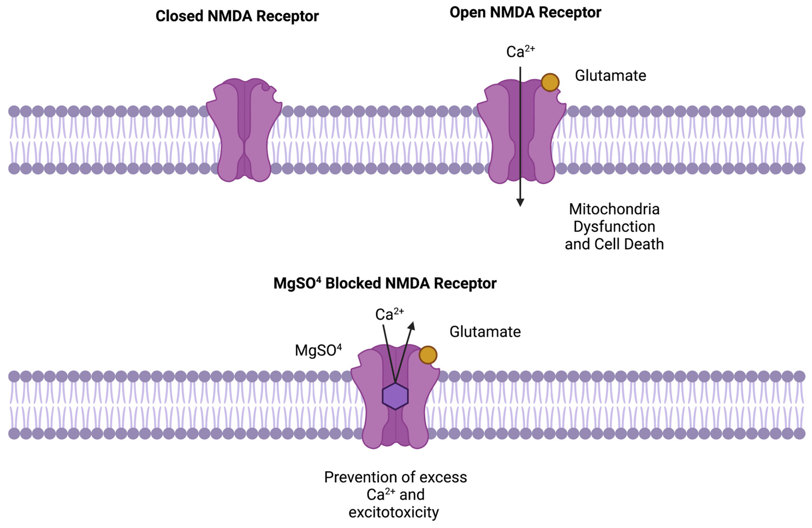
:1. Introduction
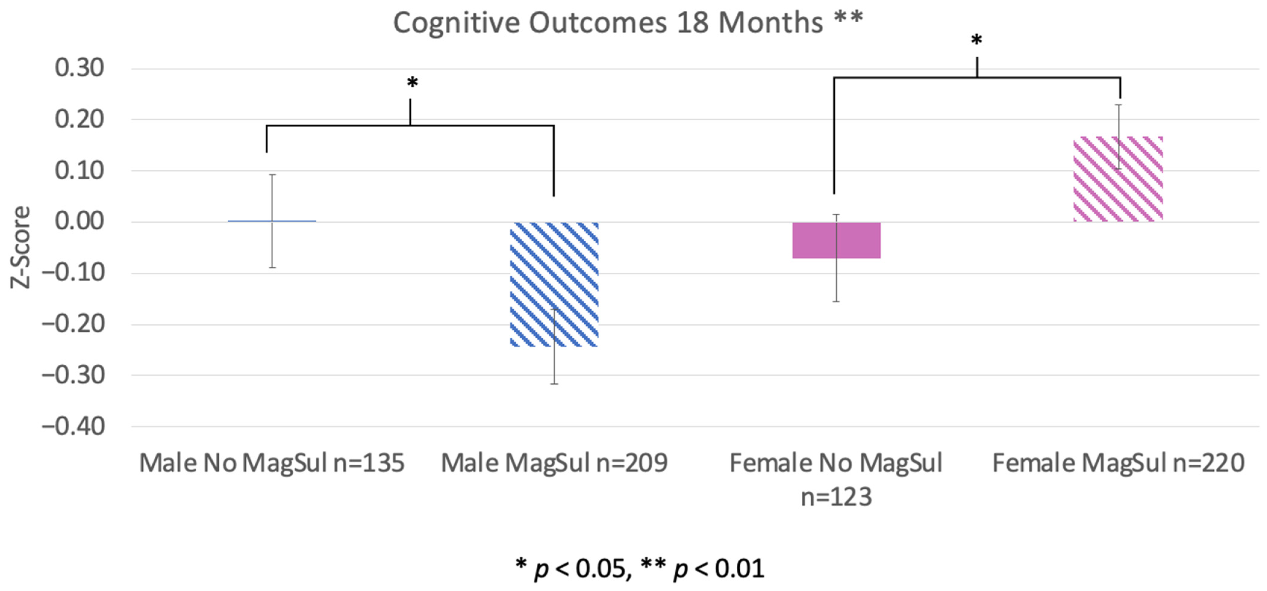
2. Results
3. Discussion
4. Materials and Methods
4.1. Subjects
4.2. Data Collection
4.3. Statistical Analysis
5. Conclusions
Author Contributions
Funding
Institutional Review Board Statement
Informed Consent Statement
Data Availability Statement
Conflicts of Interest
Abbreviations
References
- Altman, D.; Carroli, G.; Duley, L.; Farrell, B.; Moodley, J.; Neilson, J.; Smith, D.; Magpie Trial Collaboration Group. Do women with pre-eclampsia, and their babies, benefit from magnesium sulphate? The Magpie Trial: A randomised placebo-controlled trial. Lancet 2002, 359, 1877–1890. [Google Scholar] [CrossRef] [PubMed]
- Chollat, C.; Marret, S. Magnesium sulfate and fetal neuroprotection: Overview of clinical evidence. Neural Regen. Res. 2018, 13, 2044–2049. [Google Scholar] [CrossRef]
- Galinsky, R.; Dean, J.M.; Lingam, I.; Robertson, N.J.; Mallard, C.; Bennet, L.; Gunn, A.J. A Systematic Review of Magnesium Sulfate for Perinatal Neuroprotection: What Have We Learnt from the Past Decade? Front. Neurol. 2020, 11, 449. [Google Scholar] [CrossRef]
- Chollat, C.; Sentilhes, L.; Marret, S. Fetal Neuroprotection by Magnesium Sulfate: From Translational Research to Clinical Application. Front. Neurol. 2018, 9, 247. [Google Scholar] [CrossRef] [PubMed]
- Nowak, L.P.P.A.A.; Bregestovski, P.; Ascher, P.; Herbet, A.; Prochiantz, A. Magnesium gates glutamate-activated channels in mouse central neurones. Nature 1984, 307, 462–465. [Google Scholar] [CrossRef] [PubMed]
- Kang, S.W.; Choi, S.K.; Park, E.; Chae, S.J.; Choi, S.; Jin Joo, H.; Lee, G.J.; Park, H.K. Neuroprotective effects of magnesium-sulfate on ischemic injury mediated by modulating the release of glutamate and reduced of hyperreperfusion. Brain Res. 2011, 1371, 121–128. [Google Scholar] [CrossRef] [PubMed]
- Burd, I.; Breen, K.; Friedman, A.; Chai, J.; Elovitz, M.A. Magnesium sulfate reduces inflammation-associated brain injury in fetal mice. Am. J. Obstet. Gynecol. 2010, 202, 292.e1–292.e9. [Google Scholar] [CrossRef]
- Mami, A.G.; Ballesteros, J.R.; Fritz, K.I.; Kubin, J.; Mishra, O.P.; Delivoria-Papadopoulos, M. Effects of magnesium sulfate administration during hypoxia on CaM kinase IV and protein tyrosine kinase activities in the cerebral cortex of newborn piglets. Neurochem. Res. 2006, 31, 57–62. [Google Scholar] [CrossRef]
- Mami, A.G.; Ballesteros, J.; Mishra, O.P.; Delivoria-Papadopoulos, M. Effects of magnesium sulfate administration during hypoxia on Ca(2+) influx and IP(3) receptor modification in cerebral cortical neuronal nuclei of newborn piglets. Neurochem. Res. 2006, 31, 63–70. [Google Scholar] [CrossRef]
- Mishra, O.P.; Delivoria-Papadopoulos, M. Modification of modulatory sites of NMDA receptor in the fetal guinea pig brain during development. Neurochem. Res. 1992, 17, 1223–1228. [Google Scholar] [CrossRef]
- Mishra, O.P.; Delivoria-Papadopoulos, M. NMDA receptor modification in the fetal guinea pig brain during hypoxia. Neurochem. Res. 1992, 17, 1211–1216. [Google Scholar] [CrossRef] [PubMed]
- Karve, I.P.; Taylor, J.M.; Crack, P.J. The contribution of astrocytes and microglia to traumatic brain injury. Br. J. Pharmacol. 2016, 173, 692–702. [Google Scholar] [CrossRef] [PubMed]
- Galinsky, R.; Lear, C.A.; Dean, J.M.; Wassink, G.; Dhillon, S.K.; Fraser, M.; Davidson, J.O.; Bennet, L.; Gunn, A.J. Complex interactions between hypoxia-ischemia and inflammation in preterm brain injury. Dev. Med. Child Neurol. 2018, 60, 126–133. [Google Scholar] [CrossRef]
- Reiss, J.D.; Peterson, L.S.; Nesamoney, S.N.; Chang, A.L.; Pasca, A.M.; Marić, I.; Shaw, G.M.; Gaudilliere, B.; Wong, R.J.; Sylvester, K.G.; et al. Perinatal infection, inflammation, preterm birth, and brain injury: A review with proposals for future investigations. Exp. Neurol. 2022, 351, 113988. [Google Scholar] [CrossRef] [PubMed]
- Rouse, D.J.; Hirtz, D.G.; Thom, E.; Varner, M.W.; Spong, C.Y.; Mercer, B.M.; Iams, J.D.; Wapner, R.J.; Sorokin, Y.; Alexander, J.M.; et al. A randomized, controlled trial of magnesium sulfate for the prevention of cerebral palsy. N. Engl. J. Med. 2008, 359, 895–905. [Google Scholar] [CrossRef] [PubMed]
- Kamyar, M.; Manuck, T.A.; Stoddard, G.J.; Varner, M.W.; Clark, E. Magnesium sulfate, chorioamnionitis, and neurodevelopment after preterm birth. BJOG Int. J. Obstet. Gynaecol. 2016, 123, 1161–1166. [Google Scholar] [CrossRef]
- Doyle, L.W.; Anderson, P.J.; Haslam, R.; Lee, K.J.; Crowther, C.; Australasian Collaborative Trial of Magnesium Sulphate (ACTOMgSO4) Study Group. School-age outcomes of very preterm infants after antenatal treatment with magnesium sulfate vs placebo. JAMA 2014, 312, 1105–1113. [Google Scholar] [CrossRef]
- Shepherd, E.; Salam, R.A.; Manhas, D.; Synnes, A.; Middleton, P.; Makrides, M.; Crowther, C.A. Antenatal magnesium sulphate and adverse neonatal outcomes: A systematic review and meta-analysis. PLoS Med. 2019, 16, e1002988. [Google Scholar] [CrossRef]
- Salafia, C.M.; Minior, V.K.; Rosenkrantz, T.S.; Pezzullo, J.C.; Popek, E.J.; Cusick, W.; Vintzileos, A.M. Maternal, placental, and neonatal associations with early germinal matrix/intraventricular hemorrhage in infants born before 32 weeks’ gestation. Am. J. Perinatol. 1995, 12, 429–436. [Google Scholar] [CrossRef] [PubMed]
- Galinsky, R.; Dhillon, S.K.; Lear, C.A.; Yamaguchi, K.; Wassink, G.; Gunn, A.J.; Bennet, L. Magnesium sulfate and sex differences in cardiovascular and neural adaptations during normoxia and asphyxia in preterm fetal sheep. Am. J. Physiol. Regul. Integr. Comp. Physiol. 2018, 315, R205–R217. [Google Scholar] [CrossRef] [PubMed]
- Bennet, L.; Galinsky, R.; Draghi, V.; Lear, C.A.; Davidson, J.O.; Unsworth, C.P.; Gunn, A.J. Time and sex dependent effects of magnesium sulphate on post-asphyxial seizures in preterm fetal sheep. J. Physiol. 2018, 596, 6079–6092. [Google Scholar] [CrossRef] [PubMed]
- McCarthy, M.M. Sex differences in the developing brain as a source of inherent risk. Dialogues Clin. Neurosci. 2016, 18, 361–372. [Google Scholar] [CrossRef] [PubMed]
- Fan, R.; Portuguez, M.; Nunes, M. Cognition, behavior and social competence of preterm low birth weight children at school age. Clinics 2013, 68, 915–921. [Google Scholar] [CrossRef] [PubMed]
- Smith, A.L.; Alexander, M.; Rosenkrantz, T.S.; Sadek, M.L.; Fitch, R.H. Sex differences in behavioral outcome following neonatal hypoxia ischemia: Insights from a clinical meta-analysis and a rodent model of induced hypoxic ischemic brain injury. Exp. Neurol. 2014, 254, 54–67. [Google Scholar] [CrossRef] [PubMed]
- Smith, A.L.; Garbus, H.; Rosenkrantz, T.S.; Fitch, R.H. Sex differences in behavioral outcomes following temperature modulation during induced neonatal hypoxic ischemic injury in rats. Brain Sci. 2015, 5, 220–240. [Google Scholar] [CrossRef] [PubMed]
- Wood, T.R.; Gundersen, J.K.; Falck, M.; Maes, E.; Osredkar, D.; Løberg, E.M.; Thoresen, M. Variability and sex-dependence of hypothermic neuroprotection in a rat model of neonatal hypoxic–ischemic brain injury: A single laboratory meta-analysis. Sci. Rep. 2020, 10, 10833. [Google Scholar] [CrossRef]
- Rosenkrantz, T.S.; Hussain, Z.; Fitch, R.H. Sex Differences in Brain Injury and Repair in Newborn Infants: Clinical Evidence and Biological Mechanisms. Front. Pediatr. 2019, 7, 211. [Google Scholar] [CrossRef]
- McLeod, R.; Rosenkrantz, T.; Fitch, R.H. Therapeutic Interventions in Rat Models of Preterm Hypoxic Ischemic Injury: Effects of Hypothermia, Caffeine, and the Influence of Sex. Life 2022, 12, 1514. [Google Scholar] [CrossRef]
- Zhou, K.Q.; Davidson, J.O.; Gunn, A.J. Does sex materially modulate responses to therapeutic hypothermia? Pediatr. Res. 2023, 94, 1259–1260. [Google Scholar] [CrossRef]
- McLeod, R.M.; Rosenkrantz, T.S.; Fitch, R.H.; Koski, R.R. Sex Differences in Microglia Activation in a Rodent Model of Preterm Hypoxic Ischemic Injury with Caffeine Treatment. Biomedicines 2023, 11, 185. [Google Scholar] [CrossRef]
- Owen, D.; Setiawan, E.; Li, A.; McCabe, L.; Matthews, S.G. Regulation of N-methyl-D-aspartate receptor subunit expression in the fetal guinea pig brain. Biol. Reprod. 2004, 71, 676–683. [Google Scholar] [CrossRef] [PubMed]
- Thagard, A.S.; Slack, J.L.; Estrada, S.M.; Kazanjian, A.A.; Chan, S.; Burd, I.; Napolitano, P.G.; Ieronimakis, N. Long-term impact of intrauterine neuroinflammation and treatment with magnesium sulphate and betamethasone: Sex-specific differences in a preterm labor murine model. Sci. Rep. 2017, 7, 17883. [Google Scholar] [CrossRef] [PubMed]
- Daher, I.; Le Dieu-Lugon, B.; Lecointre, M.; Dupré, N.; Voisin, C.; Leroux, P.; Dourmap, N.; Gonzalez, B.J.; Marret, S.; Leroux-Nicollet, I.; et al. Time- and sex-dependent efficacy of magnesium sulfate to prevent behavioral impairments and cerebral damage in a mouse model of cerebral palsy. Neurobiol. Dis. 2018, 120, 151–164. [Google Scholar] [CrossRef] [PubMed]
- McLeod, R.M.; Rosenkrantz, T.S.; Fitch, R.H. Protective effects of early neonatal methylxanthine treatment on cognitive and language outcomes in premature infants with and without high-risk perinatal factors. Dev. Neurosci. 2024; in review. [Google Scholar]
- Li, H.; Pin, S.; Zeng, Z.; Wang, M.M.; Andreasson, K.A.; McCullough, L.D. Sex differences in cell death. Ann. Neurol. 2005, 58, 317–321. [Google Scholar] [CrossRef]
- Lang, J.T.; McCullough, L.D. Pathways to ischemic neuronal cell death: Are sex differences relevant? J. Transl. Med. 2008, 6, 33. [Google Scholar] [CrossRef]
- Liu, F.; Li, Z.; Li, J.; Siegel, C.; Yuan, R.; McCullough, L.D. Sex differences in caspase activation after stroke. Stroke 2009, 40, 1842–1848. [Google Scholar] [CrossRef]
- Chauhan, A.; Moser, H.; McCullough, L.D. Sex differences in ischaemic stroke: Potential cellular mechanisms. Clin. Sci. 2017, 131, 533–552. [Google Scholar] [CrossRef]
- Schwarz, J.M.; McCarthy, M.M. The role of neonatal NMDA receptor activation in defeminization and masculinization of sex behavior in the rat. Horm. Behav. 2008, 54, 662–668. [Google Scholar] [CrossRef]
- Johnston, M.V. Excitotoxicity in perinatal brain injury. Brain Pathol. 2005, 15, 234–240. [Google Scholar] [CrossRef]
- Damborsky, J.C.; Winzer-Serhan, U.H. Effects of sex and chronic neonatal nicotine treatment on Na(2)(+)/K(+)/Cl(−) co-transporter 1, K(+)/Cl(−) co-transporter 2, brain-derived neurotrophic factor, NMDA receptor subunit 2A and NMDA receptor subunit 2B mRNA expression in the postnatal rat hippocampus. Neuroscience 2012, 225, 105–117. [Google Scholar] [CrossRef]
- Waters, K.A.; Machaalani, R. NMDA receptors in the developing brain and effects of noxious insults. Neuro-Signals 2004, 13, 162–174. [Google Scholar] [CrossRef] [PubMed]
- McCarthy, M.M.; Davis, A.M.; Mong, J.A. Excitatory neurotransmission and sexual differentiation of the brain. Brain Res. Bull. 1997, 44, 487–495. [Google Scholar] [CrossRef] [PubMed]
- Ikonomidou, C.; Bosch, F.; Miksa, M.; Bittigau, P.; Vöckler, J.; Dikranian, K.; Tenkova, T.I.; Stefovska, V.; Turski, L.; Olney, J.W. Blockade of NMDA receptors and apoptotic neurodegeneration in the developing brain. Science 1999, 283, 70–74. [Google Scholar] [CrossRef] [PubMed]
- Dribben, W.H.; Creeley, C.E.; Wang, H.H.; Smith, D.J.; Farber, N.B.; Olney, J.W. High dose magnesium sulfate exposure induces apoptotic cell death in the developing neonatal mouse brain. Neonatology 2009, 96, 23–32. [Google Scholar] [CrossRef]
- Gray, C.; Vickers, M.H.; Dyson, R.M.; Reynolds, C.M.; Berry, M.J. Magnesium sulfate has sex-specific, dose-dependent vasodilator effects on preterm placental vessels. Biol. Sex Differ. 2015, 6, 22. [Google Scholar] [CrossRef] [PubMed]
- Bachnas, M.A.; Akbar, M.I.A.; Dachlan, E.G.; Dekker, G. The role of magnesium sulfate (MgSO4) in fetal neuroprotection. J. Matern.-Fetal Neonatal Med. Off. J. Eur. Assoc. Perinat. Med. Fed. Asia Ocean. Perinat. Soc. Int. Soc. Perinat. Obstet. 2021, 34, 966–978. [Google Scholar] [CrossRef]
- Ment, L.R.; Vohr, B.R.; Makuch, R.W.; Westerveld, M.; Katz, K.H.; Schneider, K.C.; Duncan, C.C.; Ehrenkranz, R.; Oh, W.; Philip, A.G.; et al. Prevention of intraventricular hemorrhage by indomethacin in male preterm infants. J. Pediatr. 2004, 145, 832–834. [Google Scholar] [CrossRef]
- Ohlsson, A.; Roberts, R.S.; Schmidt, B.; Davis, P.; Moddeman, D.; Saigal, S.; Solimano, A.; Vincer, M.; Wright, L.; Trial of Indomethacin Prophylaxis in Preterms Tipp Investigators. Male/female differences in indomethacin effects in preterm infants. J. Pediatr. 2005, 147, 860–862. [Google Scholar] [CrossRef]
- Schmidt, B.; Seshia, M.; Shankaran, S.; Mildenhall, L.; Tyson, J.; Lui, K.; Fok, T.; Roberts, R.; For the Trial of Indomethacin Prophylaxis in Preterms Investigators. Effects of Prophylactic Indomethacin in Extremely Low-Birth-Weight Infants With and Without Adequate Exposure to Antenatal Corticosteroids. Arch. Pediatr. Adolesc. Med. 2011, 165, 642–646. [Google Scholar] [CrossRef]
- Al-matary, A.; Abu Shaheen, A.; Abozaid, S. Use of Prophylactic Indomethacin in Preterm Infants: A Systematic Review and Meta-Analysis [Systematic Review]. Front. Pediatr. 2022, 10, 760029. [Google Scholar] [CrossRef] [PubMed]
- Azzopardi, D.; Brocklehurst, P.; Edwards, D.; Halliday, H.; Levene, M.; Thoresen, M.; Whitelaw, A.; TOBY Study Group. The TOBY Study. Whole body hypothermia for the treatment of perinatal asphyxial encephalopathy: A randomised controlled trial. BMC Pediatr. 2008, 8, 17. [Google Scholar] [CrossRef] [PubMed]
- Sewell, E.K.; Shankaran, S.; Natarajan, G.; Laptook, A.; Das, A.; McDonald, S.A.; Hamrick, S.; Baack, M.; Rysavy, M.; Higgins, R.D.; et al. Evaluation of heterogeneity in effect of therapeutic hypothermia by sex among infants with neonatal encephalopathy. Pediatr. Res. 2023, 94, 1380–1384. [Google Scholar] [CrossRef] [PubMed]
- Wachtel, R.C.; Shapiro, B.K.; Palmer, F.B.; Allen, M.C.; Capute, A.J. CAT/CLAMS A tool for the pediatric evaluation of infants and young children with developmental delay. Clinical Adaptive Test/Clinical Linguistic and Auditory Milestone Scale. Clin. Pediatr. 1994, 33, 410–415. [Google Scholar] [CrossRef] [PubMed]


| Measure | Group | Mean | SE | Significance |
|---|---|---|---|---|
| Weight (g) | No MagSul | 1044.36 | 20.02 | p > 0.05 |
| MagSul | 1001.87 | 14.06 | ||
| Gestational age (Weeks) | No MagSul | 27.43 | 0.12 | p > 0.05 |
| MagSul | 27.14 | 0.09 | ||
| Length of Stay (Weeks) | No MagSul | 83.08 | 1.88 | p > 0.05 |
| MagSul | 85.21 | 1.49 | ||
| Apgar Score (5 min) | No MagSul | 7.55 | 0.10 | p > 0.05 |
| MagSul | 7.70 | 0.07 |
| Measure | Group | Mean | SE | Significance |
|---|---|---|---|---|
| Weight (g) | Female | 934.28 | 37.95 | p = 0.014 |
| Male | 1058.88 | 33.46 | ||
| Gestational Age (Weeks) | Female | 26.61 | 0.23 | p > 0.05 |
| Male | 27.06 | 0.21 | ||
| Length of Stay (Weeks) | Female | 90.49 | 3.45 | p > 0.05 |
| Male | 89.13 | 2.35 | ||
| Apgar Score (5 min) | Female | 7.69 | 0.08 | p > 0.05 |
| Male | 7.60 | 0.09 |
| Reason for Premature Delivery | Sex | n | Significance |
|---|---|---|---|
| Preeclampsia | Female | 68 | p > 0.05 |
| Male | 52 | ||
| Premature Rupture of Membranes | Female | 103 | p > 0.05 |
| Male | 110 |
Disclaimer/Publisher’s Note: The statements, opinions and data contained in all publications are solely those of the individual author(s) and contributor(s) and not of MDPI and/or the editor(s). MDPI and/or the editor(s) disclaim responsibility for any injury to people or property resulting from any ideas, methods, instructions or products referred to in the content. |
© 2024 by the authors. Licensee MDPI, Basel, Switzerland. This article is an open access article distributed under the terms and conditions of the Creative Commons Attribution (CC BY) license (https://creativecommons.org/licenses/by/4.0/).
Share and Cite
McLeod, R.M.; Rosenkrantz, T.S.; Fitch, R.H. Antenatal Magnesium Sulfate Benefits Female Preterm Infants but Results in Poor Male Outcomes. Pharmaceuticals 2024, 17, 218. https://doi.org/10.3390/ph17020218
McLeod RM, Rosenkrantz TS, Fitch RH. Antenatal Magnesium Sulfate Benefits Female Preterm Infants but Results in Poor Male Outcomes. Pharmaceuticals. 2024; 17(2):218. https://doi.org/10.3390/ph17020218
Chicago/Turabian StyleMcLeod, Ruth M., Ted S. Rosenkrantz, and R. Holly Fitch. 2024. "Antenatal Magnesium Sulfate Benefits Female Preterm Infants but Results in Poor Male Outcomes" Pharmaceuticals 17, no. 2: 218. https://doi.org/10.3390/ph17020218