
Pharmacological Differences between Native Homomeric Transient Receptor Potential Canonical Type 4 Channels and Heteromeric Transient Receptor Potential Canonical Type 1/4 Channels in Lateral Septal Neurons

Abstract
:1. Introduction
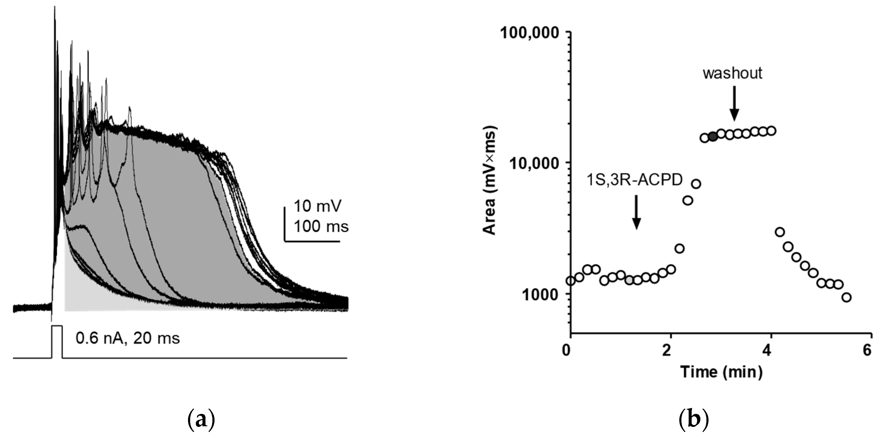
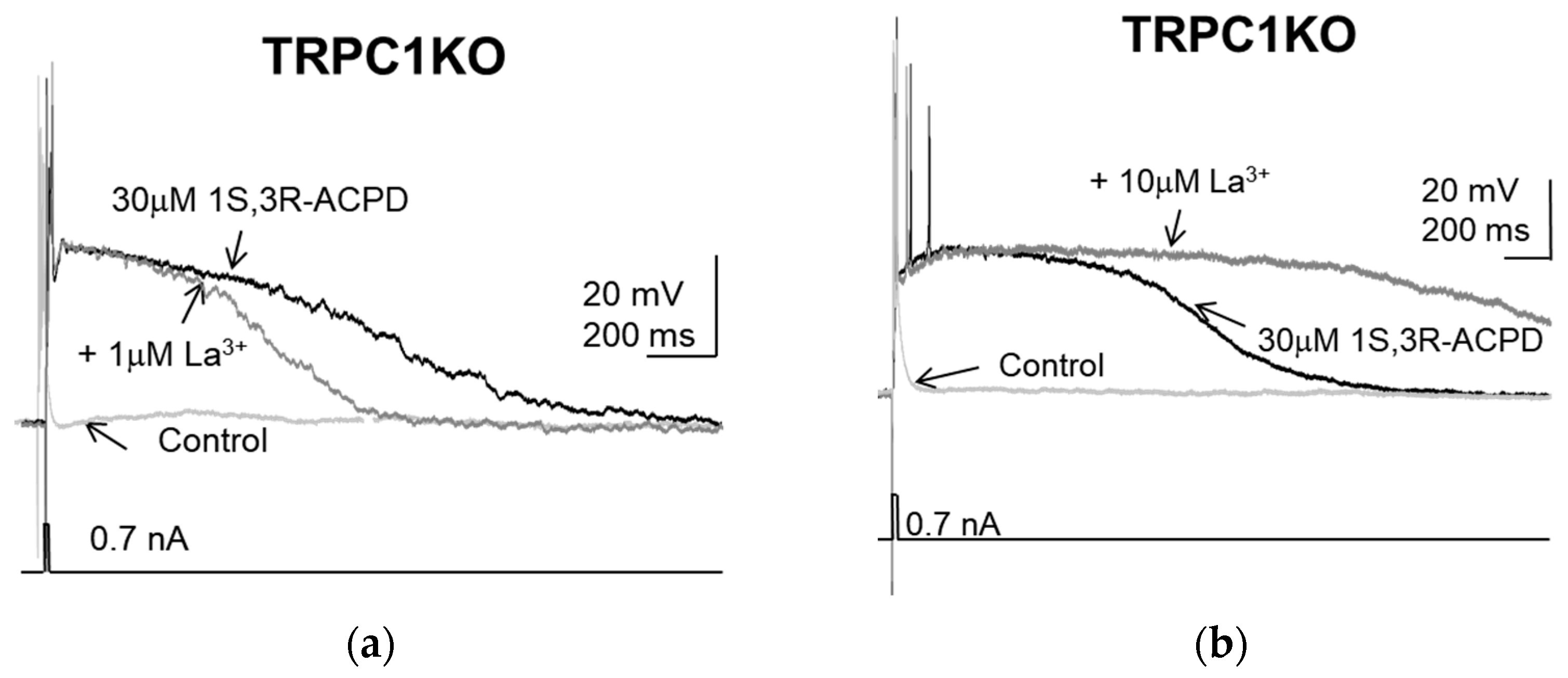
2. Results
3. Discussion
4. Materials and Methods
5. Conclusions
Author Contributions
Funding
Institutional Review Board Statement
Informed Consent Statement
Data Availability Statement
Acknowledgments
Conflicts of Interest
Abbreviations
| 1S,3R-ACPD | (1S,3R)-1-Aminocyclopentane-1,3-dicarboxylic acid; |
| 2-APB | 2-Aminoethoxydiphenylborane; |
| ACSF | artificial cerebrospinal fluid; |
| mGluR | metabotropic glutamate receptor; |
| ML204 | 4-Methyl-2-(1-piperidinyl)quinoline; |
| TRPC | Transient receptor potential channel; |
| TRPC1KO | TRPC1 knockout; |
| TRPC1/3DKO | TRPC1/3 double knockout |
References
- Vazquez, G.; Wedel, B.J.; Aziz, O.; Trebak, M.; Putney, J.W. The Mammalian TRPC Cation Channels. Biochim. Biophys. Acta-Mol. Cell Res. 2004, 1742, 21–36. [Google Scholar] [CrossRef] [PubMed]
- Clapham, D.E.; Runnels, L.W.; Strübing, C. The TRP Ion Channel Family. Nat. Rev. Neurosci. 2001, 2, 387–396. [Google Scholar] [CrossRef] [PubMed]
- Birnbaumer, L. The TRPC Class of Ion Channels: A Critical Review of Their Roles in Slow, Sustained Increases in Intracellular Ca2+ Concentrations. Annu. Rev. Pharmacol. Toxicol. 2009, 49, 395–426. [Google Scholar] [CrossRef] [PubMed]
- Montell, C. Drosophila TRP Channels. Pflügers Arch.-Eur. J. Physiol. 2005, 451, 19–28. [Google Scholar] [CrossRef] [PubMed]
- Minke, B. The History of the Drosophila TRP Channel: The Birth of a New Channel Superfamily. J. Neurogenet. 2010, 24, 216–233. [Google Scholar] [CrossRef] [PubMed]
- Montell, C. The History of TRP Channels, a Commentary and Reflection. Pflugers Arch. 2011, 461, 499–506. [Google Scholar] [CrossRef] [PubMed]
- Freichel, M.; Vennekens, R.; Olausson, J.; Hoffmann, M.; Müller, C.; Stolz, S.; Scheunemann, J.; Weißgerber, P.; Flockerzi, V. Functional Role of TRPC Proteins in Vivo: Lessons from TRPC-Deficient Mouse Models. Biochem. Biophys. Res. Commun. 2004, 322, 1352–1358. [Google Scholar] [CrossRef]
- Freichel, M.; Vennekens, R.; Olausson, J.; Stolz, S.; Philipp, S.E.; Weissgerber, P.; Flockerzi, V. Functional Role of TRPC Proteins in Native Systems: Implications from Knockout and Knock-down Studies. J. Physiol. 2005, 567, 59–66. [Google Scholar] [CrossRef]
- Nilius, B.; Owsianik, G.; Voets, T.; Peters, J.A. Transient Receptor Potential Cation Channels in Disease. Physiol. Rev. 2007, 87, 165–217. [Google Scholar] [CrossRef]
- Freichel, M.; Flockerzi, V. Biological Functions of TRPs Unravelled by Spontaneous Mutations and Transgenic Animals. Biochem. Soc. Trans. 2007, 35, 120–123. [Google Scholar] [CrossRef]
- Zheng, F.; Phelan, K.D. The Role of Canonical Transient Receptor Potential Channels in Seizure and Excitotoxicity. Cells 2014, 3, 288–303. [Google Scholar] [CrossRef] [PubMed]
- Zheng, F. TRPC Channels and Epilepsy. Adv. Exp. Med. Biol. 2017, 976, 123–135. [Google Scholar] [CrossRef] [PubMed]
- Bon, R.S.; Beech, D.J. In Pursuit of Small Molecule Chemistry for Calcium-Permeable Non-Selective TRPC Channels—Mirage or Pot of Gold? Br. J. Pharmacol. 2013, 170, 459–474. [Google Scholar] [CrossRef] [PubMed]
- Minard, A.; Bauer, C.C.; Wright, D.J.; Rubaiy, H.N.; Muraki, K.; Beech, D.J.; Bon, R.S. Remarkable Progress with Small-Molecule Modulation of TRPC1/4/5 Channels: Implications for Understanding the Channels in Health and Disease. Cells 2018, 7, 52. [Google Scholar] [CrossRef] [PubMed]
- Bon, R.S.; Wright, D.J.; Beech, D.J.; Sukumar, P. Pharmacology of TRPC Channels and Its Potential in Cardiovascular and Metabolic Medicine. Annu. Rev. Pharmacol. Toxicol. 2022, 62, 427–446. [Google Scholar] [CrossRef] [PubMed]
- Zheng, F. Canonical Transient Receptor Potential Channels as Novel Targets for Antiepileptic Drugs. In Epilepsy; Czuczwar, S.J., Ed.; Exon Publications: Brisbane, QL, Australia, 2022; pp. 79–94. ISBN 9780645332049. [Google Scholar]
- Birnbaumer, L.; Yidirim, E.; Abramowitz, J. A Comparison of the Genes Coding for Canonical TRP Channels and Their M, V and P Relatives. Cell Calcium 2003, 33, 419–432. [Google Scholar] [CrossRef] [PubMed]
- Goel, M.; Sinkins, W.G.; Schilling, W.P. Selective Association of TRPC Channel Subunits in Rat Brain Synaptosomes. J. Biol. Chem. 2002, 277, 48303–48310. [Google Scholar] [CrossRef]
- Chung, Y.H.; Sun Ahn, H.; Kim, D.; Hoon Shin, D.; Su Kim, S.; Yong Kim, K.; Bok Lee, W.; Ik Cha, C. Immunohistochemical Study on the Distribution of TRPC Channels in the Rat Hippocampus. Brain Res. 2006, 1085, 132–137. [Google Scholar] [CrossRef]
- Eder, P.; Poteser, M.; Groschner, K. TRPC3: A Multifunctional, Pore-Forming Signalling Molecule. Handb. Exp. Pharmacol. 2007, 179, 77–92. [Google Scholar] [CrossRef]
- Poteser, M.; Graziani, A.; Rosker, C.; Eder, P.; Derler, I.; Kahr, H.; Zhu, M.X.; Romanin, C.; Groschner, K. TRPC3 and TRPC4 Associate to Form a Redox-Sensitive Cation Channel. Evidence for Expression of Native TRPC3-TRPC4 Heteromeric Channels in Endothelial Cells. J. Biol. Chem. 2006, 281, 13588–13595. [Google Scholar] [CrossRef]
- Kollewe, A.; Schwarz, Y.; Oleinikov, K.; Raza, A.; Haupt, A.; Wartenberg, P.; Wyatt, A.; Boehm, U.; Ectors, F.; Bildl, W.; et al. Subunit Composition, Molecular Environment, and Activation of Native TRPC Channels Encoded by Their Interactomes. Neuron 2022, 110, 4162–4175.e7. [Google Scholar] [CrossRef] [PubMed]
- Hofmann, T.; Schaefer, M.; Schultz, G.; Gudermann, T. Subunit Composition of Mammalian Transient Receptor Potential Channels in Living Cells. Proc. Natl. Acad. Sci. USA 2002, 99, 7461–7466. [Google Scholar] [CrossRef] [PubMed]
- Ko, J.; Myeong, J.; Shin, Y.-C.; So, I. Differential PI(4,5)P2 Sensitivities of TRPC4, C5 Homomeric and TRPC1/4, C1/5 Heteromeric Channels. Sci. Rep. 2019, 9, 1849. [Google Scholar] [CrossRef] [PubMed]
- Myeong, J.; Ko, J.; Hong, C.; Yang, D.; Lee, K.P.; Jeon, J.-H.; So, I. The Interaction Domains of Transient Receptor Potential Canonical (TRPC)1/4 and TRPC1/5 Heteromultimeric Channels. Biochem. Biophys. Res. Commun. 2016, 474, 476–481. [Google Scholar] [CrossRef] [PubMed]
- Phelan, K.D.; Mock, M.M.; Kretz, O.; Shwe, U.T.T.; Kozhemyakin, M.; Greenfield, L.J.J.; Dietrich, A.; Birnbaumer, L.; Freichel, M.; Flockerzi, V.; et al. Heteromeric Canonical Transient Receptor Potential 1 and 4 Channels Play a Critical Role in Epileptiform Burst Firing and Seizure-Induced Neurodegeneration. Mol. Pharmacol. 2012, 81, 384–392. [Google Scholar] [CrossRef] [PubMed]
- Muraki, K.; Ohnishi, K.; Takezawa, A.; Suzuki, H.; Hatano, N.; Muraki, Y.; Hamzah, N.; Foster, R.; Waldmann, H.; Nussbaumer, P.; et al. Na+ Entry through Heteromeric TRPC4/C1 Channels Mediates (-)Englerin A-Induced Cytotoxicity in Synovial Sarcoma Cells. Sci. Rep. 2017, 7, 16988. [Google Scholar] [CrossRef] [PubMed]
- Ceballos, C.C.; Roque, A.C.; Leão, R.M. The Role of Negative Conductances in Neuronal Subthreshold Properties and Synaptic Integration. Biophys. Rev. 2017, 9, 827–834. [Google Scholar] [CrossRef] [PubMed]
- Philipp, S.; Hambrecht, J.; Braslavski, L.; Schroth, G.; Freichel, M.; Murakami, M.; Cavalié, A.; Flockerzi, V. A Novel Capacitative Calcium Entry Channel Expressed in Excitable Cells. EMBO J. 1998, 17, 4274–4282. [Google Scholar] [CrossRef]
- Philipp, S.; Cavalié, A.; Freichel, M.; Wissenbach, U.; Zimmer, S.; Trost, C.; Marquart, A.; Murakami, M.; Flockerzi, V. A Mammalian Capacitative Calcium Entry Channel Homologous to Drosophila TRP and TRPL. EMBO J. 1996, 15, 6166–6171. [Google Scholar] [CrossRef]
- Rychkov, G.; Barritt, G.J. TRPC1 Ca2+-Permeable Channels in Animal Cells. Handb. Exp. Pharmacol. 2007, 179, 23–52. [Google Scholar] [CrossRef]
- Beech, D.J. Canonical Transient Receptor Potential 5. Handb. Exp. Pharmacol. 2007, 179, 109–123. [Google Scholar] [CrossRef]
- Riccio, A.; Li, Y.; Moon, J.; Kim, K.-S.S.; Smith, K.S.; Rudolph, U.; Gapon, S.; Yao, G.L.; Tsvetkov, E.; Rodig, S.J.; et al. Essential Role for TRPC5 in Amygdala Function and Fear-Related Behavior. Cell 2009, 137, 761–772. [Google Scholar] [CrossRef] [PubMed]
- Von Bohlen Und Halbach, O.; Hinz, U.; Unsicker, K.; Egorov, A.V. Distribution of TRPC1 and TRPC5 in Medial Temporal Lobe Structures of Mice. Cell Tissue Res. 2005, 322, 201–206. [Google Scholar] [CrossRef] [PubMed]
- Phelan, K.D.; Shwe, U.T.T.; Abramowitz, J.; Wu, H.; Rhee, S.W.; Howell, M.D.; Gottschall, P.E.; Freichel, M.; Flockerzi, V.; Birnbaumer, L.; et al. Canonical Transient Receptor Channel 5 (TRPC5) and TRPC1/4 Contribute to Seizure and Excitotoxicity by Distinct Cellular Mechanisms. Mol. Pharmacol. 2013, 83, 429–438. [Google Scholar] [CrossRef] [PubMed]
- Jung, S.; Mühle, A.; Schaefer, M.; Strotmann, R.; Schultz, G.G.; Plant, T.D. Lanthanides Potentiate TRPC5 Currents by an Action at Extracellular Sites Close to the Pore Mouth. J. Biol. Chem. 2003, 278, 3562–3571. [Google Scholar] [CrossRef] [PubMed]
- Freichel, M.; Suh, S.H.; Pfeifer, A.; Schweig, U.; Trost, C.; Weissgerber, P.; Biel, M.; Philipp, S.; Freise, D.; Droogmans, G.; et al. Lack of an Endothelial Store-Operated Ca2+ Current Impairs Agonist-Dependent Vasorelaxation in TRP4−/− Mice. Nat. Cell Biol. 2001, 3, 121–127. [Google Scholar] [CrossRef]
- Miller, M.; Shi, J.; Zhu, Y.; Kustov, M.; Tian, J.B.; Stevens, A.; Wu, M.; Xu, J.; Long, S.; Yang, P.; et al. Identification of ML204, a Novel Potent Antagonist That Selectively Modulates Native TRPC4/C5 Ion Channels. J. Biol. Chem. 2011, 286, 33436–33446. [Google Scholar] [CrossRef]
- Tian, J.; Thakur, D.P.; Lu, Y.; Zhu, Y.; Freichel, M.; Flockerzi, V.; Zhu, M.X. Dual Depolarization Responses Generated within the Same Lateral Septal Neurons by TRPC4-Containing Channels. Pflugers Arch. 2014, 466, 1301–1316. [Google Scholar] [CrossRef]
- Gallagher, J.P.; Zheng, F.; Hasuo, H.; Shinnick-Gallagher, P. Activities of Neurons within the Rat Dorsolateral Septal Nucleus (DLSN). Prog. Neurobiol. 1995, 45, 373–395. [Google Scholar] [CrossRef]
- Schaefer, M.; Plant, T.D.; Stresow, N.; Albrecht, N.; Schultz, G. Functional Differences between TRPC4 Splice Variants. J. Biol. Chem. 2002, 277, 3752–3759. [Google Scholar] [CrossRef]
- Duan, J.; Li, J.; Zeng, B.; Chen, G.-L.; Peng, X.; Zhang, Y.; Wang, J.; Clapham, D.E.; Li, Z.; Zhang, J. Structure of the Mouse TRPC4 Ion Channel. Nat. Commun. 2018, 9, 3102. [Google Scholar] [CrossRef]
- Vinayagam, D.; Mager, T.; Apelbaum, A.; Bothe, A.; Merino, F.; Hofnagel, O.; Gatsogiannis, C.; Raunser, S. Electron Cryo-Microscopy Structure of the Canonical TRPC4 Ion Channel. Elife 2018, 7, e36615. [Google Scholar] [CrossRef]
- Duan, J.; Li, J.; Chen, G.-L.; Ge, Y.; Liu, J.; Xie, K.; Peng, X.; Zhou, W.; Zhong, J.; Zhang, Y.; et al. Cryo-EM Structure of TRPC5 at 2.8-Å Resolution Reveals Unique and Conserved Structural Elements Essential for Channel Function. Sci. Adv. 2019, 5, eaaw7935. [Google Scholar] [CrossRef]
- Vinayagam, D.; Quentin, D.; Yu-Strzelczyk, J.; Sitsel, O.; Merino, F.; Stabrin, M.; Hofnagel, O.; Yu, M.; Ledeboer, M.W.; Nagel, G.; et al. Structural Basis of TRPC4 Regulation by Calmodulin and Pharmacological Agents. Elife 2020, 9, e60603. [Google Scholar] [CrossRef]
- Wright, D.J.; Simmons, K.J.; Johnson, R.M.; Beech, D.J.; Muench, S.P.; Bon, R.S. Human TRPC5 Structures Reveal Interaction of a Xanthine-Based TRPC1/4/5 Inhibitor with a Conserved Lipid Binding Site. Commun. Biol. 2020, 3, 704. [Google Scholar] [CrossRef]


Disclaimer/Publisher’s Note: The statements, opinions and data contained in all publications are solely those of the individual author(s) and contributor(s) and not of MDPI and/or the editor(s). MDPI and/or the editor(s) disclaim responsibility for any injury to people or property resulting from any ideas, methods, instructions or products referred to in the content. |
© 2023 by the authors. Licensee MDPI, Basel, Switzerland. This article is an open access article distributed under the terms and conditions of the Creative Commons Attribution (CC BY) license (https://creativecommons.org/licenses/by/4.0/).
Share and Cite
Phelan, K.D.; Shwe, U.T.; Zheng, F. Pharmacological Differences between Native Homomeric Transient Receptor Potential Canonical Type 4 Channels and Heteromeric Transient Receptor Potential Canonical Type 1/4 Channels in Lateral Septal Neurons. Pharmaceuticals 2023, 16, 1291. https://doi.org/10.3390/ph16091291
Phelan KD, Shwe UT, Zheng F. Pharmacological Differences between Native Homomeric Transient Receptor Potential Canonical Type 4 Channels and Heteromeric Transient Receptor Potential Canonical Type 1/4 Channels in Lateral Septal Neurons. Pharmaceuticals. 2023; 16(9):1291. https://doi.org/10.3390/ph16091291
Chicago/Turabian StylePhelan, Kevin D., U Thaung Shwe, and Fang Zheng. 2023. "Pharmacological Differences between Native Homomeric Transient Receptor Potential Canonical Type 4 Channels and Heteromeric Transient Receptor Potential Canonical Type 1/4 Channels in Lateral Septal Neurons" Pharmaceuticals 16, no. 9: 1291. https://doi.org/10.3390/ph16091291