Article

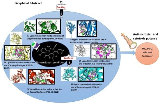
Cytotoxic and Infection-Controlled Investigations of Novel Dihydropyridine Hybrids: An Efficient Synthesis and Molecular-Docking Studies

by
Mallikarjuna R. Guda
1,2,*,
Grigory. V. Zyryanov
1,3,
Amit Dubey
4,5,
Venkata Subbaiah Munagapati
6 and
Jet-Chau Wen
6,7
1
Institute of Chemical Engineering, Ural Federal University Named after the First President of Russia B.N. Yeltsin, 28 Mira St., Yekaterinburg 620002, Russia
2
Department of Chemistry, Sri Venkateswara University, Tirupati 517502, India
3
Ural Division of the Russian Academy of Sciences, I. Ya. Postovskiy Institute of Organic Synthesis, 22 S. Kovalevskoy Street, Yekaterinburg 620219, Russia
4
Computational Chemistry and Drug Discovery Division, Quanta Calculus Pvt. Ltd., Greater Noida 201310, India
5
Department of Pharmacology, Saveetha Dental College and Hospital, Saveetha Institute of Medical and Technical Sciences, Chennai 600077, India
6
Research Centre for Soil and Water Resources and Natural Disaster Prevention (SWAN), National Yunlin University of Science and Technology, Douliou 64002, Taiwan
7
Department of Safety, Health, and Environmental Engineering, National Yunlin University of Science and Technology, Douliou 64002, Taiwan
*
Author to whom correspondence should be addressed.
Pharmaceuticals 2023, 16(8), 1159; https://doi.org/10.3390/ph16081159
Submission received: 9 July 2023 / Revised: 29 July 2023 / Accepted: 7 August 2023 / Published: 15 August 2023

Abstract

:
A sequence of novel 1,4-dihydropyridines (DHP) and their hybrids was developed using a multicomponent strategy under environmentally benign conditions. In addition, computational studies were performed, and the ligand–protein interactions calculated in different bacteria and two fungal strains. Para-hydroxy-linked DHP (5f) showed the best binding energies of 3.591, 3.916, 8.499 and 6.895 kcal/mol against various pathogens used and other substances received a good docking score. The pathogen resistance potential of the synthesized targets against four bacteria and two fungi showed that whole DHP substances exhibit different levels of resistance to each microorganism. Gram-positive bacteria, which are highly sensitive to all molecules, and the MTCC-1884-encoded fungus strongly rejected the studied compounds compared to comparator drugs. In particular, the 5f candidate showed remarkable antimicrobial activity, followed by the substances 5a, 5b, 5j, 5k and 5l. Furthermore, MIC and MBC/MFC properties showed that 5f had a minimum bacterial concentration of 12.5 μg/mL against E. coli and against two fungal pathogens, with its killing activity being effective even at low concentrations. On the other hand, whole motifs were tested for their cytotoxic activity, revealing that the methoxy and hydroxy-linked compounds (5h) showed greater cytotoxic potency, followed by the two hydroxy linked compounds (5d and 5f). Overall, this synthetic approach used represents a prototype for future nature-favored synthesis methods and these biological results serve as a guide for future therapeutic drug research. However, the computer results play an important role in the further development of biological experiments.

Graphical Abstract

1. Introduction

The selection of a key chemical compound for synthesis is important because these substances must have shown therapeutic efficacy in previous studies. Numerous investigators have reported and proven that DHP-core-owning compounds exhibit diverse biological characteristics. Also, it is used in medicines such as amlodipine to treat blood pressure and angina (for chest pain), nifedipine–calcium channel blockers, as well as to lower blood pressure in the arteries when the heart is not working much, to maintain blood circulation, and in nicardipine has a similar effect in curing ailments and diseases [1,2,3]. This substance acts as an antituberculosis agent and plays a leading role in metabolic oxidation [4]. Recently, Gomha et. al. developed novel dihydropyridines that conferred antimicrobial properties. However, this study used a well-known conventional method. [5]. The regular production conditions for the production of 1,4-dihydropyridine analogues and these produced compounds showed antitumor properties [6]. Likewise, various types of research strategies have been applied for the production of pyridines via the acid-mediated condition [7], enzymatically involved synthesis [8,9] and the heavy metal initiator method [10], nano catalyst utilization reactions [11] and molybdenum-type metal oxide with catalysts [12]. All the above solutions have numerous disadvantages such as metals, toxic reagents, alloy oxides and others that are undesirable from a green chemistry perspective.
Environmental sustainability remains a challenging aspect of all scientific investigations, particularly synthetic research [13,14,15]. Sometimes the molecule-production strategy does not fall under the twelve principles of green chemistry, resulting in [16] damage to nature from production waste. The eco-friendly synthetic approaches that support the manufacture of compounds should be designed to avoid waste and toxic pollution that affect nature and humans [17,18,19,20]. Leading portfolios of green manufacturing processes include aqua engagement synthesis, water-soluble solvent conditions, neat reactions, sonochemical techniques, multiple reactants involved in single-step reactions, microwave-heating pathways, and ball-milling strategies.
Besides synthesis and biological experiments, computational studies are becoming the crucial tool in organic research focusing on structure orientation, compound behavior, molecular properties prediction, ligand–protein interactions, binding energies, and others [21]. In recent years, numerous results related to machine-assisted studies combined with synthetic research have been published [22,23,24,25,26]. As far as we are aware, there are currently no reports combining information about DHP, its environmentally friendly manufacturing method, biological properties, and computer simulations. The present research involved the green synthesis approach, synthesized the bulk of the dihydropyridine substances and investigated their antimicrobial and cytotoxic potential. Computer-assisted characteristic curves are also discussed in detail.

2. Results and Discussion

2.1. Chemistry

As nature favors synthetic research, researchers are adopting strategies such as time saving, reactant minimization, simple solvents, one-step synthesis, cost savings, and volume yield to protect the environment. In this context, to identify the best portfolio in the area of green synthesis for the development of DHP compounds, a number of methods were used and are shown in Scheme 1 and Table 1. All processes were performed in a one-pot, multicomponent reaction using 5-amino-2-methoxyphenol, dimethylbut-2-ynedioate, several aldehydes, and malononitrile as starting reactants. It is believed that the commonly available polyethylene glycol (PEG) contemplates the production of compounds 5a, 5b and 5c to maintain economically viable conditions. The synthesis began when the starting chemicals of product 5a were added to PEG and gently heated to elevated temperatures. Product 5a was obtained in about 45 percent, followed by products 5b and 5c in yields of 39 and 41 percent, respectively, when their respective starting materials were involved in a reaction. After unexpected yields, various acid catalysts have been employed to achieve a profitable amount of end goals. In this manner, Lewis acid zinc chloride acid initiator was used under acetonitrile solvent conditions to prepare compounds 5a–c. As a result, an unsatisfactory product yield was observed (Entry 2, Table 1).
In the same solvent medium, we changed the catalyst and continued the reactions, resulting in a trace yield of compounds with values of 11% (5a), 19% (5b), and 15% (5c) in the case of indium chloride as a catalyst (Entry 3, Table 1), while the product 5a has a 32 percent yield in tin chloride promoter media, 5b has a 25% yield, and the percentage of 5c is about 21 (Entry 4, Table 1). Apart from a variety of acidic montmorillonite K-10 acid clay was considered a promoters for the production of 5a–c units using a mixed-solvent concept. The clay initiator, the 2:1 miscible ethanol–water solvent combination and all of the reactants of 5a were heated in an autoclave under argon conditions to yield the target 5a in an amount of 86%. When the reactions of 5b and 5c were successfully carried out using the same strategy, the yields of these targets were 83 percent and 88 percent, showing a high productivity (Entry 5, Table 1). Thus, with this environmentally benign reaction strategy, the products (5(d–l)) were prepared in impressive yields.

2.2. Molecular Modelling

2.2.1. Molecular-Docking Studies, Binding Pose, and Interaction Profiling

Molecular-Docking Studies

The interaction at the molecular level between the ligands and protein receptors was determined using molecular docking. The selected ligands were docked to the binding region of the germs Bacillus subtilis, Staphylococcus aureus, ATCC 8739 code bacteria, Proteus vulgaris, MTCC 1881 and Aspergillus flavus using Glide (Standard Precision Mode) in the Glide module [27]. The exact coordinates of the active sites of all protein receptors were determined using the Schrödinger grid generation method. To allow more accurate predictions for the ligand positions, using several sets of specifications, the receptors characteristics and shape were taken into consideration in a grid. The essential zone of the lattice, which designates the residue as the lattice center, should be covered by it [28,29,30,31,32]. Additional default settings were used. The optimal docking conformation of each ligand with the lowest docking score was considered.

Binding Poses and Interaction Profiling

The best docking scores for bond conformations were determined after molecular docking and compared. Furthermore, the default settings of the academic A Schrödinger-Maestro v11.4 suite were used to study molecular interactions at a distance of 4 Ao around the docked ligand (Release 2018-3 from Schrödinger: Maestro, Schrödinger, LLC, 2018, New York, NY, USA). For docked ligands and receptors, intermolecular interactions like cation, hydrophobic and hydrogen-bonding interactions that are not covalent have been identified in discovered negative contacts, positive contacts, glycine interactions and the formation of salt bridges.

2.2.2. Bacillus subtilis

Binding Mode of Best Hit Ligands 5a and 5f with Bacillus subtilis

The best projected binding energy for ligand 5a is −3.593 kcal/mol. In the active site of Bacillus subtilis, this molecule and essential amino acids have created numerous, robust connections (Figure 1). For example, the amino acids Arg205 and Asp212 can interact three times with the ligand 5a (Table 2). This ligand promotes additional receptor contacts (hydrophobic interactions, polar interactions) by building new connections. With −3.591 kcal/mol, ligand 5f has the second-best binding energy. Ligand 5f was reported to have developed some strong interactions with key amino acids in the Bacillus active site (Figure 2). For example, ligand 5f may interact three times with Ser103, Arg205, Gln270, and Hip271 (Table 2). New interactions can arise as a result of increased contacts between the ligand and receptor (hydrophobic interactions, polar interactions).

2.2.3. Staphylococcus aureus

Binding Mode of Best Ligands 5f and 5j with Staphylococcus aureus

One of the best ligands, 5f, has a binding energy of −3.916 kcal/mol. The Staphylococcus aureus active site has evolved numerous strong connections between this molecule and important amino acids (Figure 3). For example, ligand 5f may interact three times with amino acids Ser445, Asn468, and Lys499 (Table 3). This ligand creates novel connections that allow stronger hydrophobic and polar interactions with the receptor. Ligand 5j has a binding energy of −3.919 kcal/mol. This ligand has formed several, strong interactions with key amino acids at the active site of Staphylococcus aureus (Figure 4). For example, ligand 5j shows a threefold interaction with the amino acid Lys499 (Table 3). By forming new connections, this ligand allows more contacts (hydrophobic interactions, polar interactions) between the receptor Staphylococcus aureus.

2.2.4. Escherichia coli

Binding Mode of Best Ligand 5k and 5l with Escherichia coli

With a calculated binding energy of −5.414 kcal/mol, ligand 5k gave the best results by fully occupying the binding site. Some cluster residues, such as Asn247, show strong hydrogen bonding (Figure 5). The protein–ligand has also become more stable due to additional hydrophobic and polar interactions, as can be seen in Table 4. By fully occupying the binding site, 5l achieved the best result with an estimated binding energy of −5900 kcal/mol. Some residues in clusters, such as Asn210, feature two hydrogen bonds (Figure 6). Furthermore, as shown in Table 4, additional hydrophobic interactions and polar interactions increased the stability of the protein–ligand complex.

2.2.5. Proteus vulgaris

Binding Mode of Best Ligand 5f and 5k with Proteus vulgaris

The best ligand 5f has a binding energy of −8.499 kcal/mol. This ligand has formed several tight interactions with critical amino acids at the active site of Proteus vulgaris (Figure 7). For example, three hydrogen bonds were generated with amino acids Gln92, Gly98, and Gln99 by ligand 5f (Table 5). Additional interactions (hydrophobic contacts, polar interactions, etc.) between the receptors are made possible by the shapes of these ligands. The second-best ligand 5k has a binding energy of −8.322 kcal/mol. This molecule has formed several tight interactions with critical amino acids at the Proteus vulgaris active site (Figure 8). For instance, ligand 5k can interact three times with amino acids Gln92, Tyr97, Gly98, and Gln99 through strong hydrogen bonding (Table 5). This ligand creates new interactions that allow the Proteus vulgaris receptors to make more contacts (hydrophobic interactions and polar interactions).

2.2.6. Aspergillus niger

Binding Mode of Best Ligand 5b and 5f with Aspergillus niger

With a binding energy of −6.828 kcal/mol, 5b is the best ligand. At the active site of Aspergillus niger, this ligand has developed several significant interactions with important amino acids (Figure 9). Indeed, ligand 5b can interact with amino acids, such as Asn134 and Asn341 through hydrogen bonding (Table 6). This ligand creates new connections that allow strong interactions with the receptors. The binding energy of the second-best ligand 5f is −6.895 kcal/mol. At the Aspergillus niger active site, this molecule has developed numerous close contacts with crucial amino acids (Figure 10). Also, ligand 5f can participate in hydrogen bonding with amino acids such as Asn134 and Asn341 (Table 6).

2.2.7. Aspergillus flavus

Binding Mode of Best Ligand 5f and 5h with Aspergillus flavus

The binding energy of the best ligand 5f is −5.991 kcal/mol. At the Aspergillus flavus active site, this molecule, which contains an important amino acid, has created some powerful connections (Figure 11). For example, ligand 5f can form three hydrogen bonds with amino acids Arg197, Gly227, and Asp296 (Table 7). This ligand allows additional contacts (polar and hydrophobic) between the receptors. The ideal ligand is 5h, with a binding energy of −5.980 kcal/mol. In the active site of Aspergillus flavus, there are several strong interactions between ligand and critical amino acids. (Figure 12). For example, the amino acids Gly227 and Asp296 could form three separate hydrogen bonds with the ligand 5h (Table 7). The unique connections this ligand makes allow for stronger hydrophobic and polar interactions with the receptor.

2.2.8. Overall Docking Results and Discussion

The computational molecular-docking analysis of the targets revealed that the entire substances showed numerous interactions in binding energy regions of all pathogens (Figure 13, Figure 14 and Figure 15). Overall, 5f targets show strong protein–ligand interactions in all docking studies except the machine-related Escherichia coli studies. The binding potential of this substance was 3.591 kcal/mol for Bacillus subtilis, and against Staphylococcus aureus, the value was 3.916 kcal/mol, 8.499 kcal/mol for Proteus vulgaris and 6.895 kcal/mol in the case against Aspergillus niger (Figure 13 and 14). Fascinatingly, in computer experiments with the pathogen Escherichia coli, the docking score had the highest value of all at 5.900 kcal/mol, which corresponds to 5l (Figure 15). Furthermore, synthesized compound 5k showed a possible protein–ligand interaction with a binding energy level of 5.414 kcal/mol, and since it has the second-best dock value, it is of importance. In fact, 5f noted the most favorable binding energy value. Additionally, the remaining compounds showed a range of protein–ligand docking scores in the bacterial and fungal strains tested.

2.3. Biological Results

2.3.1. Antimicrobial Discussion

The compounds and their antimicrobial effectiveness against various bacterial and fungal strains were measured using established methods. Ciprofloxacin is a drug used as a standard for bacterial resistance properties and the antifungal potential of the target molecules was compared to Ketoconazole drugs. To evaluate the biological activity of compounds, the bacteria ATCC 6633 (Bacillus subtilis), ATCC 8739 (Escherichia coli), ATCC 19,433 (Staphylococcus aureus), ATCC 29,213 (Proteus vulgaris) and the fungal MTCC 1881 (Aspergillus niger), MTCC-1884 (Aspergillus flavus) were used in this study. The results of the tested substances are listed in Table 8. From the data, dihydropyridine 5f exposed higher levels of resistance to all four bacterial and two fungal strains than other compounds compared to the reference medicine. Its potential value is 40.12 mm in diameter at 200 μg/mL against B. subtilis., for the ATCC 19,433 pathogen, a resistance area of 42.14 mm, 36.19 mm against A. niger and an inhibition zone of 40.14 mm in the case of the A. flavus fungus. Moreover, compounds 5a, 5b, 5j, 5k and 5l then showed good activity against all pathogens. Although the whole tested motifs showed different resistance energies to all microbes used, the bacterial strains ATCC 6633 and ATCC 19,433 are more strongly attacked by entire tested substances than the other two bacteria. Also, in between fungal pathogens, the MTCC-1884 code strain shows stronger resistance to compounds than the A. flavus strain, which means that the screened compounds showed highly antifungal characteristics against A. flavus. On the other hand, 5c, 5e, and 5h showed moderate potency in resistance to all pathogens, while the other compounds were showed mild potency in killing fungal and bacterial pathogens (Figure 16 and Figure 17).

2.3.2. MIC and MBC/MFC Potency of Tested Compounds

Table 9 shows the test results of the smallest inhibitory dose (MIC) and smallest bacteria/fungal dose (MBC/MFC) of the five best compounds, which already showed a more pronounced zone of inhibition than other compounds against all pathogens. Among these, compound 5a has since shown the lowest bacterial concentration (MBC) of 50 μg/mL, which is twice the MI concentration in B. subtilies. In fact, composite 5b gave the lowest bacterial volume (MBC) (100 μg/mL) versus E. coli bacteria. This value corresponds to twice the MIC (50 μg/mL). Substances 5k and 5l had the lowest bacterial concentration characteristics against E. coli (25 and 50 μg/mL), which is twice the MI concentration. Further, it was found that the MB concentrations of 5f and 5l were twice the MI concentration using the S. aureus pathogen. Moreover, 5f had the lowest fungal volume (MFC) of 50 and 100 μg/mL against all fungal pathogens, which was twice the MI concentration. The 100 μg/mL value of 5a and the 25 μg/mL value of 5k were found using the fungus A. flavus. Both of these results are double their MIC. In addition, the MF concentration in 5b in the case of the pathogen A. niger is 2XMIC.

2.4. The In Vitro Cytotoxic Assay

The cytotoxic potency of the substances 5(a-l) was tested using the MTT assay against the cell lines HepG2, U937 and SKOV3. Etoposide was used as a reference and Table 10 discloses the cytotoxicity results showing that the compounds showed promising cytotoxic activity against the HepG2 cancer cell line than the other two cell lines tested. Among all the test results of substances against the three cell lines, 5h exhibited the most significant efficacy value of 29 μg/mL (HepG2), 36 (U937) and 51 μg/mL (SKOV3) compared to the standard cell line. Also, amalgams 5d, 5f and 5k showed good effectiveness at IC50s; 57, 74, 88 μg/mL, 42, 60, 75 μg/mL and 59, 83, 95 μg/mL against three cell lines. While the ortho-methyl-substituted dihydropyridine (5a), 5g (para-chloro attached DHP) and para-cyano linked DHP compound (5l) gave moderate biological potency compared to other motifs. In fact, in the case of the U937 and SKOV3 cell lines, the substance 5c displayed a very weak therapeutic activity with more than 200 μg/mL, whereas the compounds 5e and 5i gave similarly lower activity towards SKOV3 at more than 200 μg/mL. Fortunately, motif 5c resulted in an identifiable biological potency against HepG2 than the other two cell lines. Moreover, the other evaluated motifs showed some cytotoxicity related to the referee motif. The values of 86, 123, 160 μg/mL belong to 5b and for 5j to the IC50 values 84, 117, 129, respectively.
The structure–activity relationships are crucial for the relationship between the biologically active results and the targets tested. In this contradiction, most of the potency values of 5h (Table 10) associated with other motifs could be due to the presence of a methoxy group as a substituted compound at the fourth position of benzene (Figure 18). Also, the amalgams 5d and 5f displayed the most cytotoxic results next to compound 5h. The existence of a hydroxy group on the ring of benzene may be the cause of this. Moreover, the para-hydroxy benzene ring bearing the compound 5f shows the best potency compared to motif 5d bound to the ortho-hydroxybenzene ring. From the above statements, it can be concluded that the compounds linked with methoxy and hydroxy groups (5h) showed a greater therapeutic efficacy than the compounds linked with two hydroxy groups (5d and 5f).

3. Materials and Methods

3.1. Way of Preparation for the Target Compounds 5(a–l)

Malononitrile (4, 1 mmol) and methylbenzaldehyde (3a, 1 mmol) were added to an autoclave glassware that already had 7 mL of an ethanol–water solvent, and the mixture was stirred under environmental conditions for 30 min. After adding the aminomethoxyphenol (1 1mmol), dimethyl acetylenedicarboxylate (2, 1 mmol), and catalyst K-10, the process was conducted for 5 h at 70 °C. The target was obtained by means of crude filtration and extraction of crude with dichloromethane solvent followed by concentration. Utilizing 2-propanol, the color solid 5a was recrystallized. This technique achieved 5(b-l) targets.

3.1.1. Dimethyl-6-amino-5-cyano-4-(o-Tolyl)-1-(3-hydroxy-4-methoxyphenyl)-1,4-dihydropyridine-2,3-dicarboxylate (5a)

Amorphous yellowish green. M.p. 169–171; Yield 86 percent; NMR of proton in DMSO-D6 (δ) (300 MHz): = 7.15–7.17 (Doublet, 1H, aromatic), 7.23–7.39 (m, 4H, aromatic), 7.50–7.52 (Doublet, 1H, aromatic), 7.67 (Singlet, 1H, N-attached aryl hydrogen), 4.61 (s, 1H, dihydropyridine), 4.27 (s, 2H, NH2), 2.34 (s, 3H, Methyl), 3.91 (s, OCH3, 3H), 3.70 (s, OCH3, 3H), 3.43 (s, OCH3, 3H) ppm; NMR of carbon in CDCl3 + DMSO-D6 (75 MHz): δ = 167.2 (Carbonyl) 164.1 (Carbonyl), 160.8 (Amine ‘C’), 149.5 (Ring N attached ‘C’), 143.4, 141.7, 140.5, 137.2, 135.0, 131.5, 128.4, 127.1, 125.3, 124.8, 121.1, 120.2, 117.8, 114.9 (CN), 62.5 (Methoxy), 60.9 (Methoxy), 57.5 (CN attached ‘C’), 55.4 (Methoxy), 36.7 (CH), 22.3 (Methyl) ppm. Molecular formula = C24H24N3O6[(M + H)+]: HRMS m/z = Analyzed (450.1665)/Identified (450.1662).

3.1.2. Dimethyl-6-amino-5-cyano-4-(p-nitrophenyl)-1-(3-hydroxy-4-methoxyphenyl)-1,4-dihydropyridine-2,3-dicarboxylate (5b)

Amorphous yellow color. M.p. 202–204; Yield 83 percent; NMR of proton (300 MHz, DMSO-D6): δ = 7.04–7.07 (Doublet, aromatic, 1H), 7.26–7.33 (multiple, aromatic, 4H,), 7.47–7.49 (Doublet, 1H, aromatic), 7.64 (Singlet, 1H, N-attached aryl hydrogen), 4.66 (s, 1H, dihydropyridine), 4.11 (s, 2H, NH2), 3.81 (s, OCH3, 3H), 3.62 (s, OCH3, 3H), 3.50 (s, OCH3, 3H) ppm; NMR of carbon in CDCl3 + DMSO-D6 (75 MHz): δ = 167.6 (Carbonyl) 162.5 (Carbonyl), 160.2 (Amine ‘C’), 151.3 (Ring N attached ‘C’), 145.1, 143.5, 141.3, 138.6, 135.7, 133.0, 131.2, 128.5, 126.4, 123.1, 118.0, 115.2 (CN), 64.1 (Methoxy), 61.0 (Methoxy), 58.2 (CN attached ‘C’), 56.4 (Methoxy), 36.8 (CH) ppm. Molecular formula = C23H21N4O8[(M + H)+]: HRMS m/z = Analyzed (481.1359)/Identified (481.1361).

3.1.3. Dimethyl-6-amino-5-cyano-4-(o-nitrophenyl)-1-(3-hydroxy-4-methoxyphenyl)-1,4-dihydropyridine-2,3-dicarboxylate (5c)

Amorphous pale yellow. M.p. 156–158; Yield 88 percent; Proton NMR (300 MHz, DMSO D6): δ = 6.95–6.97 (Doublet, aromatic, 1H), 7.24–7.28 (multiplet, aromatic, 3H), 7.48–7.50 (Doublet, aromatic, 1H), 7.57–7.59 (Doublet, aromatic, 2H), 7.72 (Singlet, 1H, N-attached aryl hydrogen), 4.65 (s, 1H, dihydropyridine), 4.20 (s, 2H, NH2), 3.89 (s, OCH3, 3H), 3.62 (s, OCH3, 3H), 3.58 (s, OCH3, 3H) ppm; Carbon NMR in DMSO-D6 (75 MHz): δ = 167.5 (Carbonyl) 161.2 (Carbonyl), 159.1 (Amine ‘C’), 148.3 (Ring N attached ‘C’), 142.1, 140.5, 139.7, 137.8, 135.2, 133.1, 130.4, 129.2, 126.4, 124.1, 122.7, 120.5, 117.9, 114.7 (CN), 64.2 (Methoxy), 60.1 (Methoxy), 58.8 (CN attached ‘C’), 55.1 (Methoxy), 35.4 (CH), ppm. Molecular formula = C23H21N4O8[(M + H)+]: HRMS m/z = Analyzed (481.1359)/Identified (481.1356).

3.2. Protein-Structure Preparation

Six-receptor-protein X-ray crystal structure, Bacillus subtilis (PDB ID: 6UF6) [33], Staphylococcus aureus (PDB ID: 6WN9) [34], Escherichia coli (PDB ID: 1HNJ) [35], Proteus Vulgaris (PDB ID: 5HXW) [36], Aspergillus Niger (PDB ID: 1UKC) [37], and Aspergillus Flavus (PDB ID: 5ZZD) [38] used the PDB (Protein Data Bank) in order to be retrieved [39]. PDB structures were altered via the protein-preparation wizard from the Schrödinger suite [40]. Prime restored the structural integrity of the protein-receptor residues by locating the missing side-chain atoms. After destroying the water molecules’ co-factors and ions, the hydrogen elements were subsequently recovered. Protonation and the condition of the tautomer altered at pH 3.0 [41].

Ligand-Structure Preparation

Avogadro software was implemented to draw and optimize a collection of 12 molecules of ligand (named 5a–l) [42,43]. For ligand generation and three-dimensional structure transformation, the optimized ligand sets were sent to LigPrep (New York-3, LLC, Schrdinger, and LigPrep). To regulate the shapes in .mae (Maestro) layout, the standard configurations for the OPLS3e force field were employed.

3.3. Antimicrobial Experiment

Drugs like ketoconazole and ciprofloxacin were used as references in investigations on antibacterial and antifungal effects. These medications were tested for solubility in 70% buffered propanol solvent. In order to prepare the test samples, dimethylformamide (DMF) was chosen as the solvent and DMSO-liquefied composites were used at a concentration of 200 μg/mL.

3.3.1. Antibacterial Screening Test

Two different groups of Gram-positive and Gram-negative pathogens mentioned in Section 2.3.1 became accustomed to screening the assay and used the method using a disc of filter paper [44]. The fundamental medium for evaluating bacteria was nutrient agar (NA). Only DMSO reagent was used for the disc controller. The agar Petri dishes were seeded with 0.5 mL of previously organized microbial suspension at 1 × 10⁷ cells mL–1. The dishes were incubated for 12 h at 37 °C for microbes and 24 h at 5 °C for diffusion time. The diameter of the visible area of inhibition was measured on a millimeter scale as a measure of the inhibition effect.

3.3.2. Antifungal Screening

The test was performed using the poisoned diet procedure [45] with few adjustments and estimated for two plant pathogens and fungi, namely MTCC 1884 and MTCC 1881. The fungi were tested on a base medium called potato Dextrose agar (PDA). The further procedure followed the method cited in [26].

3.3.3. MIC and MBC/MFC Effectiveness

The broth-dilution method was used for MIC screening [46]. In this method, freshly prepared food broth was used as the diluent. The earlier microorganism and fungal cultures (one day old) were mixed 100-fold in dietary broth (100 µL cultures of bacterial in 10 mL of NB). The experimental tubes containing the provided bacterial and fungal cultures received increasing amounts of the test samples (40, 20, 10, 5, 205, and 1.25 µL of stock solution holds 100, 50, 25, 12.5 and 6.25 µg of the test motifs). In addition, these portions underwent cultivation for fungus and germs, respectively, at 28 °C for 48 h and 37 °C for 1 day. The solvent control and discernible turbidity of each part were checked using an NB regulator, and no test-sample controls were tested simultaneously. The MIC was measured as the minimum concentration that prevented visible progression of the organisms tested, using each test tube set in the MIC determination to screen for MBC [47] and MFC [48]. From the tubes showing no growth, a loop of the broth was removed and inoculated by streaking with sterile nutrient broth against microbes and PDA against fungi. The portions hatched at 37 °C for one day after being injected with bacteria and inoculated with fungi, followed by culturing at 28 °C for 48 h. The MBC or MFC has been restricted as the lowest concentration of the test samples intended to kill microbes or fungi.

3.4. Cytotoxic Assessment

3.4.1. Cell Lines and Cell Culture

The human histiocytic carcinoma U937, SKOV3 (human ovarian carcinoma), HepG2 (human hepatocellular carcinoma) and B16F10 (mouse melanoma) through the NCCS (National Centre for Cellular Sciences), India- Pune, cell lines were gathered. The appropriate selection media were used to cultivate the cells (U937 (1640-RPMI), MEM (HepG2), and DMEM (SKOV3, B16F10)), and 10% FBS (fetal bovine serum) that has been heat inactivated was added. A total of 100 Units/mL of penicillin, 2 mM of glutamine, 1 mM of sodium bicarbonate, and 100 g/mL of streptomycin were all used. At 37 degrees Celsius, in a humidified atmosphere with 5% CO2, all cell lines were cultivated.

3.4.2. Concentrations Tested

Before the commencement of every study, fresh stock solutions of each chemical and a positive control were made. Each chemical was produced as a stock solution at an 8 mg/mL concentration in 100% DMSO. By successive dilutions with the suitable medium for cultivation, functioning solutions of every experiment chemical were created. All substances underwent testing at the necessary concentrations ranging from 10 to 200 µg/mL.

3.4.3. Cytotoxicity

Following the Mossman protocol [49], the MTT assay [3-(4,5-dimethylthiazol-2-yl)-2,5-diphenyltetrazolium bromide] was used to quantify the cytotoxicity. In 96-well plates, cells (2x10⁴) were cultivated into every well with 100 µL of medium. An exact 100 μL of medium with various experimental doses (10 g to 200 μg/mL) was injected to the cell culture after incubation for an overnight period (at 37 °C and in an atmosphere that was humidified with 5% carbon dioxide). This equates to between 2 and 40 μg per 200 μL test quantity. After one day, the viability of cells was determined via incorporating 10 µL of MTT (5 mg/mL), each well and continuing the incubation for an additional 3 h at 37 °C. The formazan blue that had developed in the tissues disappeared in 100 μL of DMSO after the medium had been removed. At 570 nm, a spectrophotometer (MAX Plus Spectra; SOFTmax PRO-5.4 supported molecular device) was used to measure the brightness of color production. The proportion of viable cells that were inhibited was calculated in comparison to control (no test substance) readings. Mean values were evaluated using regression analysis to find the finest straight-line agreement after collecting data from three different experiments. Using appropriate regression algorithms, IC50 concentrations (resulting in a 50% reduction in cell count compared to control measurements) were determined.

4. Conclusions

An environmental favor approach was established for the synthesis of novel 1,4-dihydropyridine substances. There was good yield in the formation of whole compounds. Also, docking studies were performed for targets with four bacterial and two fungal pathogens, in which compound 5f showed a higher binding energy versus four microbes. In fact, other dihydropyridines and their docking scores against each pathogen have been widely discussed. Besides, the inhibitory activity of the compound against microbes was examined. Among other things, compound 5f was responsible for highly antimicrobial activity. Moreover, MIC and MBC/MFC data showed that 5f had a minimal bacterial concentration versus the E. coli bacterium. Also, whole motifs were tested for their cytotoxic activity. It was found that the methoxy- and hydroxy-linked compounds (5h) showed greater cytotoxic potency, followed by the two hydroxy-linked compounds (5d and 5f). A description of the biological investigation results are as follows: DHP analogs serve as fertile ground for the next generation of innovative antibacterial/cytotoxic drugs.

Author Contributions

Conceptualization, M.R.G. and G.V.Z.; methodology, M.R.G.; formal analysis, G.V.Z., V.S.M. and J.-C.W.; investigation, V.S.M. and J.-C.W.; draft preparation and editing, A.D., V.S.M., G.V.Z. and M.R.G.; visualization, A.D.; supervision, M.R.G.; funding acquisition, G.V.Z. The manuscript’s published form was approved by all authors after they had read it. All authors have read and agreed to the published version of the manuscript.

Funding

This research was funded by the Ministry of Science and Higher Education of the Russian Federation, Reference # 075-15-2022-1118, dated 29 June 2022.

Institutional Review Board Statement

Not applicable.

Informed Consent Statement

Not applicable.

Data Availability Statement

Data is contained within the article.

Acknowledgments

The author GMR and GVZ grateful of the Russian Federation’s Ministry of Science and Higher Education (Agreement # 075-15-2022-1118, dated 29 June 2022) and Ural federal university, Russia, for support. They are also thankful to Sri Venkateswara University, Tirupati, India, for collaboration.

Conflicts of Interest

The authors declare no conflict of interest.

References

  1. Kappe, C.O.; Fabian, W.M.; Semones, M.A. Conformational analysis of 4-aryl-dihydropyrimidine calcium channel modulators. A comparison of ab initio, semiempirical and X-ray crystallographic studies. Tetrahedron 1997, 53, 2803–2816. [Google Scholar] [CrossRef]
  2. Rovnyak, G.C.; Kimball, S.D.; Beyer, B.; Cucinotta, G.; DiMarco, J.D.; Gougoutas, J.; Hedberg, A.; Malley, M.; McCarthy, J.P. Calcium entry blockers and activators: Conformational and structural determinants of dihydropyrimidine calcium channel modulators. J. Med. Chem. 1995, 38, 119–129. [Google Scholar] [CrossRef]
  3. Hilgeroth, A. Dimeric 4-Aryl-1, 4-dihydropyridines: Development of a third class of nonpeptidic HIV-1 protease inhibitors. Mini-Rev. Med. Chem. 2022, 2, 235–245. [Google Scholar] [CrossRef]
  4. Nair, V.; Offerman, R.J.; Turner, G.A. Novel fluorescent 1,4-dihydropyridines. J. Am. Chem. Soc. 1986, 108, 8283–8285. [Google Scholar] [CrossRef]
  5. Gomha, S.M.; Edrees, M.M.; Muhammad, Z.A.; Kheder, N.A.; Abu-Melha, S.; Saad, A.M. Synthesis, characterization, and antimicrobial evaluation of some new 1, 4-dihydropyridines-1, 2, 4-triazole hybrid compounds. Polycycl. Aromat. Compd. 2020, 42, 173–185. [Google Scholar] [CrossRef]
  6. Pan, C.; Luo, M.; Lu, Y.; Pan, X.; Chen, X.; Ding, L.; Che, J.; He, Q.; Dong, X. Design, synthesis and biological evaluation of new dihydropyridine derivatives as PD-L1 degraders for enhancing antitumor immunity. Bioorg. Chem. 2022, 125, 105820. [Google Scholar] [CrossRef]
  7. Cabrera, D.C.; Santa-Helena, E.; Leal, H.P.; de Moura, R.R.; Nery, L.E.M.; Gonçalves, C.A.N.; Russowsky, D.; D‘Oca, M.G.M. Synthesis and antioxidant activity of new lipophilic dihydropyridines. Bioorg. Chem. 2019, 84, 1–16. [Google Scholar] [CrossRef]
  8. Sobolev, A.; Franssen, M.C.R.; Duburs, G.; de Groot, A.E. Chemoenzymatic synthesis of enantiopure 1, 4-dihydropyridine derivatives. Biocatal. Biotransform. 2004, 22, 231–252. [Google Scholar] [CrossRef]
  9. Marchalín, S.; Chudík, M.; Mastihuba, V.; Decroix, B. Use of enzymes in preparation of enantiopure 1, 4-dihydropyridines. Heterocycles 1998, 48, 1943–1958. [Google Scholar]
  10. Sadeghzadeh, S.M. Bis(4-pyridylamino) triazine-stabilised magnetite KCC-1: A chemoselective, efficient, green and reusable nanocatalyst for the synthesis of Nsubstituted 1,4-dihydropyridines. RSC Adv. 2016, 6, 99586–99594. [Google Scholar] [CrossRef]
  11. Amoozadeh, A.; Rahmani, S.; Bitaraf, M.; Abadi, F.B.; Tabrizian, E. Nano-zirconia as an excellent nano support for immobilisation of sulfonic acid: A new, efficient and highly recyclable heterogeneous solid acid nanocatalyst for multicomponent reactions. New J. Chem. 2016, 40, 770–780. [Google Scholar] [CrossRef]
  12. Hadebe, N.P.; Kerru, N.; Tukulula, M.; Jonnalagadda, S.B. A sustainable molybdenum oxide loaded on zirconia (MoO3/ZrO2) catalysed multicomponent reaction to synthesis novel dihydropyridines. Sustain. Chem. Pharm. 2022, 25, 100578. [Google Scholar] [CrossRef]
  13. Anastas, P.T. Benign by design chemistry. In Benign by Design; American Chemical Society: Washington, DC, USA, 1994; pp. 2–22. [Google Scholar]
  14. Majee, S.; Shilpa; Sarav, M.; Banik, B.K.; Ray, D. Recent Advances in the Green Synthesis of Active N-Heterocycles and Their Biological Activities. Pharmaceuticals 2023, 16, 873. [Google Scholar] [CrossRef] [PubMed]
  15. Tundo, P.; Anastas, P.T. Green Chemistry, Challenging Perspectives; Oxford University Press: Oxford, UK, 2000. [Google Scholar]
  16. Anastas, P.T.; Warner, J.C. Green Chemistry: Theory and Practice; Oxford University Press: New York, NY, USA, 1998. [Google Scholar]
  17. Karadeniz, B.; Howarth, A.J.; Stolar, T.; Islamoglu, T.; Dejanovi’c, I.; Tireli, M.; Wasson, M.C.; Moon, S.Y.; Farha, O.K.; Friscic, T.; et al. Benign by design: Green and scalable synthesis of zirconium UiO-metal-organic frameworks by water-assisted mechanochemistry. ACS Sustain. Chem. Eng. 2018, 6, 15841–15849. [Google Scholar] [CrossRef] [Green Version]
  18. Al-Zaydi, K.M.; Al-Boqami, M.; Elnagdi, N.M. Green Synthesis of Dihydropyrimidines and Pyridines Utilizing Biginelli Reaction. Polycycl. Aromat. Compd. 2022, 42, 7298–7309. [Google Scholar] [CrossRef]
  19. Draye, M.; Chatel, G.; Duwald, R. Ultrasound for Drug Synthesis: A Green Approach. Pharmaceuticals 2020, 13, 23. [Google Scholar] [CrossRef] [PubMed] [Green Version]
  20. Dige, N.C.; Pore, D.M. Green Aspect for Multicomponent Synthesis of Spiro [4H-indeno [1,2-b] pyridine-4, 3′-[3H] indoles]. Synth. Commun. 2015, 45, 2498–2510. [Google Scholar] [CrossRef]
  21. Giridhar, R. Drug discovery: Past and present. J. Adv. Pharm. Technol. Res. JAPTR 2012, 3, 2. [Google Scholar] [PubMed]
  22. Basha, N.M.; Venkatesh, B.C.; Reddy, G.M.; Zyryanov, G.V.; Subbaiah, M.V.; Wen, J.-C.; Gollakota, A.R.K. Synthesis, Antimicrobial Assay and SARs of Pyrazole Included Heterocyclic Derivatives. Polycycl. Aromat. Compd. 2023, 43, 302–316. [Google Scholar] [CrossRef]
  23. Karthick, R.; Velraj, G.; Karuppusamy, A.; Karthikeyan, S. Experimental, Molecular Docking and Molecular Dynamics Investigation on Newly Synthesized Diethyl 4-(Anthracen-9-yl)-2, 6-Dimethyl-1, 4-Dihydropyridine-3, 5-Dicarboxylate. Polycycl. Aromat. Compd. 2023, 43, 5769–5784. [Google Scholar] [CrossRef]
  24. Basha, N.M.; Seenaiah, D.; Padmaja, A.; Padmavathi, V.; Bhargav, D.S.; Vijaya, T. Synthesis and antioxidant activity of Bis and Tris Heterocycles. Arch. Der Pharm. 2014, 347, 54–67. [Google Scholar] [CrossRef] [PubMed]
  25. Veligeti, R.; Madhu, R.B.; Anireddy, J.; Pasupuleti, V.R.; Avula, V.K.R.; Ethiraj, K.S.; Uppalanchi, S.; Kasturi, S.; Perumal, Y.; Anantaraju, H.S.; et al. Syn thesis of novel cytotoxic tetracyclic acridone derivatives and study of their molecular docking, AD MET, QSAR, bioactivity and protein binding properties. Sci. Rep. 2020, 10, 20720. [Google Scholar] [CrossRef] [PubMed]
  26. Gündüz, M.G.; Dengiz, C.; Aslan, E.K.; Bogojevic, S.S.; Nikodinovic-Runic, J. Attaching azoles to Hantzsch 1, 4-dihydropyridines: Synthesis, theoretical investigation of nonlinear optical properties, antimicrobial evaluation and molecular docking studies. J. Mol. Struct. 2022, 1247, 131316. [Google Scholar] [CrossRef]
  27. Glide; Version 6.4; Schrodinger, LLC: New York, NY, USA, 2018.
  28. Dubey, A.; Marabotti, A.; Ramteke, P.W.; Facchiano, A. In silico approach to find chymase in hibitors among biogenic compounds. Future Med. Chem. 2016, 8, 841–851. [Google Scholar] [CrossRef] [PubMed]
  29. Dubey, A.; Marabotti, A.; Ramteke, P.W.; Facchiano, A. Interaction of human chymase with gink golides, terpene trilactones of Ginkgo biloba investigated by molecular docking simulations. Biochem. Biophys. Res. Commun. 2016, 473, 449–454. [Google Scholar] [CrossRef]
  30. Bharadwaj, S.; Dubey, A.; Kamboj, N.K.; Sahoo, A.K.; Kang, S.G.; Yadava, U. Drug repurposing for ligand-induced rearrangement of Sirt2 active site-based inhibitors via molecular modeling and quantum mechanics calculations. Sci. Rep. 2021, 11, 10169. [Google Scholar] [CrossRef]
  31. Dubey, A.; Dotolo, S.; Ramteke, P.W.; Facchiano, A.; Marabotti, A. Searching for Chymase Inhibitors among Chamomile Compounds Using a Computational-Based Approach. Biomolecules 2018, 9, 5. [Google Scholar] [CrossRef] [Green Version]
  32. Dubey, A.; Singh, V.; Doharey, P.K.; Sk, M.P.; Samanta, S.K.; Nema, V.; Sharma, B.; Varadwaj, P.K.; Sahoo, A.K. Modulating catalytic activity of human topoisomerase II α enzyme by fluorescent gold nanoclusters. Int. J. Biol. Macromol. 2021, 170, 523–531. [Google Scholar] [CrossRef]
  33. Li, F.K.K.; Rosell, F.I.; Gale, R.T.; Simorre, J.P.; Brown, E.D.; Strynadka, N.C.J. Crystallographic analysis of Staphylococcus aureus LcpA, the primary wall teichoic acid ligase. J. Biol. Chem. 2020, 295, 2629–2639. [Google Scholar] [CrossRef]
  34. Jones, C.S.; Sychantha, D.; Howell, P.L.; Clarke, A.J. Structural basis for the O-acetyltransferase function of the extracytoplasmic domain of OatA from Staphylococcus aureus. J. Biol. Chem. 2020, 295, 8204–8213. [Google Scholar] [CrossRef]
  35. Qiu, X.; Janson, C.A.; Smith, W.W.; Head, M.; Lonsdale, J.; Konstantinidis, A.K. Refined structures of beta-ketoacyl-acyl carrier protein synthase III. J. Mol. Biol. 2001, 307, 341–356. [Google Scholar] [CrossRef]
  36. Ju, Y.; Tong, S.; Gao, Y.; Zhao, W.; Liu, Q.; Gu, Q.; Xu, J.; Niu, L.; Teng, M.; Zhou, H. Crystal structure of a membrane-bound l-amino acid deaminase from Proteus vulgaris. J. Struct. Biol. 2016, 195, 306–315. [Google Scholar] [CrossRef] [PubMed]
  37. Bourne, Y.; Hasper, A.A.; Chahinian, H.; Juin, M.; De Graaff, L.H.; Marchot, P. Aspergillus niger protein EstA defines a new class of fungal esterases within the alpha/beta hydrolase fold superfamily of proteins. Structure 2004, 12, 677–687, Erratum in Structure 2004, 12, 1545. [Google Scholar] [CrossRef] [PubMed] [Green Version]
  38. Chang, Z.; Ansbacher, T.; Zhang, L.; Yang, Y.; Ko, T.P.; Zhang, G.; Liu, W.; Huang, J.W.; Dai, L.; Guo, R.T.; et al. Crystal structure of LepI, a multifunctional SAM-dependent enzyme which catalyzes pericyclic reactions in leporin biosynthesis. Org. Biomol. Chem. 2019, 17, 2070–2076. [Google Scholar] [CrossRef]
  39. Rose, P.W.; Prlic, A.; Bi, C.; Bluhm, W.F.; Christie, C.H.; Dutta, S.; Green, R.K.; Goodsell, D.S.; Westbrook, J.D.; Woo, J.; et al. The RCSB Protein Data B ank: Views of structural biology for basic and applied research and education. Nucl. Acids Res. 2015, 43, D345–D356. [Google Scholar] [CrossRef]
  40. Sastry, G.M.; Adzhigirey, M.; Day, T.; Annabhimoju, R.; Sherman, W. Protein and ligand preparation: Parameters, protocols, and influence on virtual screening enrichments. J. Comput. Aided Mol. Des. 2013, 27, 221–234. [Google Scholar] [CrossRef] [PubMed]
  41. Schrödinger. Release 2018-1: Schrödinger Suite 2018-1 Protein Preparation Wizard; Epik; Schrödinger, LLC: New York, NY, USA, 2016. [Google Scholar]
  42. Avogadro: An Open-Source Molecular Builder and Visualization Tool. Version 1.XX. Available online: http://avogadro.cc/ (accessed on 15 January 2022).
  43. Marcus, D.H.; Donald, E.C.; David, C.L.; Tim, V.; Eva, Z.; Geoffrey, R.H. Avogadro: An advanced semantic chemical editor, visualization, and analysis platform. J. Cheminf. 2012, 4, 17. [Google Scholar]
  44. Arima, H.; Ashida, H.; Danno, G.-I. Rutin-enhanced antibacterial activities of flavonoids against Bacillus cereus and Salmonella enteritidis. Biosci. Biotechnol. Biochem. 2002, 66, 1009–1014. [Google Scholar] [CrossRef] [PubMed] [Green Version]
  45. Miah, M.A.T.; Ahmed, H.U.; Sharma, N.R.; Ali, A.; Miah, S.A. Antifungal activity of some plant extracts. Bangladesh J. Bot. 1990, 19, 5–10. [Google Scholar]
  46. Bishnu, J.; Sunil, L.; Anuja. Antibacterial property of different medicinal plants: Ocimum sanctum, Cinnamomum zeylanicum, Xanthoxylum armatum and Origanum majorana. S. J. Sci. Eng. Technol. 2009, 5, 143–150. [Google Scholar]
  47. M7–A3; Methods for Dilution Antimicrobial Susceptibility Tests for Bacteria that Grow Aerobically Approved Standard, 3rd ed. National Committee for Clinical Laboratory Standards, NCCLS Publication: Wayne, PA, USA, 1993.
  48. M27-P; Reference Method for Broth Dilution Antifungal Susceptibility Testing of Yeasts. National Committee for Clinical Laboratory Standards: Wayne, PA, USA, 1992.
  49. Mosmann, T. Rapid colorimetric assay for cellular growth and survival: Application to proliferation and cytotoxicity assays. J. Immunol. Methods 1983, 65, 55–63. [Google Scholar] [CrossRef] [PubMed]
Scheme 1. Single-step synthesis of dihydropyridine analogues.
Pharmaceuticals 16 01159 sch001
Figure 1. The molecular interactions and binding modes of best-hit ligand 5a (ball–stick structure) at the binding site of Bacillus subtilis protein (blue cartoon structure).
Pharmaceuticals 16 01159 g001
Figure 2. The molecular interactions and binding modes of best-hit ligand 5f (ball–stick structure) at the binding site of Bacillus subtilis protein (blue cartoon structure).
Pharmaceuticals 16 01159 g002
Figure 3. The molecular interactions and binding modes of best-hit ligand 5f (ball–stick structure) at the binding site of Staphylococcus aureus protein (green cartoon structure).
Pharmaceuticals 16 01159 g003
Figure 4. The molecular interactions and binding modes of best-hit ligand 5j (ball–stick structure) at the binding site of protein (green cartoon structure).
Pharmaceuticals 16 01159 g004
Figure 5. The molecular interactions and binding modes of best-hit ligand 5k (ball–stick structure) at the binding site of Escherichia coli protein (grey cartoon structure).
Pharmaceuticals 16 01159 g005
Figure 6. The molecular interactions and binding modes of hit ligand 5l (ball–stick structure) at the binding site of Escherichia coli protein (grey cartoon structure).
Pharmaceuticals 16 01159 g006
Figure 7. The molecular interactions and binding modes of hit ligand 5f (ball–stick structure) at the binding site of Proteus vulgaris protein (cyan cartoon structure).
Pharmaceuticals 16 01159 g007
Figure 8. The molecular interactions and binding modes of hit ligand 5k (ball–stick structure) at the binding site of Proteus vulgaris protein (cyan cartoon structure).
Pharmaceuticals 16 01159 g008
Figure 9. The molecular interactions and binding modes of hit ligand 5b (ball–stick structure) at the binding site of Aspergillus niger protein (yellow cartoon structure).
Pharmaceuticals 16 01159 g009
Figure 10. The molecular interactions and binding modes of hit ligand 5f (ball–stick structure) at the binding site of Aspergillus niger protein (yellow cartoon structure).
Pharmaceuticals 16 01159 g010
Figure 11. The molecular interactions and binding modes of best-hit ligand 5f (ball–stick structure) at the binding site of Aspergillus flavus protein (red cartoon structure).
Pharmaceuticals 16 01159 g011
Figure 12. The molecular interactions and binding modes and of best-hit ligand 5h (ball–stick structure) at the binding site of Aspergillus flavus protein (red cartoon structure).
Pharmaceuticals 16 01159 g012
Figure 13. Binding energy score of prepared compounds against Gram-positive bacteria.
Pharmaceuticals 16 01159 g013
Figure 14. Binding energy score of prepared compounds against Gram-negative bacteria.
Pharmaceuticals 16 01159 g014
Figure 15. Binding energy score of prepared compounds against fungal germs.
Pharmaceuticals 16 01159 g015
Figure 16. Targets’ resistance potential towards screened bacteria.
Pharmaceuticals 16 01159 g016
Figure 17. Targets’ resistance potential towards screened fungal.
Pharmaceuticals 16 01159 g017
Figure 18. Targets’ cytotoxic potential towards screened cell line.
Pharmaceuticals 16 01159 g018
Table 1. Different strategies and yields of compound (5a–c).
Entry NoInitiatorSolventYield a
1PEG-400 b-5a (45); 5b (39), 5c (41)
2ZnCl2Cyanomethane5a (22); 5b (15), 5c (29)
3InCl3Cyanomethane5a (11); 5b (19), 5c (15)
4SnCl2.H2OCyanomethane5a (32); 5b (25), 5c (21)
5Clay acid *Ethanol–water5a (86); 5b (83), 5c (88)
a Procedure for 1–4 entries: Alkyne 2 (1 mmol) was injected into a combination of related aryl aldehyde 3 (1 mmol), malononitrile (4) (1 mmol) and respective aryl amine 1 (1 mmol) corresponding catalyst (1.5 mmol) in acetonitrile (12 mL) reflux for 5–8 h at 80 °C. followed by TLC and column to obtain corresponding products 5a, 5b and 5c. * Montmorillonite K-10 acid clay; b all respective initial compounds of products in PEG-400 and refluxed followed by purification.
Table 2. Binding energy and molecular interactions of Bacillus subtilis with twelve ligands.
Compound ID Glide Score Glide Emodel Hydrogen BondHydrophobic InteractionPolar InteractionPi–Cation InteractionPi–Pi Stacking InteractionNegative InteractionPositive InteractionGlycine Interaction
Bacillus subtilis (PDB: 6UF6)
5a−3.593−32.404Arg205, Asp212Ile88, Val102, Phe172, Phe208Ser103, Asn169, Thr171, Gln216, Gln270--Phe208Asp212, Glu272Mse101, Arg205, Arg215, Hip271--
5b−3.004−31.562Asp212, Gln216, Hip271Phe208, Ile268Asn169, Thr171, Gln216, Gln270----Asp212, Glu272Arg215, Hip271--
5c−2.805−32.360Asp212, Gln216, Gln270Phe208, Ile268Asn169, Thr171, Gln216, Gln270----Asp212, Glu272Arg205, Arg215, Hip271--
5d−3.420−28.754Asn169, Gln216, Gln270Phe208, Ile268Asn169, Thr171, Gln216, Gln270----Asp212, Glu272Arg205, Arg215, Lys269, Hip271--
5e−2.677−24.648Arg205, Gln270Val102, Phe208, Ile268Ser103, Asn169, Thr171, Gln270--Phe208Asp212, Glu272Arg205, Arg215, Lys269, Hip271--
5f−3.951−29.177Ser103, Arg205, Gln270, Hip271Val102, Phe208, Ile268Ser103, Asn169, Thr171, Gln270--Phe208Asp212, Glu272Arg205, Arg215, Lys269, Hip271--
5g−2.978−26.916Arg205, Gln270Phe208, Ile268Ser103, Asn169, Thr171, Gln270--Phe208Asp212, Glu272Arg205, Arg215, Lhip271--
5h−3.211−31.744Arg205, Asp212, Gln270Phe172, Pro204, Phe208, Ile268Asn169, Thr171, Gln216, Ser259, Gln270----Asp212Arg205, Arg215--
5i−3.103−36.652Arg205, Arg215, Gln216, Gln270Phe172, Phe208, Ile268Asn169, Thr171, Gln216, Ser259, Gln270----Asp212Arg205, Arg215--
5j−2.945−34.957Arg215, Gln216, Gln270Phe172, Phe208, Ile268Asn169, Thr171, Gln216, Ser259, Gln270----Asp212Arg205, Arg215--
5k−3.213−39.572Glu185Val168, Ile257, Ile268Thr99, Asn169, Gln216, Gln219----Glu185Arg215--
5l−2.896−34.358Arg205, Asp212, Gln270Phe172, Phe208, Ile268Asn169, Thr171, Gln216, Ser259, Gln270----Asp212Arg205, Arg215--
Table 3. Binding energy and molecular interactions of Staphylococcus aureus with twelve ligands.
Compound ID Glide Score Glide E-Model Hydrogen BondHydrophobic InteractionPolar InteractionPi–Cation InteractionPi–Pi Stacking InteractionNegative InteractionPositive InteractionGlycine Interaction
Staphylococcus aureus (PDB: 6WN9)
5a−3.910−31.859Ser445, Lys499Pro447, Phe462, Ile466, Pro467Ser445, Asn468----Glu596Lys499--
5b−3.525−38.417Asn468Pro447, Ile466,Pro467, Val592, Met595Ser445, Asn468, Gln498----Glu596Lys499--
5c−3.291−39.247Gly497Pro447, Ile466, Val592, Met595Ser445, Gln498----Glu596Lys499Gly497
5d−3.299−38.468Ser445, Gly497Pro447, Ile466 Val592, Met595Ser445, Gln498, Hie598Lys499--Glu596Lys499Gly497
5e−3.269−36.366Gly497Pro447, Phe462, Ile466, Val592, Met595Ser445, Ser446, Asn468, Gln498----Glu596Lys499Gly497
5f−3.916−44.854Ser445, Asn468, Lys499,Pro447, Phe462, Ile466, Pro467, Val592, Met595Ser445, Asn468, Gln498, Hie598Lys499--Glu596Lys465, Lys499--
5g−3.393−35.834Ser445, Gly497Pro447, Phe462, Ile466, Val592, Met595Ser445, Asn468, Gln498Lys499--Glu596Lys496, Lys499Gly497
5h−3.584−33.437Ser445, Gly497Pro447, Phe462, Ile466, Val592, Met595Ser445, Asn468, Gln498Lys499--Glu596Lys496, Lys499Gly497
5I−3.084−35.310Gly497Pro447, Ile466, Val592, Met595Ser445, Gln498, Hie598Lys499--Glu596Lys499Gly497
5j−3.919−36.527Lys499Pro447, Phe462, Ile466, Pro467, Val592, Met595Ser445, Asn468,----Glu596Lys499--
5k−3.619−38.594Asn468Pro447, Ile466, Pro467, Val592, Met595Ser445, Ser446, Asn468, Gln498----Glu596Lys499Gly497
5l−3.760−36.242Ser445, Gly497Pro447, Phe462, Ile466, Val592, Met595Ser445, Asn468, Gln498Lys499--Glu596Lys499Gly497
Table 4. Binding energy and molecular interactions of Escherichia coli with twelve ligands.
Compound ID Glide Score Glide E-Model Hydrogen BondHydrophobic InteractionPolar InteractionPi–Cation InteractionPi–Pi Stacking InteractionNegative InteractionPositive InteractionGlycine Interaction
Escherichia coli (PDB: 1HNJ)
5a−5.007−38.227Asn210 (2)Trp32, Ile155, Ile156, Met207,Val212, Phe213, Ala246, Ile250Thr37, Thr153, Thr206, Asn210, Asn247Arg36--Asp150Arg36, Arg249Gly152,
Gly209,
5b−4.810−48.379Asn247 (2)Trp32, Ile155, Ile156, Met207, Phe213, Ala246Thr28, Thr37, Asn210, Asn247Arg36,Trp32Asp27Arg36, Arg151, Arg249Gly152,
Gly209,
5c−4.797−40.220Arg36, Gly209, Gly152 Trp32, Ile156, Phe157, Met207, Val212, Phe213, Ala246, Ile250Thr37, Asn210,
Asn247, Asn274
------Arg36, Arg249Gly152,
Gly209,
5d−4.570−40.380Arg36, Gly209Trp32, Ile155, Ile156,Met207, Phe213, Ala246Thr37, Asn210,
Asn247
Arg36,Trp32--Arg36, Arg151, Arg249Gly152,
Gly209,
5e−5.119−47.951Asn247 (2)Trp32, Ile155, Ile156,Met207, Phe213, Ala246Thr28, Thr37, Asn210, Asn247--Trp32Asp27Arg36, Arg151Gly152,
Gly209,
5f−4981−44.531Asn247 (2)Trp32, Ile155, Ile156, Met207, Phe213, Ala246Thr28, Thr37, Asn210, Asn247--Trp32Asp27Arg36, Arg151Gly152,
Gly209,
5g−5.099−46.773Asn247 (2)Trp32, Ile155, Ile156, Met207, Phe213, Ala246,Thr28, Thr37, Asn210, Asn247--Trp32Asp27Arg36, Arg151,Gly152,
Gly209,
5h−5.237−42.775Asn210Trp32, Ile155, Ile156, Met207, Val212, Phe213, Ala246, Ile250Thr37, Thr153, Asn210, Asn247Arg36,--Asp150Arg36, Arg249Gly152,
Gly209,
5i−5.012−38.890Met207, Asn210 (2)Trp32, Ile155, Ile156, Met207, Ala208, Phe213, Ala246, Ile250Thr37, Thr153, Asn210, Asn247Arg36,----Arg36, Arg249Gly152,
Gly209,
5j−4.753−35.678Asn210 (2)Trp32, Ile155, Ile156, Met207, Phe213, Ala246, Ile250Thr37, Thr153, Asn210, Asn247Arg36,--Asp150Arg36, Arg249Gly152,
Gly209,
5k−5.414−46.281Asn247 (2)Trp32, Ile155, Ile156, Met207, Phe213, Ala246Thr28, Ser29, Thr37, Asn210, Asn247--Trp32Asp27Arg36, Arg151Gly152,
Gly209,
5l−5.900−49.830Asn210 (2)Trp32, Ile155, Ile156, Met207, Val212, Phe213,Ala246, Ile250Thr37, Thr153, Thr206, Asn210, Asn247Arg36,--Asp150Arg36, Arg249Gly152,
Gly209,
Table 5. Binding energy and molecular interactions of Proteus vulgaris with twelve ligands.
Compound ID Glide Score Glide E-Model Hydrogen BondHydrophobic InteractionPolar InteractionPi–Cation InteractionPi–Pi Stacking InteractionNegative InteractionPositive InteractionGlycine Interaction
Proteus vulgaris (PDB: 5HXW)
5a−8.235−58.597Gln92, Gly98,
Gln99
Ile64, Leu65, Phe96, Tyr97, Phe201,
Ile317, Trp407, Ala410, Met411, Ile413, Trp438, Met440
Gln92, Ser93, Gln99, Gln278, Gln280, Thr436, Thr441----Glu91Arg95Gly98, Gly257, Gly409, Gly437, Gly439
5b−7.815−48.721Gln92, Gly98,
Gln99, Gln278
Ile64, Leu65, Phe96, Tyr97, Phe201,
Ile317, Ala410, Met411, Trp438, Met440
Gln92, Ser93, Gln99, Gln278, Gln280, Ser313, Thr436, Thr441Tyr97----Arg95, Arg315Gly98, Gly257, Gly409, Gly437, Gly439
5c−7.076−44.009Gln92, Tyr97, Gly98, Gln99, Thr436Ile64, Leu65, Phe96, Tyr97, Phe201
Ile317, Ala410, Met411, Trp438, Met440
Gln92, Ser93, Gln99, Gln278, Gln280, Ser313, Thr436, Thr441--Trp438--Arg95Gly63, Gly98, Gly409, Gly437, Gly439
5d−7.888−49.092Thr336, Met440,Ile64, Leu65, Phe96, Tyr97, Ala100, Phe201, Ile317, Met411, Trp438, Met440Gln92, Ser93, Gln99, Gln278, Gln280, Ser313, Thr436, Thr441----Glu442Arg95Gly98, Gly409, Gly437, Gly439
5e−7.218−62.271Gly98, Gln99Ile64, Leu65, Phe96, Tyr97, Phe201, Trp407, Ala410, Met411, Trp438, Met440Gln92, Ser93, Gln99, Gln278, Gln280, Thr436, Thr441------Arg95Gly98, Gly257, Gly409, Gly437, Gly439
5f−8.499−55.594Gln92, Gly98, Gln99Ile64, Leu65, Phe96, Tyr97, Phe201,
Ile317, Trp407, Ala410, Met411, Trp438, Met440
Gln92, Ser93, Gln99, Gln278, Gln280, Thr436, Thr441--Trp438Glu91Arg95Gly98, Gly409, Gly437, Gly439
5g−7.163−60.285Gly98, Gln99Ile64, Leu65, Phe96, Tyr97, Phe201
Trp407, Ala410, Met411, Trp438, Met440
Gln92, Ser93, Gln99, Gln278, Gln280, Thr436, Thr441------Arg95Gly98, Gly257, Gly409, Gly437, Gly439
5h−7.733−38.497Gln92, Gly98, Gln99, Trp438Ile64, Leu65, Phe96, Tyr97, Phe201, Phe301, Ile317, Ala410, Met411, Trp438, Met440Gln92, Ser93, Gln99, Gln278, Gln280, Ser313--Trp438--Arg95Gly63, Gly98, Gly409, Gly437, Gly439
5i−7.213−59.216Gly98, Gln99Ile64, Leu65, Phe96, Tyr97, Phe201, Trp407, Ala410, Met411, Ile413, Trp438, Met440Gln92, Ser93, Gln99, Gln278, Gln280, Thr436, Thr441------Arg95Gly98, Gly257, Gly409, Gly437, Gly439
5j−8.053−60.362Gln92, Gly98, Gln99Ile64, Leu65, Phe96, Tyr97, Phe201, Trp407, Ala410, Met411, Ile413, Trp438, Met440Gln92, Ser93, Gln99, Gln278, Gln280, Thr436, Thr441----Glu91Arg95Gly63, Gly98, Gly409, Gly437, Gly439
5k−8.322−60.500Gln92, Tyr97, Gly98, Gln99Ile64, Leu65, Phe96, Tyr97, Phe201 Ile317, Trp407, Ala410, Met411, Trp438, Met440Gln92, Ser93, Gln99, Gln278, Gln280, Thr436, Thr441----Glu91Arg95Gly98, Gly257, Gly409, Gly437, Gly439
5l−7.866−63.768Gly98, Gln99, Thr436Ile64, Leu65, Phe96, Tyr97, Phe201, Trp407, Ala410, Met411, Trp438, Met440Gln92, Ser93, Gln99, Gln278, Gln280, Thr436, Thr441------Arg95Gly63, Gly98, Gly409, Gly437, Gly439
Table 6. Binding energy and molecular interactions of Aspergillus niger with twelve ligands.
Compound ID Glide ScoreGlide E-Model Hydrogen BondHydrophobic InteractionPolar InteractionPi–Cation InteractionPi–Pi Stacking InteractionNegative InteractionPositive InteractionGlycine Interaction
Aspergillus niger (PDB: 1UKC)
5a−6.731−64.828Asn134, Asn341Tyr137, Val209,
Trp301, Phe342, Ile436, Pro439, Phe442, Leu444, Pro445, Leu456, Tyr462
Ser133, Asn134, Ser210, Asn341, Asn431, His440, Thr441, Thr453, Ser460--------Gly126,
Gly127, Gly128
5b−6.828−70.326Asn134, Asn341Tyr137, Val209,
Trp301, Phe342, Ile436, Pro439, Phe442, Leu444, Pro445, Leu456, Tyr462
Ser133, Asn134, Ser210, Asn341,Asn431, His440, Thr441, Thr453, Ser460--------Gly126,
Gly127, Gly128
5c−4.982−45.227Asn134, Thr441, Thr453Tyr137, Val209, Phe342, Pro439, Phe442, Leu444, Pro445, Leu456, Tyr462Ser133, Asn134, Ser210, Asn341, Asn431, His440, Thr441, Thr453, Thr455, Ser457, Ser460--------Gly127, Gly454
5d−6.632−66.774Asn134, Asn341Tyr137, Val209, Phe342, Ile436, Pro439, Phe442, Leu444,Pro445, Leu456, Tyr462Ser133, Asn134, Ser210, Asn341, Asn431, His440, Thr441, Thr453--His440----Gly126,
Gly127, Gly128
5e−3.751−44.902--Ala211,Trp301,
Phe342,Ile436, Pro439,Phe442, Leu444,Pro445
Ser133, Asn134, Ser210, Asn341,His440, Thr441--------Gly126,
Gly127, Gly128
5f−6.895−67.799Asn134, Asn341Tyr137, Val209,
Trp301, Phe342, Ile436, Pro439, Phe442, Leu444, Pro445, Leu456, Tyr462
Ser133, Asn134, Ser210, Asn341, Asn431, His440, Thr441, Thr453, Ser460--------Gly126,
Gly127, Gly128
5g−6.703−68.366Ser522Tyr375, Leu381, Ala393, Val518, Pro519Thr336, Ser392, Thr401, Ser522Arg423--Ash334, Ash335, Asp337, Asp397, Asp525Arg423, Arg505Gly389
5h−3.333−46.158Ser522, Arg423Tyr375, Leu381, Phe388, Ala393, Val518, Pro519Thr336, Ser340, Ser392, Thr401, Ser522Arg423--Ash334, Ash335, Asp337, Asp525Arg423, Arg505Gly389
5i−4.785−45.484His440Tyr137, Trp301, Phe342, Ile436, Pro439, Phe442, Leu444, Pro445, Leu456, Tyr462Ser133, Asn134, Ser210, Asn341,Asn431, His440, Thr441, Thr453, Ser460--------Gly127, Gly454
5j−5.873−58.046Ser210, Asn341Tyr137, Ala211, Trp301, Phe342, Ile436, Pro439, Phe442, Leu444, Pro445, Leu456, Tyr462Ser133, Asn134, Ser210, Asn341,Asn431, His440, Thr441, Thr453, Ser460----Glh131--Gly126,
Gly127, Gly128
5k−6.435−64.546Asn134, Asn341Tyr137, Val209,
Trp301, Phe342, Ile436, Pro439, Phe442, Leu444, Pro445, Leu456, Tyr462
Ser133, Asn134, Ser210, Asn341,Asn431, His440, Thr441, Thr453, Ser460--------Gly126,
Gly127, Gly128
5l−3.167−46.817Ser522Tyr375,Leu381,Ala393, Val518, Pro519Thr336, Ser392, Ser522Arg423--Ash334, Ash335, Asp337, Asp397, Asp525Arg423, Arg505Gly389
Table 7. Binding energy and molecular interactions of Aspergillus flavus with twelve ligands.
Compound ID Glide Score Glide E-Model Hydrogen BondHydrophobic InteractionPolar InteractionPi–Cation InteractionPi–Pi Stacking InteractionNegative InteractionPositive InteractionGlycine Interaction Salt Bridge
Aspergillus flavus (PDB: 5ZZD)
5a−5.415−66.838Gly227, Asp296Phe176, Leu192, Leu193, Met230, Leu292, Ile293Hie232----Asp195, Glu196, Asp296Arg197, Arg291Gly227, Gly228, Gly229--
5b−5.000−66.164Gly227Phe176, Phe189, Leu192, Leu193, Leu253, Val256, Phe276, Leu292, Ile293------Asp195, Glu196, Asp252, Asp296Arg197, Arg291, Arg295Gly227, Gly228,Asp195
5c−5.230−65.947Gly227, Gly228, Arg291Phe176, Phe189, Leu192, Leu193, Met230, Val256, Phe276, Leu292, Ile293Hie232----Asp195, Glu196, Asp296Arg197, Arg291Gly227, Gly228, Gly229, Gly231--
5d−5.902−68.806Gly227, Gly228, Arg291Phe176, Phe189, Leu192, Leu193, Met230, Phe276, leu292, Ile293Hie232----Asp195, Glu196, Asp296Arg197, Arg291Gly227, Gly228, Gly229, Gly231--
5e−4.885−63.587Gly227, Asp296Phe176, Phe189, Leu192, Leu193, Leu292, Ile293Hie232----Asp195, Glu196, Asp296Arg197, Arg291Gly227, Gly228, Gly229--
5f−5.991−72.025Arg197, Gly227, Asp296Phe176, Phe189, Leu192, Leu193, Leu292, Ile293Hie232----Asp195, Glu196, Asp296Arg197, Arg291Gly227, Gly228, Gly229--
5g−5.362−63.817Gly227, Asp296Phe176, Phe189, Leu192, Leu193, Leu292, Ile293Hie232----Asp195, Glu196, Asp296Arg197, Arg291Gly227, Gly228, Gly229--
5h−5.980−69.543Gly227, Asp296Phe176, Phe189, Leu192, Leu193, Leu253, Leu292, Ile293Asn200,
Hie232
----Asp195, Glu196, Asp296Arg197, Arg291Gly227, Gly228, Gly229--
5i−5.032−63.403Gly227, Asp296Phe176, Phe189, Leu192, Leu193, Leu292, Ile293Hie232----Asp195, Glu196, Asp296Arg197, Arg291Gly227, Gly228, Gly229--
5j−5.249−64.825Gly227, Asp296Phe176, Phe189, Leu192, Leu193, Leu292, Ile293Hie232----Asp195, Glu196, Asp296Arg197, Arg291Gly227, Gly228, Gly229--
5k−4.893−64.395Gly227Phe176, Phe189, Leu192, Leu193, Leu292, Ile293------Asp195, Glu196, Asp252, Asp296Arg197, Arg291, Arg295Gly227, Gly229,--
5l−5.250−62.550Arg197, Gly227, Asp296Phe176, Phe189, Leu192, Leu193, Leu253, Leu292, Ile293Hie232----Asp195, Glu196, Asp252, Asp296Arg197, Arg291, Arg295Gly227, Gly228, Gly229--
Table 8. The in vitro pathogenic potential of targets 5(a–l).
Compound
No.
Zone of Inhibition for Antimicrobials (mm) after 1 Day at 200 g/mL Concentration
Bacterial and Its ResistanceFungal and Its Resistance
Gram-PositiveGram-Negative
Bacillus subtilisStaphylococcus aureusEscherichia coliProteus vulgarisAspergillus nigerA. flavus
5a35.2139.1930.0534.2132.3136.22
5b30.3133.3624.4227.2925.1529.32
5c19.1421.1914.0815.2113.2917.09
5d10.2111.3106.2908.0507.3209.20
5e16.0518.0710.0114.1610.4112.16
5f40.1242.1438.0539.2236.1940.14
5g07.1809.2304.1105.3106.0208.21
5h22.4128.1216.1519.4218.3024.05
5i05.3206.1002.1604.0104.3805.11
5j24.0229.0520.0621.33.22.2226.11
5k38.1541.0335.1137.1635.0339.05
5l34.0536.3025.2129.0730.4233.17
Ciprofloxacin41.2244.3036.2140.22--
Ketoconazole----38.4541.24
Control(DMSO)------
Table 9. MFC, MIC and MBC of substances 5a, 5b, 5f, 5k, and 5l.
Compound No.MIC
MIC (MBC/MFC) μg/mL
B. subtilisS. aureusE. coliP. vulgarisA. nigerA. flavus
5a25 (50)12.5 (100)25 (100)50 (200)12.5 (50)50 (100)
5b50 (200)25 (>200)50 (100)50 (200)12.5 (25)12.5 (100)
5f25 (100)12.5 (25)50 (>200)50 (200)25 (50)50 (100)
5k50 (200)100 (>200)12.5 (25)25 (100)50(>200)12.5 (25)
5l12.5 (200)50 (100)25 (50)50 (>200)25 (100)12.5 (100)
Ciprofloxacin12.56.2512.512.5--
Ketoconazole----12.56.25
Table 10. Compounds 5(a–l) cytotoxic potency values by using MTT assay against HepG-2, U937 and SKOV3 cell lines.
Tested TargetIC50 (μg/mL)
HepG-2U937SKOV3
Etoposide2.02 ± 0.155.23 ± 3.016.81 ± 1.56
5a65.16 ± 1.45101.49 ± 1.09119.45 ± 1.35
5b86.32 ± 1.05123.01 ± 1.65160.23 ± 1.41
5c128.71 ± 1.30>200>200
5d57.45 ± 0.9574.20 ± 2.0188.14 ± 2.20
5e147.26 ± 1.01168.71 ± 2.11>200
5f42.11 ± 2.1160.12 ± 2.6175.43 ± 0.99
5g71.94 ± 0.54>200>200
5h29.56 ± 1.2036.05 ± 1.0051.25 ± 0.22
5i112.55 ± 2.46131.33 ± 1.05>200
5j84.46 ± 0.89117.46 ± 1.19129.50 ± 2.30
5k59.78 ± 1.1283.12 ± 3.4695.09 ± 2.14
5l80.28 ± 3.01110.51 ± 0.51124.13 ± 1.02
Disclaimer/Publisher’s Note: The statements, opinions and data contained in all publications are solely those of the individual author(s) and contributor(s) and not of MDPI and/or the editor(s). MDPI and/or the editor(s) disclaim responsibility for any injury to people or property resulting from any ideas, methods, instructions or products referred to in the content.

