
Triple-Therapy of Peritoneal Metastasis—Partial-Dehydration under Hyperthermic Condition Combined with Chemotherapy: The First Preliminary In-Vitro Results


Abstract
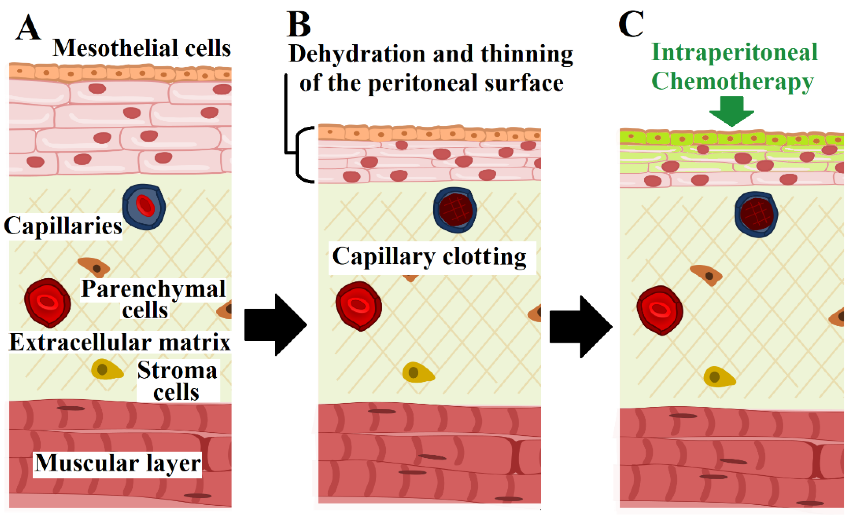
:1. Introduction
2. Results
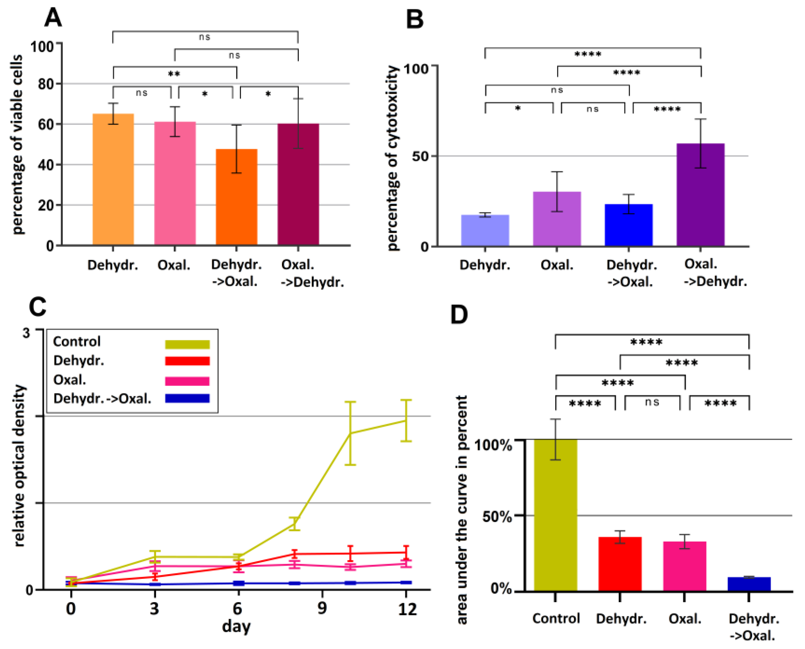
2.1. The Effect of Dehydration under Hyperthermic Conditions Combined with Chemotherapy Compared to Alternatives
2.2. The Cumulative Effect of Three-Cycles of Dehydration under Hyperthermic Conditions and Hyperthermia with Chemotherapy Compared to Alternatives
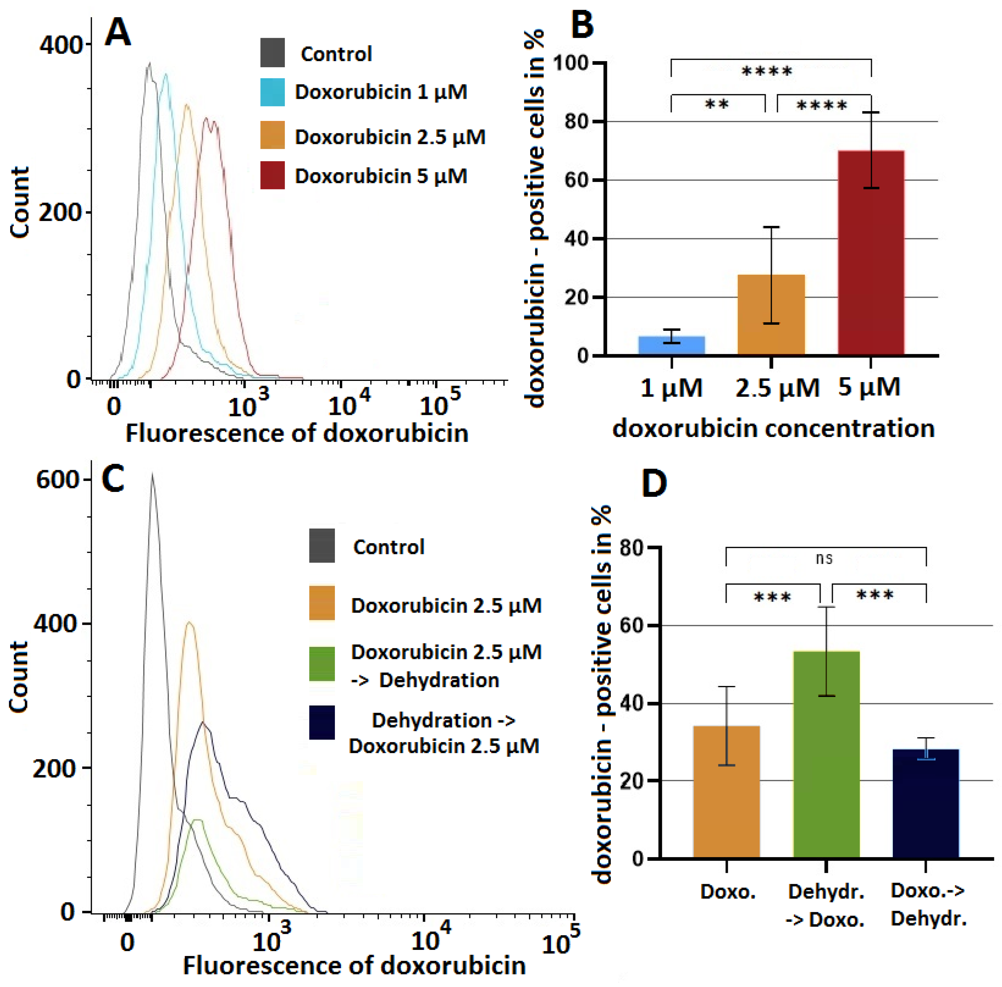
2.3. The Effect of Dehydration under Hyperthermic Conditions on the Cellular Uptake of Doxorubicin into HT-29 Cell Lines
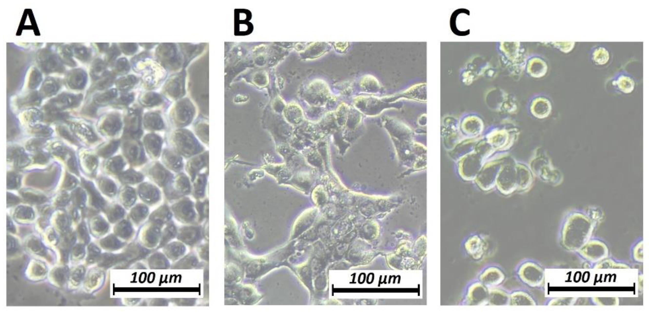
2.4. Effect of Dehydration on HT-29 Cells Phenotype Assessed by Phase Contrast Microscopy
3. Discussion
4. Materials and Methods
4.1. Cell Culture
4.2. Partial Dehydration under Hyperthermic Condition Combined with Chemotherapy
4.3. Cumulative Effect of Partial Dehydration under Hyperthermic Condition Combined with Chemotherapy and Proliferation Ability Analysis
4.4. Analysis the Effects of of Triple-Therapy and Dehydration Using Viability Testing and Cytotoxicity Assay
4.5. Analysis of Doxorubicin Penetration into HT-29 Cell Line by Flow Cytometry
4.6. Phase Contrast Microscopy
4.7. Statistical Analysis
5. Conclusions
Author Contributions
Funding
Institutional Review Board Statement
Informed Consent Statement
Data Availability Statement
Conflicts of Interest
References
- Sadeghi, B.; Arvieux, C.; Glehen, O.; Beaujard, A.C.; Rivoire, M.; Baulieux, J.; Fontaumard, E.; Brachet, A.; Caillot, J.L.; Faure, J.L.; et al. Peritoneal carcinomatosis from non-gynecologic malignancies: Results of the EVOCAPE 1 multicentric prospective study. Cancer 2000, 88, 358–363. [Google Scholar] [CrossRef]
- Anwar, A.; Kasi, A. Peritoneal Cancer. In StatPearls; StatPearls Publishing: Tampa, FL, USA, 2022. [Google Scholar]
- Heaney, R.M.; Shields, C.; Mulsow, J. Outcome following incomplete surgical cytoreduction combined with intraperitoneal chemotherapy for colorectal peritoneal metastases. World J. Gastrointest. Oncol. 2015, 7, 445–454. [Google Scholar] [CrossRef] [PubMed]
- Paul Olson, T.J.; Pinkerton, C.; Brasel, K.J.; Schwarze, M.L. Palliative surgery for malignant bowel obstruction from carcinomatosis: A systematic review. JAMA Surg. 2014, 149, 383–392. [Google Scholar] [CrossRef] [PubMed]
- Nindra, U.; Shahnam, A.; Mahon, K.L. Review of systemic chemotherapy in unresectable colorectal peritoneal carcinomatosis. Asia-Pac. J. Clin. Oncol. 2022, 18, 7–12. [Google Scholar] [CrossRef]
- Hong, S.H.; Shin, Y.R.; Roh, S.Y.; Jeon, E.K.; Song, K.Y.; Park, C.H.; Jeon, H.M.; Hong, Y.S. Treatment outcomes of systemic chemotherapy for peritoneal carcinomatosis arising from gastric cancer with no measurable disease: Retrospective analysis from a single center. Gastric Cancer 2013, 3, 290–300. [Google Scholar] [CrossRef]
- Aoyagi, T.; Terracina, K.P.; Raza, A.; Takabe, K. Current treatment options for colon cancer peritoneal carcinomatosis. World J. Gastroenterol. 2014, 20, 12493–12500. [Google Scholar] [CrossRef]
- Morano, W.F.; Khalili, M.; Chi, D.S.; Bowne, W.B.; Esquivel, J. Clinical studies in CRS and HIPEC: Trials, tribulations, and future directions-A systematic review. J. Surg. Oncol. 2018, 117, 245–259. [Google Scholar] [CrossRef]
- Sugarbaker, P.H.; Ryan, D.P. Cytoreductive surgery plus hyperthermic perioperative chemotherapy to treat peritoneal metastases from colorectal cancer: Standard of care or an experimental approach? Lancet Oncol. 2012, 13, e362–e369. [Google Scholar] [CrossRef]
- Zhou, S.; Feng, Q.; Zhang, J.; Zhou, H.; Jiang, Z.; Liu, Z.; Zheng, Z.; Chen, H.; Wang, Z.; Liang, J.; et al. High-grade postoperative complications affect survival outcomes of patients with colorectal Cancer peritoneal metastases treated with Cytoreductive surgery and Hyperthermic Intraperitoneal chemotherapy. BMC Cancer 2021, 21, 41. [Google Scholar] [CrossRef]
- Khosrawipour, T.; Khosrawipour, V.; Giger-Pabst, U. Pressurized Intra Peritoneal Aerosol Chemotherapy in patients suffering from peritoneal carcinomatosis of pancreatic adenocarcinoma. PLoS ONE 2017, 12, e0186709. [Google Scholar] [CrossRef]
- Khosrawipour, V.; Mikolajczyk, A.; Schubert, J.; Khosrawipour, T. Pressurized Intra-peritoneal Aerosol Chemotherapy (PIPAC) via Endoscopical Microcatheter System. Anticancer Res. 2018, 38, 3447–3452. [Google Scholar] [CrossRef]
- Khosrawipour, V.; Khosrawipour, T.; Kern, A.J.P.; Osma, A.; Kabakci, B.; Diaz-Carballo, D.; Förster, E.; Zieren, J.; Fakhrian, K. Distribution pattern and penetration depth of doxorubicin after pressurized intraperitoneal aerosol chemotherapy (PIPAC) in a postmortem swine model. J. Cancer Res. Clin. Oncol. 2016, 142, 2275–2280. [Google Scholar] [CrossRef]
- Khosrawipour, V.; Khosrawipour, T.; Diaz-Carballo, D.; Förster, E.; Zieren, J.; Giger-Pabst, U. Exploring the Spatial Drug Distribution Pattern of Pressurized Intraperitoneal Aerosol Chemotherapy (PIPAC). Ann. Surg. Oncol. 2016, 23, 1220–1224. [Google Scholar] [CrossRef]
- Yonemura, Y. A new bidirectional intraperitoneal and systemic induction chemotherapy (BISIC) for the peritoneal metastasis from gastric cancer in neoadjuvant setting. Integr. Cancer Sci. Therap. 2014, 1, 26–29. [Google Scholar] [CrossRef]
- Alyami, M.; Mercier, F.; Siebert, M.; Bonnot, P.-E.; Laplace, N.; Villeneuve, L.; Passot, G.; Glehen, O.; Bakrin, N.; Kepenekian, V. Unresectable peritoneal metastasis treated by pressurized intraperitoneal aerosol chemotherapy (PIPAC) leading to cytoreductive surgery and hyperthermic intraperitoneal chemotherapy. Eur. J. Surg. Oncol. 2021, 47, 128–133. [Google Scholar] [CrossRef]
- Mikolajczyk, A.; Khosrawipour, V.; Schubert, J.; Plociennik, M.; Nowak, K.; Fahr, C.; Chaudhry, H.; Khosrawipour, T. Feasibility and Characteristics of Pressurized Aerosol Chemotherapy (PAC) in the Bladder as a Therapeutical Option in Early-stage Urinary Bladder Cancer. In Vivo 2018, 32, 1369–1372. [Google Scholar] [CrossRef]
- Khosrawipour, V.; Mikolajczyk, A.; Paslawski, R.; Plociennik, M.; Nowak, K.; Kulas, J.; Arafkas, M.; Khosrawipour, T. Intrathoracic aerosol chemotherapy via spray-catheter. Mol. Clin. Oncol. 2020, 12, 350–354. [Google Scholar] [CrossRef]
- Schubert, J.; Khosrawipour, T.; Reinhard, S.; Arafkas, M.; Martino, A.; Bania, J.; Pieczka, M.; Pigazzi, A.; Khosrawipour, V. The concept of foam as a drug carrier for intraperitoneal chemotherapy, feasibility, cytotoxicity and characteristics. Sci. Rep. 2020, 10, 10341. [Google Scholar] [CrossRef]
- Schubert, J.; Khosrawipour, V.; Chaudhry, H.; Arafkas, M.; Knoefel, W.T.; Pigazzi, A.; Khosrawipour, T. Comparing the cytotoxicity of taurolidine, mitomycin C, and oxaliplatin on the proliferation of in vitro colon carcinoma cells following pressurized intra-peritoneal aerosol chemotherapy (PIPAC). World J. Surg. Oncol. 2019, 17, 93. [Google Scholar] [CrossRef]
- Khosrawipour, T.; Schubert, J.; Khosrawipour, V.; Chaudhry, H.; Grzesiak, J.; Arafkas, M.; Mikolajczyk, A. Particle stability and structure on the peritoneal surface in pressurized intra-peritoneal aerosol chemotherapy (PIPAC) analysed by electron microscopy: First evidence of a new physical concept for PIPAC. Oncol. Lett. 2019, 17, 4921–4927. [Google Scholar] [CrossRef]
- Mikolajczyk, A.; Khosrawipour, V.; Schubert, J.; Chaudhry, H.; Pigazzi, A.; Khosrawipour, T. Particle Stability During Pressurized Intra-peritoneal Aerosol Chemotherapy (PIPAC). Anticancer. Res. 2018, 38, 4645–4649. [Google Scholar] [CrossRef] [PubMed]
- Mikolajczyk, A.; Khosrawipour, V.; Lau, H.; Li, S.; Migdal, P.; Labbé, M.K.; Kielan, W.; Nicpon, J.; Stieglitz, S.; Khosrawipour, T. Exploring the potential of taurolidine in inducing mobilization and detachment of colon cancer cells: A preliminary in-vitro study. BMC Pharmacol. Toxicol. 2022, 23, 38. [Google Scholar] [CrossRef] [PubMed]
- Khosrawipour, V.; Khosrawipour, T.; Hedayat-Pour, Y.; Diaz-Carballo, D.; Bellendorf, A.; Böse-Ribeiro, H.; Mücke, R.; Mohanaraja, N.; Adamietz, I.A.; Fakhrian, K. Effect of Whole-abdominal Irradiation on Penetration Depth of Doxorubicin in Normal Tissue After Pressurized Intraperitoneal Aerosol Chemotherapy (PIPAC) in a Post-mortem Swine Model. Anticancer Res. 2017, 37, 1677–1680. [Google Scholar] [PubMed]
- Khosrawipour, V.; Giger-Pabst, U.; Khosrawipour, T.; Pour, Y.H.; Diaz-Carballo, D.; Förster, E.; Böse-Ribeiro, H.; Adamietz, I.A.; Zieren, J.; Fakhrian, K. Effect of Irradiation on Tissue Penetration Depth of Doxorubicin after Pressurized Intra-Peritoneal Aerosol Chemotherapy (PIPAC) in a Novel Ex-Vivo Model. J. Cancer 2016, 7, 910–914. [Google Scholar] [CrossRef]
- Khosrawipour, V.; Bellendorf, A.; Khosrawipour, C.; Hedayat-Pour, Y.; Diaz-Carballo, D.; Förster, E.; Mücke, R.; Kabakci, B.; Adamietz, I.A.; Fakhrian, K. Irradiation Does Not Increase the Penetration Depth of Doxorubicin in Normal Tissue After Pressurized Intra-peritoneal Aerosol Chemotherapy (PIPAC) in an Ex Vivo Model. In Vivo 2016, 30, 593–597. [Google Scholar]
- Lau, H.; Khosrawipour, T.; Mikolajczyk, A.; Frelkiewicz, P.; Nicpon, J.; Arafkas, M.; Pigazzi, A.; Knoefel, W.T.; Khosrawipour, V. Intraperitoneal chemotherapy of the peritoneal surface using high-intensity ultrasound (HIUS): Investigation of technical feasibility, safety and possible limitations. J. Cancer 2020, 11, 7209–7215. [Google Scholar] [CrossRef]
- Mikolajczyk, A.; Khosrawipour, T.; Kulas, J.; Migdal, P.; Arafkas, M.; Nicpon, J.; Khosrawipour, V. The structural effect of high intensity ultrasound on peritoneal tissue: A potential vehicle for targeting peritoneal metastases. BMC Cancer 2020, 20, 481. [Google Scholar] [CrossRef]
- Mikolajczyk, A.; Khosrawipour, V.; Kulas, J.; Kocielek, K.; Migdal, P.; Arafkas, M.; Khosrawipour, T. Release of doxorubicin from its liposomal coating via high intensity ultrasound. Mol. Clin. Oncol. 2019, 11, 483–487. [Google Scholar] [CrossRef]
- Khosrawipour, V.; Reinhard, S.; Martino, A.; Khosrawipour, T.; Arafkas, M.; Mikolajczyk, A. Increased Tissue Penetration of Doxorubicin in Pressurized Intraperitoneal Aerosol Chemotherapy (PIPAC) after High-Intensity Ultrasound (HIUS). Int. J. Surg. Oncol. 2019, 2019, 6185313. [Google Scholar] [CrossRef]
- Mikolajczyk, A.; Khosrawipour, T.; Martino, A.; Kulas, J.; Pieczka, M.; Zacharski, M.; Nicpon, J.; Khosrawipour, V. Enabling Microparticle Imprinting to Achieve Penetration and Local Endurance in the Peritoneum via High-Intensity Ultrasound (HIUS) for the Treatment of Peritoneal Metastasis. Int. J. Surg. Oncol. 2020, 2020, 9679385. [Google Scholar] [CrossRef]
- Khosrawipour, T.; Schubert, J.; Kulas, J.; Migdal, P.; Arafkas, M.; Bania, J.; Khosrawipour, V. Creating nanocrystallized chemotherapy: The differences in pressurized aerosol chemotherapy (PAC) via intracavitary (IAG) and extracavitary aerosol generation (EAG) regarding particle generation, morphology and structure. J. Cancer 2020, 11, 1308–1314. [Google Scholar] [CrossRef]
- Sugarbaker, P.H. Carcinomatosis—Is Cure an Option? J. Clin. Oncol. 2003, 21, 762–764. [Google Scholar] [CrossRef]
- Thelen, S.; Mikolajczyk-Martinez, A.; Diakun, A.; Khosrawipour, T.; Zielinski, K.; Nicpoń, J.; Kiełbowicz, Z.; Prządka, P.; Liszka, B.; Kuropka, P.; et al. Evaluating the concept of gas-based intraperitoneal hyperthermia beyond 43 °C in the treatment of peritoneal metastasis: A pilot study. Exp. Ther. Med. 2022, 24, 1–9. [Google Scholar] [CrossRef]
- Diakun, A.; Khosrawipour, T.; Mikolajczyk-Martinez, A.; Nicpoń, J.; Kiełbowicz, Z.; Prządka, P.; Liszka, B.; Kielan, W.; Zielinski, K.; Migdal, P.; et al. The Onset of In-Vivo Dehydration in Gas-Based Intraperitoneal Hyperthermia and Its Cytotoxic Effects on Colon Cancer Cells. Front. Oncol. 2022, 1, 1–9. [Google Scholar]
- Diakun, A.; Khosrawipour, T.; Mikolajczyk-Martinez, A.; Nicpoń, J.; Thelen, S.; Kiełbowicz, Z.; Prządka, P.; Liszka, B.; Kulas, J.; Zielinski, K.; et al. Safety, feasibility, and application of intraperitoneal gas-based hyperthermia beyond 43 °C in the treatment of peritoneal metastasis: An in-vivo pilot study. Front. Oncol. 2022, 12, 953920. [Google Scholar] [CrossRef]
- Diakun, A.; Khosrawipour, T.; Mikolajczyk-Martinez, A.; Kuropka, P.; Nicpoń, J.; Kiełbowicz, Z.; Prządka, P.; Liszka, B.; Li, S.; Lau, H.; et al. In-vivo thermodynamic exploration of gas-based intraperitoneal hyperthermia. Front. Oncol. 2022, 12, 925724. [Google Scholar] [CrossRef]
- Sugarbaker, P.H. Laboratory and clinical basis for hyperthermia as a component of intracavitary chemotherapy. Int. J. Hyperthermia 2007, 23, 431–442. [Google Scholar] [CrossRef]
- Mitteer, D.R.; Greer, B.D. Using GraphPad Prism’s Heat Maps for Efficient, Fine-Grained Analyses of Single-Case Data. Behav. Anal. Pract. 2022, 15, 505–514. [Google Scholar] [CrossRef]
- Cummings, B.S.; Wills, L.P.; Schnellmann, R.G. Measurement of Cell Death in Mammalian Cells. Curr. Protoc. Pharmacol. 2012, 56, 12.8.1–12.8.24. [Google Scholar] [CrossRef]
- Elmore, S. Apoptosis: A Review of Programmed Cell Death. Toxicol. Pathol. 2007, 35, 495–516. [Google Scholar] [CrossRef]
- Cookson, B.T.; Brennan, M.A. Pro-inflammatory programmed cell death. Trends Microbiol. 2001, 9, 113–114. [Google Scholar] [CrossRef] [PubMed]
- Fink, S.L.; Cookson, B.T. Apoptosis, Pyroptosis, and Necrosis: Mechanistic Description of Dead and Dying Eukaryotic Cells. Infect. Immun. 2005, 73, 1907–1916. [Google Scholar] [CrossRef] [PubMed]
- Beswick, E.J. Inflammasome activation to protect against colorectal cancer metastasis? Sci. Transl. Medicine 2015, 7, 314ec196. [Google Scholar] [CrossRef]
- Fang, Y.; Tian, S.; Pan, Y.; Li, W.; Wang, Q.; Tang, Y.; Yu, T.; Wu, X.; Shi, Y.; Ma, P.; et al. Pyroptosis: A new frontier in cancer. Biomed. Pharmacother. 2020, 121, 109595. [Google Scholar] [CrossRef]


Disclaimer/Publisher’s Note: The statements, opinions and data contained in all publications are solely those of the individual author(s) and contributor(s) and not of MDPI and/or the editor(s). MDPI and/or the editor(s) disclaim responsibility for any injury to people or property resulting from any ideas, methods, instructions or products referred to in the content. |
© 2023 by the authors. Licensee MDPI, Basel, Switzerland. This article is an open access article distributed under the terms and conditions of the Creative Commons Attribution (CC BY) license (https://creativecommons.org/licenses/by/4.0/).
Share and Cite
Khosrawipour, C.; Diakun, A.; Li, S.; Lau, H.; Kulas, J.; Khosrawipour, V.; Kielan, W.; Mikolajczyk-Martinez, A. Triple-Therapy of Peritoneal Metastasis—Partial-Dehydration under Hyperthermic Condition Combined with Chemotherapy: The First Preliminary In-Vitro Results. Pharmaceuticals 2023, 16, 763. https://doi.org/10.3390/ph16050763
Khosrawipour C, Diakun A, Li S, Lau H, Kulas J, Khosrawipour V, Kielan W, Mikolajczyk-Martinez A. Triple-Therapy of Peritoneal Metastasis—Partial-Dehydration under Hyperthermic Condition Combined with Chemotherapy: The First Preliminary In-Vitro Results. Pharmaceuticals. 2023; 16(5):763. https://doi.org/10.3390/ph16050763
Chicago/Turabian StyleKhosrawipour, Carolina, Agata Diakun, Shiri Li, Hien Lau, Joanna Kulas, Veria Khosrawipour, Wojciech Kielan, and Agata Mikolajczyk-Martinez. 2023. "Triple-Therapy of Peritoneal Metastasis—Partial-Dehydration under Hyperthermic Condition Combined with Chemotherapy: The First Preliminary In-Vitro Results" Pharmaceuticals 16, no. 5: 763. https://doi.org/10.3390/ph16050763