Article

Radiosynthesis of Stable 198Au-Nanoparticles by Neutron Activation of αvβ3-Specific AuNPs for Therapy of Tumor Angiogenesis

1
Molecular Imaging and Radiochemistry, Clinic of Radiology and Nuclear Medicine, Medical Faculty Mannheim of Heidelberg University, 68167 Mannheim, Germany
2
Biomedical Chemistry, Clinic of Radiology and Nuclear Medicine, Medical Faculty Mannheim of Heidelberg University, 68167 Mannheim, Germany
3
Mannheim Institute for Intelligent Systems in Medicine MIISM, Medical Faculty Mannheim of Heidelberg University, 68167 Mannheim, Germany
4
Research Reactor TRIGA Mainz, Institute for Nuclear Chemistry, Johannes-Gutenberg-Universität Mainz, 55128 Mainz, Germany
5
Department of Oncology, Division of Oncological Imaging, University of Alberta, Edmonton, AB T6G 2R3, Canada
6
Department of Nuclear Medicine, University Hospital Carl Gustav Carus, TU Dresden, 01307 Dresden, Germany
*
Authors to whom correspondence should be addressed.
These authors contributed equally to this work.
Pharmaceuticals 2023, 16(12), 1670; https://doi.org/10.3390/ph16121670
Submission received: 6 November 2023 / Revised: 24 November 2023 / Accepted: 28 November 2023 / Published: 30 November 2023
(This article belongs to the Special Issue Therapeutic Radionuclides in Nuclear Medicine)

Abstract

:
This paper reports on the development of stable tumor-specific gold nanoparticles (AuNPs) activated by neutron irradiation as a therapeutic option for the treatment of cancer with high tumor angiogenesis. The AuNPs were designed with different mono- or dithiol-ligands and decorated with different amounts of Arg-Gly-Asp (RGD) peptides as a tumor-targeting vector for αvβ3 integrin, which is overexpressed in tissues with high tumor angiogenesis. The AuNPs were evaluated for avidity in vitro and showed favorable properties with respect to tumor cell accumulation. Furthermore, the therapeutic properties of the [198Au]AuNPs were evaluated in vitro on U87MG cells in terms of cell survival, suggesting that these [198Au]AuNPs are a useful basis for future therapeutic concepts.

Graphical Abstract

1. Introduction

In recent years, gold nanoparticles (AuNPs) have received serious attention since their first use as radioactive 198Au-nanocolloids for nanobrachytherapy in the early 1950s [1,2]. The synthesis of ultra-small AuNPs (<5 nm) [3] with multimerization of target-specific effectors on their surface leads to a new form of targeted AuNPs with higher target avidity compared to the effectors only [4]. The combination of the target-specific accumulation and a phenomenon typically known as the “enhanced permeability and retention” (EPR) effect [5], leads to a higher tumor accumulation [6]. Therefore, AuNPs with a higher renal clearance [7] for theranostic purposes [8,9,10] were developed in recent years, equipped with small molecules [11], peptides [12], near-infrared dyes [13,14], and radionuclides [15,16,17,18,19]. PEGylation of the AuNPs leads to a higher bioavailability as it prevents the formation of a protein corona around the AuNPs in vivo [20,21]. The high affinity of sulfur for gold surfaces and the formation of stable and covalent Au-S bonds [22] allows a fast and easy functionalization of AuNPs with (di-)thiol-modified (bio)molecules [23]. In addition, the use of dithiols as a surface binding motif leads to a higher stability of the AuNPs [24]. Of particular interest is their therapeutic application [25], especially their ability to be used as radiosensitizers by Auger–Meitner electron emission induced by gamma activation [26,27,28] or by direct neutron activation of natural 197AuNPs generating [198Au]AuNPs (t1/2 = 2.69 d, βmax 961 keV, 98.99%; γ 412 keV, 95.62%) [12,29,30,31,32].
The focus of this work was the development of highly stable targeted gold nanoparticles for neutron activation [33]. Therefore, AuNPs with mono- and di-thiol linkers with low and high loading of target-specific peptides were synthesized to compare their specific avidity in cell binding assays and their stability during and after neutron irradiation. To achieve target-specific accumulation in tissues with high tumor angiogenesis, the AuNPs were functionalized with a c(RGDfK) derivative [32,34]. The Arg-Gly-Asp (RGD) peptide motif is known to bind to the transmembrane αvβ3 integrin [35,36], which is overexpressed in tumor angiogenesis in tumors of various origins, for example, on glioma cells (U87MG) [37,38,39].

2. Results

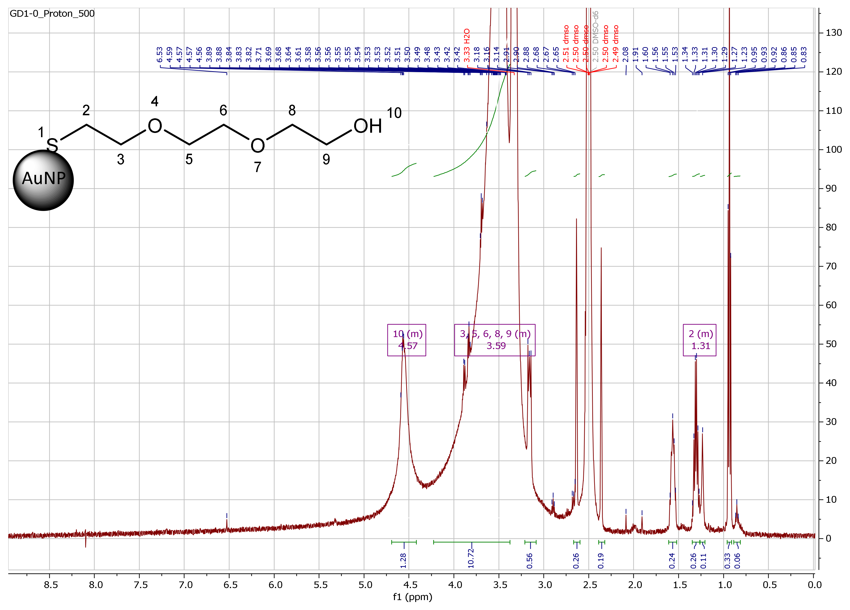
2.1. Synthesis and Functionalization of Gold Nanoparticles

Integrin αvβ3, a transmembrane protein expressed on endothelial cells, binds the RGD triple amino acid peptide motif of extracellular matrix proteins. Growing malignant tumors require continuous angiogenesis, and the integrin αvβ3 is overexpressed for this purpose. As a result, αvβ3 is preferentially expressed in tumor angiogenesis and is a potential target for AuNPs decorated with RGD peptides [36]. Therefore, ultra-small AuNPs 3 and 6 (3 ± 2 nm) were synthesized by Turcu et al. [40] and Brust and Schiffrin [41], respectively. The AuNPs contained thiol-PEG3-OH or a thioctic acid(TA)-PEG3-OH derivative 2 used as the stabilizing ligands and to achieve enhanced biocompatibility (Figure 1). The AuNPs were further functionalized by ligand exchange with low and high amounts (4–8 mg) of TA-PEG4-c(RGDfK) derivative 5 to obtain mixed AuNP-thio-PEG-dithio-PEG-RGD 7a (high RGD loading), 7b (low RGD loading) and AuNP-dithio-RGD 8a (high RGD loading), and 8b (low RGD loading), respectively. The AuNPs were purified by dialysis and size-exclusion chromatography. The size and stability of AuNPs 7a,b, and 8a,b were confirmed by UV/Vis spectroscopy and high performance liquid chromatography (HPLC).
The organic shell of the AuNPs was characterized by mass loss using thermogravimetric analyses for each functionalization step. After knowing the number of newly attached molecules, a formula by Zhu et al. was used to calculate the total molar mass of the AuNPs [42] (Table 1). A brief description of the synthesis and characterization can be found in Appendix A. All AuNPs were fully characterized by thermogravimetric analysis (TGA) (Table 1), electron microscopy (EM) (Figure A1 and Figure A2), UV/Vis spectroscopy (Figure A9), HPLC (Figure A10), and nuclear magnetic resonance spectroscopy (NMR) (Figure A11, Figure A12, Figure A13, Figure A14, Figure A15 and Figure A16). The AuNPs could be stored in lyophilized form at −20 °C for >12 months without loss of integrity. In contrast, when stored in solution at room temperature, aggregation in the form of precipitation could occur within weeks, especially for peptide-decorated particles [43].

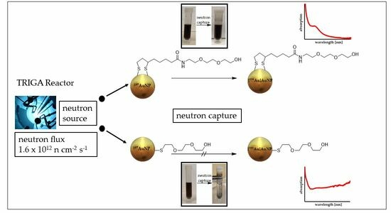
2.2. Neutron Irradiation Experiments

First neutron irradiation experiments with thermal neutrons at the TRIGA Mainz reactor were performed with non-tumor specific AuNP-dithio-PEG 3 (3-1–3-5) and AuNP-thio-PEG 6 (6-1–6-3) in different weights and concentrations. Samples were frozen and removed from the freezer immediately before irradiation. Irradiation was performed at 100 kW for 1–2 h with a neutron flux of 1.6 × 1012 cm−2 × s−1. With the reactor running, the background dose rate (DR) at the measurement position was ~2 µSv/h. Gamma measurements were not possible for probes >500 µg on the day of irradiation due to the high activity. The dead time for samples 3-5 (Table 2) was about 30 min at the end of the bombardment and still 7.5 min at 20 cm distance. Therefore, most of the gamma measurements of [198Au]AuNPs had to be performed one day after irradiation. Sample [198Au]3-5 still had ~3% dead time in 20 cm distance (Table 2). Precipitation was observed for [198Au]6 but not for [198Au]3 in the solution or on the vessel wall in any case (Figure 2). The activated samples were stored in the freezer for transport and further experiments. In addition, the half-life of [198Au]3 was determined experimentally (mean 2.80 ± 0.07 d) by measuring the activity of different concentrations with a gamma counter for 28 d (Figure A8). UV-Vis measurements showed a strong broadening of the plasmon bands for [198Au]6-1 and [198Au]6-2, indicating aggregation (Figure 3). For [198Au]3-1 and [198Au]3-2 a typical absorption for AuNPs at 514 nm was observed, indicating stable AuNPs even 5 months after neutron activation ([198Au]3-3, Figure A9). The production of ~100 MBq [198Au]3 showed stable AuNPs even at high activity concentration for at least 15 d by HPLC measurements (Figure A10).

2.3. Cell Experiments

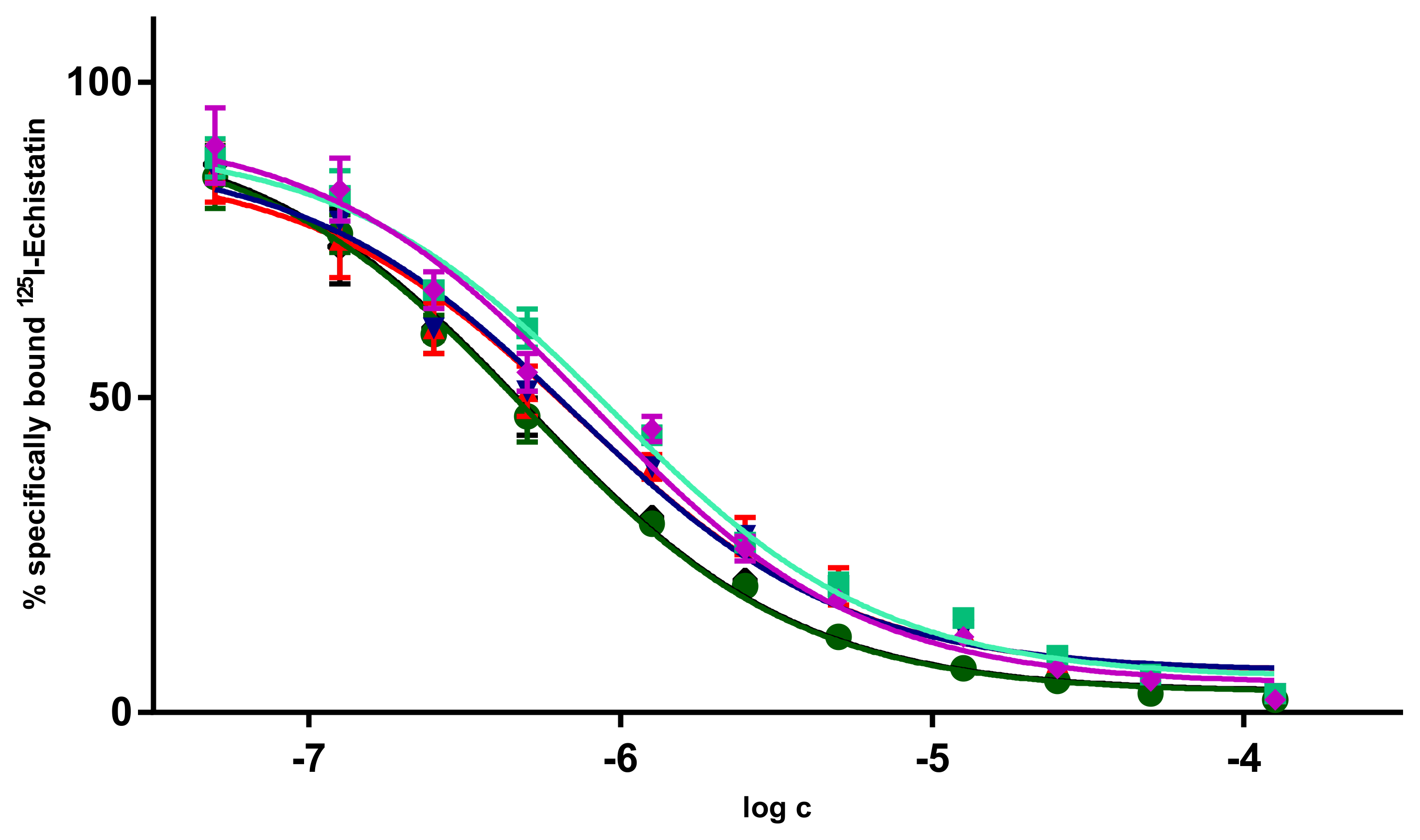
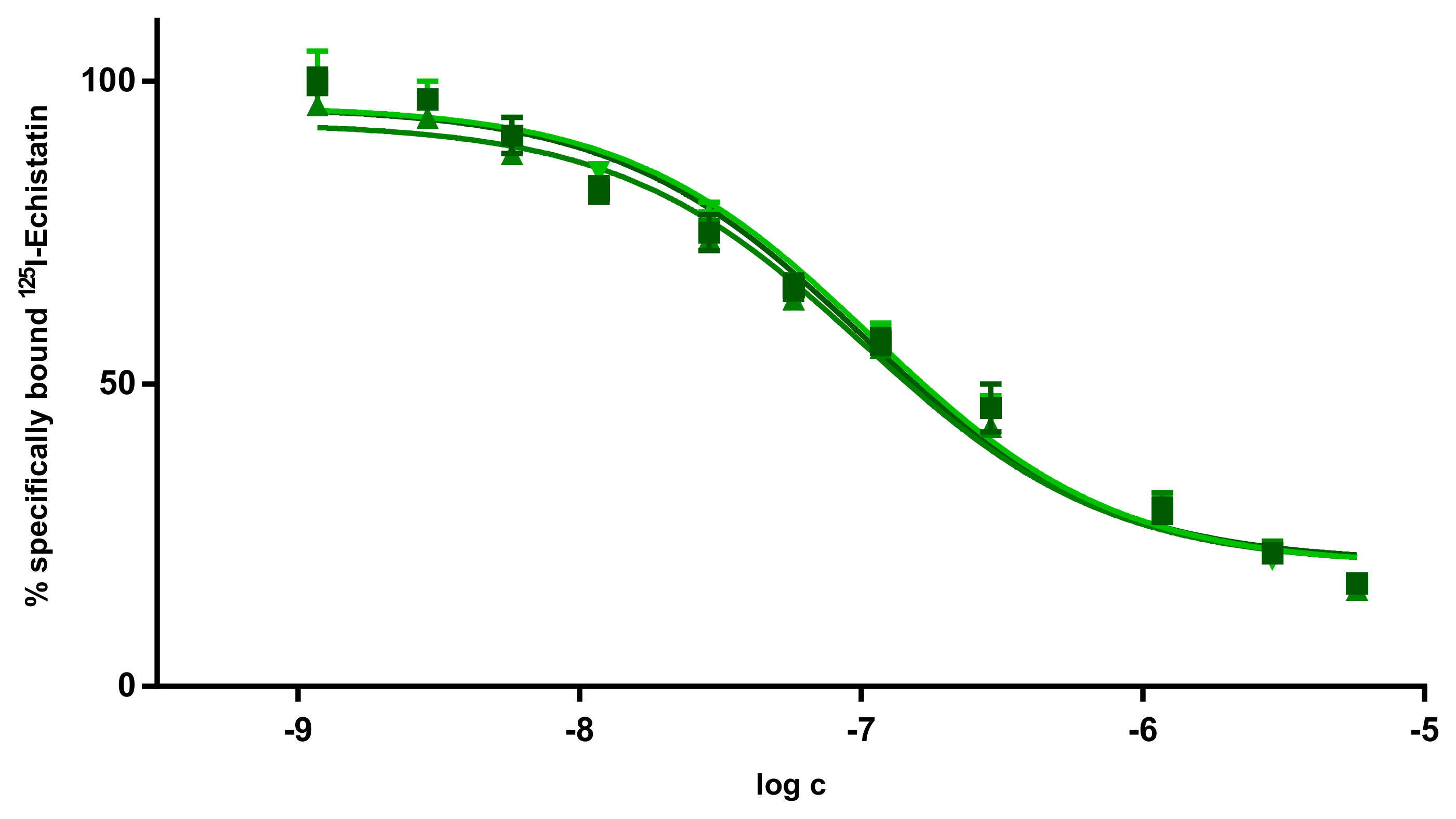
2.3.1. Determination of Target Avidities

Several different IC50 values for RGD derivatives have been reported in the literature, ranging from 0.1 nM up to 6.7 µM. The main reason for the observed differences is the assay method used to determine the IC50 values. IC50 values of 0.1–1 nM can be found for RGD peptides having been determined by ELISA assays [38] and IC50 values around 20 nM have been reported for solid-phase αvβ3 binding assays for monomeric RGD derivatives [37]. Those IC50 values were derived by non-living experiments. However, cell experiments are closer to in vivo conditions. Therefore, for the AuNPs 7 and 8, the αvβ3 integrin-avidities were determined by competitive displacement experiments on αvβ3-expressing U87MG cells using 125I-echistatin as the αvβ3-specific radioligand and competitor. The RGD monomer c(RGDfK) was evaluated as an internal reference. The evaluation of RGD derivatives by displacement experiments yielded IC50 values comparable to those reported in the literature [34]. For the c(RGDfK) monomer, a mean IC50 value of 0.7 µM was determined (Table 3, Figure A3). The multi-RGD decoration on the surface of AuNPs 7a and 7b resulted in a lower mean IC50 value of 27.8 and 38.3 nM, respectively (Figure A4 and Figure A5). Mean IC50 values of 82.4 and 103.6 nM were found for AuNPs 8a and 8b, respectively (Figure A6 and Figure A7). It was observed that the higher the loading with αvβ3-specific RGD peptide, the lower the IC50 values.

2.3.2. Determination of Cell Survival

Colony formation assays were performed with [198Au]3 with U87MG cells. For this proof-of-concept experiment, 5–10 Gy was chosen as the incubation dose. To achieve this dose, 1–2 MBq [198Au]3 per well in a 24-well plate within a 96 h incubation period was calculated using Formula (1).
D A , t = S × A T 1 / 2 ln 2 1 exp ln 2 × t T 1 / 2
Formula (1)—Calculation of dose to a cell monolayer at the bottom of a multi-well plate or Eppendorf tube for 198Au using Geant4-simulation [44]. D: energy dose, S: S-value, A: activity, T1/2: half-life of the radionuclide, t: irradiation time.
This dose corresponds to concentrations of [198Au]AuNPs of 0.515–0.939 µM, which is at least 10 times higher than the IC50 of AuNP-dithio-RGD 7 and 8. It was observed that the survival fraction (sf) of the cells was significantly reduced for [198Au]3 and that higher doses of 10 Gy (sf = 18.2%) were more effective in damaging the tumor cells than 5 Gy (sf = 33.9%) (Figure 4).

3. Discussion

c(RGDfK) is a highly potent and selective integrin αvβ3 antagonist and therefore could disrupt cell viability by inhibiting angiogenesis [45]. Radiolabeled RGD derivatives can be used as tracers for tumor angiogenesis [46]. Multimerization leads to better tumor accumulation [47,48]. Therefore, AuNPs decorated with a multitude of c(RGDfK) motifs could lead to better tumor accumulation, which is important for therapy.
Methods for the preparation of [198Au]AuNPs are already known in the literature [11,29,32]. However, the synthesis starts with neutron activation of gold foil, which is then dissolved in aqua regia, followed by nanoparticle synthesis and further functionalizations with target-specific ligands. All these steps are performed with radioactive 198Au, resulting in higher dose accumulation for the personnel and more radioactive waste as the consequence. In this work, it was decided to first complete the synthesis of tumor-specific AuNPs with a high target avidity and high stability, and to perform neutron activation as the last step in order to reduce the personnel dose and enable a highly efficient synthesis pathway, which is mandatory for high clinical relevance. The challenge was to synthesize AuNPs that withstand neutron activation without aggregation and loss of the ligand shell.
Stable αvβ3-specific AuNPs 7 and 8 were successfully synthesized with a better avidity compared to the monomeric peptide ligand c(RGDfK). During the irradiation experiments, it was observed that AuNPs containing monothiol ligands were unstable against neutron activation. However, all AuNP derivatives containing only dithiol ligands were stable against neutron activation even at the highest concentrations and irradiation times (~7.5 mg/mL within 2 h). It is known that sulfur can also be activated by neutrons via the 32S(n,p)32P reaction [49,50]. Presumably, once a sulfur atom is activated to 32P, it loses its covalent bond to the AuNP surface and a monothiol ligand is lost to the environment. In contrast, a dithiol ligand could remain bound to the surface even if a binding interaction is lost by activation of one of the sulfur atoms.
To determine the therapeutic influence of [198Au]AuNPs, cell survival was addressed by a colony formation assay. The activity and incubation time to reach relevant doses between 5 and 10 Gy were calculated for monolayer cell culture in 24-well plates (Formula (1)). To reach these doses of 5–10 Gy concentrations of a factor >10 times higher than the IC50 for AuNP-RGD 8a and 8b had to be used within 96 h of incubation. Therefore, cell viability should be considered to be very low when using such high concentrations of [198Au]8 in cell survival experiments, as the antagonist RGD may interfere with angiogenesis and thus cell viability [45]. To circumvent this problem, future experiments should use higher activity concentrations (due to longer activation of AuNPs) or longer cell incubation with lower doses >5 Gy, when evaluating cell survival. However, in the proof-of-concept cell survival experiments, non-specific [198Au]3 showed a significant effect on U87MG cells with a survival fraction as low as 18.2% at 10 Gy. Therefore, the combination of β-emission from 198Au and the antagonistic effect of RGD could dramatically reduce the therapeutically relevant dose of applied [198Au]AuNP-RGDs.

4. Materials and Methods

General procedures. All reagents and solvents were purchased from commercial suppliers (Sigma, Merck) and were used without further purification. NMR spectra were recorded on a 300 MHz Mercury Plus and a 500 MHz NMR System spectrometer (Varian, Palo Alto, CA, USA). Chemical shifts (δ) are given in ppm and are referenced to the residual solvent resonance signals relative to (CH3)4Si (1H, 13C). Mass spectra were obtained on a microflex MALDI-TOF mass spectrometer (Bruker Daltonics, Bremen, Germany) and HR-ESI-MS spectra on a LTQ FT Ultra Fourier Transform Ion Cyclotron Resonance spectrometer (Thermo Finnigan, Dreieich, Germany). When applicable, purity was determined by HPLC. The purity of all final compounds was 95% or higher. HPLC was performed on a Dionex UltiMate 3000 HPLC system (Thermo Scientific, Dreieich, Germany), equipped with a reverse phase column (Analytical: Merck Chromolith RP-18e; 100 × 4.6 mm plus a guard column 5 × 4.6 mm; semipreparative: Chromolith RP-18e; 100 × 10 mm plus a guard column 10 × 4.6 mm), and a UV-diode array detector (210 nm, 254 nm). The solvent system used was a gradient of acetonitrile:water (containing 0.1% TFA) (0–5 min: 0–100% MeCN) at a flow rate of 4 mL/min, unless otherwise stated. The purity and stability of AuNPs/[198Au]AuNPs were investigated by size exclusion HPLC using a PolySep™-SEC GFC-P 4000, LC column 300 × 7.8 mm, and a 35 mm PolySep guard column (Phenomenex, Aschaffenburg, Germany) with water (0.8 mL/min) as eluent (Figure A10). Purification of AuNPs was performed by dialysis (tubes with molecular weight cut-off of 14,000 g/mol, Visking, Roth, Karlsruhe, Germany) against distilled water and by size-exclusion chromatography using Sephadex G25 PD10 columns (Fisher Scientific, Schwerte, Germany) and distilled water as eluent.
A brief description of the AuNP syntheses can be found in Appendix A.
Determination of the number of ligands on the surface of the AuNPs. The thermogravimetric analyses were performed using a Mettler Toledo TGA 2 STARe system. AuNPs (1–2 mg) were weighed into 70-µL-aluminum oxide crucibles (Mettler Toledo, Gießen, Germany) and heated from 25–750 °C (10 K/min) in a stream of N2 or CO2 (30 mL/min). The loading of the different AuNPs is shown in Table 1 and was calculated from the different mass losses, which increase as the AuNPs are functionalized. Therefore, the amount of different ligands per particle can be calculated according to the formula of Zhu et al. [42]. Since the nanoparticles have an average diameter of ~3 nm, the calculated amount of gold atoms is ~834 Au atoms per nanoparticle. This gives a molecular weight of an AuNP of 164,298 g/mol. Using TGA, the following ligand numbers were determined:
  • The mass loss of the AuNP 6 was ~19.8%. This corresponds to ~250 PEG ligands on the AuNP surface. M~210 kDA.
  • The mass loss of AuNP-RGD 7a was ~24.8% and the RGD accounts for ~5% mass loss (~35 RGD ligands per AuNP). Therefore, the molar mass for AuNP-RGDhigh 7a was calculated to be ~239 kDa.
  • Furthermore, the AuNP-RGDlow 7b contained ~15 RGD ligands ~222 kDa.
  • The mass loss of the AuNP 3 was ~33.27%. results in ~240 PEG ligands on the AuNP surface. M~246 kDa.
  • The mass loss of AuNP-PEG-RGDhigh 8a was ~37.1% and the RGD accounts for ~4% mass loss (~24 RGD ligands per AuNP). Therefore, the molar mass for AuNP-RGDhigh 8a was calculated to be ~262 kDa.
  • Furthermore, the AuNP-RGDlow 8b contained ~18 RGD ligands ~257 kDa.
Avidity experiments. The αvβ3-binding affinities of the respective RGD peptides and AuNPs were determined using in vitro competitive displacement experiments on U87MG tumor cells (HTB-14, ATCC®, Manassas, VA, USA). U87MG cells were harvested and resuspended in the binding buffer at a cell concentration of 2 × 10⁶/mL to reach 105 cells per well.
A special binding buffer in sterile distilled water (Tris·HCl 25 mM, NaCl 150 mM, CaCl2 1 mM, MgCl2 0.5 mM, MnCl2 1 mM, pH 7.4, BSA 0.5%) was used for incubation with 0.25–0.40 kBq/well 125I-Echistatin (81.4 GBq/μmol) as the αvβ3 specific radioligand in the presence of increasing concentrations (0–100 μM) of competing c(RGDfK) peptide or c(RGDfK)-modified AuNPs (0–20 µM). IC50 values were obtained using GraphPad Prism v6.05 (nonlinear fit) software.
Neutron irradiation experiments. Production of [198Au]AuNPs by neutron activation of 0.05–15.5 mg AuNPs was performed in pneumatic transfer tube one for 1–2 h at 100 kW with a thermal neutron flux of 1.6 × 1012 cm−2 × s−1 in the TRIGA research reactor (Mainz, Germany). For calibration of the dose calibrator ISOMED 2010 (NUVIA Instruments, Dresden, Germany) 12.7 mg solid Au was irradiated for 1 h to produce 87 MBq (calculated) [198Au]Au with a measured dose rate of 57 µSv/h. 26 h later, the activity was measured with the dose calibrator, and 60 MBq was obtained (using the 137Cs-channel, 66 MBq calculated). In addition, the solid [198Au]Au (40 MBq) was carefully dissolved in 2 mL aqua regia at 50 °C within 15 min in order to find the correct calibration factors of the dose calibrator for different volumes in vials and syringes.
Irradiation of AuNPs was performed under optimized conditions in 2 mL 10% EtOH/H2O and 25 mg ascorbic acid as a stabilizer against radiolysis [51]. Theoretically, 10 mg of pure 197Au irradiated with a thermal neutron flux of 1.6 × 1012 cm−2 × s−1 would produce 48–96 MBq 198Au within 1–2 h of irradiation. In the experiment, neutron activation of 5.0 mg AuNPs 3 and 8 for 2 h produced 48 MBq [198Au]3 (66.7% Au) and 50 MBq [198Au]8 (62.9% Au). Neutron activation of 15.56 mg AuNP 3 for 2 h produced ~100 MBq [198Au]3 (67% Au). The production of 198Au was confirmed by gamma spectroscopy, which found up to three gamma lines at 411 keV (95.6%), 676 keV (0.8%), and 1088 keV (0.2%).
Colony formation assay. Three days before the experiments, 150,000 cells were seeded in 24-well plates. U87MG cells were incubated for 96 h in the presence of the αvβ3-specific or non-radioactive AuNPs or 1–2 MBq [198Au]AuNPs to achieve the calculated doses of 5–10 Gy. After incubation, the cell medium was removed, the cells were washed and harvested, and a colony formation assay was performed in triplicate for each irradiation point with 1000 cells per well in a 6-well plate. Colonies were cultured in cell medium for 28 days, then washed with 1 mL PBS, fixed with 2 mL 4% formaldehyde in PBS for 15 min, and incubated with 2 mL 0.5% crystal violet dye solution for 30 min. Afterward, colonies were washed with distilled water, dried, and counted by light microscopy. Colonies of more than 50 cells were considered viable, and the plating efficiency for each sample was estimated based on the initial number of cells seeded. Clonogenic cell survival was calculated as the relative plating efficiency of treated versus untreated samples. Triplicate samples were prepared for each treatment and experimental condition.

5. Conclusions

αvβ3-specific RGD-containing AuNPs with a higher target avidity compared to αvβ3-specific RGD were successfully synthesized. This proof-of-concept work should demonstrate, that activation of AuNPs with a ligand shell is possible without losing their organic shell and integrity. Irradiation experiments demonstrated the stability and consistency of [198Au]AuNPs with dithiol ligands compared to [198Au]AuNPs with monothiol ligands, which always aggregated at each applied concentration after neutron activation. In vitro experiments determine the therapeutic effect of [198Au]AuNPs by addressing the survival fraction of U87MG cells proved a significant influence on cell death. Therefore, the [198Au]AuNPs could serve as a tool for endoradiotherapy.
Further experiments to determine the therapeutic effects of [198Au]AuNPs in vivo by different modes of application (local vs. systemic) are currently underway.

Author Contributions

Conceptualization, G.D., C.W., M.P. and B.W.; methodology, G.D., K.E., C.G. and M.P.; software (Geant 4 v11.1.2), R.F.; validation, G.D. and M.P.; formal analysis, G.D. and K.E.; investigation, G.D.; resources, M.P. and B.W.; data curation, G.D., R.S. and M.P.; writing—original draft preparation, G.D., C.W. and M.P.; writing—review and editing, G.D., C.W., K.E., C.G., R.S., R.F. and B.W.; visualization, G.D. and M.P.; supervision, M.P. and B.W.; project administration, M.P. and B.W.; funding acquisition, C.W., M.P. and B.W. All authors have read and agreed to the published version of the manuscript.

Funding

This research was funded by Research Campus M2OLIE funded by the German Federal Ministry of Education and Research (BMBF) within the Framework “Forschungscampus: public-private partnership for Innovations”, Funding Codes 13GW0091B, 13GW0091E, 13GW0388A and 13GW0389B.

Institutional Review Board Statement

Not applicable.

Informed Consent Statement

Not applicable.

Data Availability Statement

All data can be referred to on request to the corresponding author.

Acknowledgments

The authors are grateful to Karsten Richter from DKFZ Heidelberg for performing the electron microscopy studies of the AuNPs. The NMR measurements performed by Tobias Timmermann at RKU Heidelberg and the HR-ESI measurements performed by Werner Spahl at LMU Munich are gratefully acknowledged. We also want to thank Ulrich Scherer for using the TGA spectrometer and for performing initial neutron irradiation experiments and Thorsten Röder for using the DLS at Mannheim University of Applied Sciences. We thank the mechanical workshop and the staff of the Research Reactor TRIGA Mainz for their excellent support. Moreover, the authors would like to thank Lisa Hübinger for the extensive proofreading of the manuscript.

Conflicts of Interest

The authors declare no conflict of interest.

Appendix A

Appendix A.1. Organic Syntheses

  • c(RGDfK) [52]
The peptide c(RGDfK) c(Arg-Gly-Asp-D-Phe-Lys) was synthesized according to standard protocols by solid-phase peptide synthesis on solid support using the Fmoc-strategy on H-Asp(tBu)-2-chlortrityl resin (loading 0.73 mmol/g, 137 mg, 0.1 mmol). For amino acid conjugation, HBTU (N,N,N′,N′-tetramethyl-O-(1H-benzotriazol-1-yl)uronium hexafluorophosphate) (3.9 eq., 0.39 mmol, 148.7 mg), Fmoc-protected amino acids (4.0 eq., 0.4 mmol) and DIPEA (4.0 eq., 0.4 mmol, 68 µL) were used in DMF as solvent. Each amino acid was coupled for 45 min. After coupling of the last amino acid and Fmoc-removal with 50% of piperidine solution in DMF, the linear protected peptide was cleaved from the resin using 1% TFA in CH2Cl2. After removal of the volatile components of the mixture, the crude intermediate was isolated and dissolved in dry DMF (85 mL). After addition of DIPEA (3.5 eq., 0.35 mmol, 59.5 µL) the solution was cooled to 0 °C and DPPA (1.25 eq., 0.125 mmol, 26.9 µL) was added. The reaction was allowed to warm to ambient temperature and stirred for 3 days until the cyclization was complete. The volatile components of the mixture were evaporated under reduced pressure, and the residue was treated with a mixture of TFA/TIS 97.5:2.5 for 3 h to completely deprotect the peptide. The crude product was precipitated in cold diethyl ether and washed twice with diethyl ether and dried under reduced pressure. The product was purified by semi-preparative HPLC and lyophilized to give a colorless solid (yield: 88.79%, 53.6 mg, 0.089 mmol). HR-ESI-MS (m/z) for [M + H]+ (calculated): 604.3 (604.3). MALDI-MS (m/z) for [M]+ (calculated): 603.7 (603.3).
  • TA-NHS 1 [53]
Thioctic acid (TA) (1 eq., 2.425 mmol, 0.50 g) was dissolved in acetone (12.5 mL) under an argon atmosphere. N,N-Disuccinimidyl carbonate (1.25 eq., 3.031 mmol, 0.78 g) and DIPEA (1.25 eq., 3.031 mmol, 0.5 mL) were added and the mixture was stirred overnight at room temperature. After removal of acetone under reduced pressure, the residue was dissolved in 10 mL of water and DCM (1:1). The aqueous phase was removed and the organic phase was washed twice with 4 mL water. After drying the organic phase with MgSO4, the solvent, DCM, was removed and TA-NHS was obtained as a yellowish solid. (78.01%, 574 mg, 1.89 mmol). 1H-NMR (300 MHz, DMSO-d6) δ = 3.67–3.58 (m, 1H, H-3), 3.24–3.08 (m, 2H, H-5), 2.81 (s, 4H, H-15, H-16), 2.69 (t, 3J = 7.2 Hz, 2H, H-9), 2.47–2.37 (m, 1H, H-4), 1.94–1.83 (m, 1H, H-4), 1.77–1.42 ppm (m, 6H, H-6, H-7, H-8).
  • TA-PEG3-OH 2
TA-NHS dissolved in 3 mL DCM (1.2 eq., 0.96 mmol, 0.29 g) was added to a solution of H2N-PEG3-OH (1 eq., 0.80 mmol, 0.12 g) in DCM (1 mL). After the addition of DIPEA (4 eq., 3.2 mmol, 545 µL) was added and the mixture was stirred overnight at ambient temperature. The solvent was removed under reduced pressure, and the crude product was purified by semi-preparative HPLC. Finally, the product TA-NH-PEG3-OH was isolated as yellowish viscous liquid (yield: 65.04%, 180 mg, 0.533 mol). 1H-NMR (500 MHz, DMSO-d6) δ = 7.89–7.77 (m, 1H, H-11), 3.43–3.37 (m, 12H, H-13, H-14, H-16, H-17, H-19, H-20), 3.22–3.15 (m, 2H, H-5), 2.87–2.71 (m, 2H, H-3, H-21), 2.13–2.03 (m, 2H, H-9), 1.99–1.81 (m, 2H, H-4), 1.60–1.21 ppm (m, 6H, H-6, H-7, H-8). HR-ESI-MS (m/z) for [M + H]+ (calculated): 338.1 (338. 1), [M + Na]+ (calculated): 360.1 (360.1). MALDI-MS (m/z) for [M]+ (calculated): 337.3 (337.1), [M + H]+ (calculated): 338.1 (338. 1).
  • TA-PEG4-COOH 4
TA-NHS dissolved in 2 mL DCM (1.1 eq., 0.88 mmol, 0.27 g) was added to a suspension of H2N-PEG4-COOH (1 eq., 0.80 mmol, 0.12 g) in DMF (2 mL). DIPEA (4 eq., 3.2 mmol, 545 µL) was added and the mixture was stirred overnight at ambient temperature. The solvent was removed under reduced pressure, and the crude product was purified by semi-preparative HPLC. After lyophilization the product TA-NH-PEG4-COOH was obtained as colorless solid (yield: 48.92%, 160.3 mg, 0.39 mmol). HR-ESI-MS (m/z) for [M + H]+ (calculated): 410.1 (410.1), [M + Na]+ (calculated): 432.1 (432.1). MALDI-MS (m/z) for [M]+ (calculated): 409.4 (409.1), [M + K]+ (calculated): 447.4 (448.1).
  • TA-PEG4-c(RGDfK) 5
TA-NH-PEG4-COOH dissolved in 0.5 mL of DMF (1.1 eq., 0.055 mmol, 22.4 mg) was added to a solution of PyBOP (1.9 eq., 0.094 mmol, 49.1 g) in 0.5 mL of DMF. DIPEA (3 eq., 0.149 mmol, 26 µL) was added and the mixture was stirred at ambient temperature until the reaction was completed. DMF was then removed under reduced pressure, and the crude product was purified by semi-preparative HPLC. After lyophilization the product TA-NH-PEG4-c(RGDfK) was obtained as colorless solid (yield: 28.91%, 14.3 mg, 0.014 mmol). HR-ESI-MS (m/z) for [M + H]+ (calculated): 995.4 (995.4). MALDI-MS (m/z) for [M]+ (calculated): 994.8 (994.4), [M + K]+ (calculated): 1033.7 (1033.4).
  • AuNP-dithio-PEG3-OH 3 [40]
Hydrogen tetrachloroaurate (1 eq., 0.525 mmol, 207 mg) was dissolved in 205 mL MeOH to give a bright yellow solution and under stirring a solution of TA-NH-PEG3-OH dissolved in 205 mL of MeOH was added and stirred for 2 h until the reaction color became nearly colorless. A solution of sodium borohydride dissolved in 20.5 mL of water was quickly added to the reaction. The solution immediately turned black. The reaction mixture was stirred overnight, MeOH was removed under reduced pressure, and the residue was redissolved in 9 mL of tracepure water and dialyzed in distilled water for 4 days. After lyophilisation TA-AuNP was obtained as black powder. (39.47%, 180.7 mg).
  • AuNP-thio-PEG 6 [6]
General procedure for the preparation of PEGylated AuNPs: Briefly, hydrogen tetrachloroaurate(III) trihydrate (1 eq., 0.4 mmol, 157.5 mg, ≥99.9% trace metal basis) was dissolved in 12.5 mL of trace pure water resulting in a bright yellow solution, and then extracted by mixing with 125 mL of a tetraoctylammonium bromide (TOABr, 1.2 eq., 0.48 mmol, 263 mg) toluene solution. The contents were stirred vigorously for 20 min at room temperature to facilitate the phase transfer of the Au(III) into the toluene layer, which resulted in the organic layer turning to a dark orange color and the aqueous layer becoming clear colorless. After the phase transfer was complete, the aqueous layer was removed. The organic layer was dried with MgSO4 and filtered to remove excess of water. The solution was cooled to 0 °C in an ice bath. Freshly-prepared HO-PEG3-thiol (3 eq., 1.2 mmol, 199 mg) in 6.3 mL of dichloromethane was added and stirred until the orange solution turned to colorless (~1 h). A fresh solution of tetrabutylammonium borohydride (TBABH) (10 eq., 4.0 mmol, 1.03 g) in 6.3 mL dichloromethane was then added to the rapidly stirring toluene solution over 5 s. The solution immediately turned black. The PEG-AuNP began to precipitate from the toluene after 1 h. After stirring the mixture for 16 h from 0 °C to 20 °C, 6.3 mL of trace pure water was added under slow stirring to extract the PEGylated AuNPs for 120 min. The organic layer was decanted and the aqueous layer was washed alternately with 3 × 13 mL toluene/1.3 mL MeCN and 3 × 13 mL toluene/1.3 mL isopropanol. The black aqueous layer was transferred to a Visking cellulose dialysis tube (molecular cut-off 14,000 Da) with 3 × 6.3 mL trace pure water and dialyzed in 3 × 10 L of distilled water for 1 h, 2.5 h and 16 h. The AuNPs were lyophilized to yield 69.5 mg (25.10%) of black powder. 1H-NMR (500 MHz, DMSO-d6) δ = 4.67–4.48 (m, 1H, H-10), 3.93–3.38 (m, 10H, H-3, H-5, H-6, H-8, H-9), 1.36–1.25 ppm (m, 2H, H-2).
  • AuNP-PEG-RGDs by ligand exchange
General procedure for the preparation of RGD-decorated AuNPs: Briefly, the functionalization of AuNPs 3 and 6 was performed by a place-exchange reaction with TA-PEG4-c(RGDfK) 5. TA-PEG4-c(RGDfK) 5 was dissolved in a mixture trace pure H2O:MeOH (1:1) and was added to AuNPs 3 or 6 in 2 mL trace pure H2O and stirred overnight. Purification of AuNPs was performed in two steps: First, the AuNP solution was transferred into a Visking cellulose dialysis tube (molecular cut-off 14,000 Da) with 3 × 1 mL trace pure water and dialyzed in distilled water for 4 days, and second, the AuNP solution was eluted by size-exclusion chromatography using Sephadex G25 PD10 columns and distilled water. The AuNPs were then lyophilized and obtained as a black powder.
  • AuNP-thio-PEG3-dithio-PEG4-RGDhigh 7a
TA-PEG4-c(RGDfK) 5 (8 mg, 8.04 µmol) was dissolved in 0.4 mL of trace pure H2O:MeOH (1:1) and was added to AuNP 6 (20 mg) in 2 mL of trace pure H2O and stirred overnight. After purification and lyophilization 13.1 mg (57.9%) of 7a was obtained as black powder. 1H-NMR (500 MHz, DMSO-d6) δ = 8.43–7.66 (m, 7H, H-11, H -25, H-29, H-34, H-39, H-43, H-47), 7.21–7.18 (m, 5H, H-64, H-65, H-66, H-67, H-68), 6.65 (s, 2H, H-57), 4.58–4.54 (m, 1H, H-j), 4.00–3.42 (m, 16H, H-13, H-14, H-16, H-17, H-19, H-20, H-22, H-23), 3.02–2.92 (m, 1H, H-3), 2.04–1.86 (m, 2H, H-5), 1.78–0.76 ppm (m, 10H, H-4, H-6, H-7, H-8, H-9).
  • AuNP-thio-PEG3-dithio-PEG4-RGDlow 7b
TA-PEG4-c(RGDfK) 5 (4 mg, 4.02 µmol) was dissolved in 0.4 mL of trace pure H2O:MeOH (1:1) and was added to AuNP 6 (20 mg) in 2 mL of trace pure H2O and stirred overnight. After purification and lyophilization 14.4 mg (68.4%) of 7b was obtained as black powder.
  • AuNP-dithio-PEG-RGDhigh 8a
TA-PEG4-c(RGDfK) 5 (8 mg, 8.04 µmol) was dissolved in 0.4 mL of trace pure H2O:MeOH (1:1) and was added to AuNP 3 (20 mg) in 2 mL of trace pure H2O and stirred overnight. After purification and lyophilization 20.8 (97.5%) of 8a was obtained as black powder. 1H-NMR (500 MHz, DMSO-d6) δ = 8.02–7.59 (m, 7H, H-11, H -25, H-29, H-34, H-39, H-43, H-47), 7.22–7.16 (m, 5H, H-64, H-65, H-66, H-67, H-68), 6.65 (s, 2H, H-57), 4.77–4.43 (m, 1H, H-u), 3.61–3.35 (m, 10H, H-13, H-14, H-16, H-17, H-19, H-20), 3.22–3.00 (m, 4H, H-22, H-23), 2.92–2.69 (m, 1H, H-3), 2.13–1.95 (m, 2H, H-5), 1.76–0.73 ppm (m, 10H, H-4, H-6, H-7, H-8, H-9).
  • AuNP-dithio-PEG-RGDlow 8b
TA-PEG4-c(RGDfK) 5 (4 mg, 4.02 µmol) was dissolved in 0.4 mL of trace pure H2O:MeOH (1:1) and was added to AuNP 3 (20 mg) in 2 mL of trace pure H2O and stirred overnight. After purification and lyophilization 19.7 mg (95.1%) of 8b was obtained as black powder.

Appendix A.2. Electron Microscopy

AuNP samples were diluted at will in deionized water (fade red solution), particles were adsorbed onto glow-discharged carbon-coated EM grids and directly observed by TEM (Zeiss EM912, Carl Zeiss Oberkochen, Germany). Images were digitally captured with a CCD camera (Sharp eye, TRS, Moorenweiss, Germany). Particle number and size were measured using the FIJI software (v1.50e).
Figure A1. Transmission electron microscope image of AuNP 6 and corresponding histogram of AuNP diameter distribution.
Pharmaceuticals 16 01670 g0a1
Figure A2. Transmission electron microscope image of AuNP 3 and corresponding histogram of AuNP diameter distribution.
Pharmaceuticals 16 01670 g0a2

Appendix A.3. Determination of Avidity of Non-Radioactive αvβ3-Specific AuNPs

The αvβ3-binding affinities of the respective RGD peptides and AuNPs were determined by in vitro competitive displacement experiments with U87MG tumor cells.
Figure A3. Binding curves of c(RGDfK). Different color means different experiment.
Pharmaceuticals 16 01670 g0a3
Figure A4. Binding curves of AuNPs 7a. Different color means different experiment.
Pharmaceuticals 16 01670 g0a4
Figure A5. Binding curves of AuNPs 7b. Different color means different experiment.
Pharmaceuticals 16 01670 g0a5
Figure A6. Binding curves of AuNPs 8a. Different color means different experiment.
Pharmaceuticals 16 01670 g0a6
Figure A7. Binding curves of AuNPs 8b. Different color means different experiment.
Pharmaceuticals 16 01670 g0a7

Appendix A.4. Determination of Half-Life of [198Au]3

The half-life of [198Au]3 was determined for 0.025–1.0 mg/mL by measuring five different probes for 28 days with a gamma counter (2470 WIZARD2, Perkin Elmer). A half-life of 2.80 ± 0.07 days was found (real: 2.69 days). Therefore, the half-life found in this experiment deviated by 4% from the real value (Figure A8).
Figure A8. Decay curves of [198Au]3 with different initial activities.
Pharmaceuticals 16 01670 g0a8

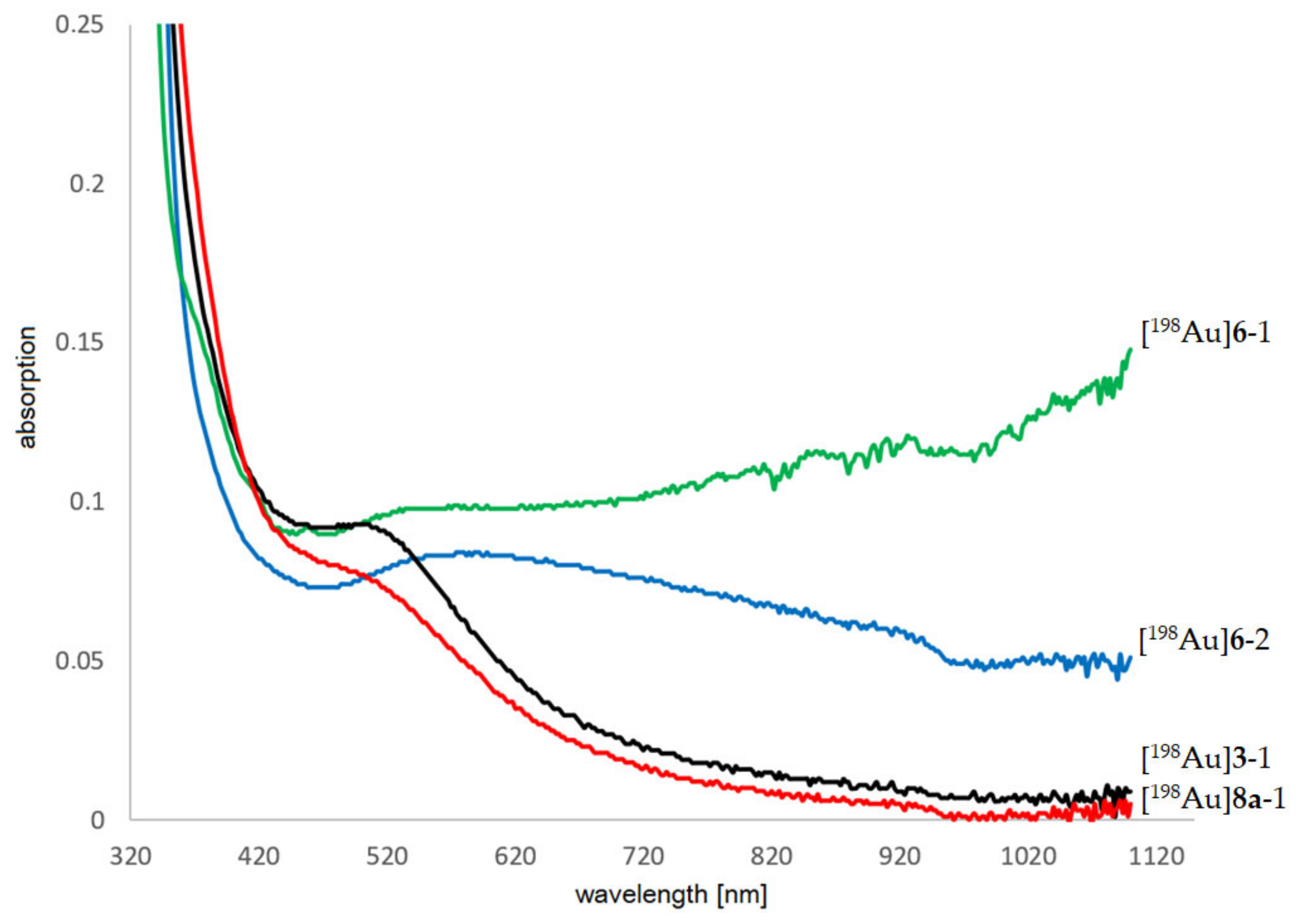
Appendix A.5. Determination of Stability of [198Au]AuNPs

Appendix A.5.1. UV/Vis Measurements

UV/Vis measurements were performed using an BioSpektrometer Kinetic (Eppendorf, Hamburg, Germany). The absorbance of [198Au]3 at a concentration of 375 µg/mL was measured for up to 5 months (Figure A9) to estimate the particle size and stability. The absorption at surface plasmon resonance (maximum, 514 nm) divided by the absorption at 450 nm (minimum) gives a factor that can be compared with tables from the literature [54].
Figure A9. Representative absorption spectrum of [198Au]3 at different time points after neutron activation with a typical absorption maximum for ultrasmall AuNPs at 514 nm indicating no aggregation of the particles.
Pharmaceuticals 16 01670 g0a9

Appendix A.5.2. HPLC Measurements

10 µL of [198Au]3 (7.75 mg/mL, 50 MBq/mL) was diluted with 50 µL H2O. 10 µL of the diluted solution was injected into an HPLC equipped with a Sephadex column. Measurements were performed up to 77 days after irradiation (Figure A10).
Figure A10. Representative (radio-)chromatogramms of [198Au]3 showing no degradation or aggregation within 77 days. The [198Au]3 tR = 5.7 min. Ascorbic acid tR = 11.6 min.
Pharmaceuticals 16 01670 g0a10

Appendix A.6. NMR Spectra

Figure A11. 1H NMR spectrum of TA-NHS 1 in d6-DMSO.
Pharmaceuticals 16 01670 g0a11
Figure A12. 1H NMR spectrum of TA-PEG3-OH 2 in d6-DMSO.
Pharmaceuticals 16 01670 g0a12
Figure A13. 1H NMR spectrum of AuNP-dithio-PEG 3 in d6-DMSO.
Pharmaceuticals 16 01670 g0a13

Appendix A.7. AuNP-Dithiol-RGD 8

Figure A14. 1H NMR spectrum of AuNP-dithio-RGDhigh 8a in d6-DMSO: characteristic signals of peptide-bond-NH (7.83 ppm), Phe-CHarom (7.22–7.16 ppm), Arg-C=NH (6.65 ppm), and Asp-CH (5.32 ppm) can be found.
Pharmaceuticals 16 01670 g0a14
Figure A15. 1H NMR spectrum of AuNP-thio-PEG 6 in d6-DMSO.
Pharmaceuticals 16 01670 g0a15

Appendix A.8. AuNP-Thiol-Dithiol-RGD 7

Figure A16. 1H NMR spectrum of AuNP-thio-dithio-RGDhigh 7a in d6-DMSO: characteristic signals of peptide-bond-NH (7.87 ppm), Phe-CHarom (7.19 ppm), Arg-C=NH (6.65 ppm), Arg-NH2 (5.92 ppm), and Asp-CH (5.32 ppm) can be found.
Pharmaceuticals 16 01670 g0a16aPharmaceuticals 16 01670 g0a16b

References

  1. Sheppard, C.W.; Goodell, J.P.B.; Hahn, P.F. Colloidal gold containing the radioactive isotope Au198 in the selective internal radiation therapy of diseases of the lymphoid system. J. Lab. Clin. Med. 1947, 12, 1437–1441. [Google Scholar]
  2. Flocks, R.H.; Kerr, H.D.; Elkins, H.B.; Culp, D. Treatment of carcinoma of the prostate by interstitial radiation with radio-active gold (Au 198): A preliminary report. J. Urol. 1952, 68, 510–522. [Google Scholar] [CrossRef] [PubMed]
  3. Frens, G. Controlled Nucleation for the Regulation of the Particle Size in Monodisperse Gold Suspensions. Nat. Phys. Sci. 1973, 241, 20–22. [Google Scholar] [CrossRef]
  4. Pretze, M.; Hien, A.; Radle, M.; Schirrmacher, R.; Wängler, C.; Wängler, B. Gastrin-releasing peptide receptor- and prostate-specific membrane antigen-specific ultrasmall gold nanoparticles for characterization and diagnosis of prostate carcinoma via fluorescence imaging. Bioconjug. Chem. 2018, 29, 1525–1533. [Google Scholar] [CrossRef] [PubMed]
  5. Maeda, H.; Fang, J.; Inutsuka, T.; Kitamoto, Y. Vascular permeability enhancement in solid tumor: Various factors, mechanisms involved and its implications. Int. Immunopharmacol. 2003, 3, 319–328. [Google Scholar] [CrossRef] [PubMed]
  6. Pretze, M.; von Kiedrowski, V.; Runge, R.; Freudenberg, R.; Hübner, R.; Davarci, G.; Schirrmacher, R.; Wängler, C.; Wängler, B. αvβ3-Specific gold nanoparticles for fluorescence imaging of tumor angiogenesis. Nanomaterials 2021, 11, 138. [Google Scholar] [CrossRef]
  7. Zarschler, K.; Rocks, L.; Licciardello, N.; Boselli, L.; Polo, E.; Garcia, K.P.; De Cola, L.; Stephan, H.; Dawson, K.A. Ultrasmall inorganic nanoparticles: State-of-the-art and perspectives for biomedical applications. Nanomed. Nanotechnol. Biol. Med. 2016, 12, 1663–1701. [Google Scholar] [CrossRef]
  8. Kim, M.S.; Lee, E.J.; Kim, J.W.; Chung, U.S.; Koh, W.G.; Keum, K.C.; Koom, W.S. Gold nanoparticles enhance anti-tumor effect of radiotherapy to hypoxic tumor. Radiat. Oncol. J. 2016, 34, 230–238. [Google Scholar] [CrossRef]
  9. Black, K.C.L.; Wang, Y.; Luehmann, H.P.; Cai, X.; Xing, W.; Pang, B.; Zhao, Y.; Cutler, C.S.; Wang, L.V.; Liu, Y.; et al. Radioactive 198Au-Doped Nanostructures with Different Shapes for In Vivo Analyses of Their Biodistribution, Tumor Uptake, and Intratumoral Distribution. ACS Nano 2014, 8, 4385–4394. [Google Scholar] [CrossRef]
  10. Cui, S.; Yin, D.; Chen, Y.; Di, Y.; Chen, H.; Ma, Y.; Achilefu, S.; Gu, Y. In vivo targeted deep-tissue photodynamic therapy based on near-infrared light triggered upconversion nanoconstruct. ACS Nano 2013, 7, 676–688. [Google Scholar] [CrossRef]
  11. Shukla, R.; Chanda, N.; Zambre, A.; Upendran, A.; Katti, K.; Kulkarni, R.R.; Nune, S.K.; Casteel, S.W.; Smith, C.J.; Vimal, J.; et al. Laminin receptor specific therapeutic gold nanoparticles (198AuNP-EGCg) show efficacy in treating prostate cancer. Proc. Natl. Acad. Sci. USA 2012, 109, 12426–12431. [Google Scholar] [CrossRef] [PubMed]
  12. Chanda, N.; Kattumuri, V.; Shukla, R.; Zambre, A.; Katti, K.; Upendran, A.; Kulkarni, R.R.; Kan, P.; Fent, G.M.; Casteel, S.W.; et al. Bombesin functionalized gold nanoparticles show in vitro and in vivo cancer receptor specificity. Proc. Natl. Acad. Sci. USA 2010, 107, 8760–8765. [Google Scholar] [CrossRef] [PubMed]
  13. Pretze, M.; Hien, A.; Roscher, M.; Richter, K.; Rädle, M.; Wängler, C.; Wängler, B. Efficient modification of GRPR-specific gold nanoparticles for fluorescence imaging of prostate carcinoma. J. Label. Compd. Radiopharm. 2017, 60, S601. [Google Scholar] [CrossRef]
  14. Hien, A.; Pretze, M.; Braun, F.; Schäfer, E.; Kümmel, T.; Roscher, M.; Schock-Kusch, D.; Waldeck, J.; Müller, B.; Wängler, C.; et al. Non-contact recognition of fluorescently labeled objects in deep tissue via optimized optical arrangement. PLoS ONE 2018, 13, e0208236. [Google Scholar] [CrossRef] [PubMed]
  15. Zhu, J.; Chin, J.; Wängler, C.; Wängler, B.; Lennox, R.B.; Schirrmacher, R. Rapid 18F-labeling and loading of PEGylated gold nanoparticles for in vivo applications. Bioconjug. Chem. 2014, 25, 1143–1150. [Google Scholar] [CrossRef] [PubMed]
  16. Zhao, Y.; Sultan, D.; Detering, L.; Cho, S.; Sun, G.; Pierce, R.; Wooley, K.L.; Liu, Y. Copper-64-alloyed gold nanoparticles for cancer imaging: Improved radiolabel stability and diagnostic accuracy. Angew. Chem. Int. Ed. 2014, 53, 156–159. [Google Scholar] [CrossRef] [PubMed]
  17. Pretze, M.; van der Meulen, N.P.; Wängler, C.; Schibli, R.; Wängler, B. Targeted 64Cu-labeled gold nanoparticles for dual imaging with positron emission tomography and optical imaging. J. Label. Comp. Radiopharm. 2019, 62, 471–482. [Google Scholar] [CrossRef]
  18. Jiménez-Mancilla, N.; Ferro-Flores, G.; Santos-Cuevas, C.; Ocampo-García, B.; Luna-Gutiérrez, M.; Azorín-Vega, E.; Isaac-Olivé, K.; Camacho-López, M.; Torres-García, E. Multifunctional targeted therapy system based on 99mTc/177Lu-labeled gold nanoparticles-Tat(49-57)-Lys3-bombesin internalized in nuclei of prostate cancer cells. J. Label. Compd. Radiopharm. 2013, 56, 663–671. [Google Scholar] [CrossRef]
  19. Eskandari, N.; Yavari, K.; Outokesh, M.; Sadjadi, S.; Ahmadi, S.J. Iodine-131 radiolabeling of poly ethylene glycol-coated gold nanorods for in vivo imaging. J. Label. Compd. Radiopharm. 2013, 56, 12–16. [Google Scholar] [CrossRef]
  20. Cui, M.; Liu, R.; Deng, Z.; Ge, G.; Liu, Y.; Xie, L. Quantitative study of protein coronas on gold nanoparticles with different surface modifications. Nano Res. 2014, 7, 345–352. [Google Scholar] [CrossRef]
  21. Dai, Q.; Walkey, C.; Chan, W.C. Polyethylene glycol backfilling mitigates the negative impact of the protein corona on nanoparticle cell targeting. Angew. Chem. Int. Ed. 2014, 53, 5093–5096. [Google Scholar] [CrossRef] [PubMed]
  22. Häkkinen, H. The gold-sulfur interface at the nanoscale. Nat. Chem. 2012, 4, 443–455. [Google Scholar] [CrossRef] [PubMed]
  23. Li, W.; Chen, X. Gold nanoparticles for photoacoustic imaging. Nanomed. Nanotechnol. Biol. Med. 2015, 10, 299–320. [Google Scholar] [CrossRef] [PubMed]
  24. Chanda, N.; Shukla, R.; Katti, K.V.; Kannan, R. Gastrin releasing protein receptor specific gold nanorods: Breast and prostate tumor avid nanovectors for molecular imaging. Nano Lett. 2009, 9, 1798–1805. [Google Scholar] [CrossRef] [PubMed]
  25. Maccora, D.; Dini, V.; Battocchio, C.; Fratoddi, I.; Cartoni, A.; Rotili, D.; Castagnola, M.; Faccini, R.; Bruno, I.; Scotognella, T.; et al. Gold nanoparticles and nanorods in nuclear medicine: A mini review. Appl. Sci. 2019, 9, 3232. [Google Scholar] [CrossRef]
  26. Mayo, R.L.; Robinson, F.R.S. Auger and secondary X-ray electrons from gold. R. Soc. Pub. 1939, 173, 192–200. [Google Scholar]
  27. Zhang, X.D.; Wu, D.; Shen, X.; Chen, J.; Sun, Y.M.; Liu, P.X.; Liang, X.J. Size-dependent radiosensitization of PEG-coated gold nanoparticles for cancer radiation therapy. Biomaterials 2012, 33, 6408–6419. [Google Scholar] [CrossRef]
  28. Hainfeld, J.F.; Slatkin, D.N.; Smilowitz, H.M. The use of gold nanoparticles to enhance radiotherapy in mice. Phys. Med. Biol. 2004, 49, N309–N315. [Google Scholar] [CrossRef]
  29. Chanda, N.; Kan, P.; Watkinson, L.D.; Shukla, R.; Zambre, A.; Carmack, T.L.; Engelbrecht, H.; Lever, J.R.; Katti, K.; Fent, G.M.; et al. Radioactive gold nanoparticles in cancer therapy: Therapeutic efficacy studies of GA-198AuNP nanoconstruct in prostate tumor-bearing mice. Nanomed. Nanotechnol. Biol. Med. 2010, 6, 201–209. [Google Scholar] [CrossRef]
  30. Säterborg, N.E. The distribution of 198Au injected intravenously as a colloid and in solution. Acta Radiol. Ther. Phys. Biol. 1973, 12, 509–528. [Google Scholar] [CrossRef]
  31. Khan, M.K.; Minc, L.D.; Nigavekar, S.S.; Kariapper, M.S.T.; Nair, B.M.; Schipper, M.; Cook, A.C.; Lesniak, W.G.; Balogh, L.P. Fabrication of {198Au0} radioactive composite nanodevices and their use for nano-brachytherapy. Nanomed. Nanotechnol. Biol. Med. 2008, 4, 57–69. [Google Scholar] [CrossRef] [PubMed]
  32. Chakravarty, R.; Chakraborty, S.; Guleria, A.; Kumar, C.; Kunwar, A.; Nair, K.V.V.; Sarma, H.D.; Dash, A. Clinical scale synthesis of intrinsically radiolabeled and cyclic RGD peptide functionalized 198Au nanoparticles for targeted cancer therapy. Nucl. Med. Biol. 2019, 72–73, 1–10. [Google Scholar] [CrossRef]
  33. Aboudzadeh, M.R.; Moassesi, M.E.; Amiri, M.; Shams, H.; Alirezapour, B.; Sadeghi, M.; Sari, M.F.; Keyvani, M. Preparation and characterization of chitosan-capped radioactive gold nanoparticles: Neutron irradiation impact on structural properties. J. Iran. Chem. Soc. 2015, 13, 339–345. [Google Scholar] [CrossRef]
  34. Lindner, S.; Michler, C.; Leidner, S.; Rensch, C.; Wangler, C.; Schirrmacher, R.; Bartenstein, P.; Wangler, B. Synthesis and in vitro and in vivo evaluation of SiFA-tagged bombesin and RGD peptides as tumor imaging probes for positron emission tomography. Bioconjug. Chem. 2014, 25, 738–749. [Google Scholar] [CrossRef] [PubMed]
  35. Horton, M.A. The αvβ3 integrin “Vitronectin receptor”. Int. J. Biochem. Cell Biol. 1997, 29, 721–725. [Google Scholar] [CrossRef] [PubMed]
  36. Liu, Z.; Wang, F.; Chen, X. Integrin αvβ3-targeted cancer therapy. Drug Dev. Res. 2008, 69, 329–339. [Google Scholar] [CrossRef] [PubMed]
  37. Dijkgraaf, I.; Yim, C.B.; Franssen, G.M.; Schuit, R.C.; Luurtsema, G.; Liu, S.; Oyen, W.J.; Boerman, O.C. PET imaging of αvβ3 integrin expression in tumours with 68Ga-labelled mono-, di- and tetrameric RGD peptides. Eur. J. Nucl. Med. Mol. Imaging 2011, 38, 128–137. [Google Scholar] [CrossRef] [PubMed]
  38. Janssen, M.; Oyen, W.J.G.; Massuger, L.F.A.G.; Frielink, C.; Dijkgraaf, I.; Edwards, D.S.; Radjopadhye, M.; Corstens, F.H.M.; Boerman, O.C. Comparison of a monomeric and dimeric radiolabeled RGD-peptide for tumor targeting. Cancer Biother. Radiopharm. 2002, 17, 641–646. [Google Scholar] [CrossRef]
  39. Zhai, C.; Franssen, G.M.; Petrik, M.; Laverman, P.; Summer, D.; Rangger, C.; Haubner, R.; Haas, H.; Decristoforo, C. Comparison of Ga-68-Labeled Fusarinine C-Based Multivalent RGD Conjugates and [68Ga]NODAGA-RGD–In Vivo Imaging Studies in Human Xenograft Tumors. Mol. Imaging Biol. 2016, 18, 758–767. [Google Scholar] [CrossRef]
  40. Turcu, I.; Zarafu, I.; Popa, M.; Chifiriuc, M.C.; Bleotu, C.; Culita, D.; Ghica, C.; Ionita, P. Lipoic acid gold nanoparticles functionalized with organic compounds as bioactive materials. Nanomaterials 2017, 7, 43. [Google Scholar] [CrossRef]
  41. Brust, M.; Walker, M.; Bethell, D.; Schiffrin, D.J.; Whyman, R. Synthesis of Thiol-derivatised Gold Nanoparticles in a Two-phase Liquid-Liquid System. J. Chem. Soc. Chem. Commun. 1994, 7, 801–802. [Google Scholar] [CrossRef]
  42. Zhu, J.; Waengler, C.; Lennox, R.B.; Schirrmacher, R. Preparation of water-soluble maleimide-functionalized 3 nm gold nanoparticles: A new bioconjugation template. Langmuir ACS J. Surf. Colloids 2012, 28, 5508–5512. [Google Scholar] [CrossRef] [PubMed]
  43. Milne, M.; Gobbo, P.; McVicar, N.; Bartha, R.; Workentin, M.S.; Hudson, R.H.E. Water-soluble gold nanoparticles (AuNP) functionalized with a gadolinium(III) chelate via Michael addition for use as a MRI contrast agent. J. Mater. Chem. B 2013, 1, 5628–5635. [Google Scholar] [CrossRef]
  44. Freudenberg, R. Monte-Carlo-Simulationen zur Dosimetrie bei der Zellexposition mit offenen Radionukliden in typischen in-vitro Bestrahlungsgeometrien. Ph.D. Thesis, Technical University Dresden, Dresden, Germany, 2012. Available online: https://eltab.ub.uni-kl.de/media/103162/ (accessed on 1 September 2023).
  45. Haubner, R.; Gratias, R.; Diefenbach, B.; Goodman, S.L.; Jonczyk, A.; Kessler, H. Structural and functional aspects of RGD-containing cyclic pentapeptides as highly potent and selective integrin αvβ3 antagonists. J. Am. Chem. Soc. 1996, 118, 7461–7472. [Google Scholar] [CrossRef]
  46. Shi, J.; Wang, F.; Liu, S. Radiolabeled cyclic RGD peptides as radiotracers for tumor imaging. Biophys. Rep. 2016, 2, 1–20. [Google Scholar] [CrossRef] [PubMed]
  47. Wängler, C.; Maschauer, S.; Prante, O.; Schäfer, M.; Schirrmacher, R.; Bartenstein, P.; Eisenhut, M.; Wängler, B. Multimerization of cRGD peptides by click chemistry: Synthetic strategies, chemical limitations, and influence on biological properties. ChemBioChem Eur. J. Chem. Biol. 2010, 11, 2168–2181. [Google Scholar] [CrossRef] [PubMed]
  48. Quigley, N.G.; Steiger, K.; Hoberück, S.; Czech, N.; Zierke, M.A.; Kossatz, S.; Pretze, M.; Richter, F.; Weichert, W.; Pox, C.; et al. PET/CT imaging of head-and-neck and pancreatic cancer in humans by targeting the “Cancer Integrin” αvβ6 with Ga-68-Trivehexin. Eur. J. Nucl. Med. Mol. Imaging 2021, 49, 1136–1147. [Google Scholar] [CrossRef]
  49. McCandless, E.L. Determination of Sulfur in ploysaccharides be neutron activation analysis. Anal. Biochem. 1964, 7, 357–365. [Google Scholar] [CrossRef]
  50. Vimalnath, K.V.; Shetty, P.; Chakraborty, S.; Das, T.; Chirayil, V.; Sarma, H.D.; Jagadeesan, K.C.; Joshi, P.V. Practicality of production of 32P by direct neutron activation for its utilization in bone pain palliation as Na3[32P]PO4. Cancer Biother. Radiopharm. 2013, 28, 423–428. [Google Scholar] [CrossRef]
  51. Zamora, P.O.; Marek, M.J. Post Labeling Stabilization of Radiolabeled Proteins and Peptides. U.S. Patent 20010055563A1, 24 June 2001. [Google Scholar]
  52. Dai, X.; Su, Z.; Liu, J.O. An improved synthesis of a selective αvβ3-integrin antagonist cyclo(-RGDfK-). Tetrahedron Lett. 2000, 41, 6295–6298. [Google Scholar] [CrossRef]
  53. Dzwonek, M.; Załubiniak, D.; Piątek, P.; Cichowicz, G.; Męczynska-Wielgosz, S.; Stępkowski, T.; Kruszewski, M.; Więckowska, A.; Bilewicz, R. Towards potent but less toxic nanopharmaceuticals—Lipoic acid bioconjugates of ultrasmall gold nanoparticles with an anticancer drug and addressing unit. RSC Adv. 2018, 8, 14947–14957. [Google Scholar] [CrossRef] [PubMed]
  54. Haiss, W.; Nguyen, T.K.T.; Aveyard, J.; Fernig, D.G. Determination of Size and Concentration of Gold Nanoparticles from UV-Vis Spectra. Anal. Chem. 2007, 79, 4215–4221. [Google Scholar] [CrossRef] [PubMed]
Figure 1. Synthesis of the different RGD-functionalized AuNPs 7a, 7b, 8a and 8b. The synthesis of c(RGDfK) is described in Appendix A.
Pharmaceuticals 16 01670 g001
Figure 2. Different amounts of AuNPs before (top) and after (bottom) neutron activation. [198Au]6-3 (right) shows a suspension immediately after irradiation (top), followed by precipitation within 0.5 min (middle) and 1 min (bottom).
Pharmaceuticals 16 01670 g002
Figure 3. UV-Vis spectra of neutron-activated AuNPs. Intact [198Au]3-1 (black line, 25 µg/mL, irradiation 60 min) and [198Au]8a-1 (red line, 25 µg/mL, irradiation 60 min). Particle aggregation can be seen as broadening of the typical plasmon band at 514 nm for [198Au]6-1 (green line, 25 µg/mL, irradiation 15 min) and [198Au]6-2 (blue line, 25 µg/mL, irradiation 60 min).
Pharmaceuticals 16 01670 g003
Figure 4. Survival fractions of the colony formation assays of 5 Gy [198Au]3 and 10 Gy [198Au]3.
Pharmaceuticals 16 01670 g004
Table 1. Calculated number of ligands and resulting molecular mass of the AuNPs.
ProbeDescriptionNumber of LigandsMolecular Mass [kDa]
6AuNP-PEG250 × thio-PEG210
7aAuNP-PEG-RGDhigh152 × thio-PEG, 35 × 5239
7bAuNP-PEG-RGDlow196 × thio-PEG, 15 × 5222
3AuNP-dithio-PEG240 × 3246
8aAuNP-dithio-PEG-RGDhigh218 × 3, 24 × 5262
8bAuNP-dithio-PEG-RGDlow220 × 3, 18 × 5257
Table 2. Summary of neutron activation of various AuNPs, mass, and calculated half-life.
ProbeWeight [mg]DR 1 [µSv/h] in 1 cm/30 cm afterPrecipitation Observedt1/2 [d] (Calc.)
5 min10 min30 min60 min
[198Au]3-10.0555/435/325/325/3no2.6866
[198Au]3-20.50125/5125/5115/4100/4no2.8177
[198Au]3-30.75170/5160/5155/4150/4no2.8525
[198Au]3-41.00250/6215/5210/5212/5no2.7837
[198Au]3-52.00500/8450/8420/8410/8no2.8761
[198Au]6-10.0576/3.7n.d. 2n.d. 2n.d. 2yesn.d. 2
[198Au]6-20.50150/n.d. 215/6n.d. 2/5.3n.d. 2yesn.d. 2
[198Au]6-35.06n.d. 2n.d. 21000/n.d. 2n.d. 2yesn.d. 2
[198Au]8a-10.05n.d. 2n.d. 215/5n.d. 2non.d. 2
1 DR: dose rate; 2 n.d.: not determined.
Table 3. Avidity experiments.
ProbeDescriptionIC50 [nM]
c(RGDfK)αvβ3 antagonist700.4 ± 155.9
7aAuNP-PEG-RGDhigh27.8 ± 3.4
7bAuNP-PEG-RGDlow38.3 ± 11.9
8aAuNP-dithio-PEG-RGDhigh82.4 ± 9.2
8bAuNP-dithio-PEG-RGDlow103.6 ± 3.5
Disclaimer/Publisher’s Note: The statements, opinions and data contained in all publications are solely those of the individual author(s) and contributor(s) and not of MDPI and/or the editor(s). MDPI and/or the editor(s) disclaim responsibility for any injury to people or property resulting from any ideas, methods, instructions or products referred to in the content.

