
Terminalia arjuna, a Cardioprotective Herbal Medicine–Relevancy in the Modern Era of Pharmaceuticals and Green Nanomedicine—A Review

Abstract
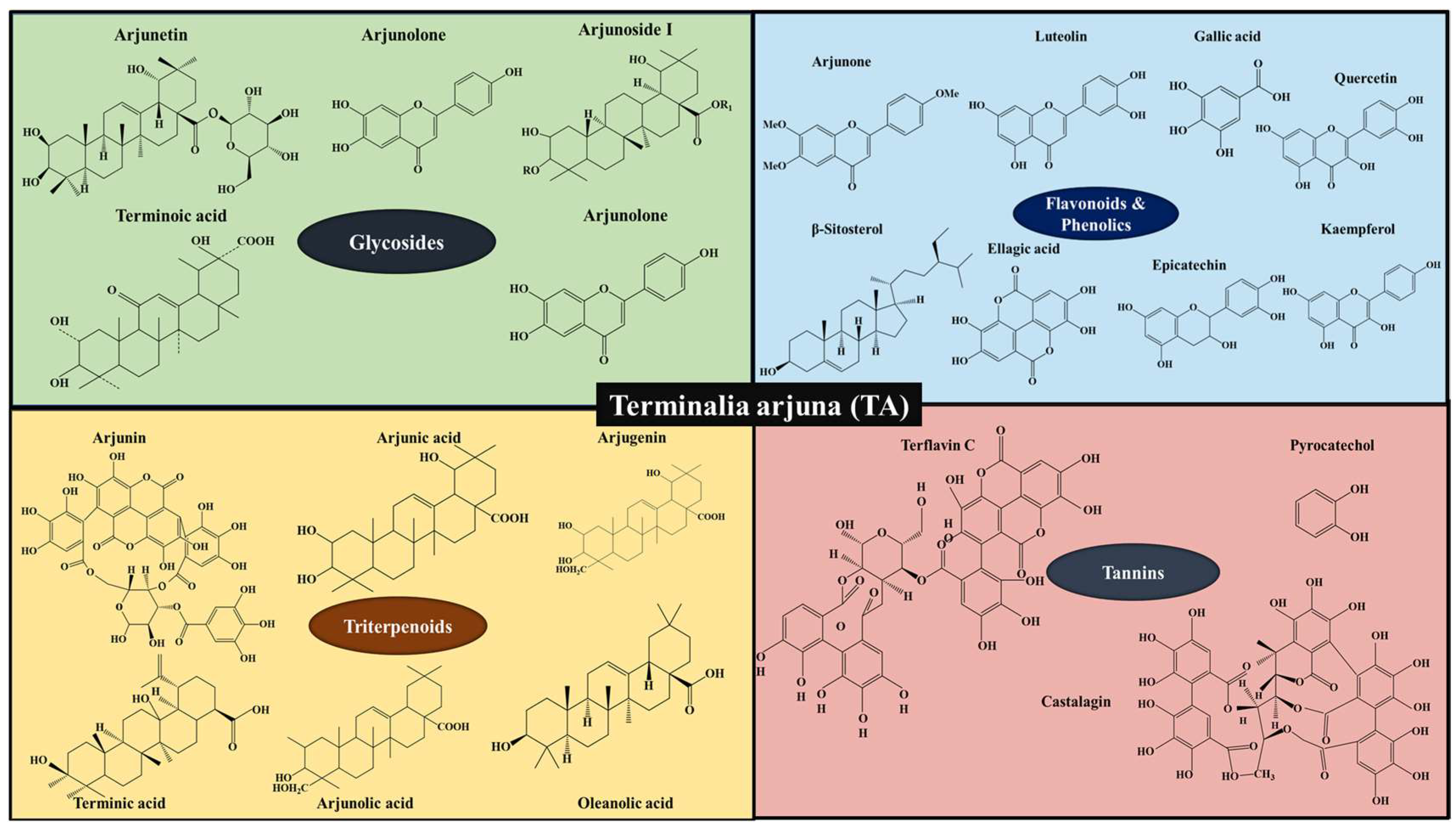
:1. Introduction
2. Extraction of Phytochemicals from TA
2.1. Pre-Extraction Process for a TA Plant
2.1.1. Drying of TA Plant Materials
2.1.2. Grinding and Powdering of TA Plant Materials
2.1.3. Effect of Solvents and the Phytochemicals Polarity on the Extraction Process Efficiency
3. Mechanism of Action
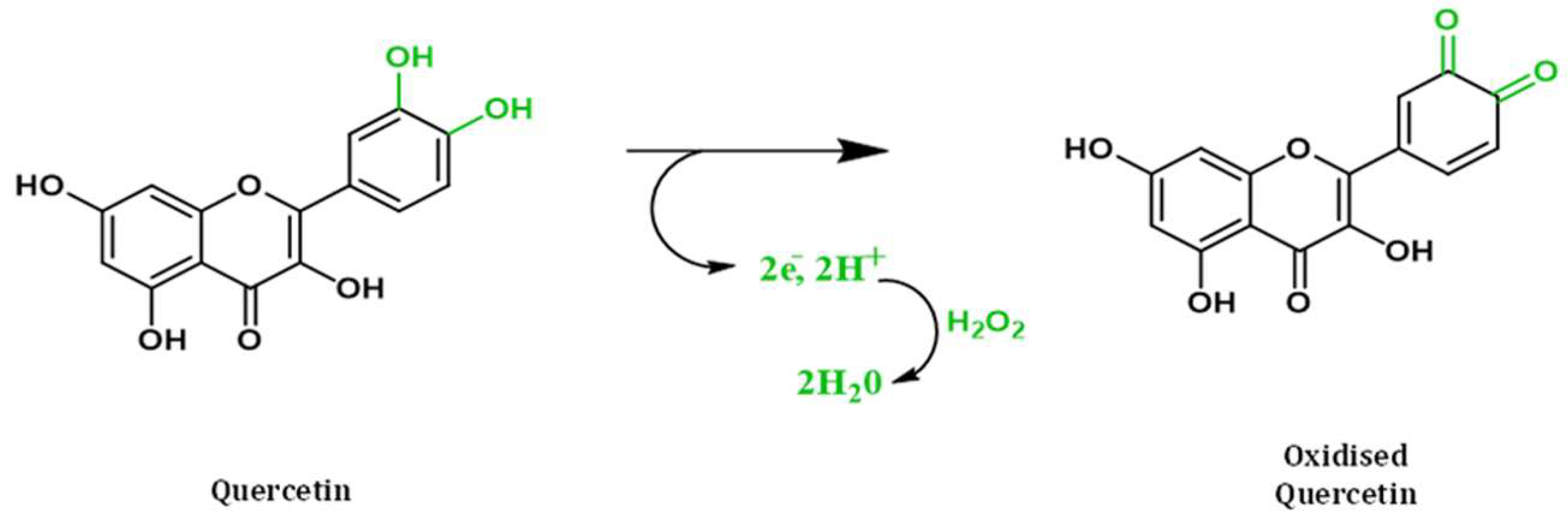
3.1. Antioxidant
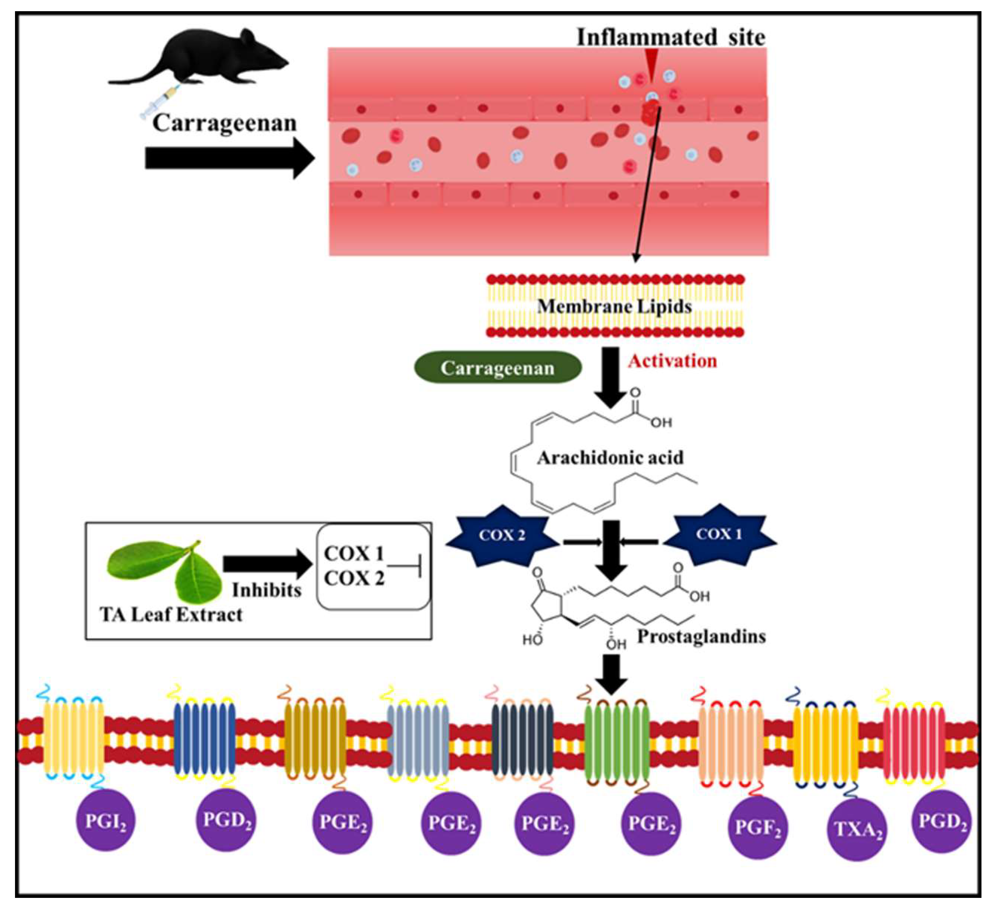
3.2. Anti-Inflammatory
3.3. Cardio Protective Property
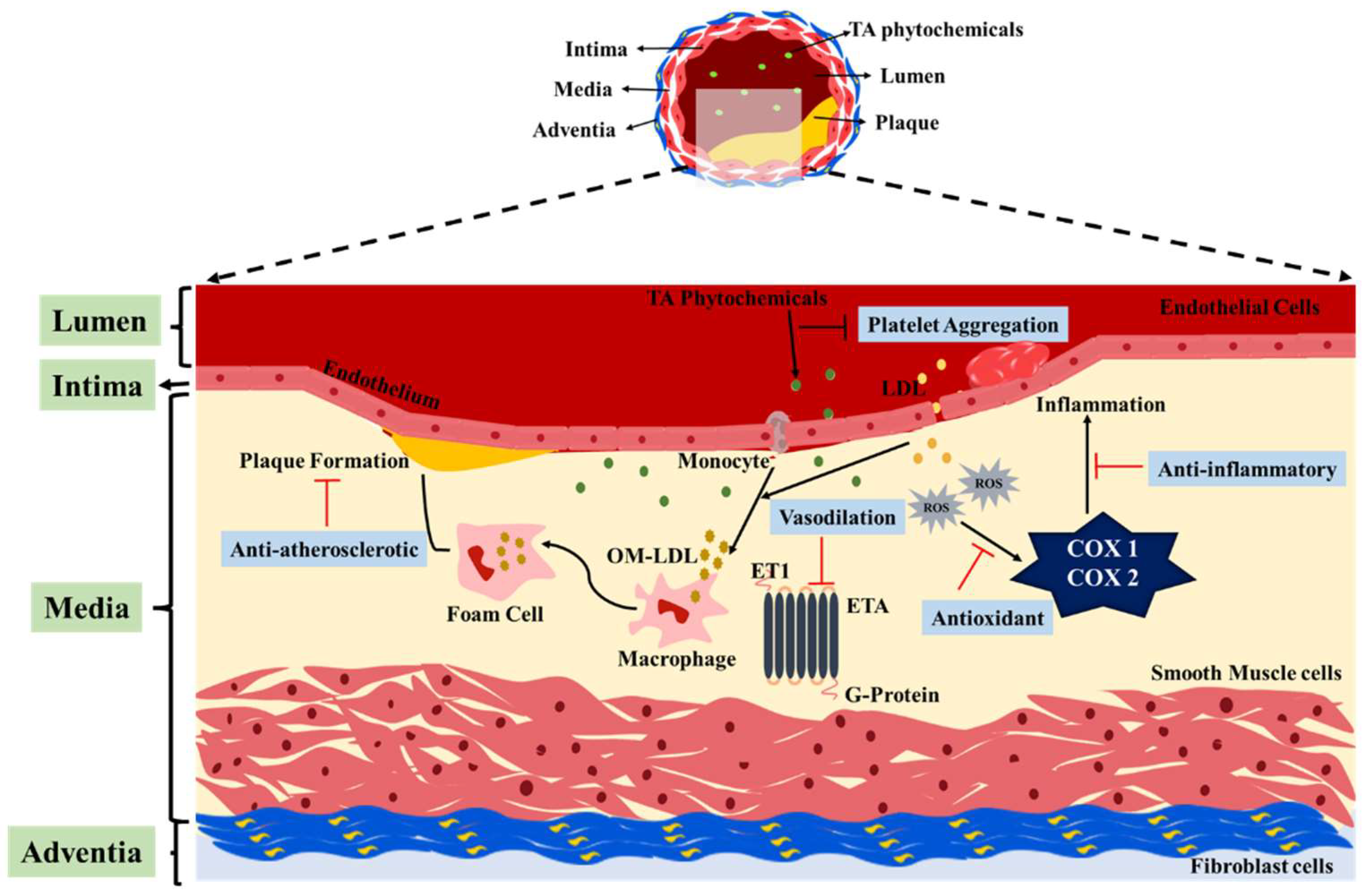
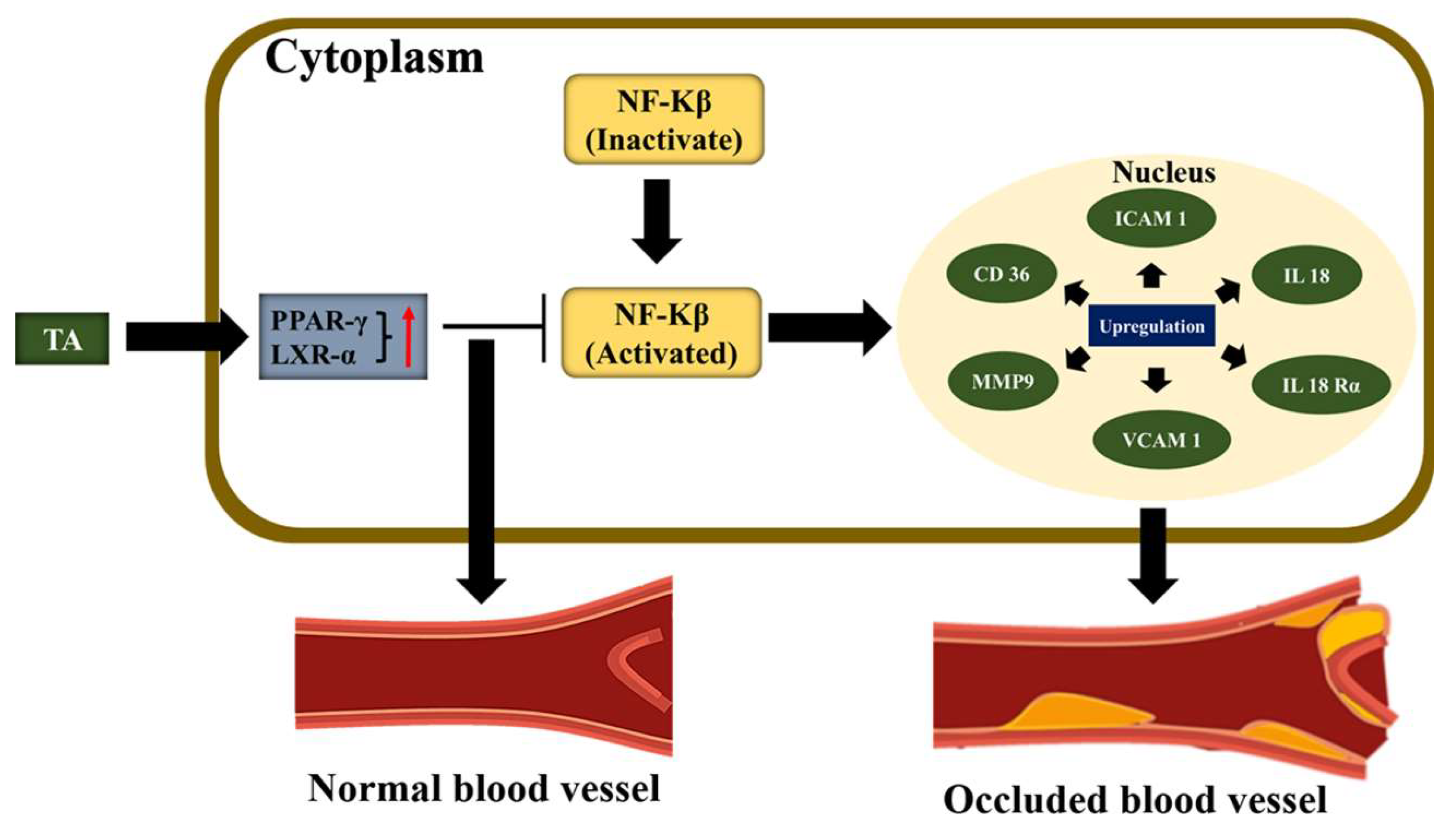
3.4. Anti–Atherosclerotic
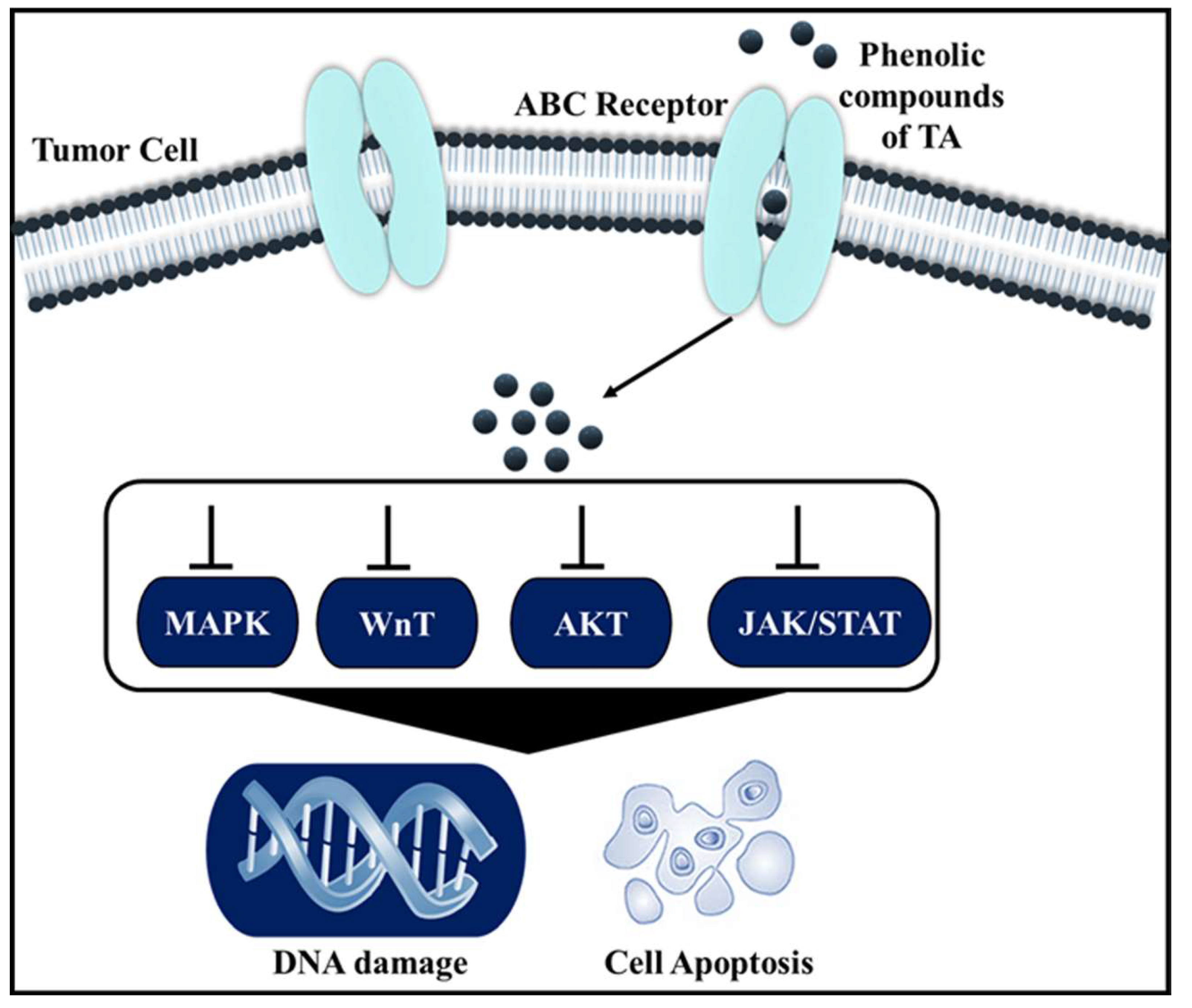
3.5. Anticancer
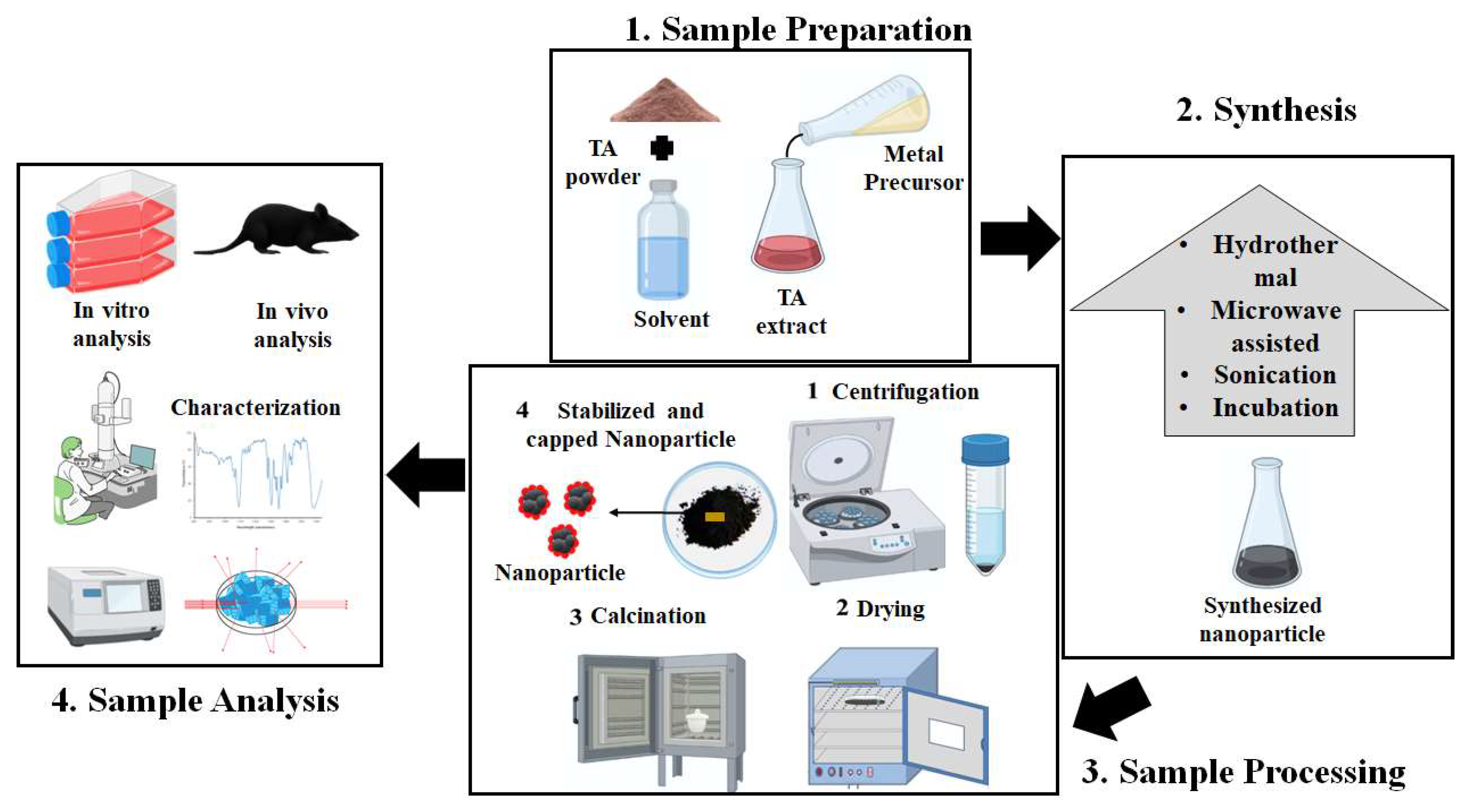
4. Green Synthesis of Nanoparticles Using TA Extracts
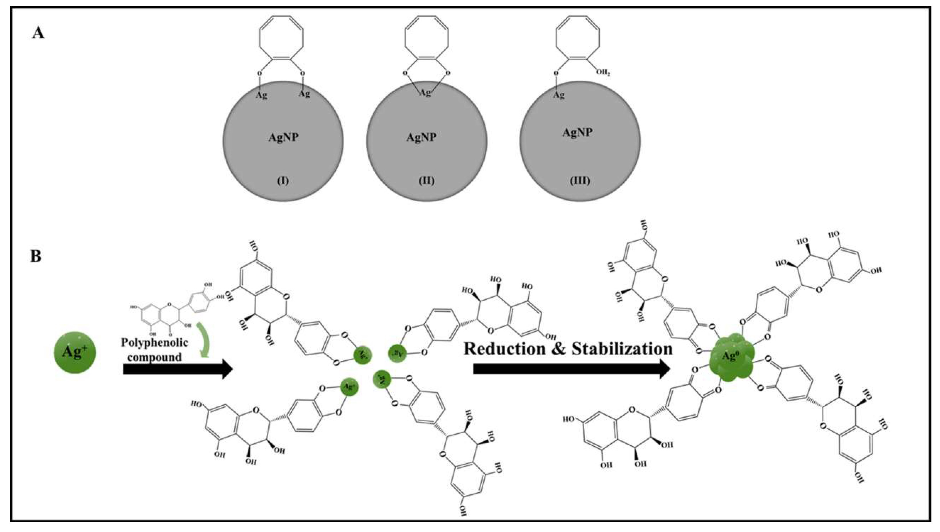
4.1. Silver Nanoparticles
4.2. Gold Nanoparticles
5. Polymeric Formulation of TA Extracts
5.1. Transdermal Delivery of TA Extracts
5.2. TA Bark Gum as a Biomaterial for Ophthalmic Application
6. Conclusions and Future Perspectives
Author Contributions
Funding
Institutional Review Board Statement
Informed Consent Statement
Data Availability Statement
Acknowledgments
Conflicts of Interest
References
- Amalraj, A.; Gopi, S. Medicinal properties of Terminalia arjuna (Roxb.) Wight & Arn.: A review. J. Tradit. Complement. Med. 2017, 7, 65–78. [Google Scholar] [CrossRef] [PubMed] [Green Version]
- Sen, T.; Samanta, S.K. Medicinal Plants, Human Health and Biodiversity: A Broad Review. In Biotechnological Applications of Biodiversity; Mukherjee, J., Ed.; Advances in Biochemical Engineering/Biotechnology; Springer: Berlin/Heidelberg, Germany, 2014; Volume 147, pp. 59–110. ISBN 978-3-662-45096-3. Available online: http://link.springer.com/10.1007/10_2014_273 (accessed on 27 July 2021).
- Demirbaş, N. Vertical Agriculture: A Review of Developments. In Proceedings of the IBANESS Congress Series, Tekirdag, Turkey, 5–6 October 2019. [Google Scholar]
- Kanthe, P.S.; Patil, B.S.; Das, K.K. Terminalia arjuna supplementation ameliorates high fat diet-induced oxidative stress in nephrotoxic rats. J. Basic Clin. Physiol. Pharmacol. 2021, 33, 409–417. [Google Scholar] [CrossRef] [PubMed]
- Dube, N.; Nimgulkar, C.; Bharatraj, D.K. Validation of therapeutic anti-inflammatory potential of Arjuna Ksheera Paka—A traditional Ayurvedic formulation of Terminalia arjuna. J. Tradit. Complement. Med. 2017, 7, 414–420. [Google Scholar] [CrossRef] [PubMed]
- Hussain, S.A.; Panjagari, N.R.; Singh, R.R.B.; Patil, G.R. Potential Herbs and Herbal Nutraceuticals: Food Applications and Their Interactions with Food Components. Crit. Rev. Food Sci. Nutr. 2015, 55, 94–122. [Google Scholar] [CrossRef]
- Peterson, C.T.; Denniston, K.; Chopra, D. Therapeutic Uses of Triphala in Ayurvedic Medicine. J. Altern. Complement. Med. 2017, 23, 607–614. [Google Scholar] [CrossRef] [Green Version]
- Soni, N.; Singh, V. Efficacy and Advancement of Terminalia arjuna in Indian Herbal Drug Research: A Review. Trends Appl. Sci. Res. 2019, 14, 233–242. [Google Scholar] [CrossRef] [Green Version]
- Ahmed, K.K.M.; Gupta, G.B.M.; Singh, N.; Kumar, A. Global Research on Terminalia arjuna: A Quantitative and Qualitative Assessment of Publications during 2004-18. Pharmacogn. Rev. 2021, 14, 45–52. [Google Scholar] [CrossRef]
- Gopinath, K.; Gowri, S.; Karthika, V.; Arumugam, A. Green synthesis of gold nanoparticles from fruit extract of Terminalia arjuna, for the enhanced seed germination activity of Gloriosa superba. J. Nanostructure Chem. 2014, 4, 115. [Google Scholar] [CrossRef] [Green Version]
- Desai, D.; Chanda, S. Pharmacognostic Study and Physicochemical Analysis of Leaves of Terminalia arjuna. Pharmacogn. J. 2014, 6, 15–19. [Google Scholar] [CrossRef] [Green Version]
- Goswami, M.; Chaturvedi, P.; Kumar Sonwani, R.; Dutta Gupta, A.; Rani Singhania, R.; Shekher Giri, B.; Nath Rai, B.; Singh, H.; Yadav, S.; Sharan Singh, R. Application of Arjuna (Terminalia arjuna) seed biochar in hybrid treatment system for the bioremediation of Congo red dye. Bioresour. Technol. 2020, 307, 123203. [Google Scholar] [CrossRef]
- Chouksey, B.K.; Srivastava, S.K. New constituent from the roots of Terminalia arjuna: Antifungal agent. NIScPR Online Period. Repos. 2001, 40, 354–356. [Google Scholar]
- Gupta, S.; Bishnoi, J.; Kumar, N.; Kumar, H.; Correspondence, J.; Nidheesh, T. Terminalia arjuna (Roxb.) Wight &Arn.: Competent source of bioactive components in functional food and drugs. Innov. J. 2018, 7, 223–231. [Google Scholar]
- Gaikwad, D.; Jadhav, N. A review on biogenic properties of stem bark of Terminalia arjuna: An update. Asian J. Pharm. Clin. Res. 2018, 11, 35–39. [Google Scholar] [CrossRef]
- Kasote, D.M.; Katyare, S.S.; Hegde, M.V.; Bae, H. Significance of Antioxidant Potential of Plants and its Relevance to Therapeutic Applications. Int. J. Biol. Sci. 2015, 11, 982–991. [Google Scholar] [CrossRef] [Green Version]
- Moulisha, B.; Biswas, K.; Karan, T.; Bhattacharya, S.; Haldar, P. Evaluation of analgesic and anti-inflammatory activities of Terminalia arjuna leaf. J. Phytol. 2011, 3, 33–38. [Google Scholar]
- Krishnamurthy, K.; Rm, S.; Karunakaran, G.; Sathish, K.; Devaraj, N. Cardioprotective effect of the alcoholic extract of Terminalia arjuna bark in an in vivo model of myocardial ischemic reperfusion injury. Life Sci. 2003, 73, 2727–2739. [Google Scholar] [CrossRef]
- Kapoor, D.; Vijayvergiya, R.; Dhawan, V. Terminalia arjuna in Coronary Artery Disease: Ethnopharmacology, Pre-Clinical, Clinical & Safety Evaluation—ScienceDirect. J. Ethnopharmacol. 2014, 155, 1029–1045. [Google Scholar]
- Subramaniam, S. Anti-hyperlipidemic and antioxidant potential of different fractions of Terminalia arjuna Roxb. bark against PX- 407 induced hyperlipidemia. Indian J. Exp. Biol. 2011, 7, 282–288. [Google Scholar]
- Sarveswaran, S.; Ilayaraja, M.; Balasubramanian, M. Antioxidant activity of Terminalia arjuna bark extract on N-nitrosodiethylamine induced hepatocellular carcinoma in rats. Mol. Cell. Biochem. 2006, 281, 87–93. [Google Scholar] [CrossRef]
- Augustine, R.; Hasan, A. Emerging applications of biocompatible phytosynthesized metal/metal oxide nanoparticles in healthcare. J. Drug Deliv. Sci. Technol. 2020, 56, 101516. [Google Scholar] [CrossRef]
- Shrivastava, P.; Jain, V.K.; Nagpal, S. Nanoparticle intervention for heavy metal detection: A review. Environ. Nanotechnol. Monit. Manag. 2022, 17, 100667. [Google Scholar] [CrossRef]
- Rahimi, H.-R.; Doostmohammadi, M.; Rahimi, H.-R.; Doostmohammadi, M. Nanoparticle Synthesis, Applications, and Toxicity. Applications of Nanobiotechnology; IntechOpen: London, UK, 2019; ISBN 978-1-78985-978-2. Available online: https://www.intechopen.com/chapters/69099 (accessed on 12 November 2022). [CrossRef] [Green Version]
- Singh, A.; Kaur, K.; Singh, A.; Kaur, K. Biological and Physical Applications of Silver Nanoparticles with Emerging Trends of Green Synthesis. Engineered Nanomaterials-Health and Safety; IntechOpen: London, UK, 2019; ISBN 978-1-83880-412-1. Available online: https://www.intechopen.com/chapters/69413 (accessed on 12 November 2022). [CrossRef] [Green Version]
- Azmir, J.; Zaidul, I.S.M.; Rahman, M.M.; Sharif, K.M.; Mohamed, A.; Sahena, F.; Jahurul, M.H.A.; Ghafoor, K.; Norulaini, N.A.N.; Omar, A.K.M. Techniques for extraction of bioactive compounds from plant materials: A review. J. Food Eng. 2013, 117, 426–436. [Google Scholar] [CrossRef]
- Safdar, M.N.; Kausar, T.; Jabbar, S.; Mumtaz, A.; Ahad, K.; Saddozai, A.A. Extraction and quantification of polyphenols from kinnow (Citrus reticulate L.) peel using ultrasound and maceration techniques. J. Food Drug Anal. 2017, 25, 488–500. [Google Scholar] [CrossRef] [PubMed]
- Gião, M.S.; González-Sanjosé, M.L.; Rivero-Pérez, M.D.; Pereira, C.I.; Pintado, M.E.; Malcata, F.X. Infusions of Portuguese medicinal plants: Dependence of Final Antioxidant Capacity and Phenol Content on Extraction Features. J. Sci. Food Agric. 2007, 87, 2638–2647. [Google Scholar] [CrossRef] [PubMed]
- Saad, R.; Murugiah, G.; Abdulhamid, J.; Yusuf, E.; Fadli, M. Comparative Study between Percolation and Ultrasonication for the Extraction of Hibiscus and Jasmine Flowers Utilizing Antibacterial Bioassay. Int. J. Pharmacogn. Phytochem. Res. 2014, 6, 6. [Google Scholar]
- Kaneria, M.; Kanani, B.; Chanda, S. Assessment of effect of hydroalcoholic and decoction methods on extraction of antioxidants from selected Indian medicinal plants. Asian Pac. J. Trop. Biomed. 2012, 2, 195–202. [Google Scholar] [CrossRef] [Green Version]
- Barriada-Pereira, M.; Concha-Graña, E.; González-Castro, M.J.; Muniategui-Lorenzo, S.; López-Mahía, P.; Prada-Rodríguez, D.; Fernández-Fernández, E. Microwave-assisted extraction versus Soxhlet extraction in the analysis of 21 organochlorine pesticides in plants. J. Chromatogr. A 2003, 1008, 115–122. [Google Scholar] [CrossRef]
- Lucchesi, M.E.; Chemat, F.; Smadja, J. Solvent-free microwave extraction of essential oil from aromatic herbs: Comparison with conventional hydro-distillation—ScienceDirect. Available online: https://www.sciencedirect.com/science/article/pii/S0021967304008672?casa_token=NNufqmRkAr4AAAAA:eQ5216C1rLAbrOyNoPY_7UqZ5jIFviE8k3WKNyHdQBS-izFkmFT8jC5CUvBZ_x6NeP1WGpxZIY-X (accessed on 14 November 2022).
- Lang, Q.; Wai, C.M. Supercritical fluid extraction in herbal and natural product studies—A practical review. Talanta 2001, 53, 771–782. [Google Scholar] [CrossRef]
- Chan, C.-H.; Yusoff, R.; Ngoh, G.-C.; Kung, F.W.-L. Microwave-assisted extractions of active ingredients from plants. J. Chromatogr. A 2011, 1218, 6213–6225. [Google Scholar] [CrossRef]
- Chemat, F.; Rombaut, N.; Sicaire, A.-G.; Meullemiestre, A.; Fabiano-Tixier, A.-S.; Abert-Vian, M. Ultrasound assisted extraction of food and natural products. Mechanisms, techniques, combinations, protocols and applications. A review. Ultrason. Sonochem. 2017, 34, 540–560. [Google Scholar] [CrossRef]
- Hammed, A.M.; Jaswir, I.; Amid, A.; Alam, Z.; Asiyanbi-H, T.T.; Ramli, N. Enzymatic Hydrolysis of Plants and Algae for Extraction of Bioactive Compounds. Food Rev. Int. 2013, 29, 352–370. [Google Scholar] [CrossRef]
- NN Azwanida A Review on the Extraction Methods Use in Medicinal Plants, Principle, Strength and Limitation. Med. Aromat. Plants 2015, 4, 1000196. Available online: http://www.omicsgroup.org/journals/a-review-on-the-extraction-methods-use-in-medicinal-plants-principle-strength-and-limitation-2167-0412-1000196.php?aid=58448 (accessed on 12 July 2021).
- trs1010_annex1.pdf. Available online: https://www.who.int/traditional-complementary-integrative-medicine/publications/trs1010_annex1.pdf (accessed on 29 July 2021).
- Jones, W.P.; Kinghorn, A.D. Extraction of Plant Secondary Metabolites. In Natural Products Isolation; Sarker, S.D., Nahar, L., Eds.; Methods in Molecular Biology; Humana Press: Totowa, NJ, USA, 2012; Volume 864, pp. 341–366. ISBN 978-1-61779-623-4. Available online: http://link.springer.com/10.1007/978-1-61779-624-1_13 (accessed on 29 July 2021).
- Makanjuola, S.A. Influence of particle size and extraction solvent on antioxidant properties of extracts of tea, ginger, and tea–ginger blend. Food Sci. Nutr. 2017, 5, 1179–1185. [Google Scholar] [CrossRef]
- Abubakar, A.R.; Haque, M. Preparation of Medicinal Plants: Basic Extraction and Fractionation Procedures for Experimental Purposes. J. Pharm. Bioallied Sci. 2020, 12, 1. [Google Scholar] [CrossRef]
- Saha, A.; Pawar, V.M.; Jayaraman, S. Characterisation of Polyphenols in Terminalia arjuna Bark Extract. Indian J. Pharm. Sci. 2012, 74, 339–347. [Google Scholar] [CrossRef]
- Mandal, S.; Patra, A.; Samanta, A.; Roy, S.; Mandal, A.; Mahapatra, T.D.; Pradhan, S.; Das, K.; Nandi, D.K. Analysis of phytochemical profile of Terminalia arjuna bark extract with antioxidative and antimicrobial properties. Asian Pac. J. Trop. Biomed. 2013, 3, 960–966. [Google Scholar] [CrossRef] [Green Version]
- Meena, D.K.; Sahoo, A.K.; Srivastava, P.P.; Sahu, N.P.; Jadhav, M.; Gandhi, M.; Swain, H.S.; Borah, S.; Das, B.K. On valorization of solvent extracts of Terminalia arjuna (arjuna) upon DNA scission and free radical scavenging improves coupling responses and cognitive functions under in vitro conditions. Sci. Rep. 2021, 11, 10656. [Google Scholar] [CrossRef]
- Akhter, S.; Hossain, M.; Haque, A.; Shahriar, M.; Bhuiyan, M. Phytochemical Screening, Antibacterial, Antioxidant and Cytotoxic Activity of the Bark Extract of Terminalia Arjuna. Eur. J. Sci. Res. 2012, 86, 543–552. [Google Scholar]
- Yallappa, S.; Manjanna, J.; Sindhe, M.A.; Satyanarayan, N.D.; Pramod, S.N.; Nagaraja, K. Microwave assisted rapid synthesis and biological evaluation of stable copper nanoparticles using T. arjuna bark extract. Spectrochim. Acta A Mol. Biomol. Spectrosc. 2013, 110, 108–115. [Google Scholar] [CrossRef]
- Kumar, V.; Sharma, N.; Sourirajan, A.; Khosla, P.K.; Dev, K. Comparative evaluation of antimicrobial and antioxidant potential of ethanolic extract and its fractions of bark and leaves of Terminalia arjuna from north-western Himalayas, India. J. Tradit. Complement. Med. 2017, 8, 100–106. [Google Scholar] [CrossRef]
- Khatkar, S.; Nanda, A.; Ansari, S.H. Improved Methods of Extraction and In Vitro Evaluation of Antimicrobial Potential of Stem Bark of Terminalia arjuna. Curr. Biochem. Eng. 2019, 5, 50–56. [Google Scholar] [CrossRef]
- Filipiak-Szok, A.; Kurzawa, M.; Szłyk, E. Simultaneous Determination of Isoquinoline Alkaloids in Medicinal Asiatic Plants by Ultrasound-Assisted Extraction and High-Performance Liquid Chromatography—Mass Spectrometry with Principal Component Analysis. Anal. Lett. 2018, 51, 2575–2585. [Google Scholar] [CrossRef]
- Oberoi, L.; Akiyama, T.; Lee, K.-H.; Liu, S.J. The aqueous extract, not organic extracts, of Terminalia arjuna bark exerts cardiotonic effect on adult ventricular myocytes. Phytomedicine 2011, 18, 259–265. [Google Scholar] [CrossRef] [PubMed]
- Bachaya, H.A.; Iqbal, Z.; Khan, M.N.; Jabbar, A.; Gilani, A.H.; Din, I. In vitro and in vivo anthelmintic activity of terminalia arjuna bark. Int. J. Agric. Biol. 2009, 11, 273. [Google Scholar]
- Sayyad, S.F. Liquisolid Compacts: An Approach to Enhance the Dissolution Rate of Nimesulide. J. Appl. Pharm. Sci. 2012, 2, 115–121. Available online: http://www.japsonline.com/vol-2-issue-5/122-124.pdf (accessed on 28 May 2012). [CrossRef]
- Barman, S.; Das, S. Hypoglycemic effect of ethanolic extract of bark of Terminalia arjuna Linn. in normal and alloxan-induced noninsulin-dependent diabetes mellitus albino rats. Int. J. Green Pharm. 2012, 6, 279. [Google Scholar] [CrossRef]
- Kumar, S.; Enjamoori, R.; Jaiswal, A.; Ray, R.; Seth, S.; Maulik, S.K. Catecholamine-induced myocardial fibrosis and oxidative stress is attenuated by Terminalia arjuna (Roxb.). J. Pharm. Pharmacol. 2009, 61, 1529–1536. [Google Scholar] [CrossRef]
- Meghwani, H.; Prabhakar, P.; Mohammed, S.A.; Seth, S.; Hote, M.P.; Banerjee, S.K.; Arava, S.; Ray, R.; Maulik, S.K. Beneficial effects of aqueous extract of stem bark of Terminalia arjuna (Roxb.), An ayurvedic drug in experimental pulmonary hypertension. J. Ethnopharmacol. 2017, 197, 184–194. [Google Scholar] [CrossRef]
- Parashar, R. Study on Osteopotential activity of Terminalia arjuna bark extract using UMR 106 cells. J. Pharmacogn. Phytochem. 2020, 9, 1690–1693. [Google Scholar]
- Uthirapathy, S. Novel Biomarkers of Atherogenic Diet Induced Dyslipidemia and Metabolic Syndrome Suppressed By Terminalia arjuna. Int. J. Pharma. Sci. Res. 2019, 10, 2528–2536. [Google Scholar] [CrossRef]
- Mojarrad, S. Responses of liver and renal function markers against arjuna tree extract in induced Hyperlipidemia Rats. Issue S 2020, 10, 6. [Google Scholar]
- Gupta, A.; Chaphalkar, S.R. Inhibition of antigen specific t cell population. J. Immunol. 2002, 169, 802–808. [Google Scholar]
- Pankaj, P.; Khamrui, K.; Devaraja, H.C.; Singh, R.R.B. The effects of alcoholic extract of Arjuna (Terminalia arjuna Wight & Arn.) bark on stability of clarified butterfat. J. Med. Plants Res. 2013, 4, 2545–2550. [Google Scholar]
- Jahan, N.; Ali, S.; Asi, M.R.; Akhtar, A. Cardioprotective Potential of Gemmomodified Extract of Terminalia arjuna against Chemically Induced Myocardial Injury in Rabbits. Pak. Vet. J. 2012, 32, 5. [Google Scholar]
- Sivalokanathan, S.; Vijayababu, M.R.; Balasubramanian, M.P. Effects of Terminalia arjuna bark extract on apoptosis of human hepatoma cell line HepG2. World J. Gastroenterol. WJG 2006, 12, 1018–1024. [Google Scholar] [CrossRef]
- Singh, G.; Singh, A.T.; Abraham, A.; Bhat, B.; Mukherjee, A.; Verma, R.; Agarwal, S.K.; Jha, S.; Mukherjee, R.; Burman, A.C. Protective effects of Terminalia arjuna against Doxorubicin-induced cardiotoxicity. J. Ethnopharmacol. 2008, 117, 123–129. [Google Scholar] [CrossRef]
- Mohammad, S.; Sadika, A.; Md, I.H.; Md, A.H.; Mohiuddin, A.B. Evaluation of in vitro antioxidant activity of bark extracts of Terminalia arjuna. J. Med. Plants Res. 2012, 6, 5286–5298. [Google Scholar] [CrossRef]
- Meena, D.; Sahoo, A.; Chowdhury, H.; Swain, H.S.; Sahu, N.; Behera, B.K.; Srivastava, P.; Das, B. Effects of extraction methods and solvent systems on extract yield, proximate composition and mineral profiling of Terminalia arjuna (Arjuna) dry powders and solvent extracts. J. Innovat. Pharmaceut. Biol. Sci. 2020, 7, 22–31. [Google Scholar]
- Zujko, M.E.; Witkowska, A.M. Antioxidant Potential and Polyphenol Content of Selected Food. Int. J. Food Prop. 2011, 14, 300–308. [Google Scholar] [CrossRef]
- Pandey, D.K.; Kaur, P. Optimization of extraction parameters of pentacyclic triterpenoids from Swertia chirata stem using response surface methodology. 3 Biotech 2018, 8, 152. [Google Scholar] [CrossRef]
- Marica Bakovic, N.H. Biologically Active Triterpenoids and Their Cardioprotective and Anti-Inflammatory Effects. J. Bioanal. Biomed. 2015, 12. Available online: https://www.omicsonline.org/open-access/biologically-active-triterpenoids-and-their-cardioprotective-and-antiinflammatory-effects-1948-593X-S12-005.php?aid=53322 (accessed on 25 November 2021). [CrossRef] [Green Version]
- Wang, W.; Ali, Z.; Shen, Y.; Li, X.C.; Khan, I.A. Ursane triterpenoids from the bark of Terminalia arjuna—ScienceDirect. Available online: https://www.sciencedirect.com/science/article/abs/pii/S0367326X10000109 (accessed on 25 November 2021).
- Ríos, J.L.; Recio, M.C.; Maáñez, S.; Giner, R.M. Natural Triterpenoids as Anti-Inflammatory Agents. In Studies in Natural Products Chemistry; Atta-ur-Rahman, Ed.; Bioactive Natural Products (Part C); Elsevier: Amsterdam, The Netherlands, 2000; Volume 22, pp. 93–143. Available online: https://www.sciencedirect.com/science/article/pii/S1572599500800241 (accessed on 25 November 2021).
- Lobo, V.; Patil, A.; Phatak, A.; Chandra, N. Free radicals, antioxidants and functional foods: Impact on human health. Pharmacogn. Rev. 2010, 4, 118–126. [Google Scholar] [CrossRef] [PubMed] [Green Version]
- Chatha, S.A.S. Bioactive Components and Antioxidant Properties of Terminalia arjuna L. Extracts. J. Food Process. Technol. 2014, 5, 1. [Google Scholar] [CrossRef]
- Papuc, C.; Goran, G.V.; Predescu, C.N.; Nicorescu, V.; Stefan, G. Plant Polyphenols as Antioxidant and Antibacterial Agents for Shelf-Life Extension of Meat and Meat Products: Classification, Structures, Sources, and Action Mechanisms: Polyphenols extending meat shelf-life. Compr. Rev. Food Sci. Food Saf. 2017, 16, 1243–1268. [Google Scholar] [CrossRef] [PubMed] [Green Version]
- Zhang, Y.-J.; Gan, R.-Y.; Li, S.; Zhou, Y.; Li, A.-N.; Xu, D.-P.; Li, H.-B. Antioxidant Phytochemicals for the Prevention and Treatment of Chronic Diseases. Molecules 2015, 20, 21138–21156. [Google Scholar] [CrossRef] [Green Version]
- Ghadigaonkar, S.; Reddy, A.G.; Kalakumar, B.; Anilkumar, B. Screening of antioxidant and free radical scavenging activities of Terminalia arjuna Roxb. Pharma Innov. J. 2021, 10, 1–5. [Google Scholar]
- Medicinal Plants with Anti-Inflammatory Activities from Selected Countries and Regions of Africa. Available online: https://www.ncbi.nlm.nih.gov/pmc/articles/PMC6086115/ (accessed on 11 July 2021).
- Halder, S.; Bharal, N.; Mediratta, P.K.; Kaur, I.; Sharma, K.K. Anti-inflammatory, immunomodulatory and antinociceptive activity of Terminalia arjuna Roxb bark powder in mice and rats. Indian J. Exp. Biol. 2009, 7, 577–583. [Google Scholar]
- Safayhi, H.; Sailer, E.-R. Anti-Inflammatory Actions of Pentacyclic Triterpenes. Planta Med. 1997, 63, 487–493. [Google Scholar] [CrossRef] [Green Version]
- Yasui, K.; Baba, A. Therapeutic potential of superoxide dismutase (SOD) for resolution of inflammation. Inflamm. Res. 2006, 55, 359–363. [Google Scholar] [CrossRef]
- Shah, S.M.A.; Akram, M.; Riaz, M.; Munir, N.; Rasool, G. Cardioprotective Potential of Plant-Derived Molecules: A Scientific and Medicinal Approach. Dose-Response 2019, 17, 1559325819852243. [Google Scholar] [CrossRef] [Green Version]
- Khaliq, F.; Fahim, M. Role of Terminalia Arjuna in Improving Cardiovascular Functions: A Review. Indian J. Physiol. Pharmacol. 2018, 62, 8–19. [Google Scholar]
- Alique, M.; Luna, C.; Carracedo, J.; Ramírez, R. LDL biochemical modifications: A link between atherosclerosis and aging. Food Nutr. Res. 2015, 59, 29240. [Google Scholar] [CrossRef] [Green Version]
- Parveen, A.; Babbar, R.; Agarwal, S.; Kotwani, A.; Fahim, M. Mechanistic Clues in the Cardioprotective Effect of Terminalia Arjuna Bark Extract in Isoproterenol-Induced Chronic Heart Failure in Rats. Cardiovasc. Toxicol. 2011, 11, 48–57. [Google Scholar] [CrossRef]
- Pawar, R.S.; Bhutani, K.K. Effect of oleanane triterpenoids from Terminalia arjuna—a cardioprotective drug on the process of respiratory oxyburst. Phytomedicine 2005, 12, 391–393. [Google Scholar] [CrossRef]
- Gholipour, S.; Sewell, R.; Lorigooini, Z.; Rafieian-kopaei, M. Medicinal Plants and Atherosclerosis: A Review on Molecular Aspects. Curr. Pharm. Des. 2018, 24, 3123–3131. [Google Scholar] [CrossRef]
- Nwodo, N.J.; Nnadi, C.O.; Ibezim, A.; Mbah, C.J. Plants with Hypolipidaemic Effects from Nigerian Flora; IntechOpen: London, UK, 2014; ISBN 978-953-51-1215-0. Available online: https://www.intechopen.com/chapters/45882 (accessed on 2 November 2022). [CrossRef]
- Alam, M.M.; Haque, A.; Begum, R. Anti-hyperglycemic and lipid lowering effect of Terminalia arjuna Bark extract on Streptozotocin induced Type-2 Diabetic Model Rats. Int. J. Pharm. Pharm. Sci. 2011, 3, 449–453. [Google Scholar]
- Bhat, O.; Kumar, P.; Rao, K.; Ahmad, A.; Dhawan, V. Terminalia arjuna prevents Interleukin-18-induced atherosclerosis via modulation of NF-κB/PPAR-γ-mediated pathway in Apo E-/- mice. Inflammopharmacology 2018, 26, 583–598. [Google Scholar] [CrossRef]
- Greenwell, M.; Rahman, P.K.S.M. Medicinal Plants: Their Use in Anticancer Treatment. Int. J. Pharm. Sci. Res. 2015, 6, 4103–4112. [Google Scholar] [CrossRef]
- Gangadevi, V.; Muthumary, J. Taxol production by Pestalotiopsis terminaliae, an endophytic fungus of Terminalia arjuna (arjun tree). Biotechnol. Appl. Biochem. 2009, 52, 9–15. [Google Scholar] [CrossRef]
- Runowicz, C.D.; Wiernik, P.H.; Einzig, A.I.; Goldberg, G.L.; Horwitz, S.B. Taxol in ovarian cancer. Cancer 1993, 71, 1591–1596. [Google Scholar] [CrossRef]
- Parker, A.L.; Kavallaris, M.; McCarroll, J.A. Microtubules and Their Role in Cellular Stress in Cancer. Front. Oncol. 2014, 4, 153. [Google Scholar] [CrossRef] [PubMed] [Green Version]
- Singh, S.; Verma, S.K.; Kumar, S. Analysis of anti-cancer potential of Terminalia arjuna. Int. J. Adv. Sci. Res. Manag. 2017, 2, 82–87. [Google Scholar]
- Anu Mary Ealia, S.; Saravanakumar, M.P. A review on the classification, characterisation, synthesis of nanoparticles and their application. IOP Conf. Ser. Mater. Sci. Eng. 2017, 263, 032019. [Google Scholar] [CrossRef]
- Parveen, K.; Banse, V.; Ledwani, L. Green synthesis of nanoparticles: Their advantages and disadvantages. AIP Conf. Proc. 2016, 1724, 020048. [Google Scholar] [CrossRef]
- Salah, N.; Habib, S.S.; Khan, Z.H.; Memic, A.; Azam, A.; Alarfaj, E.; Zahed, N.; Al-Hamedi, S. High-energy ball milling technique for ZnO nanoparticles as antibacterial material. Int. J. Nanomed. 2011, 6, 863–869. [Google Scholar] [CrossRef] [Green Version]
- Li, Z. Synthesis and Characterization of Carbon-Encapsulated Magnetic Nanoparticles via Arc-Plasma Assisted CVD. J. Nanosci. Nanotechnol. 2009, 9, 1757. Available online: http://www.ingentaconnect.com/content/asp/jnn/2009/00000009/00000012/art00133 (accessed on 12 November 2022). [CrossRef]
- Wang, W.-N.; Lenggoro, I.W.; Terashi, Y.; Kim, T.O.; Okuyama, K. One-step synthesis of titanium oxide nanoparticles by spray pyrolysis of organic precursors. Mater. Sci. Eng. B 2005, 123, 194–202. [Google Scholar] [CrossRef]
- Hatakeyama, Y.; Judai, K.; Onishi, K.; Takahashi, S.; Kimura, S.; Nishikawa, K. Anion and cation effects on the size control of Au nanoparticles prepared by sputter deposition in imidazolium-based ionic liquids. Phys. Chem. Chem. Phys. 2016, 18, 2339–2349. [Google Scholar] [CrossRef]
- Paul, S. Nanomaterials synthesis by electrodeposition techniques for high-energetic electrodes in fuel cell. Nanomater. Energy 2015, 4, 80–89. [Google Scholar] [CrossRef]
- Pradeep, A.; Priyadharsini, P.; Chandrasekaran, G. Sol–gel route of synthesis of nanoparticles of MgFe2O4 and XRD, FTIR and VSM study. J. Magn. Magn. Mater. 2008, 320, 2774–2779. [Google Scholar] [CrossRef]
- Patiño-Ruiz, D.A.; Meramo-Hurtado, S.I.; González-Delgado, Á.D.; Herrera, A. Environmental Sustainability Evaluation of Iron Oxide Nanoparticles Synthesized via Green Synthesis and the Coprecipitation Method: A Comparative Life Cycle Assessment Study. ACS Omega 2021, 6, 12410–12423. [Google Scholar] [CrossRef]
- Ying, S.; Guan, Z.; Ofoegbu, P.C.; Clubb, P.; Rico, C.; He, F.; Hong, J. Green synthesis of nanoparticles: Current developments and limitations. Environ. Technol. Innov. 2022, 26, 102336. [Google Scholar] [CrossRef]
- Govindaraju, K.; Basha, S.K.; Kumar, V.G.; Singaravelu, G. Silver, gold and bimetallic nanoparticles production using single-cell protein (Spirulina platensis) Geitler. J. Mater. Sci. 2008, 43, 5115–5122. [Google Scholar] [CrossRef]
- Omajali, J.B.; Mikheenko, I.P.; Merroun, M.L.; Wood, J.; Macaskie, L.E. Characterization of intracellular palladium nanoparticles synthesized by Desulfovibrio desulfuricans and Bacillus benzeovorans. J. Nanoparticle Res. 2015, 17, 264. [Google Scholar] [CrossRef] [Green Version]
- Saratale, R.G.; Karuppusamy, I.; Saratale, G.D.; Pugazhendhi, A.; Kumar, G.; Park, Y.; Ghodake, G.S.; Bharagava, R.N.; Banu, J.R.; Shin, H.S. A comprehensive review on green nanomaterials using biological systems: Recent perception and their future applications. Colloids Surf. B Biointerfaces 2018, 170, 20–35. [Google Scholar] [CrossRef]
- Ijaz, I.; Gilani, E.; Nazir, A.; Bukhari, A. Detail review on chemical, physical and green synthesis, classification, characterizations and applications of nanoparticles. Green Chem. Lett. Rev. 2020, 13, 223–245. [Google Scholar] [CrossRef]
- Singh, J.; Kumar, S.; Rathi, B.; Bhrara, K.; Chhikara, B.S. Therapeutic analysis of Terminalia arjuna plant extracts in combinations with different metal nanoparticles. J. Mater. Nanosci. 2015, 2, 1–7. [Google Scholar]
- Zafar, F.; Jahan, N.; Khalil-Ur-Rahman; Asi, M.R.; Zafar, W.-U.-I. Nanosuspension enhances dissolution rate and oral bioavailability of Terminalia arjuna bark extract in vivo and in vitro. Asian Pac. J. Trop. Biomed. 2020, 10, 164. [Google Scholar] [CrossRef]
- Mousumi, M.; Amit, B.; Gouriprasad, D.; Dilip, K.N. Nephroprotective Effect of Green Synthesised Gold Nanoparticles Using Bark Extract of Terminalia Arjuna on Acetaminophen Induced Nephrotoxicity in Male Albino Rat. Int. J. Pharma. Bio. Sci. 2020, 10. Available online: https://www.ijlpr.com/abstract1.php?aid=602 (accessed on 7 July 2021). [CrossRef]
- Saha, R.; Subramani, K.; Balu, K.; Suriyaprabha, R.; Siva, P.; Venkatachalam, R. Influence of the various synthesis methods on the ZnO nanoparticles property made using the bark extract of Terminalia arjuna. Mater. Chem. Phys. 2018, 209, 208–216. [Google Scholar] [CrossRef]
- Mitra, M.; Laha, J.; Nandi, D.K. Effective Role of Terminalia arjuna Reduced Gold Nanoparticles on Reproductive Dysfunction Induced by Acetaminophen in Male Wistar Rat. BioNanoScience 2020, 10, 942–949. [Google Scholar] [CrossRef]
- Sudarsan, S.; Kumar Shankar, M.; Kumar Belagal Motatis, A.; Shankar, S.; Krishnappa, D.; Mohan, C.D.; Rangappa, K.S.; Gupta, V.K.; Siddaiah, C.N. Green Synthesis of Silver Nanoparticles by Cytobacillus firmus Isolated from the Stem Bark of Terminalia arjuna and Their Antimicrobial Activity. Biomolecules 2021, 11, 259. [Google Scholar] [CrossRef]
- Ahmed, Q.; Gupta, N.; Kumar, A.; Nimesh, S. Antibacterial efficacy of silver nanoparticles synthesized employing Terminalia arjuna bark extract. Artif. Cells Nanomed. Biotechnol. 2017, 45, 1192–1200. [Google Scholar] [CrossRef] [Green Version]
- Amini, S.M.; Akbari, A. Metal nanoparticles synthesis through natural phenolic acids. IET Nanobiotechnol. 2019, 13, 771–777. [Google Scholar] [CrossRef]
- Dwivedi, P.; Narvi, S.S.; Tewari, R.P. Phytofabrication characterization and comparative analysis of Ag nanoparticles by diverse biochemicals from Elaeocarpus ganitrus Roxb., Terminalia arjuna Roxb., Pseudotsuga menzietii, Prosopis spicigera, Ficus religiosa, Ocimum sanctum, Curcuma longa. Ind. Crops Prod. 2014, 54, 22–31. [Google Scholar] [CrossRef]
- Amina, S.J.; Guo, B. A Review on the Synthesis and Functionalization of Gold Nanoparticles as a Drug Delivery Vehicle. Int. J. Nanomed. 2020, 15, 9823–9857. [Google Scholar] [CrossRef] [PubMed]
- Garibo, D.; Borbón-Nuñez, H.A.; de León, J.N.D.; García Mendoza, E.; Estrada, I.; Toledano-Magaña, Y.; Tiznado, H.; Ovalle-Marroquin, M.; Soto-Ramos, A.G.; Blanco, A.; et al. Green synthesis of silver nanoparticles using Lysiloma acapulcensis exhibit high-antimicrobial activity. Sci. Rep. 2020, 10, 12805. [Google Scholar] [CrossRef] [PubMed]
- Bag, B. Terminalia Arjuna Bark Extract Mediated Size Controlled Synthesis of Polyshaped Gold Nanoparticles and Its Application in Catalysis. Int. J. Res. Chem. Environ. 2012, 2, 338–344. [Google Scholar]
- Suganthy, N.; Sri Ramkumar, V.; Pugazhendhi, A.; Benelli, G.; Archunan, G. Biogenic synthesis of gold nanoparticles from Terminalia arjuna bark extract: Assessment of safety aspects and neuroprotective potential via antioxidant, anticholinesterase, and antiamyloidogenic effects. Environ. Sci. Pollut. Res. 2018, 25, 10418–10433. [Google Scholar] [CrossRef]
- Saiqa Ikram, S.A. Silver Nanoparticles: One Pot Green Synthesis Using Terminalia arjuna Extract for Biological Application. J. Nanomedicine Nanotechnol. 2015, 6, 1000309. Available online: https://www.omicsonline.org/open-access/silver-nanoparticles-one-pot-green-synthesis-using-terminalia-arjuna-extract-for-biological-application-2157-7439-1000309.php?aid=56647 (accessed on 6 July 2021). [CrossRef] [Green Version]
- Prasannaraj, G.; Sahi, S.V.; Ravikumar, S.; Venkatachalam, P. Enhanced Cytotoxicity of Biomolecules Loaded Metallic Silver Nanoparticles Against Human Liver (HepG2) and Prostate (PC3) Cancer Cell Lines. J. Nanosci. Nanotechnol. 2016, 16, 4948–4959. [Google Scholar] [CrossRef]
- Yallappa, S.; Manjanna, J. Biological Evaluation of Silver Nanoparticles Obtained from T. arjuna Bark Extract as Both Reducing and Capping Agent. J. Clust. Sci. 2014, 25, 1449–1462. [Google Scholar] [CrossRef]
- Dwivedi, P.; Narvi, S.; Tewari, R. Overwhelming Antibacterial Activity by Terminalia arjuna Aided Herbal Ag/CS-PVC: Nanocomposite for Overcoming Biomaterials Associated Infections. Adv. Sci. 2015, 7, 1651. [Google Scholar] [CrossRef]
- Mendeley Reference Manager. Available online: https://www.mendeley.com/reference-manager/reader/b5ebf52d-329c-328e-b600-03bb109da9a2/73013400-fb1e-17dd-1794-d9530d74df29 (accessed on 6 July 2021).
- Yallappa, S.; Manjanna, J.; Dhananjaya, B.L.; Vishwanatha, U.; Ravishankar, B.; Gururaj, H.; Niranjana, P.; Hungund, B.S. Phytochemically Functionalized Cu and Ag Nanoparticles Embedded in MWCNTs for Enhanced Antimicrobial and Anticancer Properties. Nano-Micro Lett. 2016, 8, 120–130. [Google Scholar] [CrossRef] [Green Version]
- Lachmapure, M.; Paralikar, P.; Palanisamy, M.; Alves, M.; Rai, M. Efficacy of biogenic silver nanoparticles against clinical isolates of fungi causing mycotic keratitis in humans. IET Nanobiotechnol. 2017, 11, 809–814. [Google Scholar] [CrossRef]
- Koparde, S.; Gaikwad, D. Antibacterial potential of green silver nanoparticles synthesized from medicinal plant terminalia arjuna. Int. J. Res. Biosci. Agric. Technol. 2017, 5, 258–260. [Google Scholar]
- Akther, T.; Priya, S.; Sah, S.K.; Khan, M.S.; Hemalatha, S. Ta-AgNps are potential antimicrobial resistance breakers. J. Nanostructures 2019, 9, 376–383. [Google Scholar] [CrossRef]
- Bag, B.G.; Majumdar, R. Vesicular self-assembly of a natural triterpenoid arjunolic acid in aqueous medium: Study of entrapment properties and in situ generation of gel-gold nanoparticle hybrid material. RSC Adv. 2014, 4, 53327–53334. [Google Scholar] [CrossRef]
- Mitra, M.; Bandyopadhyay, A.; Datta, G.; Nandi, D.K. Effective Dose of Herbal Gold Nanoparticles for Protection of Acetaminophen-Induced Hepatotoxicity in Male Albino Rats. BioNanoScience 2020, 10, 1094–1106. [Google Scholar] [CrossRef]
- Gopinath, K.; Venkatesh, K.S.; Ilangovan, R.; Sankaranarayanan, K.; Arumugam, A. Green synthesis of gold nanoparticles from leaf extract of Terminalia arjuna, for the enhanced mitotic cell division and pollen germination activity. Ind. Crops Prod. 2013, 50, 737–742. [Google Scholar] [CrossRef]
- Mitra, M.; Bandyopadhyay, A.; Datta, G.; Nandi, D. Protective Role of Green Synthesized Gold Nanoparticles Using Terminalia arjuna against Acetaminophen Induced Hematological Alterations in Male Wistar Rats. J. Nanomed. Nanotechnol. 2019, 10. [Google Scholar] [CrossRef]
- Dudhane, A.A.; Waghmode, S.R.; Dama, L.B.; Sonawane, A.; Katariya, S. Synthesis and Characterization of Gold Nanoparticles using Plant Extract of Terminalia arjuna with Antibacterial Activity. J. Nanosci. Nanotechnol. 2019, 15, 75–82. [Google Scholar]
- Anuradha, V.; Shankar, P.; Bhuvana, P.; SyedAli, M.; Yogananth, N. Terminalia Arjuna Bark Assisted Biosynthesis, Characterization and Bioactivity of Metal oxide Nanoparticles. J. Chem. Pharm. Res. 2017, 9, 34–46. [Google Scholar]
- Kangralkar, M.V.; Manjanna, J. Manjanna, Green Synthesis of iron nanoparticles by Terminalia arjuna bark extract and photodegradation of rose Bengal. Iran. J. Catal. 2020, 8, 181–188. [Google Scholar]
- Garai, C.; Hasan, S.N.; Barai, A.C.; Ghorai, S.; Panja, S.K.; Bag, B.G. Green synthesis of Terminalia arjuna-conjugated palladium nanoparticles (TA-PdNPs) and its catalytic applications. J. Nanostructure Chem. 2018, 8, 465–472. [Google Scholar] [CrossRef] [Green Version]
- Gaikwad, D.; Jadhav, N. Terminalia arjuna transdermal matrix formulation containing different polymer components. Asian J. Pharm. Clin. Res. 2019, 12, 266–270. [Google Scholar] [CrossRef]
- Shrivastava, V.P.; Subedi, D.P. Surface Modification of Polymeric Biomaterials by Coating Herbal Extracts. J. Nano Res. Adv. Mater. Polym. Sci. 2020, 1, 2. [Google Scholar]
- Gaikwad, D.; Jadhav, N. Formulation design and evaluation of an emulgel containing Terminalia arjuna bark extract for transdermal delivery. Pharmacogn. Mag. 2018, 14, 249. [Google Scholar] [CrossRef]
- Noreen, S.; Ghumman, S.A.; Batool, F.; Ijaz, B.; Basharat, M.; Noureen, S.; Kausar, T.; Iqbal, S. Terminalia arjuna gum/alginate in situ gel system with prolonged retention time for ophthalmic drug delivery. Int. J. Biol. Macromol. 2020, 152, 1056–1067. [Google Scholar] [CrossRef]


| S. No | Part Used | Extraction Method | Solvent Used | Phytochemicals Derived | Application | Reference |
|---|---|---|---|---|---|---|
| 1. | Bark | Refluxing | Water | Polyphenols | Polyphenol analysis | [42] |
| Methanol | Catechins Gallocatechins Ellagic acid | |||||
| 2. | Bark | Soxhlet | Methanol | Phytosterol Lactones Flavonoids Phenolic compounds Tannins Glycosides | Antimicrobial Antioxidant | [43] |
| 3. | Bark-B Leaf-L Fruit–F | Shaking incubation | Hexane | Alkaloid–F, L Steroids–F | Antioxidant DNA nicking inhibition | [44] |
| Ethyl acetate | Steroids–B, F Alkaloids-B Flavonoids-B Tannins and Phenolics–L | |||||
| Chloroform | Steroids-F Alkaloids–F, L Tannins and Phenolics-L Saponins–L | |||||
| Acetone | Tannins and phenolics Steroids Alkaloids absent only in leaf Flavonoids Saponins | |||||
| Ethanol | Steroids Alkaloids Flavonoids Saponins Tannins and phenolics | |||||
| Methanol | Steroids Alkaloids absent only in leaf Flavonoids Saponins Tannins and phenolic | |||||
| Distilled water | Tannins and phenolic Steroids–B Alkaloids–L Flavonoids–B&F Saponins | |||||
| 4. | Bark | Soxhlet | Methanol | Phenols Flavonoids Glycosides Tannin Carbohydrates Saponins Alkaloids Phytosterols | Antibacterial Antioxidant Cytotoxicity | [45] |
| Ethanol | Flavonoids Tannins Alkaloids Carbohydrate Phenols Saponins Glycosides Phytosterols | |||||
| Petroleum ether | Phenols Flavonoids Alkaloids | |||||
| n-hexane | Phytosterols Alkaloids | |||||
| Chloroform | Phenols Flavonoids Alkaloids | |||||
| Water | Carbohydrate Phenols Flavonoids Tannin Saponins Glycosides Alkaloids | |||||
| 5. | Bark | Microwave-assisted extraction | Water | Flavonoids Terpenoids | Antioxidant Antimicrobial | [46] |
| 6. | Bark Leaf | Incubation | Ethanol | Phenolics Tannins Flavonoids Phytosterols | Antioxidant Antimicrobial | [47] |
| 7. | Bark | Ultrasound-assisted extraction | Ethanol | Not mentioned | Antimicrobial | [48] |
| 8. | Bark | Ultrasound-assisted extraction | Chloroform | Not mentioned | Determination of isoquinoline alkaloid | [49] |
| 9. | Bark | Decoction | Water | Gallic acid Ellagic acid Luteolin | Cardiotonic property | [50] |
| 10. | Bark | Soxhlet | Methanol | Tannin Ellagic acid | Anthelmintic | [51] |
| 11. | Bark | Decoction | Water | Not mentioned | Ayurvedic formulation | [52] |
| 12. | Bark | Hot continuous percolation-soxhlet | Ethyl alcohol | Not mentioned | Cardioprotective effect | [18] |
| 13. | Bark | Percolation | Ethanolic | Flavonoids Tannins Glycosides Alkaloids Terpenoids | Hypoglycemic effect | [53] |
| 14. | Bark | Hot continuous percolation-soxhlet | Water | Arjunolic acid Terminoic acid | Catecholamine-induced myocardial fibrosis and oxidative stress | [54] |
| 15. | Bark | Hot continuous percolation | Water | Glycosides Flavonoids Polyphenols Saponins Terpenoids | Pulmonary hypertension | [55] |
| 16. | Bark | Hot percolation | Methanol | Tannins and Phenolic Glycosides Flavonoids Polysterols Alkaloids Carbohydrate Proteins Triterpenoids Saponins | Osteogenic activity | [56] |
| 17. | Bark | Cold percolation | Hydroalcoholic | Tannins Phenolics Sitosterol Anthraquinone Glycosides Alkaloids Flavonoids | Hypolipidemia activity | [57] |
| 18. | Bark | Hot continuous percolation-soxhlet | Ethyl alcohol | Tannins Phenolics Sitosterol Anthraquinone glycosides Alkaloids Flavonoids | Atherogenic-induced dyslipidemia and metabolic syndrome | [58] |
| 19. | Leaf | Maceration | Phosphate buffered saline | Terpenoids Flavonoids Saponins | Inhibition of T-cell antigen | [59] |
| 20. | Bark | Cold maceration | Ethyl alcohol | Phytosterol β-sitosterol | Antioxidant potential of TA | [60] |
| 21. | Bark | Reflux | Methanol | Gallic acid Catechin Chlorogenic acid Caffeic acid Ferulic acid p-coumaric acid absent in macerated Myricetin Quercetin Kaempferol | Cardioprotective Potential | [61] |
| Maceration | Glycerin/Methanol | |||||
| 22. | Bark | Maceration | Ethanol | Phenolic compounds | Anticancer activity of TA onHuman hepatoma cell (HepG2) | [62] |
| 23. | Bark | Soxhlet | Butanol | Tannins and Phenolics Triterpenoids Saponins Anthraquinone glycosides Alkaloids Flavonoids | Cardioprotective potential in doxorubicin-induced cardiotoxicity | [63] |
| 24. | Leaf | Maceration | Methanol | Alkaloids Triterpenoids Tannins Flavonoids | Analgesic and Anti-inflammatory activity | [17] |
| 25. | Bark | Soxhlet | Methanol Ethanol Petroleum Ether Chloroform n-hexane | Flavonoids | Antioxidant | [64] |
| S. No | Name | Synthesis Method | Extract/Organism Used | Color of Nanoparticles Solution | Average Nanoparticle Size (nm) | Shape of Nanoparticles | Optimal Dosage | Application | Reference |
|---|---|---|---|---|---|---|---|---|---|
| 1. | Silver | Incubation | Aqueous leaf | Yellow to dark brown | 5–20 | Spherical | Nil | Antimicrobial | [121] |
| 2. | Silver | Hot plate magnetic stirring | Aqueous bark | Dark brown | 65 | Spherical | Nil | Antibacterial | [114] |
| 3. | Silver | Shaking incubation | Endophytic bacteria from TA bark | Dark brown | 42.2 | Spherical | Nil | Antimicrobial | [113] |
| 4. | Silver | Incubation | Aqueous fruit, bark, and foliage | Dark brown and intense brown | - | Spherical and irregular shape | Nil | Antimicrobial | [116] |
| 5. | Silver | Incubation | Aqueous bark | Dark brown | 34–70 | Spherical | Nil | Cytotoxicity | [122] |
| 6. | Silver | Microwave irradiation | Aqueous bark | Dark brown | 10–15 | Spherical | Nil | Antioxidant and antimicrobial | [123] |
| 7. | Silver | Incubation | Aqueous bark | Dark brown | Non–uniform | Spherical and arbitrary | Nil | Antibacterial | [124] |
| 8. | Silver | Incubation | Aqueous bark | Dark brown | 20–50 | Spherical | Nil | Larvicidal | [125] |
| 9. | Silver | Microwave irradiation | Aqueous bark | Dark brown | 20–30 | - | Nil | Antimicrobial and anticancer | [126] |
| 10. | Silver | Incubation | Endophytic fungi from TA leaves | Dark brown | 45, 55 | - | Nil | Antifungal | [127] |
| 11. | Silver | Incubation | Aqueous bark | Dark brown | 40–50 | Spherical | Nil | Antibacterial | [128] |
| 12. | Silver | Microwave irradiation | Aqueous and methanolic bark | Dark brown | 20–50 | Spherical | Nil | Antibacterial and antibiofilm | [129] |
| 13. | Gold | Boiling | Aqueous bark | Dark brown | Nil | Nil | 175 µg/kg/day | Nephrotoxicity | [110] |
| 14. | Gold | Incubation at room temperature | Ethanolic arjunolic acid | Dark brown | 185 nm | Spherical | Nil | Entrapment study and release study of gel-gold nanoparticle | [130] |
| 15. | Gold | Boiling | Aqueous bark | Dark brown | 7–20 nm | Spherical | 175 µg/kg/day | Hepatotoxicity | [131] |
| 16. | Gold | Incubation at room temperature | Aqueous leaf | Yellow to dark red | 20–50 nm | Spherical | Nil | Mitotic cell division and pollen germination | [132] |
| 17. | Gold | Drop wise addition | Aqueous bark | Violet to pinkish red | 15–20 nm | Triangular, tetragonal, pentagonal, hexagonal, rod, and spherical | Nil | Catalysis | [119] |
| 18. | Gold | Stirring | Aqueous bark | Pale yellow to ruby red | Nil | Nil | 175 µg/kg/day | Reproductive dysfunction | [112] |
| 19. | Gold | Stirring | Aqueous bark | Pale yellow to ruby red | 20–40 nm | Spherical | 175 µg/kg/day | Hematological alterations | [133] |
| 20. | Gold | Boiling | Aqueous fruit | Yellow to reddish wine | 25 nm | Spherical | Nil | Seed germination activity | [10] |
| 21. | Gold | Incubation | Aqueous leaf | Light yellow to bright red | 15–30 nm | Spherical | Nil | Antibacterial | [134] |
| 22. | Gold | Stirring | Ethanolic bark | Yellow to pink and ruby red | 3–70 nm | Spherical and triangular | 50 µg/mL | Neuroprotective potential | [120] |
| 23. | Copper | Shaking | Aqueous bark | Blue to brown | 10–26 | Spherical | Nil | Antimicrobial | [135] |
| 24. | Copper | Microwave irradiation | Aqueous bark | Pale yellow to dark brown | 23 | Spherical | Nil | Antimicrobial and antioxidant | [46] |
| 25. | Copper | Microwave irradiation | Aqueous bark | Light yellow to black | 20–30 | Nil | Nil | Antimicrobial and anticancer | [126] |
| 26. | Iron | Microwave irradiation | Aqueous bark | Yellow to greenish black | 20–80 nm | Globular | Nil | Photo degradation | [136] |
| 27. | Palladium | Incubation | Aqueous bark | Dark brown | 4–16 | Spherical | Nil | Catalysis | [137] |
| 28. | Zinc | sonication, wet chemical, and hydrothermal | Aqueous bark | Nil | 43, 34, 21 | Spherical | 25 mg mL−1 | Toxicity and antibacterial | [111] |
Disclaimer/Publisher’s Note: The statements, opinions and data contained in all publications are solely those of the individual author(s) and contributor(s) and not of MDPI and/or the editor(s). MDPI and/or the editor(s) disclaim responsibility for any injury to people or property resulting from any ideas, methods, instructions or products referred to in the content. |
© 2023 by the authors. Licensee MDPI, Basel, Switzerland. This article is an open access article distributed under the terms and conditions of the Creative Commons Attribution (CC BY) license (https://creativecommons.org/licenses/by/4.0/).
Share and Cite
Ramesh, P.; Palaniappan, A. Terminalia arjuna, a Cardioprotective Herbal Medicine–Relevancy in the Modern Era of Pharmaceuticals and Green Nanomedicine—A Review. Pharmaceuticals 2023, 16, 126. https://doi.org/10.3390/ph16010126
Ramesh P, Palaniappan A. Terminalia arjuna, a Cardioprotective Herbal Medicine–Relevancy in the Modern Era of Pharmaceuticals and Green Nanomedicine—A Review. Pharmaceuticals. 2023; 16(1):126. https://doi.org/10.3390/ph16010126
Chicago/Turabian StyleRamesh, Purnimajayasree, and Arunkumar Palaniappan. 2023. "Terminalia arjuna, a Cardioprotective Herbal Medicine–Relevancy in the Modern Era of Pharmaceuticals and Green Nanomedicine—A Review" Pharmaceuticals 16, no. 1: 126. https://doi.org/10.3390/ph16010126