
Healing Potential of Propolis in Skin Wounds Evidenced by Clinical Studies


Abstract
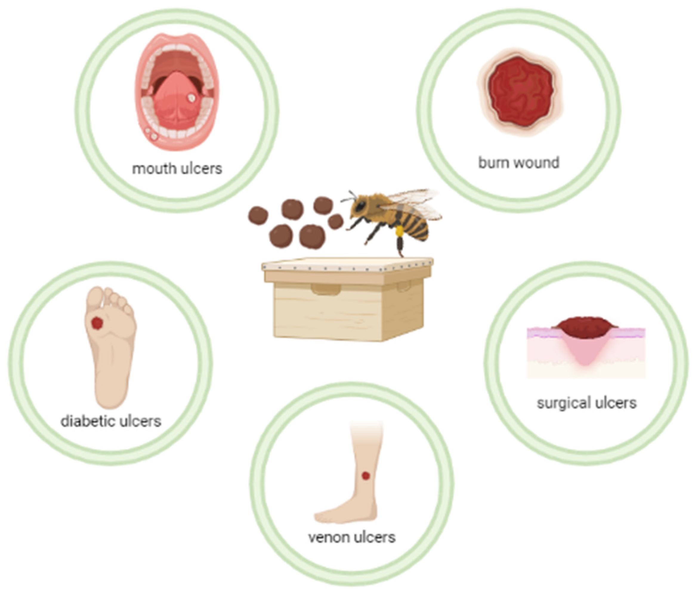
:1. Introduction
2. Results
3. Discussion
3.1. Diabetic Ulcer
3.2. Surgical Ulcer
3.3. Venous Ulcers
3.4. Wound Due to Sacroccygeal Pilonidal Disease
3.5. Burn Wounds
3.6. Mouth Ulcers
3.7. Other Studies
3.8. Propolis Wound Healing Mechanism
4. Method
5. Conclusions
Author Contributions
Funding
Institutional Review Board Statement
Informed Consent Statement
Data Availability Statement
Conflicts of Interest
References
- Resende, N.M.; Nascimento, T.C.; Lopes, F.R.F.; Prates, A.G., Jr.; Souza, N.M. Care of people with chronic wounds in Primary Health Care. J. Manag. Prim. Health Care 2017, 8, 99–108. [Google Scholar] [CrossRef]
- Alves De Lima, N.B.; Agra, G.; de Sousa, A.T.O.; de Lourdes Andre Gouveia, B. Sociodemographic, clinical and therapeutic profile of patients with acute and chronic wounds. Rev. Enferm. UFPE 2016, 10, 2005–2017. [Google Scholar]
- Vieira, C.P.D.B.; De Araújo, T.M.E. Prevalence and factors associated with chronic wounds in older adults in primary care. Rev. Esc. Enferm. USP 2018, 52, e03415. [Google Scholar] [CrossRef]
- Rabeth, S.A.N.; Caliri, M.H.L. Pressure ulcer: Strategies for disseminating knowledge in the nursing literature. Rev. Paul. Enferm. 2006, 22, 307–314. [Google Scholar]
- Batista, E.K.F.; Batista, M.D.C.D.S.; Sobrinho, J.A.N.; Trindade, H.I.D.; Silva, L.L.B.; Muller, J.B. Influence of propolis on the leukocyte and protein profiles of mice and time to close excisional wounds clean and infected by Staphylococcus aureus. Rev. Bras. Plantas Med. 2015, 17, 413–419. [Google Scholar] [CrossRef]
- Trivellato, M.L.D.M.; Kolchraiber, F.C.; Frederico, G.A.; Morales, D.C.A.M.; Silva, A.C.M.; Gamba, M.A. Advanced practices in comprehensive nursing care for people with skin ulcer. Acta Paul. Enferm. 2018, 31, 600–608. [Google Scholar] [CrossRef]
- Franco, D.; Gonçalves, L.F. Skin wounds: Choosing the appropriate dressing. Rev. Col. Bras. Cir. 2008, 35, 203–206. [Google Scholar] [CrossRef]
- Park, Y.K.; Ikegaki, M.; Abreu, J.A.; Alcici, N.M.F. Estudo da preparação dos extratos de própolis e suas aplicações. Food Sci. Technol. 1998, 18, 313–318. [Google Scholar] [CrossRef]
- Santos, M.J.D.; Vianna, L.D.A.C.; Gamba, M.A. Evaluation of the effectiveness of propolis ointment in patients with chronic wounds. Acta Paul. Enferm. 2007, 20, 199–204. [Google Scholar] [CrossRef]
- Barbosa, M.H.; Zuffi, F.B.; Maruxo, H.B.; Jorge, L.L.R. Therapeutic properties of propolis for treatment of skin lesions. Acta Paul. Enferm. 2009, 22, 318–322. [Google Scholar] [CrossRef]
- Basista-Sołtys, K. Allergy to Propolis in Beekeepers—A Literature Review. Occup. Med. Health Aff. 2013, 1, 1–3. [Google Scholar] [CrossRef]
- Bernardo, C.D.L.E.; Souza, I.A.; Colavitti, C.; Garcia, C. Propolis: Healing and natural antibiotic. Rev. Bras. Enferm. 1990, 43, 101–106. [Google Scholar] [CrossRef] [Green Version]
- Diaz, J.C.Q. Effects of drugs in surgical treatments and buccal ulcers. Rev. Cuba. Estomatol. 1996, 33, 42–48. [Google Scholar]
- Silva, A.P.R.; Soares, A.P.S.; da Silva Pessanha, C.; Roza, C.M.; Saint, L.; Tavares, C.; Silva, D.D.M.; Cardoso, M.M.; Palermo, T.A.; Dos Santos, C.M.; et al. Therapeutic use of propolis ointment in different chronic wounds. Online Perspect. Biol. Health 2017, 7, 40–46. [Google Scholar]
- Mujica, V.; Orrego, R.; Fuentealba, R.; Leiva, E.; Zúñiga-Hernández, J. Propolis as an Adjuvant in the Healing of Human Diabetic Foot Wounds Receiving Care in the Diagnostic and Treatment Centre from the Regional Hospital of Talca. J. Diabetes Res. 2019, 2019, 2507578. [Google Scholar] [CrossRef]
- Afkhamizadeh, M.; Aboutorabi, R.; Ravari, H.; Fathi Najafi, M.; Ataei Azimi, S.; Javadian Langaroodi, A.; Jaghoubi, M.A.; Sahebkar, A. Topical propolis improves wound healing in diabetic foot ulcer patients: A randomized controlled trial. Nat. Prod. Res. 2018, 32, 2096–2099. [Google Scholar] [CrossRef] [PubMed]
- Pereira Filho, J.S.; Bicalho, L.; Silva, D.A. Uso de própolis associada a outros componentes no tratamento de feridas oncológicas após excisão. Acta Biomed. Bras. 2012, 3, 15–25. [Google Scholar]
- Gregory, S.R.; Piccolo, N.; Piccolo, M.T.; Piccolo, M.S.; Heggers, J.P. Comparison of Propolis Skin Cream to Silver Sulfadiazine: A Naturopathic Alternative to Antibiotics in Treatment of Minor Burns. J. Altern. Complement. Med. 2002, 8, 77–83. [Google Scholar] [CrossRef]
- Lotfy, M.; Badra, G.; Burham, W.; Alenzi, F. Combined use of honey, bee propolis and myrrh in healing a deep, infected wound in a patient with diabetes mellitus. Br. J. Biomed. Sci. 2006, 63, 171–173. [Google Scholar] [CrossRef]
- Henshaw, F.R.; Bolton, T.; Nube, V.; Hood, A.; Veldhoen, D.; Pfrunder, L.; McKew, G.; MacLeod, C.; McLennan, S.V.; Twigg, S.M. Topical application of the bee hive protectant propolis is well tolerated and improves human diabetic foot ulcer healing in a prospective feasibility study. J. Diabetes Complicat. 2014, 28, 850–857. [Google Scholar] [CrossRef]
- Kubat, M.; Karabulut, Z.; Şengül, S. Effect of propolis on wound healing in sacrococcygeal pilonidal disease: A randomized controlled clinical trial. Pak. J. Pharm. Sci. 2021, 34, 1063–1067. [Google Scholar] [PubMed]
- Kucharzewski, M.; Kózka, M.; Urbanek, T. Topical treatment of non-healing venous leg ulcer with propolis ointment. Evid.-Based Complementary Altern. Med. 2013, 2013, 254017. [Google Scholar] [CrossRef] [PubMed]
- Stratton, I.M.; Adler, A.I.; Neil, H.A.W.; Matthews, D.R.; Manley, S.E.; Cull, C.A.; Hadden, D.; Turner, R.C.; Holman, R.R. Association of glycemia with macrovascular and microvascular complications of type 2 diabetes (UKPDS 35): Prospective observational study. BMJ 2000, 321, 405–412. [Google Scholar] [CrossRef]
- Hamilton, P.D.; Grando, G.F.; Gonzalez, A.V.; Philippsen, F.R.; Ely, P.B.; Steffen, N. Prevalence of compromised surgical margins of non-melanoma cut tumors operated by a plastic surgery service in southern Brazil in 2020. Arq. Catarin. Med. 2022, 51, 151–155. [Google Scholar]
- Carmo, S.D.S.; Castro, C.D.D.; Rios, V.S.; Sarquis, M.G.A. Atualidades na assistência de enfermagem a portadores de úlcera venosa. Rev. Eletrônica Enferm. 2007, 9, 2. [Google Scholar] [CrossRef]
- Da Silva, C.C.F.; Fayad, J.B.; Netto, L.P.P.; Carvalho, M.N.; da Silva, N.M.G.; Mesquita, V.A.C.; Veneroso, C.D.C. Surgical treatment of recurrent pilonidal sacrococcygea disease with lipocut neo v-y FLAP. J. Coloproctol. 2017, 37, 73–176. [Google Scholar]
- Hettiaratchy, S.; Dziewulski, P. Pathophysiology and types of burns. BMJ 2004, 328, 1427–1429. [Google Scholar] [CrossRef] [Green Version]
- Bezerra, K.K.S.; Dos Santos, A.; Maracaja, P.B. Antimicrobial activity of propolis in skin lesions. Sci. Agric. Semi-Arid. 2013, 9, 17–23. [Google Scholar]
- Silva, C.R.; Baltazar, F.; Almeida-Aguilar, C. Propolis: A complex natural product, with an infinity of biological activities that can be explored for the development of medicines. Evid.-Based Complementary Altern. Med. 2015, 2015, 206439. [Google Scholar]
- Yang, J.; Pi, A.; Yan, L.; Li, J.; Nan, S.; Zhang, J.; Hao, Y. Research Progress on Therapeutic Effect and Mechanism of Propolis on Wound Healing. Evid.-Based Complement. Altern. Med. 2022, 2022, 5798941. [Google Scholar] [CrossRef]
- Bouchelaghem, S. Propolis characterization and antimicrobial activities against Staphylococcus aureus and Candida albicans: A review. Saudi J. Biol. Sci. 2021, 29, 1936–1946. [Google Scholar] [CrossRef] [PubMed]
- Zulhendri, F.; Chandrasekaran, K.; Kowacz, M.; Ravalia, M.; Kripal, K.; Fearnley, J.; Perera, C. Antiviral, Antibacterial, Antifungal, and Antiparasitic Properties of Propolis: A Review. Foods 2021, 10, 1360. [Google Scholar] [CrossRef] [PubMed]
- Franchin, M.; Colón, D.F.; da Cunha, M.G.; Castanheira, F.V.S.; Saraiva, A.L.L.; Bueno-Silva, B.; Alencar, S.M.; Cunha, T.M.; Rosalen, P.L. Neovestitol, an isoflavonoid isolated from Brazilian red propolis, reduces acute and chronic inflammation: Involvement of nitric oxide and IL-6. Sci. Rep. 2016, 6, 36401. [Google Scholar] [CrossRef] [PubMed]
- Búfalo, M.C.; Ferreira, I.; Costa, G.; Francisco, V.; Liberal, J.; Cruz, M.T.; Lopez, M.S.; Batista, M.T.; Sforcin, J.M. Propolis and its constituent caffeic acid suppress LPS-stimulated pro-inflammatory response by blocking NF-κB and MAPK activation in macrophages. J. Ethnopharmacol. 2013, 149, 84–92. [Google Scholar] [CrossRef] [PubMed]


| Year | Ref. | Type of Propolis | Goals | Results |
|---|---|---|---|---|
| 2007 | [9] | Propolis ointment | To evaluate the evolution of chronic ulcers (vascular, diabetic, and pressure) with topical use of propolis. | The mean wound healing time was 13.1 weeks and the study follow-up was 20 weeks; 74.1% of ulcers healed before 13 weeks. Venous ulcers healed in 35% of patients and pressure ulcers in 10% of patients. Patients reported analgesic effects and improvement of local heat, odor, swelling, secretion, and itching. There was an improvement in the appearance of the lesions, amount of secretion, and an increase in granulation tissue. |
| 1990 | [12] | Propolis-water solution (3%) and alcoholic extract (30%) | To describe the biochemical action of propolis and to evaluate its bactericidal and bacteriostatic potential. | The presence of granulation tissue was observed in the patients’ wounds, and there was an improvement in the odor of the secretions and in pain sensitivity, demonstrating the anesthetic action of propolis. |
| 1996 | [13] | Propoline (alcoholic vehicle) and propodal (propylene glycol) | To evaluate the effects of propolis in surgical treatments and mouth ulcers. | Tissue recovered after treatment with propolis. |
| 2017 | [14] | Propolis ointment (30%) | To analyze the effect of 30% propolis ointment on the healing of different types of ulcers. | The ointment was effective as an alternative healing treatment, with a short healing time, i.e., only 45 days; 20% of patients had complete wound closure. |
| 2019 | [15] | Propolis spray (3%) in propylene glycol preparation manufactured by a bee product company in the Maule Region of Chile (Health Authorization no. 639-18 August 2009, Laboratories Rotterdam, Maule, Chile) | To evaluate the effect of propolis as an adjuvant in the healing of human diabetic foot ulcers. | Propolis promoted the closure of a diabetic foot wound and a reduction in the area of the lesion related to an increase in the deposit of extracellular matrix, which aided in healing. |
| 2018 | [16] | Propolis ointment (5%), i.e., propolis water extract was added to an ointment base (semi-solid preparations of hydrocarbons such as petrolatum, mineral oil, paraffin, and synthetic hydrocarbons), for a final concentration of 5% | To investigate the effect of topical administration of propolis on the healing of diabetic foot ulcers. | The results of the present study indicated that, although changes in erythema and ulcer secretion were not significantly altered, the area of ulceration was decreased and the wound healing process was improved within 4 weeks. |
| 2012 | [17] | Formulation containing propolis, honey, granulated sugar, butter, and powdered albumen | To report the mean healing time of oncological wounds after excision, observed with the aforementioned formulation. | The minimum healing time observed was 30 days and the maximum healing time was 45 days. |
| 2002 | [18] | Brazilian propolis ointment | To compare propolis ointment and silver sulfadiazine in mild burns. | Although silver sulfadiazine showed good results, propolis was superior, reducing the inflammatory process and promoting faster healing. |
| 2006 | [19] | Propolis (800 mg), myrrh and bee honey (50 mg) | To investigate the effect of a mixture of propolis, myrrh, and bee honey on a diabetic foot ulcer. | Effective healing was observed after four weeks. |
| 2014 | [20] | Propolis in watery liquid form, originated from Australia | To determine whether Australian propolis is effective in a pilot study of human diabetic foot ulcer healing. | Ulcer area was reduced by an average of 41% in the propolis group as compared with 16% in the control group at Week 1 and by 63% vs. 44% at Week 3, respectively. |
| 2021 | [21] | Anatolian propolis (15%) | To compare the wound healing rate between well-evaluated and standardized use of propolis (Anatolian propolis) and routine bandages in patients with sacrococcygeal pilonidal treated with marsupialization. | Ulcers had better evolutions in the 7- and 14-day intervals of the postoperative period in the investigational group. |
| 2018 | [22] | Propolis ointment (7%) | Investigation of the effectiveness of topical treatment of non-healing chronic venous leg ulcers with propolis ointment. | The ulcers healed successfully within the first 6 weeks of treatment using a two-layer bandage and topical application of propolis ointment. |
Publisher’s Note: MDPI stays neutral with regard to jurisdictional claims in published maps and institutional affiliations. |
© 2022 by the authors. Licensee MDPI, Basel, Switzerland. This article is an open access article distributed under the terms and conditions of the Creative Commons Attribution (CC BY) license (https://creativecommons.org/licenses/by/4.0/).
Share and Cite
da Rosa, C.; Bueno, I.L.; Quaresma, A.C.M.; Longato, G.B. Healing Potential of Propolis in Skin Wounds Evidenced by Clinical Studies. Pharmaceuticals 2022, 15, 1143. https://doi.org/10.3390/ph15091143
da Rosa C, Bueno IL, Quaresma ACM, Longato GB. Healing Potential of Propolis in Skin Wounds Evidenced by Clinical Studies. Pharmaceuticals. 2022; 15(9):1143. https://doi.org/10.3390/ph15091143
Chicago/Turabian Styleda Rosa, Cristiano, Ian Lucas Bueno, Ana Clara Martins Quaresma, and Giovanna Barbarini Longato. 2022. "Healing Potential of Propolis in Skin Wounds Evidenced by Clinical Studies" Pharmaceuticals 15, no. 9: 1143. https://doi.org/10.3390/ph15091143