
Ag2O Nanoparticles as a Candidate for Antimicrobial Compounds of the New Generation

 , ,
, ,
Abstract
:1. Introduction
2. Sensitive Microorganisms
3. Synthesis Methods
4. Methods for Studying Ag2O NPs
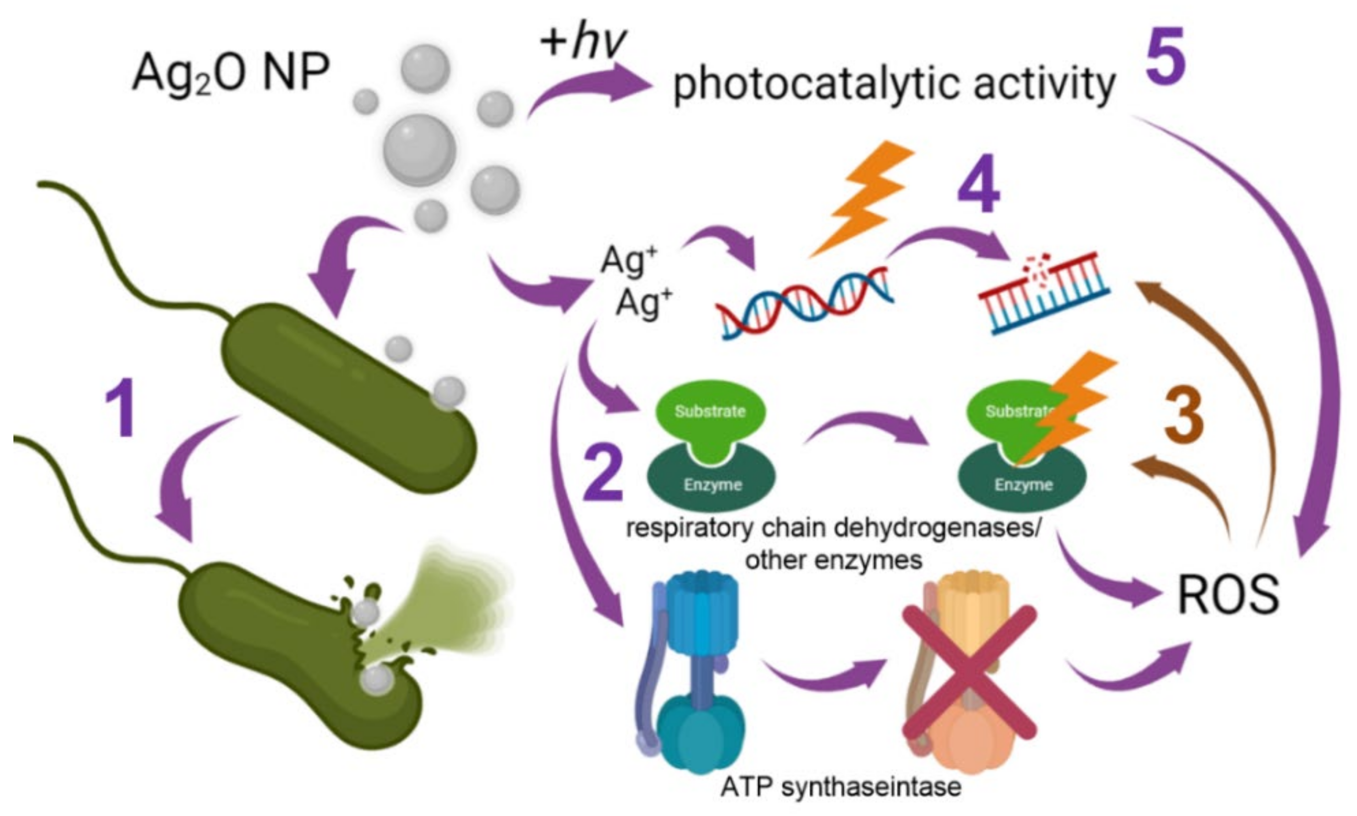
5. Mechanisms of the Antimicrobial Activity
6. Methods for Improving Antimicrobial Properties
7. Cytotoxicity to Human Cells
8. Conclusions
Author Contributions
Funding
Institutional Review Board Statement
Informed Consent Statement
Data Availability Statement
Acknowledgments
Conflicts of Interest
References
- Davies, J.; Davies, D. Origins and evolution of antibiotic resistance. Microbiol. Mol. Biol. Rev. 2010, 74, 417–433. [Google Scholar] [CrossRef] [PubMed]
- Cavalieri, F.; Tortora, M.; Stringaro, A.; Colone, M.; Baldassarri, L. Nanomedicines for antimicrobial interventions. J. Hosp. Infect. 2014, 88, 183–190. [Google Scholar] [CrossRef] [PubMed]
- Devi, L.S.; Joshi, S.R. Evaluation of the antimicrobial potency of silver nanoparticles biosynthesized by using an endophytic fungus Cryptosporiopsis ericae PS4. J. Microbiol. 2014, 52, 667–674. [Google Scholar] [CrossRef] [PubMed]
- Almatar, M.; Makky, E.A.; Var, I.; Koksal, F. The role of nanoparticles in the inhibition of multidrug-resistant bacteria and biofilms. Curr. Drug Deliv. 2018, 15, 470–484. [Google Scholar] [CrossRef]
- Solberg, C.O. Spread of Staphylococcus aureus in Hospitals: Causes and Prevention. Scand. J. Infect. Dis. 2000, 32, 587–595. [Google Scholar] [CrossRef]
- Gajbhiye, M.; Kesharwani, J.; Ingle, A.; Gade, A.; Rai, M. Fungus-mediated synthesis of silver nanoparticles and their activity against pathogenic fungi in combination with fluconazole. Nanomed. Nanotechnol. Biol. Med. 2009, 5, 382–386. [Google Scholar] [CrossRef]
- Gudkov, S.V.; Burmistrov, D.E.; Serov, D.A.; Rebezov, M.B.; Semenova, A.A.; Lisitsyn, A.B. A Mini Review of Antibacterial Properties of ZnO Nanoparticles. Front. Phys. 2021, 9, 641481. [Google Scholar] [CrossRef]
- Bongomin, F.; Gago, S.; Oladele, R.O.; Denning, D.W. Global and Multi-National Prevalence of Fungal Diseases—Estimate Precision. J. Fungi 2017, 3, 57. [Google Scholar] [CrossRef]
- Ben-Ami, R.; Kontoyiannis, D.P. Resistance to Antifungal Drugs. Infect. Dis. Clin. N. Am. 2021, 35, 279–311. [Google Scholar] [CrossRef]
- Du, W.; Gao, Y.; Liu, L.; Sai, S.; Ding, C. Striking Back against Fungal Infections: The Utilization of Nanosystems for Antifungal Strategies. Int. J. Mol. Sci. 2021, 22, 10104. [Google Scholar] [CrossRef]
- Khalil, N.M.; Abd El-Ghany, M.N.; Rodríguez-Couto, S. Antifungal and anti-mycotoxin efficacy of biogenic silver nanoparticles produced by Fusarium chlamydosporum and Penicillium chrysogenum at non-cytotoxic doses. Chemosphere 2019, 218, 477–486. [Google Scholar] [CrossRef]
- Coutard, B.; Valle, C.; De Lamballerie, X.; Canard, B.; Seidah, N.; Decroly, E. The Spike Glycoprotein of The New Coronavirus 2019-nCoV Contains A Furin-Like Cleavage Site Absent in Cov of The Same Clade. Antivir. Res. 2020, 176, 104742. [Google Scholar] [CrossRef]
- Hosseini, M.; Chin, A.W.H.; Williams, M.D.; Behzadinasab, S.; Falkinham, J.O.; Poon, L.L.M.; Ducker, W.A. Transparent Anti-SARS-CoV-2 and Antibacterial Silver Oxide Coatings. ACS Appl. Mater. Interfaces 2022, 14, 8718–8727. [Google Scholar] [CrossRef]
- Fong, J. The use of silver products in the management of burn wounds: Change in practice for the burn unit at Royal Perth Hospital. Prim. Intent. Aust. J. Wound Manag. 2005, 13, 16–22. [Google Scholar]
- Russell, F.R.; Pathm, W.B.; Hugo, A.D. Antimicrobial activity and action of silver. Prog. Med. Chem. 1994, 31, 351–371. [Google Scholar] [CrossRef]
- Uttayarat, P.; Eamsiri, J.; Tangthong, T.; Suwanmala, P. Radiolytic synthesis of colloidal silver nanoparticles for antibacterial wound dressings. Adv. Mater. Sci. Eng. 2015, 2015, 376082. [Google Scholar] [CrossRef]
- Kim, J.S.; Kuk, E.; Yu, K.N.; Kim, J.H.; Park, S.J.; Lee, H.J.; Kim, S.H.; Park, Y.K.; Park, Y.H.; Hwang, C.Y.; et al. Antimicrobial effects of silver nanoparticles. Nanomed. Nanotechnol. Biol. Med. 2007, 3, 95–101. [Google Scholar] [CrossRef]
- Chen, X.; Schluesener, H.J. Nanosilver: A nanoproduct in medical application. Toxicol. Lett. 2008, 176, 1–12. [Google Scholar] [CrossRef]
- Shen, W.; Li, P.; Feng, H.; Ge, Y.; Liu, Z.; Feng, L. The bactericidal mechanism of action against Staphylococcus aureus for AgO nanoparticles. Mater. Sci. Eng. C Mater. Biol. Appl. 2017, 75, 610–619. [Google Scholar] [CrossRef]
- Jain, S.; Mehata, M.S. Medicinal Plant Leaf Extract and Pure Flavonoid Mediated Green Synthesis of Silver Nanoparticles and their Enhanced Antibacterial Property. Sci. Rep. 2017, 7, 15867. [Google Scholar] [CrossRef]
- Ravichandran, S.; Paluri, V.; Kumar, G.; Loganathan, K.; Kokati Venkata, B.R. A novel approach for the biosynthesis of silver oxide nanoparticles using aqueous leaf extract of Callistemon lanceolatus (Myrtaceae) and their therapeutic potential. J. Exp. Nanosci. 2016, 11, 445–458. [Google Scholar] [CrossRef]
- Chakraborty, U.; Garg, P.; Bhanjana, G.; Kaur, G.; Kaushik, A.; Chaudhary, G.R. Spherical silver oxide nanoparticles for fabrication of electrochemical sensor for efficient 4-Nitrotoluene detection and assessment of their antimicrobial activity. Sci. Total Environ. 2022, 808, 152179. [Google Scholar] [CrossRef]
- Rahman, M.; Khan, S.; Jamal, A.; Faisal, M.; Asiri, A.M. Highly Sensitive Methanol Chemical Sensor Based on Undoped Silver Oxide Nanoparticles Prepared by a Solution Method. Microchim. Acta 2012, 178, 99–106. [Google Scholar] [CrossRef]
- Zhou, X.; Lu, Y.; Zhai, L.-L.; Zhao, Y.; Liu, Q.; Sun, W.-Y. Propargylamines formed from three-component coupling reactions catalyzed by silver oxide nanoparticles. RSC Adv. 2013, 3, 1732–1734. [Google Scholar] [CrossRef]
- Konop, M.; Damps, T.; Misicka, A.; Rudnicka, L. Certain Aspects of Silver and Silver Nanoparticles in Wound Care: A Minireview. J. Nanomater. 2016, 2016, 7614753. [Google Scholar] [CrossRef]
- Ghotekar, S.; Dabhane, H.; Pansambal, S.; Oza, R.; Tambade, P.; Medhane, V. A Review on Biomimetic Synthesis of Ag2O Nanoparticles using Plant Extract, Characterization and its Recent Applications. Adv. J. Chem. Sect. B 2020, 2, 102–111. [Google Scholar] [CrossRef]
- Sangappa, M.; Thiagarajan, P. Combating drug resistant pathogenic bacteria isolated from clinical infections, with silver oxide nanoparticles. Indian J. Pharm. Sci. 2015, 77, 151–155. [Google Scholar] [CrossRef]
- Ni, S.; Li, X.; Yang, P.; Ni, S.; Hong, F.; Webster, T.J. Enhanced apatite-forming ability and antibacterial activity of porous anodic alumina embedded with CaO-SiO2-Ag2O bioactive materials. Mater. Sci. Eng. C Mater. Biol. Appl. 2016, 58, 700–708. [Google Scholar] [CrossRef]
- Borges Rosa De Moura, F.; Antonio Ferreira, B.; Helena Muniz, E.; Benatti Justino, A.; Gabriela Silva, A.; De Azambuja Ribeiro, R.I.M.; Oliveira Dantas, N.; Lisboa Ribeiro, D.; De Assis Araújo, F.; Salmen Espindola, F.; et al. Antioxidant, anti-inflammatory, and wound healing effects of topical silver-doped zinc oxide and silver oxide nanocomposites. Int. J. Pharm. 2022, 617, 121620. [Google Scholar] [CrossRef]
- Iqbal, S.; Fakhar-E-Alam, M.; Akbar, F.; Shafiq, M.; Atif, M.; Amin, N.; Ismail, M.; Hanif, A.; Farooq, W.A. Application of silver oxide nanoparticles for the treatment of cancer. J. Mol. Struct. 2019, 1189, 203–209. [Google Scholar] [CrossRef]
- Salem, N.A.; Wahba, M.A.; Eisa, W.H.; El-Shamarka, M.; Khalil, W. Silver oxide nanoparticles alleviate indomethacin-induced gastric injury: A novel antiulcer agent. Inflammopharmacology 2018, 26, 1025–1035. [Google Scholar] [CrossRef] [PubMed]
- Pu, S.; Yang, Z.; Tang, J.; Ma, H.; Xue, S.; Bai, Y. Plasmonic silver/silver oxide nanoparticles anchored bismuth vanadate as a novel visible-light ternary photocatalyst for degrading pharmaceutical micropollutants. J. Environ. Sci. 2020, 96, 21–32. [Google Scholar] [CrossRef] [PubMed]
- Abbasi, B.A.; Iqbal, J.; Nasir, J.A.; Zahra, S.A.; Shahbaz, A.; Uddin, S.; Hameed, S.; Gul, F.; Kanwal, S.; Mahmood, T. Environmentally friendly green approach for the fabrication of silver oxide nanoparticles: Characterization and diverse biomedical applications. Microsc. Res. Tech. 2020, 83, 1308–1320. [Google Scholar] [CrossRef] [PubMed]
- Khatun, Z.; Lawrence, R.S.; Jalees, M.; Lawerence, K. Green synthesis and Anti-bacterial activity of Silver Oxide nanoparticles prepared from Pinus longifolia leaves extract. Int. J. Adv. Res. 2015, 3, 337–343. [Google Scholar]
- Shah, A.; Haq, S.; Rehman, W.; Waseem, M.; Shoukat, S.; Rehman, M.-U. Photocatalytic and antibacterial activities of paeonia emodi mediated silver oxide nanoparticles. Mater. Res. Express 2019, 6, 045045. [Google Scholar] [CrossRef]
- Pradheesh, G.; Suresh, S.; Suresh, J.; Alexramani, V. Antimicrobial and anticancer activity studies on green synthesized silver oxide nanoparticles from the medicinal plant cyathea nilgiriensis holttum. Int. J. Pharm. Investig. 2020, 10, 146–150. [Google Scholar] [CrossRef]
- Roy, A.; Srivastava, S.K.; Shrivastava, S.L.; Mandal, A.K. Hierarchical Assembly of Nanodimensional Silver–Silver Oxide Physical Gels Controlling Nosocomial Infections. ACS Omega 2020, 5, 32617–32631. [Google Scholar] [CrossRef]
- Jin, Y.; Dong, S. One-Pot Synthesis and Characterization of Novel Silver−Gold Bimetallic Nanostructures with Hollow Interiors and Bearing Nanospikes. J. Phys. Chem. B 2003, 107, 12902–12905. [Google Scholar] [CrossRef]
- Tripathi, S.; Mehrotra, G.K.; Dutta, P.K. Chitosan–silver oxide nanocomposite film: Preparation and antimicrobial activity. Bull. Mater. Sci. 2011, 34, 29–35. [Google Scholar] [CrossRef]
- Hu, Z.; Zhang, J.; Chan, W.L.; Szeto, Y.S. Suspension of Silver Oxide Nanoparticles in Chitosan Solution and its Antibacterial Activity in Cotton Fabrics. MRS Online Proc. Libr. 2006, 920, 203. [Google Scholar] [CrossRef]
- Hu, Z.; Chan, W.L.; Szeto, Y.S. Nanocomposite of chitosan and silver oxide and its antibacterial property. J. Appl. Polym. Sci. 2008, 108, 52–56. [Google Scholar] [CrossRef]
- Gul, S.; Rehan, Z.A.; Khan, S.A.; Akhtar, K.; Khan, M.A.; Khan, M.I.; Rashid, M.I.; Asiri, A.M.; Khan, S.B. Antibacterial PES-CA-Ag2O nanocomposite supported Cu nanoparticles membrane toward ultrafiltration, BSA rejection and reduction of nitrophenol. J. Mol. Liq. 2017, 230, 616–624. [Google Scholar] [CrossRef]
- Kakakhel, S.A.; Rashid, H.; Jalil, Q.; Munir, S.; Barkatullah, B.; Khan, S.; Ullah, R.; Shahat, A.; Mahmood, H.; A-Mishari, A.; et al. Polymers Encapsulated Aspirin Loaded Silver Oxide Nanoparticles: Synthesis, Characterization and its Bio-Applications. Sains Malays. 2019, 48, 1887–1897. [Google Scholar] [CrossRef]
- Aazem, I.; Rathinam, P.; Pillai, S.; Honey, G.; Vengellur, A.; Bhat, S.G.; Sailaja, G.S. Active bayerite underpinned Ag2O/Ag: An efficient antibacterial nanohybrid combating microbial contamination. Metallomics 2021, 13, mfab049. [Google Scholar] [CrossRef]
- Sajjad, S.; Arshad, F.; Uzair, B.; Leghari, S.A.K.; Noor, S.; Maaza, M. GO/Ag2O Composite Nanostructure as an Effective Antibacterial Agent. ChemistrySelect 2019, 4, 10365–10371. [Google Scholar] [CrossRef]
- Rajabi, A.; Ghazali, M.J.; Mahmoudi, E.; Baghdadi, A.H.; Mohammad, A.W.; Mustafah, N.M.; Ohnmar, H.; Naicker, A.S. Synthesis, Characterization, and Antibacterial Activity of Ag₂O-Loaded Polyethylene Terephthalate Fabric via Ultrasonic Method. Nanomaterials 2019, 9, 450. [Google Scholar] [CrossRef]
- Sboui, M.; Lachheb, H.; Bouattour, S.; Gruttadauria, M.; La Parola, V.; Liotta, L.F.; Boufi, S. TiO2/Ag2O immobilized on cellulose paper: A new floating system for enhanced photocatalytic and antibacterial activities. Environ. Res. 2021, 198, 111257. [Google Scholar] [CrossRef]
- Lin, Z.; Lu, Y.; Huang, J. A hierarchical Ag2O-nanoparticle/TiO2-nanotube composite derived from natural cellulose substance with enhanced photocatalytic performance. Cellulose 2019, 26, 6683–6700. [Google Scholar] [CrossRef]
- Dharmaraj, D.; Krishnamoorthy, M.; Rajendran, K.; Karuppiah, K.; Annamalai, J.; Durairaj, K.R.; Santhiyagu, P.; Ethiraj, K. Antibacterial and cytotoxicity activities of biosynthesized silver oxide (Ag2O) nanoparticles using Bacillus paramycoides. J. Drug Deliv. Sci. Technol. 2021, 61, 102111. [Google Scholar] [CrossRef]
- Li, D.; Chen, S.; Zhang, K.; Gao, N.; Zhang, M.; Albasher, G.; Shi, J.; Wang, C. The interaction of Ag2O nanoparticles with Escherichia coli: Inhibition–sterilization process. Sci. Rep. 2021, 11, 1703. [Google Scholar] [CrossRef]
- Fayyadh, A.A.; Jaduaa Alzubaidy, M.H. Green-synthesis of Ag2O nanoparticles for antimicrobial assays**. J. Mech. Behav. Mater. 2021, 30, 228–236. [Google Scholar] [CrossRef]
- Chausov, D.N.; Smirnova, V.V.; Burmistrov, D.E.; Sarimov, R.M.; Kurilov, A.D.; Astashev, M.E.; Uvarov, O.V.; Dubinin, M.V.; Kozlov, V.A.; Vedunova, M.V.; et al. Synthesis of a Novel, Biocompatible and Bacteriostatic Borosiloxane Composition with Silver Oxide Nanoparticles. Materials 2022, 15, 2. [Google Scholar] [CrossRef]
- Smirnova, V.V.; Chausov, D.N.; Serov, D.A.; Kozlov, V.A.; Ivashkin, P.I.; Pishchalnikov, R.Y.; Uvarov, O.V.; Vedunova, M.V.; Semenova, A.A.; Lisitsyn, A.B.; et al. A Novel Biodegradable Composite Polymer Material Based on PLGA and Silver Oxide Nanoparticles with Unique Physicochemical Properties and Biocompatibility with Mammalian Cells. Materials 2021, 14, 22. [Google Scholar] [CrossRef]
- Karunagaran, V.; Rajendran, K.; Sen, S. Antimicrobial Activity of Biosynthesized Silver Oxide Nanoparticles. J. Pure Appl. Microbiol. 2014, 4, 3263–3268. [Google Scholar]
- Ayanwale, A.P.; Ruíz-Baltazar, A.D.J.; Espinoza-Cristóbal, L.; Reyes-López, S.Y. Bactericidal Activity Study of ZrO2-Ag2O Nanoparticles. Dose-Response 2020, 18, 1559325820941374. [Google Scholar] [CrossRef]
- Islam, S.N.; Naqvi, S.M.A.; Parveen, S.; Ahmad, A. Application of mycogenic silver/silver oxide nanoparticles in electrochemical glucose sensing; alongside their catalytic and antimicrobial activity. 3 Biotech 2021, 11, 342. [Google Scholar] [CrossRef]
- Rokade, A.A.; Patil, M.P.; Yoo, S.I.; Lee, W.K.; Park, S.S. Pure green chemical approach for synthesis of Ag2O nanoparticles. Green Chem. Lett. Rev. 2016, 9, 216–222. [Google Scholar] [CrossRef]
- Shaffiey, S.R.; Shaffiey, S. Synthesis and evaluation of bactericidal properties of Ag2O nanoparticles against Aeromonashydrophila. Int. J. Nano Dimens. 2014, 6, 263–269. [Google Scholar] [CrossRef]
- Panáček, A.; Kvítek, L.; Prucek, R.; Kolář, M.; Večeřová, R.; Pizúrová, N.; Sharma, V.K.; Nevěčná, T.J.; Zbořil, R. Silver Colloid Nanoparticles: Synthesis, Characterization, and Their Antibacterial Activity. J. Phys. Chem. B 2006, 110, 16248–16253. [Google Scholar] [CrossRef]
- Manikandan, V.; Velmurugan, P.; Park, J.H.; Chang, W.S.; Park, Y.J.; Jayanthi, P.; Cho, M.; Oh, B.T. Green synthesis of silver oxide nanoparticles and its antibacterial activity against dental pathogens. 3 Biotech 2017, 7, 72. [Google Scholar] [CrossRef]
- Karunakaran, G.; Jagathambal, M.; Gusev, A.; Minh, N.V.; Kolesnikov, E.; Mandal, A.R.; Kuznetsov, D. Nitrobacter sp. extract mediated biosynthesis of Ag(2)O NPs with excellent antioxidant and antibacterial potential for biomedical application. IET Nanobiotechnol. 2016, 10, 425–430. [Google Scholar] [CrossRef] [PubMed]
- Haq, S.; Rehman, W.; Waseem, M.; Meynen, V.; Awan, S.U.; Saeed, S.; Iqbal, N. Fabrication of pure and moxifloxacin functionalized silver oxide nanoparticles for photocatalytic and antimicrobial activity. J. Photochem. Photobiol. B Biol. 2018, 186, 116–124. [Google Scholar] [CrossRef] [PubMed]
- Babu, P.J.; Doble, M.; Raichur, A.M. Silver oxide nanoparticles embedded silk fibroin spuns: Microwave mediated preparation, characterization and their synergistic wound healing and anti-bacterial activity. J. Colloid Interface Sci. 2018, 513, 62–71. [Google Scholar] [CrossRef] [PubMed]
- Li, R.; Chen, Z.; Ren, N.; Wang, Y.; Wang, Y.; Yu, F. Biosynthesis of silver oxide nanoparticles and their photocatalytic and antimicrobial activity evaluation for wound healing applications in nursing care. J. Photochem. Photobiol. B Biol. 2019, 199, 111593. [Google Scholar] [CrossRef]
- Elemike, E.E.; Onwudiwe, D.C.; Ekennia, A.C.; Sonde, C.U.; Ehiri, R.C. Green Synthesis of Ag/Ag₂O Nanoparticles Using Aqueous Leaf Extract of Eupatorium odoratum and Its Antimicrobial and Mosquito Larvicidal Activities. Molecules 2017, 22, 674. [Google Scholar] [CrossRef]
- Mani, M.; Harikrishnan, R.; Purushothaman, P.; Pavithra, S.; Rajkumar, P.; Kumaresan, S.; Al Farraj, D.A.; Elshikh, M.S.; Balasubramanian, B.; Kaviyarasu, K. Systematic green synthesis of silver oxide nanoparticles for antimicrobial activity. Environ. Res. 2021, 202, 111627. [Google Scholar] [CrossRef]
- Hoque, M.I.U.; Chowdhury, A.N.; Islam, M.T.; Firoz, S.H.; Luba, U.; Alowasheeir, A.; Rahman, M.M.; Rehman, A.U.; Ahmad, S.H.A.; Holze, R.; et al. Fabrication of highly and poorly oxidized silver oxide/silver/tin(IV) oxide nanocomposites and their comparative anti-pathogenic properties towards hazardous food pathogens. J. Hazard. Mater. 2021, 408, 124896. [Google Scholar] [CrossRef]
- Negi, H.; Rathinavelu Saravanan, P.; Agarwal, T.; Ghulam Haider Zaidi, M.; Goel, R. In vitro assessment of Ag2O nanoparticles toxicity against Gram-positive and Gram-negative bacteria. J. Gen. Appl. Microbiol. 2013, 59, 83–88. [Google Scholar] [CrossRef]
- Flores-Lopez, N.S.; Cervantes-Chávez, J.A.; Téllez De Jesús, D.G.; Cortez-Valadez, M.; Estévez-González, M.; Esparza, R. Bactericidal and fungicidal capacity of Ag(2)O/Ag nanoparticles synthesized with Aloe vera extract. J. Environ. Sci. Health. Part A Toxic/Hazard. Subst. Environ. Eng. 2021, 56, 762–768. [Google Scholar] [CrossRef]
- Chen, Y.; Gao, A.; Bai, L.; Wang, Y.; Wang, X.; Zhang, X.; Huang, X.; Hang, R.; Tang, B.; Chu, P.K. Antibacterial, osteogenic, and angiogenic activities of SrTiO(3) nanotubes embedded with Ag(2)O nanoparticles. Mater. Sci. Eng. C Mater. Biol. Appl. 2017, 75, 1049–1058. [Google Scholar] [CrossRef]
- Jin, Y.; Dai, Z.; Liu, F.; Kim, H.; Tong, M.; Hou, Y. Bactericidal mechanisms of Ag₂O/TNBs under both dark and light conditions. Water Res. 2013, 47, 1837–1847. [Google Scholar] [CrossRef]
- Gao, A.; Hang, R.; Huang, X.; Zhao, L.; Zhang, X.; Wang, L.; Tang, B.; Ma, S.; Chu, P.K. The effects of titania nanotubes with embedded silver oxide nanoparticles on bacteria and osteoblasts. Biomaterials 2014, 35, 4223–4235. [Google Scholar] [CrossRef]
- Khodadadi, S.; Mahdinezhad, N.; Fazeli-Nasab, B.; Heidari, M.J.; Fakheri, B.; Miri, A. Investigating the Possibility of Green Synthesis of Silver Nanoparticles Using Vaccinium arctostaphlyos Extract and Evaluating Its Antibacterial Properties. BioMed Res. Int. 2021, 2021, 5572252. [Google Scholar] [CrossRef]
- Wang, X.; Wu, H.F.; Kuang, Q.; Huang, R.B.; Xie, Z.X.; Zheng, L.S. Shape-dependent antibacterial activities of Ag2O polyhedral particles. Langmuir ACS J. Surf. Colloids 2010, 26, 2774–2778. [Google Scholar] [CrossRef]
- Kundu, S.; Sain, S.; Choudhury, P.; Sarkar, S.; Das, P.K.; Pradhan, S.K. Microstructure characterization of biocompatible heterojunction hydrogen titanate-Ag(2)O nanocomposites for superior visible light photocatalysis and antibacterial activity. Mater. Sci. Eng. C Mater. Biol. Appl. 2019, 99, 374–386. [Google Scholar] [CrossRef]
- Lekshmi, G.S.; Tamilselvi, R.; Geethalakshmi, R.; Kirupha, S.D.; Bazaka, O.; Levchenko, I.; Bazaka, K.; Mandhakini, M. Multifunctional oil-produced reduced graphene oxide - Silver oxide composites with photocatalytic, antioxidant, and antibacterial activities. J. Colloid Interface Sci. 2022, 608, 294–305. [Google Scholar] [CrossRef]
- Sajjad, S.; Uzair, B.; Shaukat, A.; Jamshed, M.; Leghari, S.A.K.; Ismail, M.; Mansoor, Q. Synergistic evaluation of AgO(2) nanoparticles with ceftriaxone against CTXM and blaSHV genes positive ESBL producing clinical strains of Uro-pathogenic E. coli. IET Nanobiotechnol. 2019, 13, 435–440. [Google Scholar] [CrossRef]
- Boopathi, S.; Gopinath, S.; Boopathi, T.; Balamurugan, V.; Rajeshkumar, R.; Sundararaman, M. Characterization and Antimicrobial Properties of Silver and Silver Oxide Nanoparticles Synthesized by Cell-Free Extract of a Mangrove-Associated Pseudomonas aeruginosa M6 Using Two Different Thermal Treatments. Ind. Eng. Chem. Res. 2012, 51, 5976–5985. [Google Scholar] [CrossRef]
- D’lima, L.; Phadke, M.; Ashok, V.D. Biogenic silver and silver oxide hybrid nanoparticles: A potential antimicrobial against multi drug-resistant Pseudomonas aeruginosa. New. J. Chem. 2020, 44, 4935–4941. [Google Scholar] [CrossRef]
- Salvadori, M.; Monezi, T.; Mehnert, D.; Corrêa, B. Antimicrobial Activity of Ag/Ag2O Nanoparticles Synthesized by Dead Biomass of Yeast and their Biocompatibility with Mammalian Cell Lines. Int. J. Res. Stud. Microbiol. Biotechnol. 2019, 5, 2454–9428. [Google Scholar] [CrossRef]
- Kayed, K.; Mansour, G. The Antimicrobial Activity of Silver Nanoparticles in Ag/Ag2O Composites Synthesized by Oxygen Plasma Treatment of Silver Thin Films. Curr. Appl. Sci. Technol. 2022, 22, 9. [Google Scholar] [CrossRef]
- Akbari, Z.; Rashidi Ranjbar, Z.; Khaleghi, M. Synthesis, characterization, and antibacterial activities of Ag2O nanoparticle and silver (I) nano-rod complex. Nanochemistry Res. 2020, 5, 233–240. [Google Scholar]
- Rashmi, B.N.; Harlapur, S.F.; Avinash, B.; Ravikumar, C.R.; Nagaswarupa, H.P.; Anil Kumar, M.R.; Gurushantha, K.; Santosh, M.S. Facile green synthesis of silver oxide nanoparticles and their electrochemical, photocatalytic and biological studies. Inorg. Chem. Commun. 2020, 111, 107580. [Google Scholar] [CrossRef]
- Phongtongpasuk, S.; Poadang, S.; Yongvanich, N. Environmental-friendly Method for Synthesis of Silver Nanoparticles from Dragon Fruit Peel Extract and their Antibacterial Activities. Energy Procedia 2016, 89, 239–247. [Google Scholar] [CrossRef]
- Aisida, S.; Ugwu, K.; Nwanya, A.; Bashir, A.K.H.; Nwankwo, U.; Ahmed, I.; Ezema, F. Biosynthesis of silver oxide nanoparticles using leave extract of Telfairia Occidentalis and its antibacterial activity. Mater. Today Proc. 2021, 36, 208–213. [Google Scholar] [CrossRef]
- Kiani, F.A.; Shamraiz, U.; Badshah, A.; Tabassum, S.; Ambreen, M.; Patujo, J.A. Optimization of Ag2O nanostructures with strontium for biological and therapeutic potential. Artif. Cells Nanomed. Biotechnol. 2018, 46 (Suppl. S3), S1083–S1091. [Google Scholar] [CrossRef]
- Elyamny, S.; Eltarahony, M.; Abu-Serie, M.; Nabil, M.M.; Kashyout, A.E.-H.B. One-pot fabrication of Ag @Ag2O core–shell nanostructures for biosafe antimicrobial and antibiofilm applications. Sci. Rep. 2021, 11, 22543. [Google Scholar] [CrossRef]
- Gudkov, S.V.; Burmistrov, D.E.; Serov, D.A.; Rebezov, M.B.; Semenova, A.A.; Lisitsyn, A.B. Do Iron Oxide Nanoparticles Have Significant Antibacterial Properties? Antibiotics 2021, 10, 884. [Google Scholar] [CrossRef] [PubMed]
- Arias, L.S.; Pessan, J.P.; Vieira, A.P.M.; Lima, T.M.T.D.; Delbem, A.C.B.; Monteiro, D.R. Iron oxide nanoparticles for biomedical applications: A perspective on synthesis, drugs, antimicrobial activity, and toxicity. Antibiotics 2018, 7, 46. [Google Scholar] [CrossRef] [PubMed]
- Apopa, P.L.; Qian, Y.; Shao, R.; Guo, N.L.; Schwegler-Berry, D.; Pacurari, M.; Porter, D.; Shi, X.; Vallyathan, V.; Castranova, V. Iron oxide nanoparticles induce human microvascular endothelial cell permeability through reactive oxygen species production and microtubule remodeling. Part. Fibre Toxicol. 2009, 6, 1. [Google Scholar] [CrossRef] [PubMed]
- Gong, Y.; Ji, Y.; Liu, F.; Li, J.; Cao, Y. Cytotoxicity, oxidative stress and inflammation induced by ZnO nanoparticles in endothelial cells: Interaction with palmitate or lipopolysaccharide. J. Appl. Toxicol. 2017, 37, 895–901. [Google Scholar] [CrossRef]
- Chatterjee, N.; Walker, G.C. Mechanisms of DNA damage, repair, and mutagenesis. Env. Mol Mutagen 2017, 58, 235–263. [Google Scholar] [CrossRef]
- Doolittle, W.F. A paradigm gets shifty. Nature 1998, 392, 15–16. [Google Scholar] [CrossRef]
- Wigley, D.B. Bacterial DNA repair: Recent insights into the mechanism of RecBCD, AddAB and AdnAB. Nat. Rev. Microbiol. 2013, 11, 9–13. [Google Scholar] [CrossRef]
- Torabi, S.; Mansoorkhani, M.J.K.; Majedi, A.; Motevalli, S. REVIEW: Synthesis, Medical and Photocatalyst Applications of Nano-Ag2O. J. Coord. Chem. 2020, 73, 1861–1880. [Google Scholar] [CrossRef]
- Sullivan, K.T.; Wu, C.; Piekiel, N.W.; Gaskell, K.; Zachariah, M.R. Synthesis and reactivity of nano-Ag2O as an oxidizer for energetic systems yielding antimicrobial products. Combust. Flame 2013, 160, 438–446. [Google Scholar] [CrossRef]
- Phan, C.M.; Nguyen, H.M. Role of Capping Agent in Wet Synthesis of Nanoparticles. J. Phys. Chem. A 2017, 121, 3213–3219. [Google Scholar] [CrossRef]
- Yong, N.L.; Ahmad, A.; Mohammad, A.W. Synthesis and characterization of silver oxide nanoparticles by a novel method. Int. J. Sci. Eng. Res. 2013, 4, 155–158. [Google Scholar]
- Jeung, D.-G.; Lee, M.; Paek, S.-M.; Oh, J.-M. Controlled Growth of Silver Oxide Nanoparticles on the Surface of Citrate Anion Intercalated Layered Double Hydroxide. Nanomaterials 2021, 11, 455. [Google Scholar] [CrossRef]
- Harish, K.; Manisha, K. Synthesis and characterization of silveroxide nanoparticles by sol-gel method. Int. J. Adv. Res. Sci. Eng. 2018, 7, 632–637. [Google Scholar]
- Wang, G.; Ma, X.; Huang, B.; Cheng, H.; Wang, Z.; Zhan, J.; Qin, X.; Zhang, X.; Dai, Y. Controlled synthesis of Ag 2 O microcrystals with facet-dependent photocatalytic activities. J. Mater. Chem. 2012, 22, 21189–21194. [Google Scholar] [CrossRef]
- Murray, B.; Li, Q.; Newberg, J.; Menke, E.; Hemminger, J.; Penner, R. Shape-and size-selective electrochemical synthesis of dispersed silver (I) oxide colloids. Nano Lett. 2005, 5, 2319–2324. [Google Scholar] [CrossRef]
- Saha, S.; Chattopadhyay, D.; Acharya, K. Preparation of silver nanoparticles by bio-reduction using nigrospora oryzae culture filtrate and its antimicrobial activity. Dig. J. Nanomater. Biostructures (DJNB) 2011, 6, 1842–3582. [Google Scholar]
- Ponnuchamy, K.; Selvi, S.; Prabha, L.; Premkumar, K.; Ganeshkumar, R.; Munisamy, G. Synthesis of Silver Nanoparticles from Sargassum Tenerrimum and Screening Phytochemicals for Its Antibacterial Activity. Nano Biomed. Eng. 2012, 4, 12–16. [Google Scholar] [CrossRef]
- Maiti, S.; Krishnan, D.; Barman, G.; Ghosh, S.K.; Laha, J.K. Antimicrobial activities of silver nanoparticles synthesized from Lycopersicon esculentum extract. J. Anal. Sci. Technol. 2014, 5, 40. [Google Scholar] [CrossRef]
- Antony, E.; Sathiavelu, M.; Arunachalam, S. Synthesis of silver nanoparticles from the medicinal plant bauhinia acuminata and biophytum sensitivum–a comparative study of its biological activities with plant extract. Int. J. Appl. Pharm. 2016, 9, 22. [Google Scholar] [CrossRef]
- Mohamed, H.E.A.; Afridi, S.; Khalil, A.T.; Zia, D.; Iqbal, J.; Ullah, I.; Shinwari, Z.K.; Maaza, M. Biosynthesis of silver nanoparticles from Hyphaene thebaica fruits and their in vitro pharmacognostic potential. Mater. Res. Express 2019, 6, 1050c9. [Google Scholar] [CrossRef]
- Kahsay, M.H.; Ramadevi, D.; Kumar, Y.P.; Mohan, B.S.; Tadesse, A.; Battu, G.; Basavaiah, K. Synthesis of silver nanoparticles using aqueous extract of Dolichos lablab for reduction of 4-Nitrophenol, antimicrobial and anticancer activities. OpenNano 2018, 3, 28–37. [Google Scholar] [CrossRef]
- Laouini, S.E.; Bouafia, A.; Soldatov, A.V.; Algarni, H.; Tedjani, M.L.; Ali, G.A.M.; Barhoum, A. Green Synthesized of Ag/Ag(2)O Nanoparticles Using Aqueous Leaves Extracts of Phoenix dactylifera L. and Their Azo Dye Photodegradation. Membranes 2021, 11, 468. [Google Scholar] [CrossRef]
- Gomaa, E.Z. Silver nanoparticles as an antimicrobial agent: A case study on Staphylococcus aureus and Escherichia coli as models for Gram-positive and Gram-negative bacteria. J. Gen. Appl. Microbiol. 2017, 63, 36–43. [Google Scholar] [CrossRef]
- Gurunathan, S.; Kalishwaralal, K.; Vaidyanathan, R.; Venkataraman, D.; Pandian, S.R.K.; Muniyandi, J.; Hariharan, N.; Eom, S.H. Biosynthesis, purification and characterization of silver nanoparticles using Escherichia coli. Colloids Surf. B Biointerfaces 2009, 74, 328–335. [Google Scholar] [CrossRef] [PubMed]
- Rao, K.; Imran, M.; Jabri, T.; Ali, I.; Perveen, S.; Shafiullah; Ahmed, S.; Shah, M.R. Gum tragacanth stabilized green gold nanoparticles as cargos for Naringin loading: A morphological investigation through AFM. Carbohydr. Polym. 2017, 174, 243–252. [Google Scholar] [CrossRef] [PubMed]
- Song, Z.; Hrbek, J.; Osgood, R. Formation of TiO2 nanoparticles by reactive-layer-assisted deposition and characterization by XPS and STM. Nano Lett. 2005, 5, 1327–1332. [Google Scholar] [CrossRef] [PubMed]
- Nagajyothi, P.C.; Sreekanth, T.V.; Tettey, C.O.; Jun, Y.I.; Mook, S.H. Characterization, antibacterial, antioxidant, and cytotoxic activities of ZnO nanoparticles using Coptidis Rhizoma. Bioorganic Med. Chem. Lett. 2014, 24, 4298–4303. [Google Scholar] [CrossRef]
- Mittal, A.K.; Chisti, Y.; Banerjee, U.C. Synthesis of metallic nanoparticles using plant extracts. Biotechnol. Adv. 2013, 31, 346–356. [Google Scholar] [CrossRef]
- Wang, T.; Lin, J.; Chen, Z.; Megharaj, M.; Naidu, R. Green synthesized iron nanoparticles by green tea and eucalyptus leaves extracts used for removal of nitrate in aqueous solution. J. Clean. Prod. 2014, 83, 413–419. [Google Scholar] [CrossRef]
- Rauwel, P.; Küünal, S.; Ferdov, S.; Rauwel, E. A Review on the Green Synthesis of Silver Nanoparticles and Their Morphologies Studied via TEM. Adv. Mater. Sci. Eng. 2015, 2015, 682749. [Google Scholar] [CrossRef]
- Singh, V.; Shrivastava, A.; Wahi, N. Biosynthesis of silver nanoparticles by plants crude extracts and their characterization using UV, XRD, TEM and EDX. Afr. J. Biotechnol. 2015, 14, 2554–2567. [Google Scholar] [CrossRef]
- Naraginti, S.; Li, Y. Preliminary investigation of catalytic, antioxidant, anticancer and bactericidal activity of green synthesized silver and gold nanoparticles using Actinidia deliciosa. J. Photochem. Photobiol. B Biol. 2017, 170, 225–234. [Google Scholar] [CrossRef]
- Sana, S.S.; Dogiparthi, L.K. Green synthesis of silver nanoparticles using Givotia moluccana leaf extract and evaluation of their antimicrobial activity. Mater. Lett. 2018, 226, 47–51. [Google Scholar] [CrossRef]
- Monshi, A.; Foroughi, M.; Monshi, M. Modified Scherrer Equation to Estimate More Accurately Nano-Crystallite Size Using XRD. World J. Nano Sci. Eng. 2012, 2, 154–160. [Google Scholar] [CrossRef]
- Zad, Z.R.; Davarani, S.S.H.; Taheri, A.; Bide, Y. A yolk shell Fe3O4@ PA-Ni@ Pd/Chitosan nanocomposite-modified carbon ionic liquid electrode as a new sensor for the sensitive determination of fluconazole in pharmaceutical preparations and biological fluids. J. Mol. Liq. 2018, 253, 233–240. [Google Scholar] [CrossRef]
- Das, B.; Dash, S.K.; Mandal, D.; Ghosh, T.; Chattopadhyay, S.; Tripathy, S.; Das, S.; Dey, S.K.; Das, D.; Roy, S. Green synthesized silver nanoparticles destroy multidrug resistant bacteria via reactive oxygen species mediated membrane damage. Arab. J. Chem. 2017, 10, 862–876. [Google Scholar] [CrossRef]
- Surma, N.; Ijuo, G.; Ogoh-Orch, B. Fuel Gases from Waste High Density Polyethylene (Hdpe) Via Low Temperature Catalytic Pyrolysis. Prog. Chem. Biochem. Res. 2020, 3, 20–30. [Google Scholar] [CrossRef]
- Venkateswarlu, S.; Natesh Kumar, B.; Prasad, C.H.; Venkateswarlu, P.; Jyothi, N.V.V. Bio-inspired green synthesis of Fe3O4 spherical magnetic nanoparticles using Syzygium cumini seed extract. Phys. B Condens. Matter 2014, 449, 67–71. [Google Scholar] [CrossRef]
- Astashev, M.E.; Sarimov, R.M.; Serov, D.A.; Matveeva, T.A.; Simakin, A.V.; Ignatenko, D.N.; Burmistrov, D.E.; Smirnova, V.V.; Kurilov, A.D.; Mashchenko, V.I.; et al. Antibacterial behavior of organosilicon composite with nano aluminum oxide without influencing animal cells. React. Funct. Polym. 2022, 170, 105143. [Google Scholar] [CrossRef]
- Von Naegelli, V. Silver nitrate: A very effective antimicrobial agent. Deut. Schr. Schweiz Nat. Ges 1893, 33, 174–182. [Google Scholar]
- Raffi, M.; Hussain, F.; Bhatti, T.; Akhter, J.; Hameed, A.; Hasan, M. Antibacterial characterization of silver nanoparticles against E. coli ATCC-15224. J. Mater. Sci. Technol. 2008, 24, 192–196. [Google Scholar]
- Sondi, I.; Salopek-Sondi, B. Silver nanoparticles as antimicrobial agent: A case study on E. coli as a model for Gram-negative bacteria. J. Colloid Interface Sci. 2004, 275, 177–182. [Google Scholar] [CrossRef]
- Belly, R.T.; Kydd, G.C. Silver resistance in microorganisms. Dev. Ind. Microbiol. 1982, 23, 567–578. [Google Scholar]
- Grigor’eva, A.; Saranina, I.; Tikunova, N.; Safonov, A.; Timoshenko, N.; Rebrov, A.; Ryabchikova, E. Fine mechanisms of the interaction of silver nanoparticles with the cells of Salmonella typhimurium and Staphylococcus aureus. Biometals Int. J. Role Met. Ions Biol. Biochem. Med. 2013, 26, 479–488. [Google Scholar] [CrossRef]
- Song, H.; Ko, K.; Oh, L.; Lee, B. Fabrication of silver nanoparticles and their antimicrobial mechanisms. Eur. Cells Mater. 2006, 11 (Suppl. S1), 58. [Google Scholar]
- Sambhy, V.; Macbride, M.M.; Peterson, B.R.; Sen, A. Silver bromide nanoparticle/polymer composites: Dual action tunable antimicrobial materials. J. Am. Chem. Soc. 2006, 128, 9798–9808. [Google Scholar] [CrossRef]
- Russell, A.D.; Hugo, W.B. 7 Antimicrobial Activity and Action of Silver Periodical 7 Antimicrobial Activity and Action of Silver [Online]. 1994, pp. 351–370. Available online: https://www.sciencedirect.com/science/article/pii/S0079646808700249 (accessed on 25 April 2022). [CrossRef]
- Xia, T.; Kovochich, M.; Brant, J.; Hotze, M.; Sempf, J.; Oberley, T.; Sioutas, C.; Yeh, J.I.; Wiesner, M.R.; Nel, A.E. Comparison of the abilities of ambient and manufactured nanoparticles to induce cellular toxicity according to an oxidative stress paradigm. Nano Lett. 2006, 6, 1794–1807. [Google Scholar] [CrossRef]
- Bruskov, V.I.; Karp, O.E.; Garmash, S.A.; Shtarkman, I.N.; Chernikov, A.V.; Gudkov, S.V. Prolongation of oxidative stress by long-lived reactive protein species induced by X-ray radiation and their genotoxic action. Free Radic. Res. 2012, 46, 1280–1290. [Google Scholar] [CrossRef]
- Bruskov, V.I.; Malakhova, L.V.; Masalimov, Z.K.; Chernikov, A.V. Heat-induced formation of reactive oxygen species and 8-oxoguanine, a biomarker of damage to DNA. Nucleic Acids Res. 2002, 30, 1354–1363. [Google Scholar] [CrossRef]
- Burmistrov, D.E.; Simakin, A.V.; Smirnova, V.V.; Uvarov, O.V.; Ivashkin, P.I.; Kucherov, R.N.; Ivanov, V.E.; Bruskov, V.I.; Sevostyanov, M.A.; Baikin, A.S.; et al. Bacteriostatic and Cytotoxic Properties of Composite Material Based on ZnO Nanoparticles in PLGA Obtained by Low Temperature Method. Polymers 2022, 14, 49. [Google Scholar] [CrossRef]
- Ocsoy, I.; Paret, M.L.; Ocsoy, M.A.; Kunwar, S.; Chen, T.; You, M.; Tan, W. Nanotechnology in Plant Disease Management: DNA-Directed Silver Nanoparticles on Graphene Oxide as an Antibacterial against Xanthomonas perforans. ACS Nano 2013, 7, 8972–8980. [Google Scholar] [CrossRef]
- Allahverdiyev, A.M.; Abamor, E.S.; Bagirova, M.; Rafailovich, M. Antimicrobial effects of TiO2 and Ag2O nanoparticles against drug-resistant bacteria and leishmania parasites. Future Microbiol. 2011, 6, 933–940. [Google Scholar] [CrossRef]
- Hua, H.; Xi, Y.; Zhao, Z.; Xie, X.; Hu, C.; Liu, H. Gram-scale wet chemical synthesis of Ag2O/TiO2 aggregated sphere heterostructure with high photocatalytic activity. Mater. Lett. 2013, 91, 81–83. [Google Scholar] [CrossRef]
- Wu, C.; Shen, L.; Cai Zhang, Y.; Huang, Q. Solvothermal synthesis of Ag/ZnO nanocomposite with enhanced photocatalytic activity. Mater. Lett. 2013, 106, 104–106. [Google Scholar] [CrossRef]
- Fernandez, C.; Thomas, A.; M, S. Green synthesis of silver oxide nanoparticle and its antimicrobial activity against organisms causing Dental plaques. Int. J. Pharma. Bio. Sci. 2016, 7, 14–19. [Google Scholar] [CrossRef]
- Bai, X.; Li, L.; Liu, H.; Tan, L.; Liu, T.; Meng, X. Solvothermal Synthesis of ZnO Nanoparticles and Anti-Infection Application in Vivo. ACS Appl. Mater. Interfaces 2015, 7, 1308–1317. [Google Scholar] [CrossRef] [PubMed]
- Salvadori, M.R.; Ando, R.A.; Nascimento, C.A.O.; Corrêa, B. Dead biomass of Amazon yeast: A new insight into bioremediation and recovery of silver by intracellular synthesis of nanoparticles. J. Environ. Sci. Health Part A 2017, 52, 1112–1120. [Google Scholar] [CrossRef]
- Vinay, S.P.; Udayabhanu; Nagaraju, G.; Chandrappa, C.P.; Chandrasekhar, N. Rauvolfia tetraphylla (Devil Pepper)-Mediated Green Synthesis of Ag Nanoparticles: Applications to Anticancer, Antioxidant and Antimitotic. J. Clust. Sci. 2019, 30, 1545–1564. [Google Scholar] [CrossRef]
- Fard, N.N.; Noorbazargan, H.; Mirzaie, A.; Hedayati Ch, M.; Moghimiyan, Z.; Rahimi, A. Biogenic synthesis of AgNPs using Artemisia oliveriana extract and their biological activities for an effective treatment of lung cancer. Artif. Cells Nanomed. Biotechnol. 2018, 46 (Suppl. S3), S1047–S1058. [Google Scholar] [CrossRef]
- Schlinkert, P.; Casals, E.; Boyles, M.; Tischler, U.; Hornig, E.; Tran, N.; Zhao, J.; Himly, M.; Riediker, M.; Oostingh, G.J.; et al. The oxidative potential of differently charged silver and gold nanoparticles on three human lung epithelial cell types. J. Nanobiotechnol. 2015, 13, 1. [Google Scholar] [CrossRef]
- Guo, C.; Buckley, A.; Marczylo, T.; Seiffert, J.; Römer, I.; Warren, J.; Hodgson, A.; Chung, K.F.; Gant, T.W.; Smith, R.; et al. The small airway epithelium as a target for the adverse pulmonary effects of silver nanoparticle inhalation. Nanotoxicology 2018, 12, 539–553. [Google Scholar] [CrossRef]
- Yeasmin, S.; Datta, H.K.; Chaudhuri, S.; Malik, D.; Bandyopadhyay, A. In-vitro anti-cancer activity of shape controlled silver nanoparticles (AgNPs) in various organ specific cell lines. J. Mol. Liq. 2017, 242, 757–766. [Google Scholar] [CrossRef]
- Miyayama, T.; Fujiki, K.; Matsuoka, M. Silver nanoparticles induce lysosomal-autophagic defects and decreased expression of transcription factor EB in A549 human lung adenocarcinoma cells. Toxicol. Vitr. Int. J. Publ. Assoc. BIBRA 2018, 46, 148–154. [Google Scholar] [CrossRef]
- Akter, M.; Sikder, M.T.; Rahman, M.M.; Ullah, A.; Hossain, K.F.B.; Banik, S.; Hosokawa, T.; Saito, T.; Kurasaki, M. A systematic review on silver nanoparticles-induced cytotoxicity: Physicochemical properties and perspectives. J. Adv. Res. 2018, 9, 1–16. [Google Scholar] [CrossRef]
- González-Vega, J.G.; García-Ramos, J.C.; Chavez-Santoscoy, R.A.; Castillo-Quiñones, J.E.; Arellano-Garcia, M.E.; Toledano-Magaña, Y. Lung Models to Evaluate Silver Nanoparticles’ Toxicity and Their Impact on Human Health. Nanomaterials 2022, 12, 2316. [Google Scholar] [CrossRef]
- García, M.C.; Torres, J.; Dan Córdoba, A.V.; Longhi, M.; Uberman, P.M. Drug delivery using metal oxide nanoparticles Periodical Drug delivery using metal oxide nanoparticles [Online]. 2022, pp. 35–83. Available online: https://www.sciencedirect.com/science/article/pii/B9780128230336000296 (accessed on 8 May 2022). [CrossRef]
- Qureshi, A.T. Silver Nanoparticles as Drug Delivery Systems. LSU Dr. Diss. 2013, 1069. [Google Scholar] [CrossRef]
- Ghiuță, I.; Cristea, D. Silver nanoparticles for delivery purposes. In Nanoengineered Biomaterials for Advanced Drug Delivery; Mozafari, M., Ed.; Elsevier: Amsterdam, The Netherlands, 2020; pp. 347–371. [Google Scholar] [CrossRef]
- Zhang, X.F.; Gurunathan, S. Combination of salinomycin and silver nanoparticles enhances apoptosis and autophagy in human ovarian cancer cells: An effective anticancer therapy. Int. J. Nanomed. 2016, 11, 3655–3675. [Google Scholar] [CrossRef]
- Yuan, Y.G.; Peng, Q.L.; Gurunathan, S. Silver nanoparticles enhance the apoptotic potential of gemcitabine in human ovarian cancer cells: Combination therapy for effective cancer treatment. Int. J. Nanomed. 2017, 12, 6487–6502. [Google Scholar] [CrossRef]
- Baram-Pinto, D.; Shukla, S.; Perkas, N.; Gedanken, A.; Sarid, R. Inhibition of herpes simplex virus type 1 infection by silver nanoparticles capped with mercaptoethane sulfonate. Bioconjugate Chem. 2009, 20, 1497–1502. [Google Scholar] [CrossRef]
- Ivanova, N.; Gugleva, V.; Dobreva, M.; Pehlivanov, I.; Stefanov, S.; Andonova, V. Silver Nanoparticles as Multi-Functional Drug Delivery Systems; IntechOpen: London, UK, 2019; pp. 71–91. [Google Scholar] [CrossRef]


| № | Composition | Particle Size, nm | Microorganism Strains | Effect | MIC/MBC | Results | Reference |
|---|---|---|---|---|---|---|---|
| 1 | Ag2O NPs coating on glass | ~1500 | Pseudomonas aeruginosa (DSM-9644), Staphylococcus aureus (ATCC no. 6538), Staphylococcus aureus (MA43300 methicillin-resistant), SARS-CoV-2 virus | Bacteriostatic Bactericidal Antiviral | 1.18 mg/mL | Coating of glass surfaces with Ag2O NPs significantly reduced the titers of the SARS-CoV-2 virus on the treated surface after 1 and 24 h. Ag2O NPs caused the death of all studied bacteria after 1 h. The activities against Gram-negative bacteria were more pronounced. | [13] |
| 2 | AgO NPs | ~170 | Staphylococcus aureus | Bactericidal | 20 µg/mL | The bactericidal action of AgO NPs realized via disruption of the bacterial cell wall integrity detectable by K+ leakage from cells, increased Ag content in cell walls and TEM data. | [19] |
| 3 | Ag2O NPs in Ag2O NPs/Ag sensor for detection of 4-nitrotoluene | 80–90 | Escherichia coli, Staphylococcus aureus | Bacteriostatic | 100 µg/mL | Ag2O NPs showed bacteriostatic effect against both studied bacteria. The antimicrobial effect against Gram-positive bacteria is much higher. | [22] |
| 4 | Ag2O NPs synthesized in Aspergillus terreus VIT 2013 culture | 500–1000 (TEM images) | Staphylococcus aureus methicillin resistant | Bacteriostatic | ~23.2 mg/mL * (0.1 mM Ag2O) | Ag2O NPs inhibited growth of all studied antibiotic-resistant S. aureus strains. | [27] |
| 5 | Ag2O NPs synthesized in Rhamnus virgate extracts | 110–120 | Aspergillus flavus, Aspergillus niger, Bacillus subtilis, Candida albicans, Escherichia coli, Fusarium solani, Klebsiella pneumonia, Mucor racemosus, Pseudomonas aeruginosa, Staphylococcus aureus | Bacteriostatic Fungistatic | 28.125–112.5 µg/mL | Antimicrobial activity significantly varied depending on the species of microorganism. Ag2O NPs decreased viability of HepG2 cell line and HUH-7 cancer cells at concentrations above 9 µg/mL. Using of ethanol extract to Ag2O NPs synthesis increased their antimicrobial activity. | [33] |
| 6 | Ag2O NPs synthesized in Pinus longifolia extract | 1–100 | Bacillus subtilis, Escherichia coli, Staphylococcus aureus | Bacteriostatic | 25 µg/mL | Ag2O NPs/P. longifolia inhibited the growth of both Gram-positive and Gram-negative bacteria equally | [34] |
| 7 | Ag2O NPs synthesized in Paeonia emodi extract | 38–86 | Bacillus subtilis, Escherichia coli, Pseudomonas aeruginosa, Staphylococcus aureus | Bacteriostatic | 0.125 µg/mL | Bacteriostatic action against Gram-negative bacteria was more pronounced. The mechanism of bacteriostatic action is a photocatalysis. | [35] |
| 8 | Ag2O NPs synthesized in Cyathea nilgiriensis extract | 8–40 | Bacillus subtilis, Escherichia coli, Klebsiella pneumonia, Micrococcus luteus, Salmonella paratyphi, Staphylococcus aureus, Aspergillus niger, Candida albicans | Bacteriostatic Fungistatic | ~100 µg/mL | Ag2O NPs/C. nilgiriensis showed bacteriostatic, antifungal and antitumor activity. | [36] |
| 9 | Natural hydrogel from Abroma augusta/Ag-Ag2O NP with varying polyphenol concentrations of 50, 100, 150 and 200 μg/mL | 20–40 | Bacillus cereus MTCC 430, C. albicans MTCC 227, Escherichia coli MTCC 443, Klebsiella pneumoniae MTCC 7162, Pseudomonas aeruginosa MTCC 741, Staphylococcus aureus MTCC 96 | Bacteriostatic Bactericidal Fungicidal | 12.5/25 µg/mL 12.5/25 µg/mL 25/50 µg/mL 25/50 µg/mL 25/50 µg/mL | Maximal antimicrobial effect of nanocomposite was observed at 200 μg/mL polyphenol concentrations. | [37] |
| 10 | Ag2O NPs mixed with chitosan solution (1% w/v in 1% acetic acid) and dried | ~5 | № | ~5.8 mg/mL (stock 0.1 M AgNO3, was used [38]; 0.05 M Ag2O was synthesized and diluted twice to 0.025 M) | Chitosan/Ag2O NPs inhibited growth of all studied bacteria. | [39] | |
| 11 | Chitosan/Ag2O NPs suspension | 10–20 | Escherichia coli, Staphylococcus aureus | Bacteriostatic | 2 µg/mL | Treating of cotton fibers by chitosan/Ag2O NPs suspension reduced Gram-negative and Gram-positive bacterial growth up to 100%. | [40] |
| 12 | Chitosan/Ag2O NPs suspension | 100–200 | Escherichia coli, Staphylococcus aureus | Bacteriostatic | 2 µg/mL | Treating of cotton fibers by chitosan/Ag2O NPs suspension reduced bacterial growth and did not change coefficient of friction of the treated fabric. | [41] |
| 13 | Polyethersulfone (PES)/cellulose acetate (CA)/Ag2O NPs nanocomposite and Cu·PES/CA/Ag2O NP membranes | 20–100 | Escherichia coli | Bacteriostatic | 8 mg/mL | PES/CA/Ag2O NPs and Cu·PES/CA/Ag2O NPs composites inhibited bacterial growth up to 20–30 and 80–90%, respectively, during 12–24 h. | [42] |
| 14 | Aspirin conjugated Ag2O NPs coated by polyvinyl alcohol (PVA) or starch | - | Apergillus niger, Citrobacter freundii, Curvularia lunata, Enterobacter aerogenes, Escherichia coli, Proteus vulgaris, Staphylococcus aureus, Vibrio cholera, Helmentiasporium maydis, Paecilomyces lilacinusby, Rhizopus nigricans | Bacteriostatic, Fungistatic | 10 µg/mL | Aspirin conjugated Ag2O NPs inhibited microbial growth above 40%. Coating of Aspirin/Ag2O NP by PVA or starch increased percent inhibition to 60%. | [43] |
| 15 | Bayerite underpinned Ag2O/Ag NPs incorporated PMMA films | - | Acinetobactor baumannii C78 and C80, Pseudomonas aeruginosa RRLP1 and RRLP2 | Bacteriostatic | 0.034 and 0.017 mg/mL | Bayerite Ag2O/Ag nanohybrid demonstrated antibacterial and antibiofilm activities against tested standard strains and clinical isolates. | [44] |
| 16 | Graphene oxide (GO)/Ag2O NPs composite | 36.3–49.9 | Escherichia coli, Staphylococcus aureus | Bacteriostatic | 20 mg/mL | GO/Ag2O NPs composite was more effective against Gram-negative bacteria. Increasing of GO wt% improved bacteriostatic activity of nanocomposite. | [45] |
| 17 | Polyethylene terephthalate (PET)/Ag2O NPs composite | 50–500 | Escherichia coli | Bacteriostatic | - | PET/Ag2O NPs inhibited bacterial growth. Bacteriostatic was same in PET/Ag2O NPs samples obtained at different pH. | [46] |
| 18 | Ag2O-TiO2 NPs | 50–150 | Escherichia coli | Bacteriostatic | 1.5 mg/mL | The nanocomposite increased photocatalytic degradation of aniline and inhibit E. coli growth. | [47] |
| 19 | Ag2O-TiO2 NPs immobilized on doped by cellulose | 10 ± 5 | - | Proposed bactericidal by photocatalysis | - | The nanocomposite increased photocatalytic degradation of methylene blue, Rhodamine B and norfloxacin under the irradiation of UV light. | [48] |
| 20 | Ag2O NPs synthesized with culture Bacillus paramycoides | 28–38 | Enterobacter sp., Micrococcus sp. Salmonella sp., Vibrio parahaemolyticus | Bactericidal | 20 µg/mL | Ag2O NPs showed significant bactericidal and antibiofilm activity through bacterial binding. Ag2O NPs had cytotoxic action versus A549 cancer cell line. | [49] |
| 21 | Precipitated Ag2O NPs | 30 | Escherichia coli | Bacteriostatic Bactericidal | 30 µg/mL 40 µg/mL | Ag2O NPs almost completely inhibited the growth of E. coli and caused lysis of bacterial cells. | [50] |
| 22 | Green synthesized Ag2O NPs with Lawsonia inermis extract | ~39 | Aspergillus sp., Candida albicans, Escherichia coli, Penicillium sp., Pseudomonas aeruginosa, Staphylococcus aureus | Bacteriostatic Fungistatic | 23.1 µg/mL * (MIC against Aspergillus sp was 0.1 M) | Ag2O NPs showed comparable bacteriostatic activity against Gram-positive and Gram-negative bacteria | [51] |
| 23 | Borosiloxane Ag2O NPs nanocomposite | 65 | Escherichia coli | Bacteriostatic Bactericidal | 1 µg/mL | Ag2O NPs doped into a borosiloxane matrix pronounced bacteriostatic and bactericidal properties via generation of ROS but did not have cytotoxicity against eukaryotic cells. | [52] |
| 24 | PLGA and Ag2O NPs nanocomposite | 35 | Escherichia coli | Bacteriostatic Bactericidal | 1 µg/mL | Ag2O NPs increased generation of H2O2 and OH-radicals, which can lead to damage to bacterial DNA and proteins but does not have cytotoxicity against mammalian cells. | [53] |
| 25 | Ag2O NPs in Bacillus thuringiensis SSV1 culture supernatant | 10–40 | Bacillus cereus, Enterococcus faecalis, Escherichia coli, Proteus mirabilis, Pseudomonas sp., Staphylococcus aureus | Bacteriostatic | 0.16 µg/mL | “Green synthesized” Ag2O NPs shower a weak bacteriostatic effect against both Gram-positive and Gram-negative bacteria. Ag2O NPs, but not B. thuringiensis induced antimicrobial action. | [54] |
| 26 | ZrO2-Ag2O NPs | 14–42 | Bacillus subtilis, Streptococcus mutans, Escherichia coli, Klebsiella oxytoca, Pseudomonas aeruginosa, Staphylococcus aureus | Bacteriostatic | 0.1 µg/mL | ZrO2 NPs enhanced the bacteriostatic effect of Ag2O NPs. The bacteriostatic effect of both Ag2O NPs and ZrO2-Ag2O depends more on the bacterial species than on belonging to Gram-positive and Gram-negative bacteria. | [55] |
| 27 | Ag2O/Ag NPs with Fusarium oxysporum components | 6–8 | Aspergillus niger, Bacillus subtilis | Bacteriostatic Fungistatic | 50 µg/mL | The antibacterial action was realized via increased ROS generation | [56] |
| 28 | Ag2O NPs conjugated with starch in different proportions | 30–110 | Bacillus cereus, Escherichia coli, Listeria monocytogenes, Proteus vulgaris, Pseudomonas putida, Salmonella typhymurium, Staphylococcus aureus, Staphylococcus saprophyticus | Bacteriostatic | 100 µg/mL | The bacteriostatic properties of starch-conjugated Ag2O NPs enhanced with increasing size and starch/Ag2O NPs ratio. | [57] |
| 29 | Ag2O NPs synthesized by precipitation method | 16 | Aeromonas hydrophila ATCC 7966T | Bacteriostatic | 60 µg/mL | Ag2O NPs starting at 60 µg/mL inhibited bacterial growth. CFU of A. hydrophila was not found on agar at concentrations of Ag2O NPs above 240 µg/mL. | [58] |
| 30 | Ag and Ag2O NPs synthesized by reduction of [Ag(NH3)2]+ and conjugated by different sugars | 25 | Enterococcus faecalis, Escherichia coli, Staphylococcus aureus, Enterococcus faecium, Klebsiella pneumonia ESBL-positive, Pseudomonas aeruginosa methicillin-susceptible, Pseudomonas aeruginosa, Staphylococcus aureus vancomycin-resistant, Staphylococcus epidermidi meithicillin-resistant, Staphylococcus epidermidis methicillin-resistant | Bacteriostatic Bactericidal | 0.68 µg/mL | Ag and Ag2O NPs showed more pronounced antimicrobial activity against Gram-negative bacteria. The addition of glucose and lactose to the NP synthesis medium significantly enhanced the antimicrobial effect of NPs. | [59] |
| 31 | Ag2O and Ag NPs synthesized using Ficus benghalensis extract | 42.7 | Lactobacilli sp., Streptococcus mutans | Bacteriostatic Bactericidal | 100 µg/mL/ 150 µg/mL | Ag2O NPs equally inhibited the growth of the studied oral pathogens, regardless of Gram staining. Ficus benghalensis extract reduced MIC/MBC by 25% compared to Ag2O NPs without extract or silver salt solution | [60] |
| 32 | Ag2O NPs synthesized using Nitrobacter sp. (strain NCIM 5067) extract | 40 | Escherichia coli, Klebsiella pneumonia, Salmonella typhimurium, Staphylococcus aureus | Bacteriostatic | 100 µg/mL | Ag2O NPs/Nitrobacter sp. extract inhibited the growth of both Gram-positive and Gram-negative bacteria equally. The degree of inhibition was comparable to the effects of streptomycin (100 µg/mL). Ag2O NPs/Nitrobacter sp. extract showed antioxidant properties. | [61] |
| 33 | Ag2O NPs conjugated with moxifloxacin | 49.76 | Aspergillus Niger, Bacillus subtilis, Candida albicans, Escherichia coli, Pseudomonas aeruginosa, Staphylococcus aureus | Bacteriostatic Fungistatic | 40–60 µg/mL * (initial 40–60 µl of suspension with 0.05 mg/mL) | The conjugation of Ag2O NPs with moxifloxacin increased the area of the zone of inhibition for all stufied microorganisms by 2–3 times compared to non-conjugated Ag2O NPs. The photocatalytic action is proposed mechanism of antimicrobial action. | [62] |
| 34 | Ag2O NPs conjugated with silk fibroin (Ag2O-SF) | 15 | Escherichia coli, Mycobacterium tuberculosis, Staphylococcus aureus | Bacteriostatic | 115.9 µg/mL * (0.5 mM Ag2O) | The conjugation of Ag2O NPs with silk fibroin enhances the bacteriostatic properties of Ag2O NPs | [63] |
| 35 | Ag2O NPs composite with Lippia citriodora plant powder | 20 | Aspergillus aureus, Staphylococcus aureus | Bacteriostatic Fungistatic | 0.1 mg/mL | Ag2O NPs/L. citriodora showed antibacterial and antifungal properties. Antibacterial activity was more pronounced and comparable to the activity of tetracycline. Ag2O NPs/L. citriodora significantly accelerated wound healing in rats compared to Ag2O NPs or controls. | [64] |
| 36 | Ag/Ag2O NPs with leaf extract of Eupatorium odoratum | 8.2–20.5 | Bacillus subtilis, Candida albicans, Escheerichua coli, Salmonella typhi, Staphylococcus aureus | Bacteriostatic Fungistatic | 25–75 µg/mL 100 µg/mL | Ag2O NPs/E. odoratum inhibited the growth of Gram-negative bacteria to a greater extent compared with Gram-positive and fungi. | [65] |
| 37 | Ag2O NPs with Cleome gynandra extract | 66 | Escheerichua coli, Staphylococcus aureus | Bacteriostatic | ~4.2 mg/mL * (20 µl suspension of 0.9 mM AgNO3) | Ag2O NPs/C. gynandra inhibited the growth of Gram-negative bacteria to a greater extent than Gram-positive ones | [66] |
| 38 | Highly or poorly oxidized AgO/Ag/SnO2 | 10–20 | Collectotrichum siamense strains BRSP08 and BRSP09, Phytophthora cactorum, Stenotrophomonas maltophilia, | Bacteriostatic Fungistatic | 0.4 µg/mL * (10 µg/spot, spot is 40 µL) | Nanocomposites with highly oxidized AgO NPs had a more pronounced bacteriostatic effect, and composites of NPs with weakly oxidized AgO NPs had a more pronounced fungistatic effects. | [67] |
| 39 | Ag2O NPs | 17.45 | Bacillus aerius, Bacillus circulans, Escherichia coli, Pseudomonas aeruginosa | Bacteriostatic Bactericidal | 5 µg/mL 7.5 µg/mL | Ag2O NPs had a more pronounced antibacterial effect against Gram-negative bacteria compared to Gram-positive ones. The mechanism of antibacterial action is inhibition of ATP synthesis. | [68] |
| 40 | Ag2O/Ag NPs synthesized in extract Aloe vera | 10–60 | Candida albicans, Candida glabrata, Candida parapsilopsis, Escherichia coli, Staphylococcus aureus | Bacteriostatic Fungistatic | 10 µg/mL | Ag2O/Ag NPs/Aloe vera inhibited the growth of Gram-negative bacteria to a greater extent than Gram-positive ones. Antimicrobial activity was comparable to 10 µg/mL carbenicillin or ampicillin. Antifungal action depended on the species of fungus. The most effective antimicrobial effect was show against C. parapsilopsi. | [69] |
| 41 | SrTiO3 nanotubes (NTs) embedded with Ag2O NTs | 10×80 | Staphylococcus aureus | Bactericidal | ― | SrTiO3 NTs/Ag2O NPs inhibited the growth of S. aureus. The antimicrobial effect was realized due to Ag2O NPs. | [70] |
| 42 | Ag2O NPs/Ti NBs | 3–10 | Bacillus subtilis | Bactericidal | 100 µg/mL | Ag2O/Ti NPs reduced the number of B. subtilis CFU compared to the control. Light enhanced the antimicrobial properties of Ag2O/Ti NBs. | [71] |
| 43 | Ag2O NPs/Ti NBs | 5–30 | Escherichia coli, Staphylococcus aureus | Bactericidal | 1.27 µg/mL | Ag2O NPs/Ti NBs killed 100% during 14–21 days. The release of Ag+ is the mechanism of its antibacterial action. | [72] |
| 44 | Ag2O/Ag NPs synthesized in Vaccinium arctostaphylos extract | 7–10 | Bacillus subtilis, Escherichia coli, Salmonella enteritidis, Staphylococcus aureus | Bacteriostatic | <116 µg/mL * (amount of NPs synthesized from 1 mM of AgNO3) | The antimicrobial effect against Gram-positive bacteria is more pronounced than against Gram-negative ones. | [73] |
| 45 | Ag2O NPs with polyhedral shape | 400–700 | Escherichia coli | Bactericidal | 10 µg/mL | The antimicrobial effect of cubic NPs is two times higher than that of octahedral NPs. | [74] |
| 46 | H2Ti3O7•2H2O/Ag2O NPs nanocomposites | 10–40 | Escherichia coli, Bacillus subtilis | Bacteriostatic Bactericidal | 25 µg/mL 50 µg/mL | The addition of Ag2O NPs to H2Ti3O7·2H2O increased the antimicrobial properties. The antibacterial action was equal against Gram-negative and Gram-positive bacteria. | [75] |
| 47 | Ag/AgO/Ag2O NPs/Coleus aromaticus extract/reduced graphene oxide | 2–4 | Escherichia coli, Klebsiella pneumonia, Staphylococcus aureus | Bacteriostatic | 50 mg/mL | Ag/AgO/Ag2O NPs improved antimicrobial properties of resulting composite. The bacteriostatic effect against Gram-positive or Gram-negative bacteria was comparable. | [76] |
| 48 | Ceftriaxone/Ag2O NPs | 35.54 | Escherichia coli | Bacteriostatic Bactericidal | 10 µg/mL | The antimicrobial activities of ceftriaxone and Ag2O NPs, assessed by zones of inhibition, were summarized. | [77] |
| 49 | Ag/Ag2O NPs synthesized in Pseudomonas aeruginosa M6 extract without cells | ~10.4 | Escherichia coli, Pseudomonas aeruginosa, Staphylococcus aureus, Candida albicans, Candida glabrata, Mycobacterium smegmatis | Bacteriostatic Fungistatic | <12 µg/mL * (100 µL suspension of P. aeruginosa M6 in 1 mM AgNO3/ml) | Antibacterial and antifungal activity significantly depended on the species of microorganisms. Interspecies differences in antibacterial action are more pronounced than differences between Gram-positive and Gram-negative bacteria. | [78] |
| 50 | Ag/Ag2O NPs synthesized in cell-free extract of Kitasatospora albolonga fungi | 20 | Pseudomonas aeruginosa multi drug resistant | Bacteriostatic | 125 µg/mL | Ag/Ag2O NPs had bacteriostatic effect and enhanced the antibacterial effect of 800 µg/mL carbenicillin. | [79] |
| 51 | Ag/Ag2O NPs synthesized in dead yeast Rhodotorula mucilaginosa biomass | 11 | Cryptococcus neoformans, Escherichia coli multi-drug resistant, Staphylococcus aureus | Bacteriostatic Bactericidal Fungistatic Fungicidal | 2 µg/mL 5 µg/mL 0.2 µg/mL 0.2 µg/mL | Ag/Ag2O NPs/R. mucilaginosa showed significant antibacterial and antifungal activity and moderate cytotoxicity against eukaryotic cell lines. Cytotoxic concentrations were 4–10 times higher than antimicrobial ones. NPs can be considered as a possible agent for the treatment of oncology. | [80] |
| 52 | Ag/Ag2O NPs synthesized in silver films under oxygen plasma treatment | 6–38 | Staphylococcus aureus | Bacteriostatic | ― | The most bacteriostatic effect was shown by Ag2O NPs with smallest size. This NP were obtained at plasma power of 1250 W. | [81] |
| 53 | Ag2O NPs and nano-rod complex (1), [Ag (3-bpdh)(NO3)]n | 45–60 | Enterococcus faecalis, Escherichia coli, Pseudomonas aeruginosa, Staphylococcus aureus | Bacteriostatic | 6.25–25 µg/mL | Ag2O NPs were equally effective against Gram-positive and Gram-negative bacteria. Least bacteriostatic effect against Escherichia coli (PTCC1330) was shown. | [82] |
| 54 | Ag2O NPs mixed with Centella Asiatica or Tridax sp. leaf powder | 11–12 | Aspergillus aureus, Aspergillus fumigates, Staphylococcus aureus, Staphylococcus epidermidis | Bacteriostatic Fungistatic | 100 µg/mL | Ag2O NPs/Tridax had a more pronounced antimicrobial effect than Ag2O NPs/Centella. The mechanism of toxicity is photocatalytic activity. | [83] |
| 55 | Ag/Ag2O NPs synthesized in Hylocereus undatus extract | 25–26 | Escherichia coli, Pseudomonas aeruginosa, Staphylococus aureus | Bacteriostatic | 500 µg/mL | Ag2O NPs/H. undatus showed more strong bacteriostatic action against Gram-positive bacteria than against Gram-negative bacteria. | [84] |
| 56 | Ag2O NPs synthesized in Telfairia occidentalis extract | 8–10 | Klebsiella pneumoniae | Bacteriostatic | 10 µg/mL | Ag2O NPs/T. occidentalis had persistent dose-dependent bacteriostatic effect. | [85] |
| 57 | Ag2O NPs with addition of 1–9% Sr | 35.7–48.4 | Enterobacter aerogens, Bordetella bronchiseptaca, Salmonella typhimurim, Aspergillus fumigatus, Aspergillus niger, Fusarium soloni | Bacteriostatic Fungistatic | ~100 µg/mL (100 μg/disc) | 3% Sr/Ag2O NPs showed maximal bacteriostatic and fungistatic activities. Antibacterial activity did not depend on species. Antifungal activity was species dependent. | [86] |
| 58 | Ag2O/Ag NPs synthesized by precipitation of AgNO3 in N-propanol | 19–60 | Bacillus cereus, Candida albicans Chlorella vulgaris, Enterococcus faecalis, Pseudomonas aeruginosa, Salmonella typhimurium, Staphylococcus aureus | Bacteriostatic Fungistatic | 5 µg/mL | Ag2O/Ag NPs inhibited growth of all studied microbes, had anti-biofilm activity. Mechanism of toxicity is Ag+ releasing. Ag2O/Ag NPs showed less cytotoxicity against Vero cell line than equal amount of AgNO3. | [87] |
Publisher’s Note: MDPI stays neutral with regard to jurisdictional claims in published maps and institutional affiliations. |
© 2022 by the authors. Licensee MDPI, Basel, Switzerland. This article is an open access article distributed under the terms and conditions of the Creative Commons Attribution (CC BY) license (https://creativecommons.org/licenses/by/4.0/).
Share and Cite
Gudkov, S.V.; Serov, D.A.; Astashev, M.E.; Semenova, A.A.; Lisitsyn, A.B. Ag2O Nanoparticles as a Candidate for Antimicrobial Compounds of the New Generation. Pharmaceuticals 2022, 15, 968. https://doi.org/10.3390/ph15080968
Gudkov SV, Serov DA, Astashev ME, Semenova AA, Lisitsyn AB. Ag2O Nanoparticles as a Candidate for Antimicrobial Compounds of the New Generation. Pharmaceuticals. 2022; 15(8):968. https://doi.org/10.3390/ph15080968
Chicago/Turabian StyleGudkov, Sergey V., Dmitriy A. Serov, Maxim E. Astashev, Anastasia A. Semenova, and Andrey B. Lisitsyn. 2022. "Ag2O Nanoparticles as a Candidate for Antimicrobial Compounds of the New Generation" Pharmaceuticals 15, no. 8: 968. https://doi.org/10.3390/ph15080968