Review

Antibiotics and Carbohydrate-Containing Drugs Targeting Bacterial Cell Envelopes: An Overview

1
Department of Medicine, Surgery and Pharmacy, University of Sassari, Via Muroni 23/A, 07100 Sassari, Italy
2
Department of Organic Chemistry, Arrhenius Laboratory, Stockholm University, S-106 91 Stockholm, Sweden
3
Department of Chemical, Physical, Mathematical and Natural Sciences, University of Sassari, Via Vienna 2, 07100 Sassari, Italy
*
Author to whom correspondence should be addressed.
These authors contributed equally to this work.
Pharmaceuticals 2022, 15(8), 942; https://doi.org/10.3390/ph15080942
Submission received: 27 June 2022 / Revised: 20 July 2022 / Accepted: 20 July 2022 / Published: 29 July 2022
(This article belongs to the Special Issue Carbohydrate-Based Drugs)

Abstract

:
Certain bacteria constitute a threat to humans due to their ability to escape host defenses as they easily develop drug resistance. Bacteria are classified into gram-positive and gram-negative according to the composition of the cell membrane structure. Gram-negative bacteria have an additional outer membrane (OM) that is not present in their gram-positive counterpart; the latter instead hold a thicker peptidoglycan (PG) layer. This review covers the main structural and functional properties of cell wall polysaccharides (CWPs) and PG. Drugs targeting CWPs are discussed, both noncarbohydrate-related (β-lactams, fosfomycin, and lipopeptides) and carbohydrate-related (glycopeptides and lipoglycopeptides). Bacterial resistance to these drugs continues to evolve, which calls for novel antibacterial approaches to be developed. The use of carbohydrate-based vaccines as a valid strategy to prevent bacterial infections is also addressed.

1. Introduction

1.1. Bacteria

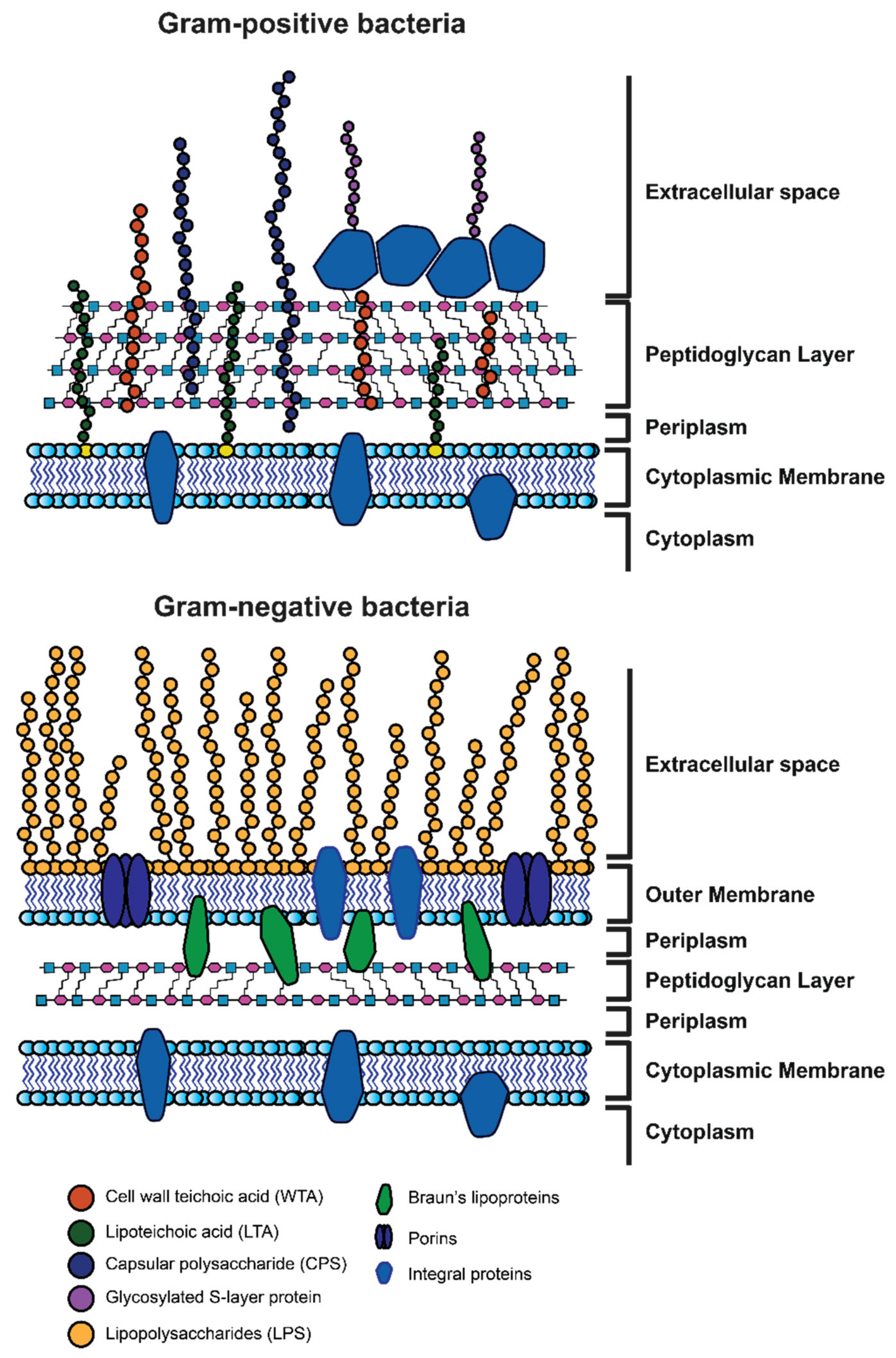
1.1.1. Gram-Positive and Gram-Negative Bacteria: General Remarks

Bacteria are microscopic single-celled organisms that are listed among the oldest known life forms on Earth. They can be found in the soil, ocean water, ice, and underneath the earth’s crust; that is because these microorganisms are often essential to other organisms’ life [1,2,3]. The worldwide bacterial population is estimated to reach 2 × 1030 cells, with a major role in the ecosystem [4]. Moreover, nitrogen-fixing bacteria are essential for the natural growth of several plant species [5,6]. The bacterial fermentation process is also fundamental for the digestive systems of sheep and cattle. In the food industry area, bacteria are involved in the manufacturing of many products, including dairy products, baked goods, and alcoholic beverages [7]. In industrial microbiology, microorganisms are grown on a large scale to produce compounds such as antibiotics, enzymes, and various chemicals, while in biotechnology, genetically-modified organisms are used to synthesize, for example, human proteins for medical use. Bacteria also live in the human and animal bodies, particularly on the skin, airways, oral cavity, and the digestive, reproductive, and urinary systems, normally without causing any harm [8]. They populate the resident flora, or “microbiota”, are essential to facilitate food digestion or prevent the proliferation of other more dangerous bacteria, and acting as saprophytes [9,10,11]. Figure 1 depicts the representative genera of the constitutive bacterial flora in humans [12,13,14,15]. The intestinal microbiota plays a central role in the development of the immune system and the modulation of its function. This has led to investigations of the mechanisms that link autoimmune diseases or allergies to changes in the microbiome [16,17,18,19]. Currently, the connections between the human body and the microbiome are not limited to the gastrointestinal or immune system but concern almost all other systems. Alzheimer’s disease, multiple sclerosis, and autism have been studied for a putative role of microbiota in their development [20,21,22,23]. Based on the bacterium-host relationship, these microorganisms can be divided into four categories: symbionts, that live and multiply in contact with the host without causing damage and establishing a beneficial reciprocal relationship; diners that live and multiply in contact with the host without causing damage; pathogens that take advantage of the host causing diseases, from mild to serious ones; and opportunists, which normally are harmless but can cause disease, even serious ones if a weakening of the defense system of the organism occurs. Among them, pathogens represent a small percentage of the entire known bacterial population.
At the beginning of the 20th century, infectious diseases originating from bacterial and viral pathogens were the main cause of death. A decrease in incidence occurred through the years thanks to a greater understanding of the infectious process, the improvement of public health services, vaccination campaigns, and last but not least, the discovery of antibiotics, which made the managing of infectious diseases possible.

1.1.2. Morphology of Gram-Positive and Gram-Negative Bacteria

Bacteria are unicellular organisms that are formed by a prokaryotic cell, with dimensions ranging from 0.2 to 2 µm. The cell is surrounded by the plasma membrane, which regulates the transport of nutrients and waste substances. The plasma membrane is covered by a wall with different characteristics, leading to the well-known division into gram-positive and gram-negative bacteria. In the gram-negative cell wall, a lipid bilayer membrane, the outer membrane (OM) is present, while the above-mentioned plasma membrane is the inner membrane (IM). In the inter-membrane space, there is a thin layer of peptidoglycan (or murein), referred to as the periplasm, a complex polymer that is made up of amino sugars and amino acids that completely envelop the cell. The outer membrane acts as a barrier against the diffusion of many compounds, making the cell more resistant to toxic substances. Gram-positive bacteria do not have an outer membrane and their wall is mainly composed of a thick layer of peptidoglycan [24].
The different composition of the bacterial wall determines the different coloration that is taken up by the bacteria with the dye that was created by the Danish microbiologist Hans Christian Gram in 1884 and thus allows one to discern the two groups of bacteria. Gram stain is still used in normal clinical practice, as the first step in the screening of a biological sample to identify the pathogenic microorganisms present [25].
In addition to the above-described cell wall, some bacteria possess additional layers of coating (S layer, capsule, or glycocalyx) and surface appendages (pili, fimbriae, flagella). The capsule or mucous layer consists of polysaccharide or protein substances that are secreted on the cell surface by bacteria for several functions, including adhesion (e.g., on solid surfaces generating biofilms) and protection.

1.1.3. The Cytoplasmic Membrane

The cytoplasmic membrane is a thin barrier that surrounds the cell and separates the cytoplasm from the extracellular environment. The membrane is made of a phospholipid bilayer: phospholipids contain both hydrophobic (fatty acid) and hydrophilic (glycerol-phosphate) components. In the phospholipid membrane, the fatty acids point inward toward each other to form the hydrophobic environment, while the hydrophilic portion remains exposed to the external environment or the cytoplasm [26]. The membrane has a thickness of 6–8 nm and can be seen using an electron microscope: it appears as two dark-colored lines (glycerophosphate) that are separated by a lighter area (fatty acids). The structure is fluid and the proteins that are immersed in the phospholipid bilayer can undergo both rotational and lateral movement. The cytoplasmic membranes of some bacteria are strengthened by molecules called hopanoids, which are rigid planar molecules and structural analogs of sterols [27]. Many membrane proteins are firmly embedded in the membrane and are named integral membrane proteins [28]. Other proteins have one portion that is anchored in the membrane and extra-membrane regions that point into or out of the cell. The so-called peripheral membrane proteins remain firmly associated with the membrane surfaces [29]. The peripheral proteins interact with integral membrane proteins in important cellular processes such as energy metabolism and transport [29].
The main function of the cytoplasmic membrane is to prevent the passive leakage of solutes into or out of the cell; it acts as a gateway for the transport of nutrients into and wastes out of the cell (permeability barrier). The membrane is also an anchor for many proteins, glucides, lipids, and complexes of the same. Some are enzymes that catalyze bioenergetic reactions whereas other transport solutes into and out of the cell [26].
Salts, sugars, amino acids, nucleotides, and many other substances need to be transported by proteins that, besides ferrying substances across the membrane, accumulate solutes against the concentration gradient. One substance that can cross the membrane in both directions is water, but its movement is accelerated by dedicated transport proteins, called “aquaporins” [30].

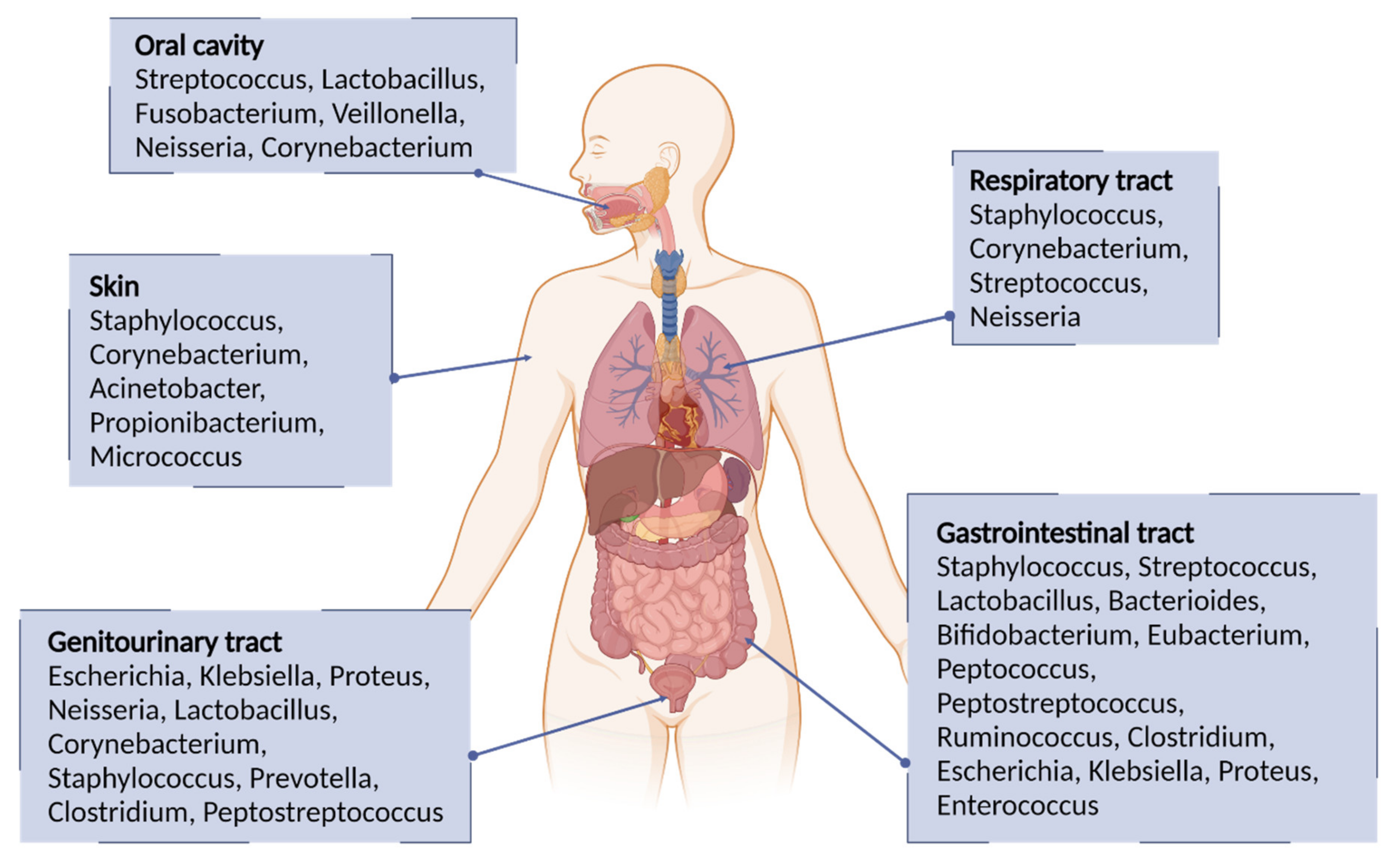
1.1.4. Pathogenic Bacteria

As previously mentioned, disease-causing bacteria are referred to as pathogens. However, in some cases, it is not possible to draw a clear distinction between pathogenic and non-pathogenic bacteria since the encounter of the human organism with the pathogen does not always generate disease. The severity and outcome depend on the complex relationship that is established between the microorganisms and the human organism. Under normal conditions, the thousands of bacteria that make up the microbiota are harmless to our health; indeed, they assist and help maintain it. The immune defenses of our body, therefore, help us to avoid the invasion of microbiota microorganisms, but when the body is compromised for various reasons, invasion can occur, and serious illness appears. Hence, the same beneficial bacteria can become pathogenic. Table 1 shows the different human pathogenic bacteria based on morphology and gram stain [24].

1.2. Bacterial Cell Wall Polysaccharides (CWPs) and Peptidoglycans (PGs): Structure and Functions

From a structural point of view, the bacterial cell wall can be classified into two different types, depending on gram-positive or gram-negative bacteria, as depicted in Figure 2. The main difference between the two cell walls is the absence of a second bilayer membrane in the gram-positive bacteria, which is compensated by a thicker peptidoglycan (PG) layer. The bilayer membrane in gram-negative bacteria is highly asymmetric; the inner leaflet is composed of phospholipids while the outer leaflet mainly contains lipopolysaccharides (LPS) whose polysaccharide chains occupy the extracellular space (Figure 2). [31,32] The PG layer in gram-positive bacteria is a fundamental component of the cell wall placed outside the cytoplasmic membrane of almost every bacterium [33,34,35], with very rare exceptions (e.g., Mycoplasmas, Planctomyces, Orientia (Rickettsia) tsutsugamushi) [36,37]. Its primary role is to maintain a sustained turgor of the cell wall, preserving the integrity and counteracting the osmotic pressure of the cell [38,39], but it also represents the anchor point for proteins and cell wall polysaccharides (e.g., teichoic acids) [40,41]. Together with the assembly of bacterial peptidoglycan hydrolases, the PG is also deeply involved in the process of cell division and cell growth [42]. Given its almost ubiquitous nature, higher eukaryotes evolved several PG recognition molecules (e.g., CD13, Nod1-2, lysozymes, and amidases) representing the first defense line against bacterial infections [38,43,44]. Moreover, PG represents a robust target for almost all effective antibiotics inhibiting bacterial cell wall synthesis [39]. The presence of such a layer is of fundamental importance for the integrity of the cell. Indeed, any degradation (e.g., by lysozymes) during cell growth or inhibition of its biosynthetic path leads irremediably to cell lysis. In addition to morphological functions, the cell wall undergoes recycling where it is constantly broken down, turned over, and remodeled [45,46,47].
PG is of major importance in gram-positive bacteria given its higher abundance which accounts for almost half of the cell wall mass, while in gram-negative bacteria, the PG layer is rather thin and not directly exposed to the extracellular space due to the presence of the outer membrane. Owing to its close to the omnipresent occurrence, PG has been one of the major antibiotic targets in recent years (e.g., penicillin, cephalosporins, carbapenems, vancomycin, teicoplanin) [48].

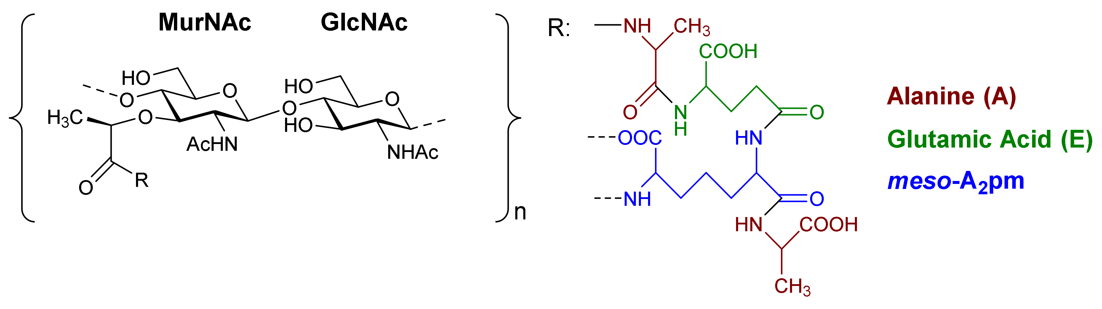
1.2.1. Chemical Structure and Variability of PG and Correlated Functions

The basic scaffold of the PG layer, as depicted in Figure 3, consists of polymeric glycan strands of two alternating β-(1→4)-linked sugars: N-acetylmuramic acid (MurNAc) and N-acetylglucosamine (GlcNAc). The different strands are arranged parallel to each other and covalently connected via peptide linkers that are substituted at the d-lactyl group of each MurNAc residue. The most common peptide linker is composed of l-Ala-γ-d-Glu-meso-A2pm (or l-Lys)-d-Ala-d-Ala. The last d-alanine residue is lost in the mature macromolecule and the previous d-Ala condenses with the corresponding d-Ala residue of another strand’s linker, thus creating the distinctive net of the PG layer.
Variations from the general structure of PG have largely been divided into three categories, viz., of: (i) the glycan strand structure and substitution pattern, (ii) the lipid linker structure and condensation point, and (iii) the glycosylation point by extracellular glycans. The most commonly reported modification in the first case is N-deacetylation and O-acetylation patterns at both GlcNAc and MurNAc, N-glycolylation of MurNAc residues, the modification of MurNAc to muramic δ-lactam, or formation of 1,6-anhydro MurNAc. The extent of these substitutions is species-dependent and has been extensively reported in previous literature works [49]. Table 2 shows the most common modifications of the PG repeating unit that have been discovered so far.
N-deacetylated sugars in the PG layer of Bacillus strains have been reported extensively for both residues [50,51,52], and more recently also for Streptococcal bacteria (e.g., S. pneumoniae, S. pyogenes, S. iniae) [53,54,55]. Deacetylation most likely occurs on the mature peptidoglycan since deacetylated precursors have not yet been reported in species containing PG deacetylases, and the latter enzymes have precise extra-cytoplasmic localization [54].
It is well known that N-deacetylation plays a key role in the protection of certain bacteria from host antimicrobial proteins such as lysozymes, which catalyzes the breakage of the β-(1→4)-linkage between GlcNAc and MurNAc leading to hydrolyzed PG layers and bacterial lysis [56]. This was shown to be related to the increased positive charge of the PG layer which decreased the efficiency of the secondary (non-enzymatic) mechanism of action of lysozymes [57], where these proteins cross the cell wall and bind to negatively charged bacterial membranes leading to its permeabilization. The lack of N-acetyl groups has been shown to provide a poor substrate for lysozyme binding, while upon chemical N-acetylation of the substrate lysozyme activity seems to be restored [54,58,59,60].
N-glycolylated muramic acids in PG, first described in Mycobacterium smegmatis [61], are used in bacterial taxonomy to classify Actinomycetales, a class of bacteria from which important antibiotics are isolated (e.g., streptomycin, actinomycin, streptothricin) [62]. Such a modification is widely present in the Mycolata taxon, a group of bacteria containing mycolic acids in their cell walls (e.g., Mycobacterium, Rhodococcus, Tsukamurella, Gordonia, Nocardia, Skermania, Dietzia) [63,64,65]. The modification is introduced during the synthesis of the UDP-precursors and generally is present only on a fraction of the muramic acid residues. Therefore, bacteria containing N-glycolylated muramic acids also contain MurNAc residues in the PG layer. The role of the N-glycoloyl group as a substituent has been the object of speculation but is still to be clarified. It has been proposed that the extra hydroxyl would engage in hydrogen bonding within the cell envelope with stabilizing effects [66].
Intramolecular amide bond formation between the amino group and the carbonyl of the lactyl group in MurNAc generates the muramic acid δ-lactam modification which appears to be quite abundant in Bacillus species and Clostridium sporogenes spores. For this modification to occur, the lactyl group must not have any peptide attached and the MurNAc residue must be N-deacetylated. This process is generally handled by just two enzymes. In B. subtilis for instance, an amidase CwlD removes the peptide that is linked to MurNAc and the peptidoglycan MurNAc N-deacetylase PdaA cleaves the acetyl group of the sugar residue [67]. Their role seems to be related to the spores’ ability to complete the germination process to produce viable cells. The muramic acid δ-lactam modification is employed by germination-specific hydrolases as a marker to recognize the spore’s PG [68,69,70].
The O-acetylation in PG appears to be uniquely present at the MurNAc residues [71,72]. First reported in Micrococcus luteus and S. faecalis, it is present in both gram-positive and gram-negative bacteria (e.g., B. cereus, Staphylococcus aureus, Enterococcus hirae, and S. pneumoniae for the gram-positive; Neisseria gonorrhoeae, Neisseria meningitidis, H. pylori, and Proteus mirabilis for the gram-negative ones). As a structural variation, O-acetylation is more prevalent than N-deacetylation and the extent of it varies between <20% and 70% depending on the species and strain that is being considered [73,74,75]. The source of acetyl groups for the O-acetylation has been a matter of discussion over the years. It has been proposed to be extracted from the N-2 position of GlcNAc or MurNAc [76]. But there are cases where acetylation in MurNAc6Ac is present in fully N-acetylated PG (e.g., P. mirabilis); alternatively, it was suggested to be obtained from peptidoglycan turnover products [73]. More recent theories have proposed acetyl-coenzyme A (CoA) or acetyl phosphate to be the source [77].
O-acetylation of PG has an important role in the ability of bacteria to produce disease. The first experimental evidence suggested this substitution to be responsible for the increased resistance towards hen egg-white lysozyme hydrolytic activity [72]. Further shreds of evidence showed the same resistance to egg white lysozyme on 14 strains of P. mirabilis containing acetylated PG, and further species lacking O-acetylation patterns were shown to be more sensitive to exogenous lysozymes [42,77,78]. Therefore, O-acetylation is a contributing factor offering resistance against antimicrobial proteins. Interestingly, the degree of O-acetylation seems to be negatively affected by β-lactam antibiotics that target proteins that are related to transpeptidation reactions in the synthesis of peptidoglycans. O-acetylation of peptidoglycan appears to play a not-yet resolved role in the cross-linking reaction [79,80,81].

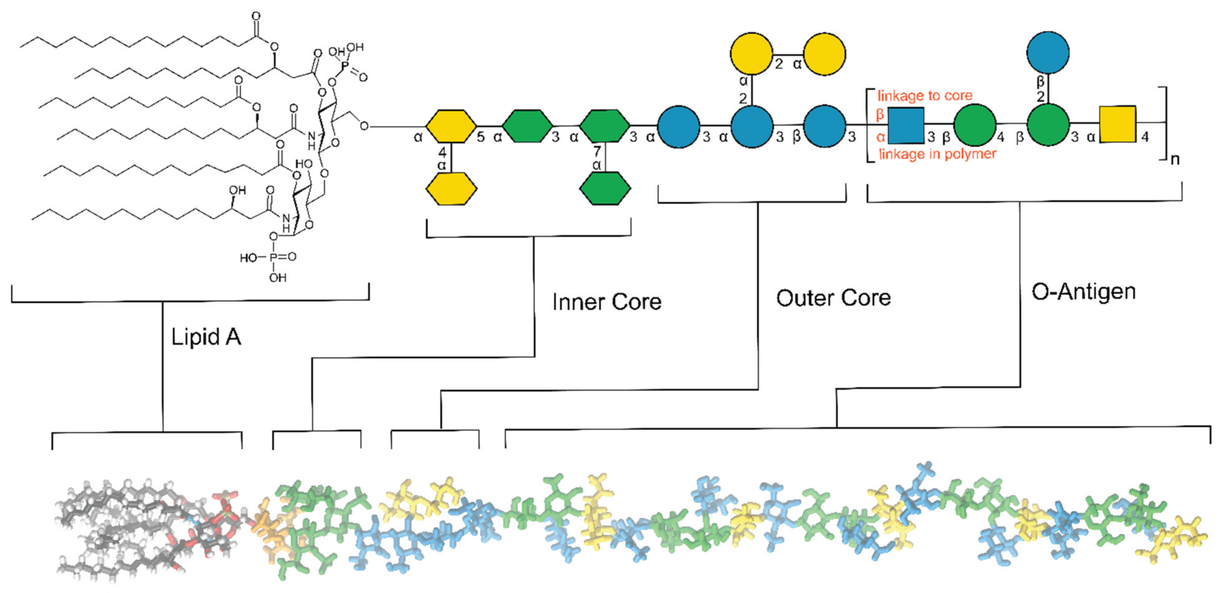
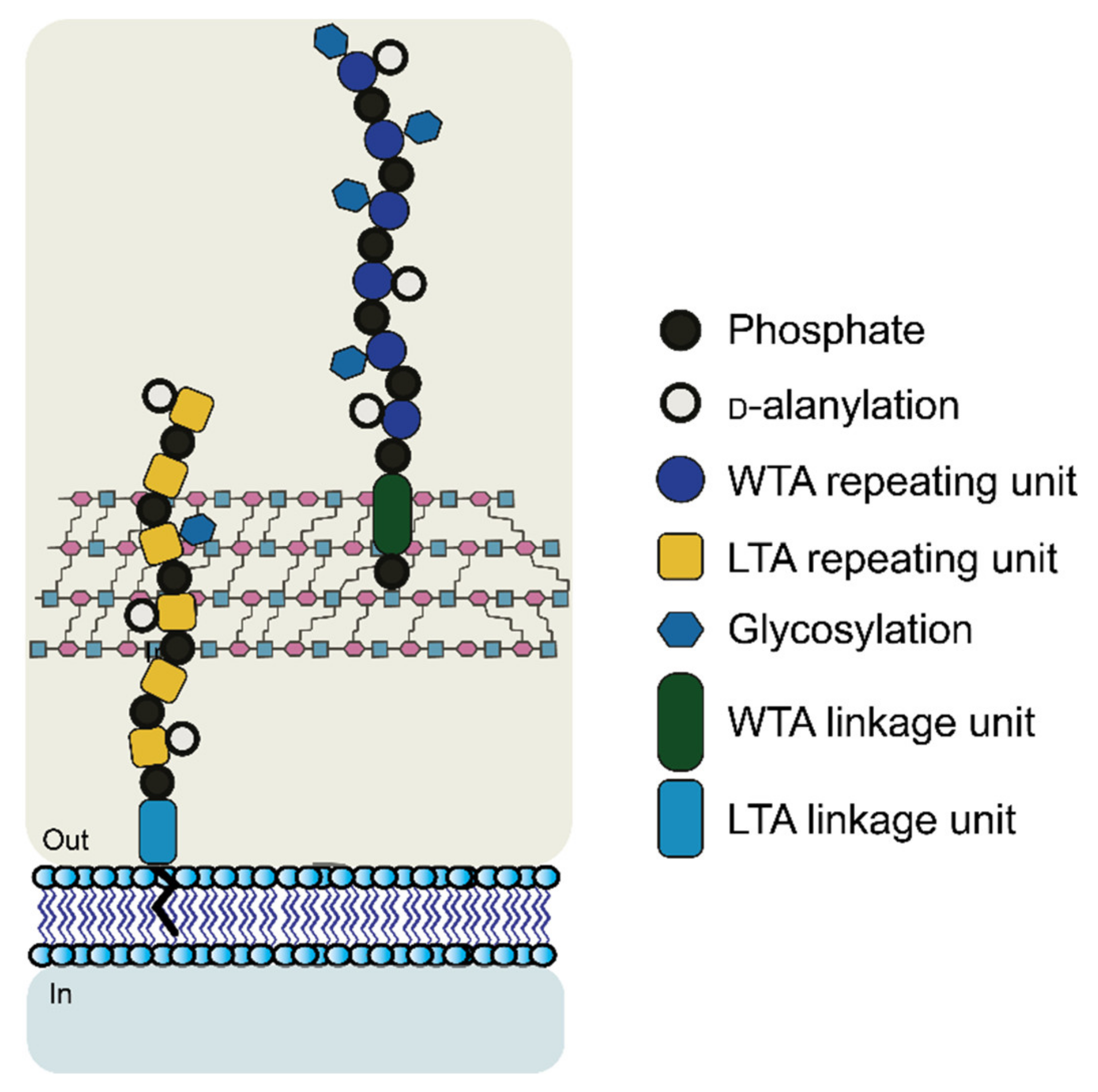
1.2.2. Cell Wall Polysaccharides: Structure and Functions

Gram-positive and gram-negative bacteria produce extracellular polysaccharides which can either be covalently bound to the cell envelope or surface proteins or be a slime that is weakly attached to the bacterial cell wall. Being present at the interface between microbes and mammalian hosts–other than representing recognition elements for pathogens–these glycoconjugates often contain antigenic moieties that trigger an immunogenic response in hosts. For this reason, these glycans and their biosynthetic pathways have experienced increased attention from the scientific community during the last decades.
As a result of Red Queen evolutionary dynamics [82], the structures of bacterial glycans present the highest variability compared to other non-bacterial classes of glycans and cannot, therefore, be easily generalized. As previously reported, the term “Red Queen” effect takes inspiration from Lewis Carroll’s quote “it takes all the running you can do, to keep in the same place” borrowed to explain the glycan structural variation that contribute to the bacterial diversity in nature. As above mentioned, bacteria have historically been divided based on the outcome of the gram staining procedure. In gram-negative bacteria, glycans are mostly present in the form of lipopolysaccharides (LPSs). A typical structure of an LPS polymer is represented in Figure 4.
LPS is generally divided into three sections: the lipid A, the core oligosaccharide (inner and outer core), and the O-antigen polysaccharide. The lipid A (or endotoxin) is the innermost and hydrophobic region of the lipopolysaccharide and possesses a conserved structure that is composed of two β-(1→6)-linked glucosamine residues often phosphorylated at the reducing end and non-reducing end residues in positions 1 and 4′, respectively. Acyl groups are condensed at the hydroxyl and amine groups in a variable number [85,86]. Due to this common architecture, most of the lipid A moieties are detected at picomolar levels by receptors that are present on immune system macrophages and endothelial animal cells [86], and a multitude of enzymes that are linked to the lipid A biosynthesis (e.g., LpxC) have over the years been validated as a target for newly developed antibiotics.
Discovered in the late 1800s and isolated from E. coli, it was confirmed that LPS was responsible for the pathogenic (endotoxic) properties, which correlated to pathological outcomes (e.g., fever and septic shock). Lipid A is part of pathogen-associated molecular patterns (PAMPs), i.e., molecules that can activate innate immune systems when they are recognized by toll-like receptors (TLRs) or other pattern recognition receptors (PRRs).
Roughly 106 lipid A residues are present in a single cell of Escherichia coli [87]. The fatty acids are somehow variable in structure and chain length but only one type of fatty acid is amide-linked, i.e., (R)-3-hydroxy fatty acids. The length of these fatty acids is 10–20 carbon atoms per chain and if the third carbon atom carries a hydroxyl group it is most often substituted by an additional fatty acid in at least one of the sugar residues [88,89,90,91].
Some species have been shown to contain a keto group in place of the hydroxyl in the N-acyl fatty acids [92,93]. Besides that, (R)-3-hydroxy fatty acids are predominant also for the ester-bound fatty acids, but these are still more variable and species-dependent. These are fatty acids from myristic and lauric acids and their hydroxylated derivatives, to even more unique structures such as cis-11-octadecenoic acid, 3-hydroxy-5-dodecenoic acid, and iso-2,3-dihydroxytetradecanoic acid [94].
The lipid A is covalently linked to heteropolysaccharides via a core region consisting of two parts, viz., first an inner core containing one to four units of Kdo (3-deoxy-d-manno-oct-2-ulosonic acid) [95], as well as l-glycero-d-manno-heptose residues, which are found in many inner cores. Additional charged elements such as phosphate and uronic acids may be present in the inner core structure providing a binding site for divalent cations (Mg2+ and Ca2+) with the effect of stabilizing the outer membrane [96].
The second portion of the core, referred to as the outer core is slightly more variable but structural modifications are still limited if compared to the O-antigen. This last complex repeating unit determines the serological and antigenic properties of the LPS and possesses a high structural variability. This diversity is reflected in the wide range of residues composing the repeating unit, which includes uronic acids, amino sugars, methylated and acetylated derivatives, deoxy sugars, but also non-sugar moieties such as amino acids and phosphate groups. The variability is further extended by the linkage diversity between the different residues in the branched and non-branched repeating units. The number of sugars in each repeating unit is typically four or five, although up to seven sugar residues have been reported for E. coli [97]. The total number of residues in a single chain can add up to several hundred, as for some bacterial species up to one hundred repeating units have been shown to be the outcome of the O-antigen biosynthesis. Hence, even a single bacterial species can account for hundreds of different O-antigen structures. E. coli is one of the most characterized species in terms of serotypes having–to date–197 reported O-antigens [97].
Over the years databases have been created to collect the different O-antigen structures of various bacterial species from a structural and genetic point of view [98,99,100]. Gram-positive bacteria contain a wide range of glycans on their cell wall. Similarly to glycans from gram-negative bacteria, these are important structural elements with significant patho/physiological relevance, other than representing key surface units for cellular recognition and signaling. Some of the most common and well-studied glycans from gram-positive bacteria are the cell wall teichoic acids (WTAs), which consist of copolymers of glycerol phosphate or ribitol phosphate and carbohydrates that are linked together by phosphodiester bridges.
These polyanionic glycopolymers play critical roles in the cell with functions that include cell morphology and division, autolytic activity, antibiotic resistance, metal ion homeostasis, phage-mediated horizontal gene transfer, and protection of bacteria from host defense peptides and antimicrobial peptides [101,102]. For their importance, these structures and their biosynthetic pathways represent attractive targets for the design of antibiotics and vaccines.
The WTA polymers can account for up to 60% of the cell wall mass containing between 40 to 60 polyol repeats. Structurally, WTA can be divided into two parts: the main chain polymer and a linkage disaccharide that acts as a bridge between the main chain and the peptidoglycan (Figure 5) [101,103,104].
The disaccharide unit is highly conserved across species and its structure is β-d-ManNAc-(1→4)-α-d-GlcNAc-1-P, where P represents a phosphodiester group that is linked to the hydroxyl group at position 6 of N-acetyl muramic acid of the PG layer. The N-acetyl-β-d-mannosamine residue is substituted in position O4 with one to two glycerol 3-phosphate units [105,106].
The most common WTA repeating units contain ribitol 5-phosphate (RboP) or glycerol 3-phosphate (GroP), but there is great variability in the WTA monomer structure with more unusual moieties [107,108,109]. Additional structural diversity may arise from the presence of substituents that are attached to the hydroxyl groups of the polyol repeating units, e.g., d-alanine, monosaccharides such as Glc or GlcNAc, or oligosaccharides [110].
The presence of these substituents is important in bacterial defense against a host and antibiotics. The d-alanylation, for instance, has been proven to protect against host-defense mechanisms [111,112]. A reduction or complete absence of d-alanine residues was shown to be correlated with an increased susceptibility to phagocytes, neutrophils, lysostaphin, and lysozymes, together with glycopeptide antibiotics and cationic antimicrobial peptides [113,114,115,116,117]. These structural changes may thus be correlated to the overall change in the charge of the membrane [116]. While the specific removal of β-d-GlcNAc modification has been reported to increase sensitivity to β-lactam antibiotics [118].
The second most characterized cell-wall glycopolymer in gram-positive species are the lipoteichoic acids (LTAs), which, similar to WTA, are zwitterionic polymers, but with a simpler structure compared to WTA. LTA typically consists of a polyglycerolphosphate (PGP) chain that is linked to the bacterial membrane via a glycolipid anchor (see Figure 5) [119]. As in WTA, the backbone of LTA is modified with alanine or glycosyl residues. Their structures also contain an initial linkage unit which is mostly represented by a disaccharide that is linked to diacylglycerol (DAG). In S. aureus and B. subtilis, the disaccharide unit is β-d-Glc(1→6)-β-d-Glc.
LTA can coexist with WTA in the same bacteria and their functions are generally similar. In bacteria containing only LTA moieties, the lack of these may lead–in addition to the same roles that are observed for WTA–to morphological defects such as the increased size of the bacteria or temperature-sensitive growth phenotypes [120,121]. Besides their location in the membrane, where LTA are tethered to the membrane via van der Waals forces and WTA are covalently attached, the LTA generally do not exceed in length past the peptidoglycan membrane while WTA extends further out into the extracellular space.
While WTA and LTA structures are possibly the most common and well-known membrane glycans in gram-positive bacteria, the cell walls of many gram-positive bacteria of the genera Streptococcus, Enterococcus and Lactococcus lack these polyanionic structures and have evolved glycopolymers that are characterized by the presence of rhamnose. These rhamnose-containing CWPs (RhaCWPs) are of particular interest due to the total absence of l-rhamnose in humans, which makes their biosynthetic pathway an attractive therapeutic target. Indeed, l-rhamnose is very often essential for bacterial virulence or viability [122].
First reported by Rebecca Lancefield in the 1930s, these structures have historically been used in the serological classification of streptococci, which initially were used to define Group A–E streptococci and later expanded to more than 20 serogroups [123,124], Figure 6 [125].
Over the years, the classification became superfluous since it was found to be unable to differentiate between different species. It is still commonly accepted that a single streptococcal species can express different Group antigens (e.g., Streptococcus dysgalactiae subsp. equisimilis strains) [126]. Similarly, to WTA, the RhaCWP comprises up to 60% of the dry cell wall mass. They are mainly localized on the outermost surface of the cell wall, but they can also be found intercalated in the peptidoglycan net [127,128,129].
Streptococcal species seem to lack orthologues of the genes that are associated with the expression of WTA (e.g., TagB, TagD, TagF) [130], while Lactococcal and Enterococcal species were reported to contain orthologues for WTA and both WTA and LTA biosynthesis, respectively, thus having a more heterogeneous expression of glycans on their surface [131,132,133,134,135]. The main constituents of the RhaCWP are rhamnose with variable combinations of Glc, GlcNAc, Gal, GalNAc, and phosphate groups, that are differently linked [136]. Even though the structure of the polymers may vary significantly across species, some exceptions show structural similarities, such as GAC and GCC. These different motifs reflect the discriminatory ability of the Lancefield scheme that is based on the diverse structural elements that are present in the different groups. Some of the known structures for the Lancefield Groups A, B, C, and G are shown in Figure 6. Variations of these structures have been reported in different works and serve as homologs of WTA in terms of biological functions [137].
The first studies on the biological functions of RhaCWP indicated a strong relationship between these and the structural identity of the cell wall [138]. The lack of RhaCWP expression was shown to lead to overgrowth and cell division abnormalities [138,139,140,141]. Inhibition of the UDP-GlcNAc:lipid phosphate transferases in S. galactiae [140] and S. pyogenes [142] was shown to cause important morphological alterations such as a reduced level of cross-linked PG, increased chain length, and mislocalization of PG hydrolases.
RhaCWP also represents an important phage receptor in many species, as demonstrated in several studies, forming epitopes in conjunction with side-chains of the polymer (e.g., GlcNAc, Glc) [143,144,145]. Additional roles are associated with the impact on virulence of the bacteria, modulated by structural modification on the RhaCWP, still maintaining unaltered bacterial physiology [133,142,146] and cell wall morphology [142]. Furthermore, knowledge of the biosynthetic pathways of RhaCWP facilitates the identification of attractive targets for developing new antimicrobial agents with limited risk of side effects due to off-target actions, since humans completely lack l-rhamnose in their system [147,148,149].

2. Drugs Targeting CWPs and PGs

2.1. Antibiotics

Common therapeutic strategies focus on the direct inhibition of the biosynthesis of the cell wall polysaccharides, blocking bacterial growth and host infection. Some widespread and well-known drugs that are recognized as inhibitors of cell wall biosynthesis are β-lactam drugs (e.g., penicillin) and glycopeptides (e.g., vancomycin and teicoplanin). Other antibacterial compounds are known for a different mechanism of action, viz., by disrupting the membrane structures of bacteria, as carried out by, e.g., polymyxins and daptomycin [150,151,152].

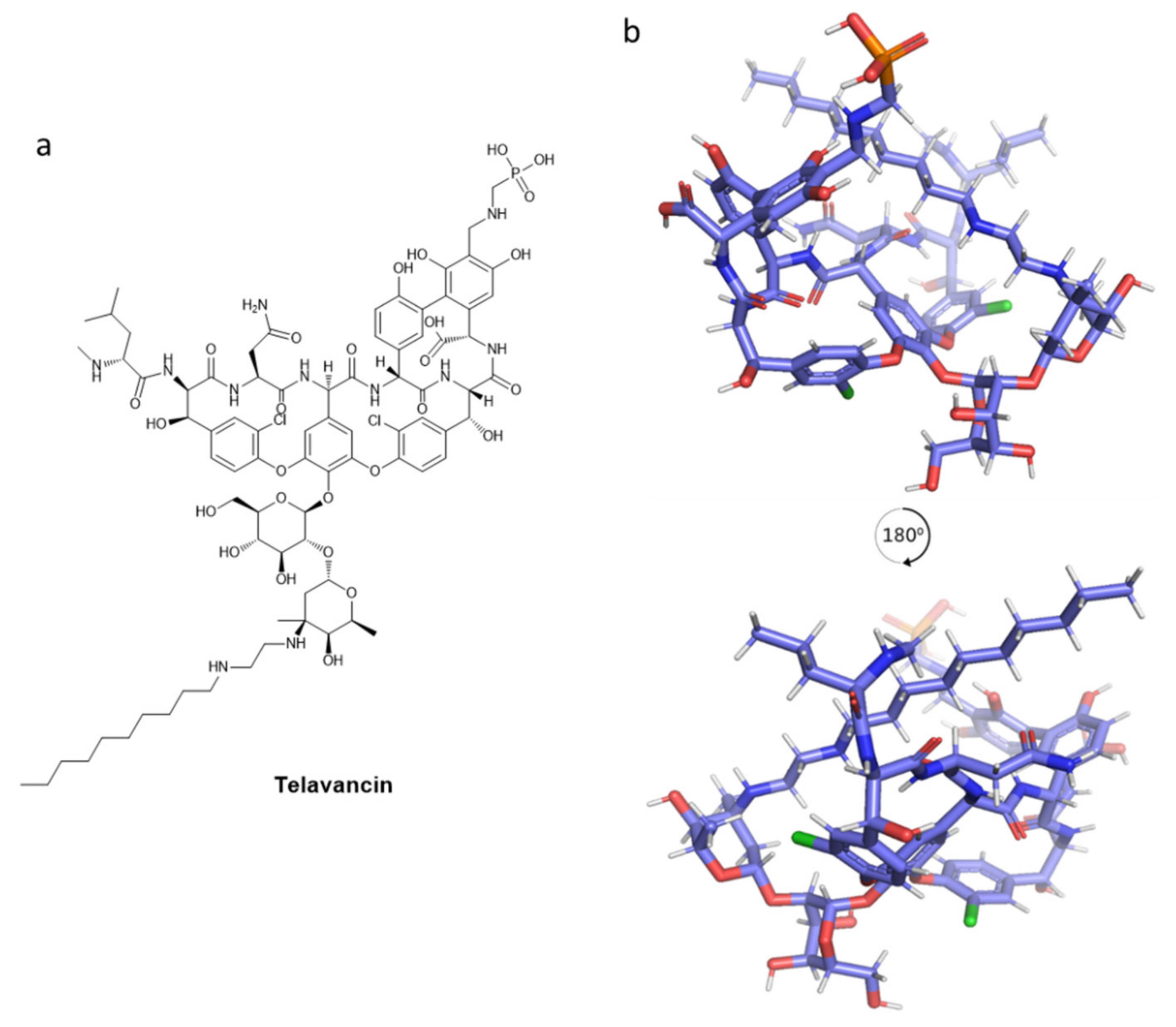
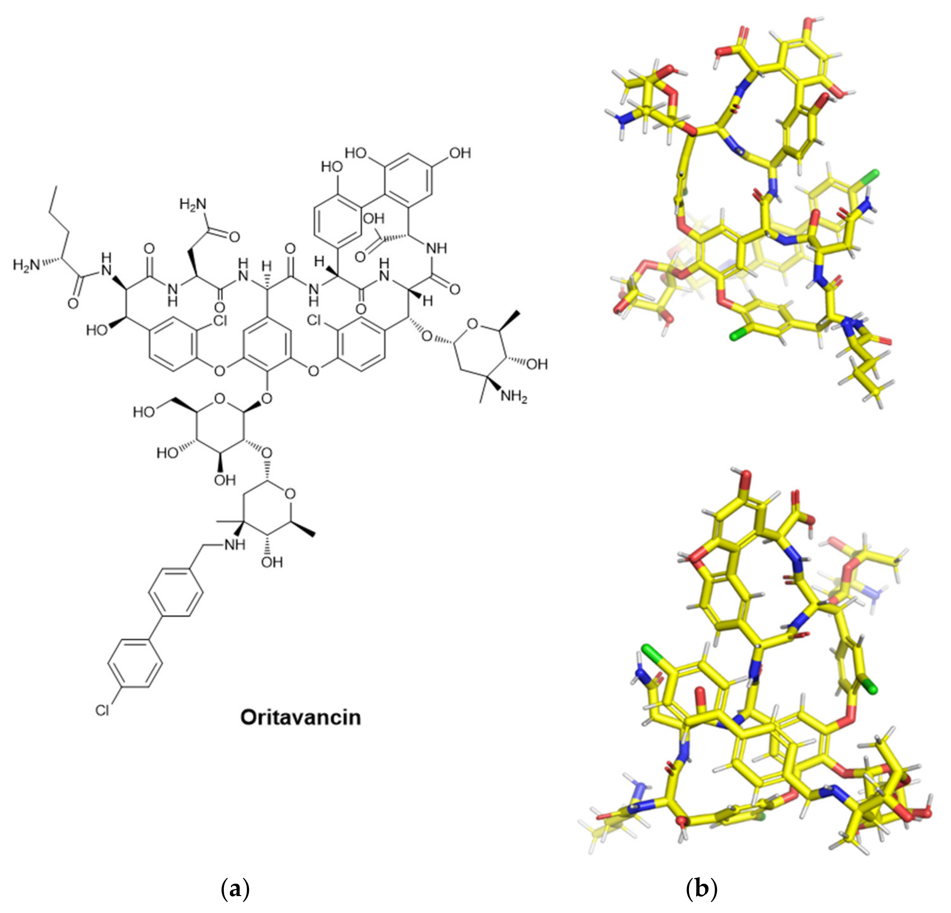
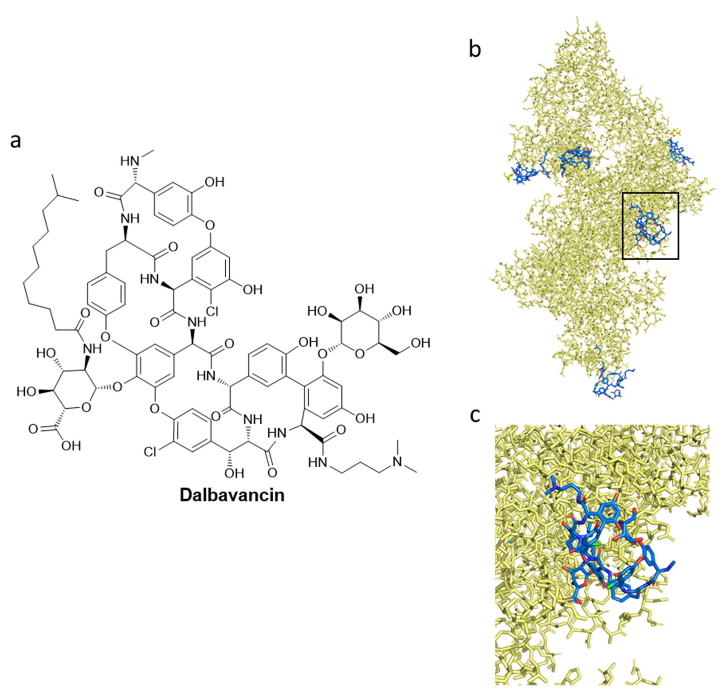
2.2. Carbohydrate-Based Antibiotics Targeting CWPs and PGs: Glycopeptides and Lipoglycopeptides

Glycopeptides and lipoglycopeptides are the representative antibiotics that are known to target the synthetic pathway of the bacterial cell wall [153]. Glycopeptide antibiotics are very important in the antibacterial treatment of gram-positive and gram-negative bacteria. In particular, lipoglycopeptides can dimerize, enhancing the binding affinity (hence the overall potency) for the peptidoglycan cell wall. The membrane anchoring is mainly achieved by the hydrophobic moieties [154].
Vancomycin, depicted in Figure 7, is a glycopeptide drug, whose name comes from the word “vanquish”. In the 1950s, it was found in Streptomyces orientalis (now Amycolaptosis) resident in the Borneo jungle [155]. It was demonstrated in 1958 to be clinically important in the treatment of gram-positive penicillin-resistant staphylococci. Since then, it has been the best treatment for hospitalized patients with severe infections [153]. Due to its potential toxicity and the rise of semisynthetic penicillin that is potent on staphylococci, the interest in vancomycin decreased. However, some drug-resistant gram-positive bacteria became a threat, such as methicillin-resistant Staphylococcus aureus (MRSA); hence, vancomycin returned as an antibiotic. In routine treatments, vancomycin has been indicated for important infections such as meningitis, endocarditis, osteomyelitis, bacteremia, and skin infections that are caused by gram-positive strains. In particular, it is crucial in cases of intolerance-allergy to β-lactam antibiotics, or in the case of MRSA [153].
In the 1980s, vancomycin-resistant strains of gram-positive bacteria spread, for example, vancomycin-resistant enterococci (VRE), even if vancomycin is recognized to have a broad spectrum of activity [156]. Vancomycin showed its potency against almost every strain of Staphylococcus aureus, including the methicillin-resistant ones. Leuconostoc spp., Listeria monocytogenes, and Lactobacillus spp. are the ones that are not sensitive to vancomycin efficacy [153]. The broad spectrum of vancomycin includes most S. epidermidis, all Streptococcus pneumoniae [157], and all Streptococcus pyogenes that have been tested, most of Streptococcus bovis and Streptococcus viridans, and Enterococcus (only a few resistant) strains. Diphtheroids, the few strains of Listeria monocytogenes, most strains of Clostridium species (but some strains of C. ramosum are slightly resistant), half of the strains of Actinomyces species are also vancomycin-sensitive. All strains of the gram-negative bacillus Flabacterium meningosepticum resulted in resistance [158].
Considering the mechanism of action of vancomycin, it has a different mode of inhibition of cell wall biosynthesis in comparison to β-lactams. It prevents the polymerization of the phosphodisaccharide-pentapeptide-lipid complex, in the second biosynthetic step consisting of cross-linking between the peptide chains [159]. Vancomycin strongly binds to peptides presenting a d-alanyl-d-alanine repeating entity at the free carboxyl end [160]. Vancomycin is a bulky drug; hence, it creates an extensive steric hindrance when it is bound to the peptide, not allowing for binding to the substrate site on the enzyme peptidoglycan synthetase. Thus, it blocks the peptidoglycan biosynthesis. As its secondary mode of action, vancomycin was also more recently shown to interfere with the cell membrane permeability [161,162].
Vancomycin (Figure 7) is still essential in the treatment of methicillin-resistant staphylococci infections, with the help of other important antibiotics: first- and second-generation cephalosporins, co-trimoxazole, clindamycin, or rifampicin. However, as already mentioned, it is relatively toxic, with a high incidence of side effects (e.g., red man syndrome). It is also not intramuscularly administrable, it is expensive, and not so active against enterococci [163].
Another important glycopeptide is teicoplanin. It was isolated from the actinomycete Actinoplanes teichomyceticus [164], with the original name of teichomycin A2. Teicoplanins refer to five structurally similar glycopeptide antibiotics, possessing a tetracyclic backbone, which is reminiscent of the tricyclic motif of vancomycin. It is a reliable alternative to vancomycin, with different advantages in comparison to the latter due to its clinical efficacy at lower dosages in severe staphylococcal infections [163]. Other advantages are the lower incidence of side effects, such as the already-described red man syndrome and the lower toxicity (in particular in co-administration with an aminoglycoside) [165]. However, teicoplanin has been commercialized in Europe and Asia, but not in the USA [153].
Many types of gram-positive bacteria are sensitive to teicoplanin, which has bacteriostatic and bactericidal activity). Some families are involved in in vitro sensitivity, such as staphylococci (even the β-lactamase-producing and methicillin-resistant strains; S. aureus, S. epidermidis, S. hominis, S. haemolyticus, S. saprophyticus), streptococci (S. pyogenes, S. pneumoniae, S. agalactiae, S. bovis, S. milleri, S. mitis, S. sanguis, etc.), enterococci, and several anaerobic ones (Clostridium difficile, Clostridium perfringens, Propionibacterium acnes, Listeria monocytogenes, and Corynebacterium jeikeium).
Teicoplanin, depicted in Figure 8, shares the same mechanism of action with vancomycin: saturation by aspecific binding to the outer layers of PG, then binding to the terminal amino acyl-d-alanyl-d-alanine in PG. It follows a blockade of PG elongation, inhibiting the biosynthetic step of transglycosylation and interfering with the function of transpeptidases [166,167,168]. This glycopeptide has an “open pocket” conformation; the “seam” is represented by the heptapeptide backbone [169]. The unit N-acyl-d-alanyl-d-alanine fits into the pocket [170]. In particular, teicoplanin inhibits the biosynthesis of the cell wall and cellular growth. Most likely, cell death is induced by cellular hydrolytic enzymes. The PG of gram-negative bacteria is sensitive to teicoplanin, but the outer lipid membrane repels this bulky and polar drug [166,170].
In a comparison of teicoplanin to vancomycin, the first is several times more potent against enterococci compared to the latter. The relationship with other antibiotics could be different; the co-administration with rifampicin is additive or sub-additive, and the mix with ampicillin antagonizes the β-lactam-based antibiotic. However, teicoplanin has a certified, but also combination-dependent, synergic effect on gram-positive cocci with other antibiotics, such as aminoglycosides, imipenem, and fosfomycin.
As above mentioned, vancomycin is crucial for antibacterial treatment, mainly for the worldwide spreading of MRSA [172,173]. Clinical failure is step by step increasing for MRSA treatment [174]; hence, it brought about the more effective semi-synthetic (lipo)glycopeptide antibiotics (with prolonged efficacy, thus less frequent dosage), such as telavancin, dalbavancin, and oritavancin [153,175]. A disadvantage is their accessibility as intravenous administration and their limited use because of their high cost. They mainly have a therapeutic indication for skin infections. The side effects are also a significant issue for these semi-synthetic lipopolysaccharide antibiotics: QT (the interval between the start of Q wave to the end of T wave measured on an electrocardiogram) prolongation and nephrotoxicity for telavancin and an increase of coagulation episodes for telavancin and oritavancin [153]. Telavancin (TD-6424, or Vibativ) is a vancomycin-derived lipoglycopeptide (for the decylaminoethyl substituent) which represents a valuable drug against the increased spreading of MRSA infections, and in general, infections that are caused by resistant gram-positive bacteria [175,176]. 2D and 3D structures of telavancin are presented in Figure 9. To potentiate the vancomycin antimicrobial activity, telavancin was developed in the context to improve the hydrophobicity at the disaccharide amine moiety. In this case, the hydrophobic portion is represented by a decylaminoethyl side chain on the vancosamine part and a phosphonomethyl aminomethyl functional group in position 4 on aminoacid 7 [177]. Telavancin offers different advantages, more related to specificity and pharmacodynamics properties. An increased affinity profile is offered by the glycopeptide moiety toward the d-Ala-d-Ala-containing peptidoglycan intermediates, and the negatively charged phosphonic acid moiety provides a more efficient urinary excretion [177,178]. The drug shows fewer problems after infusion and could be administered just once per day [176].
FDA-approved telavancin is used for complicated skin and soft tissue infections (cSSTIs), hospital-acquired bacterial pneumonia (HABP), and ventilator-associated bacterial pneumonia (VABP) that is caused by susceptible isolates of S. aureus, without current specific treatment. Different trials were conducted on telavancin for MRSA bacteremia. A randomized Phase II clinical trial (NCT00062647) confirmed that all the patients were cured [179]. It was also investigated for the treatment of Staphylococcus aureus bacteremia (SAB) [180] in a Phase III clinical trial, randomized, open-label, non-inferiority trial (NCT02208063), which failed due to insufficient statistical data [181,182]. More specific trials are needed to assess further the efficacy of telavancin, investigating the patients with complicated bacteremia more extensively [182]. Its mechanism of action, recognized to have dual activity, is mainly due to the interaction between the carboxylate binding pocket with terminal d-Ala-d-Ala residues and the decylaminoethyl side chain with the cell membrane [178,182]. Focusing on the d-Ala-d-Ala terminus, telavancin inhibits the peptidoglycan biosynthesis binding to N-acetyl-glucosamine-N-muramylpentapeptide of the peptidoglycan. The consequences of telavancin are the inhibition of different biosynthetic steps, such as the transglycosylation (PG polymerization) and the subsequent transpeptidation (generation of cross-links). In MRSA cells, telavancin is 10-fold more active than vancomycin in the inhibition of peptidoglycan synthesis [178]. The second mechanism of action is achieved by the decylaminoethyl side chain, which targets the cell membrane. This affects the cell membrane potential, through rapid concentration-dependent dissipation. The consequence is the generation of membrane pores [178], which leads to the extracellular leakage of adenosine triphosphate (ATP) and K+ ions. This second activity is specific for bacterial membranes, not for mammalian biomembranes.
Clinical and surveillance studies involving the antibiotic spectrum and activity of telavancin have been described [183,184,185]. In general, telavancin showed remarkable in vitro activity against skin and skin structure infections and pneumonia that is provoked by gram-positive bacteria (pneumonia could not be treated with daptomycin because it does not target lung tissue) [176,183]. Telavancin is particularly active against bacteria that are resistant to methicillin, daptomycin, and linezolid. It is also indicated for the treatment of vancomycin-intermediate S. aureus (VISA) and heterogeneous VISA, Clostridium species, including Clostridium perfringens and Clostridium difficile, Peptococcus anaerobius, and Bacillus anthracis. A synergic activity was demonstrated for the co-administration of telavancin with gentamycin and rifampicin [185,186,187]. To visualize telavancin, a structure representation was generated through energy minimization via Chem3D and then Avogadro (algorithm Steepest descent, force field MMFF94s), resulting in potential energy minimized 3D structure (Figure 9).
Oritavancin (previously LY333328, then KIMYRSA™, or ORBACTIV®) is a “long-acting” intravenously administered lipopolyglycopeptide that is approved by FDA for adult patients with acute bacterial skin and skin structure infections (ABSSSIs) that are caused by gram-positive bacteria. It is known for its antibiotic activity, efficacy, and safety. Other minor, but important advantages are a simpler preparation, and shorter infusion time (1 h) in a lower volume (250 mL) [188]. The initial novelty of oritavancin was its activity against vancomycin-resistant Enterococcus faecalis [189]. With its different hydrophobic and hydrophilic groups, it differs from vancomycin having an additional unsubstituted saccharide moiety and an aromatic lipophilic side chain [189]. It has a higher potency compared to vancomycin [190].
As for the mechanism of action, it acts by killing rapidly growing bacteria in a concentration-dependent way (unlike vancomycin) [154]. The inhibition of the peptidoglycan synthesis is achieved via different pathways: transglycosylation blockade, transpeptidation (cross-linking) inhibition, and membrane potential perturbation (depolarization and increased permeability) [189]. As for the spectrum of activity of oritavancin, it acts against gram-positive bacteria, remarkably against nosocomial infections that are caused by vancomycin-resistant enterococci (VRE) [191,192,193,194].
Other notable incidences were found against Micrococcus species, Listeria monocytogenes, and Corynebacterium species, but also anaerobic gram-positive bacteria such as Clostridium difficile, Clostridium perfringens, Peptostreptococcus species, Peptococcus species, and Propionibacterium acnes [191]. Oritavancin has a well-known synergy with gentamicin against VRE, and with gentamicin, linezolid, moxifloxacin, and rifampin [176,195]. There is no crystal structure involving oritavancin in the Protein Data Bank (PDB) [196]; hence, energy minimization via Chem3D and then Avogadro was performed, and the resulting conformation is displayed in Figure 10.
The last semi-synthetic lipoglycopeptide that is presented is dalbavancin (Figure 11), which has a therapeutic indication for ABSSSI that is caused by gram-positive bacteria. In 2005, a Phase II clinical trial study demonstrated that dalbavancin is beneficial as an alternative to vancomycin for catheter-related bloodstream infections, even if more data are needed in support of this [197]. Dalbavancin has a good safety profile [192], but data are needed to evaluate its importance in MRSA bacteremia [182]. A Phase II clinical trial focused on the comparison between dalbavancin and the standard of care (SOC) therapy for the treatment of complicated bacteremia or right-sided endocarditis that is caused by S. aureus will be completed in August 2023 [182]. In Figure 11b,c, several structures of dalbavancin are presented as blue sticks, in complex with human serum albumin (HAS); their association with HAS is essential for clinical use [198].

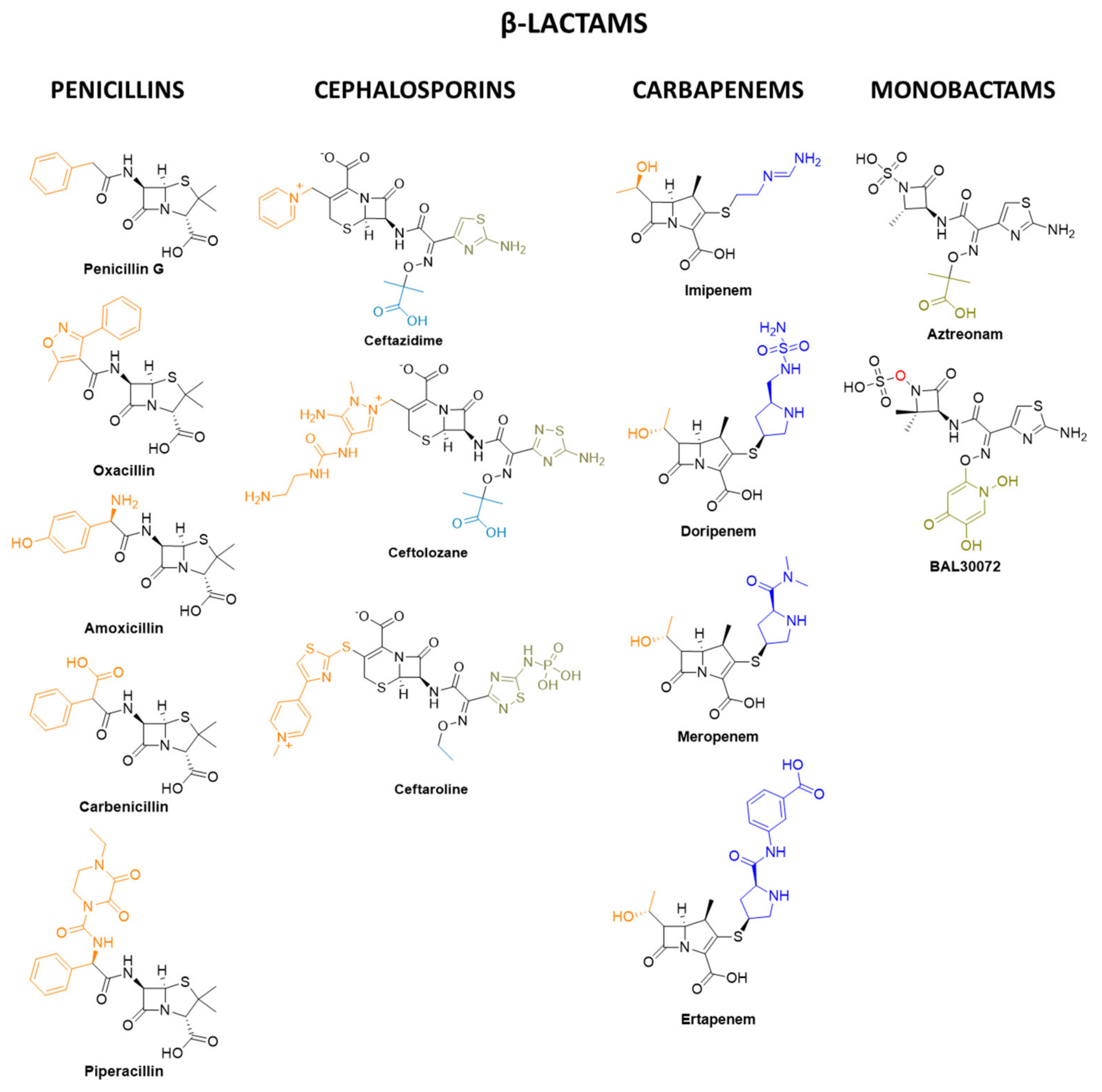
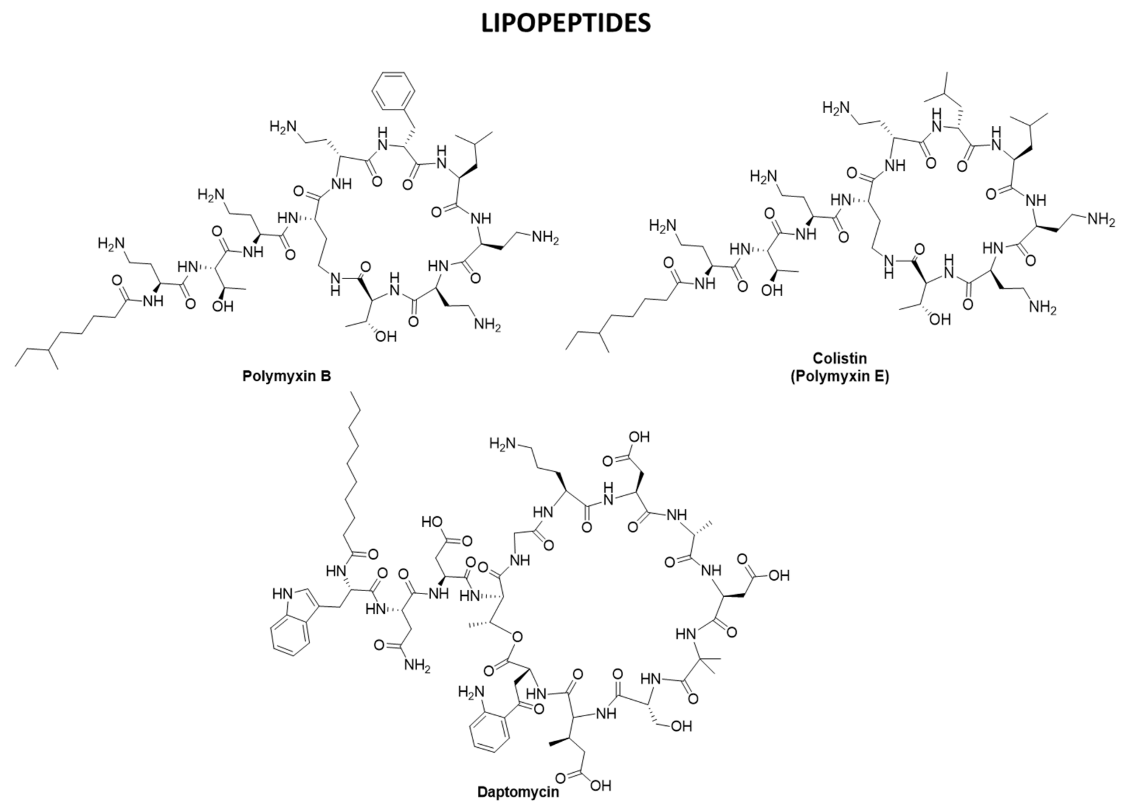
2.3. Antibiotics Targeting CWPs and PGs: β-lactams, Polymyxins, and Daptomycin

Due to the complexity of bacterial diseases (especially MDR ones), the treatment is carried out by combination therapies to gain activity through several mechanisms of action [199]. Penicillin, the “wonder drug”, is the oldest pure antibiotic worldwide that is available [200]. Penicillins (depicted in Figure 12) possess a β-lactam ring as the main chemical scaffold of the molecule.
The peptidoglycan (PG) layer generates cross-links at its C-terminus thanks to specific enzymes, transpeptidases (TPDs) [201]. The cross-linked PG polymer is essential for maintaining the membrane shape and prevents cellular osmotic rupture [150,151,201]. TPDs are PBP (penicillin-binding protein) enzymes because they can be inhibited by β-lactams such as penicillins [201]. β-lactams bind and inhibit PBP enzymes ending in bacteriolysis, hence cell death [150,151,201]. Penicillin-derived compounds are divided into natural (e.g., penicillin G), penicillinase-resistant (e.g., oxacillin), amino-, (e.g., amoxicillin), carboxy- (e.g., carbenicillin), and ureido- (e.g., piperacillin) penicillins. Natural penicillins are mostly active against gram-positive bacteria (streptococci, enterococci, and some non-β-lactamases-producing staphylococci), while synthetic ones (e.g., aminopenicillins) can be active also against some gram-negative bacteria such as non-resistant H. influenza, N. gonorrhoeae, and E. coli. Even if penicillin resistance is an increasing problem, this old class of antibiotics continues to lead to antibacterial therapy [202].
Moving forward into the family of β-lactam compounds, cephalosporins are important due to their activity against gram-negative bacilli. There are five generations of cephalosporins. Examples of cephalosporin-based therapies are available against most Enterobacteriaceae (third-generation ceftazidime + avibactam, their combined effect resulting in β-lactamase inhibition), resistant gram-negative bacilli such as P. aeruginosa (ceftolozane + tazobactam, another combination used for β-lactamase inhibition), and methicillin-resistant S. aureus (MRSA, with fifth generation ceftaroline as the only available treatment) [203].
Imipenem, doripenem, meropenem, and ertapenem (reported in Figure 12) are some of the drugs composing the well-known class of the carbapenems [203]. They derive from the naturally occurring antibiotic thienamycin, extracted by the soil microorganism Streptomyces cattleya [204,205]. They have the broadest spectrum activity compared to other β-lactam classes and are particularly important in case of severe sepsis that is caused by antibiotic-resistant bacteria. In general, they are active against both gram-positive (except methicillin-resistant S. aureus, Enterococcus faecium, and Enterococcus faecalis) and gram-negative bacteria as well as anaerobic species. [206]. The beneficial effect of carbapenems is reflected also in the significant post-antibiotic effect against gram-negative bacteria, such as P. aeruginosa [207,208]. Severe problems in using carbapenems for the treatment of infections are represented by the spread of carbapenemase-producing gram-negative bacilli [203].
Monobactams were developed in concomitance to the decrease of the efficacy of β-lactam antibiotics, mainly caused by extended-spectrum β-lactamases (ESBLs). These β-lactam-based drugs are structurally monocyclic and which makes them hydrolysis-stable compared to other β-lactam and are, therefore, interesting for new therapies [209,210]. Aztreonam (AZN, Figure 12) is currently the only available representative of this antibiotic family. It is only active against aerobic gram-negative bacilli [203]. Unfortunately, it is nephrotoxic and weakly immunogenic and is also very expensive. As with other β-lactams, aztreonam binds some transpeptidases and PBPs (only gram-negative ones), leading to cell lysis [211]. The potent activity against many gram-negative strains (also MDR) was demonstrated by BAL30072 (Figure 12), a derivative that entered Phase I clinical trials. However, BAL30072 has not progressed after Phase I in clinical trials in 2014 because of serious side effects, such as high alanine transaminase (ALT) levels in blood [212,213].
The class of lipopeptides is a broad-spectrum antibacterial-based family that is crucial to facing menaces of the highly-spreading MRSA [214] and the expansion of vancomycin-resistance MRSA infections [215,216,217,218,219,220,221].
Polymyxins are drugs of last resort but are still effective on dangerous bacteria such as carbapenem-resistant Enterobacteriaceae, MDR Pseudomonas aeruginosa, and Acinetobacter baumannii [216], also fosfomycin, ceftazidime/avibactam and meropenem–vaborbactam are an efficacious treatment for the above-mentioned bacteria [222,223]. They are known to disrupt the outer membrane of gram-negative bacteria [224], causing some antibiotic resistance problems [217]. Polymyxins [218,225] are non-ribosomal cyclic lipopeptides being secondary metabolites of a bacterium, Paenibacillus polymyxa; in particular, colistin (polymyxin E) [226,227], derives from Bacillus polymyxa var. colistinus. Structurally, they are decapeptides, consisting of a cyclic heptapeptide that is linked to a linear tripeptide side-chain acylated at the N-terminus by a fatty acid tail [228]. As for their mechanism of action, the primary electrostatic interaction with lipid A disrupts the outer membrane, following hydrophobic insertion of the fatty acyl chain of polymyxin into the membrane, making the bacterium succumb [229]. Attempts to extend the ring structure, generated by the insertion of additional Dab residues, produced compounds that are significantly less effective than polymyxins [27].
Daptomycin (Cubicin®), derived from Streptomyces roseosporus [229,230] has an in vitro selectivity against many MDR and susceptible gram-positive bacteria [231], including methicillin-sensitive and methicillin-resistant S. aureus (MRSA), streptococci, and Enterococcus (both vancomycin-susceptible strains and VRE) [153]. Daptomycin is not effective against gram-negative bacteria. In Europe, it is administered to adults intravenously once per day for cSSTI and complicated skin and skin structure infections whereas in the US it is used for the treatment of S. aureus bacteremia with or without right-sided infective endocarditis [153]. Its unique mechanism of action is extracellular and engages an oligomerization that is calcium-dependent [224]. Then, it binds to and alters the outer membrane, with a subsequent cellular potassium efflux. The final consequence is the depolarization of the membrane [153]. Figure 13 reports the 2D structure of polymyxins B, E (colistin), and daptomycin.

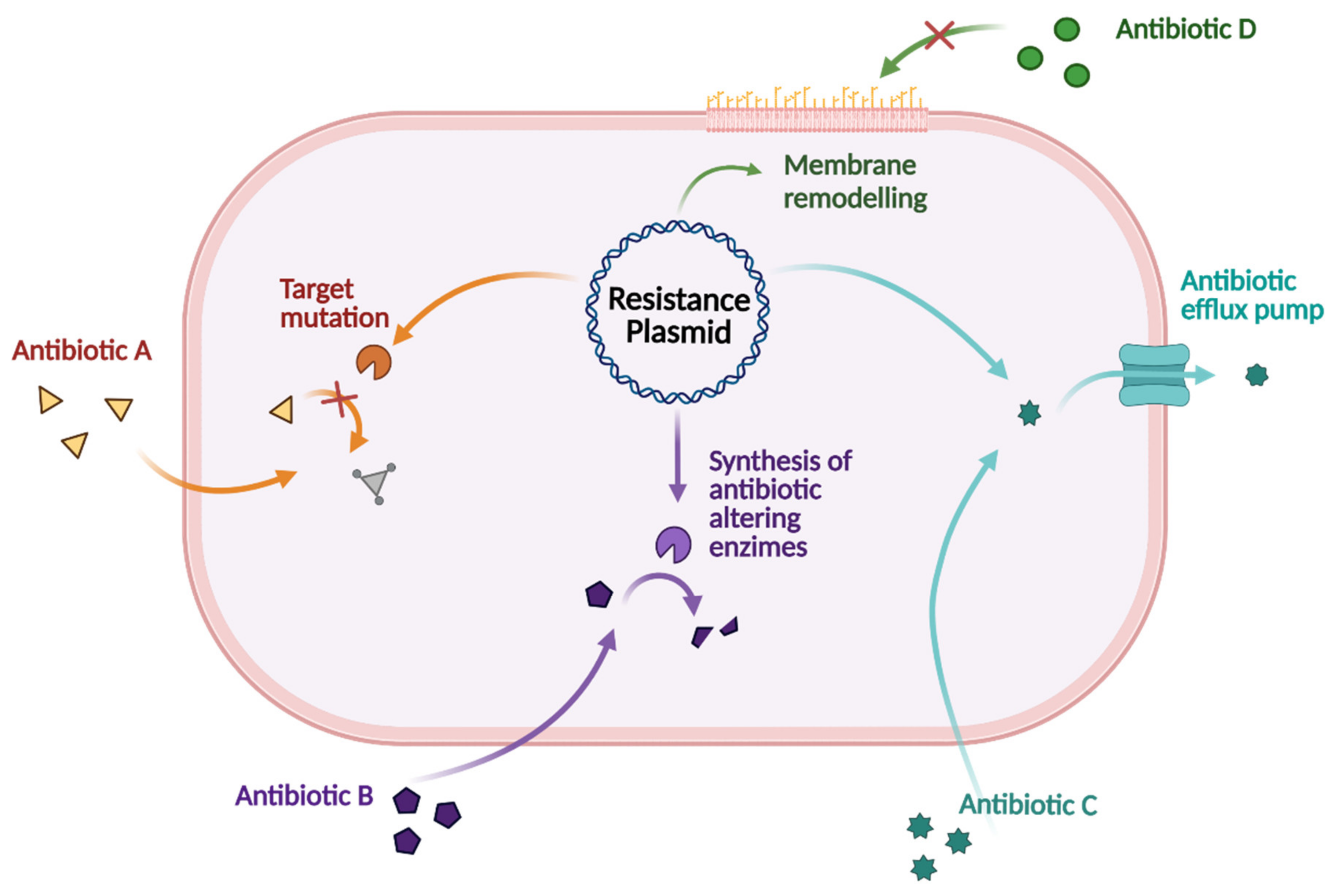
3. Antibiotic Drug Resistance

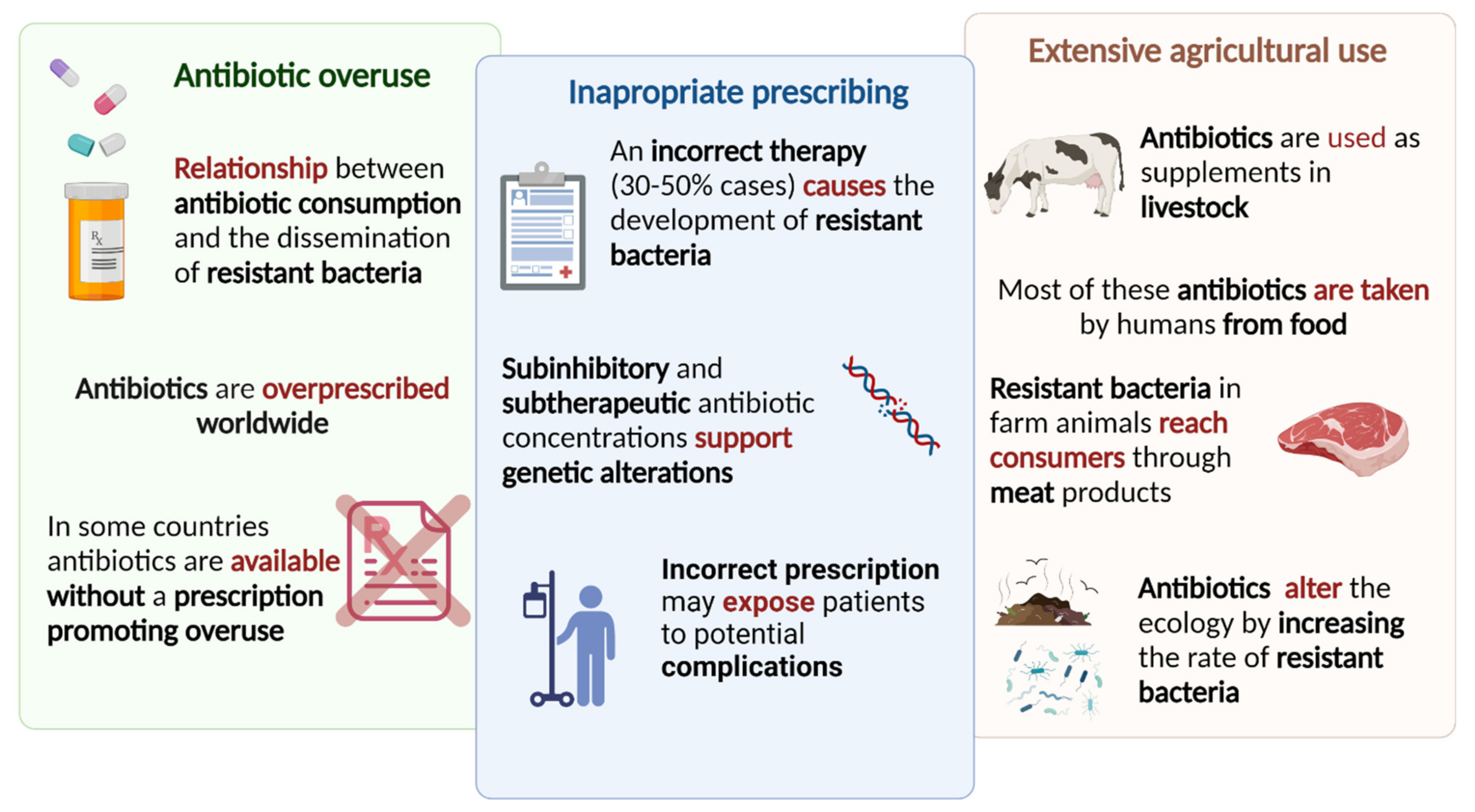
3.1. Consumption and Therapy

Since the discovery of penicillin in 1928, different classes of antibacterial drugs have been developed; unfortunately, bacteria mutate into resistant strains overcoming the mechanism of action of a specific drug. The increasing use of antibacterial agents made them gradually become less potent, also decreasing the therapeutic options that are available for patients that are affected by resistant strains. In the beginning, the problem was solved through the identification of new classes and the modification of existing antibiotics with limited cross-resistance to already in-use drugs. From the 1980s, the major scientific challenges in antibiotic discovery programs pushed large pharmaceutical companies to abandon this research area. Later, antibacterial resistance slowly became one of the main concerns in global clinics, pushing the scientific community, including some pharmaceutical companies, to re-invest money and efforts in the field. The main strategy that was pursued was to improve the potency of known antibiotic treatments, but new approaches to antibacterial drug discovery were also applied [232].
Recently, the World Health Organization (WHO) initiated a drug antibacterial drug development mapping activity and published a global clinical antibacterial pipeline report. The study highlighted that the clinical pipeline is dominated by derivatives of established classes and most development candidates display limited innovation, suggesting that the finding of new antibacterial agents without pre-existing cross-resistance should be prioritized in public funding strategies [233].
Regarding the preclinical phase, a review that was published in 2020 focusing on discovery and preclinical development projects has found, as of 1 May 2019, 407 antibacterial projects that were carried out from 314 different institutions, most of which are small and medium-sized enterprises (SMEs). The study evidenced that the preclinical pipeline contains 135 projects focusing on direct-acting small molecules belonging to innovative classes, directed towards new targets, or possessing new mechanisms of action, with high levels of diversity, and remarkable scientific concepts [234].
Together with the scarce availability of innovative drugs, the antibiotic resistance crisis has its roots in overuse; epidemiological evidence indicates a direct relationship between antibiotic consumption and the emergence and dissemination of resistant bacteria strains. Despite the warnings, antibiotics are overprescribed worldwide, and, in some countries, antibiotics are unregulated, cheap, and available without a prescription, promoting their overuse. Inappropriate prescribing play a major role in antibiotic resistance and it has been estimated that antibiotic therapy (treatment indication, choice of agent, or duration of the treatment) is incorrect in 30% to 50% of the cases, strongly contributing to the development of resistant bacteria. For instance, sub-inhibitory and sub-therapeutic antibiotic concentrations support genetic alterations, such as changes in gene expression, horizontal gene transfer (HGT), and mutagenesis. Besides promoting the development of antibiotic resistance, the incorrect prescription may also expose patients to potential complications of antibiotic therapy. The extensive agricultural use of antibiotic agents as supplements in livestock to promote growth and to prevent infection were discovered to cause an ecosystem alteration that was related to the rising of resistant bacteria. Moreover, via the handling or consuming food, antibiotics reach consumers as well as resistant bacteria that developed in farms. A schematic representation of the main causes of the antibiotic resistance crisis is depicted in Figure 14. Most of these antibiotics are taken by humans from food; moreover, resistant bacteria in farm animals reach consumers through meat products. In addition, antibiotics that are used in livestock are excreted through urine and stool, and, together with sprayed antibiotics as pesticides, pollute the environment and alter the ecology by increasing the rate of resistant versus susceptible microorganisms [235].
An emerging tool, too long underestimated, that is useful to reduce the development of drug resistance is the prevention of bacterial infections and bacterial spread. Prevention can be pursued by developing new vaccines (vide infra Section 3.3) [236,237]. The prophylactic use of vaccines by reducing the diffusion of pathogens lowers antibiotic use and reduces multidrug resistance. This new role of vaccines in fighting drug resistance has been validate by several studies [238,239].

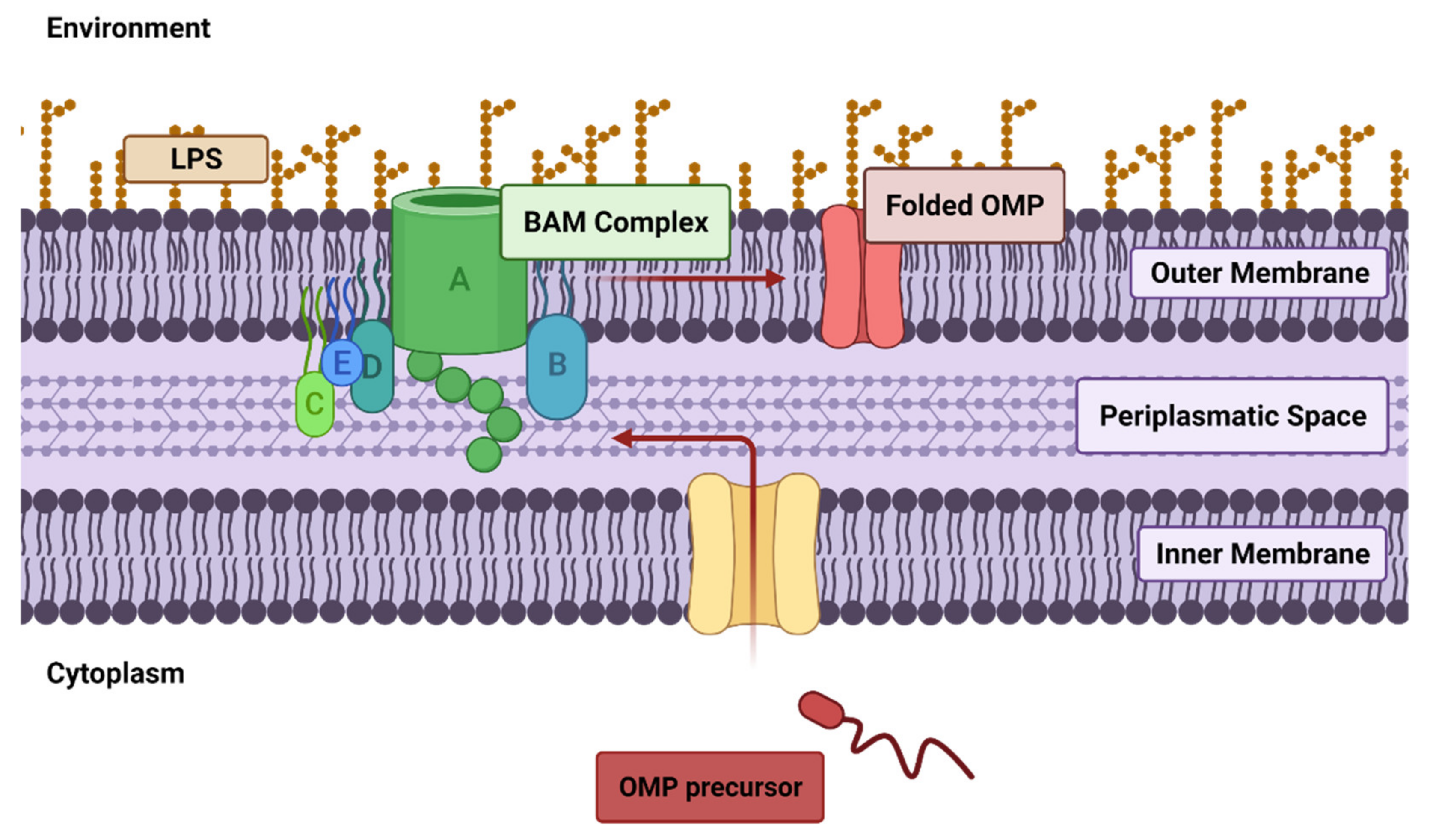
3.2. Drug Resistance Involving the Outer Membrane (OM)

Antibiotic resistance consists of a decreased sensitivity for a specific antibiotic, which allows the survival of the bacterium. Resistance to antibiotics is a natural phenomenon that has been noted since the introduction of penicillin in 1940 [240]. Very soon thereafter, the key question was raised, i.e., whether bacteria are inherently resistant to drugs or become resistant as a result of encountering the ‘antibiotic’ compounds. In search of an answer, it has been shown that bacteria can mutually transfer drug resistance to each other by passing genetic material between species. This process mainly occurs through the horizontal transfer of plasmids or bacteriophages, also known as horizontal gene transfer (HGT). Bacteriophages, i.e., viruses that are able to infect bacteria only, are also able to directly affect the bacterial genome. Moreover, the adaptation and evolution of microorganisms should also be taken into account in describing an integral view of the evolution of drug resistance [241].
There are four broad mechanisms that are indicated to lead to phenotypic resistance evolution in bacteria. A schematic representation of the bacterial mechanisms that lead to the evolution of phenotypic resistance is shown in Figure 15. The first one consists of the mutation of the target, resulting in the drug-binding inhibition. Multiple bacterial processes have been exploited as antimicrobial targets such as cell wall synthesis, nucleic acid synthesis, and metabolic pathways. All these targets could be modified by the bacteria to become resistant to the corresponding drug. A second option is related to the alteration of existing genes or the acquisition of new genes encoding drug efflux pumps. These efflux pumps extrude different classes of toxic agents, including a wide range of antibiotics, from the intracellular to the extracellular environment and their upregulation represents a crucial mechanism for acquired bacterial resistance. Also, the acquisition of new genes encoding enzymes that are able to modify or hydrolyze the specific drug could lead to a resistant phenotype. This process sequentially led to the acquisition of genes encoding for penicillinases, cephalosporinases, extended-spectrum β-lactamases, and carbapenemases. These enzymes are responsible for the decreased susceptibility of the related antibiotic class, the last of which has currently no solution. Lastly, the remodeling of the membrane could prevent drug influx at the cell surface, thus impeding access to the target that is located in the internal compartments [242,243,244].
Most antibiotics that are used in the 20th century bind specific targets that are exposed in the internal compartments of the bacterial cell, thus altering bacterial survival processes.
In gram-negative species, the impermeable LPS-bacterial outer membrane (OM) strongly limits the access into the bacterium; entry of nutrients and other water-soluble molecules, drugs included, is controlled through the “major” (i.e., the most present) porins, aqueous pore that is abundantly expressed and largely integrated into the OM. Porins present a central luminal space that serves as an aqueous pore, which selectively allows the passage of hydrophilic molecules/drugs and whose expression is finely regulated [245]. For instance, in E. coli, the two genes OmpC and OmpF encode for the major porins; they are structurally almost identical, but their expression is modulated based on environmental conditions, such as hypo/hyperosmotic conditions (Figure 16).
In general, the OM proteome is frequently subjected to remodeling through two main pathways: the degradation of the present membrane components and the insertion of new proteins. Of the two biochemical processes, the latter has been quite unraveled; newly synthesized porin precursors move from the cytoplasm, across the inner membrane and the periplasmic space before arriving at the OM, where the β-barrel assembly machinery (BAM) complex integrates the porin precursors. Moreover, porin production is regulated in response to environmental changes by a complex system of regulators at both transcriptional and post-transcriptional levels. These regulators include many factors, such as small noncoding RNAs (e.g., micF, micC, micA), or two-component signaling systems, such as OmpR and CpxR [242].
Therefore, OM makes gram-negative bacteria, which are considered urgent pathogens by WHO, intrinsically less sensitive to a wide range of antibiotics compared to their gram-positive counterparts. For instance, gram-negative bacteria show natural resistance to vancomycin and teicoplanin. These two big tricyclic glycopeptides (MW ≈ 1.45 and 1.7 kDa, respectively) interact with the terminal d-alanyl-d-alanine sequence on the pentapeptide side-chains of the peptidoglycan precursor, hence inhibiting peptidoglycan synthesis. Porins in the OM of gram-negative bacteria selectively allow hydrophilic molecules with MW < 600 Da, thus impeding vancomycin and teicoplanin to reach their target. Therefore, they are considered gram-positive selective antibiotics [246].
Reduction in the number of porins and/or mutations modulating the selectivity of the porin channel could also contribute to limiting drug uptake. For instance, carbapenem resistance in some strains of Enterobacteriaceae was aimed at the reduction or abrogation of certain porins production. Conversely, imipenem and cephalosporins resistance in E. aerogenes and resistance to β-lactams and tetracycline in Neisseria gonorrhoeae were related to a modification in porins selectivity [243].
Since antibiotic resistance frequently results in a complex phenotype, the specific targeting of membrane remodeling, i.e., the alteration of membrane components (proteins or lipids) to allow bacterial adaptation to a new environmental condition, could offer a valid strategy to restore bacterial sensitivity to the available antibiotics. For instance, both the BAM and the transcriptional/post-transcriptional regulators could serve as a drug target, offering an innovative strategy to target OM remodeling.
Notably, OM has been identified as a potential target for nanoparticles that are designed for this purpose. For instance, it was recently reported that carbohydrate-coated silica nanoparticles were used to target E. coli. The study showed that gluconamide moieties on a nanoparticle coating interact with LPS molecules in the OM, allowing for penetration into the membrane without causing cytotoxic effects, thereby enabling drug delivery in a controlled way [247].

3.3. Carbohydrate-Based Antibacterial Vaccines

Vaccination is one of the key achievements in the fight against infectious diseases that has improved the length and the quality of human lives. It is the most efficient and least expensive strategy to prevent infectious diseases. It is based on the principle that stimulating an antigen-specific immune response provides protection from the respective eventual infection. The induced immune response includes the production of specific antibodies with long life and the formation of long-lasting memory B- and T-cells and it will be activated upon encounter with the pathogen [248].
Although vaccines were discovered and evolved from 1796 and onward it was only in the 1920s that the carbohydrate-based vaccines were developed. Avery and Heidelberger discovered that capsular polysaccharides (CPSs) from Streptococcus pneumoniae are immunoreactive components of the pathogen. This initiated the use of CPSs as antigens in the development of carbohydrate-based vaccines. Microbial pathogens carry a variously composed pattern of polysaccharides on their surface, based on oligosaccharide repeating units, in part functioning to evade the host immune system.
CPS antigens were discovered to give an age-dependent immune response and to be thymus-independent antigens and thus do not activate T-cells. The sole B-cell activation results in the production of low-affinity antibodies [249]. Protein-conjugated polysaccharides, zwitterionic polysaccharides and glycolipids, being thymus-dependent antigens, are capable of stimulating both B- and T-cells thereby generating high-affinity antibodies and immunological memory. The first CPS-based vaccine was a six-valent vaccine that was developed to prevent S. pneumoniae and was approved in 1947 but it was only in the 1970–80s that the more efficient CPS-conjugate vaccines were developed and approved [250,251]. The immunogenicity and efficacy of carbohydrate-based vaccines were effectively enhanced by coupling CPSs to carrier proteins. The proteins that were generally selected as carriers are denatured bacterial toxoids.
At present, several glycoconjugate vaccines are licensed and are commonly used to prevent Haemophilus influenzae type b, Neisseria meningitidis, Streptococcus pneumoniae, and Salmonella typhi [252]; a selection of FDA-approved vaccines are listed in Table 3, https://www.fda.gov/vaccines-blood-biologics/vaccines (Accessed on 29 April 2022).
The zwitterionic polysaccharides (ZPSs) contain both positive and negative charges in their repeating units. ZPSs activate the T-cells via electrostatic interactions, but only a small number of bacteria express zwitterionic polysaccharides on their surface. Therefore, chemical modification of a polysaccharide to incorporate also positively charged functional groups was performed to obtain a ZPS [253,254].
Glycolipids were discovered to have an immunomodulatory activity that is capable of activating a singular subset of T-lymphocytes, also known as invariant natural killer T (iNKT)-cells [255,256]. The immune potentiating properties of iNKT cells are currently being explored for the development of potential vaccines against metastasis, autoimmune, and infectious diseases [257,258]. The successful development of CPS-based vaccines first and glycoconjugate vaccines later stimulated the research on preventing further diseases such as cancer or infections that are caused by viruses, fungi, protozoan parasites, helminths, or other bacteria using carbohydrate-based vaccines [259,260,261].

4. Conclusions

Bacterial drug resistance is a worldwide threat to human health and both gram-positive and gram-negative bacteria can bypass host defenses to avoid the chemotherapeutic treatments of infection. The bacterial cell membrane functions as a barrier to the surroundings and whereas gram-positive bacteria have only a single cell membrane that is surrounded by a thick layer of peptidoglycan. Those that are defined as gram-negative contain two membranes, with only a thin peptidoglycan layer in between, and lipopolysaccharides in the outer leaflet of the outer membrane. Cell wall polysaccharides such as lipoteichoic acids and wall teichoic acids further complement the architecture of gram-positive bacteria. Therapeutic strategies to combat bacterial infection rely on the inhibition of cell wall biosynthesis employing β-lactam drugs such as penicillins or glycopeptides such as vancomycin and teicoplanin, whereas other approaches function by disrupting the membrane of bacteria, a mode of action that is used by polymyxins and daptomycin. Carbohydrate-based drugs that are used to treat infection that are caused by gram-positive bacteria include vancomycin, which blocks peptidoglycan biosynthesis. However, as vancomycin-resistant enterococci have emerged, treatment of these using existing antimicrobial agents poses significant difficulties. Furthermore, although drugs that are utilized in targeting gram-positive bacteria have been used in strategies for combating infections from gram-negative bacteria, by employing a combined use of rifampicin and colistin in combination with β-lactam drugs, the challenge of restraining and battling antibiotic resistance in gram-negative bacteria is paramount in future research.

Author Contributions

Conceptualization, F.R. and R.I.; writing—original draft preparation, F.R., A.R., R.I., I.L., S.S. and S.P.; writing—review and editing, F.R., R.I., A.R. and G.W.; supervision, G.W. and A.C.; project administration, R.I., G.W. and A.C.; funding acquisition, A.C. All authors have read and agreed to the published version of the manuscript.

Funding

This research was funded by Regione Autonoma della Sardegna (Sardinia, Italy), through “Legge Regionale 7 Agosto 2007:CRP1_574, 22/41 del 2017” grant number RASSR01499; and by University of Sassari, grant number FARPIRAS2020.

Institutional Review Board Statement

Not applicable.

Informed Consent Statement

Not applicable.

Data Availability Statement

Data sharing not applicable.

Acknowledgments

The authors acknowledge the funders for the financial support.

Conflicts of Interest

The authors declare no conflict of interest.

Abbreviations

Outer membrane (OM); inner membrane (IM); peptidoglycan (PG); cell wall polysaccharides (CWPs); N-acetylmuramic acid (MurNAc); N-acetylglucosamine (GlcNAc); lipopolysaccharide (LPS); pathogen-associated molecular patterns (PAMPs); toll-like receptors (TLRs); pattern recognition receptors (PRRs); cell wall teichoic acid (WTA); ribitol 5-phosphate (RboP); glycerol 3-phosphate (GroP); lipoteichoic acids (LTAs); polyglycerolphosphate (PGP); diacylglycerol (DAG); rhamnose-containing CWPs (RhaCWPs); multidrug-resistant (MDR); methicillin-resistant Staphylococcus aureus (MRSA); vancomycin-resistant enterococci (VRE); complicated skin and soft tissue infections (cSSTIs); hospital-acquired bacterial pneumonia (HABP); ventilator-associated bacterial pneumonia (VABP); Staphylococcus aureus bacteremia (SAB); vancomycin intermediate S. aureus (VISA); acute bacterial skin and skin structure infections (ABSSSIs); horizontal gene transfer (HGT); Protein Data Bank (PDB); standard of care (SOC); human serum albumin (HAS); transpeptidases (TPDs); penicillin-binding protein (PBP); extended-spectrum β-lactamases (ESBLs); alanine transaminase (ALT); capsular polysaccharide (CPS); zwitterionic polysaccharide (ZPS); invariant natural killer T (iNKT); small and medium-sized enterprises (SMEs); β-barrel assembly machinery (BAM); outer membrane protein (OMP).

References

  1. Shi, Y.; Jiang, Y.; Wang, S.; Wang, X.; Zhu, G. Biogeographic Distribution of Comammox Bacteria in Diverse Terrestrial Habitats. Sci. Total Environ. 2020, 717, 137257. [Google Scholar] [CrossRef] [PubMed]
  2. Mckay, R.M.L.; Prášil, O.; Pechar, L.; Lawrenz, E.; Rozmarynowycz, M.J.; Bullerjahn, G.S. Freshwater Ice as Habitat: Partitioning of Phytoplankton and Bacteria between Ice and Water in Central European Reservoirs. Environ. Microbiol. Rep. 2015, 7, 887–898. [Google Scholar] [CrossRef] [PubMed]
  3. Sul, W.J.; Oliver, T.A.; Ducklow, H.W.; Amaral-Zettlera, L.A.; Sogin, M.L. Marine Bacteria Exhibit a Bipolar Distribution. Proc. Natl. Acad. Sci. USA 2013, 110, 2342–2347. [Google Scholar] [CrossRef] [PubMed] [Green Version]
  4. Louca, S.; Mazel, F.; Doebeli, M.; Parfrey, L.W. A Census-Based Estimate of Earth’s Bacterial and Archaeal Diversity. PLoS Biol. 2019, 17, e3000106. [Google Scholar] [CrossRef] [Green Version]
  5. Barney, B.M. Aerobic Nitrogen-Fixing Bacteria for Hydrogen and Ammonium Production: Current State and Perspectives. Appl. Microbiol. Biotechnol. 2020, 104, 1383–1399. [Google Scholar] [CrossRef]
  6. Reis, V.M.; Teixeira, K.R.D.S. Nitrogen Fixing Bacteria in the Family Acetobacteraceae and Their Role in Agriculture. J. Basic Microbiol. 2015, 55, 931–949. [Google Scholar] [CrossRef]
  7. Kurth, J.M.; den Camp, H.J.M.O.; Welte, C.U. Several Ways One Goal—Methanogenesis from Unconventional Substrates. Appl. Microbiol. Biotechnol. 2020, 104, 6839–6854. [Google Scholar] [CrossRef]
  8. Tropini, C.; Earle, K.A.; Huang, K.C.; Sonnenburg, J.L. The Gut Microbiome: Connecting Spatial Organization to Function. Cell Host Microbe 2017, 21, 433–442. [Google Scholar] [CrossRef] [Green Version]
  9. Lombardo, R.; Tema, G.; Cornu, J.N.; Fusco, F.; McVary, K.; Tubaro, A.; de Nunzio, C. The Urothelium, the Urinary Microbioma and Men LUTS: A Systematic Review. Minerva Urol. E Nefrol. 2020, 72, 712–722. [Google Scholar] [CrossRef]
  10. Annalisa, N.; Alessio, T.; Claudette, T.D.; Erald, V.; Antonino, D.L.; Nicola, D.D. Gut Microbioma Population: An Indicator Really Sensible to Any Change in Age, Diet, Metabolic Syndrome, and Life-Style. Mediat. Inflamm. 2014, 2014, 901308. [Google Scholar] [CrossRef]
  11. Requena, T.; Velasco, M. The Human Microbiome in Sickness and in Health. Rev. Clínica Española 2021, 221, 233–240. [Google Scholar] [CrossRef] [PubMed]
  12. Swidsinski, A.; Ladhoff, A.; Pernthaler, A.; Swidsinski, S.; Loening–Baucke, V.; Ortner, M.; Weber, J.; Hoffmann, U.; Schreiber, S.; Dietel, M.; et al. Mucosal Flora in Inflammatory Bowel Disease. Gastroenterology 2002, 122, 44–54. [Google Scholar] [CrossRef] [PubMed] [Green Version]
  13. Hart, A.L.; Stagg, A.J.; Frame, M.; Graffner, H.; Glise, H.; Falk, P.; Kamm, M.A. The Role of the Gut Flora in Health and Disease, and Its Modification as Therapy. Aliment. Pharmacol. Ther. 2002, 16, 1383–1393. [Google Scholar] [CrossRef] [PubMed]
  14. Larsen, B.; Monif, G.R.G. Understanding the Bacterial Flora of the Female Genital Tract. Clin. Infect. Dis. 2001, 32, e69–e77. [Google Scholar] [CrossRef] [PubMed] [Green Version]
  15. Byrd, A.L.; Belkaid, Y.; Segre, J.A. The Human Skin Microbiome. Nat. Rev. Microbiol. 2018, 16, 143–155. [Google Scholar] [CrossRef]
  16. Kataoka, K. The Intestinal Microbiota and Its Role in Human Health and Disease. J. Med. Investig. 2016, 63, 27–37. [Google Scholar] [CrossRef] [Green Version]
  17. Kåhrström, C.T.; Pariente, N.; Weiss, U. Intestinal Microbiota in Health and Disease. Nature 2016, 535, 47. [Google Scholar] [CrossRef] [Green Version]
  18. Patterson, E.E.; Ryan, P.M.; Cryan, J.F.; Dinan, T.G.; Paul Ross, R.; Fitzgerald, G.F.; Stanton, C.E. Gut Microbiota, Obesity and Diabetes. Postgrad. Med. J. 2016, 92, 286–300. [Google Scholar] [CrossRef]
  19. Dabke, K.; Hendrick, G.; Devkota, S. The Gut Microbiome and Metabolic Syndrome. J. Clin. Investig. 2019, 129, 4050–4057. [Google Scholar] [CrossRef]
  20. Spielman, L.J.; Gibson, D.L.; Klegeris, A. Unhealthy Gut, Unhealthy Brain: The Role of the Intestinal Microbiota in Neurodegenerative Diseases. Neurochem. Int. 2018, 120, 149–163. [Google Scholar] [CrossRef]
  21. Jiang, C.; Li, G.; Huang, P.; Liu, Z.; Zhao, B. The Gut Microbiota and Alzheimer’s Disease. J. Alzheimer’s Dis. 2017, 58, 1–15. [Google Scholar] [CrossRef]
  22. Kesika, P.; Suganthy, N.; Sivamaruthi, B.S.; Chaiyasut, C. Role of Gut-Brain Axis, Gut Microbial Composition, and Probiotic Intervention in Alzheimer’s Disease. Life Sci. 2021, 264, 118627. [Google Scholar] [CrossRef]
  23. Mirza, A.; Forbes, J.D.; Zhu, F.; Bernstein, C.N.; Van Domselaar, G.; Graham, M.; Waubant, E.; Tremlett, H. The Multiple Sclerosis Gut Microbiota: A Systematic Review. Mult. Scler. Relat. Disord. 2020, 37, 101427. [Google Scholar] [CrossRef]
  24. Riedel, S.; Morse, S.A.; Mietzner, T.; Miller, S. Jawetz Melnick & Adelbergs Medical Microbiology, 28th ed.; McGraw Hill: Columbus, OH, USA, 2019. [Google Scholar]
  25. Madigan, M.T.; Martinko, J.M.; Bender, K.S.; Buckley, D.H.; Stahl, D.A. Brock Biology of Microorganisms, 14th ed.; Pearson Education, Inc.: London, UK, 2015. [Google Scholar]
  26. Singer, S.J.; Nicolson, G.L. The Fluid Mosaic Model of the Structure of Cell Membranes. Science 1972, 175, 720–731. [Google Scholar] [CrossRef]
  27. Sáenz, J.P.; Grosser, D.; Bradley, A.S.; Lagny, T.J.; Lavrynenko, O.; Broda, M.; Simons, K. Hopanoids as Functional Analogues of Cholesterol in Bacterial Membranes. Proc. Natl. Acad. Sci. USA 2015, 112, 11971–11976. [Google Scholar] [CrossRef] [Green Version]
  28. Mbamala, E.C.; Ben-Shaul, A.; May, S. Domain Formation Induced by the Adsorption of Charged Proteins on Mixed Lipid Membranes. Biophys. J. 2005, 88, 1702–1714. [Google Scholar] [CrossRef] [Green Version]
  29. Whited, A.M.; Johs, A. The Interactions of Peripheral Membrane Proteins with Biological Membranes. Chem. Phys. Lipids 2015, 192, 51–59. [Google Scholar] [CrossRef] [Green Version]
  30. Tanghe, A.; van Dijck, P.; Thevelein, J.M. Why Do Microorganisms Have Aquaporins? Trends Microbiol. 2006, 14, 78–85. [Google Scholar] [CrossRef]
  31. Horne, J.E.; Brockwell, D.J.; Radford, S.E. Role of the Lipid Bilayer in Outer Membrane Protein Folding in Gram-Negative Bacteria. J. Biol. Chem. 2020, 295, 10340–10367. [Google Scholar] [CrossRef]
  32. Henderson, J.C.; Zimmerman, S.M.; Crofts, A.A.; Boll, J.M.; Kuhns, L.G.; Herrera, C.M.; Trent, M.S. The Power of Asymmetry: Architecture and Assembly of the Gram-Negative Outer Membrane Lipid Bilayer. Annu. Rev. Microbiol. 2016, 70, 255–278. [Google Scholar] [CrossRef]
  33. Chattaway, F. Microbial Cell Walls and Membranes. Biochem. Educ. 1982, 10, 33. [Google Scholar] [CrossRef]
  34. Mengin-Lecreulx, D.; Lemaitre, B. Structure and Metabolism of Peptidoglycan and Molecular Requirements Allowing Its Detection by the Drosophila Innate Immune System. J. Endotoxin Res. 2005, 11, 105–111. [Google Scholar] [CrossRef] [PubMed] [Green Version]
  35. Vollmer, W.; Höltje, J.V. Morphogenesis of Escherichia coli. Curr. Opin. Microbiol. 2001, 4, 625–633. [Google Scholar] [CrossRef]
  36. Moulder, J.W. Why Is Chlamydia Sensitive to Penicillin in the Absence of Peptidoglycan? Infect. Agents. Dis. 1993, 2, 87–99. [Google Scholar] [PubMed]
  37. Tamura, A.; Ohashi, N.; Urakami, H.; Miyamura, S. Classification of Rickettsia tsutsugamushi in a New Genus, Orientia Gen. Nov., as Orientia tsutsugamushi Comb. Nov. Int. J. Syst. Bacteriol. 1995, 45, 589–591. [Google Scholar] [CrossRef] [Green Version]
  38. Schleifer, K.H.; Kandler, O. Peptidoglycan Types of Bacterial Cell Walls and Their Taxonomic Implications. Bacteriol. Rev. 1972, 36, 407–477. [Google Scholar] [CrossRef] [PubMed]
  39. Doyle, R.J.; Dziarski, R. The Bacterial Cell: Peptidoglycan. J. Clin. Pathol. 2002, 1, 137–153. [Google Scholar] [CrossRef] [Green Version]
  40. Dramsi, S.; Magnet, S.; Davison, S.; Arthur, M. Covalent Attachment of Proteins to Peptidoglycan. FEMS Microbiol. Rev. 2008, 32, 307–320. [Google Scholar] [CrossRef] [Green Version]
  41. Neuhaus, F.C.; Baddiley, J. A Continuum of Anionic Charge: Structures and Functions of d -Alanyl-Teichoic Acids in Gram-Positive Bacteria. Microbiol. Mol. Biol. Rev. 2003, 67, 686–723. [Google Scholar] [CrossRef] [Green Version]
  42. Vollmer, W.; Joris, B.; Charlier, P.; Foster, S. Bacterial Peptidoglycan (Murein) Hydrolases. FEMS Microbiol. Rev. 2008, 32, 259–286. [Google Scholar] [CrossRef] [Green Version]
  43. Dziarski, R. Recognition of Bacterial Peptidoglycan by the Innate Immune System. Cell. Mol. Life Sci. 2003, 60, 1793–1804. [Google Scholar] [CrossRef]
  44. Rosenthal, R.S.; Dziarski, R. Isolation of Peptidoglycan and Soluble Peptidoglycan Fragments. Methods 1994, 235, 253–285. [Google Scholar]
  45. Park, J.T.; Uehara, T. How Bacteria Consume Their Own Exoskeletons (Turnover and Recycling of Cell Wall Peptidoglycan). Microbiol. Mol. Biol. Rev. 2008, 72, 211–227. [Google Scholar] [CrossRef] [Green Version]
  46. Reith, J.; Mayer, C. Peptidoglycan Turnover and Recycling in Gram-Positive Bacteria. Appl. Microbiol. Biotechnol. 2011, 92, 1–11. [Google Scholar] [CrossRef]
  47. Mayer, C.; Kluj, R.M.; Mühleck, M.; Walter, A.; Unsleber, S.; Hottmann, I.; Borisova, M. Bacteria’s Different Ways to Recycle Their Own Cell Wall. Int. J. Med. Microbiol. 2019, 309, 151326. [Google Scholar] [CrossRef]
  48. Kohanski, M.A.; Dwyer, D.J.; Collins, J.J. How Antibiotics Kill Bacteria: From Targets to Networks. Nat. Rev. Microbiol. 2010, 8, 423–435. [Google Scholar] [CrossRef] [Green Version]
  49. Vollmer, W. Structural Variation in the Glycan Strands of Bacterial Peptidoglycan. FEMS Microbiol. Rev. 2008, 32, 287–306. [Google Scholar] [CrossRef]
  50. Zipperle, G.F.; Ezzell, J.W.; Doyle, R.J. Glucosamine Substitution and Muramidase Susceptibility in Bacillus Anthracis. Can. J. Microbiol. 1984, 30, 553–559. [Google Scholar] [CrossRef]
  51. Hayashi, H.; Araki, Y.; Ito, E. Occurrence of Glucosamine Residues with Free Amino Groups in Cell Wall Peptidoglycan from Bacilli as a Factor Responsible for Resistance to Lysozyme. J. Bacteriol. 1973, 113, 592–598. [Google Scholar] [CrossRef] [Green Version]
  52. Atrih, A.; Bacher, G.; Allmaier, G.; Williamson, M.P.; Foster, S.J. Analysis of Peptidoglycan Structure from Vegetative Cells of Bacillus subtilis 168 and Role of PBP 5 in Peptidoglycan Maturation. J. Bacteriol. 1999, 181, 3956–3966. [Google Scholar] [CrossRef] [Green Version]
  53. Ohno, N.; Yadomae, T.; Miyazaki, T. Identification of 2-Amino-2-Deoxyglucose Residues in the Peptidoglucan of Streptococcus pneumoniae. Carbohydr. Res. 1982, 107, 152–155. [Google Scholar] [CrossRef]
  54. Vollmer, W.; Tomasz, A. The PgdA Gene Encodes for a Peptidoglycan N-Acetylglucosamine Deacetylase in Streptococcus pneumoniae. J. Biol. Chem. 2000, 275, 20496–20501. [Google Scholar] [CrossRef] [PubMed] [Green Version]
  55. Rush, J.S.; Parajuli, P.; Ruda, A.; Li, J.; Pohane, A.A.; Zamakhaeva, S.; Rahman, M.M.; Chang, J.C.; Gogos, A.; Kenner, C.W.; et al. PplD Is a De-N-Acetylase of the Cell Wall Linkage Unit of Streptococcal Rhamnopolysaccharides. Nat. Commun. 2022, 13, 590. [Google Scholar] [CrossRef] [PubMed]
  56. Schindler, M.; Sharon, N.; Assaf, Y.; Chipman, D.M. Mechanism of Lysozyme Catalysis: Role of Ground-State Strain in Subsite D in Hen Egg-White and Human Lysozymes. Biochemistry 1977, 16, 423–431. [Google Scholar] [CrossRef]
  57. Ragland, S.A.; Criss, A.K. From Bacterial Killing to Immune Modulation: Recent Insights into the Functions of Lysozyme. PLoS Pathog. 2017, 13, e1006512. [Google Scholar] [CrossRef]
  58. Amano, K.; Hayashi, H.; Araki, Y.; Ito, E. The Action of Lysozyme on Peptidoglycan with N-Unsubstituted Glucosamine Residues. Eur. J. Biochem. 1977, 76, 299–307. [Google Scholar] [CrossRef]
  59. Amano, K.-I.; Araki, Y.; Ito, E. Effect of N-Acyl Substitution at Glucosamine Residues on Lysozyme-Catalyzed Hydrolysis of Cell-Wall Peptidoglycan and Its Oligosaccharides. Eur. J. Biochem. 1980, 107, 547–553. [Google Scholar] [CrossRef]
  60. Westmacott, D.; Perkins, H.R. Effects of Lysozyme on Bacillus cereus 569: Rupture of Chains of Bacteria and Enhancement of Sensitivity to Autolysins. J. Gen. Microbiol. 1979, 115, 1–11. [Google Scholar] [CrossRef] [Green Version]
  61. Adam, A.; Petit, J.F.; Wietzerbin-Falszpan, J.; Sinay, P.; Thomas, D.W.; Lederer, E. L’acide N-Glycolyl-Muramique, Constituant Des Parois de Mycobacterium smegmatis: Identification Par Spectrometrie de Masse. FEBS Lett. 1969, 4, 87–92. [Google Scholar] [CrossRef] [Green Version]
  62. Uchida, K.; Kudo, T.; Suzuki, K.I.; Nakase, T. A New Rapid Method of Glycolate Test by Diethyl Ether Extraction, Which Is Applicable to a Small Amount of Bacterial Cells of Less than One Milligram. J. Gen. Appl. Microbiol. 1999, 45, 49–56. [Google Scholar] [CrossRef] [Green Version]
  63. Sutcliffe, I.C. Cell Envelope Composition and Organisation in the Genus Rhodococcus. Antonie Van Leeuwenhoek Int. J. Gen. Mol. Microbiol. 1998, 74, 49–58. [Google Scholar] [CrossRef]
  64. Azuma, I.; Thomas, D.; Adam, A.; Ghuysen, J.; Bonaly, R.; Petit, J.; Lederer, E. Occurence of N-Glucosylmuramic Acid in Bacterial Cell Walls. Biochim. Biophys. Acta 1970, 20, 444–451. [Google Scholar] [CrossRef]
  65. Waksman, S.A.; Schatz, A.; Reynolds, D.M. Production of Antibiotic Substances by Actinomycetes. Ann. N. Y. Acad. Sci. 2010, 1213, 112–124. [Google Scholar] [CrossRef]
  66. Brennan, P.J.; Nikaido, H. The Envelope of Mycobacteria. Annu. Rev. Biochem. 1995, 64, 29–63. [Google Scholar] [CrossRef]
  67. Sekiguchi, J.; Akeo, K.; Yamamoto, H.; Khasanov, F.K.; Alonso, J.C.; Kuroda, A. Nucleotide Sequence and Regulation of a New Putative Cell Wall Hydrolase Gene, CwlD, Which Affects Germination in Bacillus subtilis. J. Bacteriol. 1995, 177, 5582–5589. [Google Scholar] [CrossRef] [Green Version]
  68. Popham, D.L.; Helin, J.; Costello, C.E.; Setlow, P. Muramic Lactam in Peptidoglycan of Bacillus subtilis Spores Is Required for Spore Outgrowth but Not for Spore Dehydration or Heat Resistance. Proc. Natl. Acad. Sci. USA 1996, 93, 15405–15410. [Google Scholar] [CrossRef] [Green Version]
  69. Woodward, R.; Yi, W.; Li, L.; Zhao, G.; Eguchi, H.; Sridhar, P.R.; Guo, H.; Song, J.K.; Motari, E.; Cai, L.; et al. In Vitro Bacterial Polysaccharide Biosynthesis: Defining the Functions of Wzy and Wzz. Nat. Chem. Biol. 2010, 6, 418–423. [Google Scholar] [CrossRef]
  70. Chirakkal, H.; O’Rourke, M.; Atrih, A.; Foster, S.J.; Moir, A. Analysis of Spore Cortex Lytic Enzymes and Related Proteins in Bacillus subtilis Endoscope Germination. Microbiology 2002, 148, 2383–2392. [Google Scholar] [CrossRef] [Green Version]
  71. Abrams, A. O-Acetyl Groups in the Cell Wall of Streptococcus faecalis. J. Biol. Chem. 1958, 230, 949–959. [Google Scholar] [CrossRef]
  72. Brumfitt, W.; Wardlaw, A.C.; Park, J.T. Development of Lysozyme-Resistance in Micrococcus lysodiekticus and Its Association with an Increased O-Acetyl Content of the Cell Wall. Nature 1958, 181, 1783–1784. [Google Scholar] [CrossRef]
  73. Clarke, A.J.; Dupont, C. O-Acetylated Peptidoglycan: Its Occurrence, Pathobiological Significance, and Biosynthesis. Can. J. Microbiol. 1992, 38, 85–91. [Google Scholar] [CrossRef] [PubMed]
  74. Clarke, A.J. Extent of Peptidoglycan O Acetylation in the Tribe Proteeae. J. Bacteriol. 1993, 175, 4550–4553. [Google Scholar] [CrossRef] [PubMed] [Green Version]
  75. Weadge, J.T.; Pfeffer, J.M.; Clarke, A.J. Identification of a New Family of Enzymes with Potential O-Acetylpeptidoglycan Esterase Activity in Both Gram-Positive and Gram-Negative Bacteria. BMC Microbiol. 2005, 5, 1–15. [Google Scholar] [CrossRef] [PubMed] [Green Version]
  76. Dupont, C.; Clarke, A.J. Evidence for N → O Acetyl Migration as the Mechanism for O Acetylation of Peptidoglycan in Proteus mirabilis. J. Bacteriol. 1991, 173, 4318–4324. [Google Scholar] [CrossRef] [PubMed] [Green Version]
  77. Bera, A.; Herbert, S.; Jakob, A.; Vollmer, W.; Götz, F. Why Are Pathogenic Staphylococci so Lysozyme Resistant? The Peptidoglycan O-Acetyltransferase OatA Is the Major Determinant for Lysozyme Resistance of Staphylococcus aureus. Mol. Microbiol. 2005, 55, 778–787. [Google Scholar] [CrossRef]
  78. Dupont, C.; Clarke, A.J. Dependence of Lysozyme-Catalyzed Solubilization of Proteus mirabilis Peptidoglycan on the Extent of O-Acetylation. Eur. J. Biochem. 1991, 195, 763–769. [Google Scholar] [CrossRef]
  79. Sauvage, E.; Kerff, F.; Terrak, M.; Ayala, J.A.; Charlier, P. The Penicillin-Binding Proteins: Structure and Role in Peptidoglycan Biosynthesis. FEMS Microbiol. Rev. 2008, 32, 234–258. [Google Scholar] [CrossRef] [Green Version]
  80. Burghaus, P.; Johannsen, L.; Naumann, D.; Labischinski, H.; Bradaczek, H.; Giesbrecht, P. The Influence of Different Antibiotics on the Degree of O-Acetylation of Staphylococcal Cell Walls. In The Target of Penicillin; De Gruyter: Berlin, Germany, 1983; pp. 317–322. [Google Scholar] [CrossRef]
  81. Sidow, T.; Johannsen, L.; Labischinski, H. Penicillin-Induced Changes in the Cell Wall Composition of Staphylococcus aureus before the Onset of Bacteriolysis. Arch. Microbiol. 1990, 154, 73–81. [Google Scholar] [CrossRef]
  82. Varki, A. Nothing in Glycobiology Makes Sense, except in the Light of Evolution. Cell 2006, 126, 841–845. [Google Scholar] [CrossRef] [Green Version]
  83. Lee, J.; Patel, D.S.; Ståhle, J.; Park, S.-J.; Kern, N.R.; Kim, S.; Lee, J.; Cheng, X.; Valvano, M.A.; Holst, O.; et al. CHARMM-GUI Membrane Builder for Complex Biological Membrane Simulations with Glycolipids and Lipoglycans. J. Chem. Theory Comput. 2019, 15, 775–786. [Google Scholar] [CrossRef] [Green Version]
  84. Furevi, A. Structural and Conformational Analysis of Bacterial Polysaccharides Using NMR Spectroscopy. Ph.D. Thesis, Stockholm University, Stockholm, Sweden, 2022. [Google Scholar]
  85. Brade, H.; Opal, S.M.; Vogel, S.N.; Morrison, D.C. Endotoxin in Health and Disease; Marcel Dekker: New York, NY, USA, 1999. [Google Scholar]
  86. Raetz, C.R.H.; Whitfield, C. Lipopolysaccharide Endotoxins. Annu. Rev. Biochem. 2002, 71, 635–700. [Google Scholar] [CrossRef] [Green Version]
  87. Wyckoff, T.J.O.; Raetz, C.R.H. The Active Site of Escherichia coli UDP-N-Acetylglucosamine Acyltransferase. J. Biol. Chem. 1999, 274, 27047–27055. [Google Scholar] [CrossRef] [Green Version]
  88. Rietschel, E.T. Handbook of Endotoxin, Vol.1; Elsevier: Amsterdam, The Netherlands, 1985. [Google Scholar]
  89. Lugtenberg, B.; Van Alphen, L. Molecular Architecture and Functioning of the Outer Membrane of Escherichia Coli and Other Gram-Negative Bacteria. BBA Rev. Biomembr. 1983, 737, 51–115. [Google Scholar] [CrossRef]
  90. Weintraub, A.; Zähringer, U.; Wollenweber, H.W.; Seydel, U.; Rietschel, E. Structural Characterization of the Lipid A Component of Bacteroides fragilis Strain NCTC 9343 Lipopolysaccharide. Eur. J. Biochem. 1989, 183, 425–431. [Google Scholar] [CrossRef]
  91. Wollenweber, H.W.; Rietschel, E.T. Analysis of Lipopolysaccharide (Lipid A) Fatty Acids. J. Microbiol. Methods 1990, 11, 195–211. [Google Scholar] [CrossRef]
  92. Salimath, P.V.; Weckesser, J.; Strittmatter, W.; Mayer, H. Structural Studies on the Non-Toxic Lipid A from Rhodopseudomonas sphaeroides ATCC 17023. Eur. J. Biochem. 1983, 136, 195–200. [Google Scholar] [CrossRef]
  93. Weckesser, J.; Mayer, H. Different Lipid A Types in Lipopolysaccharides of Phototrophic and Related Non-Phototrophic Bacteria. FEMS Microbiol. Lett. 1988, 54, 143–153. [Google Scholar] [CrossRef]
  94. Moran, A.P.; Zähringer, U.; Seydel, U.; Scholz, D.; Stutz, P.; Rietschel, E.T. Structural Analysis of the Lipid A Component of Campylobacter jejuni CCUG 10936 (Serotype O:2) Lipopolysaccharide. Eur. J. Biochem. 1991, 198, 459–469. [Google Scholar] [CrossRef]
  95. Zhou, X.-Y.; Li, L.-X.; Zhang, Z.; Duan, S.-C.; Huang, Y.-W.; Luo, Y.-Y.; Mu, X.-D.; Chen, Z.-W.; Qin, Y.; Hu, J.; et al. Chemical Synthesis and Antigenic Evaluation of Inner Core Oligosaccharides from Acinetobacter baumannii Lipopolysaccharide. Angew. Chem. Int. Ed. 2022, 61, e202204420. [Google Scholar] [CrossRef]
  96. Vaara, M. Agents That Increase the Permeability of the Outer Membrane. Microbiol. Rev. 1992, 56, 395–411. [Google Scholar] [CrossRef]
  97. Liu, B.; Furevi, A.; Perepelov, A.V.; Guo, X.; Cao, H.; Wang, Q.; Reeves, P.R.; Knirel, Y.A.; Wang, L.; Widmalm, G. Structure and Genetics of Escherichia coli O Antigens. FEMS Microbiol. Rev. 2020, 44, 655–683. [Google Scholar] [CrossRef] [Green Version]
  98. Lundborg, M.; Modhukur, V.; Widmalm, G. Glycosyltransferase Functions of E. coli O-Antigens. Glycobiology 2009, 20, 366–368. [Google Scholar] [CrossRef] [Green Version]
  99. Li, Y.; Huang, J.; Wang, X.; Xu, C.; Han, T.; Guo, X. Genetic Characterization of the O-Antigen and Development of a Molecular Serotyping Scheme for Enterobacter cloacae. Front. Microbiol. 2020, 11, 1–12. [Google Scholar] [CrossRef]
  100. Toukach, P.V.; Egorova, K.S. Carbohydrate Structure Database Merged from Bacterial, Archaeal, Plant and Fungal Parts. Nucleic Acids Res. 2016, 44, D1229–D1236. [Google Scholar] [CrossRef]
  101. Brown, S.; Santa Maria, J.P.; Walker, S. Wall Teichoic Acids of Gram-Positive Bacteria. Annu. Rev. Microbiol. 2013, 67, 313–336. [Google Scholar] [CrossRef] [Green Version]
  102. Weidenmaier, C.; Peschel, A. Teichoic Acids and Related Cell-Wall Glycopolymers in Gram-Positive Physiology and Host Interactions. Nat. Rev. Microbiol. 2008, 6, 276–287. [Google Scholar] [CrossRef]
  103. Swoboda, J.G.; Campbell, J.; Meredith, T.C.; Walker, S. Wall Teichoic Acid Function, Biosynthesis, and Inhibition. ChemBioChem 2009, 11, 35–45. [Google Scholar] [CrossRef] [PubMed] [Green Version]
  104. Schaefer, K.; Owens, T.W.; Kahne, D.; Walker, S. Substrate Preferences Establish the Order of Cell Wall Assembly in Staphylococcus aureus. J. Am. Chem. Soc. 2018, 140, 2442–2445. [Google Scholar] [CrossRef]
  105. Araki, Y.; Ito, E. Linkage Units in Cell Walls of Gram-Positive Bacteria. Crit. Rev. Microbiol. 1989, 17, 121–135. [Google Scholar] [CrossRef] [PubMed]
  106. Kojima, N.; Araki, Y.; Ito, E. Structure of the Linkage Units between Ribitol Teichoic Acids and Peptidoglycan. J. Bacteriol. 1985, 161, 299–306. [Google Scholar] [CrossRef] [Green Version]
  107. Endl, J.; Seidl, H.P.; Fiedler, F.; Schleider, K.H. Chemical Composition and Structure of Cell Wall Teichoic Acids of Staphylococci. Arch. Microbiol. 1983, 135, 215–223. [Google Scholar] [CrossRef] [PubMed]
  108. Endl, J.; Seidl, P.H.; Fiedler, F.; Schleifer, K.H. Determination of Cell Wall Teichoic Acid Structure of Staphylococci by Rapid Chemical and Serological Screening Methods. Arch. Microbiol. 1984, 137, 272–280. [Google Scholar] [CrossRef]
  109. Naumova, I.B.; Shashkov, A.S.; Tul’Skaya, E.M.; Streshinskaya, G.M.; Kozlova, Y.I.; Potekhina, N.V.; Evtushenko, L.I.; Stackebrandt, E. Cell Wall Teichoic Acids: Structural Diversity, Species Specificity in the Genus Nocardiopsis, and Chemotaxonomic Perspective. FEMS Microbiol. Rev. 2001, 25, 269–283. [Google Scholar] [CrossRef] [PubMed]
  110. Shashkov, A.S.; Potekhina, N.V.; Naumova, I.B.; Evtushenko, L.I.; Widmalm, G. Cell Wall Teichoic Acids of Actinomadura Viridis VKM Ac-1315T. Eur. J. Biochem. 1999, 262, 688–695. [Google Scholar] [CrossRef]
  111. Kristian, S.A.; Datta, V.; Weidenmaier, C.; Kansal, R.; Fedtke, I.; Peschel, A.; Gallo, R.L.; Nizet, V. D-Alanylation of Teichoic Acids Promotes Group A Streptococcus Antimicrobial Peptide Resistance, Neutrophil Survival, and Epithelial Cell Invasion. J. Bacteriol. 2005, 187, 6719–6725. [Google Scholar] [CrossRef] [Green Version]
  112. Kristian, S.A.; Lauth, X.; Nizet, V.; Goetz, F.; Neumeister, B.; Peschel, A.; Landmann, R. Alanylation of Teichoic Acids Protects Staphylococcus aureus against Toll-like Receptor 2-Dependent Host Defense in a Mouse Tissue Cage Infection Model. J. Infect. Dis. 2003, 188, 414–423. [Google Scholar] [CrossRef] [Green Version]
  113. Collins, L.V.; Kristian, S.A.; Weidenmaier, C.; Faigle, M.; Van Kessel, K.P.M.; Van Strijp, J.A.G.; Goütz, F.; Neumeister, B.; Peschel, A. Staphylococcus aureus Strains Lacking D-Alanine Modifications of Teichoic Acids Are Highly Susceptible to Human Neutrophil Killing and Are Virulence Attenuated in Mice. J. Infect. Dis. 2002, 186, 214–219. [Google Scholar] [CrossRef] [Green Version]
  114. Peschel, A.; Otto, M.; Jack, R.W.; Kalbacher, H.; Jung, G.; Götz, F. Inactivation of the Dlt Operon in Staphylococcus aureus Confers Sensitivity to Defensins, Protegrins, and Other Antimicrobial Peptides. J. Biol. Chem. 1999, 274, 8405–8410. [Google Scholar] [CrossRef] [Green Version]
  115. Peschel, A.; Vuong, C.; Otto, M.; Gotz, F. The D-Alanine Residues of Staphylococcus aureus Teichoic Acids Alter the Susceptibility to Vancomycin and the Activity of Autolytic Enzymes. Antimicrob. Agents Chemother. 2000, 44, 2845–2847. [Google Scholar] [CrossRef] [Green Version]
  116. Vadyvaloo, V.; Arous, S.; Gravesen, A.; Héchard, Y.; Chauhan-Haubrock, R.; Hastings, J.W.; Rautenbach, M. Cell-Surface Alterations in Class IIa Bacteriocin-Resistant Listeria Monocytogenes Strains. Microbiology 2004, 150, 3025–3033. [Google Scholar] [CrossRef] [Green Version]
  117. Walter, J.; Loach, D.M.; Alqumber, M.; Rockel, C.; Hermann, C.; Pfitzenmaier, M.; Tannock, G.W. D-Alanyl Ester Depletion of Teichoic Acids in Lactobacillus reuteri 100-23 Results in Impaired Colonization of the Mouse Gastrointestinal Tract. Environ. Microbiol. 2007, 9, 1750–1760. [Google Scholar] [CrossRef] [PubMed]
  118. Brown, S.; Xia, G.; Luhachack, L.G.; Campbell, J.; Meredith, T.C.; Chen, C.; Winstel, V.; Gekeler, C.; Irazoqui, J.E.; Peschel, A.; et al. Methicillin Resistance in Staphylococcus aureus Requires Glycosylated Wall Teichoic Acids. Proc. Natl. Acad. Sci. USA 2012, 109, 18909–18914. [Google Scholar] [CrossRef] [PubMed] [Green Version]
  119. Fisher, W. Glycolipids, Phosphoglycolipids, and Sulfoglycolipids. In Handbook of Lipid Research, Vol.6; Morris, K., Ed.; Prenum Press: New York, NY, USA, 1990; pp. 123–234. [Google Scholar]
  120. Gründling, A.; Schneewind, O. Synthesis of Glycerol Phosphate Lipoteichoic Acid in Staphylococcus aureus. Proc. Natl. Acad. Sci. USA 2007, 104, 8478–8483. [Google Scholar] [CrossRef] [PubMed] [Green Version]
  121. Webb, A.J.; Karatsa-Dodgson, M.; Gründling, A. Two-Enzyme Systems for Glycolipid and Polyglycerolphosphate Lipoteichoic Acid Synthesis in Listeria Monocytogenes. Mol. Microbiol. 2009, 74, 299–314. [Google Scholar] [CrossRef] [Green Version]
  122. Mäki, M.; Renkonen, R. Biosynthesis of 6-Deoxyhexose Glycans in Bacteria. Glycobiology 2004, 14, 1R–15R. [Google Scholar] [CrossRef] [Green Version]
  123. Facklam, R. What Happened to the Streptococci: Overview of Taxonomic and Nomenclature Changes. Clin. Microbiol. Rev. 2002, 15, 613–630. [Google Scholar] [CrossRef] [Green Version]
  124. Köhler, W. The Present State of Species within the Genera Streptococcus and Enterococcus. Int. J. Med. Microbiol. 2007, 297, 133–150. [Google Scholar] [CrossRef]
  125. Neelamegham, S.; Aoki-Kinoshita, K.; Bolton, E.; Frank, M.; Lisacek, F.; Lütteke, T.; O’Boyle, N.; Packer, N.H.; Stanley, P.; Toukach, P.; et al. Updates to the Symbol Nomenclature for Glycans Guidelines. Glycobiology 2019, 29, 620–624. [Google Scholar] [CrossRef]
  126. McMillan, D.J.; Vu, T.; Bramhachari, P.V.; Kaul, S.Y.; Bouvet, A.; Shaila, M.S.; Karmarkar, M.G.; Sriprakash, K.S. Molecular Markers for Discriminating Streptococcus pyogenes and S. dysgalactiae Subspecies equisimilis. Eur. J. Clin. Microbiol. Infect. Dis. 2010, 29, 585–589. [Google Scholar] [CrossRef]
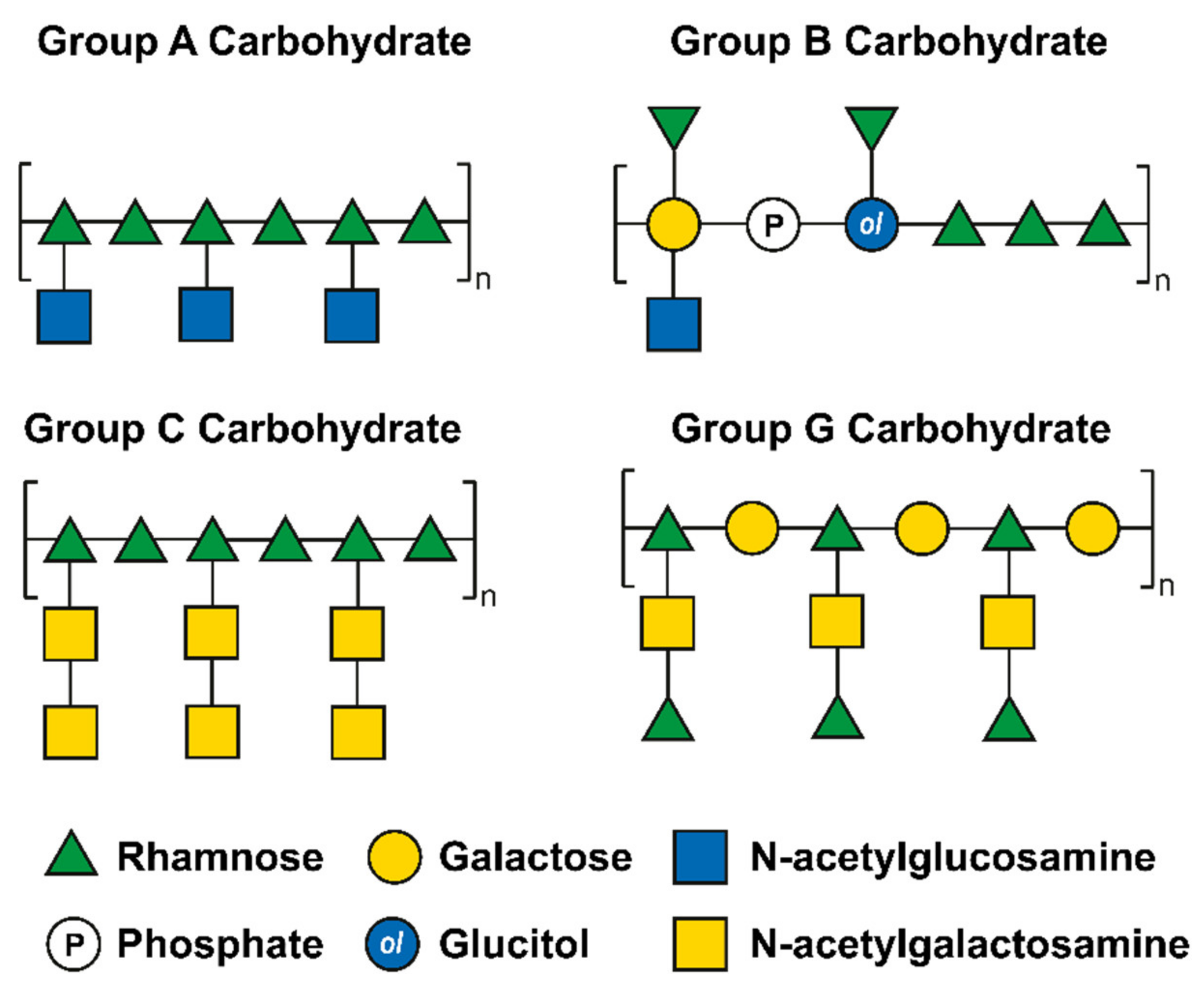
  127. Swanson, J.; Gotschlich, E.C. Electron Microscopic Studies on Streptococci II. Group A Carbohydrate. J. Exp. Med. 1973, 138, 245–258. [Google Scholar] [CrossRef] [Green Version]
  128. Wagner, M.; Wagner, B.; Ryc, M. An Electron Microscopic Study of the Location of Peptidoglycan in Group A and C Streptococcal Cell Walls. J. Gen. Microbiol. 1978, 108, 283–294. [Google Scholar] [CrossRef] [PubMed]
  129. Wagner, B.; Wagner, M.; Kubin, V.; Ryc, M. Immunoelectron Microscopic Study of the Location of Group-Specific and Protein Type-Specific Antigens of Group B Streptococci. J. Gen. Microbiol. 1980, 118, 95–105. [Google Scholar] [CrossRef] [PubMed] [Green Version]
  130. Sutcliffe, I.C.; Black, G.W.; Harrington, D.J. Bioinformatic Insights into the Biosynthesis of the Group B Carbohydrate in Streptococcus agalactiae. Microbiology 2008, 154, 1354–1363. [Google Scholar] [CrossRef] [Green Version]
  131. Chapot-Chartier, M.P. Interactions of the Cell-Wall Glycopolymers of Lactic Acid Bacteria with Their Bacteriophages. Front. Microbiol. 2014, 5, 1–10. [Google Scholar] [CrossRef] [PubMed] [Green Version]
  132. Hancock, L.E.; Gilmore, M.S. The Capsular Polysaccharide of Enterococcus faecalis and Its Relationship to Other Polysaccharides in the Cell Wall. Proc. Natl. Acad. Sci. USA 2002, 99, 1574–1579. [Google Scholar] [CrossRef] [PubMed] [Green Version]
  133. Teng, F.; Singh, K.V.; Bourgogne, A.; Zeng, J.; Murray, B.E. Further Characterization of the Epa Gene Cluster and Epa Polysaccharides of Enterococcus faecalis. Infect. Immun. 2009, 77, 3759–3767. [Google Scholar] [CrossRef] [PubMed] [Green Version]
  134. Thurlow, L.R.; Thomas, V.C.; Hancock, L.E. Capsular Polysaccharide Production in Enterococcus faecalis and Contribution of CpsF to Capsule Serospecificity. J. Bacteriol. 2009, 191, 6203–6210. [Google Scholar] [CrossRef] [Green Version]
  135. Theilacker, C.; Holst, O.; Lindner, B.; Huebner, J.; Kaczyński, Z. The Structure of the Wall Teichoic Acid Isolated from Enterococcus faecalis Strain 12030. Carbohydr. Res. 2012, 354, 106–109. [Google Scholar] [CrossRef]
  136. Pritchard, D.G.; Coligan, J.E.; Speed, S.E.; Gray, B.M. Carbohydrate Fingerprints of Streptococcal Cells. J. Clin. Microbiol. 1981, 13, 89–92. [Google Scholar] [CrossRef] [Green Version]
  137. Mistou, M.Y.; Sutcliffe, I.C.; Van Sorge, N.M. Bacterial Glycobiology: Rhamnose-Containing Cell Wall Polysaccharides in Gram-Positive Bacteria. FEMS Microbiol. Rev. 2016, 40, 464–479. [Google Scholar] [CrossRef] [Green Version]
  138. McCarty, M. The Lysis of Group A Hemolytic Streptococci by Extracellular Enzymes of Streptomyces albus. J. Exp. Med. 1952, 96, 569–581. [Google Scholar] [CrossRef] [PubMed]
  139. Tsukioka, Y.; Yamashita, Y.; Nakano, Y.; Oho, T.; Koga, T. Identification of a Fourth Gene Involved in DTDP-Rhamnose Synthesis in Streptococcus mutans. J. Bacteriol. 1997, 179, 4411–4414. [Google Scholar] [CrossRef] [PubMed] [Green Version]
  140. Caliot, É.; Dramsi, S.; Chapot-Chartier, M.P.; Courtin, P.; Kulakauskas, S.; Péchoux, C.; Trieu-Cuot, P.; Mistou, M.Y. Role of the Group B Antigen of Streptococcus agalactiae: A Peptidoglycan-Anchored Polysaccharide Involved in Cell Wall Biogenesis. PLoS Pathog. 2012, 8, e1002756. [Google Scholar] [CrossRef] [PubMed]
  141. Van der Beek, S.L.; Le Breton, Y.; Ferenbach, A.T.; Chapman, R.N.; van Aalten, D.M.F.; Navratilova, I.; Boons, G.J.; Mciver, K.S.; van Sorge, N.M.; Dorfmueller, H.C. GacA Is Essential for Group A Streptococcus and Defines a New Class of Monomeric DTDP-4-Dehydrorhamnose Reductases (RmlD). Mol. Microbiol. 2015, 98, 946–962. [Google Scholar] [CrossRef] [Green Version]
  142. Van Sorge, N.M.; Cole, J.N.; Kuipers, K.; Henningham, A.; Aziz, R.K.; Kasirer-Friede, A.; Lin, L.; Berends, E.T.M.; Davies, M.R.; Dougan, G.; et al. The Classical Lancefield Antigen of Group A Streptococcus Is a Virulence Determinant with Implications for Vaccine Design. Cell Host Microbe 2014, 15, 729–740. [Google Scholar] [CrossRef] [PubMed] [Green Version]
  143. Krause, R.M. Studies on Bacteriophages of Hemolytic Streptococci. I. Factors Influencing the Interaction of Phage and Susceptible Host Cell. J. Exp. Med. 1957, 106, 365–384. [Google Scholar] [CrossRef] [Green Version]
  144. Fischetti, V.A.; Zabriskie, J.B. Studies on Streptococcal Bacteriophages II. Adsorpion Studies on Group A and Group C Streptococcal Bacteriophages. J. Exp. Med. 1967, 127, 489–505. [Google Scholar] [CrossRef]
  145. Shibata, Y.; Yamashita, Y.; Van Der Ploeg, J.R. The Serotype-Specific Glucose Side Chain of Rhamnose-Glucose Polysaccharides Is Essential for Adsorption of Bacteriophage M102 to Streptococcus mutans. FEMS Microbiol. Lett. 2009, 294, 68–73. [Google Scholar] [CrossRef] [Green Version]
  146. Xu, Y.; Singh, K.V.; Qin, X.; Murray, B.E.; Weinstock, G.M. Analysis of a Gene Cluster of Enterococcus faecalis Involved in Polysaccharide Biosynthesis. Infect. Immun. 2000, 68, 815–823. [Google Scholar] [CrossRef] [Green Version]
  147. Alphey, M.S.; Pirrie, L.; Torrie, L.S.; Boulkeroua, W.A.; Gardiner, M.; Sarkar, A.; Maringer, M.; Oehlmann, W.; Brenk, R.; Scherman, M.S.; et al. Allosteric Competitive Inhibitors of the Glucose-1-Phosphate Thymidylyltransferase (RmlA) from Pseudomonas aeruginosa. ACS Chem. Biol. 2013, 8, 387–396. [Google Scholar] [CrossRef]
  148. Sewell, E.W.C.; Brown, E.D. Taking Aim at Wall Teichoic Acid Synthesis: New Biology and New Leads for Antibiotics. J. Antibiot. 2014, 67, 43–51. [Google Scholar] [CrossRef] [Green Version]
  149. Campbell, J.; Singh, A.K.; Santa Maria, J.P.; Kim, Y.; Brown, S.; Swoboda, J.G.; Mylonakis, E.; Wilkinson, B.J.; Walker, S. Synthetic Lethal Compound Combinations Reveal a Fundamental Connection between Wall Teichoic Acid and Peptidoglycan Biosyntheses in Staphylococcus aureus. ACS Chem. Biol. 2011, 6, 106–116. [Google Scholar] [CrossRef] [Green Version]
  150. Nicolau, D.P. Carbapenems: A Potent Class of Antibiotics. Expert Opin. Pharmacother. 2008, 9, 23–37. [Google Scholar] [CrossRef]
  151. Zhanel, G.G.; Wiebe, R.; Dilay, L.; Thomson, K.; Rubinstein, E.; Hoban, D.J.; Noreddin, A.M.; Karlowsky, J.A. Comparative Review of the Carbapenems. Drugs 2007, 67, 1027–1052. [Google Scholar] [CrossRef]
  152. Tenover, F.C. Mechanisms of Antimicrobial Resistance in Bacteria. Am. J. Med. 2006, 119, S3–S10. [Google Scholar] [CrossRef]
  153. Watkins, R.R. A Primer on Antimicrobials; Elsevier Inc.: Amsterdam, The Netherlands, 2022. [Google Scholar]
  154. Belley, A.; McKay, G.A.; Arhin, F.F.; Sarmiento, I.; Beaulieu, S.; Fadhil, I.; Parr, T.R.; Moeck, G. Oritavancin Disrupts Membrane Integrity of Staphylococcus aureus and Vancomycin-Resistant Enterococci to Effect Rapid Bacterial Killing. Antimicrob. Agents Chemother. 2010, 54, 5369–5371. [Google Scholar] [CrossRef] [Green Version]
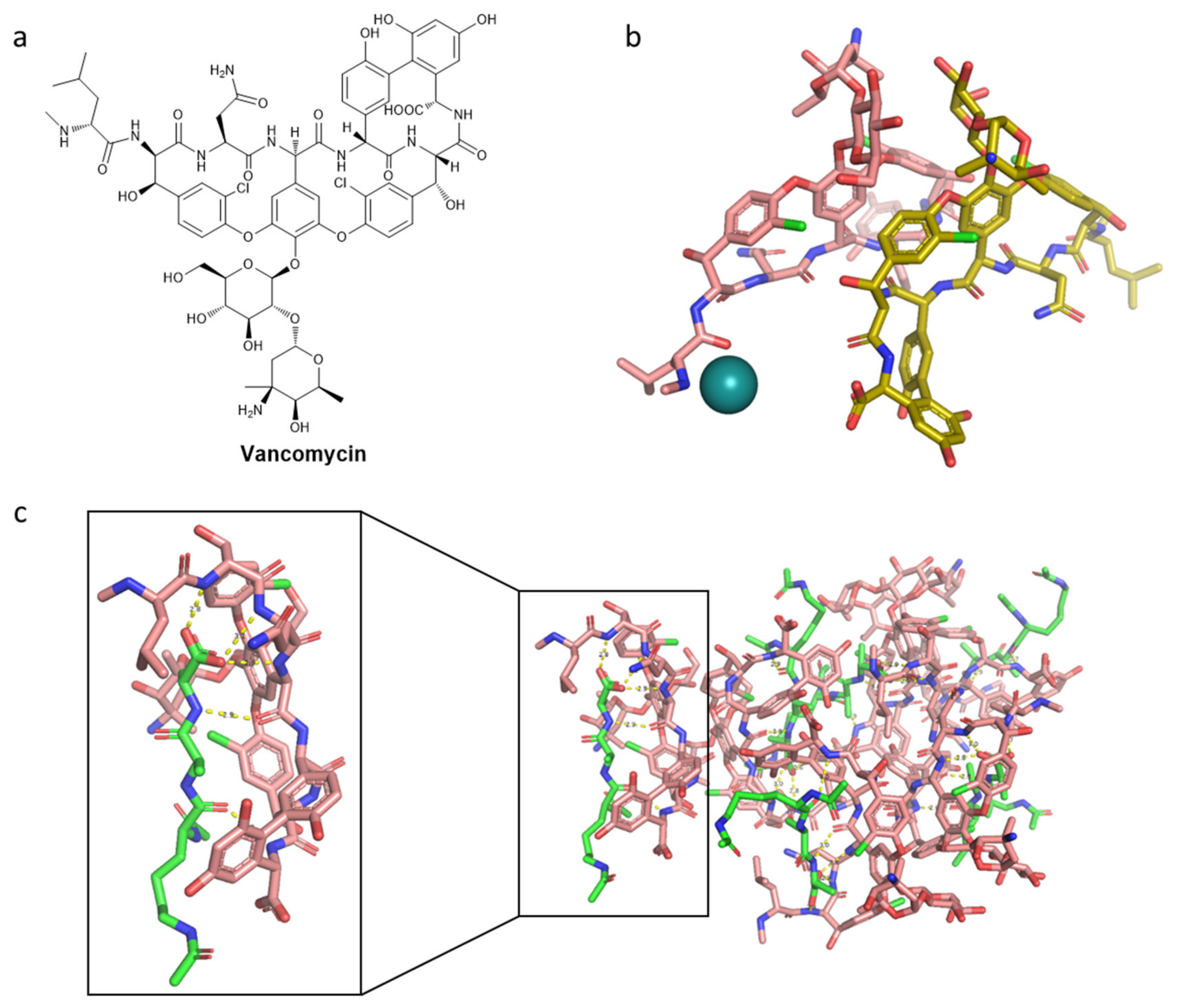
  155. Griffith, R.S. Vancomycin Use—An Historical Review. J. Antimicrob. Chemother. 1984, 14, 1–5. [Google Scholar] [CrossRef]
  156. Wilhelm, M.P.; Estes, L. Symposium on Antimicrobial Agents—Part XII. Vancomycin. Mayo Clin. Proc. 1999, 74, 928–935. [Google Scholar] [CrossRef]
  157. Jacobs, M.R. Emergence of Multiple Resistant Pneumococci. J. Occup. Environ. Med. 1980, 22, 559. [Google Scholar] [CrossRef]
  158. Aber, R.C.; Wennersten, C.; Moellering, R.C. Antimicrobial Susceptibility of Flavobacteria. Antimicrob. Agents Chemother. 1978, 14, 483–487. [Google Scholar] [CrossRef] [Green Version]
  159. Anderson, J.S.; Matsuhashi, M.; Haskin, M.A.; Strominger, J.L. Lipid-Phosphoacetylmuramyl-Pentapeptide and Lipid-Phosphodisaccharide-Pentapeptide: Presumed Membrane Transport Intermediates in Cell Wall Synthesis. Proc. Natl. Acad. Sci. USA 1965, 53, 881–889. [Google Scholar] [CrossRef] [Green Version]
  160. Perkins, H.R. Specificity of Combination between Mucopeptide Precursors and Vancomycin or Ristocetin. Biochem. J. 1969, 111, 195–205. [Google Scholar] [CrossRef] [Green Version]
  161. Hancock, R.; Fitz-James, P.C. Some Differences in the Action of Penicillin, Bacitracin, and Vancomycin on Bacillus megaterium. J. Bacteriol. 1964, 87, 1044–1050. [Google Scholar] [CrossRef] [Green Version]
  162. Jordan, D.C.; Mallory, H.D. Site of Action of Action of Vancomycin on Staphylococcus aureus. Antimicrob. Agents Chemother. 1964, 10, 489–494. [Google Scholar]
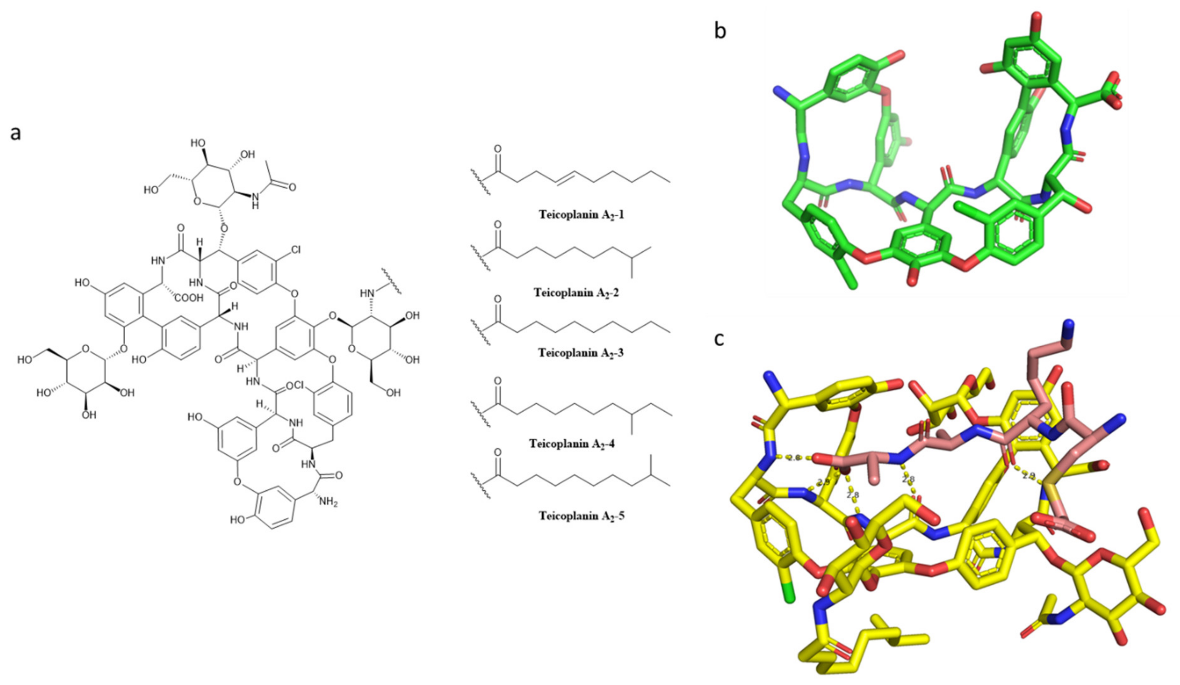
  163. Campoli-Richards, D.M.; Brogden, R.N.; Faulds, D. Teicoplanin. A Review of Its Antibacterial Activity, Pharmacokinetic Properties and Therapeutic Potential. Drugs 1990, 40, 449–486. [Google Scholar] [CrossRef]
  164. Parenti, F.; Beretta, G.; Berti, M.; Arioli, V. Teichomycins, New Antibiotics from Actinoplanes Teichomyceticus Nov. Sp. I. Description of the Producer Strain, Fermentation Studies and Biological Properties. J. Antibiot. 1978, 31, 276–283. [Google Scholar] [CrossRef] [PubMed] [Green Version]
  165. Janknegt, R. Teicoplanin in Perspective A Critical Comparison with Vancomycin. Pharm. Weekbl. Sci. 1991, 13, 153–160. [Google Scholar] [CrossRef] [PubMed]
  166. Parenti, F. Structure and Mechanism of Action of Teicoplanin. J. Hosp. Infect. 1986, 7, 79–83. [Google Scholar] [CrossRef]
  167. Somma, S.; Gastaldo, L.; Corti, A. Teicoplanin, a New Antibiotic from Actinoplanes teichomyceticus Nov. Sp. Antimicrob. Agents Chemother. 1984, 26, 917–923. [Google Scholar] [CrossRef] [Green Version]
  168. Reynolds, P.E.; Somner, E.A. Comparison of the Target Sites and Mechanisms of Action of Glycopeptide and Lipoglycodepsipeptide Antibiotics. Drugs Exp. Clin. Res. 1990, 16, 385–389. [Google Scholar]
  169. Barna, J.C.J.; Williams, D.H.; Stone, D.J.M.; Leung, T.W.C.; Doddrell, D.M. Structure Elucidation of the Teicoplanin Antibiotics. J. Am. Chem Soc. 1984, 106, 4895–4902. [Google Scholar] [CrossRef]
  170. Parenti, F. Glycopeptide Antibiotics. J. Clin. Pharmacol. 1988, 28, 136–140. [Google Scholar] [CrossRef] [PubMed]
  171. Bolognino, I.; Carrieri, A.; Purgatorio, R.; Catto, M.; Caliandro, R.; Carrozzini, B.; Belviso, B.D.; Majellaro, M.; Sotelo, E.; Cellamare, S.; et al. Enantiomeric Separation and Molecular Modelling of Bioactive 4-Aryl-3,4-Dihydropyrimidin-2(1H)-One Ester Derivatives on Teicoplanin-Based Chiral Stationary Phase. Separations 2022, 9, 7. [Google Scholar] [CrossRef]
  172. Kirst, H.A.; Thompson, D.G.; Nicas, T.I. Historical Yearly Usage of Vancomycin. Antimicrob. Agents Chemother 1998, 42, 1303–1304. [Google Scholar] [CrossRef] [Green Version]
  173. Pakyz, A.L.; Macdougall, C.; Oinonen, M.; Polk, R.E. Trends in Antibacterial Use in US Academic Health Centers. Arch. Intern. Med. 2008, 168, 2254–2260. [Google Scholar] [CrossRef] [Green Version]
  174. Steinkraus, G.; White, R.; Friedrich, L. Vancomycin MIC Creep in Non-Vancomycin-Intermediate Staphylococcus aureus (VISA), Vancomycin-Susceptible Clinical Methicillin-Resistant S. Aureus (MRSA) Blood Isolates from 2001 05. J. Antimicrob. Chemother. 2007, 60, 788–794. [Google Scholar] [CrossRef] [Green Version]
  175. King, A.; Phillips, I.; Kaniga, K. Comparative in Vitro Activity of Telavancin (TD-6424), a Rapidly Bactericidal, Concentration-Dependent Anti-Infective with Multiple Mechanisms of Action against Gram-Positive Bacteria. J. Antimicrob. Chemother. 2004, 53, 797–803. [Google Scholar] [CrossRef] [Green Version]
  176. Saravolatz, L.D.; Stein, G.E.; Johnson, L.B. Telavancin: A Novel Lipoglycopeptide. Clin. Infect. Dis. 2009, 49, 1908–1914. [Google Scholar] [CrossRef] [Green Version]
  177. Leadbetter, M.R.; Adams, S.M.; Bazzini Kevin Krause, B.M.; T Lam, B.M.; Linsell, M.S.; Kelly Quast, M.; Shaw, J.; Soriano, E.; Terry Wu, S.X.; Christensen, B.G.; et al. Hydrophobic Vancomycin Derivatives with Improved ADME Properties: Discovery of Telavancin (TD-6424). J. Antibiot. 2004, 57, 326–336. [Google Scholar] [CrossRef] [Green Version]
  178. Higgins, D.L.; Chang, R.; Debabov, D.V.; Leung, J.; Wu, T.; Krause, K.M.; Sandvik, E.; Hubbard, J.M.; Kaniga, K.; Schmidt, D.E.; et al. Telavancin, a Multifunctional Lipoglycopeptide, Disrupts Both Cell Wall Synthesis and Cell Membrane Integrity in Methicillin-Resistant Staphylococcus aureus. Antimicrob. Agents Chemother. 2005, 49, 1127–1134. [Google Scholar] [CrossRef] [Green Version]
  179. Stryjewski, M.E.; Lentnek, A.; O’Riordan, W.; Pullman, J.; Tambyah, P.A.; Miró, J.M.; Fowler, V.G.; Barriere, S.L.; Kitt, M.M.; Corey, G.R. A Randomized Phase 2 Trial of Telavancin versus Standard Therapy in Patients with Uncomplicated Staphylococcus aureus Bacteremia: The ASSURE Study. BMC Infect. Dis. 2014, 14, 289. [Google Scholar] [CrossRef] [Green Version]
  180. Holland, T.L.; Chambers, H.F.; Boucher, H.W.; Corey, G.R.; Coleman, R.; Castaneda-Ruiz, B.; Fowler, V.G. Considerations for Clinical Trials of Staphylococcus aureus Bloodstream Infection in Adults. Clin. Infect. Dis. 2019, 68, 865–872. [Google Scholar] [CrossRef] [PubMed]
  181. A Phase 3 Telavancin Staphylococcus aureus (S. aureus) Bacteremia Trial. 2020. Available online: https://Clinicaltrials.Gov/Ct2/Show/Results/NCT02208063 (accessed on 15 February 2022).
  182. Burgin, D.J.; Liu, R.; Hsieh, R.C.; Heinzinger, L.R.; Otto, M. Investigational Agents for the Treatment of Methicillin-Resistant Staphylococcus aureus (MRSA) Bacteremia: Progress in Clinical Trials. Expert Opin. Investig. Drugs 2022, 31, 263–279. [Google Scholar] [CrossRef] [PubMed]
  183. Draghi, D.C.; Benton, B.M.; Krause, K.M.; Thornsberry, C.; Pillar, C.; Sahm, D.F. Comparative Surveillance Study of Telavancin Activity against Recently Collected Gram-Positive Clinical Isolates from across the United States. Antimicrob. Agents Chemother. 2008, 52, 2383–2388. [Google Scholar] [CrossRef] [PubMed] [Green Version]
  184. Saravolatz, L.D.; Pawlak, J.; Johnson, L.B. Comparative Activity of Telavancin against Isolates of Community-Associated Methicillin-Resistant Staphylococcus aureus. J. Antimicrob. Chemother. 2007, 60, 406–409. [Google Scholar] [CrossRef] [Green Version]
  185. Goldstein, E.J.C.; Citron, D.M.; Merriam, C.V.; Warren, Y.A.; Tyrrell, K.L.; Fernandez, H.T. In Vitro Activities of the New Semisynthetic Glycopeptide Telavancin (TD-6424), Vancomycin, Daptomycin, Linezolid, and Four Comparator Agents against Anaerobic Gram-Positive Species and Corynebacterium Spp. Antimicrob. Agents Chemother. 2004, 48, 2149–2152. [Google Scholar] [CrossRef] [Green Version]
  186. Leuthner, K.D.; Cheung, C.M.; Rybak, M.J. Comparative Activity of the New Lipoglycopeptide Telavancin in the Presence and Absence of Serum against 50 Glycopeptide Non-Susceptible Staphylococci and Three Vancomycin-Resistant Staphylococcus aureus. J. Antimicrob. Chemother. 2006, 58, 338–343. [Google Scholar] [CrossRef]
  187. Leonard, S.N.; Vidaillac, C.; Rybak, M.J. Activity of Telavancin against Staphylococcus aureus Strains with Various Vancomycin Susceptibilities in an In Vitro Pharmacokinetic/Pharmacodynamic Model with Simulated Endocardial Vegetations. Antimicrob. Agents Chemother. 2009, 53, 2928–2933. [Google Scholar] [CrossRef] [Green Version]
  188. Heo, Y.A. Oritavancin (KIMYRSA™) in Acute Bacterial Skin and Skin Structure Infections: A Profile of Its Use in the USA. Drugs Ther. Perspect. 2022, 38, 57–63. [Google Scholar] [CrossRef]
  189. Zhanel, G.G.; Schweizer, F.; Karlowsky, J.A. Oritavancin: Mechanism of Action. Clin. Infect. Dis. 2012, 54, S214–S219. [Google Scholar] [CrossRef] [Green Version]
  190. Guskey, M.T.; Tsuji, B.T. A Comparative Review of the Lipoglycopeptides: Oritavancin, Dalbavancin, and Telavancin. Pharmacotherapy 2010, 30, 80–94. [Google Scholar] [CrossRef]
  191. Mendes, R.E.; Farrell, D.J.; Sader, H.S.; Jones, R.N. Oritavancin Microbiologic Features and Activity Results from the Surveillance Program in the United States. Clin. Infect. Dis. 2012, 54, 203–216. [Google Scholar] [CrossRef] [Green Version]
  192. Billeter, M.; Zervos, M.J.; Chen, A.Y.; Dalovisio, J.R.; Kurukularatne, C. Dalbavancin: A Novel Once-Weekly Lipoglycopeptide Antibiotic. Clin. Infect. Dis. 2008, 46, 577–583. [Google Scholar] [CrossRef]
  193. Mendes, R.E.; Farrell, D.J.; Sader, H.S.; Flamm, R.K.; Jones, R.N. Baseline Activity of Telavancin against Gram-Positive Clinical Isolates Responsible for Documented Infections in U.S. Hospitals (2011–2012) as Determined by the Revised Susceptibility Testing Method. Antimicrob. Agents Chemother. 2015, 59, 702–706. [Google Scholar] [CrossRef] [Green Version]
  194. Saravolatz, L.D.; Pawlak, J.; Johnson, L.B. In Vitro Susceptibilities and Molecular Analysis of Vancomycin-Intermediate and Vancomycin-Resistant Staphylococcus aureus Isolates. Clin. Infect. Dis. 2012, 55, 582–586. [Google Scholar] [CrossRef] [Green Version]
  195. Belley, A.; Neesham-Grenon, E.; Arhin, F.F.; McKay, G.A.; Parr, T.R.; Moeck, G. Assessment by Time-Kill Methodology of the Synergistic Effects of Oritavancin in Combination with Other Antimicrobial Agents against Staphylococcus aureus. Antimicrob. Agents Chemother. 2008, 52, 3820–3822. [Google Scholar] [CrossRef] [Green Version]
  196. Berman, H.M.; Battistuz, T.; Bhat, T.N.; Bluhm, W.F.; Bourne, P.E.; Burkhardt, K.; Feng, Z.; Gilliland, G.L.; Iype, L.; Jain, S.; et al. The Protein Data Bank. Acta Crystallogr. Sect. D Biol. Crystallogr. 2002, 58, 899–907. [Google Scholar] [CrossRef]
  197. Raad, I.; Darouiche, R.; Vazquez, J.; Lentnek, A.; Hachem, R.; Hanna, H.; Goldstein, B.; Henkel, T.; Seltzer, E. Efficacy and Safety of Weekly Dalbavancin Therapy for Catheter-Related Bloodstream Infection Caused by Gram-Positive Pathogens. Clin. Infect. Dis. 2005, 40, 374–380. [Google Scholar] [CrossRef]
  198. Ito, S.; Senoo, A.; Nagatoishi, S.; Ohue, M.; Yamamoto, M.; Tsumoto, K.; Wakui, N. Structural Basis for the Binding Mechanism of Human Serum Albumin Complexed with Cyclic Peptide Dalbavancin. J. Med. Chem. 2020, 63, 14045–14053. [Google Scholar] [CrossRef]
  199. Xu, X.; Xu, L.; Yuan, G.; Wang, Y.; Qu, Y.; Zhou, M. Synergistic Combination of Two Antimicrobial Agents Closing Each Other’s Mutant Selection Windows to Prevent Antimicrobial Resistance OPEN. Sci. Rep. 2018, 8, 7237. [Google Scholar] [CrossRef]
  200. Henderson, J.W. The Yellow Brick Road to Penicillin: A Story of Serendipity. Mayo Clin. Proc. 1997, 72, 683–687. [Google Scholar] [CrossRef]
  201. Terrak, M. Peptidoglycan Glycosyltransferase Inhibition: New Perspectives for an Old Target. Anti-Infect. Agents Med. Chem. 2008, 7, 180–192. [Google Scholar] [CrossRef]
  202. Miller, E.L. The Penicillins: A Review and Update. J. Midwifery Women’s Health 2002, 47, 426–434. [Google Scholar] [CrossRef]
  203. Watkins, R.R. Chapter 5—A Primer on Antimicrobials; Elsevier Inc.: Amsterdam, The Netherlands, 1941. [Google Scholar]
  204. Kahan, F.M.; Kropp, H.; Sundelof, J.G.; Birnbaum, J. Thienamycin: Development of Imipenem-Cilastatin. J. Antimicrob. Chemother. 1983, 12, 1–35. [Google Scholar] [CrossRef]
  205. Geddes, A.M.; Stille, W. Imipenem: The First Thienamycin Antibiotic. Rev. Infect. Dis. 1985, 7, S353–S356. [Google Scholar] [CrossRef]
  206. Papp-Wallace, K.M.; Endimiani, A.; Taracila, M.A.; Bonomo, R.A. Carbapenems: Past, Present, and Future. Antimicrob. Agents Chemother. 2011, 55, 4943–4960. [Google Scholar] [CrossRef] [Green Version]
  207. Chambers, H.F. Other Beta-Lactam Antibiotics. In Mandell, Douglas, and Bennett’s Principles and Practice of Infectious Diseases, 5th ed.; Mandell, G.L., Bennett, J.E., Dolin, R., Eds.; Churchill Livingstone: Philadelphia, PA, USA, 2000; Volume 1, pp. 291–293. [Google Scholar]
  208. Neu, H.C.; Novelli, A.; Chin, N.X. In Vitro Activity and Beta-Lactamase Stability of a New Carbapenem, SM-7338. Antimicrob. Agents Chemother. 1989, 33, 1009–1018. [Google Scholar] [CrossRef] [Green Version]
  209. Sakurai, Y.; Yoshida, Y.; Saitoh, K.; Nemoto, M.; Yamaguchi, A.; Sawai, T. Characteristics of Aztreonam as a Substrate, Inhibitor and Inducer for Beta-Lactamases. J. Antibiot. 1990, 43, 403–410. [Google Scholar] [CrossRef] [PubMed] [Green Version]
  210. Sykes, R.B.; Bonner, D.P. Discovery and Development of the Monobactams. Rev. Infect. Dis. 1985, 7, S579–S593. [Google Scholar] [CrossRef]
  211. Gutmann, L.; Vincent, S.; Billot-Klein, D.; Acar, J.F.; Mrèna, E.; Williamson, R. Involvement of Penicillin-Binding Protein 2 with Other Penicillin-Binding Proteins in Lysis of Escherichia coli by Some β-Lactam Antibiotics Alone and in Synergistic Lytic Effect of Amdinocillin (Mecillinam). Antimicrob. Agents Chemother. 1986, 30, 906–912. [Google Scholar] [CrossRef] [Green Version]
  212. Paech, F.; Messner, S.; Spickermann, J.; Wind, M.; Schmitt-Hoffmann, A.H.; Witschi, A.T.; Howell, B.A.; Church, R.J.; Woodhead, J.; Engelhardt, M.; et al. Mechanisms of Hepatotoxicity Associated with the Monocyclic β-Lactam Antibiotic BAL30072. Arch. Toxicol. 2017, 91, 3647–3662. [Google Scholar] [CrossRef] [PubMed]
  213. Prasad, N.K.; Seiple, I.B.; Cirz, R.T.; Rosenberg, O.S. Leaks in the Pipeline: A Failure Analysis of Gram-Negative Antibiotic Development from 2010 to 2020. Antimicrob. Agents Chemother. 2022, 66, e00054-22. [Google Scholar] [CrossRef]
  214. Li, J.; Milne, R.W.; Nation, R.L.; Turnidge, J.D.; Coulthard, K. Stability of Colistin and Colistin Methanesulfonate in Aqueous Media and Plasma as Determined by High-Performance Liquid Chromatography. Antimicrob. Agents Chemother. 2003, 47, 1364–1370. [Google Scholar] [CrossRef] [PubMed] [Green Version]
  215. Aeschlimann, J.R.; Hershberger, E.; Rybak, M.J. Analysis of Vancomycin Population Susceptibility Profiles, Killing Activity, and Postantibiotic Effect against Vancomycin-Intermediate Staphylococcus aureus. Antimicrob. Agents Chemother. 1999, 43, 1914–1918. [Google Scholar] [CrossRef] [Green Version]
  216. Baddour, L.M.; Wilson, W.R.; Bayer, A.S.; Fowler, V.G.; Tleyjeh, I.M.; Rybak, M.J.; Barsic, B.; Lockhart, P.B.; Gewitz, M.H.; Levison, M.E.; et al. Infective Endocarditis in Adults: Diagnosis, Antimicrobial Therapy, and Management of Complications: A Scientific Statement for Healthcare Professionals from the American Heart Association. Circulation 2015, 132, 1435–1486. [Google Scholar] [CrossRef]
  217. Stevens, D.L.; Bisno, A.L.; Chambers, H.F.; Everett, E.D.; Dellinger, P.; Goldstein, E.J.C.; Gorbach, S.L.; Hirschmann, J.V.; Kaplan, E.L.; Montoya, J.G.; et al. Practice Guidelines for the Diagnosis and Management of Skin and Soft-Tissue Infections. Clin. Infect. Dis. 2005, 41, 1373–1406. [Google Scholar] [CrossRef]
  218. Gemmell, C.G.; Edwards, D.I.; Fraise, A.P.; Gould, F.K.; Ridgway, G.L.; Warren, R.E. Guidelines for the Prophylaxis and Treatment of Methicillin-Resistant Staphylococcus aureus (MRSA) Infections in the UK. J. Antimicrob. Chemother. 2006, 57, 589–608. [Google Scholar] [CrossRef]
  219. Mermel, L.A.; Farr, B.M.; Sherertz, R.J.; Raad, I.I.; O’Grady, N.; Harris, J.S.; Craven, D.E. Guidelines for the Management of Intravascular Catheter-Related Infections. Clin. Infect. Dis. 2001, 32, 1249–1272. [Google Scholar] [CrossRef] [Green Version]
  220. Horstkotte, D.; Follath, F.; Gutschik, E.; Lengyel, M.; Oto, A.; Pavie, A.; Soler-Soler, J.; Thiene, G.; Von Graevenitz, A. Guidelines on Prevention, Diagnosis and Treatment of Infective Endocarditis Executive Summary: The Task Force on Infective Endocarditis of the European Society of Cardiology. Eur. Heart J. 2004, 25, 267–276. [Google Scholar] [CrossRef] [Green Version]
  221. Elliott, T.S.J.; Foweraker, J.; Gould, F.K.; Perry, J.D.; Sandoe, J.A.T. Guidelines for the Antibiotic Treatment of Endocarditis in Adults: Report of the Working Party of the British Society for Antimicrobial Chemotherapy. J. Antimicrob. Chemother. 2004, 54, 971–981. [Google Scholar] [CrossRef]
  222. Eliopoulos, G.M. Antimicrobial Agents for Treatment of Serious Infections Caused by Resistant Staphylococcus aureus and Enterococci. Eur. J. Clin. Microbiol. Infect. Dis. 2005, 24, 826–831. [Google Scholar] [CrossRef]
  223. FDA. FDA SSSI Guidance for Industry Acute Bacterial Skin and Skin Structure Infections: Developing Drugs for Treatment. In US Food and Drug Administration. Guidance for Industry. Acute Bacterial Skin and Skin Structure Infections: Developing Drugs for Treatment; FDA: Washington, DC, USA, 2013. [Google Scholar]
  224. Tally, F.P.; DeBruin, M.F. Development of Daptomycin for Gram-Positive Infections. J. Antimicrob. Chemother. 2000, 46, 523–526. [Google Scholar] [CrossRef] [PubMed] [Green Version]
  225. Weems, J.J. The Many Faces of Staphylococcus aureus Infection: Recognizing and Managing Its Life-Threatening Manifestations. Postgraduate Med. 2001, 110, 24–36. [Google Scholar] [CrossRef] [PubMed]
  226. King, M.D.; Humphrey, B.J.; Wang, Y.F.; Kourbatova, E.V.; Ray, S.M. Annals of Internal Medicine Article Staphylococcus aureus USA 300 Clone as the Predominant Cause of Skin and Soft-Tissue Infections. Ann. Intern. Med. 2006, 144, 309–318. [Google Scholar] [CrossRef] [Green Version]
  227. Li, J.; Rayner, C.R.; Nation, R.L.; Owen, R.J.; Spelman, D.; Tan, K.E.; Liolios, L. Heteroresistance to Colistin in Multidrug-Resistant Acinetobacter baumannii. Antimicrob. Agents Chemother. 2006, 50, 2946–2950. [Google Scholar] [CrossRef] [Green Version]
  228. Cathy, A.; Petti, M.D.A.; Vance, G.; Fowler, M.D., Jr. Optimal Treatment of Complicated Skin and Skin Structure Infections. Infect. Dis. Clin. N. Am. 2002, 16, 413–435. [Google Scholar]
  229. Eisenstein, B.I.; Oleson, F.B., Jr.; Baltz, R.H. Daptomycin: From the Mountain to the Clinic, with Essential Help from Francis Tally, MD. Clin. Infect. Dis. 2010, 50, S10–S15. [Google Scholar] [CrossRef] [Green Version]
  230. Fenton, C.; Keating, G.M.; Curran, M.P. Daptomycin. Drugs 2004, 64, 445–455. [Google Scholar] [CrossRef]
  231. Streit, J.M.; Jones, R.N.; Sader, H.S. Daptomycin Activity and Spectrum: A Worldwide Sample of 6737 Clinical Gram-Positive Organisms. J. Antimicrob. Chemother. 2004, 53, 669–674. [Google Scholar] [CrossRef] [Green Version]
  232. Niccolai, D.; Tarsi, L.; Thomas, R.J. The Renewed Challenge of Antibacterial Chemotherapy. Chem. Commun. 1997, 24, 2333–2342. [Google Scholar] [CrossRef]
  233. Theuretzbacher, U.; Gottwalt, S.; Beyer, P.; Butler, M.; Czaplewski, L.; Lienhardt, C.; Moja, L.; Paul, M.; Paulin, S.; Rex, J.H.; et al. Analysis of the Clinical Antibacterial and Antituberculosis Pipeline. Lancet Infect. Dis. 2019, 19, e40–e50. [Google Scholar] [CrossRef]
  234. Theuretzbacher, U.; Outterson, K.; Engel, A.; Karlén, A. The Global Preclinical Antibacterial Pipeline. Nat. Rev. Microbiol. 2020, 18, 275–285. [Google Scholar] [CrossRef] [PubMed] [Green Version]
  235. Ventola, C.L. The Antibiotic Resistance Crisis: Part 1: Causes and Threats. Pharm. Ther. 2015, 40, 277–283. [Google Scholar]
  236. Buchy, P.; Ascioglu, S.; Buisson, Y.; Datta, S.; Nissen, M.; Tambyah, P.A.; Vong, S. Impact of Vaccines on Antimicrobial Resistance. Int. J. Infect. Dis. 2020, 90, 188–196. [Google Scholar] [CrossRef] [Green Version]
  237. Micoli, F.; Bagnoli, F.; Rappuoli, R.; Serruto, D. The Role of Vaccines in Combatting Antimicrobial Resistance. Nat. Rev. Microbiol. 2021, 19, 287–302. [Google Scholar] [CrossRef]
  238. Marchetti, F.; Prato, R.; Viale, P. Survey among Italian Experts on Existing Vaccines’ Role in Limiting Antibiotic Resistance. Hum. Vaccines Immunother. 2021, 17, 4283–4290. [Google Scholar] [CrossRef]
  239. Jansen, K.U.; Anderson, A.S. The Role of Vaccines in Fighting Antimicrobial Resistance (AMR). Hum. Vaccines Immunother. 2018, 14, 2142–2149. [Google Scholar] [CrossRef] [Green Version]
  240. Lobanovska, M.; Pilla, G. Penicillin’s Discovery and Antibiotic Resistance: Lessons for the Future? Yale J. Biol. Med. 2017, 90, 135–145. [Google Scholar]
  241. Guevara Salazar, J.A.; Morán Díaz, J.R.; Ramírez Segura, E.; Trujillo Ferrara, J.G. What Are the Origins of Growing Microbial Resistance? Both Lamarck and Darwin Were Right. Expert Rev. Anti-Infect. Ther. 2021, 19, 563–569. [Google Scholar] [CrossRef]
  242. Rosas, N.C.; Lithgow, T. Targeting Bacterial Outer-Membrane Remodelling to Impact Antimicrobial Drug Resistance. Trends Microbiol. 2021, 30, 544–552. [Google Scholar] [CrossRef]
  243. Reygaert, W.C. An Overview of the Antimicrobial Resistance Mechanisms of Bacteria. AIMS Microbiol. 2018, 4, 482–501. [Google Scholar] [CrossRef]
  244. Li, X.-Z.; Plésiat, P.; Nikaido, H. The Challenge of Efflux-Mediated Antibiotic Resistance in Gram-Negative Bacteria. Clin. Microbiol. Rev. 2015, 28, 337–418. [Google Scholar] [CrossRef] [Green Version]
  245. Ghai, I.; Ghai, S. Understanding Antibiotic Resistance via Outer Membrane Permeability. Infect. Drug Resist. 2018, 11, 523–530. [Google Scholar] [CrossRef] [Green Version]
  246. Johnson, A.P.; Uttley, A.H.C.; Woodford, N.; George, R.C. Resistance to Vancomycin and Teicoplanin: An Emerging Clinical Problem. Clin. Microbiol. Rev. 1990, 3, 280–291. [Google Scholar] [CrossRef]
  247. Capeletti, L.B.; de Oliveira, J.F.A.; Loiola, L.M.D.; Galdino, F.E.; da Silva Santos, D.E.; Soares, T.A.; de Oliveira Freitas, R.; Cardoso, M.B. Gram-Negative Bacteria Targeting Mediated by Carbohydrate–Carbohydrate Interactions Induced by Surface-Modified Nanoparticles. Adv. Funct. Mater. 2019, 29, 1–11. [Google Scholar] [CrossRef]
  248. Plotkin, S. History of Vaccination. Proc. Natl. Acad. Sci. USA 2014, 111, 12283–12287. [Google Scholar] [CrossRef] [Green Version]
  249. Weintraub, A. Immunology of Bacterial Polysaccharide Antigens. Carbohydr. Res. 2003, 338, 2539–2547. [Google Scholar] [CrossRef]
  250. Sun, L.; Middleton, D.R.; Wantuch, P.L.; Ozdilek, A.; Avci, F.Y. Carbohydrates as T-Cell Antigens with Implications in Health and Disease. Glycobiology 2016, 26, 1029–1040. [Google Scholar] [CrossRef] [Green Version]
  251. Werdelin, O.; Meldal, M.; Jensen, T. Processing of Glycans on Glycoprotein and Glycopeptide Antigens in Antigen-Presenting Cells. Proc. Natl. Acad. Sci. USA 2002, 99, 9611–9613. [Google Scholar] [CrossRef] [Green Version]
  252. Khatun, F.; Toth, I.; Stephenson, R.J. Immunology of Carbohydrate-Based Vaccines. Adv. Drug Deliv. Rev. 2020, 165–166, 117–126. [Google Scholar] [CrossRef]
  253. Gallorini, S.; Berti, F.; Mancuso, G.; Cozzi, R.; Tortoli, M.; Volpini, G.; Telford, J.L.; Beninati, C.; Maione, D.; Wack, A. Toll-like Receptor 2 Dependent Immunogenicity of Glycoconjugate Vaccines Containing Chemically Derived Zwitterionic Polysaccharides. Proc. Natl. Acad. Sci. USA 2009, 106, 17481–17486. [Google Scholar] [CrossRef] [Green Version]
  254. Gallorini, S.; Berti, F.; Parente, P.; Baronio, R.; Aprea, S.; D’Oro, U.; Pizza, M.; Telford, J.L.; Wack, A. Introduction of Zwitterionic Motifs into Bacterial Polysaccharides Generates TLR2 Agonists Able to Activate APCs. J. Immunol. 2007, 179, 8208–8215. [Google Scholar] [CrossRef] [Green Version]
  255. Carreño, L.J.; Saavedra-Ávila, N.A.; Porcelli, S.A. Synthetic Glycolipid Activators of Natural Killer T Cells as Immunotherapeutic Agents. Clin. Transl. Immunol. 2016, 5, e69. [Google Scholar] [CrossRef] [Green Version]
  256. Kim, S.; Lalani, S.; Parekh, V.V.; Wu, L.; Van Kaer, L. Glycolipid Ligands of Invariant Natural Killer T Cells as Vaccine Adjuvants. Expert Rev. Vaccines 2008, 7, 1519–1532. [Google Scholar] [CrossRef] [Green Version]
  257. Avci, F.Y.; Li, X.; Tsuji, M.; Kasper, D.L. Carbohydrates and T Cells: A Sweet Twosome. Semin. Immunol. 2013, 25, 146–151. [Google Scholar] [CrossRef] [Green Version]
  258. Cavallari, M.; Stallforth, P.; Kalinichenko, A.; Rathwell, D.C.K.; Gronewold, T.M.A.; Adibekian, A.; Mori, L.; Landmann, R.; Seeberger, P.H.; De Libero, G. Asemisynthetic Carbohydrate-Lipid Vaccine That Protects against S. Pneumoniae in Mice. Nat. Chem. Biol. 2014, 10, 950–956. [Google Scholar] [CrossRef]
  259. Oppenheimer, S.B.; Alvarez, M.; Nnoli, J. Carbohydrate-Based Experimental Therapeutics for Cancer, HIV/AIDS and Other Diseases. Acta Histochem. 2008, 110, 6–13. [Google Scholar] [CrossRef] [Green Version]
  260. Yin, Z.; Huang, X. Recent Development in Carbohydrate Based Anticancer Vaccines. J. Carbohydr. Chem. 2012, 31, 143–186. [Google Scholar] [CrossRef] [Green Version]
  261. Hossain, F.; Andreana, P.R. Developments in Carbohydrate-Based Cancer Therapeutics. Pharmaceuticals 2019, 12, 84. [Google Scholar] [CrossRef] [Green Version]
Figure 1. Schematic representation of target human districts of the main gram-positive and gram-negative bacteria. Created with BioRender.com (Accessed on 29 April 2022).
Pharmaceuticals 15 00942 g001
Figure 2. Schematic representation of the cell wall structure of gram-positive and gram-negative bacteria.
Pharmaceuticals 15 00942 g002
Figure 3. Key structural elements that are representative of the peptidoglycan region. The strands of PG are linked via peptide chains that condense with each other forming amide bonds via either the meso-A2pm carboxyl group or amine group.
Pharmaceuticals 15 00942 g003
Figure 4. Lipopolysaccharide with its O-antigen from E. coli O6 built with LPS modeler [83] and rendered in VMD. Figure adapted from Furevi [84].
Pharmaceuticals 15 00942 g004
Figure 5. Schematic representation of LTA and WTA in the gram-positive cell wall. The WTA extends beyond the PG layer while LTA may not be able to become elongated past it.
Pharmaceuticals 15 00942 g005
Figure 6. Different types of streptococcal glycan repeating units according to the Lancefield classification. The representation follows the shapes and colors of the SNFG representation for carbohydrates [125].
Pharmaceuticals 15 00942 g006
Figure 7. (a) 2D structure of vancomycin. (b) PyMOL 3D structure of two vancomycin molecules with a Zn2+ ion (PDB code 5M2K, ethanediol was removed); (c) 3D representation of the complex of vancomycin (colored in salmon) and d-Ala-d-Ala (in green, PDB ID: IFVM).
Pharmaceuticals 15 00942 g007
Figure 8. (a) 2D structure of teicoplanin, in its different forms. (b) 3D structure of teicoplanin aglycone (TAG: PDB ID 6TOV), DMS (dimethyl sulfoxide was removed from the original pdb file of the crystal structure) [171], and (c) teicoplanin in complex with d-Ala-d-Ala residue with polar contacts (PDB ID: 3VFJ).
Pharmaceuticals 15 00942 g008
Figure 9. (a) Telavancin 2D structure and (b) 3D conformation of the minimized structure of telavancin (purple) that was obtained with PyMOL.
Pharmaceuticals 15 00942 g009
Figure 10. (a) 2D representation of oritavancin; (b) 3D energy minimized structure of oritavancin (yellow), made with PyMOL.
Pharmaceuticals 15 00942 g010
Figure 11. (a) Chemical structure of dalbavancin; (b,c) crystal structure (entire structure and close-up, respectively) of dalbavancin (blue) in complex with human serum albumin (HAS, yellow), PDB ID: 6M5E [198]. Figure made by ChemDraw and PyMOL.
Pharmaceuticals 15 00942 g011
Figure 12. Structure of representative penicillins (penicillin G, oxacillin, amoxicillin, carbenicillin, and piperacillin); cephalosporins (ceftazidime, ceftolozane, and ceftaroline); carbapenems (imipenem, doripenem, meropenem, and ertapenem); and monobactams (aztreonam and BAL30072).
Pharmaceuticals 15 00942 g012
Figure 13. Structure of polymyxin B, colistin (polymyxin E), and daptomycin.
Pharmaceuticals 15 00942 g013
Figure 14. Summary of the roots of the antibiotic resistance crisis. Created with BioRender.com (Accessed on 29 April 2022).
Pharmaceuticals 15 00942 g014
Figure 15. Scheme of mechanisms that lead phenotypic resistance evolution in bacteria. The highlighted paths include the mutation of the target (orange), the synthesis of antibiotic-altering enzymes (purple), the synthesis of genes encoding drug efflux pumps (light blue), and the membrane remodeling (green). Created with BioRender.com (Accessed on 29 April 2022).
Pharmaceuticals 15 00942 g015
Figure 16. Biogenesis of bacterial OMP. Newly synthesized OMP precursors (red) move from the cytoplasm across the inner membrane and the periplasmic space. The β-barrel assembly machinery (BAM) complex (green), located in the OM, integrates the OMP precursors, creating the folded OMP (orange). Created with BioRender.com (Accessed on 29 April 2022).
Pharmaceuticals 15 00942 g016
Table 1. Pathogenic bacteria and their associated infections. Examples of gram-negative and gram-positive pathogenic bacteria and their associated infections.
GRAM NEGATIVEILLNESSES
Cocci
Moraxella catarrhalisRespiratory tract infections
Neisseria gonorrhoeaeGonorrhea
Neisseria meningitidisMeningococcal Meningitis
Chlamydia trachomatisChlamydia
Cocci-bacilli
Rickettsia prowazekii
Rickettsia typhi
Epidemic typhus
Murine typhus
Cocci-bacilli and bacilli
BrucellaBrucellosis
BordetellaWhooping cough
LegionellaPneumonia
Bacilli
Escherichia coliGastroenteric and urinary tract infections
Salmonella typhiTyphoid fever
Salmonella spp. Diarrhea
Haemophilus influenzaeOtitis, bronchitis, pneumonia, meningitis
Flagellated bacilli
Helicobacter pyloriStomach and duodenal ulcers
Vibrions
Vibrio choleraeCholera
Spirochetes
Borrelia burgdorferi
Leptospira
Lyme disease
Leptospirosis
Non-fermenting Bacilli
Pseudomonas aeruginosaLungs, blood or heart valves infections
GRAM POSITIVEILLNESSES
Cocci
Staphylococcus aureus, S. epidermisSkin, heart valve and bone infections, pneumonia
Enterococcus faecalis, E. faeciumHospital Intestinal infections
Streptococcus B: S. agalactiaeInfections of various systems
Streptococcus A: S. pyogenesNecrotizing fasciitis, Acute rheumatic fever, Acute glomerulus nephritis, Scarlet fever
Streptococcus pneumoniaePneumococcal infections
Bacilli
Clostridium perfrigensFood poisoning
Clostridium tetaniTetanus
Clostridium botulinumBotulism
Corynebacterium diphtheriae
Corynebacterium jeikeium
Diphtheria
Severe infections in the hospitalized patient
ActinomycesActinomycosis
Bacillus anthracisAnthrax
Listeria monocytogenesListeriosis
Acid-fast bacilli
Mycobacterium avium complexLung infections
Mycobacterium tuberculosisTuberculosis
Mycobacterium LepraeLeprosy
Nocardia spp. Nocardiosis
Table 2. Known modification of the PG repeating unit.
Repeating UnitModifications
Pharmaceuticals 15 00942 i001Unmodified unit
Pharmaceuticals 15 00942 i002N-Deacetylation
Pharmaceuticals 15 00942 i003Glycolylation
Pharmaceuticals 15 00942 i004O-Acetylation
Pharmaceuticals 15 00942 i005MurN-δ-lactam
Pharmaceuticals 15 00942 i006MurNAc-6-phosphate
Pharmaceuticals 15 00942 i007Linkage at GlcNAc
Table 3. Approved carbohydrate-based vaccines; a selection from FDA-approved vaccines. https://www.fda.gov/vaccines-blood-biologics/vaccines (Accessed on 29 April 2022).
PathogenVaccineManufacturer and Trade Name
Haemophilus influenzae type b (Hib)Glycoconjugate, polysaccharide with tetanus toxoid (TT)Sanofi Pasteur (ActHIB®);
GlaxoSmithKline Biologicals (Hiberix®)
Diphtheria toxoid (DT), TT and acellular pertussis adsorbed, inactivated poliovirus and Hib–TT conjugate vaccineSanofi Pasteur (Pentacel®)
Hib conjugate (meningococcal protein conjugate)Merck & Co (PedvaxHIB®)
Hib conjugate (meningococcal protein conjugate) and hepatitis B (recombinant) vaccineMerck & Co (Comvax®)
Neisseria meningitidis
A, C, Y and W-135
Glycoconjugate, meningococcal polysaccharide with DTSanofi Pasteur (Menactra®)
Meningococcal polysaccharideSanofi Pasteur (Menomune-A/C/Y/W-135®)
De-O-acetylated polysaccharide (C11 strain of MenC)Pfizer (NeisVac-C®)
Capsular oligosaccharide (C11 strain of MenC)GSK (Menjugate®)
Salmonella typhiVi capsular polysaccharideSanofi Pasteur (TYPHIm Vi®)
Streptococcus pneumoniae
4, 6B, 9V, 14, 18C, 19F and 23F
Pneumococcal polysaccharide 7-valent–CRm197 conjugateWyeth Pharmaceuticals (Prevnar®)
Streptococcus pneumoniae
1, 2, 3, 4, 5, 6B, 7F, 8, 9N, 9V, 10A, 11A, 12F, 14, 15B, 17F, 18C,19F, 19A, 20, 22F, 23F and 33F
Pneumococcal polysaccharide, 23-valentMerck & Co (Pneumovax 23®)
Publisher’s Note: MDPI stays neutral with regard to jurisdictional claims in published maps and institutional affiliations.

