
Copper-67-Labeled Bombesin Peptide for Targeted Radionuclide Therapy of Prostate Cancer

Abstract
:1. Introduction
2. Results
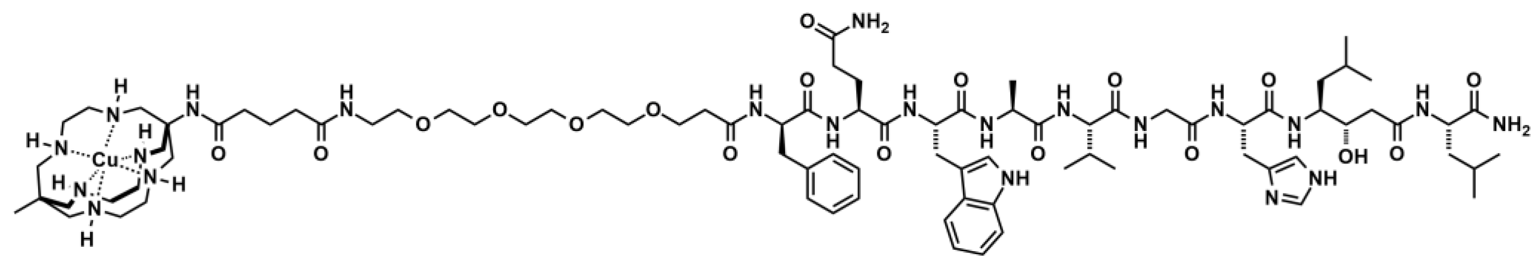
2.1. Radiochemistry
2.2. Serum Stability
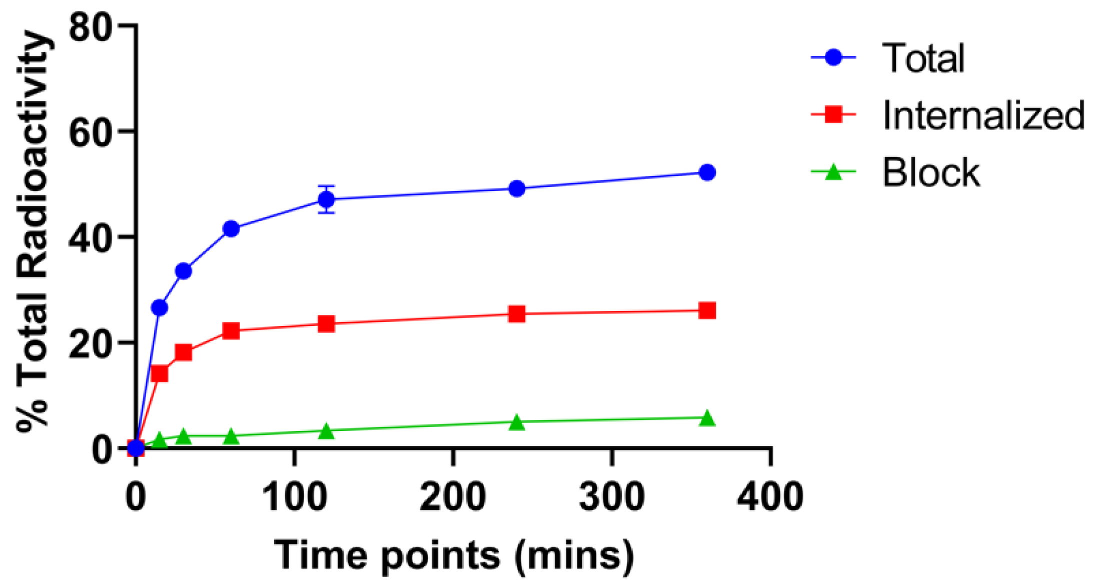
2.3. Cell Binding and Internalization
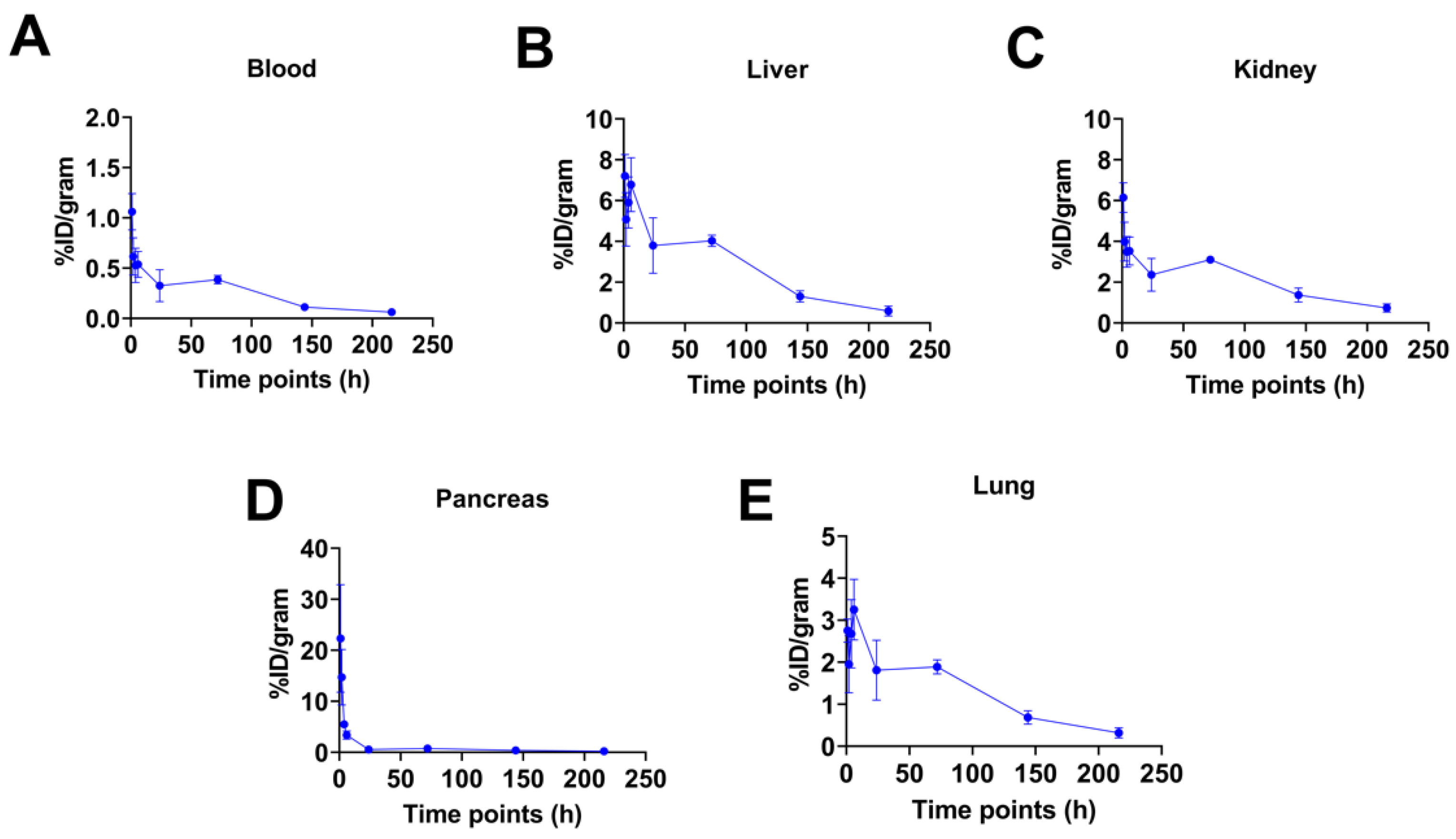
2.4. Biodistribution Studies
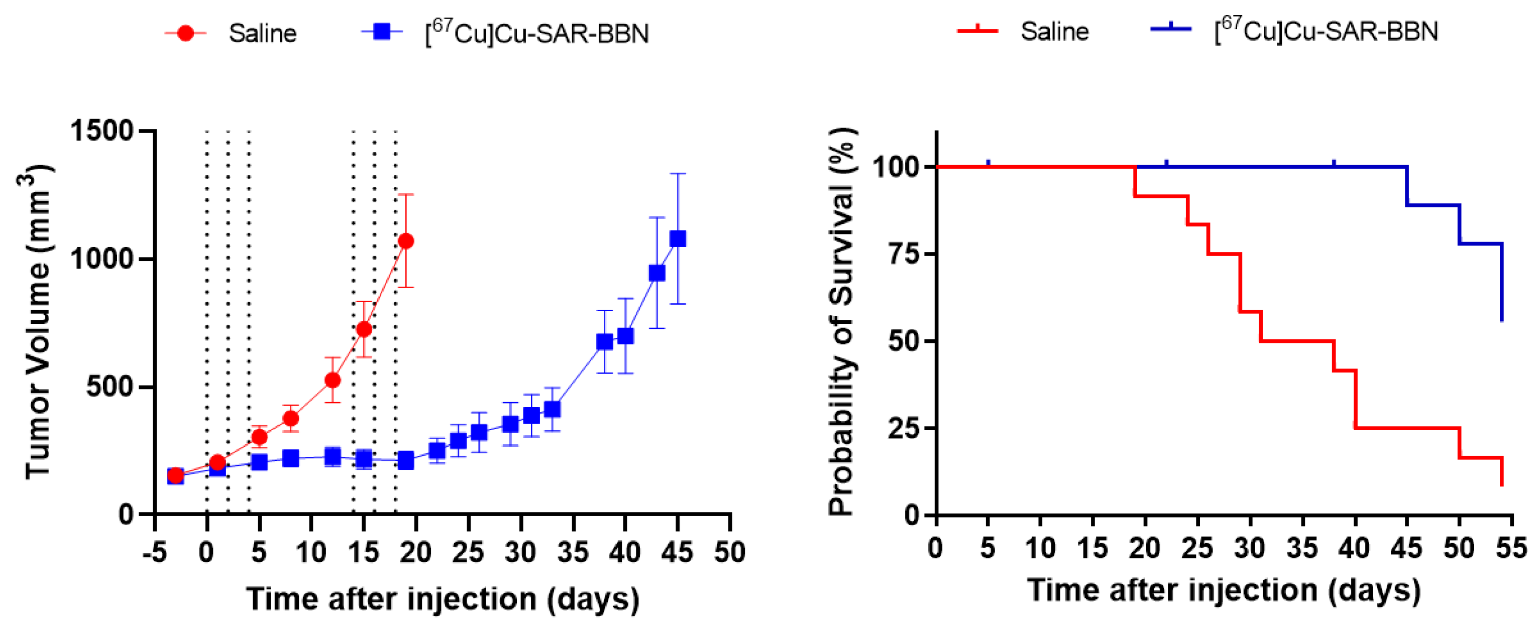
2.5. Therapy Studies
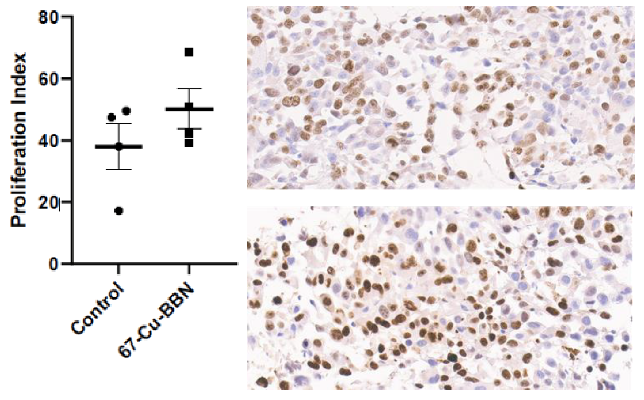
2.6. Immunohistochemical Staining Assay
3. Discussion
4. Materials and Methods
4.1. General Information
4.2. Cell Culture
4.3. Radiochemistry
4.4. Serum Stability
4.5. Cell Binding and Internalization
4.6. Biodistribution Studies
4.7. Therapy Studies
4.8. Immunohistochemical Staining Assay
4.9. Statistical Analysis
5. Conclusions
Supplementary Materials
Author Contributions
Funding
Institutional Review Board Statement
Informed Consent Statement
Data Availability Statement
Acknowledgments
Conflicts of Interest
References
- Breeman, W.A.; de Jong, M.; Erion, J.L.; Bugaj, J.E.; Srinivasan, A.; Bernard, B.F.; Kwekkeboom, D.J.; Visser, T.J.; Krenning, E.P. Preclinical comparison of (111)In-labeled DTPA- or DOTA-bombesin analogs for receptor-targeted scintigraphy and radionuclide therapy. J. Nucl. Med. 2002, 43, 1650–1656. [Google Scholar]
- Biddlecombe, G.B.; Rogers, B.E.; de Visser, M.; Parry, J.J.; de Jong, M.; Erion, J.L.; Lewis, J.S. Molecular imaging of gastrin-releasing peptide receptor-positive tumors in mice using 64Cu- and 86Y-DOTA-(Pro1,Tyr4)-bombesin(1–14). Bioconjug. Chem. 2007, 18, 724–730. [Google Scholar] [CrossRef] [PubMed]
- de Barros, A.L.B.; das Graças Mota, L.; de Aguiar Ferreira, C.; Corrêa, N.C.R.; de Góes, A.M.; Oliveira, M.C.; Cardoso, V.N. 99mTc-labeled bombesin analog for breast cancer identification. J. Radioanal. Nucl. Chem. 2013, 295, 2083–2090. [Google Scholar] [CrossRef]
- Mansi, R.; Wang, X.; Forrer, F.; Waser, B.; Cescato, R.; Graham, K.; Borkowski, S.; Reubi, J.C.; Maecke, H.R. Development of a potent DOTA-conjugated bombesin antagonist for targeting GRPr-positive tumours. Eur. J. Nucl. Med. Mol. Imaging 2011, 38, 97–107. [Google Scholar] [CrossRef] [PubMed] [Green Version]
- Fuscaldi, L.L.; de Barros, A.L.B.; Santos, C.R.d.P.; de Oliveira, M.C.; Fernandes, S.O.A.; Cardoso, V.N. Feasibility of the 99mTc-HYNIC-βAla-Bombesin(7–14) for detection of LNCaP prostate tumour in experimental model. J. Radioanal. Nucl. Chem. 2015, 305, 379–386. [Google Scholar] [CrossRef]
- Pujatti, P.B.; Foster, J.M.; Finucane, C.; Hudson, C.D.; Burnet, J.C.; Pasqualoto, K.F.M.; Mengatti, J.; Mather, S.J.; de Araújo, E.B.; Sosabowski, J.K. Evaluation and comparison of a new DOTA and DTPA-bombesin agonist in vitro and in vivo in low and high GRPR expressing prostate and breast tumor models. Appl. Radiat. Isot. 2015, 96, 91–101. [Google Scholar] [CrossRef]
- Marostica, L.L.; de Barros, A.L.; Silva, J.O.; Lopes, S.C.; Salgado, B.S.; Chondrogiannis, S.; Rubello, D.; Cassali, G.D.; Schenkel, E.P.; Cardoso, V.N.; et al. Feasibility study with 99mTc-HYNIC-βAla-Bombesin(7–14) as an agent to early visualization of lung tumour cells in nude mice. Nucl. Med. Commun. 2016, 37, 372–376. [Google Scholar] [CrossRef]
- Xiao, D.; Wang, J.; Hampton, L.L.; Weber, H.C. The human gastrin-releasing peptide receptor gene structure, its tissue expression and promoter. Gene 2001, 264, 95–103. [Google Scholar] [CrossRef]
- Aranda-Lara, L.; Ferro-Flores, G.; Azorín-Vega, E.; Ramírez, F.d.M.; Jiménez-Mancilla, N.; Ocampo-García, B.; Santos-Cuevas, C.; Isaac-Olivé, K. Synthesis and evaluation of Lys1(α,γ-Folate)Lys3(177Lu-DOTA)-Bombesin(1–14) as a potential theranostic radiopharmaceutical for breast cancer. Appl. Radiat. Isot. 2016, 107, 214–219. [Google Scholar] [CrossRef]
- Aranda-Lara, L.; Ferro-Flores, G.; Ramírez Fde, M.; Ocampo-García, B.; Santos-Cuevas, C.; Díaz-Nieto, L.; Isaac-Olivé, K. Improved radiopharmaceutical based on 99mTc-Bombesin-folate for breast tumour imaging. Nucl. Med. Commun. 2016, 37, 377–386. [Google Scholar] [CrossRef]
- Manrique-Arias, J.C.; Pitalua-Cortes, Q.; Pedrero-Piedras, R.; Rodríguez-Mena, G.i.; López, T.; Cabezas-Ortiz, C.; García-Pérez, O. Synthesis and Radiation Dosimetry of [68Ga]-Ga-Lys1, Lys3-DOTA-Bombesin (1,14) Antagonist for PET-Imaging, as a Potential Theragnostic Tracer in Oncology. J. Encapsulation Adsorpt. Sci. 2020, 10, 13. [Google Scholar]
- Abd-Elgaliel, W.R.; Gallazzi, F.; Garrison, J.C.; Rold, T.L.; Sieckman, G.L.; Figueroa, S.D.; Hoffman, T.J.; Lever, S.Z. Design, synthesis, and biological evaluation of an antagonist-bombesin analogue as targeting vector. Bioconjug. Chem. 2008, 19, 2040–2048. [Google Scholar] [CrossRef] [Green Version]
- Cescato, R.; Maina, T.; Nock, B.; Nikolopoulou, A.; Charalambidis, D.; Piccand, V.; Reubi, J.C. Bombesin Receptor Antagonists May Be Preferable to Agonists for Tumor Targeting. J. Nucl. Med. 2008, 49, 318–326. [Google Scholar] [CrossRef] [Green Version]
- Gourni, E.; Del Pozzo, L.; Kheirallah, E.; Smerling, C.; Waser, B.; Reubi, J.-C.; Paterson, B.M.; Donnelly, P.S.; Meyer, P.T.; Maecke, H.R. Copper-64 Labeled Macrobicyclic Sarcophagine Coupled to a GRP Receptor Antagonist Shows Great Promise for PET Imaging of Prostate Cancer. Mol. Pharm. 2015, 12, 2781–2790. [Google Scholar] [CrossRef]
- Liu, F.; Zhu, H.; Yu, J.; Han, X.; Xie, Q.; Liu, T.; Xia, C.; Li, N.; Yang, Z. (68)Ga/(177)Lu-labeled DOTA-TATE shows similar imaging and biodistribution in neuroendocrine tumor model. Tumour Biol. 2017, 39, 1010428317705519. [Google Scholar] [CrossRef] [Green Version]
- Bentancor, N.; Trindade, V.; Vasilskis, E.; Sanz, I.s.; Balter, H.; Engler, H. 68Ga-PSMA and 177Lu-PSMA of high specific activity for targeted diagnosis and therapy of prostate cancer in patients. J. Nucl. Med. 2017, 58, 686. [Google Scholar]
- Dalm, S.U.; Bakker, I.L.; de Blois, E.; Doeswijk, G.N.; Konijnenberg, M.W.; Orlandi, F.; Barbato, D.; Tedesco, M.; Maina, T.; Nock, B.A.; et al. 68Ga/177Lu-NeoBOMB1, a Novel Radiolabeled GRPR Antagonist for Theranostic Use in Oncology. J. Nucl. Med. 2017, 58, 293–299. [Google Scholar] [CrossRef] [Green Version]
- Zia, N.; Cullinane, C.; VanZuylekom, J.; Waldeck, K.; McInnes, L.; Buncic, G.; Haskali, M.; Roselt, P.; Hicks, R.; Donnelly, P. A Bivalent Inhibitor of Prostate Specific Membrane Antigen Radiolabeled with Copper-64 with High Tumor Uptake and Retention. Angew. Chem. Int. Ed. 2019, 58, 14991–14994. [Google Scholar] [CrossRef]
- McInnes, L.E.; Cullinane, C.; Roselt, P.D.; Jackson, S.; Blyth, B.J.; van Dam, E.M.; Zia, N.A.; Harris, M.J.; Hicks, R.J.; Donnelly, P.S. Therapeutic Efficacy of a Bivalent Inhibitor of Prostate-Specific Membrane Antigen Labeled with 67Cu. J. Nucl. Med. 2021, 62, 829. [Google Scholar] [CrossRef]
- Grünberg, J.; Novak-Hofer, I.; Honer, M.; Zimmermann, K.; Knogler, K.; Bläuenstein, P.; Ametamey, S.; Maecke, H.R.; Schubiger, P.A. In vivo Evaluation of 177Lu- and 67/64Cu-Labeled Recombinant Fragments of Antibody chCE7 for Radioimmunotherapy and PET Imaging of L1-CAM-Positive Tumors. Clin. Cancer Res. 2005, 11, 5112. [Google Scholar] [CrossRef] [Green Version]
- Kelly, J.M.; Ponnala, S.; Amor-Coarasa, A.; Zia, N.A.; Nikolopoulou, A.; Williams, C.; Schlyer, D.J.; DiMagno, S.G.; Donnelly, P.S.; Babich, J.W. Preclinical Evaluation of a High-Affinity Sarcophagine-Containing PSMA Ligand for 64Cu/67Cu-Based Theranostics in Prostate Cancer. Mol. Pharm. 2020, 17, 1954–1962. [Google Scholar] [CrossRef]
- Voss, S.D.; Smith, S.V.; DiBartolo, N.; McIntosh, L.J.; Cyr, E.M.; Bonab, A.A.; Dearling, J.L.J.; Carter, E.A.; Fischman, A.J.; Treves, S.T.; et al. Positron emission tomography (PET) imaging of neuroblastoma and melanoma with 64Cu-SarAr immunoconjugates. Proc. Natl. Acad. Sci. USA 2007, 104, 17489–17493. [Google Scholar] [CrossRef] [Green Version]
- Cai, H.; Fissekis, J.; Conti, P.S. Synthesis of a novel bifunctional chelator AmBaSar based on sarcophagine for peptide conjugation and (64)Cu radiolabelling. Dalton Trans. 2009, 27, 5395–5400. [Google Scholar] [CrossRef]
- Cai, H.; Li, Z.; Huang, C.W.; Shahinian, A.H.; Wang, H.; Park, R.; Conti, P.S. Evaluation of copper-64 labeled AmBaSar conjugated cyclic RGD peptide for improved microPET imaging of integrin alphavbeta3 expression. Bioconjug. Chem. 2010, 21, 1417–1424. [Google Scholar] [CrossRef]
- Smith, S.V. Molecular imaging with copper-64. J. Inorg. Biochem. 2004, 98, 1874–1901. [Google Scholar] [CrossRef]
- Kähkönen, E.; Jambor, I.; Kemppainen, J.; Lehtiö, K.; Grönroos, T.J.; Kuisma, A.; Luoto, P.; Sipilä, H.J.; Tolvanen, T.; Alanen, K.; et al. In Vivo Imaging of Prostate Cancer Using [68Ga]-Labeled Bombesin Analog BAY86-7548. Clin. Cancer Res. 2013, 19, 5434. [Google Scholar] [CrossRef] [Green Version]
- Sah, B.-R.; Burger, I.A.; Schibli, R.; Friebe, M.; Dinkelborg, L.; Graham, K.; Borkowski, S.; Bacher-Stier, C.; Valencia, R.; Srinivasan, A.; et al. Dosimetry and First Clinical Evaluation of the New 18F-Radiolabeled Bombesin Analogue BAY 864367 in Patients with Prostate Cancer. J. Nucl. Med. 2015, 56, 372. [Google Scholar] [CrossRef] [Green Version]
- Van de Wiele, C.; Dumont, F.; Vanden Broecke, R.; Oosterlinck, W.; Cocquyt, V.; Serreyn, R.; Peers, S.; Thornback, J.; Slegers, G.; Dierckx, R.A. Technetium-99m RP527, a GRP analogue for visualisation of GRP receptor-expressing malignancies: A feasibility study. Eur. J. Nucl. Med. 2000, 27, 1694–1699. [Google Scholar] [CrossRef]
- Lohrmann, C.; Zhang, H.; Thorek, D.L.J.; Desai, P.; Zanzonico, P.B.; O’Donoghue, J.; Irwin, C.P.; Reiner, T.; Grimm, J.; Weber, W.A. Cerenkov Luminescence Imaging for Radiation Dose Calculation of a 90Y-Labeled Gastrin-Releasing Peptide Receptor Antagonist. J. Nucl. Med. 2015, 56, 805. [Google Scholar] [CrossRef] [Green Version]
- Bandara, N.; Stott Reynolds, T.J.; Schehr, R.; Bandari, R.P.; Diebolder, P.J.; Krieger, S.; Xu, J.; Miao, Y.; Rogers, B.E.; Smith, C.J. Matched-pair, (86)Y/(90)Y-labeled, bivalent RGD/bombesin antagonist, [RGD-Glu-[DO3A]-6-Ahx-RM2], as a potential theranostic agent for prostate cancer. Nucl. Med. Biol. 2018, 62–63, 71–77. [Google Scholar] [CrossRef]
- Kurth, J.; Krause, B.J.; Bergner, C.; Schwarzenboeck, S.; Heuschkel, M. First in human dosimetry of [177Lu]RM2: A gastrin-releasing peptide receptor antagonist for targeted radiotherapy of metastasized castration resistant prostate cancer. J. Nucl. Med. 2019, 60, 271. [Google Scholar]
- Cui, L.; Liu, Z.; Jin, X.; Jia, B.; Li, F.; Wang, F. Evaluation of 188Re-MAG2-RGD-bombesin for potential prostate cancer therapy. Nucl. Med. Biol. 2013, 40, 182–189. [Google Scholar] [CrossRef]
- Cooper, M.S.; Ma, M.T.; Sunassee, K.; Shaw, K.P.; Williams, J.D.; Paul, R.L.; Donnelly, P.S.; Blower, P.J. Comparison of (64)Cu-complexing bifunctional chelators for radioimmunoconjugation: Labeling efficiency, specific activity, and in vitro/in vivo stability. Bioconjug. Chem. 2012, 23, 1029–1039. [Google Scholar] [CrossRef] [PubMed] [Green Version]
- Dumont, R.A.; Tamma, M.; Braun, F.; Borkowski, S.; Reubi, J.C.; Maecke, H.; Weber, W.A.; Mansi, R. Targeted radiotherapy of prostate cancer with a gastrin-releasing peptide receptor antagonist is effective as monotherapy and in combination with rapamycin. J. Nucl. Med. 2013, 54, 762–769. [Google Scholar] [CrossRef] [PubMed] [Green Version]
- Montemagno, C.; Raes, F.; Ahmadi, M.; Bacot, S.; Debiossat, M.; Leenhardt, J.; Boutonnat, J.; Orlandi, F.; Barbato, D.; Tedesco, M.; et al. In Vivo Biodistribution and Efficacy Evaluation of NeoB, a Radiotracer Targeted to GRPR, in Mice Bearing Gastrointestinal Stromal Tumor. Cancers 2021, 13, 1051. [Google Scholar] [CrossRef]


Publisher’s Note: MDPI stays neutral with regard to jurisdictional claims in published maps and institutional affiliations. |
© 2022 by the authors. Licensee MDPI, Basel, Switzerland. This article is an open access article distributed under the terms and conditions of the Creative Commons Attribution (CC BY) license (https://creativecommons.org/licenses/by/4.0/).
Share and Cite
Huynh, T.T.; van Dam, E.M.; Sreekumar, S.; Mpoy, C.; Blyth, B.J.; Muntz, F.; Harris, M.J.; Rogers, B.E. Copper-67-Labeled Bombesin Peptide for Targeted Radionuclide Therapy of Prostate Cancer. Pharmaceuticals 2022, 15, 728. https://doi.org/10.3390/ph15060728
Huynh TT, van Dam EM, Sreekumar S, Mpoy C, Blyth BJ, Muntz F, Harris MJ, Rogers BE. Copper-67-Labeled Bombesin Peptide for Targeted Radionuclide Therapy of Prostate Cancer. Pharmaceuticals. 2022; 15(6):728. https://doi.org/10.3390/ph15060728
Chicago/Turabian StyleHuynh, Truc T., Ellen M. van Dam, Sreeja Sreekumar, Cedric Mpoy, Benjamin J. Blyth, Fenella Muntz, Matthew J. Harris, and Buck E. Rogers. 2022. "Copper-67-Labeled Bombesin Peptide for Targeted Radionuclide Therapy of Prostate Cancer" Pharmaceuticals 15, no. 6: 728. https://doi.org/10.3390/ph15060728