Article

An Albumin-Binding PSMA Ligand with Higher Tumor Accumulation for PET Imaging of Prostate Cancer

1
Key Laboratory of Carcinogenesis and Translational Research (Ministry of Education/Beijing), Key Laboratory for Research and Evaluation of Radiopharmaceuticals (National Medical Products Administration), Department of Nuclear Medicine, Peking University Cancer Hospital & Institute, Beijing 100142, China
2
School of Medicine, Guizhou University, Guiyang 550025, China
*
Authors to whom correspondence should be addressed.
These authors contributed equally to this work.
Pharmaceuticals 2022, 15(5), 513; https://doi.org/10.3390/ph15050513
Submission received: 16 March 2022 / Revised: 7 April 2022 / Accepted: 19 April 2022 / Published: 22 April 2022

Abstract

:
Prostate-specific membrane antigen (PSMA) is an ideal target for the diagnosis and treatment of prostate cancer. Due to the short half-life in blood, small molecules/peptides are rapidly cleared by the circulatory system. Prolonging the half-life of PSMA probes has been considered as an effective strategy to improve the tumor detection. Herein, we reported a 64Cu-labeled PSMA tracer conjugating with maleimidopropionic acid (MPA), 64Cu-PSMA-CM, which showed an excellent ability to detect PSMA-overexpressing tumors in delayed time. Cell experiments in PSMA-positive 22Rv1 cells, human serum albumin binding affinity, and micro-PET imaging studies in 22Rv1 model were performed to investigate the albumin binding capacity and PSMA specificity. Comparisons with 64Cu-PSMA-BCH were performed to explore the influence of MPA on the biological properties. 64Cu-PSMA-CM could be quickly prepared within 30 min. The uptake of 64Cu-PSMA-CM in 22Rv1 cells increased over time and it could bind to HSA with a high protein binding ratio (67.8 ± 1.5%). When compared to 64Cu-PSMA-BCH, 64Cu-PSMA-CM demonstrated higher and prolonged accumulation in 22Rv1 tumors, contributing to high tumor-to-organ ratios. These results showed that 64Cu-PSMA-CM was PSMA specific with a higher tumor uptake, which demonstrated that MPA is an optional strategy for improving the radioactivity concentration in PSMA-expressing tumors and for developing the ligands for PSMA radioligand therapy.

1. Introduction

According to the 2018 global cancer statistics, prostate cancer (PCa) ranks second in male cancer incidence and fifth in mortality [1]. Due to its high expression in prostate cancer cells, prostate-specific membrane antigen (PSMA) has been reported as an effective target for specific imaging and targeted therapy of prostate cancer [2]. In the past 20 years, extensive small-molecule inhibitors of PSMA have emerged, which demonstrated high binding affinity and PSMA specificity. Among them, radiolabeled small molecules/peptides including 68Ga/177Lu/225Ac-PSMA-617, 68Ga/177Lu/225Ac-PSMA-I&T, 68Ga-PSMA-11, 99mTc-MIP-1404, 99mTc-PSMA-I&S, 18F-DCFPyL, Al18F-PSMA-BCH, and 18F-PSMA-1007 currently dominate in clinical trials for PCa imaging or PSMA radioligand therapy (PRLT) [3,4,5,6,7,8,9,10,11,12]. In recent years, PRLT has seen a pandemic of metastatic castration-resistant prostate cancer (mCRPC) lesions, leading to at least a 50% reduction in prostate-specific antigens and showing an acceptable safety profile with negligible side effects [13]. However, as a small molecule compound, the fast clearance in blood and the particularly insufficient dose delivery to tumors leads to higher dosage administration or more frequent treatments, which may increase the possibility of adverse reactions and limit [14] the effectiveness of β-particle radiotherapy [14].
In order to improve the utilization efficiency of nuclides and enhance the tumor inhibition efficiency, some strategies for albumin binding have been developed to prolong the blood circulation time and increase radioactive accumulation in tumors [15,16]. Human serum albumin (HSA, MW 66 kDa), the most abundant protein in plasma, composed of seventeen pairs of disulfide bonds and one free cysteine (cysteine-34), is known to be a versatile carrier for drug targeting [17,18]. There has been long-term interest in improving the in vivo pharmacokinetic properties of small-molecule drugs or imaging probes by utilizing albumin’s long physiologic half-life (19 days), thereby enhancing therapeutic effect. Cysteine-34, the only free thiol group in albumin, located in the hydrophobic crevice, holds an excellent chemical characteristic for further structural modification to produce novel bioactive constructs with an extended half-life [19,20,21]. For instance, when conjugated with 4-(P-iodophenyl) butyric acid and Evans blue—both of which can bind to albumin—PSMA inhibitors could availably improve the accumulation in kidneys and increase the uptake in tumors with PSMA positive expression [15,22].
Maleimidopropionic acid (MPA) modification provides another attractive platform for targeted drug delivery, allowing for binding with albumin cysteine-34 specifically and covalently to form a thiosuccinimide bond [23]. Several successful combinations have demonstrated the benefits of combining macromolecular therapies. Timms et al. [24] confirmed that the presence of the reactive maleimidopropionic acid group, which covalently linked the peptide CJC-1295 to free cysteine-34 in albumin, could significantly prolong the activity retention of peptides in the bloodstream. Xie et al. [25] chemically modified the peptide of HIV fusion inhibitor with a single MPA exhibiting potent anti-HIV activity and a remarkably extended half-life in vivo. Feng et al. [26] developed an MPA-conjugated peptide which exhibited a prolonged half-life and enhanced anti-tumor efficiency in vivo.
We recently also reported a novel albumin-based PSMA probe Al18F-PSMA-CM. The probe was modified with MPA, which showed higher PSMA specificity and longer half-life in blood, contributing to a distinct accumulation in 22Rv1 tumors, with increasing uptake in 4 h [11,27]. These results indicated MPA may be a promising choice to develop radiopharmaceuticals for PRLT of prostate cancer. But the half-life of 18F was not enough to match the circulation of MPA-conjugated PSMA-CM, which cannot evaluate the efficiency of PSMA-CM. Compared with 18F (T1/2, 109.77 min; β+, 96.7%), 64Cu (T1/2, 12.7 h; β+, 17.4% [Emax, 0.656 MeV]; β, 39% [Emax, 0.573 MeV]) was considered to be a diagnostic nuclide with its unique decay characteristics of the matched pair 64Cu/67Cu for theranostic applications. In addition, the longer half-life of 64Cu provides an advantage for delay imaging, which may effectively monitor the uptake of radiotracers in tumors [28]. Hence, in order to match the prolonged half-life of PSMA-CM, we radiolabeled PSMA-CM with 64Cu and evaluated 64Cu-PSMA-CM in mice bearing 22Rv1 tumors, showing great potential for detecting prostate lesions in delayed imaging.

2. Results

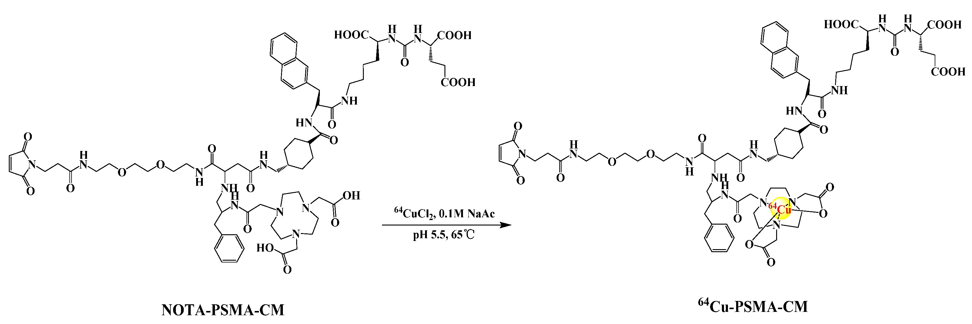
2.1. Radiosynthesis and Quality Control

The radiolabeling process of 64Cu-PSMA-CM is depicted in Figure 1. The radiochemical yield of 64Cu-PSMA-CM was over 75% and radiochemical purity was >99% after a Sep-Pak C18 cartridge purification (Figure S1). The specific activity was 15.97 ± 2.01 GBq/μmol.

2.2. In Vitro Stability

The in vitro stability of 64Cu-PSMA-CM was tested in 5% human serum albumin (HSA) and saline at 37.0 °C. As shown in Figure 2, the radiochemical purity (RCP) of 64Cu-PSMA-CM was over 95% after more than 48 h incubation in saline and 5% HSA, indicating that 64Cu-PSMA-CM was quite stable and can be applied for further study.

2.3. Partition Coefficient

The partition coefficient of 64Cu-PSMA-CM was calculated, and the log p-value was −2.25 ± 0.05, indicating that 64Cu-PSMA-CM was highly hydrophilic.

2.4. Western Blotting and In Vitro Cell Experiments

The expression of PSMA in 22Rv1 and PC3 cells were measured by a Western blot assay. The 22Rv1 cells showed significantly higher expression level of PSMA than PC3 cells with relative values of 1.23 ± 0.07 and 0.283 ± 0.07 (p = 0.00018), which indicates the high expression of PSMA in 22Rv1 cells (Figure 3a,b).
Cell uptake experiments were performed on PSMA (+) 22Rv1 and PSMA (−) PC-3 cells, as shown in Figure 3c. The uptake of 64Cu-PSMA-CM in 22Rv1 cells increased with time, it was comparable to 64Cu-PSMA-BCH (5.45 ± 0.42 %IA/106 cells vs. 6.31 ± 0.60 %IA/106 cells at 120 min, respectively). The uptake in 22Rv1 cells can be significantly blocked by ZJ-43 (blocked to 3.38 ± 0.04 IA%/106 cells, p = 0.02). However, the uptake values of 64Cu-PSMA-CM in PC-3 cells were lower than that in 22Rv1 cells, and the uptake cannot be blocked by excess of ZJ-43 (p > 0.05) (Figure 3d).

2.5. Binding Affinity Assay

Human serum albumin binding assay was performed to test the albumin binding ability of 64Cu-PSMA-CM. The protein binding ratio of 64Cu-PSMA-BCH and 64Cu-PSMA-CM were calculated as 55.9 ± 4.0% and 67.8 ± 1.5% (p = 0.003).
The binding affinity of 64Cu-PSMA-CM and 64Cu-PSMA-BCH to PSMA were conducted on 22Rv1 cells. The dissociation constant values of 64Cu-PSMA-BCH and 64Cu-PSMA-CM were 0.59 nM and 4.58 nM, respectively (Figure 4).

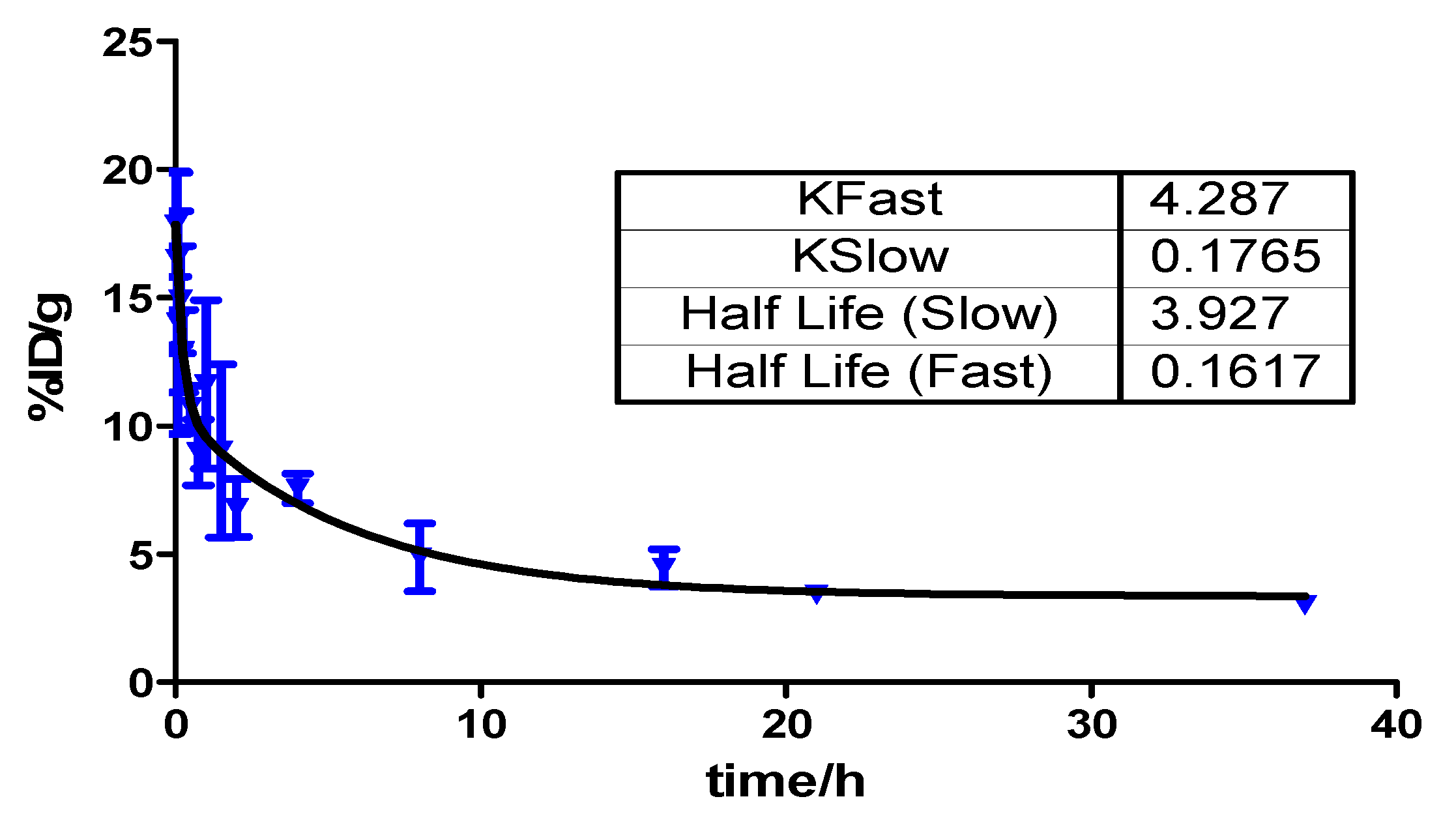
2.6. Pharmacokinetics

The pharmacokinetics profile of 64Cu-PSMA-CM in blood circulation was tested by injecting 3.7 MBq of 64Cu-PSMA-CM into Balb/c male mice (n = 4). As shown in Figure 5, the blood activity–time profile of 64Cu-PSMA-CM was: Ct = 7.21e−4.287t + 7.29e−0.176t. The half-life of the distribution phase and elimination phase were 0.16 h and 3.93 h, respectively, which significantly delayed the elimination phase compared to 64Cu-PSMA-BCH (0.208 min) [29].

2.7. Biodistribution Studies

The biodistribution studies of 64Cu-PSMA-CM were performed in 22Rv1-beared male mice. As shown in Figure 6a and Table S1, within 1 h, 64Cu-PSMA-CM rapidly accumulated in the blood pool (16.95 ± 3.05 ID%/g), liver (5.34 ± 1.13 ID%/g), and kidneys (95.84 ± 3.33 %ID/g). Subsequently, the clearance in the kidneys was fast with uptake values of 8.92 ± 0.41 %ID/g at 24 h p.i. and was accompanied by the decline of radioactivity in other organs, such as heart, lung, spleen, and blood, demonstrating that 64Cu-PSMA-CM was mainly excreted from the urinary system. The uptake in 22Rv1 tumor was increased within 24 h with a highest uptake value of 14.29 ± 1.44 %ID/g at 24 h p.i., and it decreased at 48 h p.i., advantageously providing a high tissue contrast at 24 h p.i. (Figure 6b).

2.8. Micro-PET Imaging

Micro-PET imaging of 64Cu-PSMA-CM and a comparison with 64Cu-PSMA-BCH were demonstrated on mice bearing 22Rv1 tumors. As shown in Figure 7a and Figure S2a, the heart, liver, kidneys, salivary glands, and 22Rv1 tumors were observed. Kidneys had the highest uptake of 64Cu-PSMA-CM within 12 h, followed by the heart and liver, and the radioactivity of these organs rapidly decreased over time. The accumulation in 22Rv1 tumors continuously increased until 24 h p.i. and then decreased at 48 h, which was similar to the results of previous biodistribution studies. When co-injected with excess ZJ-43 (50 μg), the uptake in tumors and kidneys significantly decreased (p < 0.001). Due to the fast clearance of 64Cu-PSMA-CM from the nontumor organs, relatively selective tumor images become quite clear at 12 h, and good contrast images in tumors was obtained at 24 h. Generally, the tumor-to-non-target organ ratios of 64Cu-PSMA-CM increased with time, and the excellent tumor-to-kidneys ratio (1.29 ± 0.05) and tumor-to-liver ratio (1.76 ± 0.08) were at 24 h p.i. (Figure S3). When compared to 64Cu-PSMA-BCH, a tracer without MPA, 64Cu-PSMA-CM demonstrated prolonged circulation in vivo and significantly higher uptake in 22Rv1 tumor and organs, such as heart, liver, and kidneys (Figure 7a,c and Figure S2). As previously mentioned, 64Cu-PSMA-CM showed the highest accumulation in 22Rv1 tumor with an SUV mean value of 1.88 ± 0.04 at 24 h p.i., whereas the peak uptake of 64Cu-PSMA-BCH was 0.84 ± 0.02 at 4 h p.i. (Figure S2). In addition, due to the fast clearance of 64Cu-PSMA-BCH from the kidneys, the tumor-to-kidney ratio was higher than 64Cu-PSMA-CM, while the tumor-to-liver ratio of 64Cu-PSMA-CM was superior to 64Cu-PSMA-BCH (Figure S3). Furthermore, immunohistochemical staining results indicated the positive expression of PSMA in 22Rv1 tumor tissue (Figure 7d), further confirming the targeted accumulation of the probe in 22Rv1 tumor.

2.9. Estimation of Radiation Dosimetry

Human organ radiation dosimetry was estimated based on the biodistribution data in mice bearing 22Rv1 tumors. As shown in Table 1, the liver received the highest absorbed dose (0.107 mGy/MBq), followed by the kidneys and gallbladder wall with absorbed doses of 0.0997 mGy/MBq and 0.0414 mGy/MBq, respectively. The effective dose of whole body is 0.0276 mSv/MBq, which was similar to 64Cu-PSMA-BCH (0.0292 mSv/MBq) [29].

3. Discussion

Due to the fast clearance in blood and particularly insufficient dose delivery to tumor of small molecule compounds 177Lu/225Ac-PSMA-617, there have been long-term efforts in developing a general strategy which can effectively prolong the compounds’ half-life in vivo. Among the reported methods, the combination of pharmaceuticals and albumin-binding molecules is one of the most commonly used methods, which has high efficiency and few side effects [17]. In the past five years, many albumin-based PSMA probes have been developed, but most of the studies focused on 4-(p-iodophenyl)butyric acid and Evans blue derivatives, aiming to improve metabolic behavior in the body [22,30,31,32]. The clinical studies of 177Lu-EB-PSMA-617 had been performed and achieved promising results in the therapy of mCRPC [21,32]. We previously confirmed maleimidopropionic acid (MPA) modification, offering an another effective platform for binding albumin and conjugating with the PSMA tracer [27]. Here, benefiting from the moderate half-life of copper-64, we developed a 64Cu-labeled MPA-modified PSMA tracer and evaluated the targeting capability of the PSMA-tumor.
64Cu-PSMA-CM was prepared with high radiochemical yield, high radiochemical purity, and moderate specific activity. It was quite stable in vitro, which meets the criteria of quality control and can be used for further studies.
When compared with 64Cu-PSMA-BCH, a PSMA probe without MPA moiety, 64Cu-PSMA-CM showed a higher human serum albumin binding ratio and longer half-life. This indicated that the conjugation of MPA indeed enhanced the binding ability of the tracer to HSA and extended the circulation in blood, which was expected to offset the defect of the rapid clearance of small molecule inhibitors. Although the binding affinity of 64Cu-PSMA-CM to PSMA decreased, the Kd value was still at the level of nanomole, which was comparable with other reported PSMA probes.
64Cu-PSMA-CM was specifically accumulated in PSMA (+) 22Rv1 cells and increased over time with the highest uptake value of 5.45 ± 0.42 IA%/106 cells at 120 min, which was significantly higher than that in PSMA (−) PC-3 cells (1.79 ± 0.20 IA%/106 cells). It can be specifically blocked to 3.38 ± 0.04 IA%/106 cells by PSMA inhibitor, ZJ-43, indicating the high specificity of 64Cu-PSMA-CM to PSMA. The comparable uptake values of 64Cu-PSMA-CM and 64Cu-PSMA-BCH in PSMA (+) 22Rv1 cells indicated that the conjugation of MPA moiety had little effect on the specificity and binding affinity, or for the uptake in cells.
Both biodistribution and micro-PET imaging studies in 22Rv1-beared mice confirmed that 64Cu-PSMA-CM was highly accumulated in PSMA (+) tumors and washed out from normal organs at delayed time points. This resulted in a continuous increase of tumor-to-organ ratio, prospectively facilitating the observation of more tumor lesions. longer circulation in the blood resulted in longer retention of radioactivity in the kidneys and liver.
When compared with 64Cu-PSMA-BCH, 22Rv1 tumors showed excellent permeability and retention of 64Cu-PSMA-CM, which may improve the contrast of tumors to non-target organs. The higher uptake in tumors and comparable uptake in cells indicated that the higher uptake in PSMA (+) positive tumors was due to the prolonged circulation of 64Cu-PSMA-CM in the blood. In addition, the significantly higher tumor accumulation in delayed time demonstrated that the compound fulfilled the prerequisites for dosimetry in the course of therapy planning with 67Cu. It also showed that modification with MPA is a good strategy to improve radioactivity accumulation in PSMA (+) tumors, which may effectively guide the development of 177Lu or 225Ac labeled tracers for PRLT.

4. Materials and Methods

4.1. General Materials

64Cu was produced by the HM-20 cyclotron via 64Ni (p,n) 64Cu reaction at Beijing Cancer Hospital. Human serum albumin (HSA) was purchased from Chengdu Rongsheng Pharmaceuticals Co., Ltd. (Chengdu, China). ZJ-43 (N-[[[(1S)1-Carboxy-3-methylbutyl]amino]-carbonyl]-L-glutamic acid) was purchased from Tocris Bioscience (Bristol, UK). The precursor, NOTA-PSMA-BCH (BCH is Beijing Cancer Hospital) and PSMA-CM (CM stands for Covalent binding with Maleimidopropionic acid) were acquired from the China Peptides Company. Acetonitrile and trifluoroacetic acid (TFA) were purchased from Honeywell International Inc. (Elmsford, New York, NY, USA) and Shanghai Aladdin Biochemical Technology Co., Ltd. (Shanghai, China), respectively. Sep-Pak C18-Light cartridges were purchased from Waters (Leinster, Ireland). The Radio-TLC was conducted on an AR 2000 system (Bioscan, Poway, CA, USA). Filter paper was used as carrier and the eluant was a mixture of saturated EDTA and saline. Micro-PET was performed on Super Nova PET/CT (PINGSENG, Shanghai, China).

4.2. Radiosynthesis and Quality Control of 64Cu-PSMA-CM

The precursor, NOTA-PSMA-CM, was synthesized using a solid phase platform. Briefly speaking, the PSMA-targeting urea-based pharmacophore L-Glu-NH-CO-NH-L-Lys- was protected by resinimmobilized ε-allyloxycarbonyl lysine. The dehydrated product of 3-maleimidopropionic acid (3-(2,5-dioxo-2,5-dihydro-1H-pyrrol-1-yl)propanoic acid) and 1-hydroxy-1h-pyrrole -2,5-dione reacted with 2,2’-(ethane-1,2-diylbis(oxy))bis(Ethan-1-amine) to form substrates 1-(3-methylene-6-(2-propoxyethoxy) hexyl) 1H-pyrrole-2, 5-dione3-Maleimidopropionic acid. And then, the compound was reacted with NOTA-chelated 2,5-dioxo-2,5-dihydro-1H-pyrrol-1-yl4-(allyloxy)-2-(2-formamido-3-phenylpropanamido)pent-4-enoate. After the catalytic reaction of tetrakis(Triphenylphosphine)palladium and benzylamine, the compound was mixed with the reagents of Fmoc-2-Nal-OH (2-({[(9H-fluoren9-yl)methoxy]carbonyl}amino) -3-(naphthalen-2-yl), Fmoc-tranexamic acid {(1r,4r)-4-[({[(9H-fluoren-9- yl)methoxy]carbonyl}amino)methyl]cyclohexane-1-carboxylic acid} and then 2,2’- (7- (4-benzyl-7-(2-((((1S, 4s)–4-(((R)-1–carboxy–2-(naphthalen–2-yl)ethyl)carbamoyl) cyclohexyl)methyl)amino)–2-oxoethyl)–21-(2,5–dioxo-2,5-dihydro-1H-pyrrol-1-y)-2,8,19-trioxo-12,15-dioxa-3,6,9,18-tetraazahenicosyl)-1,4,7-triazonane-1,4-diyl)diacetic acid was obtained. Finally, the compound was reacted with PSMA-targeting urea-based pharmacophore L-Glu-NH-CO-NH-L-Lys- by a series of chemical reactions and carboxyl deprotection to obtain the final product, (((1S)–5-((2R)–2-((1s,4S)–4 -(5-((2-(2-(4,7-bis(carboxymethyl)-1,4,7–triazonan–1-yl)acetamido)–3-phenylpropyl)amino)–19-(2,5-dioxo-2,5-dihydro-1H–pyrrol–1-yl)-3,6,17–trioxo-10,13–dioxa-2,7,16-triazanonadecyl)cyclohexane-1-carboxamido)-3-(naphthalen-2-yl)propanamido)-1-carboxypentyl)carbamoyl)-L-glutamic acid (NOTA-PSMA-CM). The details of the synthesis refer to previous reports [27].64CuCl2 was obtained as solution in 0.1 M HCl. 50 μL 64CuCl2 solution, 200 μL NaAc buffer (0.1 M, pH = 5.5) and 4 μL PSMA-CM (4 mM in water, 16 nM) were mixed and the mixture was heated at 95 °C for 15 min. After reaction, the mixture was loaded into the syringe with 2 mL of H2O, then passed through a Sep-Pak C18-Light Cartridge (pretreated with 5 mL ethanol and 10 mL H2O, respectively) and the C18-Light Cartridge was washed with 5 mL of H2O. The product was obtained by eluting the C18-Light Cartridge with 0.5 mL 80% ethanol, diluting with 4 mL 0.9% saline.
Quality control of 64Cu-PSMA-CM was performed by radio-high-performance liquid chromatography (radio-HPLC) and radio-thin-layer-chromatography (radio-TLC). Radio-HPLC was performed on a C18 cartridge (ZORBAX 300SB-C18, 4.6 mm × 250 mm, 5 μm; Agilent, Palo Alto, California, CA, USA) with the mobile phases of (A) H2O and (B) acetonitrile mixed with 0.1% trifluoracetic acid (TFA) using a linear AB gradient (15–60% of B in 15 min) with a flow of 1 mL/min and the UV of 280 nm. Radio-TLC was conducted on an AR 2000 systemwith the carrier of filter paper and the eluant of a mixture of saturated EDTA and saline.

4.3. In Vitro Stability

64Cu-PSMA-CM was incubated in saline or 5% human serum albumin (HSA) at 37 °C and the radiochemical purity was determined by radio-HPLC at different points in time (1 h, 4 h, 12 h, 24 h, and 48 h). For the stability in 5% HSA, 15 μL of mixture was taken out and precipitated by ethanol, filtered, and the supernatant was analyzed.

4.4. Partition Coefficient

Amounts of 10 μL of 64Cu-PSMA-CM in saline (0.37 MBq), 990 μL of phosphate-buffered saline (PBS, 0.1 M, pH 7.4), and 1mL of octanol were mixed in a tube. The mixture was vortexed for 3 min and centrifuged (3000 rpm × 5 min). Then, five samples (100 μL) from each phase were removed and the radioactivity was measured. The experiment was repeated three times. The partition coefficient value was calculated as below: P = (average of counts in octanol/average of counts in PBS), the value was expressed as log P ± SD.

4.5. Cell Culture and Animal Models

Human prostate cancer cell lines (22Rv1 and PC-3) were obtained from the Stem Cell Bank, Chinese Academy of Science. Both cell lines were cultured in RPMI 1640 medium containing 10% fetal bovine serum (FBS) and 1% penicillin–streptomycin (Biological Industries, Shanghai, China). The cells were cultured in a humidified incubator at 37 °C with 5% CO2.
Healthy male KM mice and BALB/c nude mice aged 6–8 weeks were acquired from Vital River Laboratory Animal Technology Co., Ltd. (Beijing, China). Healthy male KM mice were used for the pharmacokinetics experiment. About 2 × 10⁶ cells of 22Rv1 in RPMI 1640 medium were subcutaneously injected into the front left of the BALB/c nude mice. When the tumors reached a size of approximately 0.5–1 cm3 in volume, the mice were used for further animal studies including biodistribution and micro-PET imaging. All animal experiments were conducted with the approval of the Ethics Committee of Peking University Cancer Hospital.

4.6. Western Blotting and Cell Uptake

The Western blotting assay was conducted on 22Rv1 and PC3 cell lines to verify the expression of PSMA. The details are described in the supporting information.
The 22Rv1 and PC-3 cell lines were plated on 24-well plates (2 × 10⁵ cells/well). After incubating for 24 h, the medium was removed and washed with PBS (phosphate buffer saline, 0.01 M). Then, 500 μL medium with 74 KBq of 64Cu-PSMA-CM was added to the well plates and co-incubated with cells at 37 °C for 10 min, 30 min, 60 min, and 120 min. The medium was then removed and the cells were washed twice with 1 mL of cold PBS. After that, cells were lysed with 1M NaOH, and then the NaOH solution was collected for analysis. For the blocking experiment, cells were co-incubated with ZJ-43 (1 µg/well), a PSMA inhibitor, for 120 min. Five samples of 64Cu-PSMA-CM were taken out and were thought to be standard. The result was expressed as %IA/106 cells. For comparison, the cell uptake experiment of 64Cu-PSMA-BCH in 22Rv1 cells was performed.

4.7. Binding Affinity Assay

In order to test the HSA protein binding ratio, 10 μL of 64Cu-PSMA-CM (0.37 MBq) was added to 0.2 mL of 20% HSA (n = 5) and incubated at 37 °C for 4 h. Then the protein was precipitated by 0.5 mL of ethanol, centrifuged (5000 rpm), washed by saline (2 × 0.2 mL), and centrifuged twice, then measured for the radioactivity of the precipitation and supernatant by a γ-counter. The result was presented as the percentage of precipitation counts/(precipitation counts + supernatant counts). The human serum albumin binding ability of 64Cu-PSMA-BCH was measured with the same protocol.
The dissociation constant of 64Cu-PSMA-CM was performed on 22Rv1 cells by adding different concentrations of 64Cu-PSMA-CM (0.185–74 kBq/mL, 400 μL/well, 4 well/group) to the 24-well plate (2 × 10⁵ cells/well). After incubation for 2 h at 37 °C, the medium was removed, the cells were washed twice with cold PBS and then lysed by 0.5 mL NaOH. The radioactivity of NaOH solution was measured and the dissociation constant was calculated using the one-site total model in Graph Pad software (Graph Pad prism 5). Similarly, the dissociation constant of 64Cu-PSMA-CM was determined with the same protocol.

4.8. Pharmacokinetics in Blood

An amount of 3.7 MBq (250 μL) of 64Cu-PSMA-CM was intravenously injected into Babl/c male mice (n = 4). Blood was collected from the ophthalmic artery at different time points, then the radioactivity of blood was weighed and measured by a γ-counter. An injection of 2.5 μL was taken out as standard. The results were expressed as the percentage of injected dose per gram (%ID/g).
A two-phase decay Equation (1) in Graph Pad software (Graph Pad prism 5) was used to serve as the two-compartment model which models biological process of the probe by fitting the ID%/g versus to time of 64Cu-PMA-CM to describe blood pharmacokinetics.
Ct = Aeαt + Beβt
where A and B are relevant constants, and α and β are rate constants which were used to calculate as ln(2)/α and ln(2)/β for the half-life of distribution phase and elimination phase, respectively.

4.9. Biodistribution

Twelve 22Rv1 tumor model mice were randomly divided into four groups and then 64Cu-PSMA-CM (200 μL, 0.74 MBq/mouse) was injected via tail vein. After 1 h, 6 h, 24 h, and 48 h, the mice were sacrificed by cervical dislocation. Organs of interest including blood, heart, liver, spleen, lung, kidney, small intestine, large intestine, muscle, brain, and 22Rv1 tumor were collected. The organs were then weighed and measured for their radioactivity. Five samples of the 1% injection volume were taken out as a standard. The final uptake values of each organ were expressed as the percentage of injected dose per gram (%ID/g).

4.10. Micro-PET Imaging and Immunohistochemical Staining

An amount of 200 μL of 64Cu-PSMA-CM (9.32 MBq) was injected into mice bearing 22Rv1 xenograft tumors via tail vein. For blocking, the mice were co-injected with 50 μg of ZJ-43. Imaging was performed on Super Nova PET/CT (PINGSENG, Shanghai, China). The mice were anaesthetized with 3% (v/v) isoflurane, then fixed on the bed at 4 h, 8 h, 12 h, 24 h, and 48 h p.i. Micro-PET scans were conducted with continuous 1% (v/v) isoflurane for 15 min. The images of 64Cu-PSMA-CM were obtained at different time points and SUV mean values of regions of interest (ROIs) over the heart, kidney, muscle, liver, and tumor were collected. The ratio of tumor-to-non-target organs was then calculated at different time points. To better assess its anti-tumor efficiency in vivo, a comparison with 64Cu-PSMA-BCH (5.55 MBq/mouse) of micro-PET imaging was performed in mice bearing 22Rv1 tumor models at 4 h p.i., 12 h p.i., and 24 h p.i. The mean standardized uptake value (SUVmean) in each organ was calculated according to following Formula (2) [7].
SUV = Activity VOI (MBq/mL)/Injected dose (MBq) × Body weight (g)
After micro-PET imaging, the 22Rv1 mice were sacrificed by cervical dislocation, and the 22Rv1 tumor tissues were quickly collected and immersed in formalin solution for further immunohistochemical staining. The details are presented in the supporting information.

4.11. Estimation of Radiation Dosimetry in Human Organs

The fraction of radioactivity uptake in human tissues was calculated according to the biodistribution results in mice bearing 22Rv1 tumors. The time–activity curves of various organs and the whole body were obtained and then calculated the area under the curves (AUCs) of different organs with origin 2019b software (OriginLab, Northampton, MA, USA). The results were further analyzed with OLINDA/EXM software (version 2.2; HERMES Medical Solutions AB) to estimate the radiation dosimetry of each organ and effective dose.

4.12. Statistical Analysis

All quantified data were expressed as the mean ± SD. The significant differences of cell uptake and human serum albumin binding affinity were calculated by two-tailed, paired t-test. Quantified ROI data based on the results of micro-PET imaging were analyzed using two-way ANOVA. Statistical analysis was performed using the Microsoft Excel 2016 software program (Microsoft Corporation) or Graph Pad software (Graph Pad prism 5). The p values less than 0.05 were considered statistically significant.

5. Conclusions

In this study, we successfully developed an albumin-based PSMA probe conjugated with MPA, 64Cu-PSMA-CM. It exhibited good physicochemical and biological properties. It showed a longer half-life in blood, a high affinity to PSMA, and a higher uptake in PSMA (+) tumors—which effectively monitored the change of radioactivity accumulation of PSMA-targeted tracers in tumors. The probe offers a good strategy and was initially verified for the potential to develop radiotracers for PRLT for prostate cancer.

Supplementary Materials

The following supporting information can be downloaded at: https://www.mdpi.com/article/10.3390/ph15050513/s1, Figure S1: Quality control of 64Cu-PSMA-CM. (a) The radio-chemical purification of 64Cu-PSMA-CM by Radio-HPLC analysis before purification. The radio-chemical purification of 64Cu-PSMA-CM after purification by Radio-HPLC (b) and radio-TLC (c) analysis; Figure S2: The SUV mean values of 64Cu-PSMA-CM (a) and 64Cu-PSMA-BCH (b) in organs and 22Rv1 tumor at different time points according to images in 22Rv1 xenograft nude mice. ***: p < 0.001; Figure S3: The tumor-to-kidney (a) and tumor-to-liver (b) ratios of 64Cu-PSMA-CM and 64Cu-PSMA-BCH at different time points according to images in 22Rv1 xenograft nude mice; Table S1: Biodistribution of 64Cu-PSMA-CM in 22Rv1 male mice (ID%/g, X ± SD, n = 3).

Author Contributions

Conceptualization, T.L., H.Z. and Z.Y.; data curation, Y.R., T.L., C.L. and X.G.; formal analysis, Y.R., X.G. and F.W.; funding acquisition, T.L., C.L., H.Z. and Z.Y.; investigation, Y.R., T.L., C.L., X.G. and F.W.; methodology, Y.R., T.L., C.L., H.Z. and Z.Y.; software, X.G. and F.W.; supervision, H.Z. and Z.Y.; validation, Y.R., T.L., C.L., X.G. and F.W.; visualization, Y.R., T.L. and C.L.; writing—original draft, Y.R. and T.L.; writing—review & editing, H.Z. and Z.Y. All authors have read and agreed to the published version of the manuscript.

Funding

This research was funded by the Peking University Medicine Fund of Fostering Young Scholars’ Scientific & Technological Innovation (No. BMU2022PY011), the National Natural Science Foundation of China projects (No. 82102092), the Beijing Natural Science Foundation (No. 7202028), the Beijing Municipal Administration of Hospitals-Yangfan Project (No. ZYLX201816), the Beijing Hospitals Authority Dengfeng Project (No. DFL20191102), and the Beijing Millions of Talent Projects A Level Funding (No. 2019A38).

Institutional Review Board Statement

The animal study was reviewed and approved by Peking University Cancer Hospital Animal Care and Use Committee (approval number: EAEC201910, approval date: 25 October 2019).

Informed Consent Statement

Informed consent was obtained from all subjects involved in the study.

Data Availability Statement

Data is contained within the article and supplementary material.

Conflicts of Interest

The authors declare no conflict of interest.

References

  1. Bray, F.; Ferlay, J.; Soerjomataram, I.; Siegel, R.L.; Torre, L.A.; Jemal, A. Global cancer statistics 2018: GLOBOCAN estimates of incidence and mortality worldwide for 36 cancers in 185 countries. CA Cancer J. Clin. 2018, 68, 394–424. [Google Scholar] [CrossRef] [PubMed] [Green Version]
  2. Okarvi, S.M. Recent developments of prostate-specific membrane antigen (PSMA)-specific radiopharmaceuticals for precise imaging and therapy of prostate cancer: An overview. Clin. Transl. Imaging 2019, 7, 189–208. [Google Scholar] [CrossRef]
  3. Hofman, M.S.; Emmett, L.; Sandhu, S.; Iravani, A.; Joshua, A.M.; Goh, J.C.; Pattison, D.A.; Tan, T.H.; Kirkwood, I.D.; Ng, S.; et al. [177Lu]Lu-PSMA-617 versus cabazitaxel in patients with metastatic castration-resistant prostate cancer (TheraP): A randomised, open-label, phase 2 trial. Lancet 2021, 397, 797–804. [Google Scholar] [CrossRef]
  4. Zacho, H.D.; Ravn, S.; Afshar-Oromieh, A.; Fledelius, J.; Ejlersen, J.A.; Petersen, L.J. Added value of (68)Ga-PSMA PET/CT for the detection of bone metastases in patients with newly diagnosed prostate cancer and a previous (99m)Tc bone scintigraphy. EJNMMI Res. 2020, 10, 31. [Google Scholar] [CrossRef] [PubMed]
  5. Werner, P.; Neumann, C.; Eiber, M.; Wester, H.J.; Schottelius, M. [(99cm)Tc]Tc-PSMA-I&S-SPECT/CT: Experience in prostate cancer imaging in an outpatient center. EJNMMI Res. 2020, 10, 45. [Google Scholar] [CrossRef]
  6. Schmidkonz, C.; Götz, T.I.; Atzinger, A.; Ritt, P.; Prante, O.; Kuwert, T.; Bäuerle, T.; Goebell, P.; Cordes, M. 99mTc-MIP-1404 SPECT/CT for Assessment of Whole-Body Tumor Burden and Treatment Response in Patients with Biochemical Recurrence of Prostate Cancer. Clin. Nucl. Med. 2020, 45, e349–e357. [Google Scholar] [CrossRef]
  7. Piron, S.; Verhoeven, J.; Descamps, B.; Kersemans, K.; De Man, K.; Van Laeken, N.; Pieters, L.; Vral, A.; Vanhove, C.; De Vos, F. Intra-individual dynamic comparison of (18)F-PSMA-11 and (68)Ga-PSMA-11 in LNCaP xenograft bearing mice. Sci. Rep. 2020, 10, 21068. [Google Scholar] [CrossRef]
  8. Liu, C.; Liu, T.; Zhang, Z.; Zhang, N.; Du, P.; Yang, Y.; Liu, Y.; Yu, W.; Li, N.; Gorin, M.A.; et al. PSMA PET/CT and standard plus PET/CT-Ultrasound fusion targeted prostate biopsy can diagnose clinically significant prostate cancer in men with previous negative biopsies. J. Nucl. Med. 2020, 61, 1314–1319. [Google Scholar] [CrossRef]
  9. Lawal, I.O.; Mokoala, K.M.G.; Mahapane, J.; Kleyhans, J.; Meckel, M.; Vorster, M.; Ebenhan, T.; Rösch, F.; Sathekge, M.M. A prospective intra-individual comparison of [68Ga]Ga-PSMA-11 PET/CT, [68Ga]Ga-NODAGAZOL PET/CT, and [99mTc]Tc-MDP bone scintigraphy for radionuclide imaging of prostate cancer skeletal metastases. Eur. J. Nucl. Med. Mol. Imaging 2021, 48, 134–142. [Google Scholar] [CrossRef]
  10. Hofman, M.S.; Lawrentschuk, N.; Francis, R.J.; Tang, C.; Vela, I.; Thomas, P.; Rutherford, N.; Martin, J.M.; Frydenberg, M.; Shakher, R.; et al. Prostate-specific membrane antigen PET-CT in patients with high-risk prostate cancer before curative-intent surgery or radiotherapy (proPSMA): A prospective, randomised, multicentre study. Lancet 2020, 395, 1208–1216. [Google Scholar] [CrossRef]
  11. Liu, T.; Liu, C.; Xu, X.; Liu, F.; Guo, X.; Li, N.; Wang, X.; Yang, J.; Yang, X.; Zhu, H.; et al. Preclinical Evaluation and Pilot Clinical Study of Al18F-PSMA-BCH for Prostate Cancer PET Imaging. J. Nucl. Med. 2019, 60, 1284–1292. [Google Scholar] [CrossRef] [PubMed] [Green Version]
  12. Giesel, F.L.; Hadaschik, B.; Cardinale, J.; Radtke, J.; Vinsensia, M.; Lehnert, W.; Kesch, C.; Tolstov, Y.; Singer, S.; Grabe, N. F-18 labelled PSMA-1007: Biodistribution, radiation dosimetry and histopathological validation of tumor lesions in prostate cancer patients. Eur. J. Nucl. Med. Mol. Imaging 2017, 44, 678–688. [Google Scholar] [CrossRef] [PubMed] [Green Version]
  13. Hofman, M.S.; Violet, J.; Hicks, R.J.; Ferdinandus, J.; Thang, S.P.; Akhurst, T.; Iravani, A.; Kong, G.; Ravi Kumar, A.; Murphy, D.G.; et al. [(177)Lu]-PSMA-617 radionuclide treatment in patients with metastatic castration-resistant prostate cancer (LuPSMA trial): A single-centre, single-arm, phase 2 study. Lancet Oncol. 2018, 19, 825–833. [Google Scholar] [CrossRef]
  14. Kurth, J.; Krause, B.J.; Schwarzenbock, S.M.; Stegger, L.; Schafers, M.; Rahbar, K. External radiation exposure, excretion, and effective half-life in (177)Lu-PSMA-targeted therapies. EJNMMI Res. 2018, 8, 32. [Google Scholar] [CrossRef] [PubMed] [Green Version]
  15. Kelly, J.M.; Amor-Coarasa, A.; Nikolopoulou, A.; Wustemann, T.; Barelli, P.; Kim, D.; Williams, C., Jr.; Zheng, X.; Bi, C.; Hu, B.; et al. Dual-Target Binding Ligands with Modulated Pharmacokinetics for Endoradiotherapy of Prostate Cancer. J. Nucl. Med. 2017, 58, 1442–1449. [Google Scholar] [CrossRef] [Green Version]
  16. Choy, C.J.; Ling, X.; Geruntho, J.J.; Beyer, S.K.; Latoche, J.D.; Langton-Webster, B.; Anderson, C.J.; Berkman, C.E. (177)Lu-Labeled Phosphoramidate-Based PSMA Inhibitors: The Effect of an Albumin Binder on Biodistribution and Therapeutic Efficacy in Prostate Tumor-Bearing Mice. Theranostics 2017, 7, 1928–1939. [Google Scholar] [CrossRef]
  17. Liu, Z.; Chen, X. Simple bioconjugate chemistry serves great clinical advances: Albumin as a versatile platform for diagnosis and precision therapy. Chem. Soc. Rev. 2016, 45, 1432–1456. [Google Scholar] [CrossRef] [PubMed] [Green Version]
  18. Jacobson, O.; Kiesewetter, D.O.; Chen, X. Albumin-Binding Evans Blue Derivatives for Diagnostic Imaging and Production of Long-Acting Therapeutics. Bioconjug. Chem. 2016, 27, 2239–2247. [Google Scholar] [CrossRef]
  19. Umbricht, C.A.; Benešová, M.; Schibli, R.; Müller, C. Preclinical Development of Novel PSMA-Targeting Radioligands: Modulation of Albumin-Binding Properties to Improve Prostate Cancer Therapy. Mol. Pharm. 2018, 15, 2297–2306. [Google Scholar] [CrossRef]
  20. Benesova, M.; Umbricht, C.A.; Schibli, R.; Muller, C. Albumin-Binding PSMA Ligands: Optimization of the Tissue Distribution Profile. Mol. Pharm. 2018, 15, 934–946. [Google Scholar] [CrossRef]
  21. Zang, J.; Fan, X.; Wang, H.; Liu, Q.; Wang, J.; Li, H.; Li, F.; Jacobson, O.; Niu, G.; Zhu, Z.; et al. First-in-human study of (177)Lu-EB-PSMA-617 in patients with metastatic castration-resistant prostate cancer. Eur. J. Nucl. Med. Mol. Imaging 2019, 46, 148–158. [Google Scholar] [CrossRef] [PubMed]
  22. Wang, Z.; Tian, R.; Niu, G.; Ma, Y.; Lang, L.; Szajek, L.P.; Kiesewetter, D.O.; Jacobson, O.; Chen, X. Single Low-Dose Injection of Evans Blue Modified PSMA-617 Radioligand Therapy Eliminates Prostate-Specific Membrane Antigen Positive Tumors. Bioconjug. Chem. 2018, 29, 3213–3221. [Google Scholar] [CrossRef] [PubMed]
  23. Gunnoo, S.B.; Madder, A. Chemical Protein Modification through Cysteine. ChemBioChem 2016, 17, 529–553. [Google Scholar] [CrossRef] [PubMed] [Green Version]
  24. Timms, M.; Ganio, K.; Forbes, G.; Bailey, S.; Steel, R. An immuno polymerase chain reaction screen for the detection of CJC-1295 and other growth-hormone-releasing hormone analogs in equine plasma. Drug Test. Anal. 2019, 11, 804–812. [Google Scholar] [CrossRef]
  25. Xie, D.; Yao, C.; Wang, L.; Min, W.; Xu, J.; Xiao, J.; Huang, M.; Chen, B.; Liu, B.; Li, X.; et al. An albumin-conjugated peptide exhibits potent anti-HIV activity and long in vivo half-life. Antimicrob. Agents Chemother. 2010, 54, 191–196. [Google Scholar] [CrossRef] [Green Version]
  26. Feng, J.; Zhao, C.; Wang, L.; Qu, L.; Zhu, H.; Yang, Z.; An, G.; Tian, H.; Shou, C. Development of a novel albumin-based and maleimidopropionic acid-conjugated peptide with prolonged half-life and increased in vivo anti-tumor efficacy. Theranostics 2018, 8, 2094–2106. [Google Scholar] [CrossRef]
  27. Liu, T.; Liu, C.; Ren, Y.; Guo, X.; Jiang, J.; Xie, Q.; Xia, L.; Wang, F.; Zhu, H.; Yang, Z. Development of an Albumin-Based PSMA Probe with Prolonged Half-Life. Front. Mol. Biosci. 2020, 7, 585024. [Google Scholar] [CrossRef]
  28. Carlos Dos Santos, J.; Beijer, B.; Bauder-Wust, U.; Schafer, M.; Leotta, K.; Eder, M.; Benesova, M.; Kleist, C.; Giesel, F.; Kratochwil, C.; et al. Development of Novel PSMA Ligands for Imaging and Therapy with Copper Isotopes. J. Nucl. Med. 2020, 61, 70–79. [Google Scholar] [CrossRef]
  29. Liu, T.; Liu, C.; Zhang, Z.; Zhang, N.; Guo, X.; Xia, L.; Jiang, J.; Xie, Q.; Yan, K.; Rowe, S.P.; et al. (64)Cu-PSMA-BCH: A new radiotracer for delayed PET imaging of prostate cancer. Eur. J. Nucl. Med. Mol. Imaging 2021, 48, 4508–4516. [Google Scholar] [CrossRef]
  30. Kelly, J.; Amor-Coarasa, A.; Ponnala, S.; Nikolopoulou, A.; Williams, C., Jr.; Schlyer, D.; Zhao, Y.; Kim, D.; Babich, J.W. Trifunctional PSMA-targeting constructs for prostate cancer with unprecedented localization to LNCaP tumors. Eur. J. Nucl. Med. Mol. Imaging 2018, 45, 1841–1851. [Google Scholar] [CrossRef]
  31. Kelly, J.M.; Amor-Coarasa, A.; Ponnala, S.; Nikolopoulou, A.; Williams, C., Jr.; DiMagno, S.G.; Babich, J.W. Albumin-Binding PSMA Ligands: Implications for Expanding the Therapeutic Window. J. Nucl. Med. 2019, 60, 656–663. [Google Scholar] [CrossRef] [PubMed] [Green Version]
  32. Zang, J.; Liu, Q.; Sui, H.; Wang, R.; Jacobson, O.; Fan, X.; Zhu, Z.; Chen, X. (177)Lu-EB-PSMA Radioligand Therapy with Escalating Doses in Patients with Metastatic Castration-Resistant Prostate Cancer. J. Nucl. Med. 2020, 61, 1772–1778. [Google Scholar] [CrossRef] [PubMed]
Figure 1. The radio-labeling process of 64Cu-PSMA-CM.
Pharmaceuticals 15 00513 g001
Figure 2. In vitro stability of 64Cu-PSMA-CM. The radio-HPLC patterns of (a) 64Cu-PSMA-CM after incubation in saline and (b) 5% HSA for 12 h. (c) The radio-chemical purity of 64Cu-PSMA-CM in saline (blue) and 5% HSA (red) at 1 h, 4 h, 12 h, 24 h, and 48 h.
Pharmaceuticals 15 00513 g002
Figure 3. (a,b) were relative PSMA expression in 22Rv1 and PC3 cell lines. The expression of PSMA in 22Rv1 cells was significantly higher than in PC3 cells. The cell uptake of 64Cu-PSMA-BCH and 64Cu-PSMA-CM in (c) 22Rv1 cells or (d) PC-3 cells. *: p < 0.05, ***: p < 0.001, NS = not statistically significant. Block = 1 μg of ZJ-43.
Pharmaceuticals 15 00513 g003
Figure 4. The binding affinity of 64Cu-PSMA-CM and 64Cu-PSMA-BCH to PSMA in 22Rv1 cells. The dissociation constant values of 64Cu-PSMA-BCH and 64Cu-PSMA-CM were 0.59 nM and 4.58 nM, respectively.
Pharmaceuticals 15 00513 g004
Figure 5. Blood activity–time profile of 64Cu-PSMA-CM in normal Kunming mice. The half-life of the distribution phase and elimination phase were 0.16 h and 3.93 h, respectively.
Pharmaceuticals 15 00513 g005
Figure 6. (a) Bio-distribution of 64Cu-PSMA-CM in 22Rv1 mice at 1 h, 6 h, 24 h, and 48 h. (b) Tumor-to-organ ratios according to the distribution. LI—large intestine; SI—small intestine; T—tumor.
Pharmaceuticals 15 00513 g006
Figure 7. Micro-PET images of 64Cu-PSMA-CM and comparison with 64Cu-PSMA-BCH in 22Rv1 xenograft nude mice. Maximum intensity projection (MIP) images of 64Cu-PSMA-CM in mice bearing 22Rv1 tumors (a) without or (b) with the co-injection of blocker ZJ-43 at 4 h, 8 h, 12 h, 24 h, and 48 h p.i., and (c) images of 64Cu-PSMA-BCH at 4 h p.i. 12 h and 24 h p.i. in mice bearing 22Rv1 tumors. White arrows indicated 22Rv1 tumors. Immunohistochemical staining of (d) 22Rv1 xenografts tumor showed PSMA-positive (20 × amplifcation).
Pharmaceuticals 15 00513 g007
Table 1. Human organ radiation dosimetry estimation of 64Cu-PSMA-CM.
Target OrganAbsorbed Dose (mGy/MBq)
Adrenals3.94 × 10−2
Brain2.84 × 10−2
Esophagus2.95 × 10−2
Eyes2.53 × 10−2
Gallbladder Wall4.14 × 10−2
Left colon3.16 × 10−2
Small Intestine4.00 × 10−2
Stomach Wall3.09 × 10−2
Right colon3.33 × 10−2
Rectum3.80 × 10−2
Heart Wall3.25 × 10−2
Kidneys9.97 × 10−2
Liver1.07 × 10−1
Lungs3.27 × 10−2
Pancreas3.32 × 10−2
Prostate3.08 × 10−2
Salivary Glands2.80 × 10−2
Red Marrow2.40 × 10−2
Osteogenic Cells3.46 × 10−2
Spleen1.74 × 10−2
Testes2.73 × 10−2
Thymus2.83 × 10−2
Thyroid2.82 × 10−2
Urinary Bladder Wall3.06 × 10−2
Total Body3.09 × 10−2
Effective Dose (mSv/MBq)2.76 × 10−2
Publisher’s Note: MDPI stays neutral with regard to jurisdictional claims in published maps and institutional affiliations.

