
Current and Future Prospective of Injectable Hydrogels—Design Challenges and Limitations


Abstract
:
1. Introduction
1.1. History of Biodegradable Hydrogel Formulations
1.2. Why Hydrogels?
1.3. Classification of Hydrogels
1.4. Synthesis and Generation of Hydrogels
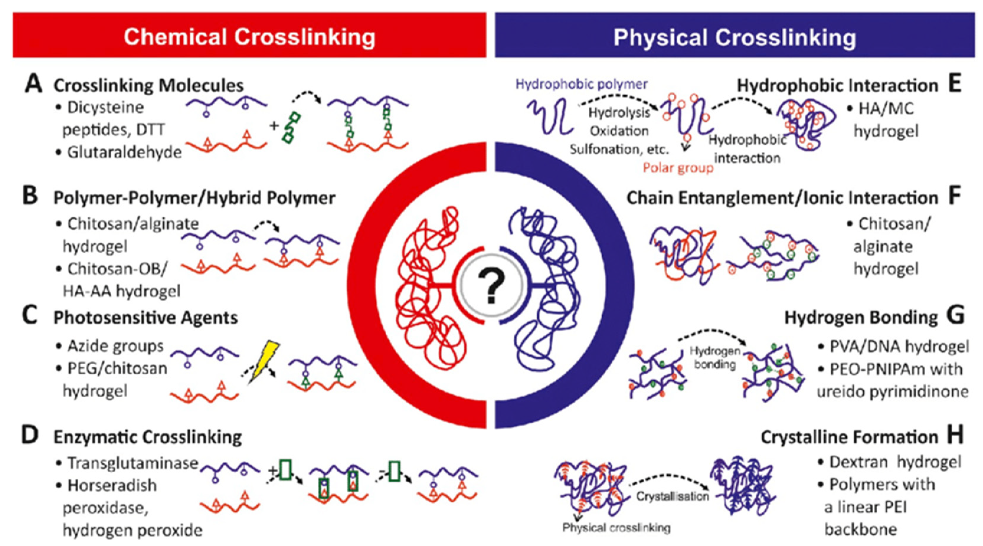
1.4.1. Chemical Crosslinking
Photo-Crosslinked Polymerization
Click Chemistry
Schiff’s Base Reaction
Enzyme-Catalyzed Reactions
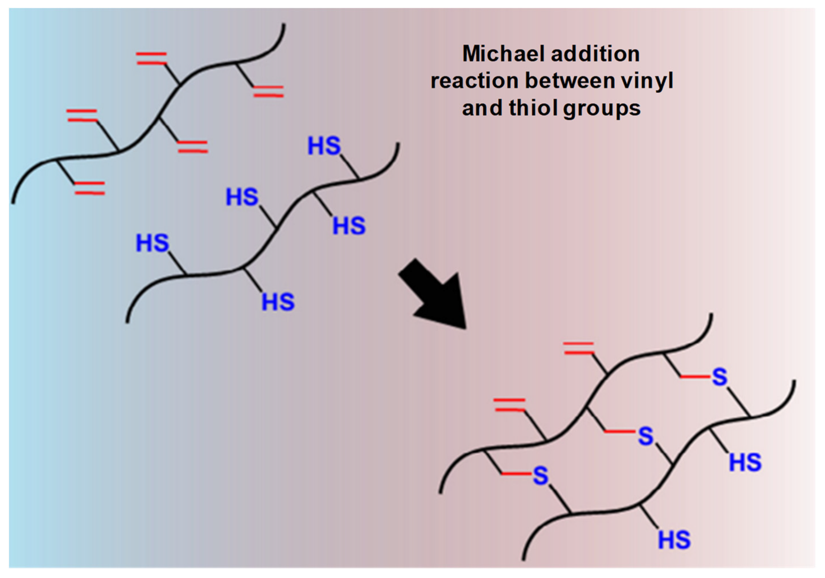
Thiol-Based Michael Reaction
1.4.2. Physical Crosslinking
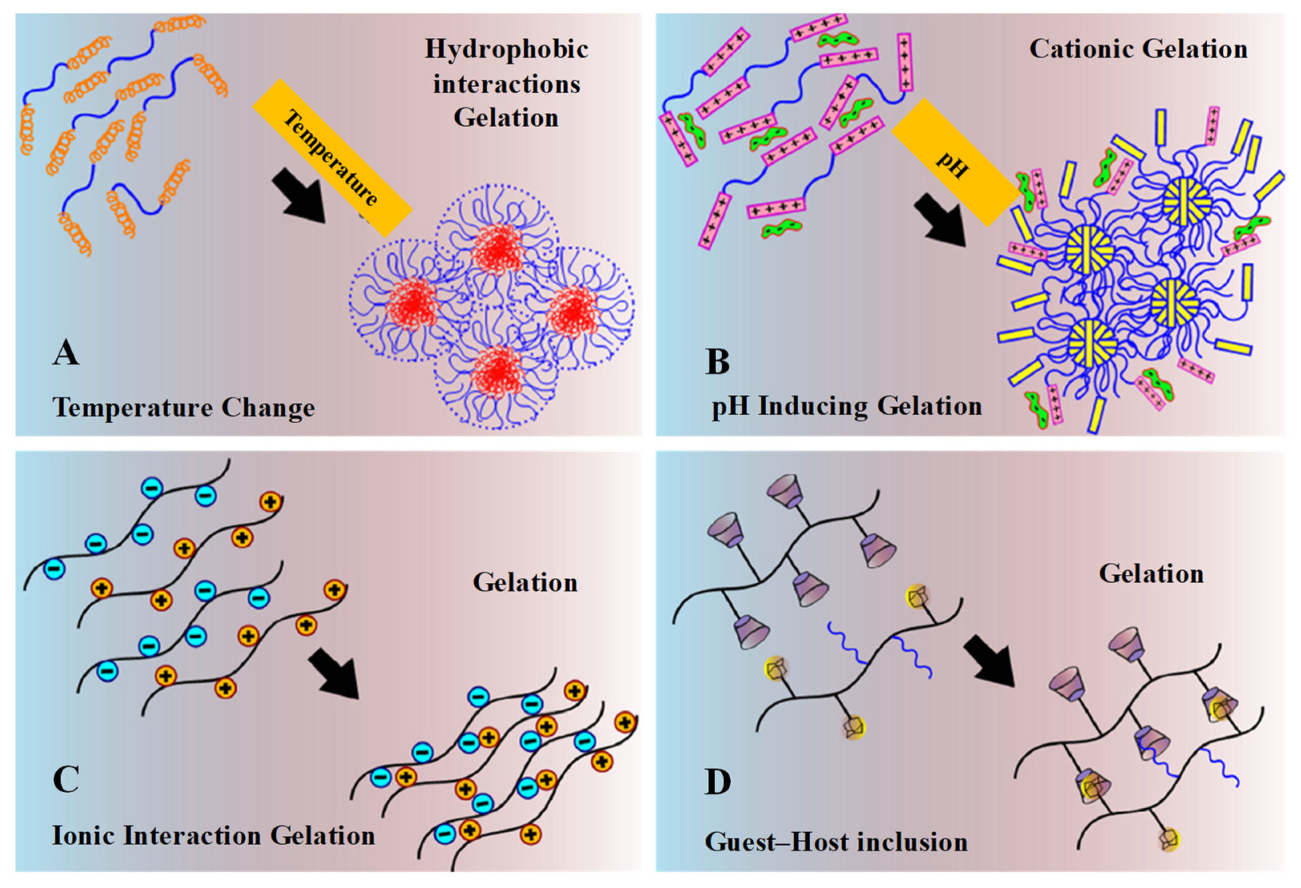
Temperature-Induced
pH-Induced
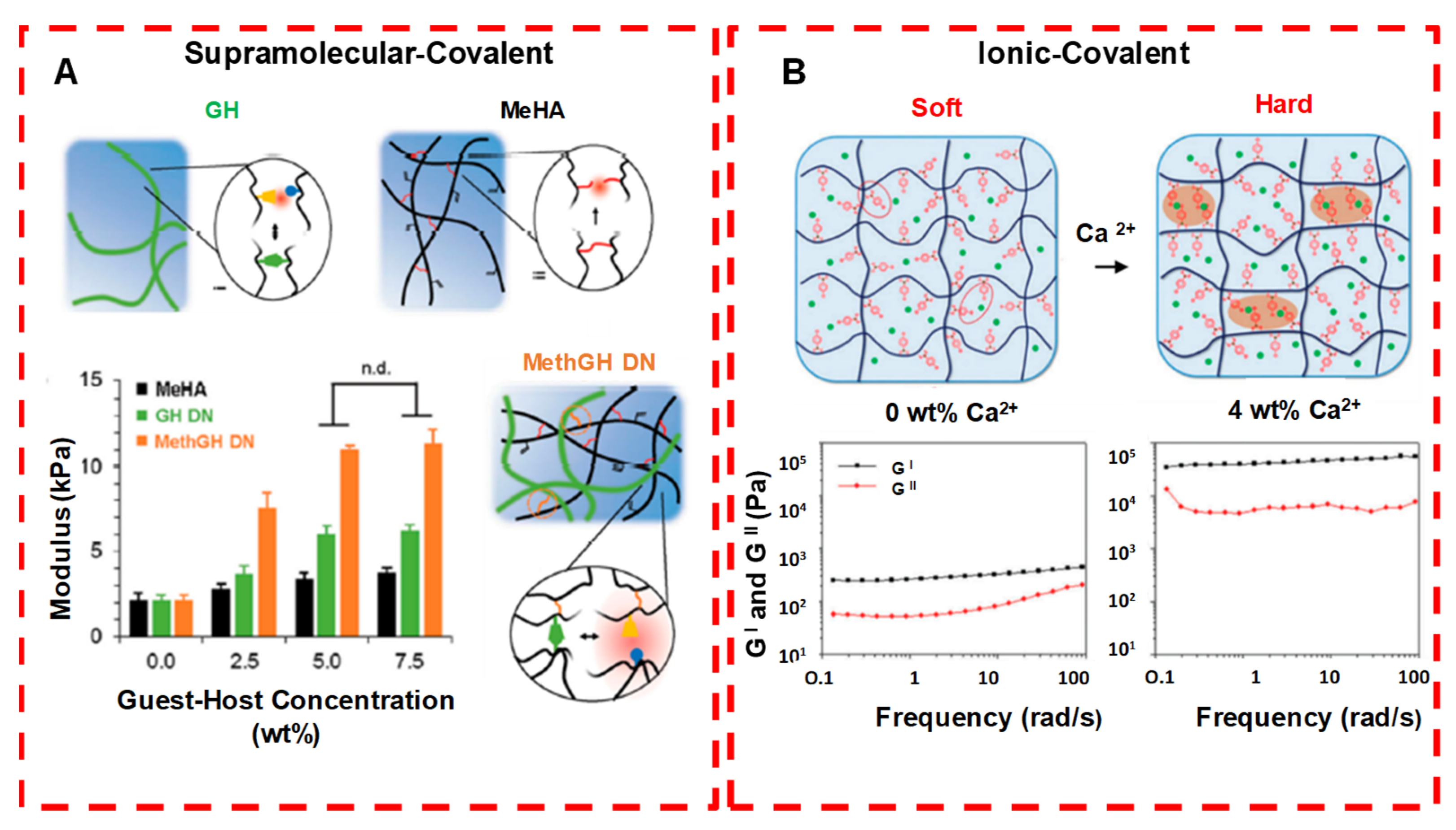
Ionic Interactions
Guest–Host Inclusion
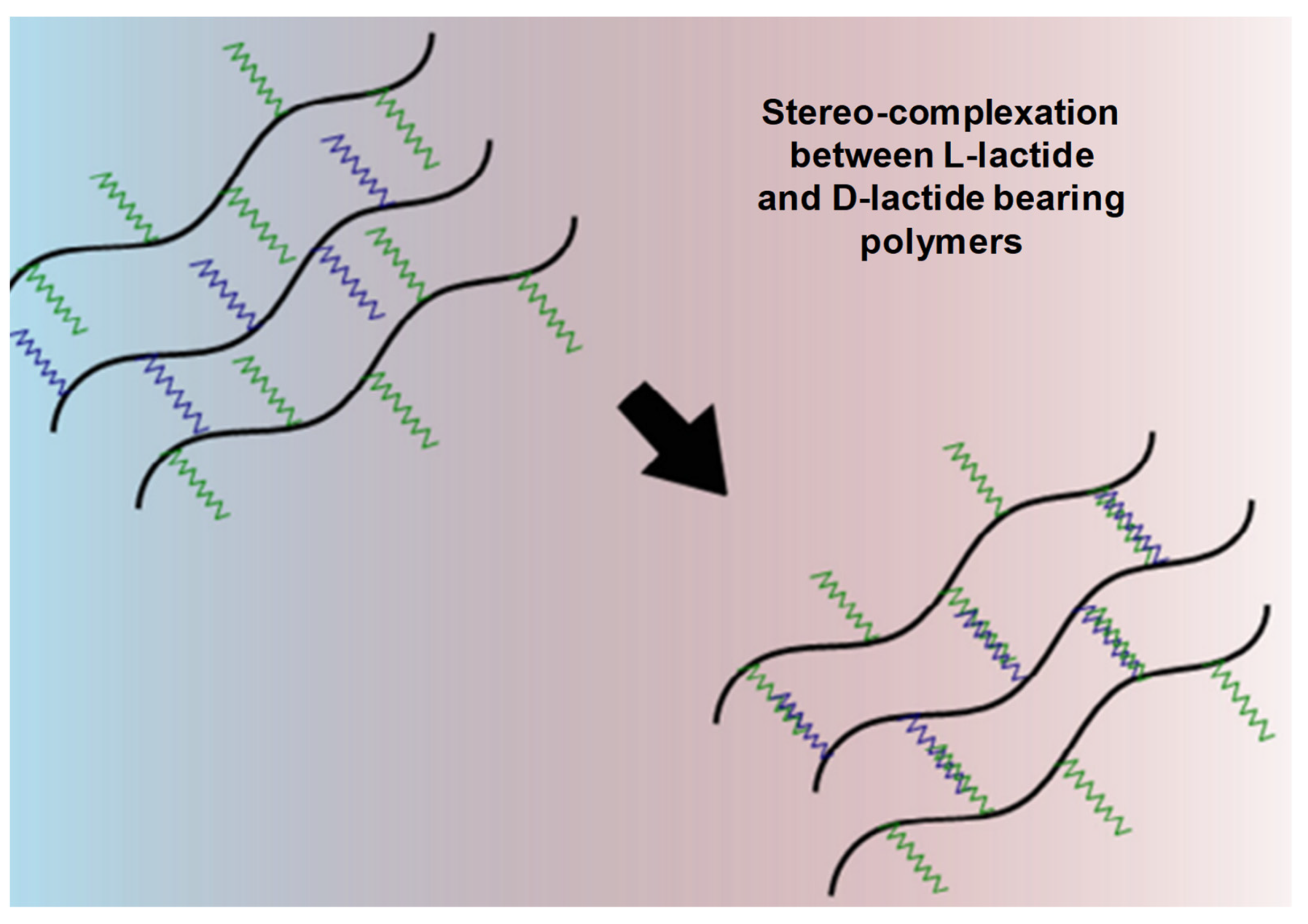
Stereo-Complexation
Complementary Binding
1.5. Surface Chemistry, Internal Bonding and Characterization
1.6. Commercially Available Hydrogel-Based Dosage Forms
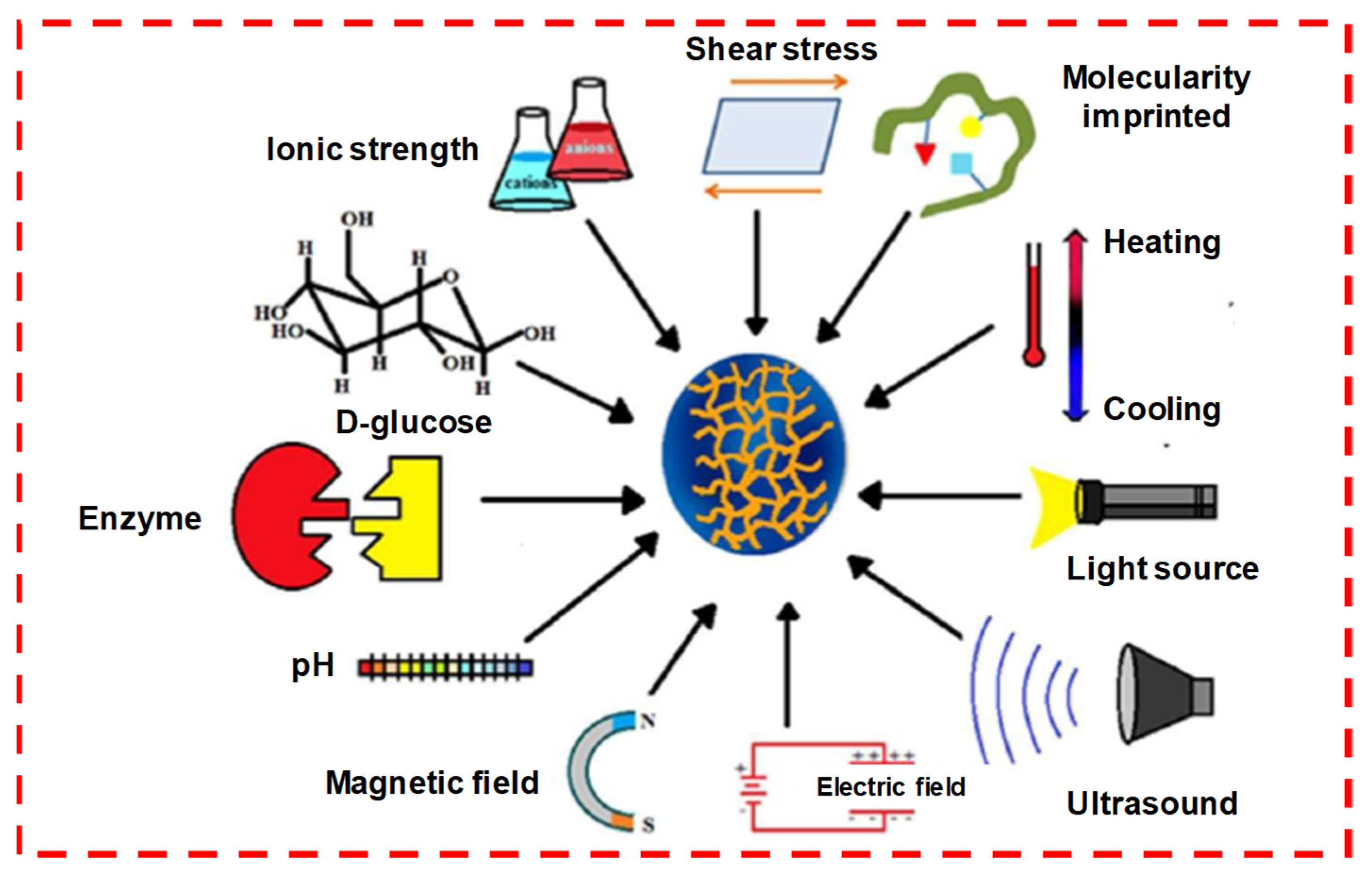
1.7. Responsive Released Studies
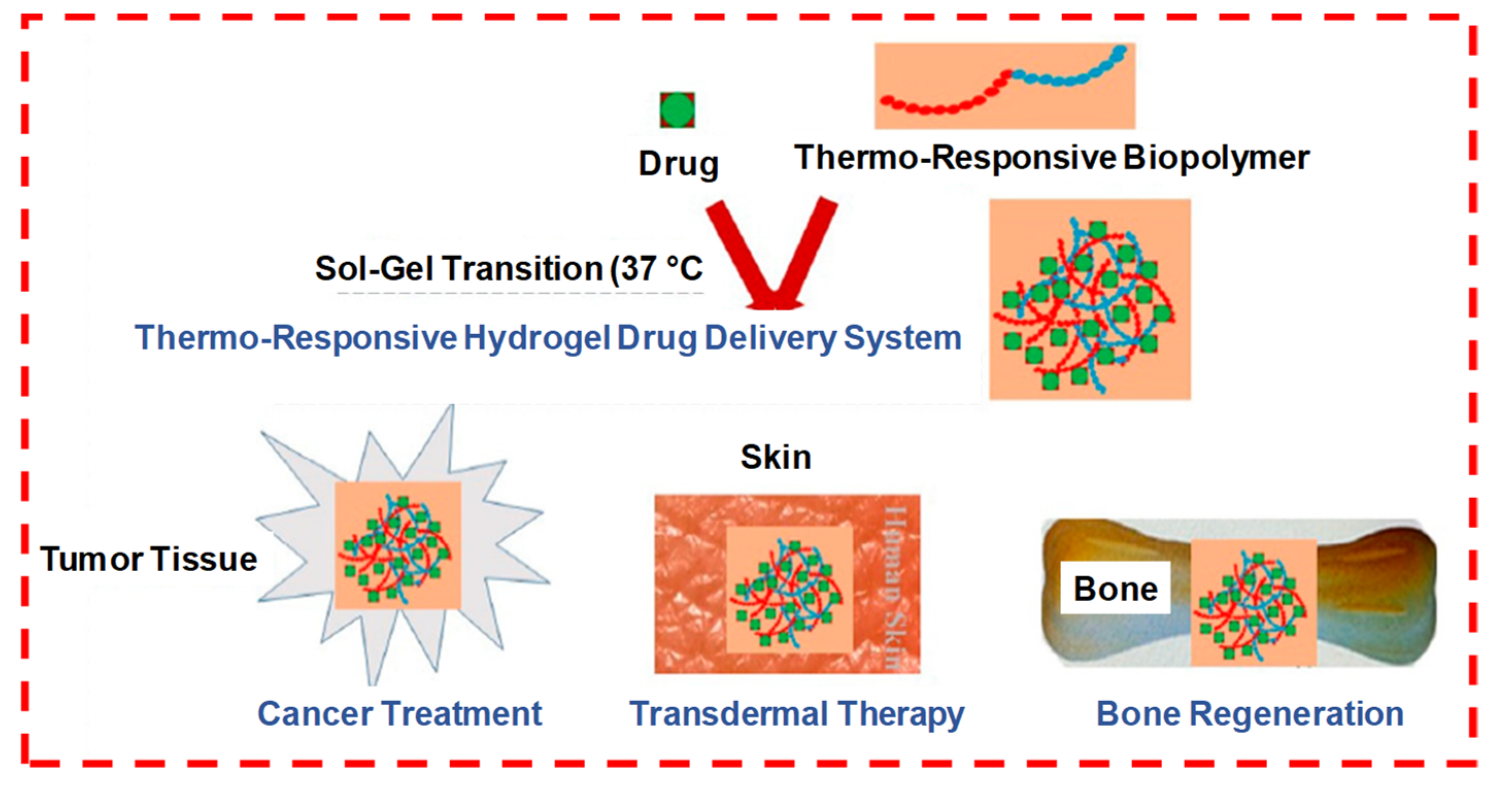
1.7.1. Thermosensitive
1.7.2. Temperature Sensitive
1.7.3. pH-Sensitive
1.7.4. Photosensitive
1.7.5. Enzyme Sensitive
1.7.6. Dual-Sensitive Hydrogel
1.7.7. Glucose
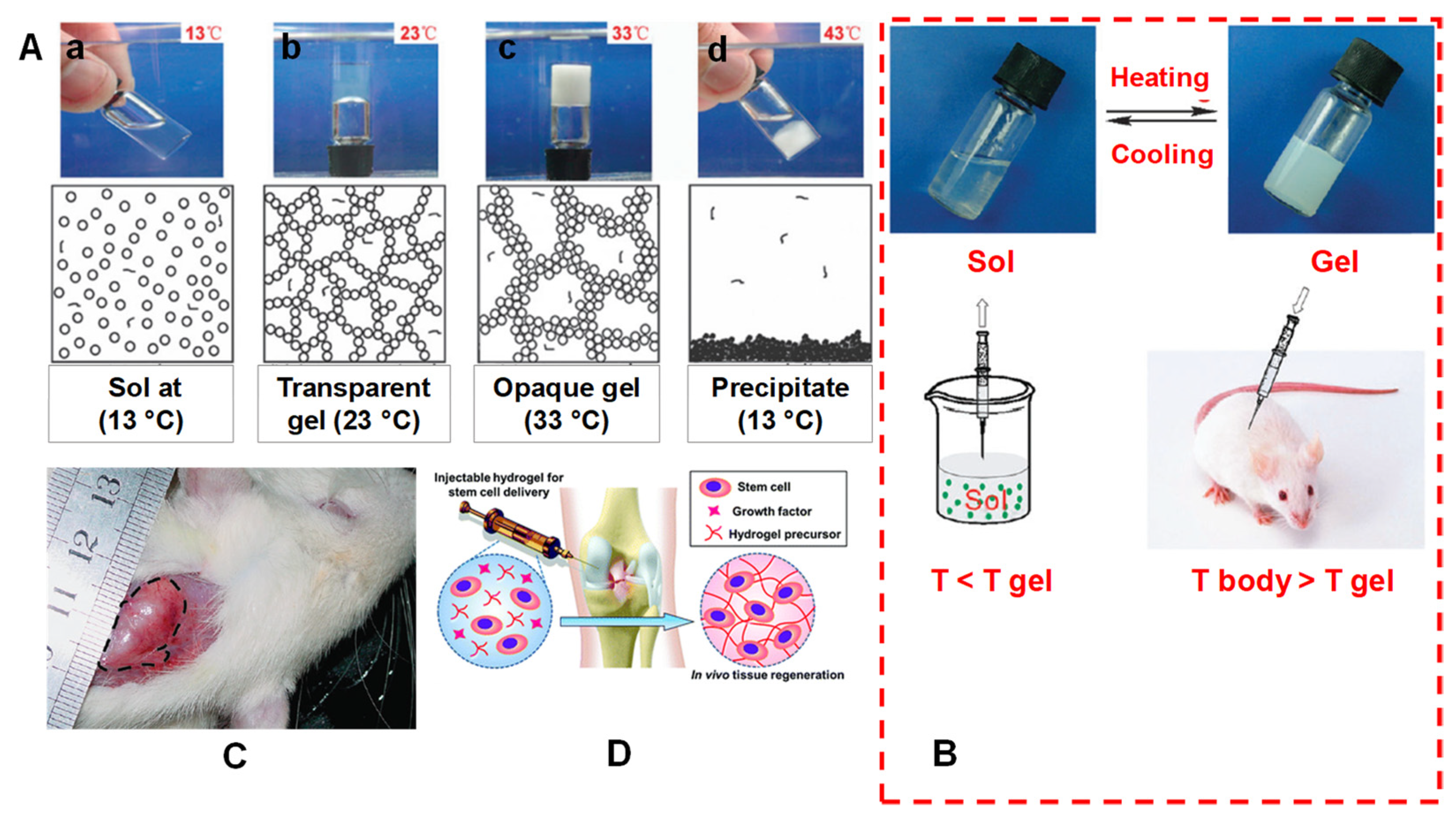
1.8. Sol–Gel Transition State of IHs
2. Current Trends
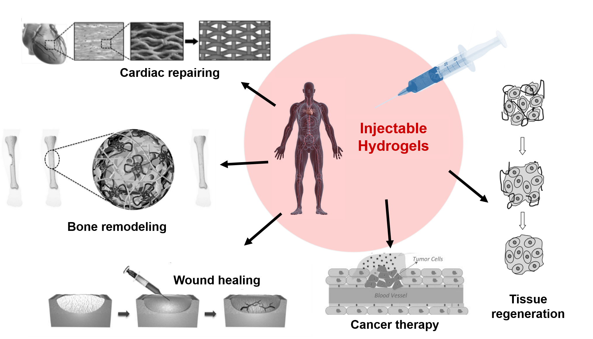
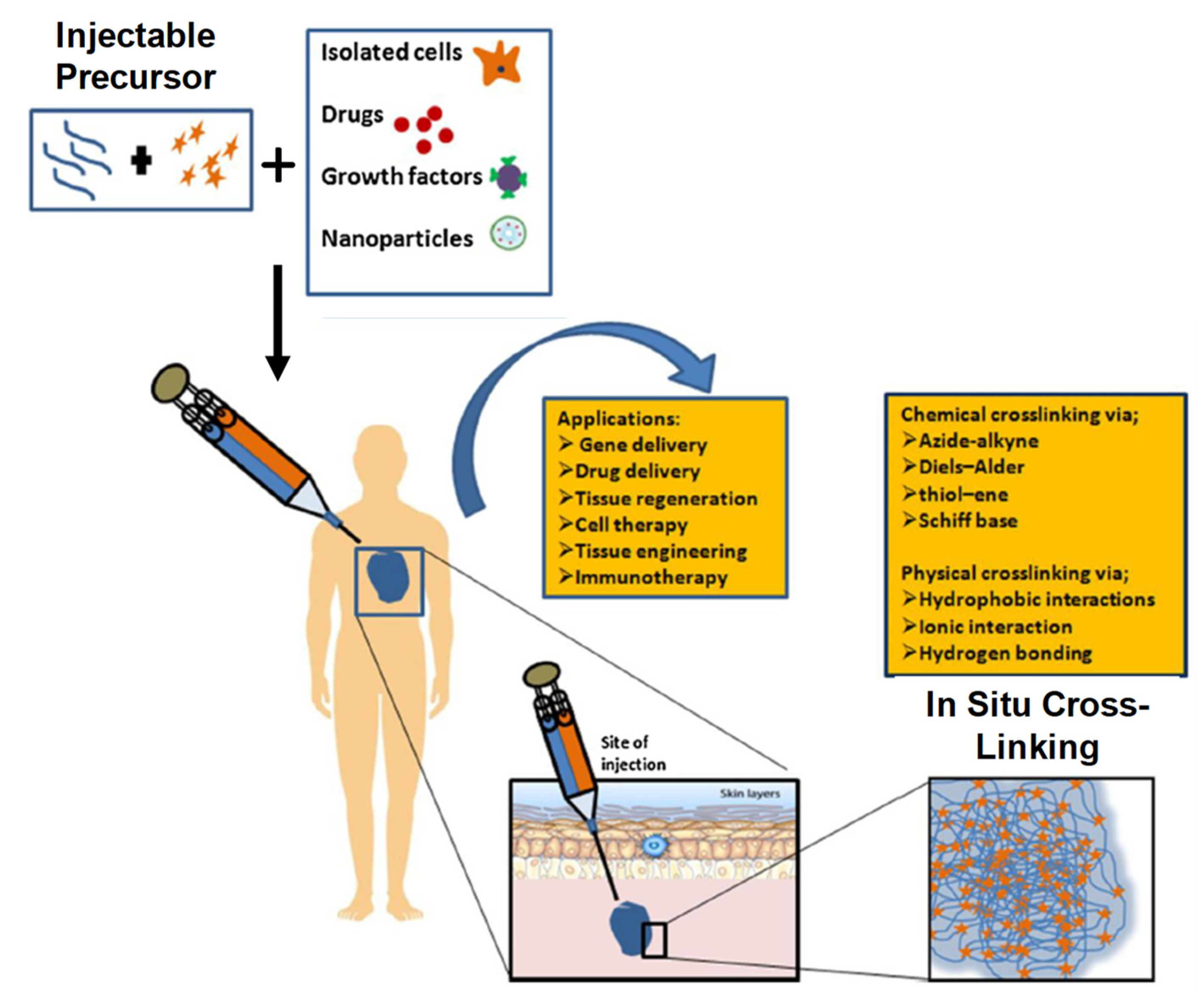
2.1. IHs and Its Application in Drug Delivery System and Biomedical Engineering Applications
2.2. Drug Delivery
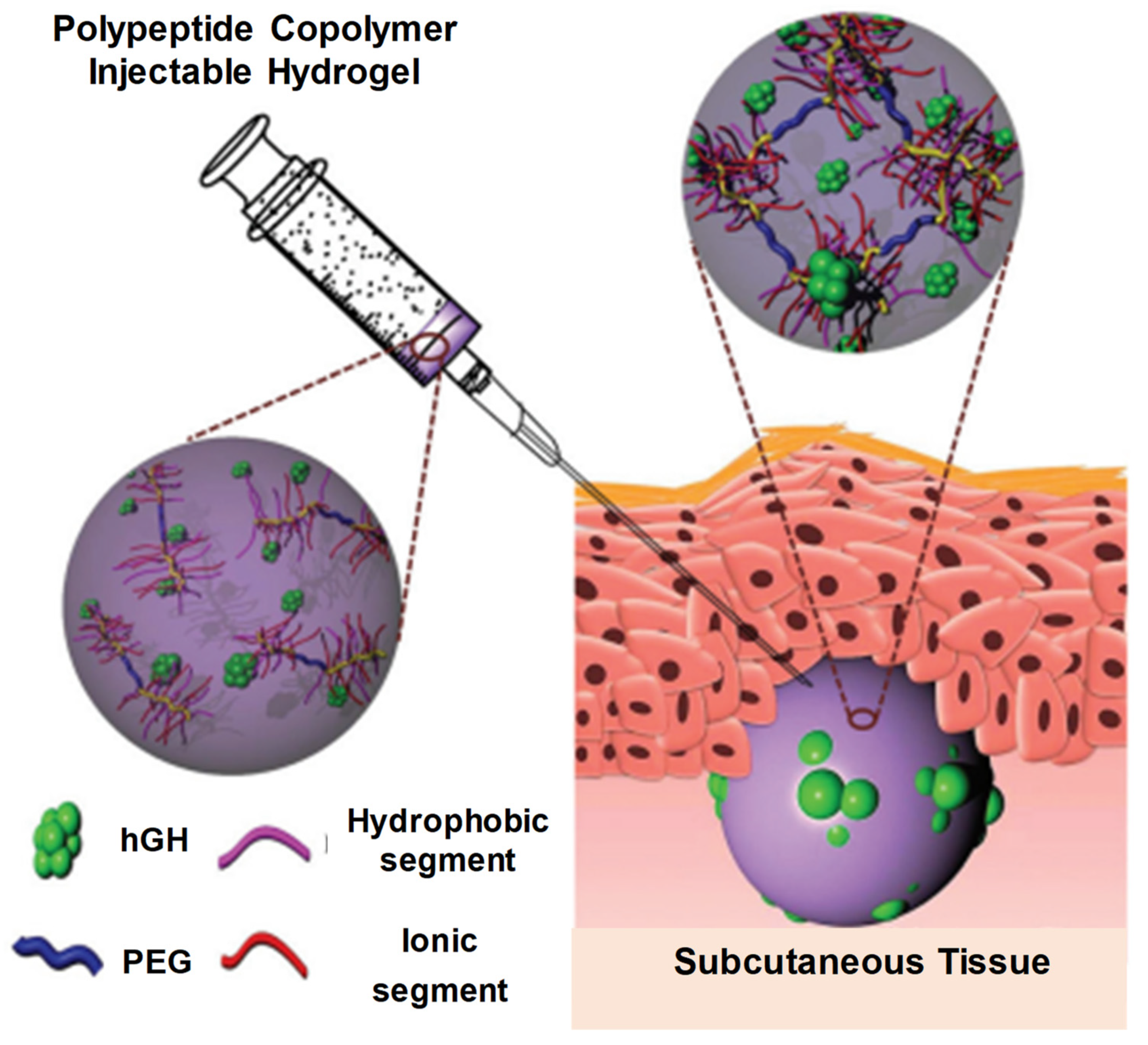
2.2.1. Protein Delivery
2.2.2. DNA/Gene Delivery
2.2.3. Vaccine Delivery
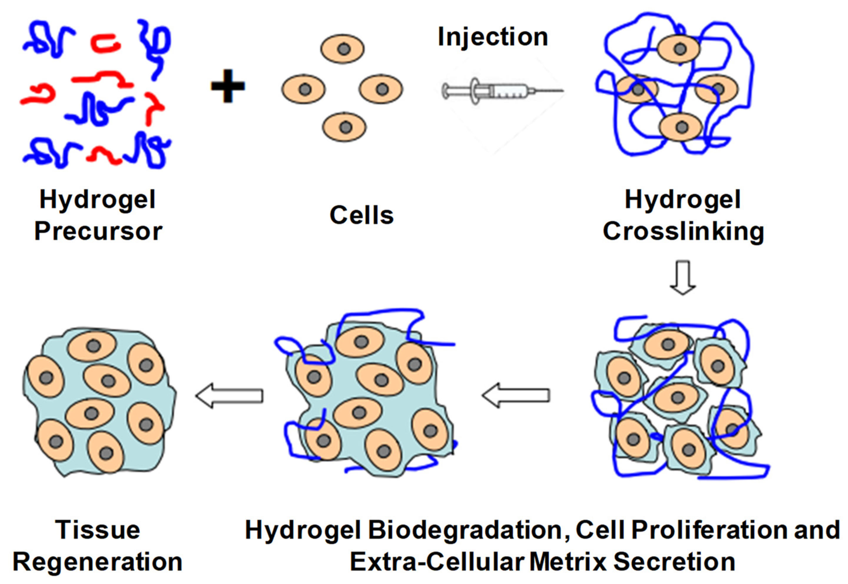
2.2.4. Tissue Engineering
2.2.5. Regenerative Medicines
2.3. Therapeutic Applications
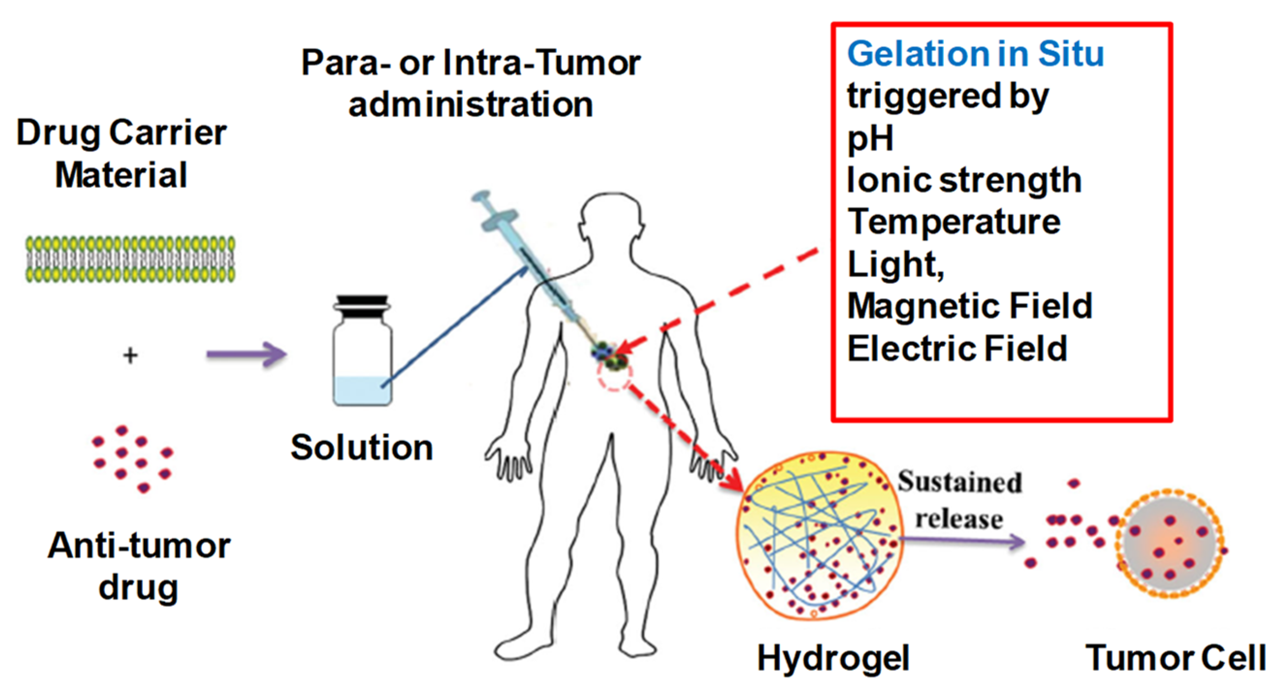
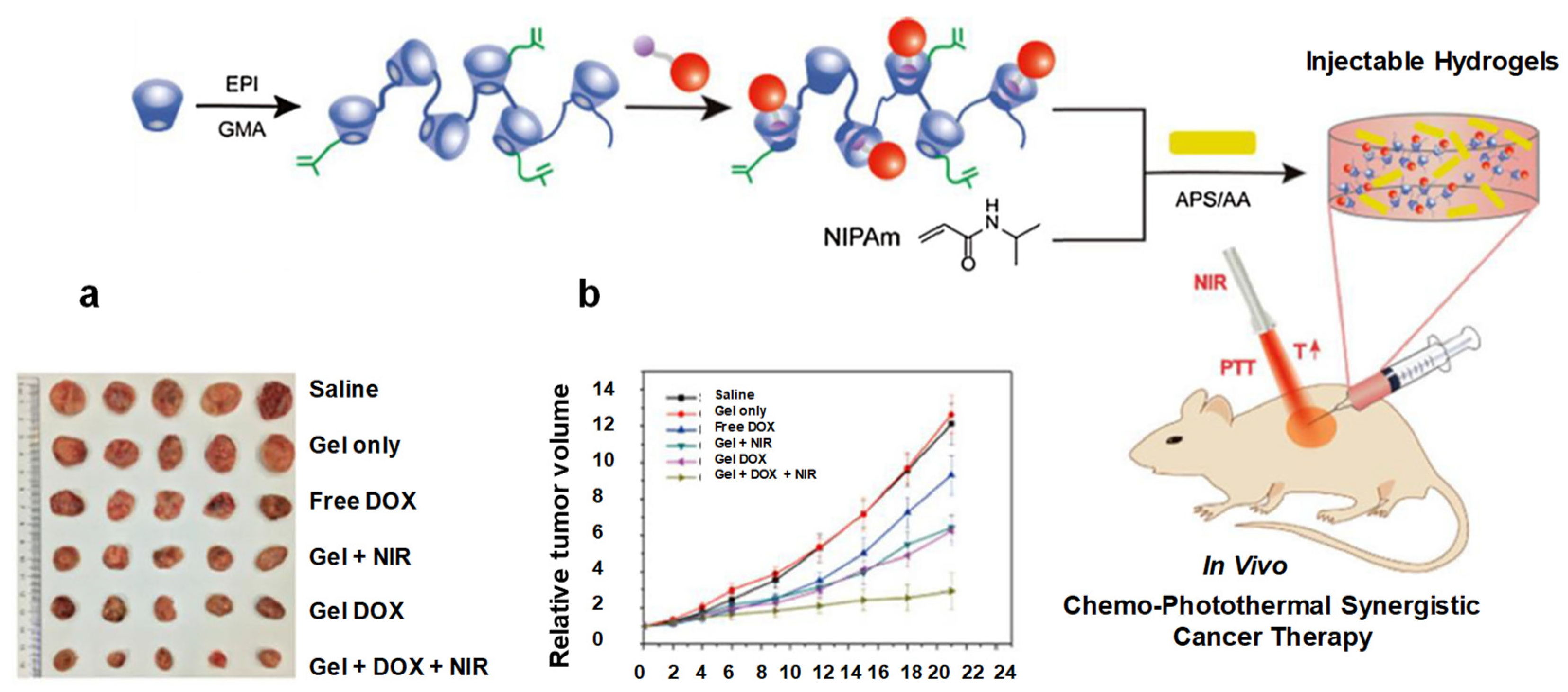
2.3.1. Cancer Therapy
2.3.2. Wound Healings
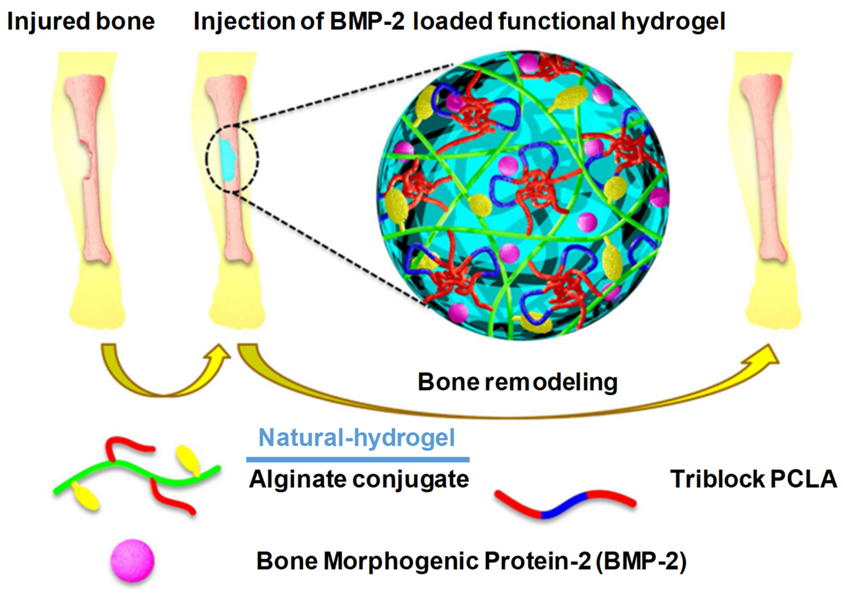
2.3.3. Bone Regeneration
2.4. Biodegradable Hydrogel Injectables That Are Undergoing Clinical Trials
2.5. FDA-Approved Hydrogel Formulations
3. Future Prospects
3.1. Limitations and Outcomes/Overcomes
3.2. Injectable Formulation Challenges
3.2.1. Mechanical Robustness
3.2.2. Loading and Release of Therapeutic Agents
3.2.3. Hydrogel Bioactivity
3.2.4. Immunological Compatibility
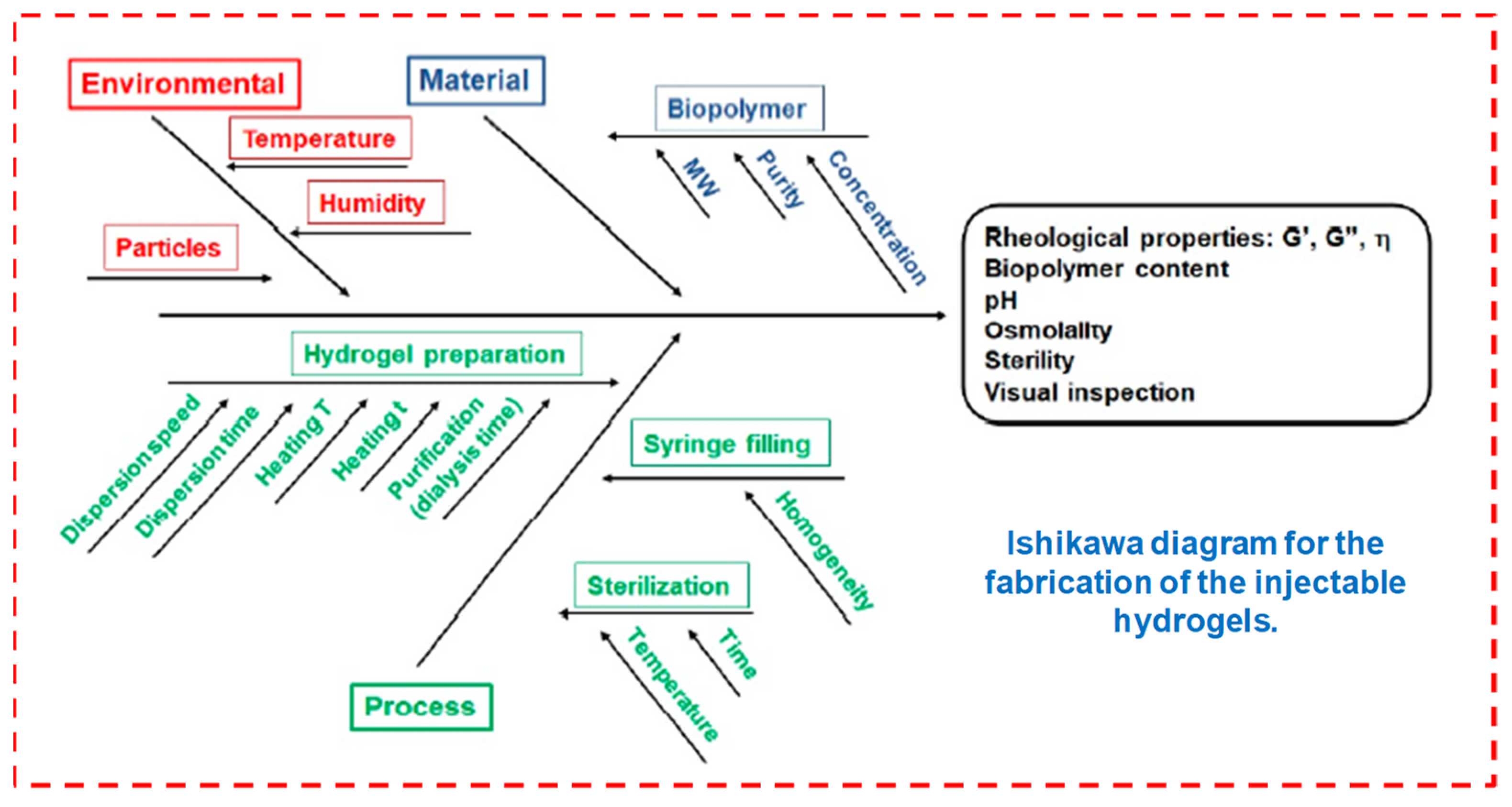
3.2.5. Technological Challenges
3.2.6. Scale-Up Strategies and GMP Processes
3.2.7. Regulatory Approvals
4. Conclusions
Author Contributions
Funding
Institutional Review Board Statement
Informed Consent Statement
Data Availability Statement
Acknowledgments
Conflicts of Interest
References
- Lee, J.H. Injectable hydrogels delivering therapeutic agents for disease treatment and tissue engineering. Biomater. Res. 2018, 22, 27. [Google Scholar] [CrossRef] [PubMed] [Green Version]
- Chao, Y.; Chen, Q.; Liu, Z. Smart Injectable Hydrogels for Cancer Immunotherapy. Adv. Funct. Mater. 2020, 30, 1902785. [Google Scholar] [CrossRef]
- Wang, S.; Zheng, H.; Zhou, L.; Cheng, F.; Liu, Z.; Zhang, H.; Wang, L.; Zhang, Q. Nanoenzyme-Reinforced Injectable Hydrogel for Healing Diabetic Wounds Infected with Multi-Drug Resistant Bacteria. Nano Lett. 2020, 20, 5149–5158. [Google Scholar] [CrossRef]
- Liu, S.; Guo, R.; Li, C.; Lu, C.; Yang, G.; Wang, F.; Nie, J.; Ma, C.; Gao, M. POSS hybrid hydrogels: A brief review of synthesis, properties and applications. Eur. Polym. J. 2021, 143, 110180. [Google Scholar] [CrossRef]
- Chatterjee, S.; Hui, P.C.L. Review of applications and future prospects of stimuli-responsive hydrogel based on thermo-responsive biopolymers in drug delivery systems. Polymers 2021, 13, 2086. [Google Scholar] [CrossRef]
- Tan, H.; Marra, K.G. Injectable, biodegradable hydrogels for tissue engineering applications. Materials 2010, 3, 1746–1767. [Google Scholar] [CrossRef]
- Li, Y.; Yang, H.Y.; Lee, D.S. Advances in biodegradable and injectable hydrogels for biomedical applications. J. Control. Release 2021, 330, 151–160. [Google Scholar] [CrossRef]
- Nguyen, Q.V.; Huynh, D.P.; Park, J.H.; Lee, D.S. Injectable polymeric hydrogels for the delivery of therapeutic agents: A review. Eur. Polym. J. 2015, 72, 602–619. [Google Scholar] [CrossRef]
- Alonso, J.M.; Del Olmo, J.A.; Gonzalez, R.P.; Saez-martinez, V. Injectable hydrogels: From laboratory to industrialization. Polymers 2021, 13, 650. [Google Scholar] [CrossRef]
- Yao, Q.; Lan, Q.H.; Jiang, X.; Du, C.C.; Zhai, Y.Y.; Shen, X.; Xu, H.L.; Xiao, J.; Kou, L.; Zhao, Y.Z. Bioinspired biliverdin/silk fibroin hydrogel for antiglioma photothermal therapy and wound healing. Theranostics 2020, 10, 11719–11736. [Google Scholar] [CrossRef]
- Yu, L.; Ding, J. Injectable hydrogels as unique biomedical materials. Chem. Soc. Rev. 2008, 37, 1473–1481. [Google Scholar] [CrossRef]
- Du, X.; Zhou, J.; Shi, J.; Xu, B. Supramolecular Hydrogelators and Hydrogels: From Soft Matter to Molecular Biomaterials. Chem. Rev. 2015, 115, 13165–13307. [Google Scholar] [CrossRef] [PubMed]
- Kim, S.H.; Thambi, T.; Giang Phan, V.H.; Lee, D.S. Modularly engineered alginate bioconjugate hydrogel as biocompatible injectable scaffold for in situ biomineralization. Carbohydr. Polym. 2020, 233, 115832. [Google Scholar] [CrossRef] [PubMed]
- Sun, Y.; Nan, D.; Jin, H.; Qu, X. Recent Advances of Injectable Hydrogels for Drug Delivery and. Polym. Test. 2020, 81, 106283. [Google Scholar] [CrossRef]
- Liang, K.; Bae, K.H.; Kurisawa, M. Recent advances in the design of injectable hydrogels for stem cell-based therapy. J. Mater. Chem. B 2019, 7, 3775–3791. [Google Scholar] [CrossRef]
- Mansoor, S.; Kondiah, P.P.D.; Choonara, Y.E. Advanced Hydrogels for the Controlled Delivery of Insulin. Pharmaceutics 2021, 13, 2113. [Google Scholar] [CrossRef] [PubMed]
- Kaith, B.S.; Singh, A.; Sharma, A.K.; Sud, D. Hydrogels: Synthesis, Classification, Properties and Potential Applications—A Brief Review. J. Polym. Environ. 2021, 29, 3827–3841. [Google Scholar] [CrossRef]
- Sharma, S.; Tiwari, S. A review on biomacromolecular hydrogel classification and its applications. Int. J. Biol. Macromol. 2020, 162, 737–747. [Google Scholar] [CrossRef]
- Khansari, M.M.; Sorokina, L.V.; Mukherjee, P.; Mukhtar, F.; Shirdar, M.R.; Shahidi, M.; Shokuhfar, T. Classification of Hydrogels Based on Their Source: A Review and Application in Stem Cell Regulation. JOM 2017, 69, 1340–1347. [Google Scholar] [CrossRef]
- Suflet, D.M.; Popescu, I.; Pelin, I.M.; Ichim, D.L.; Daraba, O.M.; Constantin, M.; Fundueanu, G. Dual cross-linked chitosan/pva hydrogels containing silver nanoparticles with antimicrobial properties. Pharmaceutics 2021, 13, 1461. [Google Scholar] [CrossRef]
- Hu, C.; Zhang, F.; Long, L.; Kong, Q.; Luo, R.; Wang, Y. Dual-responsive injectable hydrogels encapsulating drug-loaded micelles for on-demand antimicrobial activity and accelerated wound healing. J. Control. Release 2020, 324, 204–217. [Google Scholar] [CrossRef] [PubMed]
- George, J.; Hsu, C.C.; Nguyen, L.T.B.; Ye, H.; Cui, Z. Neural tissue engineering with structured hydrogels in CNS models and therapies. Biotechnol. Adv. 2020, 42, 107370. [Google Scholar] [CrossRef] [PubMed]
- Sawhney, A.S.; Pathak, C.P.; Hubbell, J.A. Bioerodible Hydrogels Based on Photopolymerized Poly(ethylene glycol)-co-poly (α-hydroxy acid) Diacrylate Macromers. Macromolecules 1993, 26, 581–587. [Google Scholar] [CrossRef]
- Patterson, J.; Martino, M.M.; Hubbell, J.A. Biomimetic materials in tissue engineering. Mater. Today 2010, 13, 14–22. [Google Scholar] [CrossRef]
- Mandal, A.; Clegg, J.R.; Anselmo, A.C.; Mitragotri, S. Hydrogels in the clinic. Bioeng. Transl. Med. 2020, 5, e10158. [Google Scholar] [CrossRef] [Green Version]
- Moreira Teixeira, L.S.; Feijen, J.; van Blitterswijk, C.A.; Dijkstra, P.J.; Karperien, M. Enzyme-catalyzed crosslinkable hydrogels: Emerging strategies for tissue engineering. Biomaterials 2012, 33, 1281–1290. [Google Scholar] [CrossRef]
- Kurisawa, M.; Lee, F.; Wang, L.S.; Chung, J.E. Injectable enzymatically crosslinked hydrogel system with independent tuning of mechanical strength and gelation rate for drug delivery and tissue engineering. J. Mater. Chem. 2010, 20, 5371–5375. [Google Scholar] [CrossRef]
- Qiu, B.; Stefanos, S.; Ma, J.; Lalloo, A.; Perry, B.A.; Leibowitz, M.J.; Sinko, P.J.; Stein, S. A hydrogel prepared by in situ cross-linking of a thiol-containing poly(ethylene glycol)-based copolymer: A new biomaterial for protein drug delivery. Biomaterials 2003, 24, 11–18. [Google Scholar] [CrossRef]
- Eslahi, N.; Abdorahim, M.; Simchi, A. Smart Polymeric Hydrogels for Cartilage Tissue Engineering: A Review on the Chemistry and Biological Functions. Biomacromolecules 2016, 17, 3441–3463. [Google Scholar] [CrossRef]
- Qureshi, D.; Nayak, S.K.; Maji, S.; Anis, A.; Kim, D.; Pal, K. Environment sensitive hydrogels for drug delivery applications. Eur. Polym. J. 2019, 120, 109220. [Google Scholar] [CrossRef]
- Jacob, S.; Nair, A.B.; Shah, J.; Sreeharsha, N.; Gupta, S.; Shinu, P. Emerging role of hydrogels in drug delivery systems, tissue engineering and wound management. Pharmaceutics 2021, 13, 357. [Google Scholar] [CrossRef] [PubMed]
- Guvendiren, M.; Lu, H.D.; Burdick, J.A. Shear-thinning hydrogels for biomedical applications. Soft Matter 2012, 8, 260–272. [Google Scholar] [CrossRef]
- Kabir, S.M.F.; Sikdar, P.P.; Haque, B.; Bhuiyan, M.A.R.; Ali, A.; Islam, M.N. Cellulose-based hydrogel materials: Chemistry, properties and their prospective applications. Prog. Biomater. 2018, 7, 153–174. [Google Scholar] [CrossRef] [PubMed] [Green Version]
- De France, K.J.; Cranston, E.D.; Hoare, T. Mechanically Reinforced Injectable Hydrogels. ACS Appl. Polym. Mater. 2020, 2, 1016–1030. [Google Scholar] [CrossRef]
- Zhao, C.; Zhou, L.; Chiao, M.; Yang, W. Antibacterial hydrogel coating: Strategies in surface chemistry. Adv. Colloid Interface Sci. 2020, 285, 102280. [Google Scholar] [CrossRef]
- Mathew, A.P.; Uthaman, S.; Cho, K.H.; Cho, C.S.; Park, I.K. Injectable hydrogels for delivering biotherapeutic molecules. Int. J. Biol. Macromol. 2018, 110, 17–29. [Google Scholar] [CrossRef]
- Yu, S.; He, C.; Chen, X. Injectable Hydrogels as Unique Platforms for Local Chemotherapeutics-Based Combination Antitumor Therapy. Macromol. Biosci. 2018, 18, e1800240. [Google Scholar] [CrossRef]
- Dimatteo, R.; Darling, N.J.; Segura, T. In situ forming injectable hydrogels for drug delivery and wound repair. Adv. Drug Deliv. Rev. 2018, 127, 167–184. [Google Scholar] [CrossRef]
- Wan, J.; Geng, S.; Zhao, H.; Peng, X.; Xu, J.; Wei, M.; Mao, J.; Zhou, Y.; Zhu, Q.; Zhao, Y.; et al. Precise synchronization of hyperthermia-chemotherapy: Photothermally induced on-demand release from injectable hydrogels of gold nanocages. Nanoscale 2018, 10, 20020–20030. [Google Scholar] [CrossRef]
- Overstreet, D.J.; Dutta, D.; Stabenfeldt, S.E.; Vernon, B.L. Injectable hydrogels. J. Polym. Sci. Part B Polym. Phys. 2012, 50, 881–903. [Google Scholar] [CrossRef]
- Liu, C.; Zhang, Q.; Zhu, S.; Liu, H.; Chen, J. Preparation and applications of peptide-based injectable hydrogels. RSC Adv. 2019, 9, 28299–28311. [Google Scholar] [CrossRef] [Green Version]
- Poustchi, F.; Amani, H.; Ahmadian, Z.; Niknezhad, S.V.; Mehrabi, S.; Santos, H.A.; Shahbazi, M.A. Combination Therapy of Killing Diseases by Injectable Hydrogels: From Concept to Medical Applications. Adv. Healthc. Mater. 2021, 10, e2001571. [Google Scholar] [CrossRef] [PubMed]
- Zhang, X.; Tan, B.; Wu, Y.; Zhang, M.; Liao, J. A review on hydrogels with photothermal effect in wound healing and bone tissue engineering. Polymers 2021, 13, 2100. [Google Scholar] [CrossRef] [PubMed]
- Ornell, K.J.; Lozada, D.; Phan, N.V.; Coburn, J.M. Controlling methacryloyl substitution of chondroitin sulfate: Injectable hydrogels with tunable long-term drug release profiles. J. Mater. Chem. B 2019, 7, 2151–2161. [Google Scholar] [CrossRef]
- Hafeez, S.; Islam, A.; Gull, N.; Ali, A.; Khan, S.M.; Zia, S.; Anwar, K.; Khan, S.U.; Jamil, T. γ-Irradiated chitosan based injectable hydrogels for controlled release of drug (Montelukast sodium). Int. J. Biol. Macromol. 2018, 114, 890–897. [Google Scholar] [CrossRef]
- Zhao, L.; Niu, L.; Liang, H.; Tan, H.; Liu, C.; Zhu, F. PH and Glucose Dual-Responsive Injectable Hydrogels with Insulin and Fibroblasts as Bioactive Dressings for Diabetic Wound Healing. ACS Appl. Mater. Interfaces 2017, 9, 37563–37574. [Google Scholar] [CrossRef]
- Shen, Y.; Li, X.; Huang, Y.; Chang, G.; Cao, K.; Yang, J.; Zhang, R.; Sheng, X.; Ye, X. pH and redox dual stimuli-responsive injectable hydrogels based on carboxymethyl cellulose derivatives. Macromol. Res. 2016, 24, 602–608. [Google Scholar] [CrossRef]
- Fathi, M.; Alami-Milani, M.; Geranmayeh, M.H.; Barar, J.; Erfan-Niya, H.; Omidi, Y. Dual thermo-and pH-sensitive injectable hydrogels of chitosan/(poly(N-isopropylacrylamide-co-itaconic acid)) for doxorubicin delivery in breast cancer. Int. J. Biol. Macromol. 2019, 128, 957–964. [Google Scholar] [CrossRef]
- Murugaraj, K.M.R. Structural, electrical, and weak ferromagnetic-to-antiferromagnetic nature of Ni and La co-doped BaTiO3 by sol-gel combustion route. J. Sol-Gel Sci. Technol. 2020, 95, 11–21. [Google Scholar] [CrossRef]
- Sakai, T.; Katashima, T.; Matsushita, T.; Chung, U. Sol-gel transition behavior near critical concentration and connectivity. Polym. J. 2016, 48, 629–634. [Google Scholar] [CrossRef]
- Ilochonwu, B.C.; Urtti, A.; Hennink, W.E.; Vermonden, T. Intravitreal hydrogels for sustained release of therapeutic proteins. J. Control. Release 2020, 326, 419–441. [Google Scholar] [CrossRef] [PubMed]
- Fan, D.Y.; Tian, Y.; Liu, Z.J. Injectable Hydrogels for Localized Cancer Therapy. Front. Chem. 2019, 7, 675. [Google Scholar] [CrossRef] [PubMed]
- Giang Phan, V.H.; Duong, H.T.T.; Thambi, T.; Nguyen, T.L.; Turabee, M.H.; Yin, Y.; Kim, S.H.; Kim, J.; Jeong, J.H.; Lee, D.S. Modularly engineered injectable hybrid hydrogels based on protein-polymer network as potent immunologic adjuvant in vivo. Biomaterials 2019, 195, 100–110. [Google Scholar] [CrossRef]
- Nandi, R.; Yucknovsky, A.; Mazo, M.M.; Amdursky, N. Exploring the inner environment of protein hydrogels with fluorescence spectroscopy towards understanding their drug delivery capabilities. J. Mater. Chem. B 2020, 8, 6964–6974. [Google Scholar] [CrossRef] [PubMed]
- Xiao, Y.; Gu, Y.; Qin, L.; Chen, L.; Chen, X.; Cui, W.; Li, F.; Xiang, N.; He, X. Colloids and Surfaces B: Biointerfaces Injectable thermosensitive hydrogel-based drug delivery system for local cancer therapy. Colloids Surf. B Biointerfaces 2021, 200, 111581. [Google Scholar] [CrossRef]
- Afinjuomo, F.; Abdella, S.; Youssef, S.H. Inulin and Its Application in Drug Delivery. Pharmaceuticals 2021, 14, 855. [Google Scholar] [CrossRef]
- Gil, M.S.; Cho, J.; Thambi, T.; Giang Phan, V.H.; Kwon, I.; Lee, D.S. Bioengineered robust hybrid hydrogels enrich the stability and efficacy of biological drugs. J. Control. Release 2017, 267, 119–132. [Google Scholar] [CrossRef]
- Ma, H.; He, C.; Cheng, Y.; Li, D.; Gong, Y.; Liu, J.; Tian, H.; Chen, X. PLK1shRNA and doxorubicin co-loaded thermosensitive PLGA-PEG-PLGA hydrogels for osteosarcoma treatment. Biomaterials 2014, 35, 8723–8734. [Google Scholar] [CrossRef]
- Mo, F.; Jiang, K.; Zhao, D.; Wang, Y.; Song, J.; Tan, W. DNA hydrogel-based gene editing and drug delivery systems. Adv. Drug Deliv. Rev. 2021, 168, 79–98. [Google Scholar] [CrossRef]
- Koetting, M.C.; Guido, J.F.; Gupta, M.; Zhang, A.; Peppas, N.A. PH-responsive and enzymatically-responsive hydrogel microparticles for the oral delivery of therapeutic proteins: Effects of protein size, crosslinking density, and hydrogel degradation on protein delivery. J. Control. Release 2016, 221, 18–25. [Google Scholar] [CrossRef] [Green Version]
- Gilbert, T.; Smeets, N.M.B.; Hoare, T. Injectable Interpenetrating Network Hydrogels via Kinetically Orthogonal Reactive Mixing of Functionalized Polymeric Precursors. ACS Macro Lett. 2015, 4, 1104–1109. [Google Scholar] [CrossRef]
- Umeki, Y.; Mohri, K.; Kawasaki, Y.; Watanabe, H.; Takahashi, R.; Takahashi, Y.; Takakura, Y.; Nishikawa, M. Induction of Potent Antitumor Immunity by Sustained Release of Cationic Antigen from a DNA-Based Hydrogel with Adjuvant Activity. Adv. Funct. Mater. 2015, 25, 5758–5767. [Google Scholar] [CrossRef] [Green Version]
- Liu, Y.; Xiao, L.; Joo, K.I.; Hu, B.; Fang, J.; Wang, P. In situ modulation of dendritic cells by injectable thermosensitive hydrogels for cancer vaccines in mice. Biomacromolecules 2014, 15, 3836–3845. [Google Scholar] [CrossRef] [Green Version]
- Branco, M.C.; Pochan, D.J.; Wagner, N.J.; Schneider, J.P. The effect of protein structure on their controlled release from an injectable peptide hydrogel. Biomaterials 2010, 31, 9527–9534. [Google Scholar] [CrossRef] [Green Version]
- Kong, L.; Wu, Z.; Zhao, H.; Cui, H.; Shen, J.; Chang, J.; Li, H.; He, Y. Bioactive Injectable Hydrogels Containing Desferrioxamine and Bioglass for Diabetic Wound Healing. ACS Appl. Mater. Interfaces 2018, 10, 30103–30114. [Google Scholar] [CrossRef]
- Won, J.E.; Wi, T.I.; Lee, C.M.; Lee, J.H.; Kang, T.H.; Lee, J.W.; Shin, B.C.; Lee, Y.J.; Park, Y.M.; Han, H.D. NIR irradiation-controlled drug release utilizing injectable hydrogels containing gold-labeled liposomes for the treatment of melanoma cancer. Acta Biomater. 2021, 136, 508–518. [Google Scholar] [CrossRef] [PubMed]
- Wei, W.; Li, H.; Yin, C.; Tang, F. Research progress in the application of in situ hydrogel system in tumor treatment. Drug Deliv. 2020, 27, 460–468. [Google Scholar] [CrossRef] [PubMed] [Green Version]
- Okay, O. Self-healing hydrogels formed via hydrophobic interactions. In Supramolecular Polymer Networks and Gels; Springer: Cham, Switzerland, 2015; Volume 268, ISBN 9783319154046. [Google Scholar]
- Lin, J.; Zheng, S.Y.; Xiao, R.; Yin, J.; Wu, Z.L.; Zheng, Q.; Qian, J. Constitutive behaviors of tough physical hydrogels with dynamic metal-coordinated bonds. J. Mech. Phys. Solids 2020, 139, 103935. [Google Scholar] [CrossRef]
- Hennink, W.E.; van Nostrum, C.F. Novel crosslinking methods to design hydrogels. Adv. Drug Deliv. Rev. 2012, 64, 223–236. [Google Scholar] [CrossRef]
- Ahmed, E.M. Hydrogel: Preparation, characterization, and applications: A review. J. Adv. Res. 2015, 6, 105–121. [Google Scholar] [CrossRef] [Green Version]
- Qiu, X.; Hu, S. “Smart” materials based on cellulose: A review of the preparations, properties, and applications. Materials 2013, 6, 738–781. [Google Scholar] [CrossRef] [PubMed] [Green Version]
- Caló, E.; Khutoryanskiy, V.V. Biomedical applications of hydrogels: A review of patents and commercial products. Eur. Polym. J. 2015, 65, 252–267. [Google Scholar] [CrossRef] [Green Version]
- Taha, T.A. Optical properties of PVC/Al2O3 nanocomposite films. Polym. Bull. 2019, 76, 903–918. [Google Scholar] [CrossRef]
- Said, H.M.; Alla, S.G.A.; El-Naggar, A.W.M. Synthesis and characterization of novel gels based on carboxymethyl cellulose/acrylic acid prepared by electron beam irradiation. React. Funct. Polym. 2004, 61, 397–404. [Google Scholar] [CrossRef]
- Zhang, L.; Ren, X.; Zhang, Y.; Zhang, K. Step-Growth Polymerization Method for Ultrahigh Molecular Weight Polymers. ACS Macro Lett. 2019, 8, 948–954. [Google Scholar] [CrossRef]
- Sun, Y.; Su, J.; Liu, G.; Chen, J.; Zhang, X.; Zhang, R.; Jiang, M.; Qiu, M. Advances of blood cell-based drug delivery systems. Eur. J. Pharm. Sci. 2017, 96, 115–128. [Google Scholar] [CrossRef] [PubMed]
- Su, J.; Zhang, R.; Lian, Y.; Kamal, Z.; Cheng, Z.; Qiu, Y.; Qiu, M. Preparation and characterization of erythrocyte membrane-camouflaged berberine hydrochloride-loaded gelatin nanoparticles. Pharmaceutics 2019, 11, 93. [Google Scholar] [CrossRef] [PubMed] [Green Version]
- Liu, W.; Ruan, M.; Wang, Y.; Song, R.; Ji, X.; Xu, J.; Dai, J.; Xue, W. Light-Triggered Biomimetic Nanoerythrocyte for Tumor-Targeted Lung Metastatic Combination Therapy of Malignant Melanoma. Small 2018, 14, e1801754. [Google Scholar] [CrossRef]
- Malhotra, S.; Dumoga, S.; Sirohi, P.; Singh, N. Red Blood Cells-Derived Vesicles for Delivery of Lipophilic Drug Camptothecin. ACS Appl. Mater. Interfaces 2019, 11, 22141–22151. [Google Scholar] [CrossRef]
- Kamal, Z.; Su, J.; Qiu, M. Erythrocytes modified (coated) gold nanoparticles for effective drug delivery. In Metal Nanoparticles for Drug Delivery and Diagnostic Applications; Micro and Nanotechnologies; Elsevier: Amsterdam, The Netherlands, 2020; pp. 13–29. ISBN 978-0-12-816960-5. [Google Scholar]
- Li, L.L.; Xu, J.H.; Qi, G.B.; Zhao, X.; Yu, F.; Wang, H. Core-shell supramolecular gelatin nanoparticles for adaptive and “on-demand” antibiotic delivery. ACS Nano 2014, 8, 4975–4983. [Google Scholar] [CrossRef]
- Pornpattananangkul, D.; Zhang, L.; Olson, S.; Aryal, S.; Obonyo, M.; Vecchio, K.; Huang, C.M.; Zhang, L. Bacterial toxin-triggered drug release from gold nanoparticle-stabilized liposomes for the treatment of bacterial infection. J. Am. Chem. Soc. 2011, 133, 4132–4139. [Google Scholar] [CrossRef] [PubMed] [Green Version]
- Wang, F.; Gao, W.; Thamphiwatana, S.; Luk, B.T.; Angsantikul, P.; Zhang, Q.; Hu, C.M.J.; Fang, R.H.; Copp, J.A.; Pornpattananangkul, D.; et al. Hydrogel retaining toxin-absorbing nanosponges for local treatment of methicillin-resistant Staphylococcus aureus infection. Adv. Mater. 2015, 27, 3437–3443. [Google Scholar] [CrossRef] [PubMed] [Green Version]
- Lin, A.; Liu, Y.; Zhu, X.; Chen, X.; Liu, J.; Zhou, Y.; Qin, X.; Liu, J. Bacteria-Responsive Biomimetic Selenium Nanosystem for Multidrug-Resistant Bacterial Infection Detection and Inhibition. ACS Nano 2019, 13, 13965–13984. [Google Scholar] [CrossRef] [PubMed]
- Huang, P.; Wang, D.; Su, Y.; Huang, W.; Zhou, Y.; Cui, D.; Zhu, X.; Yan, D. Combination of small molecule prodrug and nanodrug delivery: Amphiphilic drug-drug conjugate for cancer therapy. J. Am. Chem. Soc. 2014, 136, 11748–11756. [Google Scholar] [CrossRef] [PubMed]
- Chen, S. Biomimetic Nanoscale Systems for pH-Triggered Intracellular Drug Delivery. Ph.D. Thesis, Imperial College London, London, UK, 2017. [Google Scholar]
- Chu, M.; Zhang, M.B.; Liu, Y.C.; Kang, J.R.; Chu, Z.Y.; Yin, K.L.; Ding, L.Y.; Ding, R.; Xiao, R.X.; Yin, Y.N.; et al. Role of berberine in the treatment of methicillin-resistant staphylococcus aureus infections. Sci. Rep. 2016, 6, 24748. [Google Scholar] [CrossRef] [PubMed] [Green Version]
- Gao, M.; Liang, C.; Song, X.; Chen, Q.; Jin, Q.; Wang, C.; Liu, Z. Erythrocyte-membrane-enveloped perfluorocarbon as nanoscale artificial red blood cells to relieve tumor hypoxia and enhance cancer radiotherapy. Adv. Mater. 2017, 29, 1701429. [Google Scholar] [CrossRef]
- Zhang, Y.; Zhang, J.; Chen, W.; Angsantikul, P.; Spiekermann, K.A.; Fang, R.H.; Gao, W.; Zhang, L. Erythrocyte membrane-coated nanogel for combinatorial antivirulence and responsive antimicrobial delivery against Staphylococcus aureus infection. J. Control. Release 2017, 263, 185–191. [Google Scholar] [CrossRef]


| Classification of Hydrogel | Types with Examples | |||
|---|---|---|---|---|
| Source | Natural Agarose, Alginate, Chitosan, Collagen I, Fibrin, Gelatin, Hyaluronic acid, Matrigel | Synthetic Gelatin methacryloyl, Pluronic, Polyethylene glycol (PEG), Polyamides, Poly (acrylic acid) | ||
| Structure | Inter-penetrating network | Co-polymer network | Homopolymer network | Double network |
| Crosslinking method | Chemical crosslinking | Physical crosslinking | ||
| Charge | Anionic | Cationic | Amphoteric | Non-ionic |
| Biodegradable method | Non-biodegradable: Poly(2-hydroxyethyl methacrylate, Trimethylolpropane trimethacrylate), | Biodegradable natural: Collagen/Gelatin, Chitosan, Hyaluronic acid, Chondroitin sulfate, Alginate, Agar/Agarose, Fibrin Synthetic: Polyethylene glycol, polyethylene oxide, poly-vinyl alcohol, Poly(aldehyde guluronate), Polyanhydrides | ||
| Brand/Commercial Product | Polymer | Active Constituents | Dosage Form | Application | Manufacturer | |
|---|---|---|---|---|---|---|
| Hydrogel-Based Oral Dosage Form | Buccastem® M | Povidone K30, xanthan gum, locust bean gum | Prochlorperazine maleate | Tablet | Nausea and vomiting in migraine | Alliance Pharmaceuticals, Chippenham, UK |
| Biotene® | Carbomer and hydroxyethyl cellulose | Nil | Gel | Oral moisturizing agent in dry mouth | Glaxo SmithKline, London, UK | |
| Gengigel® | Hyaluronan | Nil | Gel | Mouth and gum care—oral ulcers | Pharmaniaga Berhad, Selangor, Malaysia | |
| Hydrogel 15% | Carbomer in ozonized sunflower oil | Ozone | Gel | Oral health | Honest 03, Dimondale, Michigan, USA | |
| Lubrajel™ BA | Glyceryl acrylate and glyceryl polyacrylate | Nil | Gel | Oral moisturizing agent | Ashland Global Specialty Chemicals Inc., North Calorina, USA | |
| Nicorette® | Hydroxypropyl methylcellulose | Nicotine | Chewing gum | Smoking cessation | Glaxo SmithKline, London, UK | |
| Nicotinell® | Xanthan gum and gelatin | Nicotine | Chewing gum | Smoking cessation | Glaxo SmithKline, London, UK | |
| Zilactin-B Gel® | Hydroxypropyl cellulose | Benzocaine | Gel | Local anesthetic in minor oral problems | Blairex laboratories Inc, Indiana, USA | |
| Zuplenz TM | Polyethylene glycol 1000, polyvinyl alcohol and rice starch | Ondansetron | Soluble oral film | Chemotherapy, radiation, surgery-induced nausea and vomiting | Galena Bipharma Inc., Portland, USA | |
| Hydrogel-Based Ocular Dosage form Dosage Form | Biofinity® | Comifilcon A | Silicone hydrogel | Ocular | Continuous wear up to 7 days, corrects near sightedness and far sightedness | Cooper Vision, California, USA |
| Air Optix® night and day aqua | Lotrafilcon-A | Fluoro-silicone hydrogel | Contact lenses | Continuous wear up to 7 days, corrects near sightedness and far sightedness | Alcon, Texas, USA | |
| Retisert® | Silicone elastomer and polyvinyl alcohol membrane | Fluocinolone acetonide | Intraocular implant | Deliver long-term control of inflammation | Bausch and Lomb, New York, USA | |
| Lacrisert® | Hydroxypropyl cellulose | Nil | Ophthalmic insert | Moderate to severe dry eyes | Bausch and Lomb, New York, USA | |
| Systane® | Propylene glycol | Aminomethylpropanol | Ocular lubricant | For use as a lubricant to prevent further irritation or to relieve dryness of the eye | Alcon, Texas, USA | |
| Restasis® | carbomer copolymer Type A | Cyclosporine | Insert into eye | Indicated to increase tear production | Allergan, California, USA | |
| Proclear® (Omafilcon B) | 2-Hydroxy-ethylmethacrylate and 2-methacryloxyethyl phosphorylcholine crosslinked with ethylene glycol dimethacrylate | Nil | Contact lenses | Indicated for daily wear for the correction of visual acuity | Cooper Vision, California, USA | |
| Clintas Hydrate® | Carbomer | Nil | Eye | Lubricating eye gel for occasional dry eye discomfort | Altacor, Cambridge, UK | |
| Dailies® AquaComfort | Nelfilcon A polymer (polyvinyl alcohol partially acetalized with N-formylmethyl acrylamide) | Nil | Contact lenses | Optical correction of refractive ametropia | Ciba vision, Atlanata, Georgia | |
| Systane® gel drops | Polyethylene glycol 400, propylene glycol | Nil | Eye instillation | For the temporary relief of burning and irritation due to dryness of the eye | Alcon, Texas, USA | |
| Hylo® gel | Sodium hyaluronate, citrate buffer, sorbitol | Nil | Eye instillation | Long lasting dry eye relief | Candorvision, Quebec, Canada, | |
| Iluvien® | Polyvinyl alcohol, and silicone adhesives | Fluocinolone acetonide | Intravitreal implant | Treatment of diabetic macular edema | Alimera Sciences, Alpharetta, Georgia | |
| Yutiq™ | Polyvinyl alcohol | Fluocinolone acetonide | Intravitreal implant | Treatment of chronic non-infectious uveitis affecting the posterior segment of the eye | EyePoint Pharmaceuticals Inc, Massachusetts, USA | |
| Ozurdex® | Poly (D,L-lactide-co-glycolid) | Dexamethasone | Intravitreal implant | Macular edema, non-infectious uveitis | Allergan, California, USA | |
| Hydrogel-Based Wound Dressing Dosage Form | Helix3-cm® | Type 1 native bovine collagen | Nil | Dermal gauze pad | Management of burns, sores, blisters, ulcers and other wounds | Amerx Health Care Corp, Florida, USA |
| 3M™ Tegaderm™ hydrogel wound filler | Propylene glycol, a hydrocolloid dressing | Nil | Dermal wound filler | Low to moderate draining wounds, partial and full-thickness dermal ulcers | 3M Health Care Ltd., Minnesota, USA | |
| AquaSite® amorphous hydrogel dressing | Glycerin-based hydrocolloid dressing | Nil | Wound dressing | Provide moist heat healing environment and autolytic debridement | Integra Life Science Corp, New Jersey, USA | |
| Algicell® Ag calcium alginate dressing with antimicrobial silver | Calcium alginate ionic silver | Silver | Infective wound dressing | Effective against a broad range of bacteria and more absorption of drainage | Integra Life Science Corp, New Jersey, USA | |
| INTRASITE® gel hydrogel wound dressing | Modified carboxymethyl cellulose, propylene glycol | Nil | Necrotic wound dressing | Re-hydrates necrotic tissue, facilitating autolytic debridement minor burns, superficial lacerations, cuts and abrasions | Smith &Nephew Healthcare Limited, Watford, UK | |
| Microcyn® skin and wound hydrogel | Hypochlorous acid | Nil | Wound dressing | All types of chronic and acute wounds and all types of burns | Microsafe Group, Adelaide, Australia | |
| Prontosan® wound gel | Glycerol, Hydroxyethylcellulose | Polyhexamethylene biguanide and undecylenamidopr-opyl betaine | Wound gel | Cleansing and moisturizing of skin wounds and burns | B. Braun, Melsungen, Germany | |
| Purilon® gel, Regenecare® wound gel | Collagen, aloe and sodium alginate | Lidocaine (2%) | Wound gel | Pressure ulcers, cuts, burns and abrasions | MPM Medical, Texas, USA | |
| Cutimed® gel | Carbomer 940 | Nil | Wound dresser and gel | Supports autolytic debridement in necrotic and sloughy wounds | BSN Medical, Hamburg, Germany | |
| Viniferamine® wound hydrogel Ag | Glycerin metallic silver | Silver | Infective wound dressing | Partial and full thickness wounds with signs of infection and little to no exudate | McKesson, Texas, USA | |
| HemCon® bandage PRO | Chitosan | Nil | Bandage | Providing hemostasis, antibacterial barrier against wide range of microorganisms | TriCol Biomedical Inc., Oregon, USA | |
| Hyalofill®-F and R | Hyaluronic acid in fleece and rope | Nil | Wound care and treatment | Absorbs wound exudate, promotes granulation tissue formulation, supports healing process | Anika, Padua, Italy | |
| CMC fiber dressing | Carboxymethyl cellulose | Nil | Wound dressing | Absorptive dressing for moderate to heavy exudate | Gentell, Pennsylvania, USA | |
| Inadine™ (PVP-1) non-adherent dressing | Polyethylene glycol | Povidone iodine | Wound dressing | Ulcers deriving from different etiologies, chronic wounds | 3M Health Care Ltd., Minnesota, USA |
| Name/Sponsor Company | Gelation Mechanism | Hydrogel Material (Types) | Injection Type | Indications | Clinical Trail/Phase |
|---|---|---|---|---|---|
| Argiform (Research Centre BIOFORM, Moscow, Russia) | Chemical reaction | Polyacrylamide/silver ions (Synthetic) | Intra-articular | Knee osteoarthritis | NCT03897686 (NA) |
| Aquamid (Henning Bliddal, Copenhagen, Denmark) | Chemical reaction | Polyacrylamide (Synthetic) | Intra-articular | Knee osteoarthritis | NCT03060421 (NA) |
| PAAG-OA (Contura, Copenhagen, Denmark) | Chemical reaction | Polyacrylamide (Synthetic) | Intra-articular | Knee osteoarthritis | NCT04045431 (NA) |
| Aquamid (A2 Reumatologi Og Idrætsmedicin, Holte, Denmark) | Chemical reaction | Polyacrylamide (Synthetic) | Intra-articular | Knee osteoarthritis | NCT03067090 (NA) |
| GelStix® Nucleus augmentation device (Dr med. Paolo Maino Viceprimario Anestesiologia, Germany) | Chemical reaction | Polyacrylonitrile (Synthetic) | Intra-discal | Degenerative disc disease | NCT02763956 (NA) |
| Hymovis Viscoelastic Hydrogel (Fidia Farmaceutici s.p.a., Italy) | Physical interaction | High molecular weight hyaluronan (Natural) | Intra-articular | Osteoarthritis | NCT01372475 (Phase III) |
| HYADD® 4 Hydrogel (Fidia Farmaceutici s.p.a., Italy) | Physical interaction | Non-crosslinked hyaluronic acid alkylamide (Natural) | Intra-articular | Knee osteoarthritis | NCT02187549 (NA) |
| Promedon (Kolbermoor, Germany) | Physical interaction | Hydroxyethyl cellulose (Natural) | Knee | Osteoarthritis | NCT04061733 (NA) |
| Algisyl-LVR® device (LoneStar Heart, Inc., California, USA) | Physical interaction | Alginate (Natural) | Intra-myocardial | Heart failure and dilated cardiomyopathy | NCT01311791 (Phase II/III) |
| Algisyl device (LoneStar Heart, Inc., California, USA) | Physical interaction | Alginate (Natural) | Intra-myocardial | Moderate to severe heart failure | NCT03082508 (NA) |
| Neo-kidney augment (inRegen, California, USA) | Chemical reaction | Gelatin with selected renal cells (Natural) | Kidney | Type 2 diabetes and chronic kidney disease | NCT02525263 (Phase II) |
| Renal autologous cell therapy (inRegen, California, USA) | Chemical reaction | Gelatin with renal autologous cells (Natural) | Renal cortex | Chronic kidney disease from congenital anomalies of kidney and urinary tract | NCT04115345 (Phase I) |
| The Second Affiliated Hospital of Chongqing Medical University (China) | Mechanism unknown | Unknown/human amniotic epithelial cells (Natural) | Uterine cavity | Asherman’s syndrome | NCT03223454 (Phase I) |
| Naofumi Takehara (Hiroshima, Japan) | Mechanism unknown | Gelatin with basic fibroblast growth factor (Natural) | Intra-myocardial | Ischemic cardiomyopathy | NCT00981006 (Phase I) |
| VentriGel (Ventrix, Inc., California, USA) | Physical interaction | Native myocardial extracellular matrix (Natural) | Trans-endocardial | Myocardial infarction | NCT02305602 (Phase I) |
| Absorbable Radiopaque Tissue Marker (Sidney Kimmel Comprehensive Cancer Center at Johns Hopkins, Baltimore, USA) | Chemical reaction | Polyethylene glycol/TraceIT® (Synthetic) | Between pancreas and duodenum | Imaging of pancreatic adenocarcinoma | NCT03307564 |
| Memorial Sloan Kettering Cancer Center, New York, USA | Chemical reaction | Polyethylene glycol (Synthetic) | Visceral pleura Lung | Biopsy | NCT02224924 (Phase III) |
| Absorbable Radiopaque Tissue Marker (Washington University School of Medicine, USA) | Chemical reaction | Polyethylene glycol/TraceIT® (Synthetic) | Resection bed | Imaging of oropharyngeal cancer | NCT03713021 (Phase I) |
| Absorbable Radiopaque Hydrogel Spacer (Thomas, Pennsylvania, USA) | Chemical reaction | Polyethylene glycol/TraceIT® (Synthetic) | Spacing in radiation therapy for rectal cancer | NCT03258541 (NA) | |
| Augmenix, Inc. Bedford, USA | Chemical reaction | Polyethylene glycol/SpaceOAR® (Synthetic) | Between the rectum and prostate | Spacing in radiation therapy for prostate cancer | NCT01538628 (Phase III) |
| Royal North Shore Hospital, Australia | Chemical reaction | Polyethylene glycol/SpaceOAR® (Synthetic) | Between the rectum and prostate | Spacing in radiation therapy for prostate cancer | NCT02212548 (NA) |
| University of Washington, USA | Chemical reaction | Polyethylene glycol/TraceIT® (Synthetic) | Around circumference of the tumor bed | Imaging of bladder carcinoma | NCT03125226 |
| Gut Guarding Gel (National Cheng-Kung University Hospital, Tainan city, Taiwan) | Physical interaction | Sodium alginate/calcium lactate (Natural) | Submucosal | Gastroenterological tumor and polyps | NCT03321396 (NA) |
| Bulkamid (Karolinska Institutet, Stockholm, Sweden) | Chemical reaction | Polyacrylamide (Synthetic) | Transurethral | Midurethral sling surgery | NCT02776423 |
| Bulkamid (Cantonal Hospital, Frauenfeld, Frauenfeld, Switzerland) | Chemical reaction | Polyacrylamide/botulinum toxin A (Synthetic) | Intra-vesical | Mixed urinary incontinence | NCT02815046 (NA) |
| Bulkamid (Contura, Copenhagen, Denmark) | Chemical reaction | Polyacrylamide (Synthetic) | Transurethral | Stress urinary incontinence | NCT00629083 (NA) |
| Bulkamid (Helsinki University Central Hospital, Finland) | Chemical reaction | Polyacrylamide (Synthetic) | Transurethral | Stress urinary incontinence | NCT02538991 (NA) |
| Bulkamid (Karolinska Institute, Huddinge, Sweden) | Chemical reaction | Polyacrylamide (Synthetic) | Submucosal | Anal incontinence | NCT02550899 (Phase IV) |
| Ocular Therapeutix, Inc., Massachusetts, USA | Chemical reaction | Polyethylene glycol/OTX-TKI (Synthetic) | Intra-vitreal | Neovascular age-related macular degeneration) | NCT03630315 (Phase I) |
| EUTROPHILL hydrogel (Assistance Publique-Hôpitaux de Paris, France) | Chemical reaction | Polyacrylamide (Synthetic) | Subcutaneous | HIV-related facial lipoatrophy | NCT01077765 (Phase III) |
| Frequency Therapeutics, Massachusetts, USA | Physical interaction | Poloxamer/FX-322 (Synthetic) | Intra-tympanic | Sensorineural hearing loss | NCT04120116 (Phase II) |
| Brand Name/Company | Gelation Mechanism | Hydrogel Material (Types) | APIs | Injection Type | Indications | FDA Approved/Application No. |
|---|---|---|---|---|---|---|
| Zyplast(R)® and Zyderm(R)® (Inamed Corporation/Allergan, Inc., California, USA) | Chemical reaction | Bovine collagen | Bovine | Dermis | For correction of contour deficiencies | 1981/FDA and EMA |
| Fibrel® (Serono Laboratories, Geneva, Switzerland) | Physical interaction | Collagen (Natural) | Dermis | For correction of depressed cutaneous scars | 1988/FDA | |
| Fibrel® (Serono Laboratories, Geneva, Switzerland) | Physical interaction | Collagen (natural) | Dermis | Correction of depressed cutaneous scars | 1988/P850053 | |
| Sandostatin® Novartis Pharm. Corp., Basil, Switzerland) | Temperature | PLGA | Octreotide acetate | Acromegaly | 1998/021-008 | |
| Atridox® Atrix Lab. Inc., London, UK | Temperature | PLGA | Doxycycline hyclate (10%) | Adult periodontitis | 1998/50751 | |
| Atrisorb D® Atrix Lab. Inc., London, UK | Temperature | PLGA | Doxycycline hyclate | Periodontal tissue regeneration | 2000/K982865 | |
| Osteogenic protein 1(OP-1®) implant, OP-1® Putty (Stryker Biotech, Michigan, USA) | Physical interaction | Collagen, carboxymethylcellulose, and recombinant OP-1 (Natural) | Spinal injection | Posterolateral lumbar spinal fusion | 2001/FDA | |
| INFUSE® bone graft (Medtronic Sofamor Danek USA, Inc., Tennessee, USA) | Physical interaction | Collagen and recombinant human bone morphogenetic protein-2 (Natural) | Spinal injection | Spinal fusion and spine, oral-maxillofacial and orthopedic trauma surgeries | 2002 for first indication/FDA | |
| Collagen Implant, CosmoDerm® 1 human-based collagen, CosmoDerm® 2 human-based collagen CosmoPlast® human-based collagen (Inamed Corporation/Allergan, Inc., California, USA) | Cosmo Derm: Physical interaction, Cosmo Plast: Chemical reaction | Human collagen (Natural) | Superficial papillary dermis | For correction of soft tissue contour deficiencies, such as wrinkles and acne scars | 2003/FDA and EMA | |
| Radiesse® (Bioform Medical, Inc., San Mateo, USA) | Physical interaction) | Hydroxylapatite, carboxymethyl-cellulose (Synthetic) | Dermis | For correction of facial folds and wrinkles, signs of facial fat loss and volume loss | 2004/EMA 2006/FDA (for first indication) | |
| UFLEXXA® (Ferring Pharmaceuticals Inc., Saint-Prex, Switzerland) | Physical interaction | Hyaluronic acid (Natural) | Intra-articular | Knee osteoarthritis) | 2004/FDA 2005/EMA | |
| Hylaform® (Hylan B gel), Captique Injectable Gel, Prevelle Silk (Genzyme Biosurgery., Massachusetts, USA) | Chemical reaction) | Modified hyaluronic acid derived from a bird (avian) source (Natural) | Dermis | Correction of moderate to severe facial wrinkles and folds | 1995/EMA 2004/FDA | |
| Sculptra® (Sanofi Aventis, New Jersy, USA) | Physical interaction | Poly-L-lactic acid (Synthetic) | Dermis | For correction of signs of facial fat loss, shallow to deep contour deficiencies and facial wrinkles | 2000/EMA 2004/FDA (for first indication) | |
| Coaptite® (BioForm Medical, Inc., San Mateo, USA) | Physical interaction | Calcium hydroxylapatite, sodium carboxymethylcellulose, glycerin (Synthetic) | SC | Female stress urinary incontinence) | 2001/EMA 2005/FDA | |
| Artefill® (Suneva Medical, Inc., California, USA) | Physical interaction | Polymethylmethacrylate beads, collagen and lidocaine (Synthetic) | Dermis | Facial wrinkles and folds | 2006/FDA | |
| Juvéderm®/Voluma XC/Ultra XC/Volbella XC/Vollure XC (Allergan, Inc., California, USA) | Chemical reaction | Hyaluronic acid (Natural) | Facial tissue, cheek, lips | For correction of facial wrinkles and folds, volume loss and lip augmentation. EMA (2000) FDA (2006 for first indication) | 2000/EMA 2006/FDA (for first indication) | |
| Bulkamid® hydrogel (Searchlight Medical Inc., New York, USA) | Chemical reaction | Polyacrylamide | Transurethral | Female stress urinary incontinence | 2003/EMA 2006/FDA | |
| Elevess® (Anika Therapeutics, Massachusetts, USA) | Chemical reaction | Hyaluronic acid with lidocaine (Natural) | Dermis | Moderate to severe facial wrinkles and folds | 2006/FDA 2007/EMA | |
| Supprelin LA® (Indevus Pharmaceuticals, Inc., Massachusetts, USA) | Chemical reaction | Histrelin acetate, Poly (2-hydroxyethyl methacrylate) (Synthetic) | SC | Central precocious puberty | 2005/EMA 2005/FDA | |
| Evolence® Collagen Filler (Colbar Lifescience, Herzliya, Israel) | Chemical reaction | Collagen (Natural) | Dermis | Moderate to deep facial wrinkles and folds | 2004/EMA 2008/FDA | |
| Belotero Balance® (Merz Pharmaceuticals., Frankfurt, Germany) | Chemical reaction | Hyaluronic acid (Natural) | Dermis | Moderate to severe facial wrinkles and folds | 2004/EMA 2011/FDA | |
| Juvéderm® XC (Allergan, Inc., California, USA) | Chemical reaction | Hyaluronic acid with lidocaine (Natural) | Facial tissue | Correction of facial wrinkles and folds | 2010/FDA | |
| SpaceOAR® Hydrogel (Augmenix, Inc., Massachusetts, USA) | Chemical reaction | Polyethylene glycol (Synthetic) | Percutaneous | For protecting vulnerable tissues during prostate cancer radiotherapy | 2010/EMA 2015/FDA | |
| Restylane® Lyft, Restylane® Refyne, Restylane® Defyne (Galderma Laboratories, L.P., Texas, USA) Restylane® Silk (Valeant Pharmaceuticals North America LLC/Medicis, USA) Restylane® Injectable Gel (Medicis Aesthetics Holdings, Inc., New Jersy, USA) | Chemical reaction | Hyaluronic acid with Lidocaine (Natural) | SC, dermis, lips | For correction of volume deficit, facial folds and wrinkles, midface contour deficiencies and perioral rhytids | 2010/EMA 2012/FDA (for first indication) | |
| TraceIT® Hydrogel Tissue Marker (Augmenix, Inc., Massachusetts, USA) | Chemical reaction | Polyethylene glycol (Synthetic) | Percutaneous | Improved soft tissue alignment for image guided therapy | 2013/FDA | |
| Algisyl-LVR® Hydrogel Implant (LoneStar Heart, Inc., California, USA) | Physical interaction | Alginate (Natural) | Percutaneous | Advanced heart failure | 2014/EMA | |
| Vantas® (Endo Pharmaceuticals., Pennsylvania, USA) | Chemical reaction | Histrelin acetate, poly (2-hydroxyethyl methacrylate), poly(2-hydroxypropyl methacrylate) and gonadotropin releasing hormone (Synthetic) | SC | Palliative treatment of prostate cancer | 2004/FDA2005/EMA | |
| Radiesse® (+) (Merz Pharmaceuticals., Frankfurt, Germany) | Physical interaction | Hydroxylapatite, carboxymethyl-cellulose with Lidocaine (Synthetic) | Dermis | Correction of wrinkles and folds, stimulation of natural collagen production | 2015/FDA | |
| Teosyal® RHA (Teoxane SA., Geneva, Switzerland) | Chemical reaction | Hyaluronic acid (Natural) | Dermis | Facial wrinkles and folds | 2015/EMA 2017/FDA | |
| Revanesse® Versa/Revanesse® Ultra (Prollenium Medical Technologies Inc., Aurora, Canada) | Chemical reaction | Hyaluronic acid (Natural) | Dermis | Moderate to severe facial wrinkles and creases | 2017/FDA | |
| Revanesse® Versa., California, USA | Chemical reaction | Hyaluronic acid with lidocaine (Natural) | Dermis | Moderate to severe facial wrinkles and creases | 2018/FDA | |
| Belotero balance® (+) Lidocaine (Merz Pharmaceutical., Frankfurt, Germany) | Chemical reaction | Hyaluronic acid with lidocaine (Natural) | Dermis | Moderate to severe facial wrinkles and folds | 2019/FDA |
| Chemical/Physical Crosslinking | Types of Hydrogel Material | Hydrogel Synthesis Procedure | Applications and Advantages | Limitations and Disadvantages | Reference |
|---|---|---|---|---|---|
| Hydrophobic interaction | Hydrophilic monomers and hydrophobic co-monomers | Free radical copolymerization of a hydrophilic monomer with a hydrophobic co-monomer | Absence of crosslinking agents and relative ease of production | Poor mechanical characteristics | [65] |
| Ionic interaction | Solution and multivalent ions of opposite charge | Polyelectrolyte ionic interaction through simple ion exchange mechanisms and complex formation | Crosslinking takes place at room temperature and physiological pH Properties can be fine-tuned by cationic and anionic constituents | Limited to ionic polymers and sensitive to impurities | [66] |
| Hydrogen bond | Polymeric functional groups of high electron density with electron-deficient hydrogen atom | Self-assembly through secondary molecular interactions | Increase in polymer concentration can increase the stability of gel | Influx of water can disperse/dissolve the gel within short duration | [67] |
| Bulk polymerization | Monomers and monomer-soluble initiators | The polymerization reaction is initiated with radiation, ultraviolet or chemical catalysts at low rate of conversions | A simple and versatile technique for preparing hydrogels with desired physical properties and forms | Increase in viscosity during high rate of polymerization reaction can generate heat Weak polymer structure | [68] |
| Solution polymerization | Ionic or neutral monomers with the multifunctional crosslinking agent | Reaction initiated thermally with UV irradiation or by redox initiator system | Control of temperature Performed in non-toxic aqueous medium at room temperature High polymerization rate | To be washed to eliminate reactants, the polymers and other impurities | [69] |
| Suspension polymerization | Hydrophilic monomers, initiators, cross-linkers and suspending agent | The monomers and initiator are dispersed in the organic phase as a homogenous mixture | Directly usable as powders, beads or microspheres Restricted to water insoluble polymer | Cooling jacket required to dissipate heat Requirement of agitators and dispersant | [70] |
| Grafting | Viny polymers, initiators and crosslinking agents | Covalent bonding of monomers on free radicals generated on stronger support structures | Improve functional properties of the polymer | Difficulty of characterizing side chains | [71] |
| Irradiation | High energy gamma beams and electron beams as initiators | Irradiation of aqueous polymer solution results in the formation of radicals and macroradicals on the polymer chains | Pure, sterile, residue-free hydrogel Does not require catalyst and other additives Irradiation dose can control swelling capacity | Irradiation can cause polymer degradation via chain scission and crosslinking events | [72] |
| Step growth polymerization | Bi- or multifunctional monomers and each with attest two sites for bonding | Multifunctional monomers react to form oligomers resulting in long chain polymers | No initiator is required to start the polymerization and termination reactions | Prolonged reaction times required to achieve a high degree of conversion and high molecular weights | [73] |
Publisher’s Note: MDPI stays neutral with regard to jurisdictional claims in published maps and institutional affiliations. |
© 2022 by the authors. Licensee MDPI, Basel, Switzerland. This article is an open access article distributed under the terms and conditions of the Creative Commons Attribution (CC BY) license (https://creativecommons.org/licenses/by/4.0/).
Share and Cite
Almawash, S.; Osman, S.K.; Mustafa, G.; El Hamd, M.A. Current and Future Prospective of Injectable Hydrogels—Design Challenges and Limitations. Pharmaceuticals 2022, 15, 371. https://doi.org/10.3390/ph15030371
Almawash S, Osman SK, Mustafa G, El Hamd MA. Current and Future Prospective of Injectable Hydrogels—Design Challenges and Limitations. Pharmaceuticals. 2022; 15(3):371. https://doi.org/10.3390/ph15030371
Chicago/Turabian StyleAlmawash, Saud, Shaaban K. Osman, Gulam Mustafa, and Mohamed A. El Hamd. 2022. "Current and Future Prospective of Injectable Hydrogels—Design Challenges and Limitations" Pharmaceuticals 15, no. 3: 371. https://doi.org/10.3390/ph15030371