Article

Discovery of a Novel Template, 7-Substituted 7-Deaza-4′-Thioadenosine Derivatives as Multi-Kinase Inhibitors

1
Research Institute of Pharmaceutical Sciences, College of Pharmacy, Seoul National University, Seoul 08826, Korea
2
Future Medicine Co., Ltd., Seongnam 13449, Korea
*
Author to whom correspondence should be addressed.
Pharmaceuticals 2021, 14(12), 1290; https://doi.org/10.3390/ph14121290
Submission received: 12 October 2021 / Revised: 7 December 2021 / Accepted: 8 December 2021 / Published: 10 December 2021
(This article belongs to the Special Issue Anticancer Drugs 2021)

Abstract

:
The development of anticancer drugs remains challenging owing to the potential for drug resistance. The simultaneous inhibition of multiple targets involved in cancer could overcome resistance, and these agents would exhibit higher potency than single-target inhibitors. Protein kinases represent a promising target for the development of anticancer agents. As most multi-kinase inhibitors are heterocycles occupying only the hinge and hydrophobic region in the ATP binding site, we aimed to design multi-kinase inhibitors that would occupy the ribose pocket, along with the hinge and hydrophobic region, based on ATP-kinase interactions. Herein, we report the discovery of a novel 4′-thionucleoside template as a multi-kinase inhibitor with potent anticancer activity. The in vitro evaluation revealed a lead 1g (7-acetylene-7-deaza-4′-thioadenosine) with potent anticancer activity, and marked inhibition of TRKA, CK1δ, and DYRK1A/1B kinases in the kinome scan assay. We believe that these findings will pave the way for developing anticancer drugs.

1. Introduction

Although cancer has been extensively investigated, drug resistance remains a major challenge in the clinical progress of anticancer drugs [1]. It is frequently responsible for treatment failure in patients with cancer undergoing monotherapy. Under these circumstances, a polypharmacological strategy may overcome the drug resistance crisis. The question then arises: How would it work? Cancer cells are dependent not only on a single oncogene but also on cells enclosing it. Therefore, inhibition of a single target produces mutations that promote cancer cell survival, in advanced cancers [2]. Rationally designed multi-target inhibitors that could hit more than one oncogenic target may surpass the effect mediated by single-target inhibitors, as they would obstruct cancer cell proliferation and, secondly, block the microenvironment that facilitates oncogenesis [3]. This would more comprehensively inhibit the pathway involved, simultaneously reducing the negative impact on tumor cells to acquire a resistance mutation. Accordingly, the synergistic effect of inhibiting multiple targets would induce less resistance and greater efficacy [3]. As cancer is a polygenic disease, it is worth noting that a single drug acting synchronously on multiple targets is advantageous over the combined use of individual single-target drugs [3]. Accordingly, smaller doses are required for simultaneous targets to produce desired effects as the molecule will be concurrently present in tissues (at the site of action). On the other hand, combination therapy complicates the dosing schedule, increases the risk of toxicity arising from drug-drug interactions, and negatively impacts patient adherence, contrary to multi-target drugs [4,5]. These well-established facts motivate our interest in the current study. Polypharmacology has become more appealing in recent years and is currently a hot topic in this field [6,7,8].
One main factor underlying cancer growth is the presence of kinase mutations. Protein kinases play a crucial role in cellular functions by mediating protein phosphorylation. These enzymes transfer the terminal phosphoryl group of adenosine triphosphate (ATP) to a protein substrate, ultimately resulting in processes, such as signal transduction, gene regulation, and metabolism. Therefore, dysregulation of kinases is often associated with several diseases, including cancer [9]. The protein kinase domain is the most common domain encoded by cancer genes [10] and is linked to cancer onset and progression [10,11]. Several multi-kinase inhibitors are currently in clinical use, indicating growing attention for multi-kinase inhibitors [12]; for example, multi-targeted receptor tyrosine kinase inhibitors, such as sorafenib—approved for the treatment of renal cell carcinoma (RCC) and hepatocellular carcinoma, and sunitinib—approved for the treatment of RCC and imatinib-resistant gastrointestinal stromal tumor have been developed [3,13,14]. Recently, the multi-target kinase inhibitor, entrectinib was approved by the Food and Drug Administration for the treatment of ROS1 (c-ros oncogene 1)-positive, metastatic non-small cell lung cancer and solid tumors with neurotrophic receptor tyrosine kinase (NTRK) fusions [15,16].
The majority of the kinase inhibitors are heterocycles, which are ATP-competitive [17,18,19], i.e., they act by competing with ATP to bind to the ATP-binding site of kinases, and therefore, block the phosphorylation process. The ATP binding site of protein kinases is illustrated in Figure 1A. The catalytic domain of all protein kinases encompasses two lobes, linked by a flexible hinge region. ATP binds to the cleft, between the two lobes, a highly conserved catalytic structure in protein kinases [20], where the transfer of γ-phosphate of ATP to protein substrate is catalyzed by kinases in their active DFG-in conformation. The adenine ring from ATP forms two hydrogen bonds with the amino acids in the hinge region [21]. The ATP pocket also contains the unoccupied hydrophobic pockets and a hydrophilic ribose region. Most of the ATP-competitive inhibitors known, commonly occupy the hinge and hydrophobic regions I, II [22,23] but rarely the ribose pocket. Nevertheless, it is worthy to note that, occupying the ribose region results in improved binding towards kinases as demonstrated by Gandin et al. [23]. Since ATP binding site is conserved in protein kinases, it could be a challenging task to design selective multi-kinase inhibitors [24] as it can often lead to off-target interactions [22,25]. It may be advantageous in treating polygenic diseases like cancer, where polypharmacological agents are more effective [3]. However, to maintain the safety profile of a multi-kinase inhibitor, only specific kinases should be targeted [22]. It would be of great importance to find out the combination of kinases whose inhibition would result in therapeutic benefits without unwanted side effects [26,27]. The state-of-the-art of kinase inhibitors in human trials have been provided by Klaeger et al. [28].
In the present study, we aimed to design multi-kinase inhibitors that would interact with the hinge region, hydrophobic pocket I, also known as buried region adjacent to the hinge region and ribose pocket at the same time. The interactions with the hydrophobic pocket have frequently been utilized to achieve inhibitor selectivity over kinases [23]. On this basis, we attempted to design novel kinase inhibitors with a nucleoside skeleton as it’s an ATP-mimic by modifying the hydrophobic residue (R), based on ATP-kinase interactions, as illustrated in Figure 1B. These compounds are expected to simultaneously inhibit several kinases, given the sequence and structural homology among the ATP binding sites of kinases [20]. Nevertheless, we wanted to determine the most suitable substituent for the hydrophobic pocket which is not occupied by ATP, whether acting as a pharmacophore for selective kinase inhibition. To achieve this goal, we selected a 7-deazaadenine scaffold, as it serves as a good template for functionalization at the 7-position to occupy the adjacent hydrophobic pocket, thereby enhancing the interactions with the kinase. It is interesting to note how a subtle structural variation in the nucleobase of adenosine exerts cytotoxic biological properties, as demonstrated by a natural product tubercidin (7-deazaadenosine) [29,30]. The 7-substituted-tubercidin analogs also showed very interesting anti-cancer activity [31]. The sugar pocket is predominantly hydrophilic and conserved in most protein kinases. It is well known that the bioisosteric replacement of oxygen with sulfur on furanose imparts chemotherapeutic properties to its respective sulfur analog [32,33,34,35] with metabolic stability [36]. Like the ribose ring in ATP, the hydrophilic polar hydroxyl group of the 4-thiosugar moiety will form a hydrogen bond with the sugar region enabling the molecule to fit in it, resulting in enhanced binding [23].
All the synthesized compounds were evaluated for their anticancer activity by employing a sulforhodamine B (SRB) colorimetric assay and the most potent compound 1g (7-acetylene-7-deaza-4′-thioadenosine) was screened for kinase panel assay. Accordingly, compound 1g was found to inhibit TRKA (neurotrophic tyrosine receptor kinase 1, NTRK1), DYRK1A/1B (dual specificity tyrosine-phosphorylation-regulated kinase 1A and 1B), and CK1δ (casein kinase 1 delta, CSNK1D) kinases, reportedly associated with overexpression in cancer cells [11,37,38,39]. To the best of our knowledge, we report for the first time the design and synthesis of 7-substituted 7-deaza-4′-thionucleoside analogs that are supposed to simultaneously occupy hinge, hydrophobic, and ribose regions and their structure-activity relationship as a multi-kinase inhibitor against TRKA, DYRK1A, DYRK1B, and CK1δ with potent anticancer activity.

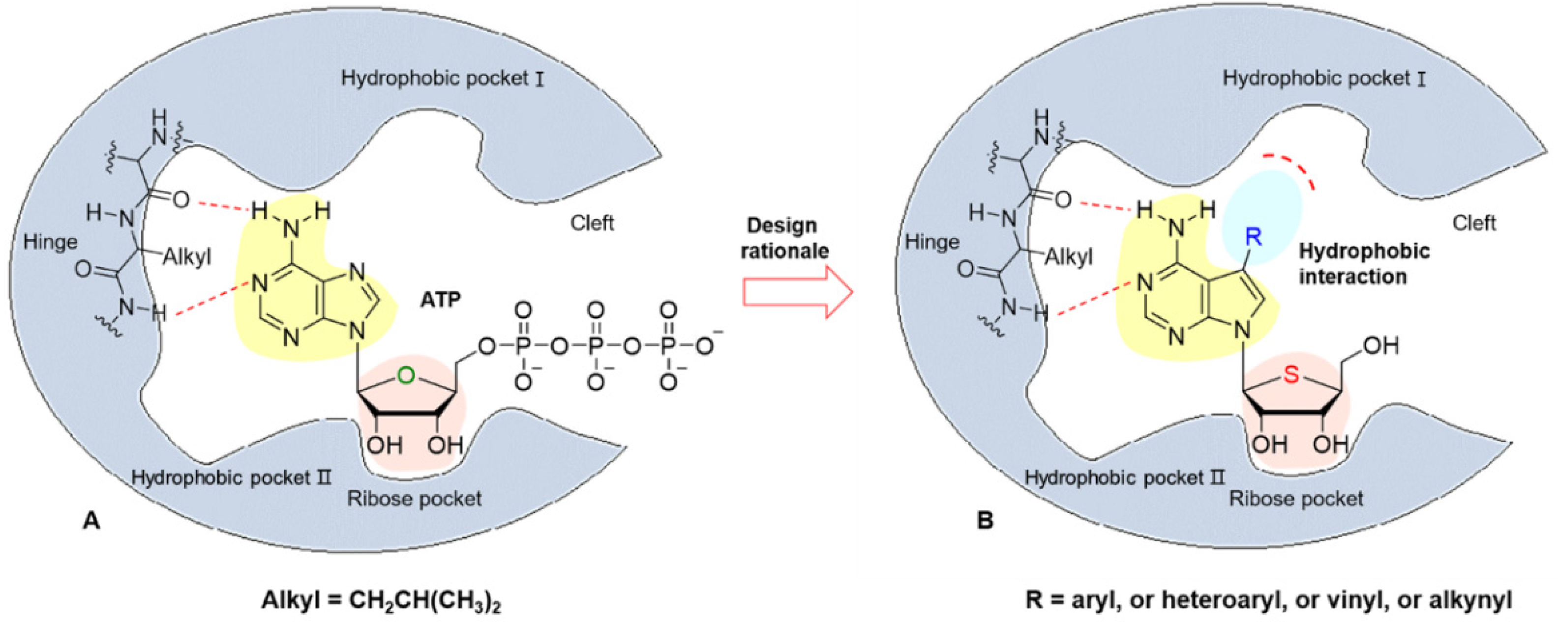
2. Results and Discussion
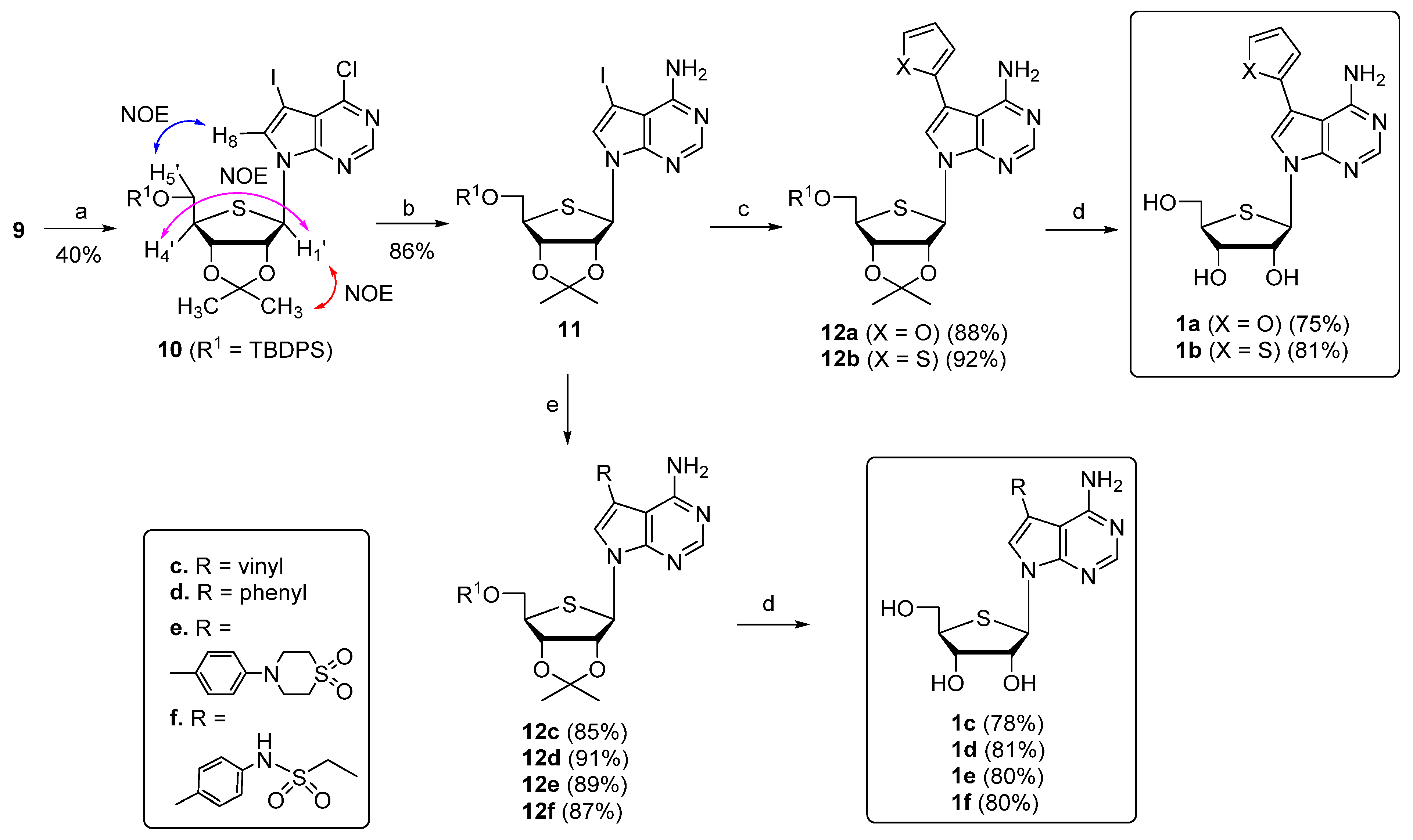
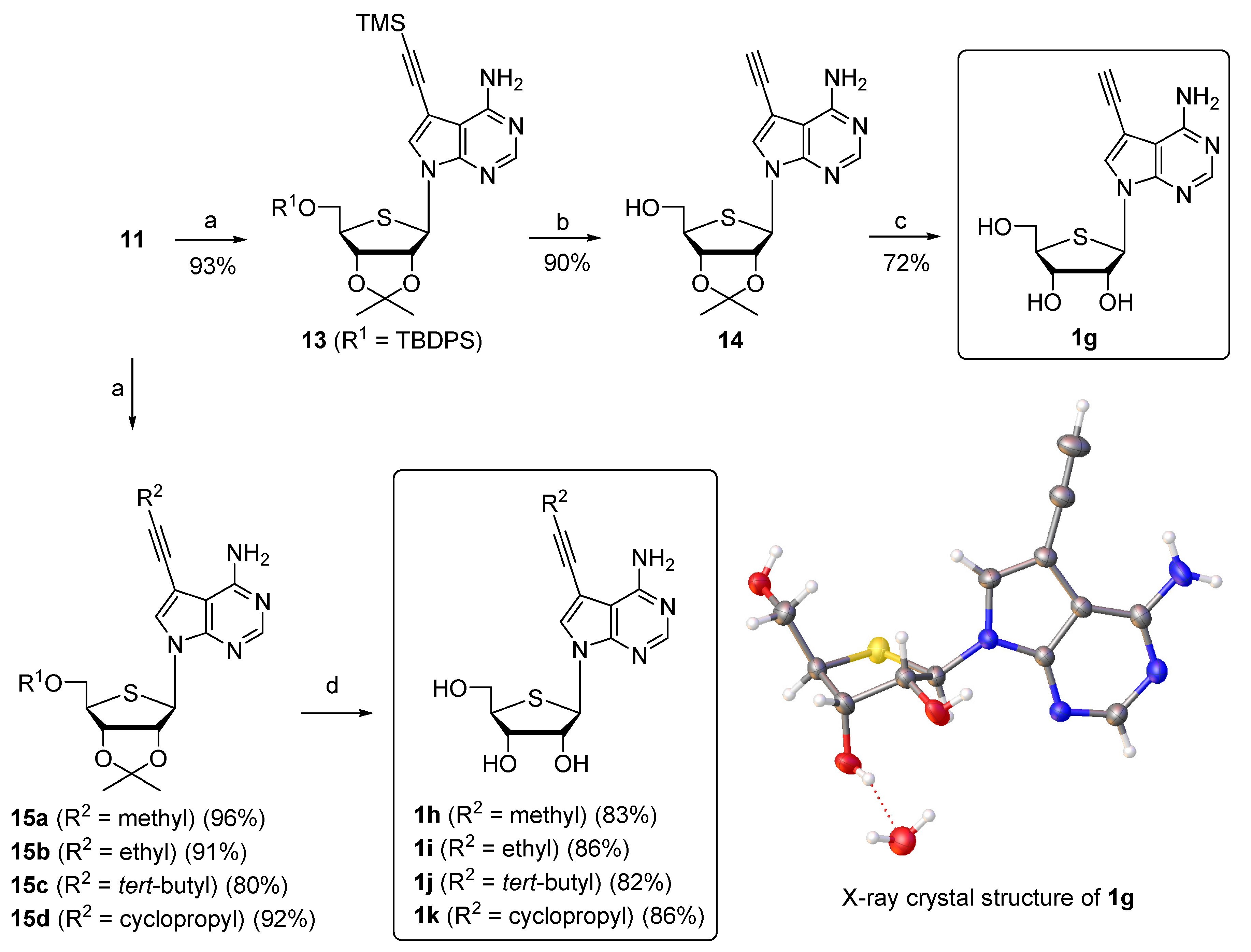
2.1. Chemistry

The structures of synthesized compounds are represented in Figure 2.
As shown in Scheme 1, to synthesize the final nucleoside 1, we first synthesized the glycosyl donor 9 from commercially available D-ribose.
D-Ribose was converted to 2,3-O-isopropylidene-D-ribonic γ-lactone (2) using the two steps protocol as described previously [40]. First, D-ribose was converted to D-ribonolactone using bromine/water in the presence of potassium carbonate and later treated with acetone in the presence of a catalytic amount of concentrated sulfuric acid at room temperature to afford 2. Following the reported general protocol [41] for thiosugar 7, the inversion of configuration at the C4 chiral center of D-ribonolactone was achieved by treating 2 initially with mesyl chloride and subjecting it to base hydrolysis using aqueous potassium hydroxide solution to give 3 with inverted stereochemistry. Protection of the C5-hydroxyl of 3 with tert-butyldiphenylsilyl (TBDPS), followed by reduction of the resulting compound 4 with NaBH4, afforded diol 5. Sulfur heterocyclization was performed by converting 5 to dimesylate 6, immediately reacted with sodium sulfide nonahydrate at 90 °C to give 7 at a 28% overall yield from 2. Compound 7 was subjected to mCPBA oxidation at −78 °C to give sulfoxide 8 (82% yield). Pummerer rearrangement of 8 occurred upon heating with acetic anhydride to afford glycosyl donor 9 as a 1:1.6 α/β anomeric mixture.
The glycosyl donor 9 was then condensed with silylated 7-deaza-7-iodo-6-chloropurine under heating at 80 °C for 1 h in the presence of a Lewis acid, TMSOTf, to afford the desired β-stereoisomer 10 as a single stereoisomer (40% yield; Scheme 2); however, the same reaction at room temperature failed to afford the desired product. The β configuration of condensed nucleoside 10 was easily determined by 2D NOESY experiments. The NOESY spectrum revealed a correlation between 1′-H and 4′-H, as well as between 1′-H and one of the two methyl groups of the acetonide group. A correlation between 5′-H and H-8 was also observed, confirming the presence of the β-D-anomer (see the Supporting Information). Ammonolysis of 10 in tert-butanolic ammonia at 90 °C produced key intermediate 11, which was ready for functionalization with hydrophobic groups at the C7 position via palladium-catalyzed cross-coupling reactions. Pd-catalyzed Stille coupling of 7-iodo derivative 11 with 2-tributylstannylfuran and 2-tributylstannylthiophene in the presence of PdCl2(PPh3)2 yielded 7-furanyl and 7-thiofuranyl derivatives 12a and 12b, respectively. Removal of acetonides of 12a and 12b with 50% aqueous trifluoroacetic acid (TFA) afforded the final nucleosides, 1a, and 1b, respectively.
Further Suzuki coupling reactions were performed to introduce other hydrophobic groups, such as vinyl, phenyl, and 4-substituted-phenyl to 11. Coupling of 11 with vinyl boronic ester in the presence of PdCl2(PPh3)2 in DMF/H2O gave 12c in 85% yield. In this Suzuki coupling reaction, water was used as a co-solvent to avoid side reactions, resulting from Heck coupling [42]. The desired Suzuki coupled product was obtained as a single product. Similar Suzuki reactions of 11 with phenyl and 4-substituted phenyl boronic esters afforded 7-phenyl and 7-(4-substituted)phenyl derivatives, 12df. Treatment of 12cf with 50% aqueous TFA yielded the final nucleosides 1cf, respectively.
Next, to introduce other linear hydrophobic groups, such as acetylene, Sonogashira coupling was employed, as shown in Scheme 3. Treatment of 11 with trimethylsilyl acetylene in the presence of a palladium catalyst and copper iodide afforded 13 (93% yield). Removal of the silyl groups of 13 with 1 M TBAF solution in THF afforded 14, which was treated with 2 N HCl to yield the 7-acetylene derivative 1g. The molecular structure of 1g was confirmed by a single X-ray crystal analysis (CCDC 1575257); further evidence supporting the β-configuration is provided in the Supporting Information [43]. Several 2-substituted acetylene analogs 15ad were also synthesized from 11 by employing the same Sonogashira coupling conditions. The final propylene, butylene, tert-butyl acetylene, and cyclopropyl acetylene analogs 1hk were obtained by treating 15ad with 50% aqueous TFA at room temperature.

2.2. Biological Evaluation

2.2.1. Antiproliferative Activity

All synthesized compounds 1ak were evaluated for their antiproliferative activity against six different cancer cell lines, including human lung (A549), colon (HCT116), breast (MDA-MB-231), liver (SK-HEP-1), stomach (SNU638), and prostate (PC-3) cancer cells, using SRB colorimetric assay [44,45]. As demonstrated in Table 1, compounds 1a and 1b possessing furanyl and thiofuranyl moieties at the 7-position, respectively, exhibited moderate antiproliferative activity. Compound 1c, with a vinyl substituent at the 7-position, displayed potent anticancer activity, whereas bulky groups, such as phenyl (1d) and substituted phenyls (1e and 1f) showed low to no anticancer activity. In contrast, the linear acetylene moiety (1g) at the 7-position exhibited excellent anticancer activity in the nanomolar range. Surprisingly, 2-substituted acetylene derivatives 1hk abolished antiproliferative activity. This result demonstrates that a small and linear hydrophobic group, such as acetylene is necessary for potent anticancer activity. Since compound 1g exhibited the most potent antiproliferative activity against cancer cells (IC50 = 0.004–0.06 μM), the antiproliferative activity of 1g against normal cells was additionally evaluated in primary dermal fibroblast cells. Compound 1g also showed considerable growth inhibition in cultured human normal dermal fibroblast cells (IC50 = 0.15 μM). Although rather toxic, compound 1g seems to be more potent in the antiproliferative activity of cancer cells than normal cells.

2.2.2. Kinome Scan Profile

To characterize the kinase inhibition profile of the most potent compound 1g, it was profiled against a panel of 96 kinases at a concentration of 1 μM (Figure 3). The results revealed that compound 1g exhibited strong inhibitory activities against four kinases at 1 μM (<20% activity remaining), i.e., TRKA (NTRK1), DYRK1B, and CK1δ (CSNK1D) among the panel (see the Supporting Information for tabular representation of kinome scan data, Table S1). Since compound 1g showed strong inhibition of DYRK1B, it was evaluated for its isoform, DYRK1A inhibition. Compound 1g displayed potent inhibition of DYRK1A (IC50 = 43 nM, see the Supporting information, Table S2). These kinases are reportedly involved in cancer initiation and progression [11,37,38,39] and represent promising targets for cancer therapy. Based on this result, it could be inferred that the hydrophobic pocket in the ATP binding site of these four kinases could accommodate only small and linear hydrophobic groups such as acetylene for kinase inhibition. Next, the concentration-dependent inhibitory activities of compounds 1ak were investigated and the half-maximal inhibitory concentration (IC50) was determined against four kinases, TRKA, DYRK1A, DYRK1B, and CK1δ (see the Supporting information, Table S2). The kinase inhibition trend observed for 1ak was almost similar to that of their antiproliferative activity. In general, compounds 1a, 1b, and 1g exhibited excellent kinase inhibition activities, whereas compounds 1c and 1d exhibited moderate kinase inhibition. Among compounds tested, compounds 1a and 1b with 7-furanyl and 7-thiofuranyl substituents, respectively showed excellent kinase inhibition activity against NTRK1, whereas compounds 1a and 1g with 7-furanyl and 7-acetylene substituents, respectively showed excellent kinase inhibition activity against DYRK1A and DYRK1B. Compounds 1b and 1d with 7-thiofuranyl and 7-phenyl substituents, respectively showed excellent inhibition against CSNK1D. However, sterically demanding compounds 1e and 1f and the 2-substituted acetylene derivatives 1hk demonstrated weak to no inhibitory activity. Compound 1g showed the best antiproliferative activity, but compound 1a was discovered as the best inhibitory compound against the above-mentioned four kinases, indicating that the anti-cancer effect of 1g might occur by unexpected mode of action.

2.2.3. Antiproliferative Activity against KM12 and ACHN Cell Lines

The effect of 1g on cell growth was also determined in KM12, a colon cancer cell line that highly expresses NTRK1/2/3 and DYRK3. Compared to doxorubicin (IC50 = 0.12 μM), compound 1g (IC50 = 0.07 μM) showed marked activity against the cell growth of KM12. Likewise, IC50 of 1g on cell growth of ACHN, a renal cancer cell line, expressing CK1δ and DYRK2 was approximately 0.04 μM, when compared with 0.04 μM of doxorubicin, suggesting the highly effective and selective anti-proliferative activity of 1g in cancer cell lines expressing NTRK, DYRK2 or CK1δ; However, we can’t exclude the possibility that anti-cancer phenotype might be driven through some other mechanism.

2.2.4. Metabolic Stability and CYP Inhibition

Based on anticancer and kinase inhibition data, compound 1g was examined for its microsomal stability and CYP isozyme inhibition in vitro (Table 2). Compound 1g was metabolically stable in human liver microsomes. Moreover, it showed no CYP isozyme inhibition against the five major drug-metabolizing cytochrome P450 isozymes.

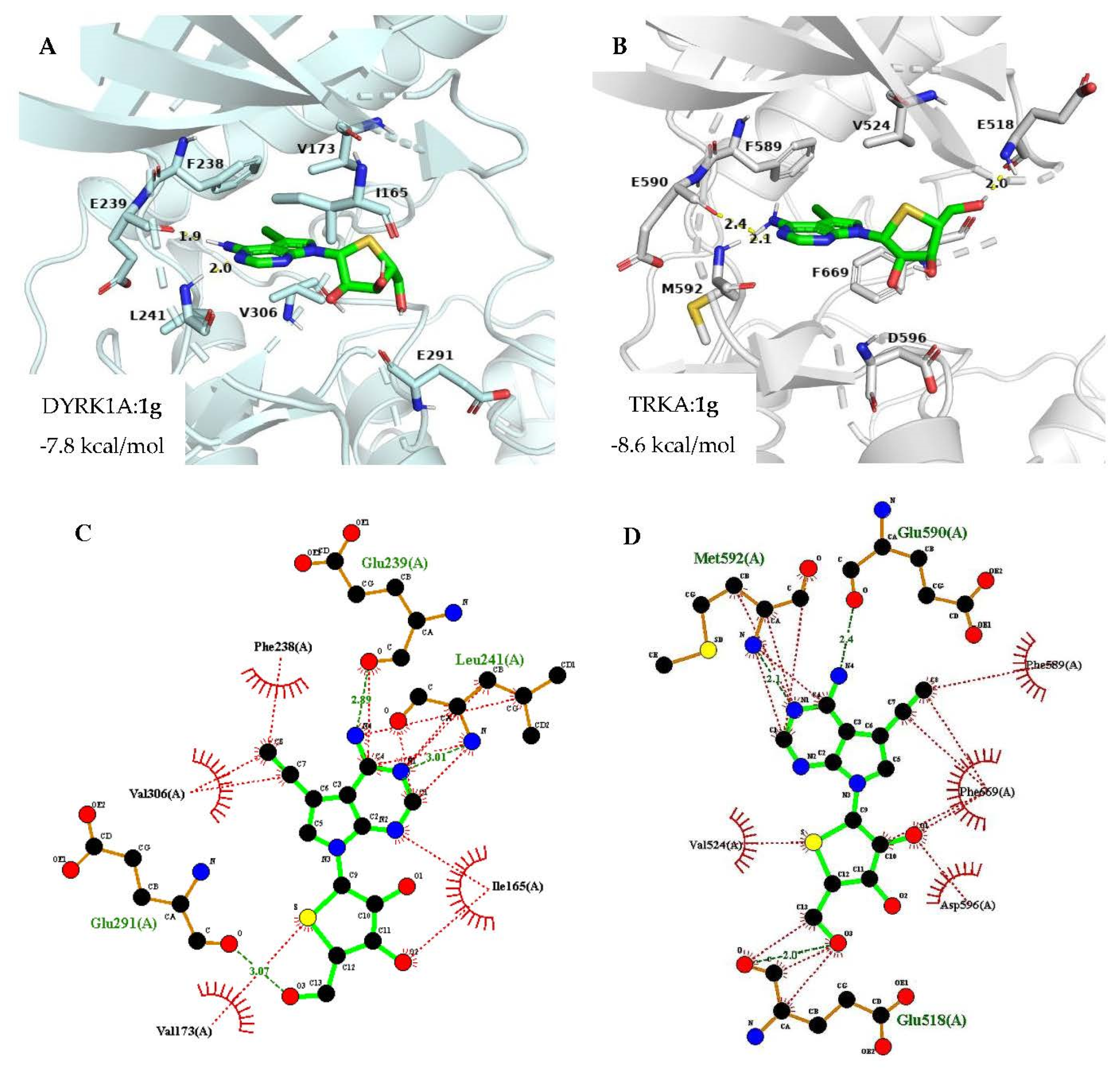
2.3. Docking Analysis

Next, to justify the rationale of this design strategy, the ligand-bound DYRK1A (PDB ID: 7A51) [46] and TRKA (PDB ID: 5JFV) [47] crystal structures were used for the molecular docking study (Figure 4). The docked pose of compound 1g into the ATP binding site of DYRK1A and TRKA, as depicted in Figure 4A and 4B respectively, revealed that the purine ring formed two key hydrogen bonds with hinge residue Leu 241, and Glu 239 of DYRK1A, whereas in the case of TRKA the purine ring of 1g formed hydrogen bonding with Met 592 and Glu 590, in a manner comparable with that of ATP. Furthermore, hydrogen bonding interaction was seen between 5′-OH of 1g and Glu 291 of DYRK1A (Figure 4C). Similarly, hydrogen bonding interaction between 5′-OH of 1g and Glu 518 of TRKA was observed. In addition, the acetylene moiety occupied the hydrophobic pocket as supposed and displayed hydrophobic interactions with gatekeeper residue, Phe 238 and Val 306 of DYRK1A (Figure 4C). While, in the TRKA: 1g docked pose acetylene was found to form hydrophobic interactions with the gatekeeper residue, Phe 589 and Phe 669 (Figure 4D). Additionally, a hydrophobic interaction of 4-thio of the sugar moiety with Val 173 and Val 524 of DYRK1A and TRKA, respectively was observed. Thus, it can be concluded that compound 1g fits markedly well in the ATP binding sites of DYRK1A and TRKA, thus providing a new scaffold that inhibits the activity of these enzymes.

3. Materials and Methods

3.1. General Methods

Proton (1H) and carbon (13C) NMR spectra were recorded on a JEOL JNM-GCX (400/100 MHz), Bruker AMX-500 (500/125 MHz), or JEOL JNM-ECA 600 (600/150 MHz) spectrometer. Chemical shifts are given in parts per million (δ), calibrated to the solvent peak, and coupling constants (J) in hertz (Hz). High-resolution mass (HRMS) measurements were recorded on a Thermo LCQ XP instrument. UV spectra were recorded in methanol on a U-3000 made by Hitachi. Optical rotations were measured on Jasco III in an appropriate solvent and [α]25D values are given in 10−1 deg cm2 g−1. Melting points were determined on a Barnstead electrothermal 9100 instrument and are uncorrected. Microwave-assisted reactions were conducted in Biotage Initiator+ US/JPN (part no. 356007) microwave reactor. The TLC spots were examined under ultraviolet light at 254 nm and further visualized by p-anisaldehyde or phosphomolybdic acid stain solution. Column chromatography was performed using silica gel (Kieselgel 60, 70–230 mesh, Merck). The purity of all tested compounds was determined by high-performance liquid chromatography (HPLC) analysis, confirming ≥95% purity.

3.2. Chemical Synthesis

3.2.1. (3aR,6S,6aR)-6-(Hydroxymethyl)-2,2-dimethyldihydrofuro[3,4-d][1,3]dioxol-4(3aH)-one (3)

To an ice-cooled solution of 2 (50 g, 0.265 mol) in pyridine (330 mL), methanesulfonyl chloride (52.34 g, 0.45 mol) was dropwise added under a nitrogen atmosphere and the solution was stirred at room temperature for 4 h. The reaction was quenched with slow addition of saturated aqueous NaHCO3 (520 mL) with stirring until no effervescence and extracted with dichloromethane (2 L). The combined organic layer was washed successively with water and brine, dried (MgSO4), and concentrated in vacuo below 25 °C to give the crude product. To this crude mesylate, a solution of potassium hydroxide (36.74 g, 0.65 mol) in water (250 mL) was added using a dropping funnel, maintaining the temperature below 30 °C. This reaction mixture was stirred at room temperature for 12 h and then adjusted to pH 3.0 to 4.0 by adding 3 M hydrochloric acid (260 mL). The acidic solution was concentrated under reduced pressure to afford a solid mass. The solid mass was triturated with acetone (2 L) and heated to 50 °C for 30 min. The acetone was decanted, dried over anhydrous MgSO4, and filtered. The filtrate was concentrated to obtain crude 3 (Rf = 0.45, TLC eluent = CH2Cl2/MeOH, 19:1).

3.2.2. (3aR,6S,6aR)-6-(((tert-butyldiphenylsilyl)oxy)methyl)-2,2-dimethyldihydrofuro[3,4-d][1,3]dioxol-4(3aH)-one (4)

To a solution of 3 in methylene chloride (240 mL), imidazole (8.78 g, 129 mmol) was added, followed by dropwise addition of tert-butyldiphenylsilyl chloride (26 g, 94.26 mmol) at 0 °C. After being stirred at room temperature for 12 h, the reaction mixture was partitioned between methylene chloride (3 × 600 mL) and water (760 mL). The layers were separated and the combined organic layer was dried (MgSO4), filtered, and evaporated to give crude 4 (Rf = 0.50, TLC eluent = hexane/ethyl acetate, 4:1).

3.2.3. (S)-2-((tert-butyldiphenylsilyl)oxy)-1-((4S,5R)-5-(hydroxymethyl)-2,2-dimethyl-1,3-dioxolan-4-yl)ethan-1-ol (5)

The crude 4 was dissolved in THF-MeOH (285 mL-54 mL) and to this, sodium borohydride (14.64 g, 387.07 mmol) was added portion wise at 0 °C. After stirring for 2 h at room temperature, the reaction mixture was quenched with glacial acetic acid (26 mL) and evaporated. The residue was diluted with 20% aqueous potassium sodium tartrate (500 mL) and the aqueous layer was extracted with ethyl acetate (3 × 600 mL). The organic layer was washed with brine, dried over MgSO4, and evaporated to obtain crude 5 (Rf = 0.50, TLC eluent = hexane/ethyl acetate, 3:2).

3.2.4. (S)-2-((tert-butyldiphenylsilyl)oxy)-1-((4R,5R)-2,2-dimethyl-5-(((methylsulfonyl)oxy)methyl)-1,3-dioxolan-4-yl)ethyl Methanesulfonate (6)

To a solution of 5 in methylene chloride (277 mL), 4-dimethylaminopyridine (0.36 g, 2.96 mmol) and triethylamine (106.40 g, 1051.53 mmol) were added and the solution was cooled to 0 °C. To this methanesulfonyl chloride (59.11 g, 516.09 mmol) was added dropwise and the reaction mixture was stirred at room temperature for 2 h. The reaction was quenched with saturated aqueous NaHCO3 (300 mL) until no effervescence and extracted with methylene chloride (3 × 520 mL). The combined organic layer was washed with brine (150 mL), dried (MgSO4), and passed through silica to remove any inorganic impurities and evaporated below 25 °C to give crude di-O-mesylate 6 (Rf = 0.55, TLC eluent = hexane/ethyl acetate, 3:2).

3.2.5. tert-Butyl(((3aS,4R,6aR)-2,2-dimethyltetrahydrothieno[3,4-d][1,3]dioxol-4 yl)methoxy)diphenylsilane (7)

To a stirred solution of 6 in DMF (1.5 L) crushed Na2S⋅9H2O (43.37g, 180.60 mmol) was added and the reaction mixture was transferred to a preheated bath at 90 °C. After being stirred for 15 h, the reaction mixture was cooled to room temperature and quenched with water (1 L). The aqueous layer was extracted with n-hexane (3 × 1 L). The organic layer was combined, washed with brine, dried over anhydrous MgSO4, and concentrated. The residue was purified by column chromatography (silica gel, hexane/ethyl acetate, 19:1) to afford 7 (31.6 g, 28% in 5 steps) as pale yellow syrup; [α]25D 45.17 (c 14.5, CH3OH); UV (CH3OH) λmax 264.94 nm; 1H NMR (CDCl3, 500 MHz): δ 7.67–7.63 (m, 4H), 7.42–7.35 (m, 6H), 4.79 (d, J = 1.7 Hz, 2H), 3.76 (dd, J = 10.6, 4.9 Hz, 1H), 3.60 (dd, J = 10.6, 6.8 Hz, 1H), 3.38–3.35 (m, 1H), 3.13–3.10 (m, 1H), 2.82 (merged dd, J1 = J2 = 12.6 Hz, 1H), 1.50 (s, 3H), 1.30 (s, 3H), 1.04 (s, 9H); 13C NMR (CDCl3, 125 MHz): δ 135.56, 135.55, 133.0, 132.9, 129.8, 129.7, 127.7, 110.6, 85.9, 83.7, 65.9, 55.4, 38.3, 26.8, 26.4, 24.5, 19.1; HRMS (ESI-Q-TOF) m/z [M + NH4]+ for C24H36NO3SSi calculated 446.218, found 446.2175.

3.2.6. (3aS,4R,5S,6aR)-4-(((tert-butyldiphenylsilyl)oxy)methyl)-2,2-dimethyltetrahydrothieno[3,4-d][1,3]dioxole 5-oxide (8)

A solution of 3-chloroperbenzoic acid (5.56 g, 32.25 mmol) in methylene chloride (110 mL) was added dropwise to a stirred solution of 7 (27.65 g, 64.50 mmol) in methylene chloride (110 mL) at −78 °C under a nitrogen atmosphere and stirred at the same temperature for 45 min. The reaction mixture was quenched with saturated NaHCO3 solution and extracted with methylene chloride (3 × 650 mL). The combined organic layer was washed with brine, dried over MgSO4, evaporated, and the residue was purified by silica gel column chromatography (hexane/ethyl acetate, 2:3) to afford 8 (23 g, 82%) as a colorless syrup; [α]25D 17.87 (c 3.75, CH3OH); UV (CH3OH) λmax 264.94 nm; 1H NMR (CDCl3, 500 MHz): δ 7.59–7.54 (m, 4H), 7.46–7.43 (m, 2H), 7.40–7.37 (m, 4H), 5.21 (t, J = 5.5 Hz, 1H), 4.97 (d, J = 5.3 Hz, 1H), 4.09 (dd, J = 11.2, 2.5 Hz, 1H), 3.82 (dd, J = 11.2, 3.3 Hz, 1H), 3.58 (s, 1H), 3.39 (merged dd, J1 = J2 = 14.8 Hz, 1H), 3.25 (dd, J = 14.8, 6.0 Hz, 1H), 1.56 (s, 3H), 1.33 (s, 3H), 1.01 (s, 9H); 13C NMR (CDCl3, 125 MHz): δ 135.4, 135.3, 131.7, 131.6, 130.1, 130.0, 127.9, 127.8, 112.2, 86.3, 84.7, 74.7, 61.7, 58.3, 26.7, 26.6, 24.3, 18.9; HRMS (ESI-Q-TOF) m/z [M + H]+ for C24H33O4SSi calculated 445.1863, found 445.1862.

3.2.7. (3aR,6R,6aS)-6-(((tert-butyldiphenylsilyl)oxy)methyl)-2,2-dimethyltetrahydrothieno[3,4-d][1,3]dioxol-4-yl acetate (9)

A solution of 8 (16 g, 35.98 mmol) in acetic anhydride (142 mL) was transferred to a preheated bath at 110 °C and stirred at the same temperature for 4 h. After concentration under reduced pressure, the residue was neutralized with aqueous sat. NaHCO3 until pH 7.0 and stirred for 15 min. To the solution brine was added and extracted with ethyl acetate (3 × 600 mL). The organic layer was combined and washed with brine, dried (MgSO4), filtered, and evaporated under reduced pressure. The crude residue obtained was purified by silica gel column chromatography (hexane/ethyl acetate, 19:1) to give 9 (12.6 g, 72%) as a colorless syrup: 1:1.6 α/β mixture of anomers; UV (CH3OH) λmax 259.85 nm; 1H NMR (CDCl3, 500 MHz): δ 7.70–7.62 (m, 6H), 7.43–7.40 (m, 3H), 7.39–7.36 (m, 7H), 6.09 (d, J = 5.3 Hz, 0.4H), 5.90 (s, 1H), 4.99 (d, J = 5.4 Hz, 1H), 4.82–4.80 (m, 0.6H), 4.64–4.61 (m, 1.5H), 3.82–3.80 (m, 1H), 3.77–3.72 (m, 1.6H), 3.60–3.52 (m, 2H), 2.13 (s, 1.4H), 1.81 (s, 3H), 1.53 (s, 1.9H), 1.48 (s, 3H), 1.30 (s, 1.6H), 1.28 (s, 3H), 1.06 (s, 9H), 1.04 (s, 5H); HRMS (ESI-Q-TOF) m/z [M + Na]+ for C26H34NaO5SSi calculated 509.1788, found 509.1791.

3.2.8. 7-((3aR,4R,6R,6aS)-6-(((tert-butyldiphenylsilyl)oxy)methyl)-2,2-dimethyltetrahydrothieno[3,4-d][1,3]dioxol-4-yl)-4-chloro-5-iodo-7H-pyrrolo[2,3-d]pyrimidine (10)

N,O-Bis(trimethylsilyl)acetamide (BSA, 2.5 mL, 10.27 mmol) was added to a stirred suspension of 4-chloro-5-iodo-7H-pyrrolo[2,3-d]pyrimidine (2.6 g, 9.33 mmol) in anhydrous acetonitrile (67 mL) under nitrogen atmosphere. The resulting suspension was stirred at room temperature for 10 min until a homogeneous solution was obtained. To this clear solution a solution of 9 (5 g, 10.27 mmol) in anhydrous acetonitrile (50 mL) were added followed by dropwise addition of trimethylsilyl trifluoromethanesulfonate (1.5 mL, 8.40 mmol). The reaction mixture was stirred at room temperature for 15 min before transferring it to a preheated bath at 80 °C. After stirring at the same temperature for 1 h, the reaction mixture was cooled to room temperature and diluted with ethyl acetate (700 mL). The organic layer was washed with aqueous sat. NaHCO3 (3 × 250 mL) and brine (100 mL), dried over MgSO4, filtered, and concentrated. The residue was purified by column chromatography (silica gel, hexane/ethyl acetate, 50:3) to give 10 (2.9 g, 40%) as a pale yellow sticky mass; [α]25D −7.03 (c 0.6, CH3OH); UV (CH3OH) λmax 310.78 nm; 1H NMR (CD3OD, 500 MHz): δ 8.46 (s, 1H), 7.87 (s, 1H), 7.64–7.58 (m, 4H), 7.40–7.33 (m, 4H), 7.31–7.28 (m, 2H), 6.24 (d, J = 2.2 Hz, 1H), 5.10 (dd, J = 5.5, 2.2 Hz, 1H), 4.96 (dd, J = 5.5, 2.1 Hz, 1H), 3.88 (dd, J = 10.4, 7.2 Hz, 1H), 3.81 (dd, J = 10.4, 7.3 Hz, 1H), 3.75 (td, J = 7.2, 2.1 Hz, 1H), 1.55 (s, 3H), 1.28 (s, 3H), 1.04 (S, 9H); 13C NMR (CD3OD, 100 MHz): δ 154.2, 152.5, 152.3, 137.5, 137.4, 136.2, 135.0, 134.7, 131.9, 131.8, 129.7, 129.6, 119.9, 114.0, 90.9, 87.0, 70.1, 67.7, 58.9, 53.5, 28.3, 28.1, 26.1, 20.8; HRMS (ESI-Q-TOF) m/z [M + H]+ for C30H34ClIN3O3SSi calculated 706.0818, found 706.0798.

3.2.9. 7-((3aR,4R,6R,6aS)-6-(((tert-butyldiphenylsilyl)oxy)methyl)-2,2-dimethyltetrahydrothieno[3,4-d][1,3]dioxol-4-yl)-5-iodo-7H-pyrrolo[2,3-d]pyrimidin-4-amine (11)

A solution of 10 (2.9 g, 4.11 mmol) in saturated solution of NH3/t-BuOH (30 mL) contained in a stainless steel bomb reactor was transferred to a preheated bath at 90 °C and stirred at the same temperature for 24 h. The steel bomb containing reaction mixture was cooled to room temperature and solvent was evaporated under reduced pressure. The residue was purified by silica gel column chromatography (hexane/ethyl acetate, 13:7) to obtain 11 (2.42 g, 86%) as a sticky mass; [α]25D −45.49 (c 2.4, CH3OH); UV (CH3OH) λmax 286.04 nm; 1H NMR (CDCl3, 500 MHz): δ 8.22 (s, 1H), 7.64–7.62 (m, 4H), 7.43–7.40 (m, 2H), 7.37–7.33 (m, 5H), 6.21 (d, J = 2.7 Hz, 1H), 5.88 (br s, 2H), 4.88 (dd, J = 5.6, 2.8 Hz, 1H), 4.78 (dd, J = 5.6, 2.8 Hz, 1H), 3.86–3.79 (m, 2H), 3.77–3.75 (m, 1H), 1.58 (s, 3H), 1.27 (s, 3H), 1.07 (s, 9H); 13C NMR (CDCl3, 125 MHz): δ 156.7, 151.9, 150.1, 135.5, 135.5, 132.9, 132.8, 129.9, 129.8, 127.8, 127.0, 112.4, 104.5, 89.0, 84.0, 66.3, 65.1, 55.7, 50.5, 27.3, 26.8, 25.1, 19.2; HRMS (ESI-Q-TOF) m/z [M + H]+ for C30H36IN4O3SSi calculated 687.1317, found 687.1301.
General Procedure of Stille Coupling for the Synthesis of 12a and 12b. To the compound 11 (1 equiv) in a microwave vial equipped with a septum, catalyst PdCl2(PPh3)2 (15 mol %) was added and degassed THF (2.8 mL/mmol) under nitrogen atmosphere. The resulting solution was degassed for 5 min and a corresponding 2-(tributylstannyl)hetaryl (2.5 equiv) was added. After stirring the reaction mixture in a microwave for 1 h at 70 °C, it was quenched by adding water and brine. The aqueous layer was extracted with ethyl acetate thrice and organics were concentrated. The residue was purified by column chromatography.

3.2.10. 7-((3aR,4R,6R,6aS)-6-(((tert-butyldiphenylsilyl)oxy)methyl)-2,2-dimethyltetrahydrothieno[3,4-d][1,3]dioxol-4-yl)-5-(furan-2-yl)-7H-pyrrolo[2,3-d]pyrimidin-4-amine (12a)

Compound 12a (0.16 g, 88%) was obtained from 11 (0.2 g, 0.29 mmol) as a pale yellow sticky mass; silica gel column chromatography (hexane/ethyl acetate, 3:2); [α]25D −31.0 (c 0.2, CH3OH); UV (CH3OH) λmax 291.76 nm; 1H NMR (CDCl3, 400 MHz): δ 8.27 (s, 1H), 7.64–7.61 (m, 4H), 7.47–7.46 (m, 1H), 7.39–7.36 (m, 3H), 7.33–7.29 (m, 4H), 6.45–6.44 (m, 1H), 6.28–6.27 (m, 2H), 5.98 (br s, 2H), 4.95 (dd, J = 5.9, 3.2 Hz, 1H), 4.82 (dd, J = 5.9, 3.2 Hz, 1H), 3.92–3.82 (m, 2H), 3.79–3.75 (m, 1H), 1.59 (s, 3H), 1.28 (s, 3H), 1.05 (s, 9H); 13C NMR (CDCl3, 100 MHz): δ 157.2, 152.9, 151.2, 149.0, 141.3, 135.77, 135.74, 133.17, 133.11, 130.1, 130.0, 127.9, 120.0, 112.7, 112.1, 107.3, 105.6, 100.9, 89.2, 84.2, 66.3, 65.3, 55.8, 27.6, 27.0, 25.4, 19.5; HRMS (ESI-Q-TOF) m/z [M + H]+ for C34H39N4O4SSi calculated 627.2456, found 627.2463.

3.2.11. 7-((3aR,4R,6R,6aS)-6-(((tert-butyldiphenylsilyl)oxy)methyl)-2,2-dimethyltetrahydrothieno[3,4-d][1,3]dioxol-4-yl)-5-(thiophen-2-yl)-7H-pyrrolo[2,3-d]pyrimidin-4-amine (12b)

Compound 12b (0.21 g) was afforded from 11 (0.25 g, 0.36 mmol) in 92% yield as colorless sticky mass; silica gel column chromatography (hexane/ethyl acetate, 3:2); [α]25D −51.36 (c 0.31, CH3OH); UV (CH3OH) λmax 287.13 nm; 1H NMR (CDCl3, 400 MHz): δ 8.28 (s, 1H), 7.62–7.59 (m, 4H), 7.37–7.34 (m, 3H), 7.32–7.27 (m, 4H), 7.23 (s, 1H), 7.10–7.07 (m, 1H), 7.00–6.99 (m, 1H), 6.28 (d, J = 2.7 Hz, 1H), 5.41 (br s, 2H), 4.94 (dd, J = 5.5, 3.2 Hz, 1H), 4.81 (dd, J = 5.9, 3.2 Hz, 1H), 3.91–3.82 (m, 2H), 3.79–3.74 (m, 1H), 1.59 (s, 3H), 1.28 (s, 3H), 1.03 (s, 9H); 13C NMR (CDCl3, 100 MHz): δ 156.7, 152.1, 150.7, 135.68, 135.63, 129.97, 129.94, 128.0, 127.8, 126.5, 125.8, 121.9, 112.6, 109.4, 102.1, 89.0, 84.1, 66.2, 65.3, 55.6, 27.5, 26.9, 25.3, 19.3; HRMS (ESI-Q-TOF) m/z [M + H]+ for C34H39N4O3S2Si calculated 643.2227, found 643.2218.
General Procedure for the Synthesis of 1a and 1b. To an ice-cooled solution of 12a and 12b (1 equiv) in THF (3 mL/mmol) 50% aqueous trifluoroacetic acid (19.5 mL/mmol) was dropwise added and the resulting mixture was stirred at room temperature for 12 h. Acidic solution was basified using a weakly basic anion-exchange resin (Dowex® 66 free base) and stirred for an additional 3 h, filtered, and concentrated under vacuum. The residue was purified by silica gel column chromatography (CH2Cl2/MeOH, 47:3) to give 1a and 1b respectively.

3.2.12. (2R,3R,4S,5R)-2-(4-Amino-5-(furan-2-yl)-7H-pyrrolo[2,3-d]pyrimidin-7-yl)-5-(hydroxymethyl)tetrahydrothiophene-3,4-diol (1a)

It was obtained in 75% yield as white solid; mp 197–199 °C; [α]25D −21.14 (c 0.05, CH3OH); UV (CH3OH) λmax 290.77 nm; 1H NMR (DMSO-d6, 500 MHz): δ 8.13 (s, 1H), 7.95 (s, 1H), 7.78 (s, 1H), 6.90 (br s, 2H, D2O exchange), 6.72 (d, J = 3.0 Hz, 1H), 6.62–6.61 (m, 1H), 6.17 (d, J = 7.0 Hz, 1H), 5.43 (d, J = 6.4 Hz, 1H, D2O exchange), 5.27 (d, J = 4.4 Hz, 1H, D2O exchange), 5.18 (t, J = 5.5 Hz, 1H, D2O exchange), 4.48–4.45 (m, 1H), 4.19–4.18 (m, 1H), 3.78–3.75 (m, 1H), 3.63–3.58 (m, 1H), 3.29–3.28 (m, 1H); 13C NMR (DMSO-d6, 100 MHz): δ 157.6, 152.5, 151.6, 149.1, 142.3, 120.7, 112.3, 106.8, 105.6, 99.4, 77.7, 73.6, 63.9, 61.3, 53.4; HRMS (ESI-Q-TOF) m/z [M + H]+ for C15H17N4O4S calculated 349.0965, found 349.0974; purity ≥95%.

3.2.13. (2R,3R,4S,5R)-2-(4-Amino-5-(thiophen-2-yl)-7H-pyrrolo[2,3-d]pyrimidin-7-yl)-5-(hydroxymethyl)tetrahydrothiophene-3,4-diol (1b)

It was afforded in 81% yield as white solid; mp 166–172 °C; [α]25D −23.93 (c 0.09, CH3OH); UV (CH3OH) λmax 286.41 nm; 1H NMR (CD3OD, 500 MHz): δ 8.13 (s, 1H), 7.66 (s, 1H), 7.42 (d, J = 5.0 Hz, 1H), 7.14–7.11 (m, 2H), 6.22 (d, J = 5.9 Hz, 1H), 4.52 (dd, J = 5.7, 3.7 Hz, 1H), 4.29 (merged dd, J1 = J2 = 3.8 Hz, 1H), 3.88–3.80 (m, 2H), 3.48 (dd, J = 9.4, 4.8 Hz, 1H); 13C NMR (CD3OD, 125 MHz): δ 159.5, 153.5, 152.6, 137.5, 129.8, 128.5, 127.6, 124.5, 111.6, 103.5, 80.8, 76.3, 65.1, 64.5, 54.8; HRMS (ESI-Q-TOF) m/z [M + H]+ for C15H17N4O3S2 calculated 365.0737, found 365.0749; purity ≥95%.
General Procedure of Suzuki Coupling for the Synthesis of 12cf. A degassed mixture of DMF/H2O (6.9 mL/2.7 mL/mmol) was added to a microwave vial containing compound 11 (1 equiv), corresponding boronic ester (1.2 equiv), PdCl2(PPh3)2 (6 mol %), and sodium carbonate (2 equiv). The resulting reaction mixture was heated in a microwave at 70 °C for 1 h. After quenching with water, the reaction mixture was extracted with ethyl acetate thrice. The organic layers were combined, washed with brine, dried over MgSO4, filtered, and evaporated. The residue obtained was purified by column chromatography.

3.2.14. 7-((3aR,4R,6R,6aS)-6-(((tert-butyldiphenylsilyl)oxy)methyl)-2,2-dimethyltetrahydrothieno[3,4-d][1,3]dioxol-4-yl)-5-vinyl-7H-pyrrolo[2,3-d]pyrimidin-4-amine (12c)

Compound 12c was obtained in 85% yield as yellow sticky mass; silica gel column chromatography (hexane/ethyl acetate, 13:7); [α]25D −38.33 (c 1.1, CH3OH); UV (CH3OH) λmax 288.95 nm; 1H NMR (CD3OD, 400 MHz): δ 8.03 (s, 1H), 7.64 (t, J = 9.2 Hz, 4H), 7.42–7.39 (m, 3H), 7.37–7.31 (m, 4H), 6.91 (dd, J = 17.2, 10.8 Hz, 1H), 6.21 (s, 1H), 5.44 (d, J = 17.6 Hz, 1H), 5.20 (d, J = 10.8 Hz, 1H), 5.07–5.05 (m, 1H), 4.96–4.95 (m, 1H), 3.93–3.89 (m, 1H), 3.86–3.81 (m, 1H), 3.73–3.69 (m, 1H), 1.55 (s, 3H), 1.29 (s, 3H), 1.04 (s, 9H); 13C NMR (CD3OD, 100 MHz): δ 157.8, 151.1, 150.1, 135.37, 135.35, 133.0, 132.7, 129.74, 129.71, 128.1, 127.58, 127.54, 119.4, 115.8. 114.4, 112.0, 101.4, 88.4, 84.5, 66.0, 65.5, 55.7, 26.3, 26.0, 24.0, 18.7; HRMS (ESI-Q-TOF) m/z [M + H]+ for C32H39N4O3SSi calculated 587.2507, found 587.2506.

3.2.15. 7-((3aR,4R,6R,6aS)-6-(((tert-butyldiphenylsilyl)oxy)methyl)-2,2-dimethyltetrahydrothieno[3,4-d][1,3]dioxol-4-yl)-5-phenyl-7H-pyrrolo[2,3-d]pyrimidin-4-amine (12d)

It was obtained in 91% yield as colorless sticky mass; silica gel column chromatography (hexane/ethyl acetate, 1:1); [α]25D −46.89 (c 0.14, CH3OH); UV (CH3OH) λmax 283.50 nm; 1H NMR (CD3OD, 400 MHz): δ 8.11 (s, 1H), 7.62–7.59 (m, 4H), 7.43–7.41 (m, 3H), 7.39–7.33 (m, 4H), 7.29–7.26 (m, 4H), 7.23 (s, 1H), 6.27 (d, J = 2.7 Hz, 1H), 5.09 (dd, J = 5.4, 2.7 Hz, 1H), 4.96 (dd, J = 5.5, 2.3 Hz, 1H), 3.92–3.82 (m, 2H), 3.75–3.71 (m, 1H), 1.57 (s, 3H), 1.30 (s, 3H), 1.01 (s, 9H); 13C NMR (CD3OD, 100 MHz): δ 157.4, 151.2, 150.1, 135.38, 135.34, 134.1, 132.9, 132.7, 129.7, 128.7, 128.6, 127.55, 127.50, 127.1, 120.7, 117.9, 111.9, 101.3, 88.6, 84.7, 66.5, 65.6, 56.0, 26.2, 25.9, 24.0, 18.6; HRMS (ESI-Q-TOF) m/z [M + H]+ for C36H41N4O3SSi calculated 637.2663, found 637.2646.

3.2.16. 4-(4-(4-Amino-7-((3aR,4R,6R,6aS)-6-(((tert-butyldiphenylsilyl)oxy)methyl)-2,2-dimethyltetrahydrothieno[3,4-d][1,3]dioxol-4-yl)-7H-pyrrolo[2,3-d]pyrimidin-5-yl)phenyl)thiomorpholine 1,1-dioxide (12e)

It was afforded in 89% yield as colorless sticky mass; silica gel column chromatography (hexane/ethyl acetate, 11:9); [α]25D −28.78 (c 0.38, CH3OH); UV (CH3OH) λmax 270.04 nm; 1H NMR (CDCl3, 400 MHz): δ 8.27 (s, 1H), 7.61–7.59 (m, 4H), 7.40–7.34 (m, 4H), 7.32–7.27 (m, 4H), 7.14 (s, 1H), 6.94–6.92 (m, 2H), 6.31 (d, J = 3.6 Hz, 1H), 5.28 (br s, 2H), 4.99 (dd, J = 5.9, 3.2 Hz, 1H), 4.82 (dd, J = 5.9, 2.7 Hz, 1H), 3.93–3.86 (m, 4H), 3.84–3.82 (m, 2H), 3.79–3.75 (m, 1H), 3.13–3.11 (m, 4H), 1.60 (s, 3H), 1.29 (s, 3H), 1.03 (s, 9H); 13C NMR (CDCl3, 100 MHz): δ 156.6, 151.5, 150.7, 146.9, 135.66, 135.62, 133.0, 130.3, 129.96, 129.92, 127.8, 126.5, 120.6, 116.8, 116.6, 112.7, 101.9, 88.9, 84.0, 66.0, 65.2, 55.5, 50.5, 47.6, 27.5, 26.9, 25.3, 19.3; HRMS (ESI-Q-TOF) m/z [M + H]+ for C40H48N5O5S2Si calculated 770.2861, found 770.2865.

3.2.17. N-(4-(4-Amino-7-((3aR,4R,6R,6aS)-6-(((tert-butyldiphenylsilyl)oxy)methyl)-2,2-dimethyltetrahydrothieno[3,4-d][1,3]dioxol-4-yl)-7H-pyrrolo[2,3-d]pyrimidin-5-yl)phenyl)ethanesulfonamide (12f)

Compound 12f was obtained in 87% yield as colorless sticky mass; silica gel column chromatography (hexane/ethyl acetate, 2:3); [α]25D −48.16 (c 0.09, CH3OH); UV (CH3OH) λmax 286.04 nm; 1H NMR (CDCl3, 500 MHz): δ 8.29 (s, 1H), 7.60–7.59 (m, 4H), 7.39–7.34 (m, 2H), 7.32–7.30 (m, 4H), 7.28–7.27 (m, 2H), 7.25–7.24 (m, 2H), 7.20 (s, 1H), 6.66 (br s, 1H), 6.30 (d, J = 2.9 Hz, 1H), 5.37 (br s, 2H), 4.97 (dd, J = 5.6, 3.0 Hz, 1H), 4.82 (dd, J = 5.6, 2.7 Hz, 1H), 3.90–3.82 (m, 2H), 3.80–3.78 (m, 1H), 3.15 (q, J = 7.3 Hz, 2H), 1.60 (s, 3H), 1.39 (t, J = 7.3 Hz, 3H), 1.29 (s, 3H), 1.02 (s, 9H); 13C NMR (CDCl3, 100 MHz): δ 156.3, 151.1, 150.7, 136.4, 135.63, 135.60, 132.8, 130.9, 130.0, 127.8, 121.3, 120.9, 116.4, 112.6, 101.7, 89.1, 84.1, 66.4, 65.2, 55.6, 46.3, 27.5, 26.9, 25.3, 19.3, 8.4; HRMS (ESI-Q-TOF) m/z [M + H]+ for C38H46N5O5S2Si calculated 744.2704, found 744.2698.

3.2.18. (2R,3R,4S,5R)-2-(4-Amino-5-vinyl-7H-pyrrolo[2,3-d]pyrimidin-7-yl)-5-(hydroxymethyl)tetrahydrothiophene-3,4-diol (1c)

Compound 12c (0.12 g, 0.20 mmol) was converted to 1c as described for 1a, affording white solid (47.5 mg, 78%); silica gel column chromatography (CH2Cl2/MeOH, 19:1); mp 110–112 °C; [α]25D −41.86 (c 0.20, CH3OH); UV (CH3OH) λmax 288.50 nm; 1H NMR (DMSO-d6, 500 MHz): δ 8.04 (s, 1H), 7.77 (s, 1H), 7.10 (dd, J = 17.2, 10.9 Hz, 1H), 6.70 (br s, 2H, D2O exchange), 6.12 (d, J = 7.0 Hz, 1H), 5.59 (d, J = 17.1 Hz, 1H), 5.38 (d, J = 6.4 Hz, 1H, D2O exchange), 5.25 (d, J = 4.4 Hz, 1H, D2O exchange), 5.17 (t, J = 5.5 Hz, 1H, D2O exchange), 5.11 (d, J = 11.0 Hz, 1H), 4.46–4.41 (m, 1H), 4.18–4.14 (m, 1H), 3.77–3.72 (m, 1H), 3.61–3.55 (m, 1H), 3.27–3.24 (m, 1H); 13C NMR (CD3OD, 100 MHz): δ 157.8, 150.9, 150.6, 128.3, 119.9, 115.3, 113.9, 78.6, 74.1, 63.0, 62.2, 52.6; HRMS (ESI-Q-TOF) m/z [M + H]+ for C13H17N4O3S calculated 309.1016, found 309.1018; purity ≥95%.

3.2.19. (2R,3R,4S,5R)-2-(4-Amino-5-phenyl-7H-pyrrolo[2,3-d]pyrimidin-7-yl)-5-(hydroxymethyl)tetrahydrothiophene-3,4-diol (1d)

Compound 1d was prepared from 12d (0.16 g, 0.25 mmol) as described for 1a, affording white solid (72.5 mg, 81%); silica gel column chromatography (CH2Cl2/MeOH, 19:1); mp 102–104 °C; [α]25D −46.30 (c 0.06, CH3OH); UV (CH3OH) λmax 280.95 nm; 1H NMR (DMSO-d6, 500 MHz): δ 8.14 (s, 1H), 7.66 (s, 1H), 7.50–7.46 (m, 4H), 7.38–7.34 (m, 1H), 6.18 (d, J = 6.9 Hz, 1H), 5.42 (d, J = 6.4 Hz, 1H), 5.28 (d, J = 4.5 Hz, 1H), 5.16 (t, J = 5.5 Hz, 1H), 4.51–4.47 (m, 1H), 4.19–4.16 (m, 1H), 3.77–3.71 (m, 1H), 3.61–3.56 (m, 1H), 3.29–3.25 (m, 1H); 13C NMR (CD3OD, 125 MHz): δ 159.5, 153.1, 152.8, 136.6, 130.9, 130.7, 129.2, 123.4, 119.7, 103.2, 80.9, 76.4, 65.2, 64.5, 54.8; HRMS (ESI-Q-TOF) m/z [M + H]+ for C17H19N4O3S calculated 359.1172, found 359.1177; purity ≥95%.

3.2.20. 4-(4-(4-Amino-7-(3,4-dihydroxy-5-(hydroxymethyl)tetrahydrothiophen-2-yl)-7H-pyrrolo[2,3-d]pyrimidin-5-yl)phenyl)thiomorpholine 1,1-dioxide (1e)

It was obtained from 12e (0.1 g, 0.12 mmol) as described for 1a, as white solid (47.1 mg, 80%); silica gel column chromatography (CH2Cl2/MeOH, 93:7); mp 188–192 °C; [α]25D −24.29 (c 0.05, CH3OH); UV (CH3OH) λmax 270.04 nm; 1H NMR (DMSO-d6, 500 MHz): δ 8.13 (s, 1H), 7.54 (s, 1H), 7.35 (merged dd, J1 = J2 = 8.4 Hz, 2H), 7.13 (merged dd, J1 = J2 = 8.5 Hz, 2H), 6.18 (d, J = 6.9 Hz, 1H), 5.40 (d, J = 6.4 Hz, 1H), 5.26 (d, J = 4.3 Hz, 1H), 5.16 (t, J = 5.4 Hz, 1H), 4.49–4.47 (m, 1H), 4.18–4.17 (m, 1H), 3.87–3.82 (m, 4H), 3.75–3.72 (m, 1H), 3.60–3.57 (m, 1H), 3.29–3.27 (m, 1H), 3.17–3.12 (m, 4H); 13C NMR (DMSO-d6, 125 MHz): δ 157.2, 151.6, 151.0, 146.2, 129.4, 125.1, 120.1, 116.2, 116.0, 100.3, 77.3, 73.3, 63.3, 60.9, 52.8, 49.7, 46.5; HRMS (ESI-Q-TOF) m/z [M + H]+ for C21H26N5O5S2 calculated 492.137, found 492.1388; purity ≥95%.

3.2.21. N-(4-(4-Amino-7-(3,4-dihydroxy-5-(hydroxymethyl)tetrahydrothiophen-2-yl)-7H-pyrrolo[2,3-d]pyrimidin-5-yl)phenyl)ethanesulfonamide (1f)

It was afforded from 12f (90 mg, 0.12 mmol) as described for 1a, as white solid (44.6 mg, 80%); silica gel column chromatography (CH2Cl2/MeOH, 91:9); mp 127–132 °C; [α]25D −35.60 (c 0.05, CH3OH); UV (CH3OH) λmax 283.86 nm; 1H NMR (DMSO-d6, 500 MHz): δ 9.87 (s, 1H), 8.14 (s, 1H), 7.60 (s, 1H), 7.42 (merged dd, J1 = J2 = 8.2 Hz, 2H), 7.31 (merged dd, J1 = J2 = 8.1 Hz, 2H), 6.18 (d, J = 6.9 Hz, 1H), 5.40 (d, J = 6.4 Hz, 1H), 5.26 (d, J = 4.3 Hz, 1H), 5.15 (t, J = 5.4 Hz, 1H), 4.49–4.46 (m, 1H), 4.19–4.17 (m, 1H), 3.76–3.72 (m, 1H), 3.61–3.57 (m, 1H), 3.29–3.27 (m, 1H), 3.13 (q, J = 7.2 Hz, 2H), 1.22 (t, J = 7.2 Hz, 3H); 13C NMR (DMSO-d6, 125 MHz): δ 157.2, 151.6, 151.1, 137.1, 129.7, 129.2, 120.7, 119.8, 115.9, 100.1, 77.3, 73.3, 63.3, 60.9, 52.8, 45.0, 8.02; HRMS (ESI-Q-TOF) m/z [M + H]+ for C19H24N5O5S2 calculated 466.1213, found 466.1225; purity ≥95%.
General Procedure of Sonogashira Coupling for the Synthesis of 13. To a microwave vial equipped with a septum, containing 11 (1 equiv), CuI (25 mol %), and PdCl2(PPh3)2 (10 mol %) a degassed mixture of DMF-Et3N (6.1 mL/mmol, 4:1) was added. The resulting reaction mixture was degassed with nitrogen for 5 min before adding corresponding alkyne (1.1 equiv) and heated in a microwave at 50 °C for 1 h. The reaction mixture was partitioned between ethyl acetate and water. The combined organic layer was dried over MgSO4, filtered, and evaporated. The resulting residue was purified by silica gel chromatography to afford the respective compounds.

3.2.22. 7-((3aR,4R,6R,6aS)-6-(((tert-butyldiphenylsilyl)oxy)methyl)-2,2-dimethyltetrahydrothieno[3,4-d][1,3]dioxol-4-yl)-5-((trimethylsilyl)ethynyl)-7H-pyrrolo[2,3-d]pyrimidin-4-amine (13)

The desired compound 13 was obtained from 11 (1.67 g, 2.43 mmol) in 93% yield as sticky mass; silica gel column chromatography (hexane/ethyl acetate, 4:1); [α]25D −74.11 (c 0.65, CH3OH); UV (CH3OH) λmax 284.59 nm; 1H NMR (CDCl3, 500 MHz): δ 8.24 (s, 1H), 7.65–7.62 (m, 4H), 7.42–7.39 (m, 3H), 7.37–7.34 (m, 4H), 6.19 (d, J = 2.6 Hz, 1H), 5.73 (br s, 2H), 4.89 (dd, J = 5.6, 2.6 Hz, 1H), 4.78 (dd, J = 5.6, 2.3 Hz, 1H), 3.87–3.84 (m, 1H), 3.79–3.73 (m, 2H), 1.57 (s, 3H), 1.27 (s, 3H), 1.07 (s, 9H), 0.24 (s, 9H); 13C NMR (CDCl3, 100 MHz): δ 157.3, 153.0, 149.7, 135.7, 135.6, 133.0, 132.9, 130.0, 127.9, 126.9, 112.5, 104.0, 98.4, 97.7, 96.2, 89.3, 84.2, 66.5, 65.2, 56.0, 27.4, 26.9, 25.2, 19.3, 0.01; HRMS (ESI-Q-TOF) m/z [M + H]+ for C35H45N4O3SSi2 calculated 657.2745, found 657.2739.

3.2.23. ((3aS,4R,6R,6aR)-6-(4-Amino-5-ethynyl-7H-pyrrolo[2,3-d]pyrimidin-7-yl)-2,2-dimethyltetrahydrothieno[3,4-d][1,3]dioxol-4-yl)methanol (14)

To the stirred solution of 13 (0.58 g, 0.88 mmol) in anhydrous THF (5.8 mL) a solution of 1 M TBAF in THF (2.64 mL, 2.64 mmol) was added under nitrogen atmosphere and the resulting reaction mixture was stirred at room temperature for 40 min. The reaction was quenched with saturated aqueous NH4Cl solution (5 mL) and extracted with ethyl acetate (3 × 150 mL). The combined organic layer was washed with brine (100 mL), dried over MgSO4, filtered, and concentrated to give the residue. Upon silica gel column chromatography (methylene chloride/methanol, 97:3), compound 14 (0.27 g, 90%) was obtained as pale yellow syrup; [α]25D −51.62 (c 1.15, CH3OH); UV (CH3OH) λmax 280.95 nm; 1H NMR (CD3OD, 500 MHz): δ 8.12 (s, 1H), 7.78 (s, 1H), 6.27 (d, J = 3.0 Hz, 1H), 5.14 (dd, J = 5.3, 3.1 Hz, 1H), 4.97 (dd, J = 5.4, 2.2 Hz, 1H), 3.79–3.74 (m, 2H), 3.73 (s, 1H), 3.71–3.68 (m, 1H), 1.58 (s, 3H), 1.32 (s, 3H); 13C NMR (CD3OD, 125 MHz): δ 159.9, 154.4, 151.2, 129.7, 114.1, 105.2, 97.7, 91.1, 87.4, 77.9, 68.7, 65.8, 57.7, 28.6, 26.2; HRMS (ESI-Q-TOF) m/z [M + H]+ for C16H19N4O3S calculated 347.1172, found 347.1168.

3.2.24. (2R,3R,4S,5R)-2-(4-Amino-5-ethynyl-7H-pyrrolo[2,3-d]pyrimidin-7-yl)-5-(hydroxymethyl)tetrahydrothiophene-3,4-diol (1g)

To a solution of 14 (0.21 g, 0.60 mmol) in THF (6 mL) 2 N HCl solution (6 mL) was added dropwise at 0 °C and the reaction mixture was stirred at room temperature for 15 h. A weakly basic anion-exchange resin (Dowex® 66 free base) was added to the resulting solution to neutralize HCl and stirred for additional 3 h. The solution was filtered, evaporated, and the residue was purified by silica gel column chromatography (methylene chloride/methanol, 47:3) to afford 1g (0.13 g, 72%) as white solid; mp 235–237 °C; [α]25D −47.30 (c 0.05, CH3OH); UV (CH3OH) λmax 280.59 nm; 1H NMR (DMSO-d6, 500 MHz): δ 8.12 (s, 1H), 7.93 (s, 1H), 6.06 (d, J = 6.9 Hz, 1H), 5.43 (d, J = 6.3 Hz, 1H), 5.27 (d, J = 4.3 Hz, 1H), 5.17 (t, J = 5.3 Hz, 1H), 4.45–4.42 (m, 1H), 4.28 (s, 1H), 4.16–4.14 (m, 1H), 3.76–3.71 (m, 1H), 3.61–3.57 (m, 1H), 3.28–3.25 (m, 1H); 13C NMR (DMSO-d6, 150 MHz): δ 157.3, 152.6, 149.8, 127.6, 102.2, 94.0, 83.0, 77.34, 77.30, 73.2, 63.1, 61.1, 53.0; HRMS (ESI-Q-TOF) m/z [M + H]+ for C13H15N4O3S calculated 307.0859, found 307.0863; purity ≥95%.

3.2.25. 7-((3aR,4R,6R,6aS)-6-(((tert-butyldiphenylsilyl)oxy)methyl)-2,2-dimethyltetrahydrothieno[3,4-d][1,3]dioxol-4-yl)-5-(prop-1-yn-1-yl)-7H-pyrrolo[2,3-d]pyrimidin-4-amine (15a)

Following the procedure described to synthesize 13, compound 11 (200 mg, 0.29 mmol) afforded 15a (164 mg, 96%) as sticky mass; silica gel column chromatography (hexane/ethyl acetate, 3:1); [α]25D −63.53 (c 0.195, CH3OH); UV (CH3OH) λmax 282.77 nm; 1H NMR (CDCl3, 400 MHz): δ 8.22 (s, 1H), 7.66–7.63 (m, 4H), 7.41–7.40 (m, 2H), 7.38–7.34 (m, 4H), 7.30 (s, 1H), 6.20 (d, J = 2.8 Hz, 1H), 5.74 (br s, 2H), 4.90 (dd, J = 5.6, 2.8 Hz, 1H), 4.80 (dd, J = 6.0, 5.8 Hz, 1H), 3.88 (dd, J = 10, 6.8 Hz, 1H), 3.82–3.75 (m, 2H), 2.07 (s, 3H), 1.58 (s, 3H), 1.28 (s, 3H), 1.08 (s, 9H); 13C NMR (CDCl3, 100 MHz): δ 157.0, 152.3, 149.4, 135.7, 135.6, 133.0, 132.9, 129.9, 127.8, 125.8, 112.6, 103.8, 97.0, 89.1, 88.4, 84.1, 72.5, 66.3, 65.2, 55.9, 27.4, 26.9, 25.2, 19.3, 4.6; HRMS (ESI-Q-TOF) m/z [M + H]+ for C33H39N4O3SSi calculated 599.2507, found 599.2512.

3.2.26. 5-(But-1-yn-1-yl)-7-((3aR,4R,6R,6aS)-6-(((tert-butyldiphenylsilyl)oxy)methyl)-2,2-dimethyltetrahydrothieno[3,4-d][1,3]dioxol-4-yl)-7H-pyrrolo[2,3-d]pyrimidin-4-amine (15b)

It was obtained from 11 (234 mg, 0.34 mmol) as described for 13, as sticky mass (189 mg, 91%); silica gel column chromatography (hexane/ethyl acetate, 7:3); [α]25D −71.76 (c 0.06, CH3OH); UV (CH3OH) λmax 283.50 nm; 1H NMR (CDCl3, 500 MHz): δ 8.22 (s, 1H), 7.65–7.62 (m, 4H), 7.42–7.39 (m, 2H), 7.37–7.34 (m, 4H), 7.30 (s, 1H), 6.19 (d, J = 2.7 Hz, 1H), 5.68 (br s, 2H), 4.89 (dd, J = 5.7, 2.8 Hz, 1H), 4.79 (dd, J = 5.6, 2.8 Hz, 1H), 3.87–3.85 (m, 1H), 3.80–3.77 (m, 1H), 3.76–3.74 (m, 1H), 2.43 (q, J = 14.9, 7.5 Hz, 2H), 1.57 (s, 3H), 1.27 (s, 3H), 1.23 (t, J = 7.5 Hz, 3H), 1.06 (s, 9H); 13C NMR (CDCl3, 100 MHz): δ 157.0, 152.4, 149.4, 135.6, 135.5, 132.99, 132.9, 129.8, 127.8, 127.7, 125.6, 112.5, 103.8, 96.8, 94.1, 89.0, 84.0, 72.7, 66.1, 65.1, 55.8, 27.3, 26.8, 25.1, 19.2, 13.9, 13.2; HRMS (ESI-Q-TOF) m/z [M + H]+ for C34H41N4O3SSi calculated 613.2663, found 613.2669.

3.2.27. 7-((3aR,4R,6R,6aS)-6-(((tert-butyldiphenylsilyl)oxy)methyl)-2,2-dimethyltetrahydrothieno[3,4-d][1,3]dioxol-4-yl)-5-(3,3-dimethylbut-1-yn-1-yl)-7H-pyrrolo[2,3-d]pyrimidin-4-amine (15c)

For the synthesis of 15c, compound 11 (300 mg, 0.43 mmol) was treated as described for 13, yielding sticky mass (224 mg, 80%); silica gel column chromatography, hexane/ethyl acetate, 13:7; [α]25D −67.09 (c 0.265, CH3OH); UV (CH3OH) λmax 283.86 nm; 1H NMR (CDCl3, 500 MHz): δ 8.21 (s, 1H), 7.64 (t, J = 6.4 Hz, 4H), 7.42–7.40 (m, 2H), 7.38–7.34 (m, 4H), 7.32 (s, 1H), 6.20 (d, J = 2.6 Hz, 1H), 5.83 (br s, 2H), 4.88 (dd, J = 5.6, 2.7 Hz, 1H), 4.77 (dd, J = 5.5, 2.5 Hz, 1H), 3.87 (dd, J = 9.7, 6.2 Hz, 1H), 3.79–3.73 (m, 2H), 1.57 (s, 3H), 1.31 (s, 9H), 1.26 (s, 3H), 1.07 (s, 9H); 13C NMR (CDCl3, 100 MHz): δ 157.2, 152.6, 149.6, 135.7, 135.6, 133.0, 132.9, 129.9, 127.9, 125.3, 112.5, 103.9, 101.1, 96.9, 89.2, 84.1, 72.1, 66.2, 65.2, 55.9, 31.0, 28.3, 27.4, 26.9, 25.2, 19.3; HRMS (ESI-Q-TOF) m/z [M + H]+ for C36H45N4O3SSi calculated 641.2976, found 641.2991.

3.2.28. 7-((3aR,4R,6R,6aS)-6-(((tert-butyldiphenylsilyl)oxy)methyl)-2,2-dimethyltetrahydrothieno[3,4-d][1,3]dioxol-4-yl)-5-(cyclopropylethynyl)-7H-pyrrolo[2,3-d]pyrimidin-4-amine (15d)

It was obtained from 11 (214 mg, 0.31 mmol) as described for 13, as sticky mass (180 mg, 92%); silica gel column chromatography, hexane/ethyl acetate, 13:7; [α]25D −70.38 (c 0.10, CH3OH); UV (CH3OH) λmax 284.50 nm; 1H NMR (CDCl3, 400 MHz): δ 8.21 (s, 1H), 7.66–7.63 (m, 4H), 7.44–7.40 (m, 2H), 7.39–7.34 (m, 4H), 7.31 (s, 1H), 6.19 (d, J = 3.2 Hz, 1H), 5.93 (br s, 2H), 4.88 (dd, J = 5.9, 3.2 Hz, 1H), 4.79 (dd, J = 5.9, 2.7 Hz, 1H), 3.89–3.85 (m, 1H), 3.82–3.73 (m, 2H), 1.58 (s, 3H), 1.49–1.44 (m, 1H), 1.27 (s, 3H), 1.07 (s, 9H), 0.92–0.85 (m, 2H), 0.79–0.75 (m, 2H); 13C NMR (CDCl3, 100 MHz): δ 156.4, 149.0, 135.6, 135.5, 132.9, 132.8, 129.9, 127.8, 126.2, 112.5, 103.8, 97.1, 89.1, 84.1, 68.0, 66.3, 65.1, 55.8, 27.3, 26.8, 25.1, 19.2, 8.7, 0.2; HRMS (ESI-Q-TOF) m/z [M + H]+ for C35H41N4O3SSi calculated 625.2663, found 625.2667.

3.2.29. (2R,3R,4S,5R)-2-(4-Amino-5-(prop-1-yn-1-yl)-7H-pyrrolo[2,3-d]pyrimidin-7-yl)-5-(hydroxymethyl)tetrahydrothiophene-3,4-diol (1h)

Compound 15a (150 mg, 0.25 mmol) was converted to 1h (66.4 mg, 83%) yielding white solid, using the procedure described for 1a; silica gel column chromatography (CH2Cl2/MeOH, 47:3); mp 190–192 °C; [α]25D −51.14 (c 0.05, CH3OH); UV (CH3OH) λmax 282.77 nm; 1H NMR (DMSO-d6, 500 MHz): δ 8.10 (s, 1H), 7.75 (s, 1H), 6.05 (d, J = 6.7 Hz, 1H), 5.40 (d, J = 6.2 Hz, 1H), 5.25 (d, J = 4.1 Hz, 1H), 5.16 (t, J = 5.3 Hz, 1H), 4.42–4.39 (m, 1H), 4.16–4.13 (m, 1H), 3.75–3.70 (m, 1H), 3.61–3.56 (m, 1H), 3.27–3.24 (m, 1H), 2.08 (s, 3H); 13C NMR (DMSO-d6, 125 MHz): δ 157.4, 152.5, 149.7, 125.8, 102.1, 95.7, 88.4, 77.3, 73.2, 72.6, 63.1, 61.1, 52.9, 4.2; HRMS (ESI-Q-TOF) m/z [M + H]+ for C14H17N4O3S calculated 321.1016, found 321.1013; purity ≥95%.

3.2.30. (2R,3R,4S,5R)-2-(4-Amino-5-(but-1-yn-1-yl)-7H-pyrrolo[2,3-d]pyrimidin-7-yl)-5-(hydroxymethyl)tetrahydrothiophene-3,4-diol (1i)

Following the procedure described for 1a; compound 15b (176 mg, 0.28 mmol) yielded 1i (80.4 mg, 86%) as white solid; silica gel column chromatography (CH2Cl2/MeOH, 47:3); mp 161–162 °C; [α]25D −32.48 (c 0.05, CH3OH); UV (CH3OH) λmax 283.86 nm; 1H NMR (DMSO-d6, 500 MHz): δ 8.10 (s, 1H), 7.76 (s, 1H), 6.06 (d, J = 6.8 Hz, 1H), 5.40 (J = 6.3 Hz, 1H), 5.26 (J = 4.4 Hz, 1H), 5.16 (t, J = 5.4 Hz, 1H), 4.43–4.40 (m, 1H), 4.16–4.13 (m, 1H), 3.75–3.70 (m, 1H), 3.60–3.56 (m, 1H), 3.27–3.24 (m, 1H), 2.47 (q, J = 14.9, 7.4 Hz, 2H), 1.17 (t, J = 7.4 Hz, 3H); 13C NMR (CD3OD, 100 MHz): δ 157.6, 151.6, 149.2, 126.0, 103.1, 97.0, 93.8, 78.8, 74.2, 72.0, 63.0, 62.5, 52.7, 12.9, 12.5; HRMS (ESI-Q-TOF) m/z [M + H]+ for C15H19N4O3S calculated 335.1172, found 335.1157; purity ≥95%.

3.2.31. (2R,3R,4S,5R)-2-(4-Amino-5-(3,3-dimethylbut-1-yn-1-yl)-7H-pyrrolo[2, 3-d]pyrimidin-7-yl)-5-(hydroxymethyl)tetrahydrothiophene-3,4-diol (1j)

Compound 15c (200 mg, 0.31 mmol) was converted to 1j (91.8 mg, 82%) as white solid, by following the procedure described for 1a; silica gel column chromatography (CH2Cl2/MeOH, 47:3); mp 144–146 °C; [α]25D −61.72 (c 0.06, CH3OH); UV (CH3OH) λmax 287.50 nm; 1H NMR (DMSO-d6, 500 MHz): δ 8.11 (s, 1H), 7.76 (s, 1H), 6.06 (d, J = 7.0 Hz, 1H), 5.39 (d, J = 6.4 Hz, 1H), 5.27 (d, J = 4.4 Hz, 1H), 5.16 (t, J = 5.5 Hz, 1H), 4.45–4.41 (m, 1H), 4.16–4.13 (m, 1H), 3.76–3.71 (m, 1H), 3.61–3.55 (m, 1H), 3.27–3.24 (m, 1H), 1.31 (s, 9H); 13C NMR (DMSO-d6, 100 MHz): δ 157.5, 152.6, 149.8, 125.7, 102.2, 100.3, 95.3, 77.3, 73.2, 72.5, 63.2, 60.9, 52.9, 30.6, 27.8; HRMS (ESI-Q-TOF) m/z [M + H]+ for C17H23N4O3S calculated 363.1485, found 363.1492; purity ≥95%.

3.2.32. (2R,3R,4S,5R)-2-(4-Amino-5-(cyclopropylethynyl)-7H-pyrrolo[2,3-d]pyrimidin-7-yl)-5-(hydroxymethyl)tetrahydrothiophene-3,4-diol (1k)

Compound 15d (156 mg, 0.24 mmol) was converted to 1k (71.4 mg, 86%) as white solid, by following the procedure described for 1a; silica gel column chromatography (CH2Cl2/MeOH, 47:3); mp 166–168 °C; [α]25D −50.34 (c 0.05, CH3OH); UV (CH3OH) λmax 284.22 nm; 1H NMR (CD3OD, 400 MHz): δ 8.09 (s, 1H), 7.67 (s, 1H), 6.11 (d, J = 5.6 Hz, 1H), 4.44 (dd, J = 5.6, 3.6 Hz, 1H), 4.24 (merged dd, J1 = J2 = 4.0 Hz, 1H), 3.89–3.79 (m, 2H), 3.49–3.44 (m, 1H), 1.57–1.50 (m, 1H), 0.94–0.88 (m, 2H), 0.78–0.74 (m, 2H); 13C NMR (CD3OD, 100 MHz): δ 159.2, 153.2, 150.7, 127.8, 104.7, 98.3, 97.1, 80.2, 75.6, 69.1, 64.5, 64.0, 54.2, 9.1, 0.9; HRMS (ESI-Q-TOF) m/z [M + H]+ for C16H19N4O3S calculated 347.1172, found 347.1159; purity ≥95%.

3.3. Cell Proliferation Inhibition Assay (SRB Assay)

Human lung cancer cells (A549), colorectal cancer (HCT116) cells, breast cancer cells (MDA-MB-231), liver cancer cells (SK-HEP-1), and prostate cancer cells (PC-3) were purchased from the American Type Culture Collection (Manassas, VA, USA). Human gastric cancer cells (SNU-638) were purchased from the Korean Cell Line Bank (Seoul, Korea). Cells were cultured in medium (Dulbecco’s modified Eagle’s medium for MDA-MB-231 and SK-HEP-1 cells; Roswell Park Memorial Institute 1640 for A549, HCT116, SNU-638, PC-3 cells) supplemented with penicillin-streptomycin and 10% fetal bovine serum at 37 °C in a humidified incubator with 5% carbon dioxide. Cells were seeded at a density of 4–7 × 10⁴ cells/mL in 96-well culture plates, and then treated with indicated compounds for 72 h. At the end of the experiment, cells were fixed with 10% trichloroacetic acid (TCA) solution and subjected to sulforhodamine B (SRB) assay to determine cell proliferation [42]. The percentage of cell proliferation was calculated with the following formula:
Cell proliferation (%) = 100 × [(A treated − A zero day)/(A control − A zero day)],
where A is the average absorbance. The IC50 values were calculated through non-linear regression analysis using TableCurve 2D v5.01 (Systat Software Inc., San Jose, CA, USA). All experiments were performed in triplicate and data shown are representative of two or three independent experiments.

Cell Culture

The human colon cancer (KM12) and renal cancer (ACHN) cell lines were obtained from the Korean Cell Line Bank (Seoul, Korea). Cells were grown in DMEM supplemented with 10% fetal bovine serum (FBS) and antibiotics-antimycotics (PSF: 100 units/mL penicillin G sodium, 100 µg/mL streptomycin and 250 ng/mL amphotericin B). All cells were incubated at 37 °C in a humidified atmosphere containing 5% CO2 and subcultured twice a week.

3.4. Kinome Scan Assays

The kinome scan assays were carried out at Eurofins DiscoverX Corporation. For kinome scan profiling of compound 1g, it was screened at 1μM against 96 kinases (N = 2 independent experiments) [26]. The results for binding interactions are reported as % inhibition, where higher values indicate strong affinity; see the Supporting Information, Table S1 for full kinome profile. For kinase inhibition profile of compounds 1ak [see the Supporting information, Table S2, whose IC50 values were determined using an 11-point 3-fold serial dilution of each test compound using their KINOMEscan assay and Ki was determined by the Cheng-Prusoff equation..

3.5. Metabolic Stability

Phosphate buffer (0.1 M, pH 7.4) containing human liver microsomes (0.5 mg/mL) and test compound (a final concentration of 1 μM) were pre-incubated for 5 min at 37 °C. NADPH regeneration system solution was added to it and incubated for 30 min at 37 °C. Acetonitrile solution containing chlorpropamide was added at the end of the reaction. The sample was centrifuged for 5 min (14,000× g rpm, 4 °C) and the supernatant was injected into the LC-MS/MS system for the analysis. The amount of substrate that remained after the reaction was analyzed using the Shimadzu Nexera XR system and TSQ vantage (Thermo). Kinetex C18 column (2.1 × 100 mm, 2.6 μm particle size; Phenomenex) was used for HPLC. The mobile phase used contained 0.1% formic acid in distilled water (A) and 0.1% formic acid containing acetonitrile (B). Xcalibur (version 1.6.1) was used for data analysis. Verapamil was used as a positive control [48,49].

3.6. CYP Inhibition Assay

Human liver microsomes (0.25 mg/mL), 0.1 M phosphate buffer (pH 7.4), a cocktail of five coenzyme substrates (Phenacetin 50 μM, Diclofenac 10 μM, S-mephenytoin 100 μM, Dextromethorphan 5 μM, Midazolam 2.5 μM), and test compound (10 μM concentration) was pre-incubated for 5 min at 37 °C. NADPH generation system solution was added and incubated for 15 min at 37 °C. In order to terminate the reaction, acetonitrile solution containing an internal standard (Terfenadine) was added and centrifuged for 5 min (14,000× g rpm, 4 °C). The supernatant was injected into the LC-MS/MS system to analyze the metabolites of the substrates simultaneously. Metabolites of each substrate produced during the reaction were analyzed using the Shimadzu Nexera XR system and TSQ vantage (Thermo). Kinetex C18 column (2.1 × 100 mm, 2.6 μm particle size; Phenomenex, USA) was used for HPLC. The mobile phase used contained 0.1% formic acid in distilled water (A) and 0.1% formic acid containing acetonitrile (B). The generated metabolites were quantified using MRM (Multiple Reaction Monitoring) and Xcalibur (version 1.6.1) was used for data analysis [50,51].

3.7. Computational Docking Simulation

Ligand binding site for docking was defined as a 30 Å3 grid box for DYRK1A and 20 × 24 × 20 Å3 grid box for TRKA centered on the centroid of co-crystallized native ligands. The crystal structures of DYRK1A (PDB ID: 7A51) [46] and TRKA (PDB ID: 5JFV) [47] were downloaded from RCSB PDB and computational docking was performed using AutoDock Vina version 1.5.6 (The Scripps Research Institute, La Jolla, CA, USA) [52]. For the macromolecule-ligand pair, the binding model of the ligand with the lowest binding free energy (kcal/mol) was used for further analysis. Figure to show the molecular modeling results were visualized using PyMOL (Schrödinger, LLC, New York, NY, USA) [53]. LIGPLOT+ (version 2.2.4) was used to view the interactions between amino acid residues of enzyme and compound [54].

4. Conclusions

Protein kinases represent a promising target for the development of anticancer agents due to their association with cancer growth and progression [10,11,12]. In the present study, we designed molecules using the nucleoside skeleton with the intention to simultaneously occupy the hinge and the hydrophobic region I (buried region), along with the ribose region of the ATP-binding site. We sought to identify whether the hydrophobic pocket I acts as a pharmacophore in kinase inhibition. Thus, we designed and synthesized 7-substituted 7-deaza-4′-thioadenosine derivatives 1 with a nucleoside skeleton by modifying the hydrophobic residue (R), based on ATP-kinase interactions. Among all the synthesized compounds, compound 1g with acetylene at the 7-position of 7-deaza-4′-thioadenosine (R = acetylene) exhibited markedly potent anticancer activity in vitro against six different cancer cell lines and potent kinase inhibition of TRKA, DYRK1A/1B, and CK1δ at a concentration of 1 μM among the panel of 96 kinases. The results showed that the C-7 substituent of 7-deazaadenine was optimal for substituting extremely small and linear acetylene, indicating that a very small linear hydrophobic group is required to inhibit TRKA, DYRK1A/1B, and CK1δ. These results will contribute greatly to the further development of new anticancer agents with multi-kinase inhibition.

Supplementary Materials

The following are available online at https://www.mdpi.com/article/10.3390/ph14121290/s1, 1H and 13C NMR spectra, HRMS (ESI-Q-TOF) data for 1g, Figure S1: ORTEP diagram of compound 1g showing thermal ellipsoid at 50% probability, X-ray crystallographic data for 1g, HPLC chromatograms, Table S1: Kinome scan data of compound 1g, Table S2: Kinase inhibition profile of 1ak against TRKA, CK1δ, and DYRK1A/1B.

Author Contributions

Conceptualization, L.S.J.; methodology, K.K.M., K.S., S.K.T., Y.A.Y., H.W.L., J.Y.K. and M.K.; software, K.K.M. and G.K.; validation, L.S.J.; formal analysis, K.K.M., W.S.B., H.K. and S.A.; investigation, K.K.M., W.S.B., H.K., S.A., E.-J.K., M.N. and S.K.L.; data curation, K.K.M., W.S.B., H.K. and S.A.; writing—original draft preparation, K.K.M. and L.S.J.; writing—review and editing, L.S.J.; supervision, L.S.J.; project administration, L.S.J.; funding acquisition, L.S.J. All authors have read and agreed to the published version of the manuscript.

Funding

This work was supported by the National Research Foundation (NRF) grants (NRF-2021R1A2B5B02001544) of Korea.

Institutional Review Board Statement

Not applicable.

Informed Consent Statement

Not applicable.

Data Availability Statement

Data is contained within the article and supplementary files.

Conflicts of Interest

The authors declare no conflict of interest.

References

  1. Nikolaou, M.; Pavlopoulou, A.; Georgakilas, A.G.; Kyrodimos, E. The challenge of drug resistance in cancer treatment: A current overview. Clin. Exp. Metastasis 2018, 35, 309–318. [Google Scholar] [CrossRef]
  2. Khamisipour, G.; Jadidi-Niaragh, F.; Jahromi, A.S.; Zandi, K.; Hojjat-Farsangi, M. Mechanisms of tumor cell resistance to the current targeted-therapy agents. Tumor Biol. 2016, 37, 10021–10039. [Google Scholar] [CrossRef] [Green Version]
  3. Potapova, O.; Laird, A.D.; Nannini, M.A.; Barone, A.; Li, G.; Moss, K.G.; Cherrington, J.M.; Mendel, D.B. Contribution of individual targets to the antitumor efficacy of the multitargeted receptor tyrosine kinase inhibitor SU11248. Mol. Cancer Ther. 2006, 5, 1280–1289. [Google Scholar] [CrossRef] [Green Version]
  4. Peters, J.-U. Polypharmacology-foe or friend? J. Med. Chem. 2013, 56, 8955–8971. [Google Scholar] [CrossRef]
  5. Anighoro, A.; Bajorath, J.; Rastelli, G. Polypharmacology: Challenges and opportunities in drug discovery. J. Med. Chem. 2014, 57, 7874–7887. [Google Scholar] [CrossRef]
  6. Bolognesi, M.L.; Cavalli, A. Multitarget drug discovery and polypharmacology. Chem. Med. Chem. 2016, 11, 1190–1192. [Google Scholar] [CrossRef] [PubMed] [Green Version]
  7. Proschak, E.; Stark, H.; Merk, D. Polypharmacology by design: A medicinal chemist’s perspective on multitargeting compounds. J. Med. Chem. 2019, 62, 420–444. [Google Scholar] [CrossRef]
  8. Chaudhari, R.; Fong, L.W.; Tan, Z.; Huang, B.; Zhang, S. An up-to-date overview of computational polypharmacology in modern drug discovery. Expert Opin. Drug Discov. 2020, 15, 1025–1044. [Google Scholar] [CrossRef] [PubMed]
  9. Cicenas, J.; Zalyte, E.; Bairoch, A.; Gaudet, P. Kinases and cancer. Cancers 2018, 10, 63. [Google Scholar] [CrossRef] [Green Version]
  10. Futreal, P.A.; Coin, L.; Marshall, M.; Down, T.; Hubbard, T.; Wooster, R.; Rahman, N.; Stratton, M.R. A census of human cancer genes. Nat. Rev. Cancer 2004, 4, 177–183. [Google Scholar] [CrossRef] [PubMed]
  11. Du, Z.; Lovly, C.M. Mechanisms of receptor tyrosine kinase activation in cancer. Mol. Cancer 2018, 17, 58. [Google Scholar] [CrossRef] [PubMed]
  12. Ferguson, F.M.; Gray, N.S. Kinase inhibitors: The road ahead. Nat. Rev. Drug Discov. 2018, 17, 353–377. [Google Scholar] [CrossRef] [PubMed]
  13. Bracarda, S.; Caserta, C.; Sordini, L.; Rossi, M.; Hamzay, A.; Crino, L. Protein kinase inhibitors in the treatment of renal cell carcinoma: Sorafenib. Ann. Oncol. 2007, 18, vi22–vi25. [Google Scholar] [CrossRef]
  14. Demetri, G.D.; van Oosterom, A.T.; Garrett, C.R.; Blackstein, M.E.; Shah, M.H.; Verweij, J.; McArthur, G.; Judson, I.R.; Heinrich, M.C.; Morgan, J.A.; et al. Efficacy and safety of sunitinib in patients with advanced gastrointestinal stromal tumour after failure of imatinib: A randomised controlled trial. Lancet 2006, 368, 1329–1338. [Google Scholar] [CrossRef]
  15. Ardini, E.; Menichincheri, M.; Banfi, P.; Bosotti, R.; De Ponti, C.; Pulci, R.; Ballinari, D.; Ciomei, M.; Texido, G.; Degrassi, A.; et al. Entrectinib, a pan−TRK, ROS1, and ALK inhibitor with activity in multiple molecularly defined cancer indications. Mol. Cancer Ther. 2016, 15, 628–639. [Google Scholar] [CrossRef] [Green Version]
  16. Roskoski, R., Jr. Properties of FDA-approved small molecule protein kinase inhibitors: A 2021 update. Pharmacol. Res. 2021, 165, 105463. [Google Scholar] [CrossRef]
  17. Bhullar, K.S.; Lagarón, N.O.; McGowan, E.M.; Parmar, I.; Jha, A.; Hubbard, B.P.; Rupasinghe, H.P.V. Kinase-targeted cancer therapies: Progress, challenges and future directions. Mol. Cancer 2018, 17, 48. [Google Scholar] [CrossRef]
  18. Okamoto, K.; Ikemori-Kawada, M.; Jestel, A.; von Konig, K.; Funahashi, Y.; Matsushima, T.; Tsuruoka, A.; Inoue, A.; Matsui, J. Distinct binding mode of multikinase inhibitor lenvatinib revealed by biochemical characterization. ACS Med. Chem. Lett. 2015, 6, 89–94. [Google Scholar] [CrossRef] [PubMed] [Green Version]
  19. Attwood, M.M.; Fabbro, D.; Sokolov, A.V.; Knapp, S.; Schiöth, H.B. Trends in kinase drug discovery: Targets, indications and inhibitor design. Nat. Rev. Drug Discov. 2021, 20, 839–861. [Google Scholar] [CrossRef]
  20. Vulpetti, A.; Bosotti, R. Sequence and structural analysis of kinase ATP pocket residues. IL Farmaco 2004, 59, 759–765. [Google Scholar] [CrossRef]
  21. Zheng, J.; Knighton, D.R.; Ten Eyck, L.F.; Karlsson, R.; Xuong, N.; Taylor, S.S.; Sowadski, J.M. Crystal structure of the catalytic subunit of cAMP-dependent protein kinase complexed with MgATP and peptide inhibitor. Biochemistry 1993, 32, 2154–2161. [Google Scholar] [CrossRef]
  22. Davis, M.I.; Hunt, J.P.; Herrgard, S.; Ciceri, P.; Wodicka, L.M.; Pallares, G.; Hocker, M.; Treiber, D.K.; Zarrinkar, P.P. Comprehensive analysis of kinase inhibitor selectivity. Nat. Biotechnol. 2011, 29, 1046–1051. [Google Scholar] [CrossRef]
  23. Gandin, V.; Ferrarese, A.; Dalla Via, M.; Marzano, C.; Chilin, A.; Marzaro, G. Targeting kinases with anilinopyrimidines: Discovery of N-phenyl-N′-[4-(pyrimidin-4-ylamino)phenyl]urea derivatives as selective inhibitors of class III receptor tyrosine kinase subfamily. Sci. Rep. 2015, 5, 16750. [Google Scholar] [CrossRef] [Green Version]
  24. Morphy, R. Selectively nonselective kinase inhibition: Striking the right balance. J. Med. Chem. 2010, 53, 1413–1437. [Google Scholar] [CrossRef] [PubMed]
  25. Anastassiadis, T.; Deacon, S.W.; Devarajan, K.; Ma, H.; Peterson, J.R. Comprehensive assay of kinase catalytic activity reveals features of kinase inhibitor selectivity. Nat. Biotechnol. 2011, 29, 1039–1045. [Google Scholar] [CrossRef] [Green Version]
  26. Fabian, M.A.; Biggs, W.H., III; Treiber, D.K.; Atteridge, C.E.; Azimioara, M.D.; Benedetti, M.G.; Carter, T.A.; Ciceri, P.; Edeen, P.T.; Floyd, M.; et al. A small molecule-kinase interaction map for clinical kinase inhibitor. Nat. Biotechnol. 2005, 23, 329–336. [Google Scholar] [CrossRef] [PubMed]
  27. Wells, C.I.; Al-Ali, H.; Andrews, D.M.; Asquith, C.R.M.; Axtman, A.D.; Dikic, I.; Ebner, D.; Ettmayer, P.; Fischer, C.; Frederiksen, M.; et al. The kinase chemogenomic set (KCGS): An open science resource for kinase vulnerability identification. Int. J. Mol. Sci. 2021, 22, 566. [Google Scholar] [CrossRef]
  28. Klaeger, S.; Heinzlmeir, S.; Wilhelm, M.; Polzer, H.; Vick, B.; Koenig, P.-A.; Reinecke, M.; Ruprecht, B.; Petzoldt, S.; Meng, C.; et al. The target landscape of clinical kinase drugs. Science 2017, 358, 1148. [Google Scholar] [CrossRef] [PubMed] [Green Version]
  29. Biabani, M.F.; Gunasekera, S.P.; Longley, R.E.; Wright, A.E.; Pomponi, S.A. Tubercidin, a cytotoxic agent from the marine sponge Caulospongia biflabellata. Pharm. Biol. 2002, 40, 302–303. [Google Scholar] [CrossRef]
  30. Grage, T.B.; Rochlin, D.B.; Weiss, A.J.; Wilson, W.L. Clinical studies with tubercidin administered after absorption into human erythrocytes. Cancer Res. 1970, 30, 79–81. [Google Scholar] [PubMed]
  31. Bourderioux, A.; Nauš, P.; Perlíková, P.; Pohl, R.; Pichová, I.; Votruba, I.; Džubák, P.; Konečný, P.; Hajdúch, M.; Stray, K.M.; et al. Synthesis and significant cytostatic activity of 7-hetaryl-7-deazaadenosines. J. Med. Chem. 2011, 54, 5498–5507. [Google Scholar] [CrossRef] [PubMed]
  32. Tiwari, K.N.; Shortnacy-fowler, A.T.; Cappellacci, L.; Parker, W.B.; Waud, W.R.; Montgomery, J.A.; Secrist, J.A., III. Synthesis of 4′-thio-β-D-arabinofuranosylcytosine (4′-thio-ara-C) and comparison of its anticancer activity with that of ara-C. Nucleosides Nucleotides Nucleic Acids 2000, 19, 329–340. [Google Scholar] [CrossRef] [PubMed]
  33. Shelton, J.; Lu, X.; Hollenbaugh, J.A.; Cho, J.H.; Amblard, F.; Schinazi, R.F. Metabolism, biochemical actions, and chemical synthesis of anticancer nucleosides, nucleotides, and base analogs. Chem. Rev. 2016, 116, 14379–14455. [Google Scholar] [CrossRef] [PubMed]
  34. Reist, E.J.; Gueffroy, D.E.; Goodman, L. Synthesis of 4-thio-D- and -L-ribofuranose and the corresponding adenine nucleosides. J. Am. Chem. Soc. 1964, 86, 5658–5663. [Google Scholar] [CrossRef]
  35. Gunaga, P.; Moon, H.R.; Choi, W.J.; Shin, D.H.; Park, J.G.; Jeong, L.S. Recent advances in 4′-thionucleosides as potential antiviral and antitumor agents. Curr. Med. Chem. 2004, 11, 2585–2637. [Google Scholar] [CrossRef]
  36. Parker, W.B.; Shaddix, S.C.; Rose, L.M.; Waud, W.R.; Shewach, D.S.; Tiwari, K.N.; Secrist, J.A., III. Metabolism of 4′-thio-β-D-arabinofuranosylcytosine in CEM cells. Biochem. Pharmacol. 2000, 60, 1925–1932. [Google Scholar] [CrossRef]
  37. Radhakrishnan, A.; Nanjappa, V.; Raja, R.; Sathe, G.; Puttamallesh, V.N.; Jain, A.P.; Pinto, S.M.; Balaji, S.A.; Chavan, S.; Sahasrabuddhe, N.A.; et al. A dual specificity kinase, DYRK1A, as a potential therapeutic target for head and neck squamous cell carcinoma. Sci. Rep. 2016, 6, 36132. [Google Scholar] [CrossRef] [Green Version]
  38. Friedman, E. Mirk/Dyrk1B in cancer. J. Cell. Biochem. 2007, 102, 274–279. [Google Scholar] [CrossRef]
  39. Knippschild, U.; Wolff, S.; Giamas, G.; Brockschmidt, C.; Wittau, M.; Wurl, P.U.; Eismann, T.; Stoter, M. The role of the casein kinase 1 (CK1) family in different signaling pathways linked to cancer development. Onkologie 2005, 28, 508–514. [Google Scholar] [CrossRef]
  40. Batra, H.; Moriarty, R.M.; Penmasta, R.; Sharma, V.; Stanciuc, G.; Staszewski, J.P.; Tuladhar, S.M.; Walsh, D.A.; Datla, S.; Krishnaswamy, S. A concise, efficient and production-scale synthesis of a protected L-lyxonolactone derivative: An important aldonolactone core. Org. Process Res. Dev. 2006, 10, 484–486. [Google Scholar] [CrossRef]
  41. Haraguchi, K.; Shimada, H.; Kimura, K.; Akutsu, G.; Tanaka, H.; Abe, H.; Hamasaki, T.; Baba, M.; Gullen, E.A.; Dutschman, G.E.; et al. Synthesis of 4′-ethynyl-2′-deoxy-4′-thioribonucleosides and discovery of a highly potent and less toxic NRTI. ACS Med. Chem. Lett. 2011, 2, 692–697. [Google Scholar] [CrossRef] [Green Version]
  42. Molloy, J.J.; Seath, C.P.; West, M.J.; McLaughlin, C.; Fazakerley, N.J.; Kennedy, A.R.; Nelson, D.J.; Watson, A.J.B. Interrogating Pd(II) anion metathesis using a bifunctional chemical probe: A transmetalation switch. J. Am. Chem. Soc. 2018, 140, 126–130. [Google Scholar] [CrossRef] [Green Version]
  43. The CIF file for X-ray crystal structure of 1g has been deposited at the Cambridge Crystallographic Data Centre (CCDC, 12 Union Road, Cambridge, CB2 1EZ (UK); Tel: (+44)1223-336-408, Fax: (+44) 1223-336-033, e-mail: Deposit@ccdc.cam.ac.uk) with deposition number: CCDC 1575257.
  44. Vichai, V.; Kirtikara, K. Sulforhodamine B colorimetric assay for cytotoxicity screening. Nat. Protoc. 2006, 1, 1112–1116. [Google Scholar] [CrossRef]
  45. Song, J.; Yu, J.; Jeong, L.S.; Lee, S.K. A novel cytarabine analog evokes synthetic lethality by targeting MK2 in p53-deficient cancer cells. Cancer Lett. 2021, 497, 54–65. [Google Scholar] [CrossRef]
  46. Walmsley, D.L.; Murray, J.B.; Dokurno, P.; Massey, A.J.; Benwell, K.; Fiumana, A.; Foloppe, N.; Ray, S.; Smith, J.; Surgenor, A.E.; et al. Fragment-derived selective inhibitors of dual-specificity kinases DYRK1A and DYRK1B. J. Med. Chem. 2021, 64, 8971–8991. [Google Scholar] [CrossRef]
  47. Skerratt, A.E.; Andrews, M.; Bagal, S.K.; Bilsland, J.; Brown, D.; Bungay, P.J.; Cole, S.; Gibson, K.R.; Jones, R.; Morao, I.; et al. The discovery of a potent, selective, and peripherally restricted pan-Trk inhibitor (PF-06273340) for the treatment of pain. J. Med. Chem. 2016, 59, 10084–10099. [Google Scholar] [CrossRef] [PubMed] [Green Version]
  48. Di, L.; Kerns, E.H.; Gao, N.; Li, S.Q.; Huang, Y.; Bourassa, J.L.; Huryn, D.M. Experimental design on single-time-point high-throughput microsomal stability assay. J. Pharm. Sci. 2004, 93, 1537–1544. [Google Scholar] [CrossRef]
  49. Kerns, E.H.; Di, L. (Eds.) Chapter 29—Metabolic Stability Methods. Drug-like Properties: Concepts, Structure Design and Methods; Academic Press: San Diego, CA, USA, 2008; pp. 329–347. [Google Scholar]
  50. Kim, M.-J.; Kim, H.; Cha, I.-J.; Park, J.-S.; Shon, J.-H.; Liu, K.-H.; Shin, J.-G. High-throughput screening of inhibitory potential of nine cytochrome P450 enzymes in vitro using liquid chromatography/tandem mass spectrometry. Rapid Commun. Mass Spectrom. 2005, 19, 2651–2658. [Google Scholar] [CrossRef] [PubMed]
  51. Kerns, E.H.; Di, L. (Eds.) Chapter 32—CYP Inhibition Methods. Drug-like Properties: Concepts, Structure Design and Methods; Academic Press: San Diego, CA, USA, 2008; pp. 360–371. [Google Scholar]
  52. Trott, O.; Olson, A.J. AutoDock Vina: Improving the speed and accuracy of docking with a new scoring function, efficient optimization and multithreading. J. Comput. Chem. 2010, 31, 455–461. [Google Scholar] [CrossRef] [Green Version]
  53. DeLano, W.L. The PyMOL Molecular Graphics System (Version 2.4.0); Schrödinger, LLC: New York, NY, USA, 2002. [Google Scholar]
  54. Laskowski, R.A.; Swindells, M.B. LigPlot+: Multiple ligand-protein interaction diagrams for drug discovery. J. Chem. Inf. Model. 2011, 51, 2778–2786. [Google Scholar] [CrossRef] [PubMed]
Figure 1. The rationale for the design of multi-kinase inhibitors with a nucleoside skeleton. (A) Schematic representation displaying catalytic domain of typical protein kinases binding ATP. Important interactions are highlighted with either colored or dashed lines, indicating hydrophobic interaction/hydrogen bonding. (B) Rational design of kinase inhibitors based on ATP-kinase interactions.
Pharmaceuticals 14 01290 g001
Figure 2. The structures of target nucleoside analogs were modified at the C7 position.
Pharmaceuticals 14 01290 g002
Scheme 1. Synthesis of glycosyl donor 9 from D-ribose. Reagents and conditions: (a) (i) MsCl, pyridine, 0 °C, 4 h; (ii) KOH, H2O, rt, 12 h; (b) TBDPSCl, imidazole, DMAP, CH2Cl2, 0 °C to rt, 12 h; (c) NaBH4, THF/MeOH, 0 °C to rt, 2 h; (d) MsCl, Et3N, CH2Cl2, 0 °C to rt, 2 h; (e) Na2S.9H2O, DMF, 90 °C, 15 h; (f) mCPBA, CH2Cl2, −78 °C, 45 min; (g) acetic anhydride, 110 °C, 4 h.
Pharmaceuticals 14 01290 sch001
Scheme 2. Synthesis of 7-substituted 7-deaza-4′-thioadenosine derivatives 1a–f. Reagents and conditions: (a) 7-Deaza-7-iodo-6-chloropurine, BSA, TMSOTf, CH3CN, rt to 80 °C, 1 h; (b) NH3/tert-BuOH, 90 °C, 12 h; (c) corresponding 2-tributylstannylheteroaryl, PdCl2(PPh3)2, THF, MW, 70 °C, 1 h; (d) 50% TFA/H2O, THF, rt, 12 h; (e) corresponding boronic ester, PdCl2(PPh3)2, Na2CO3, DMF/H2O, MW, 70 °C, 1 h.
Pharmaceuticals 14 01290 sch002
Scheme 3. Synthesis of 7-substituted 7-deaza-4′-thioadenosine derivatives 1gk. Reagents and conditions: (a) corresponding alkyne, PdCl2(PPh3)2, CuI, Et3N, DMF, MW, 50 °C, 1 h; (b) 1 M TBAF, THF, rt, 40 min; (c) 2 N HCl, THF, rt, 15 h; (d) 50% TFA/H2O, THF, rt, 12 h.
Pharmaceuticals 14 01290 sch003
Figure 3. Kinase inhibition profile of compound 1g. Kinome scan assays were performed for compound 1g (1 μM) against 96 kinases. **—strong inhibition of target kinases (<20% activity remaining).
Pharmaceuticals 14 01290 g003
Figure 4. (A) Proposed binding mode of 1g (represented in green) in the ATP binding site of (A) DYRK1A (represented in pale cyan; PDB ID: 7A51). (B) TRKA (represented in gray; PDB ID: 5JFV). Hydrogen bonding is represented as yellow dashed lines. Non-polar hydrogen atoms are omitted. (B) 2D interaction diagram of 1g (represented in green) with (C) DYRK1A amino acid residues. (D) TRKA amino acid residues.
Pharmaceuticals 14 01290 g004
Table 1. Anticancer activity of the final 7-substituted 7-deaza-4′-thionucleosides 1ak against several human cancer cell lines.
Pharmaceuticals 14 01290 i001
Compound No.IC50 (μM) a
A549 bHCT116 cMDA-MB-231 dSK-HEP-1 eSNU638 fPC-3 g
1a1.022.163.313.381.952.46
1b1.651.742.721.382.412.48
1c0.970.560.310.220.470.2
1d7.576.358.837.816.329.21
1e>50>50>50>50>50>50
1f41.630.630.110.327.515.4
1g0.060.030.050.050.030.004
1h>50>5045.56.05>50>50
1i>50>50>50>50>50>50
1j>50>5029.6>50>50>50
1k>50>5030.68.31>50>50
Etoposide h0.361.114.90.910.4123.4
Gemcitabine i0.30.21.10.20.13.6
a Measured using SRB assay. b Human lung cancer cells. c Human colon cancer cells. d Human breast cancer cells. e Human liver cancer cells. f Human stomach cancer cell. g Human prostate cancer cells. h Etoposide was used as positive control. i Gemcitabine was used as positive control [44].
Table 2. Human liver microsomal stability and CYP isozyme inhibition activity of 1g.
Compound No.Metabolic Stability (%) aCYP Inhibition, IC50 (μM) b
CYP1A2CYP2C9CYP2C19CYP2D6CYP3A4
1g82.3>20>20>20>20>20
Verapamil15.3-----
Ketoconazole d-95.7 c93.6 c93.6 c96.0 c27.2 c
a % Remaining during 30 min. b IC50 < 1 μM—potent inhibition; 1 μM < IC50 < 10 μM—moderate inhibition; IC50 > 10 μM—no or weak inhibition. c Percentage of enzyme remaining after inhibition. d CYP3A4 inhibitor (0.1 μM).
Publisher’s Note: MDPI stays neutral with regard to jurisdictional claims in published maps and institutional affiliations.

