Effectiveness of Riboflavin and Rose Bengal Photosensitizer Modified Adhesive Resin for Orthodontic Bonding

Abstract
:1. Introduction
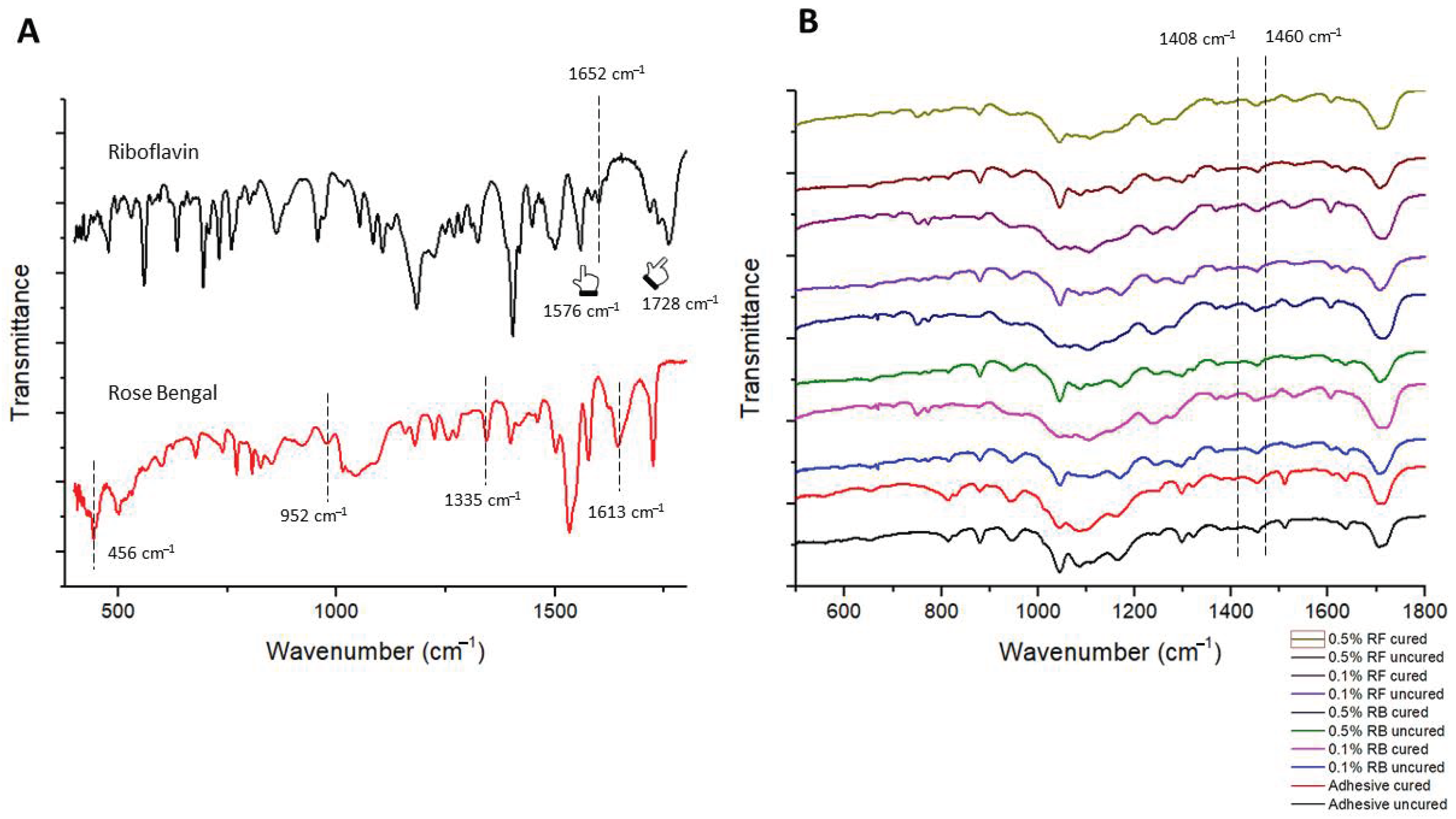
2. Results
3. Discussion
4. Materials and Methods
4.1. Materials and Chemicals
4.2. Specimen Preparation
4.3. Photosensitizer-Modified Experimental Orthodontic Adhesives
4.4. Groups
4.5. Photodynamic Therapy (PDT) Protocol
4.6. Degree of Conversion
4.7. Anti-Bacterial Testing
4.8. Placement of Brackets on Enamel Surface
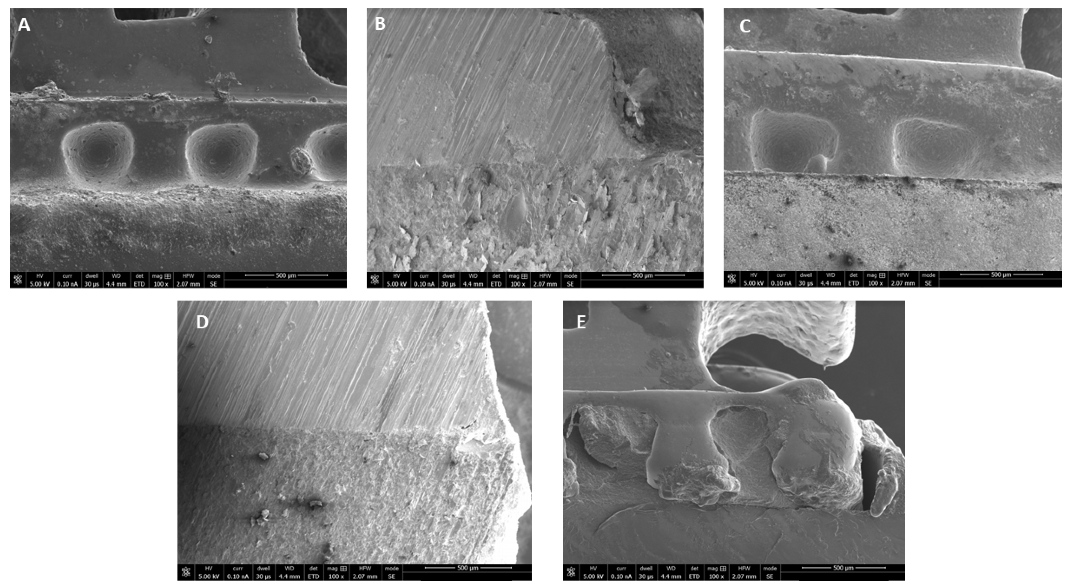
4.9. Scanning Electron Microscopy (SEM)
4.10. Adhesive Remnant Index (ARI) Assessment
4.11. Statistical Analysis
5. Conclusions
Funding
Institutional Review Board Statement
Informed Consent Statement
Data Availability Statement
Conflicts of Interest
References
- Mei, L.; Chieng, J.; Wong, C.; Benic, G.; Farella, M. Factors affecting dental biofilm in patients wearing fixed orthodontic appliances. Prog. Orthod. 2017, 18, 4. [Google Scholar] [CrossRef] [PubMed] [Green Version]
- Zhang, M.; McGrath, C.; Hägg, U. Changes in oral health-related quality of life during fixed orthodontic appliance therapy. Am. J. Orthod. Dentofacial Orthop. 2008, 133, 25–29. [Google Scholar] [CrossRef] [PubMed]
- Pachevska, A.V.; Filimonov, Y.V.; Filimonov, V.Y.; Dudik, O.P.; Popova, O.I.; Drachuk, N.V.; Kasianenko, D.M.; Biloshitska, A.V.; Istoshyn, V.M. Clinical and laboratory assessment the levels of oral hygiene, total protein, hydrogen sulfide and nitrogen metabolites in oral fluid in the development of inflammatory complications during orthodontic treatment of children. Wiad Lek 2019, 72, 744–747. [Google Scholar] [CrossRef] [PubMed]
- Ristic, M.; Vlahovic Svabic, M.; Sasic, M.; Zelic, O. Clinical and microbiological effects of fixed orthodontic appliances on periodontal tissues in adolescents. Orthod. Craniofacial Res. 2007, 10, 187195. [Google Scholar] [CrossRef] [PubMed]
- Verrusio, C.; Iorio-Siciliano, V.; Blasi, A.; Leuci, S.; Adamo, D.; Nicolò, M. The effect of orthodontic treatment on periodontal tissue inflammation: A systematic review. Quintessence Int. 2018, 49, 69–77. [Google Scholar] [PubMed]
- Panhóca, V.H.; Esteban Florez, F.L.; Corrêa, T.Q.; Paolillo, F.R.; de Souza, C.W.; Bagnato, V.S. Oral decontamination of orthodontic patients using photodynamic therapy mediated by bluelight irradiation and curcumin associated with sodium dodecyl sulfate. Photomed. Laser Surg. 2016, 34, 411–417. [Google Scholar] [CrossRef] [PubMed]
- Martha, K.; Mezei, T.; Janos, K. A histological analysis of gingival condition associated with orthodontic treatment. Rom. J. Morphol. Embryol. 2013, 54, 823–827. [Google Scholar] [PubMed]
- Al Nazeh, A.; Alshahrani, A.A.; Almoammar, S.; Kamran, M.A.; Togoo, R.A.; Alshahrani, I. Application of photodynamic therapy against periodontal bacteria in established gingivitis lesions in adolescent patients undergoing fixed orthodontic treatment. Photodiag. Photodyn. Ther. 2020, 1, 101904. [Google Scholar] [CrossRef]
- Huang, J.; Li, C.Y.; Jiang, J.H. Effects of fixed orthodontic brackets on oral malodor: A systematic review and meta-analysis according to the preferred reporting items for systematic reviews and meta-analyses guidelines. Medicine (Baltimore) 2018, 97, e0233. [Google Scholar] [CrossRef]
- Gómez, C.; Abellán, R.; Palma, J.C. Efficacy of photodynamic therapy versus ultrasonic scaler for preventing gingival inflammation and white spot lesions during orthodontic treatment. Photodiag. Photodyn. Ther. 2018, 24, 377–383. [Google Scholar] [CrossRef]
- Julien, K.C.; Buschang, P.H.; Campbell, P.M. Prevalence of white spot lesion formation during orthodontic treatment. Angle Orthod. 2013, 83, 641–647. [Google Scholar] [CrossRef] [PubMed]
- Akram, Z.; Raffat, M.A.; Shafqat, S.S.; Mirza, S.; Ikram, S. Clinical efficacy of photodynamic therapy as an adjunct to scaling and root planing in the treatment of chronic periodontitis among cigarette smokers: A systematic review and meta-analysis. Photodiag. Photodyn. Ther. 2019, 26, 334–341. [Google Scholar] [CrossRef] [PubMed]
- Akram, Z.; Shafqat, S.S.; Niaz, M.O.; Raza, A.; Naseem, M. Clinical efficacy of photodynamic therapy and laser irradiation as an adjunct to open flap debridement in the treatment of chronic periodontitis: A systematic review and meta-analysis. Photodermatol. Photoimmunol. Photomed. 2020, 36, 3–13. [Google Scholar] [CrossRef] [PubMed]
- Kamran, M.A. Clinical, microbiological and immunological outcomes with photodynamic therapy as an adjunct to full-mouth scaling in patients undergoing fixed orthodontic treatment. Photodiag. Photodyn. Ther. 2020, 29, 101585. [Google Scholar] [CrossRef]
- Akram, Z.; Javed, F.; Hosein, M.; Al-Qahtani, M.A.; Alshehri, F.; Alzahrani, A.I.; Vohra, F. Photodynamic therapy in the treatment of symptomatic oral lichen planus: A systematic review. Photodermatol. Photoimmunol. Photomed. 2018, 34, 167–174. [Google Scholar] [CrossRef] [Green Version]
- Almohareb, T.; Alhamoudi, N.; Al Deeb, M.; Bin-Shuwaish, M.S.; Mokeem, S.A.; Shafqat, S.S.; Vohra, F.; Abduljabbar, T. Clinical efficacy of photodynamic therapy as an adjunct to mechanical debridement in the treatment of per-implantitis with abscess. Photodiagnosis Photodyn. Ther. 2020, 30, 101750. [Google Scholar] [CrossRef]
- Mirza, S.; Khan, A.A.; Al-Kheraif, A.A.; Khan, S.Z.; Shafqat, S.S. Efficacy of adjunctive p.hotodynamic therapy on the clinical periodontal, HbA1c and advanced glycation end product levels among mild to moderate chronic periodontal disease patients with type 2 diabetes mellitus: A randomized controlled clinical trial. Photodiagnosis Photodyn Ther. 2019, 28, 177–182. [Google Scholar] [CrossRef]
- Alhamoudi, N.; Mokeem, S.; Shafqat, S.S.; Vohra, F.; Abduljabbar, T. Effectiveness of antimicrobial photodynamic therapy as an adjunct to open flap debridement in patients with aggressive periodontitis. Photodiagnosis Photodyn Ther. 2020, 3, 102075. [Google Scholar] [CrossRef]
- Javed, F.; Salehpoor, D.; Al-Dhafeeri, T.; Yousuf, M.; Malmstrom, H.; Khan, J.; Akram, Z. Is adjunctive photodynamic therapy more effective than scaling and root planing alone in the treatment of periodontal disease in hyperglycemic patients? A systematic review. Photodiagnosis Photodyn. Ther. 2018, 22, 1–6. [Google Scholar] [CrossRef]
- Vohra, F.; Akram, Z.; Bukhari, I.A.; Sheikh, S.A.; Javed, F. Short-term effects of adjunctive antimicrobial photodynamic therapy in obese patients with chronic periodontitis: A randomized controlled clinical trial. Photodiagnosis Photodyn. Ther. 2018, 21, 10–15. [Google Scholar] [CrossRef]
- Arboleda, A.; Miller, D.; Cabot, F.; Taneja, M.; Aguilar, M.C.; Alawa, K.; Amescua, G.; Yoo, S.H.; Parel, J.M. Assessment of rose bengal versus riboflavin photodynamic therapy for inhibition of fungal keratitis isolates. Am. J. Ophthalmol. 2014, 158, 64–70. [Google Scholar] [CrossRef] [PubMed] [Green Version]
- Cheng, L.; Zhang, K.; Zhang, N.; Melo, M.; Weir, M.; Zhou, X.; Bai, Y.; Reynolds, M.; Xu, H. Developing a new generation of antimicrobial and bioactive dental resins. J. Dent. Res. 2017, 96, 855–863. [Google Scholar] [CrossRef] [PubMed]
- Chenicheri, S.R.U.; Ramachandran, R.; Thomas, V.; Wood, A. Insight into Oral Biofilm: Primary, Secondary and Residual Caries and Phyto-Challenged Solutions. Open Dent. J. 2017, 11, 312–333. [Google Scholar] [CrossRef] [PubMed] [Green Version]
- Holzmeier, M.; Schaubmayr, M.; Dasch, W.; Hirschfelder, U. A New Generation of Self-etching Adhesives: Comparison with Traditional Acid Etch Technique. J. Orofac Orthop 2008, 69, 78–93. [Google Scholar] [CrossRef]
- Miller, J.S. Rose bengal-sensitized photooxidation of 2-chlorophenol in water using solar simulated light. Water Res. 2005, 39, 412–422. [Google Scholar] [CrossRef]
- Insińska-Rak, M.; Sikorski, M. Riboflavin interactions with oxygen-a survey from the photochemical perspective. Chemistry 2014, 20, 15280–15291. [Google Scholar] [CrossRef]
- Abrahamse, H.; Hamblin, M.R. New photosensitizers for photodynamic therapy. Biochem. J. 2016, 473, 347–364. [Google Scholar] [CrossRef] [Green Version]
- Costa, A.C.; Rasteiro, V.M.; Pereira, C.A.; Rossoni, R.D.; Junqueira, J.C.; Jorge, A.O. The effects of rose bengal- and erythrosine-mediated photodynamic therapy on Candida albicans. Mycoses 2012, 55, 56–63. [Google Scholar] [CrossRef]
- Spikes, J.D.; Shen, H.R.; Kopecková, P.; Kopecek, J. Photodynamic crosslinking of proteins. III. Kinetics of the FMN- and rose bengal-sensitized photooxidation and intermolecular crosslinking of model tyrosine-containing N-(2-hydroxypropyl) methacrylamide copolymers. Photochem Photobiol. 1999, 70, 130–137. [Google Scholar] [CrossRef]
- Akram, Z.; Abduljabbar, T.; Sauro, S.; Daood, U. Effect of photodynamic therapy and laser alone as adjunct to scaling and root planing on gingival crevicular fluid inflammatory proteins in periodontal disease: A systematic review. Photodiagnosis Photodyn. Ther. 2016, 16, 142–153. [Google Scholar] [CrossRef]
- Kwiatkowski, S.; Knap, B.; Przystupski, D.; Saczko, J.; Kędzierska, E.; Knap-Czop, K.; Kotlińska, J.; Michel, O.; Kotowski, K.; Kulbacka, J. Photodynamic therapy—Mechanisms, photosensitizers and combinations. Biomed. Pharmacother. 2018, 106, 1098–1107. [Google Scholar] [CrossRef] [PubMed]
- Prażmo, E.J.; Kwaśny, M.; Łapiński, M.; Mielczarek, A. Photodynamic Therapy as a Promising Method Used in the Treatment of Oral Diseases. Adv. Clin. Exp. Med. 2016, 25, 799–807. [Google Scholar] [CrossRef] [PubMed] [Green Version]
- Kundu, B.; Sarkar, D.; Ray, N.; Talukdar, A. Understanding the riboflavin biosynthesis pathway for the development of antimicrobial agents. Med. Res. Rev. 2019, 39, 1338–1371. [Google Scholar] [PubMed]
- Khan, S.; Rayis, M.; Rizvi, A.; Alam, M.M.; Rizvi, M.; Naseem, I. ROS mediated antibacterial activity of photoilluminated riboflavin: A photodynamic mechanism against nosocomial infections. Toxicol. Rep. 2019, 9, 136–142. [Google Scholar] [CrossRef]
- Anju, V.T.; Paramanantham, P.; Lal, S.B.S.; Sharan, A.; Alsaedi, M.H.; Dawoud, T.M.S.; Asad, S.; Busi, S. Antimicrobial photdynamic activity of rose bengal conjugated multi walled carbon nanotubes against planktonic cells and biofilm of Escherichia coli. Photodiagnosis Photodyn. Ther. 2018, 24, 300–310. [Google Scholar]
- Fuentes-Lemus, E.; Mariotti, M.; Reyes, J.; Leinisch, F.; Hägglund, P.; Silva, E.; Davies, M.J.; López-Alarcón, C. Photo-oxidation of lysozyme triggered by riboflavin is O2-dependent, occurs via mixed type 1 and type 2 pathways, and results in inactivation, site-specific damage and intra- and inter-molecular crosslinks. Free Radic. Biol. Med. 2020, 152, 61–73. [Google Scholar] [CrossRef]
- Leinisch, F.; Mariotti, M.; Rykaer, M.; Lopez-Alarcon, C.; Hägglund, P.; Davies, M.J. Peroxyl radical- and photo-oxidation of glucose 6-phosphate dehydrogenase generates cross-links and functional changes via oxidation of tyrosine and tryptophan residues. Free Radic. Biol. Med. 2017, 112, 240–252. [Google Scholar] [CrossRef] [Green Version]
- Alkhudhairy, F.; AlKheraif, A.A.; Naseem, M.; Vohra, F.; Khan, R. Degree of conversion and depth of cure of Ivocerin containing photo-polymerized resin luting cement in comparison to conventional luting agents. Pak. J. Med. Sci. 2018, 34, 253–259. [Google Scholar] [CrossRef]
- Hashem, M. Antimicrobial capacity and physico-chemical characteristics of adhesive resin containing riboflavin after photodynamic therapy. Photodiag. Photodyn. Ther. 2020. [Google Scholar] [CrossRef]
- Daood, U.; Omar, H.; Qasim, S.; Nogueira, L.P.; Pichika, M.R.; Mak, K.K.; Steier, L.; Cky, Y.; Lin, S.L.; Fawzy, A.S. New antimicrobial and collagen crosslinking formulated dentin adhesive with improved bond durability. J. Mech. Behav. Biomed. Mater. 2020, 110, 103927. [Google Scholar] [CrossRef]
- Khan, S.; Arif, S.H.; Naseem, I. Interaction of aminophylline with photoilluminated riboflavin leads to ROS mediated macromolecular damage and cell death in benzopyrene induced mice lung carcinoma. Chem. Biol. Interact. 2019, 302, 135–142. [Google Scholar] [CrossRef] [PubMed]


| Groups | Degree of Conversion (%Mean ± SD) | Tukey (p < 0.05) |
|---|---|---|
| Transbond XT | n = 48.5 ± 6.6 | A |
| 0.1% RB-PDT adhesive | n = 46.8 ± 7.3 | A |
| 0.1% RF-PDT adhesive | n = 46.2 ± 5.8 | A |
| 0.5% RB-PDT adhesive | n = 41.3 ± 4.3 | B |
| 0.5% RF-PDT adhesive | n = 39.7 ± 5.1 | B |
| Groups (Time Point) | Mean ARI | p-Value |
|---|---|---|
| Transbond XT Immediately after After 24 h After thermocycling | n = 1.33 ± 0.57 n = 1.27 ± 0.69 n = 1.24 ± 0.71 | n = 0.022 |
| 0.1% RB-PDT adhesive Immediately after After 24 h After thermocycling | n = 1.47 ± 0.65 n = 1.42 ± 0.71 n = 1.37 ± 0.75 | |
| 0.1% RF-PDT adhesive Immediately after After 24 h After thermocycling | n = 1.72 ± 0.55 n = 1.68 ± 0.46 n = 1.61 ± 0.84 | |
| 0.5% RB-PDT adhesive Immediately after After 24 h After thermocycling | n = 1.91 ± 0.85 n = 1.73 ± 0.77 n = 1.66 ± 0.90 | |
| 0.5% RF-PDT adhesive Immediately after After 24 h After thermocycling | n = 1.66 ± 0.83 n = 1.62 ± 0.97 n = 1.58 ± 0.74 |
Publisher’s Note: MDPI stays neutral with regard to jurisdictional claims in published maps and institutional affiliations. |
© 2021 by the author. Licensee MDPI, Basel, Switzerland. This article is an open access article distributed under the terms and conditions of the Creative Commons Attribution (CC BY) license (http://creativecommons.org/licenses/by/4.0/).
Share and Cite
Alqerban, A. Effectiveness of Riboflavin and Rose Bengal Photosensitizer Modified Adhesive Resin for Orthodontic Bonding. Pharmaceuticals 2021, 14, 48. https://doi.org/10.3390/ph14010048
Alqerban A. Effectiveness of Riboflavin and Rose Bengal Photosensitizer Modified Adhesive Resin for Orthodontic Bonding. Pharmaceuticals. 2021; 14(1):48. https://doi.org/10.3390/ph14010048
Chicago/Turabian StyleAlqerban, Ali. 2021. "Effectiveness of Riboflavin and Rose Bengal Photosensitizer Modified Adhesive Resin for Orthodontic Bonding" Pharmaceuticals 14, no. 1: 48. https://doi.org/10.3390/ph14010048