

Cognitive Effort during Visuospatial Problem Solving in Physical Real World, on Computer Screen, and in Virtual Reality
 , ,
, , 
Abstract
:1. Introduction
2. Materials and Methods
2.1. Participants
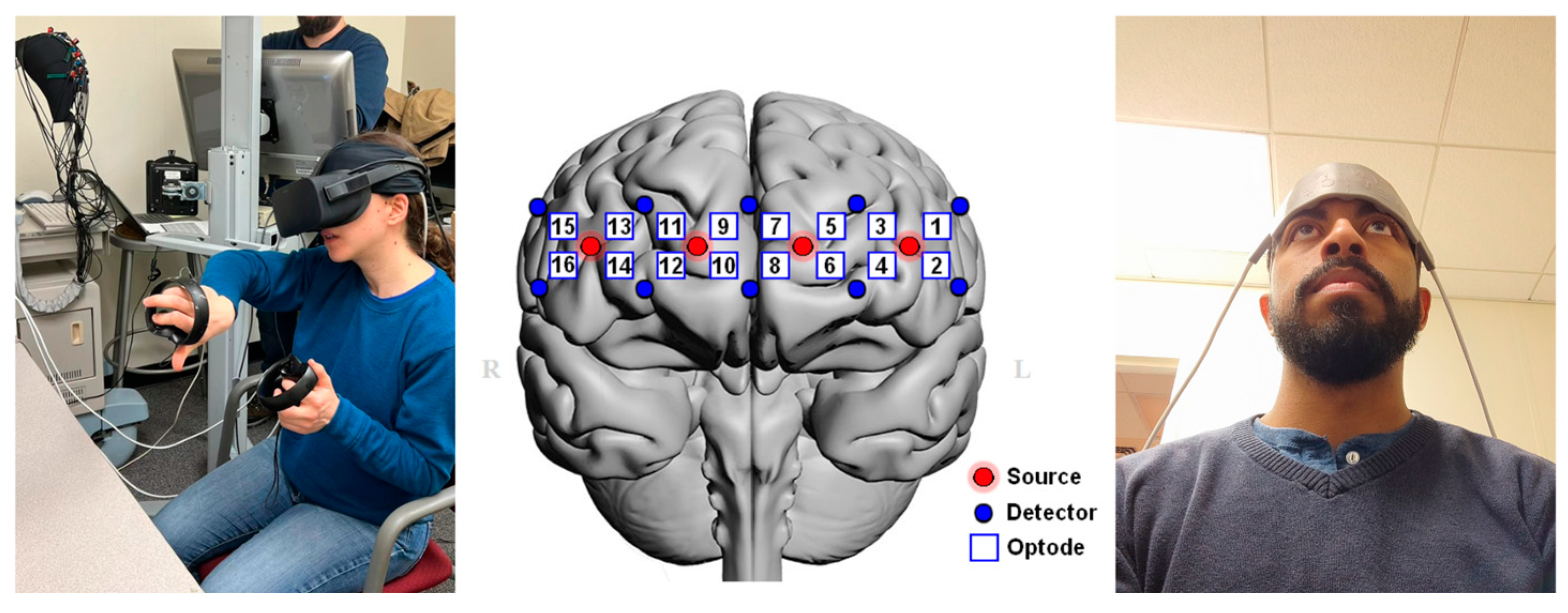
2.2. VR Equipment
2.3. Behavioral Measurements
2.4. fNIRS Data Acquisition
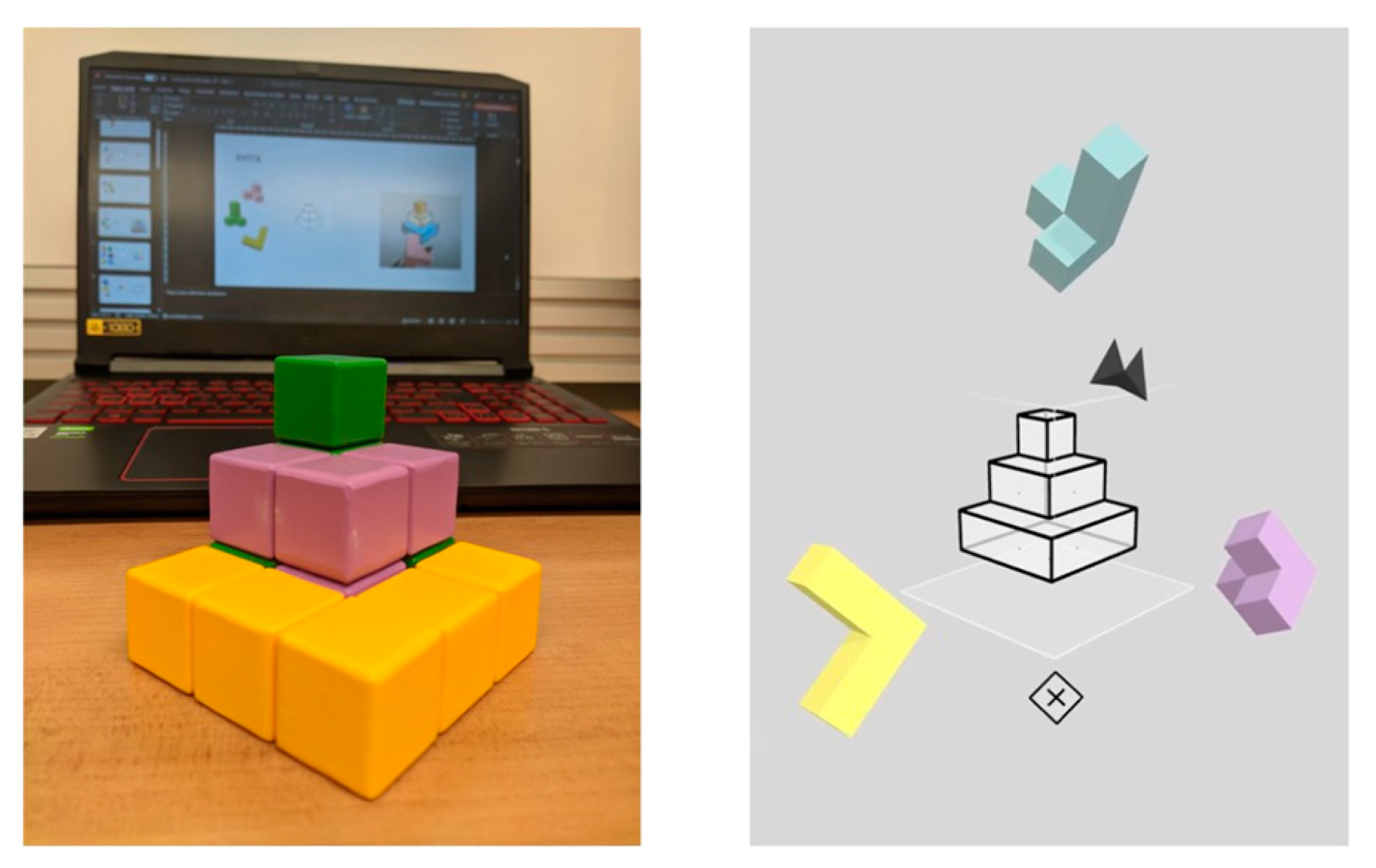
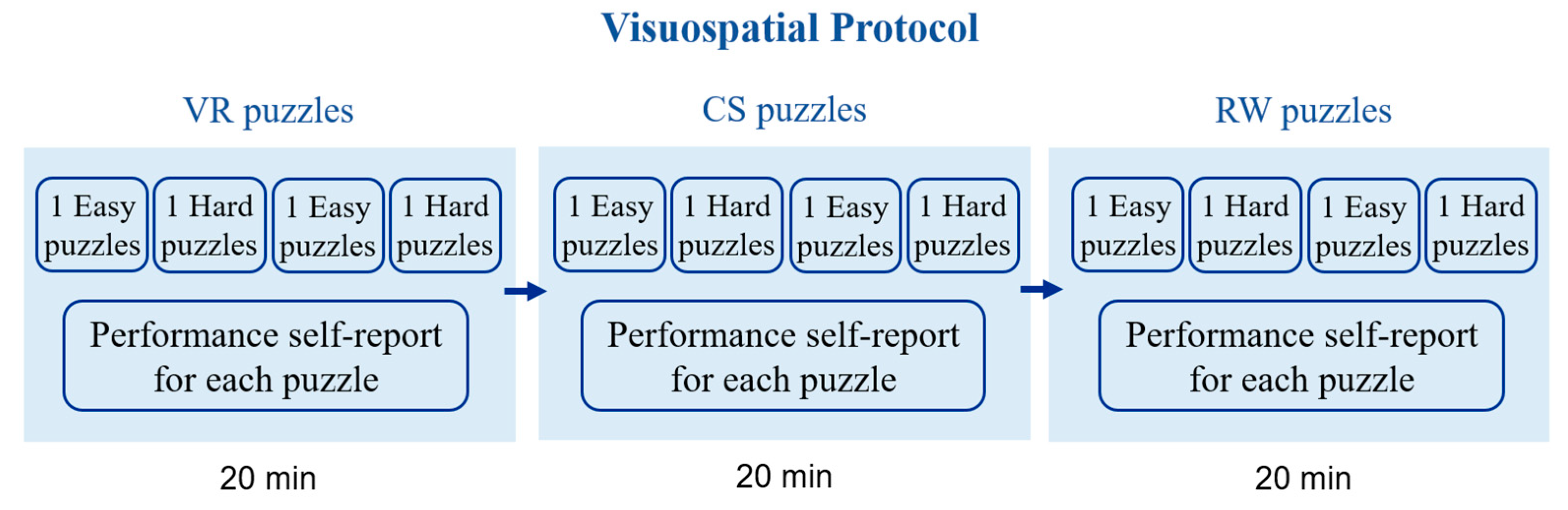
2.5. Visuospatial Task
2.6. Experimental Setup
2.7. Data Analysis
3. Results
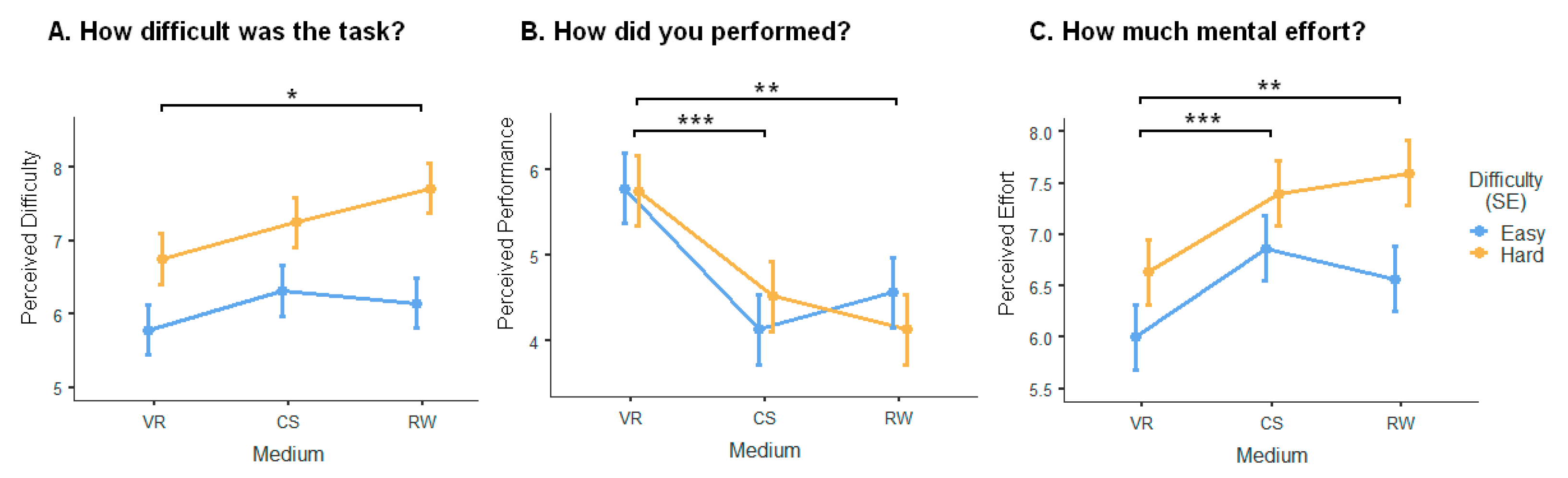
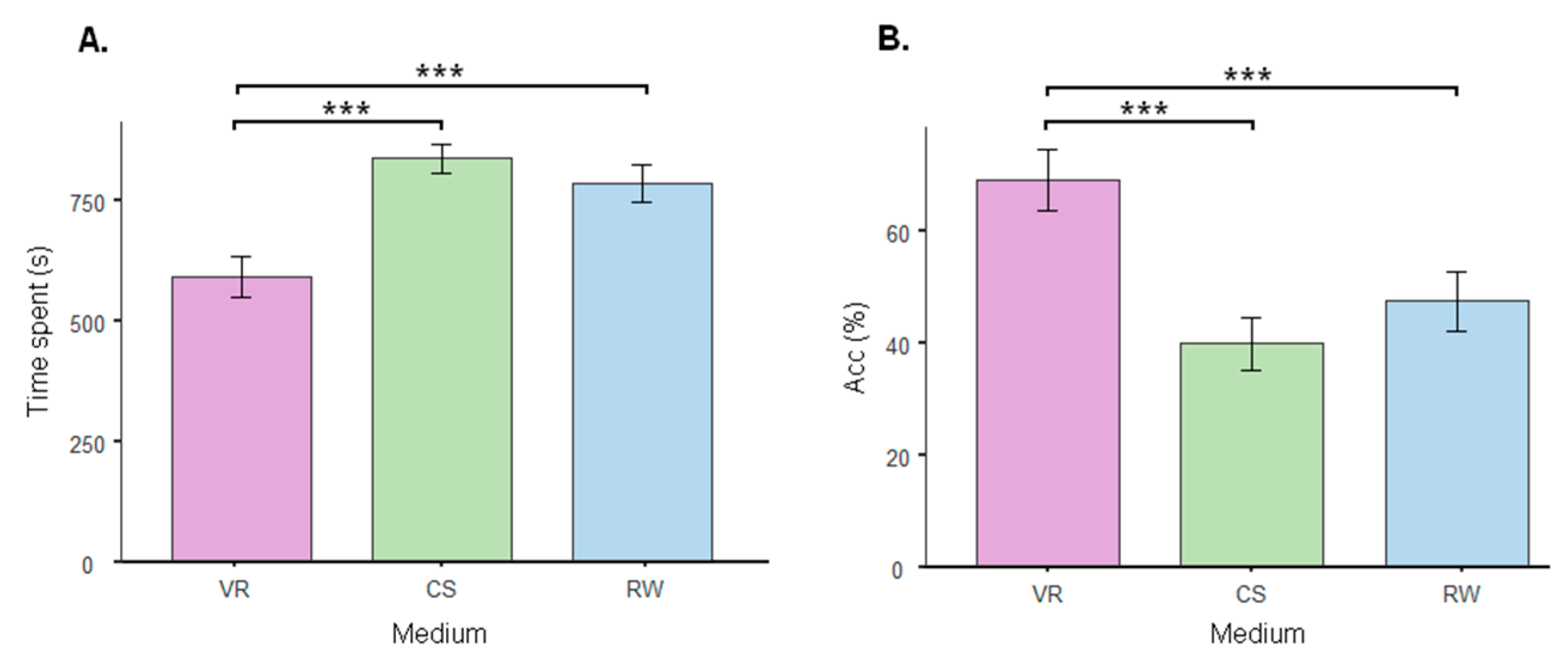
3.1. Self-Assessment Survey
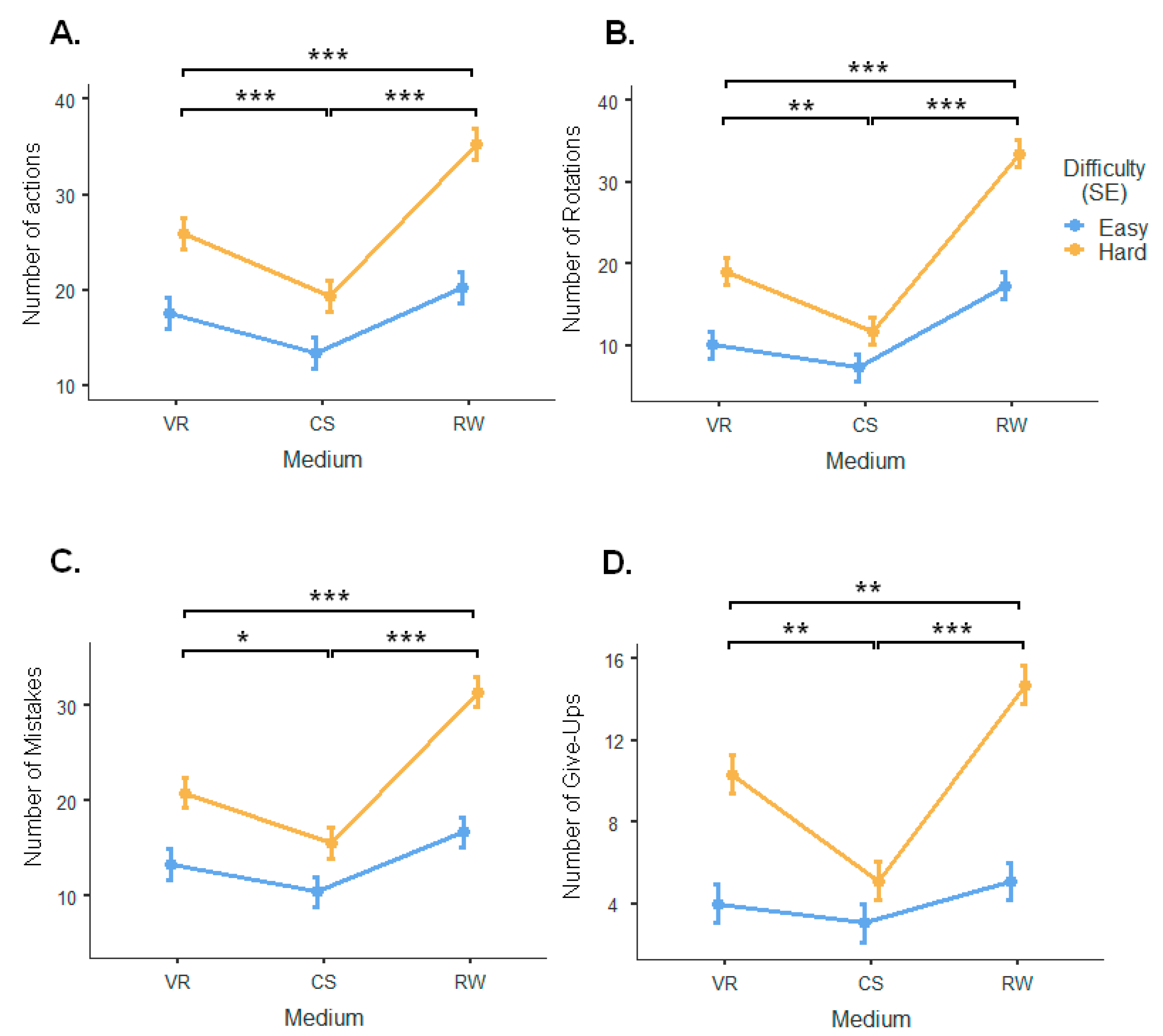
3.2. Performance and Behavioral Measures
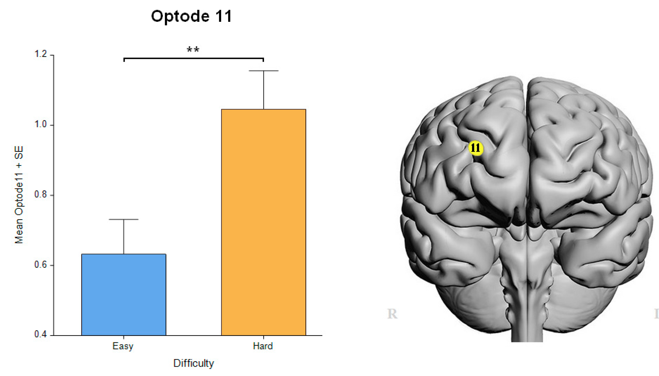
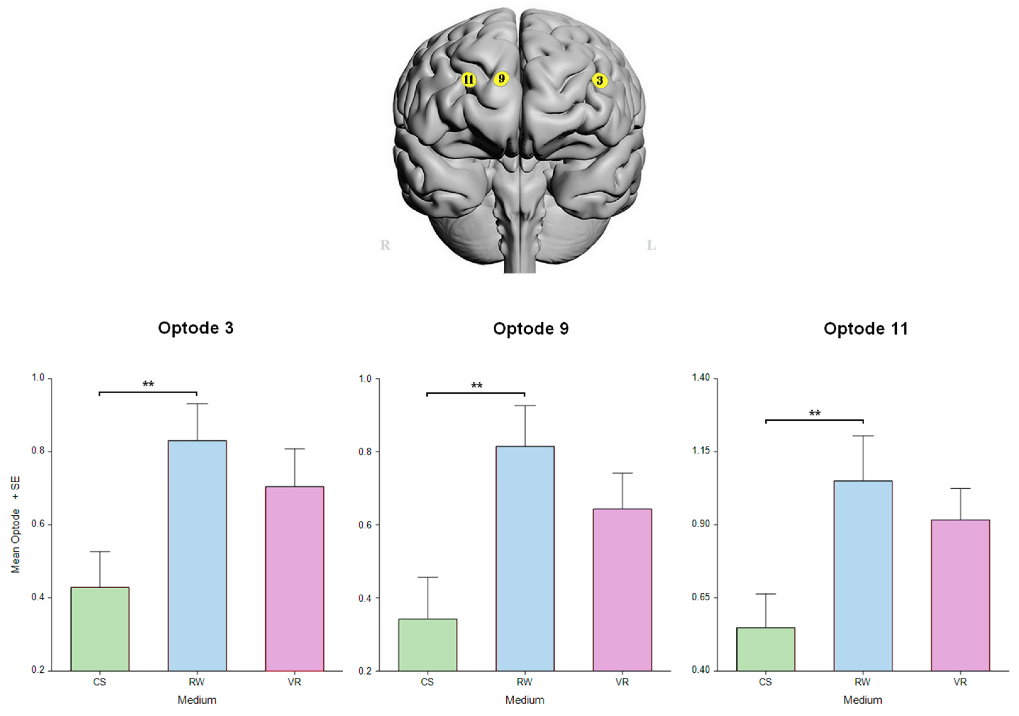
3.3. fNIRS Measures
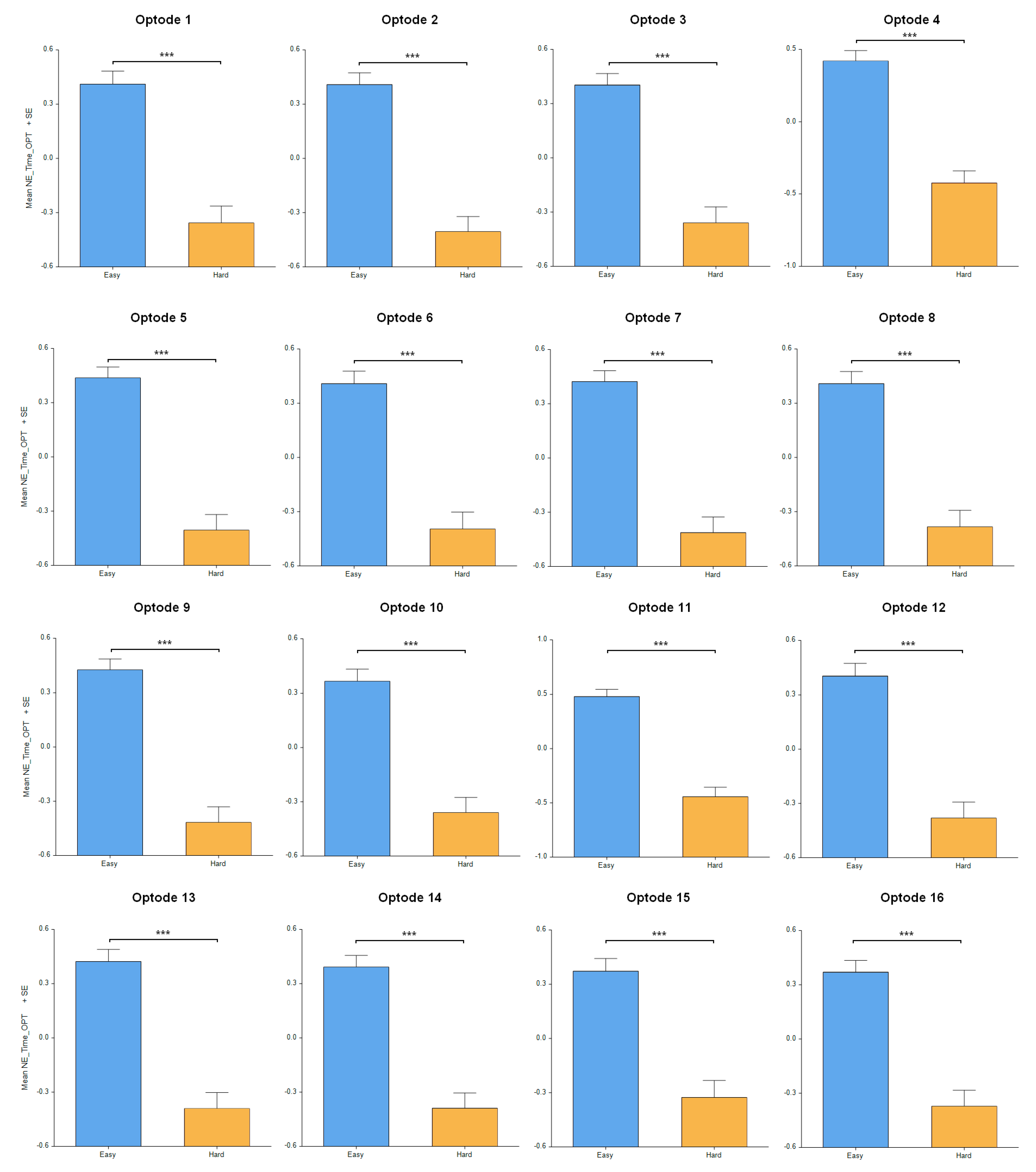
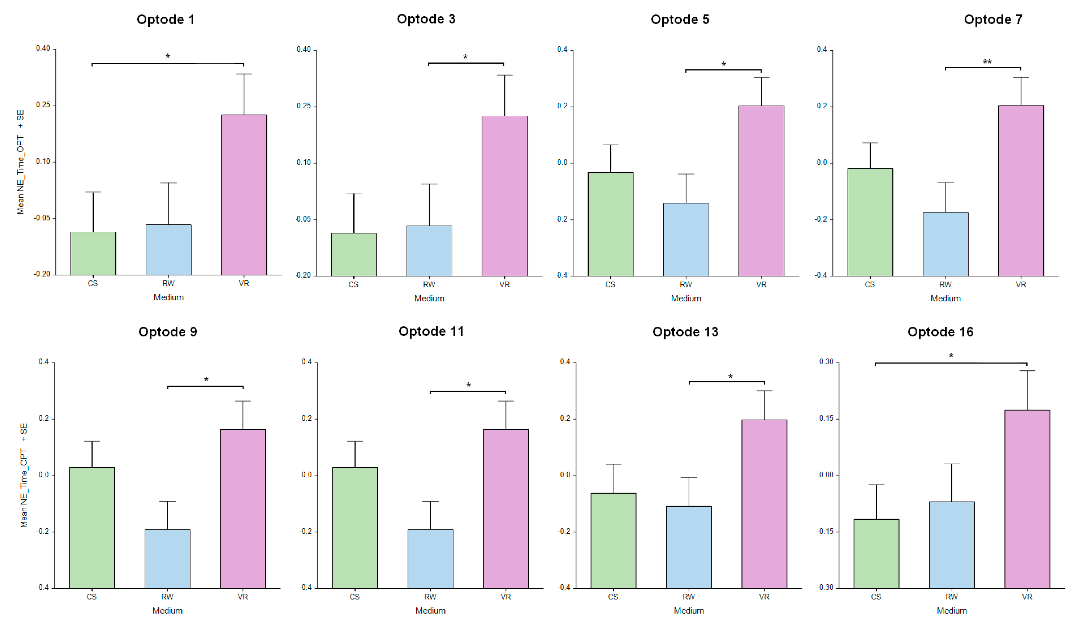
3.4. Neural Efficiency Measures
4. Discussion
5. Conclusions
Author Contributions
Funding
Institutional Review Board Statement
Informed Consent Statement
Data Availability Statement
Conflicts of Interest
References
- Linn, M.C.; Petersen, A.C. A Meta-Analysis of Gender Differences in Spatial Ability: Implications for Mathematics and Science Achievement. Psychol. Gend. Adv. Through Meta-Anal. 1986, 8, 67–101. [Google Scholar]
- Wai, J.; Lubinski, D.; Benbow, C.P. Spatial Ability for STEM Domains: Aligning over 50 Years of Cumulative Psychological knowledge Solidifies Its Importance. J. Educ. Psychol. 2009, 101, 817–835. [Google Scholar] [CrossRef]
- Khine, M.S. Spatial Cognition: Key to STEM Success. In Visual-Spatial Ability in STEM Education; Khine, M.S., Ed.; Springer: Cham, Switzerland, 2017; pp. 3–8. ISBN 978-3-319-44384-3. [Google Scholar]
- Mix, K.S.; Cheng, Y.-L. The Relation between Space and Math: Developmental and Educational Implications. Adv. Child Dev. Behav. 2012, 42, 197–243. [Google Scholar] [PubMed]
- Lubinski, D.; Benbow, C.P. Study of Mathematically Precocious Youth after 35 Years: Uncovering Antecedents for the Development of Math-Science Expertise. Perspect. Psychol. Sci. 2006, 1, 316–345. [Google Scholar] [CrossRef] [PubMed]
- Uttal, D.H.; Meadow, N.G.; Tipton, E.; Hand, L.L.; Alden, A.R.; Warren, C.; Newcombe, N.S. The Malleability of Spatial Skills: A Meta-Analysis of Training Studies. Psychol. Bull. 2013, 139, 352–402. [Google Scholar] [CrossRef] [PubMed]
- Newcombe, N.S. Picture This: Increasing Math and Science Learning by Improving Spatial Thinking. Am. Educ. 2010, 34, 29. [Google Scholar]
- Gilligan-Lee, K.A.; Hawes, Z.C.K.; Mix, K.S. Spatial Thinking as the Missing Piece in Mathematics Curricula. Npj Sci. Learn. 2022, 7, 10. [Google Scholar] [CrossRef]
- Türkman, B. The Evolution of the Term of Giftedness & Theories to Explain Gifted Characteristics. J. Gift. Educ. Creat. 2020, 7, 17–24. [Google Scholar]
- Posner, M.I.; Dehaene, S. Attentional Networks. Trends Neurosci. 1994, 17, 75–79. [Google Scholar] [CrossRef]
- Sato, J.R.; Kozasa, E.H.; Russell, T.A.; Radvany, J.; Mello, L.E.A.M.; Lacerda, S.S.; Amaro, E. Brain Imaging Analysis Can Identify Participants under Regular Mental Training. PLoS ONE 2012, 7, e39832. [Google Scholar] [CrossRef]
- Sato, J.R.; Salum, G.A.; Gadelha, A.; Crossley, N.; Vieira, G.; Manfro, G.G.; Zugman, A.; Picon, F.A.; Pan, P.M.; Hoexter, M.Q.; et al. Default Mode Network Maturation and Psychopathology in Children and Adolescents. J. Child Psychol. Psychiatry 2016, 57, 55–64. [Google Scholar] [CrossRef] [PubMed]
- Voyer, D.; Voyer, S.; Bryden, M.P. Magnitude of Sex Differences in Spatial Abilities: A Meta-Analysis and Con-sideration of Critical Variables. Psychol. Bull. 1995, 117, 250. [Google Scholar] [CrossRef] [PubMed]
- Haier, R.J.; Karama, S.; Leyba, L.; E Jung, R. MRI assessment of cortical thickness and functional activity changes in adolescent girls following three months of practice on a visual-spatial task. BMC Res. Notes 2009, 2, 174. [Google Scholar] [CrossRef]
- Dehaene, S.; Spelke, E.; Pinel, P.; Stanescu, R.; Tsivkin, S. Sources of Mathematical Thinking: Behavioral and Brain-Imaging Evidence. Science 1999, 284, 970–974. [Google Scholar] [CrossRef]
- Dehaene, S.; Piazza, M.; Pinel, P.; Cohen, L. Three parietal circuits for number processing. Cogn. Neuropsychol. 2003, 20, 487–506. [Google Scholar] [CrossRef] [PubMed]
- Ischebeck, A.; Schocke, M.; Delazer, M. The Processing and Representation of Fractions within the Brain: An fMRI Investigation. NeuroImage 2009, 47, 403–413. [Google Scholar] [CrossRef] [PubMed]
- Zack, M.H. Rethinking the Knowledge-Based Organization. MIT Sloan Manag. Rev. 2003, 44, 67–71. [Google Scholar]
- Hugdahl, K.; Thomsen, T.; Ersland, L. Sex Differences in Visuo-Spatial Processing: An fMRI Study of Mental Rotation. Neuropsychologia 2006, 44, 1575–1583. [Google Scholar] [CrossRef]
- Pinti, P.; Aichelburg, C.; Gilbert, S.; Hamilton, A.; Hirsch, J.; Burgess, P.; Tachtsidis, I. A Review on the Use of Wearable Functional Near-Infrared Spectroscopy in Naturalistic Environments. Jpn. Psychol. Res. 2018, 60, 347–373. [Google Scholar] [CrossRef]
- Balardin, J.B.; Morais, G.A.Z.; Furucho, R.A.; Trambaiolli, L.; Vanzella, P.; Biazoli, C.; Sato, J.R. Imaging Brain Function with Functional Near-Infrared Spectroscopy in Unconstrained Environments. Front. Hum. Neurosci. 2017, 11, 258. [Google Scholar] [CrossRef]
- Villringer, A.; Chance, B. Non-Invasive Optical Spectroscopy and Imaging of Human Brain Function. Trends Neurosci. 1997, 20, 435–442. [Google Scholar] [CrossRef]
- Izzetoglu, M.; Izzetoglu, K.; Bunce, S.; Ayaz, H.; Devaraj, A.; Onaral, B.; Pourrezaei, K. Functional Near-Infrared Neuroimaging. IEEE Trans. Neural Syst. Rehabil. Eng. 2005, 13, 153–159. [Google Scholar] [CrossRef] [PubMed]
- Ferrari, M.; Quaresima, V. A Brief Review on the History of Human Functional Near-Infrared Spectroscopy (fNIRS) Development and Fields of Application. NeuroImage 2012, 63, 921–935. [Google Scholar] [CrossRef] [PubMed]
- Ayaz, H.; Baker, W.B.; Blaney, G.; Boas, D.A.; Bortfeld, H.; Brady, K.; Brake, J.; Brigadoi, S.; Buckley, E.M.; Carp, S.A.; et al. Optical Imaging and Spectroscopy for the Study of the Human Brain: Status Report. Neurophotonics 2022, 9, S24001. [Google Scholar] [CrossRef] [PubMed]
- Curtin, A.; Ayaz, H. The Age of Neuroergonomics: Towards Ubiquitous and Continuous Measurement of Brain Function with fNIRS. Jpn. Psychol. Res. 2018, 60, 374–386. [Google Scholar] [CrossRef]
- Ayaz, H.; Onaral, B.; Izzetoglu, K.; Shewokis, P.A.; McKendrick, R.; Parasuraman, R. Continuous Monitoring of Brain Dynamics with Functional near Infrared Spectroscopy as a Tool for Neuroergo-nomic Research: Empirical Examples and a Technological Development. Front. Hum. Neurosci. 2013, 7, 871. [Google Scholar] [CrossRef]
- Ayaz, H.; Shewokis, P.A.; Izzetoglu, M.; Cakir, M.P.; Onaral, B. Tangram Solved? Prefrontal Cortex Activation Analysis during Geometric Problem Solving. In Proceedings of the 2012 Annual International Conference of the IEEE Engineering in Medicine and Biology Society, San Diego, CA, USA, 28 August–1 September 2012; IEEE: Piscataway, NJ, USA; pp. 4724–4727. [Google Scholar]
- Dehais, F.; Lafont, A.; Roy, R.; Fairclough, S. A Neuroergonomics Approach to Mental Workload, Engagement and Human Performance. Front. Neurosci. 2020, 14, 268. [Google Scholar] [CrossRef]
- Shewokis, P.A.; Ayaz, H.; Izzetoglu, M.; Bunce, S.; Gentili, R.J.; Sela, I.; Izzetoglu, K.; Onaral, B. Brain in the Loop: Assessing Learning Using fNIR in Cognitive and Motor Tasks. In Foundations of Augmented Cognition. Directing the Future of Adaptive Systems; Schmorrow, D.D., Fidopiastis, C.M., Eds.; Lecture Notes in Computer Science; Springer: Berlin/Heidelberg, Germany, 2011; Volume 6780, pp. 240–249. ISBN 978-3-642-21851-4. [Google Scholar]
- Ayaz, H.; Shewokis, P.A.; Curtin, A.; Izzetoglu, M.; Izzetoglu, K.; Onaral, B. Using MazeSuite and Functional near Infrared Spectroscopy to Study Learning in Spatial Navigation. J. Vis. Exp. 2011, 56, e3443. [Google Scholar]
- Çakır, M.P.; Ayaz, H.; Izzetoğlu, M.; Shewokis, P.A.; İzzetoğlu, K.; Onaral, B. Bridging Brain and Educational Sciences: An Optical Brain Imaging Study of Visuospatial Reasoning. Procedia Soc. Behav. Sci. 2011, 29, 300–309. [Google Scholar] [CrossRef]
- Barreto, C.; Bruneri, G.d.A.; Brockington, G.; Ayaz, H.; Sato, J.R. A New Statistical Approach for fNIRS Hyperscanning to Predict Brain Activity of Preschoolers’ Using Teacher’s. Front. Hum. Neurosci. 2021, 15, 622146. [Google Scholar] [CrossRef] [PubMed]
- Battro, A.M.; Calero, C.I.; Goldin, A.P.; Holper, L.; Pezzatti, L.; Shalóm, D.E.; Sigman, M. The Cognitive Neuroscience of the Teacher-Student Interaction: Cognitive Neuroscience of the Teacher-Student Interaction. Mind Brain Educ. 2013, 7, 177–181. [Google Scholar] [CrossRef]
- Brockington, G.; Balardin, J.B.; Morais, G.A.Z.; Malheiros, A.; Lent, R.; Moura, L.M.; Sato, J.R. From the Laboratory to the Classroom: The Potential of Functional Near-Infrared Spectroscopy in Educational Neuroscience. Front. Psychol. 2018, 9, 1840. [Google Scholar] [CrossRef] [PubMed]
- Gateau, T.; Ayaz, H.; Dehais, F. In silico vs. Over the Clouds: On-the-Fly Mental State Estimation of Aircraft Pilots, Using a Functional Near Infrared Spectroscopy Based Passive-BCI. Front. Hum. Neurosci. 2018, 12, 187. [Google Scholar] [CrossRef] [PubMed]
- McKendrick, R.; Parasuraman, R.; Murtza, R.; Formwalt, A.; Baccus, W.; Paczynski, M.; Ayaz, H. Into the Wild: Neuroergonomic Differentiation of Hand-Held and Augmented Reality Wearable Displays during Outdoor Navigation with Functional near Infrared Spectroscopy. Front. Hum. Neurosci. 2016, 10, 216. [Google Scholar] [CrossRef] [PubMed]
- Liu, J.; Zhang, H.; Chen, C.; Chen, H.; Cui, J.; Zhou, X. The Neural Circuits for Arithmetic Principles. NeuroImage 2017, 147, 432–446. [Google Scholar] [CrossRef] [PubMed]
- Mark, J.A.; Kraft, A.E.; Ziegler, M.D.; Ayaz, H. Neuroadaptive Training via fNIRS in Flight Simulators. Front. Neuroergon. 2022, 3, 8205233. [Google Scholar] [CrossRef] [PubMed]
- Perrey, S. Evaluating Brain Functioning with NIRS in Sports: Cerebral Oxygenation and Cortical Activation Are Two Sides of the Same Coin. Front. Neuroergonom. 2022, 3, 1022924. [Google Scholar] [CrossRef] [PubMed]
- von Lühmann, A.; Zheng, Y.; Ortega-Martinez, A.; Kiran, S.; Somers, D.C.; Cronin-Golomb, A.; Awad, L.N.; Ellis, T.D.; Boas, D.A.; Yücel, M.A. Toward Neuroscience of the Everyday World (NEW) using functional near-infrared spectroscopy. Curr. Opin. Biomed. Eng. 2021, 18, 100272. [Google Scholar] [CrossRef] [PubMed]
- Lent, R.; Ribeiro, S.; Sato, J.R. Neuroplasticity: From Cells to Circuits and Brains Towards the Classroom. In Learning Under the Lens; Carroll, A., Cunnington, R., Nugent, A., Eds.; Routledge: Vienna, Austria, 2020; pp. 47–62. ISBN 978-0-429-02783-3. [Google Scholar]
- Rypma, B.; Berger, J.S.; D’Esposito, M. The Influence of Working-Memory Demand and Subject Performance on Prefrontal Cortical Activity. J. Cogn. Neurosci. 2002, 14, 721–731. [Google Scholar] [CrossRef]
- Prat, C.S.; Keller, T.A.; Just, M.A. Individual Differences in Sentence Comprehension: A Functional Magnetic Resonance Imaging Investigation of Syntactic and Lexical Processing Demands. J. Cogn. Neurosci. 2007, 19, 1950–1963. [Google Scholar] [CrossRef]
- Ayaz, H.; Shewokis, P.A.; Bunce, S.; Izzetoglu, K.; Willems, B.; Onaral, B. Optical Brain Monitoring for Operator Training and Mental Workload Assessment. NeuroImage 2012, 59, 36–47. [Google Scholar] [CrossRef]
- Fishburn, F.A.; Norr, M.E.; Medvedev, A.V.; Vaidya, C.J. Sensitivity of fNIRS to Cognitive State and Load. Front. Hum. Neurosci. 2014, 8, 76. [Google Scholar] [CrossRef] [PubMed]
- Causse, M.; Chua, Z.; Peysakhovich, V.; Del Campo, N.; Matton, N. Mental Workload and Neural Efficiency Quantified in the Prefrontal Cortex Using fNIRS. Sci. Rep. 2017, 7, 5222. [Google Scholar] [CrossRef] [PubMed]
- Shewokis, P.A.; Shariff, F.U.; Liu, Y.; Ayaz, H.; Castellanos, A.; Lind, D.S. Acquisition, Retention and Transfer of Simulated Laparoscopic Tasks Using fNIR and a Contextual Interference Paradigm. Am. J. Surg. 2017, 213, 336–345. [Google Scholar] [CrossRef] [PubMed]
- Getchell, N.; Shewokis, P. Understanding the Role of Cognitive Effort within Contextual Interference Para-digms: Theory, Measurement, and Tutorial. Braz. J. Mot. Behav. 2023, 17, 59–69. [Google Scholar] [CrossRef]
- Curtin, A.; Ayaz, H.; Tang, Y.; Sun, J.; Wang, J.; Tong, S. Enhancing Neural Efficiency of Cognitive Processing Speed via Training and Neurostimulation: An fNIRS and TMS Study. NeuroImage 2019, 198, 73–82. [Google Scholar] [CrossRef] [PubMed]
- Curtin, A.; Ayaz, H. Neural Efficiency Metrics in Neuroergonomics: Theory and Applications. In Neuroergonomics; Elsevier: Amsterdam, The Netherlands, 2019; pp. 133–140. [Google Scholar]
- McIntire, J.P.; Havig, P.R.; Geiselman, E.E. What Is 3D Good for? A Review of Human Performance on Stereo-scopic 3D Displays. In Head- and Helmet-Mounted Displays XVII; and Display Technologies and Applications for Defense, Security, and Avionics VI; SPIE: Bellingham, WA, USA, 2012; Volume 8383, pp. 280–292. [Google Scholar]
- Dan, A.; Reiner, M. EEG-Based Cognitive Load of Processing Events in 3D Virtual Worlds Is Lower than Processing Events in 2D Display. Int. J. Psychophysiol. 2017, 122, 75–84. [Google Scholar] [CrossRef]
- Lochhead, I.; Hedley, N.; Çöltekin, A.; Fisher, B. The Immersive Mental Rotations Test: Evaluating Spatial Ability in Virtual Reality. Front. Virtual Real. 2022, 3, 820237. [Google Scholar] [CrossRef]
- Makransky, G.; Borre-Gude, S.; Mayer, R.E. Motivational and Cognitive Benefits of Training in Immersive Virtual Reality Based on Multiple Assessments. J. Comput. Assist. Learn. 2019, 35, 691–707. [Google Scholar] [CrossRef]
- Kaimal, G.; Carroll-Haskins, K.; Topoglu, Y.; Ramakrishnan, A.; Arslanbek, A.; Ayaz, H. Exploratory fNIRS Assessment of Differences in Activation in Virtual Reality Visual Self-Expression Including with a Fragrance Stimulus. Art Ther. 2021, 39, 128–137. [Google Scholar] [CrossRef]
- Fekete, T.; Rubin, D.; Carlson, J.M.; Mujica-Parodi, L.R. The NIRS Analysis Package: Noise Reduction and Statistical Inference. PLoS ONE 2011, 6, e24322. [Google Scholar] [CrossRef]
- Yücel, M.A.; Selb, J.; Aasted, C.M.; Lin, P.-Y.; Borsook, D.; Becerra, L.; Boas, D.A. Mayer Waves Reduce the Ac-curacy of Estimated Hemodynamic Response Functions in Functional Near-Infrared Spectroscopy. Biomed. Opt. Express 2016, 7, 3078–3088. [Google Scholar] [CrossRef] [PubMed]
- Ayaz, H.; Izzetoglu, M.; Shewokis, P.A.; Onaral, B. Sliding-Window Motion Artifact Rejection for Functional near-Infrared Spectroscopy. In Proceedings of the 2010 Annual International Conference of the IEEE Engineering in Medicine and Biology, Buenos Aires, Argentina, 31 August–4 September 2010; IEEE: Piscataway, NJ, USA, 2010; pp. 6567–6570. [Google Scholar]
- Benjamini, Y.; Hochberg, Y. Controlling the False Discovery Rate: A Practical and Powerful Approach to Multiple Testing. J. R. Stat. Soc. Ser. B Methodol. 1995, 57, 289–300. [Google Scholar] [CrossRef]
- Singh, A.K.; Dan, I. Exploring the False Discovery Rate in Multichannel NIRS. NeuroImage 2006, 33, 542–549. [Google Scholar] [CrossRef] [PubMed]
- Tuovinen, J.E.; Paas, F. Exploring Multidimensional Approaches to the Efficiency of Instructional Conditions. Instr. Sci. 2004, 32, 133–152. [Google Scholar] [CrossRef]
- Soares, R.d.S.; Oku, A.Y.A.; Barreto, C.S.F.; Sato, J.R. Applying Functional Near-Infrared Spectroscopy and Eye-Tracking in a Naturalistic Educational Environment to Investigate Physiological Aspects That Underlie the Cognitive Effort of Children during Mental Rotation Tests. Front. Hum. Neurosci. 2022, 16, 889806. [Google Scholar] [CrossRef] [PubMed]
- Bunce, S.C.; Izzetoglu, K.; Ayaz, H.; Shewokis, P.; Izzetoglu, M.; Pourrezaei, K.; Onaral, B. Implementation of fNIRS for Monitoring Levels of Expertise and Mental Workload. In Foundations of Augmented Cognition. Directing the Future of Adaptive Systems; Schmorrow, D.D., Fidopiastis, C.M., Eds.; Lecture Notes in Computer Science; Springer: Berlin/Heidelberg, Germany, 2011; Volume 6780, pp. 13–22. ISBN 978-3-642-21851-4. [Google Scholar]
- Loomis, J.M.; Blascovich, J.J.; Beall, A.C. Immersive Virtual Environment Technology as a Basic Research Tool in Psychology. Behav. Res. Methods Instrum. Comput. 1999, 31, 557–564. [Google Scholar] [CrossRef] [PubMed]
- Yaremych, H.E.; Persky, S. Tracing Physical Behavior in Virtual Reality: A Narrative Review of Applications to Social Psychology. J. Exp. Soc. Psychol. 2019, 85, 103845. [Google Scholar] [CrossRef] [PubMed]
- Fusco, A.; Tieri, G. Challenges and Perspectives for Clinical Applications of Immersive and Non-Immersive Virtual Reality. J. Clin. Med. 2022, 11, 4540. [Google Scholar] [CrossRef]
- Laver, K.E.; Lange, B.; George, S.; Deutsch, J.E.; Saposnik, G.; Crotty, M. Virtual Reality for Stroke Rehabilitation. Cochrane Database Syst. Rev. 2017, 11, CD008349. [Google Scholar] [CrossRef]
- Nicholson, D.T.; Chalk, C.; Funnell, W.R.J.; Daniel, S.J. Can Virtual Reality Improve Anatomy Education? A Randomised Controlled Study of a Computer-Generated Three-Dimensional Anatomical Ear Model. Med. Educ. 2006, 40, 1081–1087. [Google Scholar] [CrossRef] [PubMed]
- Paas, F.G.W.C.; Van Merriënboer, J.J.G. The Efficiency of Instructional Conditions: An Approach to Combine Mental Effort and Performance Measures. Hum. Factors 1993, 35, 737–743. [Google Scholar] [CrossRef]
- van Gog, T.; Paas, F. Instructional Efficiency: Revisiting the Original Construct in Educational Research. Educ. Psychol. 2008, 43, 16–26. [Google Scholar] [CrossRef]
- Todorov, E.; Shadmehr, R.; Bizzi, E. Augmented Feedback Presented in a Virtual Environment Accelerates Learning of a Difficult Motor Task. J. Mot. Behav. 1997, 29, 147–158. [Google Scholar] [CrossRef] [PubMed]
- Zimmerli, L.; Duschau-Wicke, A.; Mayr, A.; Riener, R.; Lunenburger, L. Virtual Reality and Gait Rehabilitation Augmented Feedback for the Lokomat. In Proceedings of the 2009 Virtual Rehabilitation International Conference, Lafayette, LA, USA, 14–18 March 2009; IEEE: Piscataway, NJ, USA, 2009; pp. 150–153. [Google Scholar]
- Robertson, J.V.G.; Roby-Brami, A. Augmented Feedback, Virtual Reality and Robotics for Designing New Re-habilitation Methods. In Rethinking Physical and Rehabilitation Medicine; Collection de L’Académie Européenne de Médecine de Réadaptation; Springer: Paris, France, 2010; pp. 223–245. ISBN 978-2-8178-0033-2. [Google Scholar]
- Milla, K.; Bakhshipour, E.; Bodt, B.; Getchell, N. Does Movement Matter? Prefrontal Cortex Activity During 2D vs. 3D Performance of the Tower of Hanoi Puzzle. Front. Hum. Neurosci. 2019, 13, 156. [Google Scholar] [CrossRef] [PubMed]
- Wenk, N.; Penalver-Andres, J.; Buetler, K.A.; Nef, T.; Müri, R.M.; Marchal-Crespo, L. Effect of Immersive Visualization Technologies on Cognitive Load, Motivation, Usability, and Embodiment. Virtual Real. 2023, 27, 307–331. [Google Scholar] [CrossRef] [PubMed]
- Lee, J.H.; Ku, J.; Cho, W.; Hahn, W.Y.; Kim, I.Y.; Lee, S.-M.; Kang, Y.; Kim, D.Y.; Yu, T.; Wiederhold, B.K.; et al. A Virtual Reality System for the Assessment and Rehabilitation of the Activities of Daily Living. CyberPsychol. Behav. 2003, 6, 383–388. [Google Scholar] [CrossRef]
- Bailenson, J.N.; Yee, N.; Blascovich, J.; Beall, A.C.; Lundblad, N.; Jin, M. The Use of Immersive Virtual Reality in the Learning Sciences: Digital Transformations of Teachers, Students, and Social Context. J. Learn. Sci. 2008, 17, 102–141. [Google Scholar] [CrossRef]
- Rose, T.; Nam, C.S.; Chen, K.B. Immersion of Virtual Reality for Rehabilitation—Review. Appl. Ergon. 2018, 69, 153–161. [Google Scholar] [CrossRef]
- Bailey, J.O.; Bailenson, J.N. Immersive Virtual Reality and the Developing Child. In Cognitive Development in Digital Contexts; Elsevier: Amsterdam, The Netherlands, 2017; pp. 181–200. [Google Scholar]
- George, C.; Spitzer, M.; Hussmann, H. Training in IVR: Investigating the Effect of Instructor Design on Social Presence and Performance of the VR User. In Proceedings of the 24th ACM Symposium on Virtual Reality Software and Technology, Tokyo, Japan, 28 November–1 December 2018; Association for Computing Machinery: New York, NY, USA, 2018; pp. 1–5. [Google Scholar]
- Slater, M.; Spanlang, B.; Sanchez-Vives, M.V.; Blanke, O. First Person Experience of Body Transfer in Virtual Reality. PLoS ONE 2010, 5, e10564. [Google Scholar] [CrossRef]
- Freire, P. Cultural Action for Freedom; Harvard Educational Press: Cambridge, MA, USA, 1970. [Google Scholar]
- von Glasersfeld, E. Cognition, Construction of Knowledge, and Teaching. Synthese 1989, 80, 121–140. [Google Scholar] [CrossRef]
- Fosnot, C.T.; Perry, R.S. Constructivism: A Psychological Theory of Learning. Constr. Theory Perspect. Pract. 1996, 2, 8–33. [Google Scholar]
- Weiss, R.E. Designing Problems to Promote Higher-Order Thinking. New Dir. Teach. Learn. 2003, 2003, 25–31. [Google Scholar] [CrossRef]
- Heilbronner, N.N.; Connell, E.E.; Dobyns, S.M.; Reis, S.M. The “Stepping Stone Phenomenon”: Exploring the Role of Positive Attrition at an Early College Entrance Program. J. Adv. Acad. 2010, 21, 392–425. [Google Scholar] [CrossRef]
- Lyons, I.M.; Beilock, S.L. When Math Hurts: Math Anxiety Predicts Pain Network Activation in Anticipation of Doing Math. PLoS ONE 2012, 7, e48076. [Google Scholar] [CrossRef] [PubMed]
- Mayer, R.E. Principles of Multimedia Learning Based on Social Cues: Personalization, Voice, and Image Principles; Cambridge Uni-versity Press: Cambridge, MA, USA, 2005. [Google Scholar]
- Mayer, R.E. Multimedia Learning: Guiding Visuospatial Thinking with Instructional Animation; Cambridge University Press: Cambridge, MA, USA, 2005. [Google Scholar]
- Hofmann, S.M.; Klotzsche, F.; Mariola, A.; Nikulin, V.; Villringer, A.; Gaebler, M. Decoding Subjective Emotional Arousal from EEG during an Immersive Virtual Reality Experience. eLife 2021, 10, e64812. [Google Scholar] [CrossRef] [PubMed]
- Castiblanco Jimenez, I.A.; Marcolin, F.; Ulrich, L.; Moos, S.; Vezzetti, E.; Tornincasa, S. Interpreting Emotions with EEG: An Experimental Study with Chromatic Variation in VR. In Advances on Mechanics, Design Engineering and Manufacturing IV; Gerbino, S., Lanzotti, A., Martorelli, M., Mirálbes Buil, R., Rizzi, C., Roucoules, L., Eds.; Lecture Notes in Mechanical Engineering; Springer: Cham, Switzerland, 2023; pp. 318–329. ISBN 978-3-031-15927-5. [Google Scholar]
- Teo, W.-P.; Muthalib, M.; Yamin, S.; Hendy, A.M.; Bramstedt, K.; Kotsopoulos, E.; Perrey, S.; Ayaz, H. Does a Combination of Virtual Reality, Neuromodulation and Neuroimaging Provide a Comprehensive Platform for Neurorehabilitation?—A Narrative Review of the Literature. Front. Hum. Neurosci. 2016, 10, 284. [Google Scholar] [CrossRef] [PubMed]
- Cui, X.; Bray, S.; Bryant, D.M.; Glover, G.H.; Reiss, A.L. A Quantitative Comparison of NIRS and fMRI across Multiple Cognitive Tasks. NeuroImage 2011, 54, 2808–2821. [Google Scholar] [CrossRef] [PubMed]
- Jurcak, V.; Tsuzuki, D.; Dan, I. 10/20, 10/10, and 10/5 systems revisited: Their validity as relative head-surface-based positioning systems. NeuroImage 2007, 34, 1600–1611. [Google Scholar] [CrossRef]
- Ayaz, H.; Dehais, F. Neuroergonomics: The Brain at Work and Everyday Life; Elsevier: Amsterdam, The Netherlands, 2019. [Google Scholar]
- Dehais, F.; Karwowski, W.; Ayaz, H. Brain at Work and in Everyday Life as the Next Frontier: Grand Field Challenges for Neuroergonomics. Front. Neuroergon. 2020, 1, 583733. [Google Scholar] [CrossRef]
- Ayaz, H.; Dehais, F. Neuroergonomics. In Handbook of Human Factors and Ergonomics; Salvendy, G., Karwowski, W., Eds.; Wiley: Hoboken, NJ, USA, 2021; pp. 816–841. ISBN 978-1-119-63608-3. [Google Scholar]
- Goble, M.; Caddick, V.; Patel, R.; Modi, H.; Darzi, A.; Orihuela-Espina, F.; Leff, D.R. Optical Neuroimaging and Neurostimulation in Surgical Training and Assessment: A State-of-the-Art Review. Front. Neuroergon. 2023, 4, 1142182. [Google Scholar] [CrossRef] [PubMed]
- Aksoy, E.; Izzetoglu, K.; Baysoy, E.; Agrali, A.; Kitapcioglu, D.; Onaral, B. Performance Monitoring via Functional near Infrared Spectroscopy for Virtual Reality Based Basic Life Support Training. Front. Neurosci. 2019, 13, 1336. [Google Scholar] [CrossRef] [PubMed]
- Hansen, M.L.; Rasmussen, M.I.; Rubin, S.; Pellicer, A.; Cheng, G.; Xu, X.; Zhaoqing, Y.; Zoffmann, V.; Greisen, G. Pilot Test of an Online Training Module on Near-Infrared Spectroscopy Monitoring for the Randomised Clinical Trial SafeBoosC-III. Trials 2020, 21, 356. [Google Scholar] [CrossRef]
- Cho, B.-H.; Kim, S.; Shin, D.I.; Lee, J.H.; Lee, S.M.; Kim, I.Y.; Kim, S.I. Neurofeedback Training with Virtual Reality for Inattention and Impulsiveness. CyberPsychol. Behav. 2004, 7, 519–526. [Google Scholar] [CrossRef]
- Hubbard, R.; Sipolins, A.; Zhou, L. Enhancing Learning through Virtual Reality and Neurofeedback: A First Step. In Proceedings of the Seventh International Learning Analytics & Knowledge Conference, Arlington, TX, USA, 13–17 March 2017; Association for Computing Machinery: New York, NY, USA, 2017; pp. 398–403. [Google Scholar]
- Vourvopoulos, A.; Pardo, O.M.; Lefebvre, S.; Neureither, M.; Saldana, D.; Jahng, E.; Liew, S.-L. Effects of a Brain-Computer Interface with Virtual Reality (VR) Neurofeedback: A Pilot Study in Chronic Stroke Patients. Front. Hum. Neurosci. 2019, 13, 210. [Google Scholar] [CrossRef] [PubMed]
- Berger, L.M.; Wood, G.; Kober, S.E. Effects of Virtual Reality-Based Feedback on Neurofeedback Training Performance—A Sham-Controlled Study. Front. Hum. Neurosci. 2022, 16, 952261. [Google Scholar] [CrossRef] [PubMed]


Disclaimer/Publisher’s Note: The statements, opinions and data contained in all publications are solely those of the individual author(s) and contributor(s) and not of MDPI and/or the editor(s). MDPI and/or the editor(s) disclaim responsibility for any injury to people or property resulting from any ideas, methods, instructions or products referred to in the content. |
© 2024 by the authors. Licensee MDPI, Basel, Switzerland. This article is an open access article distributed under the terms and conditions of the Creative Commons Attribution (CC BY) license (https://creativecommons.org/licenses/by/4.0/).
Share and Cite
da Silva Soares, R., Jr.; Ramirez-Chavez, K.L.; Tufanoglu, A.; Barreto, C.; Sato, J.R.; Ayaz, H. Cognitive Effort during Visuospatial Problem Solving in Physical Real World, on Computer Screen, and in Virtual Reality. Sensors 2024, 24, 977. https://doi.org/10.3390/s24030977
da Silva Soares R Jr., Ramirez-Chavez KL, Tufanoglu A, Barreto C, Sato JR, Ayaz H. Cognitive Effort during Visuospatial Problem Solving in Physical Real World, on Computer Screen, and in Virtual Reality. Sensors. 2024; 24(3):977. https://doi.org/10.3390/s24030977
Chicago/Turabian Styleda Silva Soares, Raimundo, Jr., Kevin L. Ramirez-Chavez, Altona Tufanoglu, Candida Barreto, João Ricardo Sato, and Hasan Ayaz. 2024. "Cognitive Effort during Visuospatial Problem Solving in Physical Real World, on Computer Screen, and in Virtual Reality" Sensors 24, no. 3: 977. https://doi.org/10.3390/s24030977