1. Introduction
Mastication is a physiological process in which the food introduced into the mouth is turned into an alimentary bolus [
1]. During mastication, rhythmic activity of the muscles that open and close the mouth is produced in the brainstem [
2]. A system of sensory feedback, formed using a variety of muscular, intraoral and articular receptors, interacts with the central system to adapt mandible movement to the food characteristics [
3,
4]. Changes in masticatory movements can have repercussions on quality of life because people avoid certain foods if they are unable to chew them, or swallow the food without it being properly crushed [
5]. This is more notorious in older people [
6]. The analysis of masticatory characteristics can be used to design specific diets [
7]. Hence, it is of interest to analyze movement patterns during mastication.
Various recording methods have been proposed to analyze mandibular movements during mastication. Video recording has been used as a simple method to analyze mastication by counting the number of masticatory cycles, the duration of each cycle and the duration of complete mastication [
8,
9]. Here, the subject is filmed during mastication and later, an observer determines the chewing strokes and the total duration of every cycle with a chronometer. The use of external markers to analyze movement is a technique that can describe mandibular movement using spatial coordinates, recording the trajectory of these markers and analyzing them using software (Optotrak®, Northern Digital, Waterloo, ON, Canada) [
10]. These are mounted on structures fixed to the mandible, commonly on the lower incisors [
11,
12,
13]. This technology makes kinematic analysis possible, since by obtaining the coordinates of the markers, it is possible to know the displacement, speed and acceleration of the mandible in three dimensions [
14]. Another method to record three-dimensional movement during mastication is using a kinesiograph [
15], which consists of a facebow, where the sensors are mounted and a magnet is adhered to the vestibular part of the lower incisors [
16]. The sensors measure the variation in the magnetic field of the magnet and its position is calculated using software (K5−R Mandibular Kinesiograph, Myo-Tronics Research Inc., Seattle, WA, USA) [
17]. The electromagnetic articulograph uses a similar principle. It uses electromagnetic waves that induce currents in small coils that act as sensors. Depending on the distance between the receiver coil and the emitter coil, the induced current intensity will vary. Based on this, the equipment determines the position of the coil within the measurement area [
18]. The sensors are attached to the patient with tissue adhesive [
19].
This work discusses the methods developed to analyze mandibular movement in the Oral Physiology Laboratory of the Dental Sciences Research Center (Centro de Investigación en Ciencias Odontológicas—CICO), in the Faculty of Dentistry at the Universidad de La Frontera (Temuco, Chile) using electromagnetic articulography with the AG501 3D EMA articulograph (Carstens Medizinelektronik, Lenglern, Germany) and MATLAB® routines (R2020a, version 9.8.0, The MathWorks Inc., Natick, MA, USA). In the following paragraphs, EMA will be described in detail together with these protocols and compared to other devices used to analyze the same parameters. This will show that electromagnetic articulography can encompass various aspects of the analysis of mandibular movement that usually are covered using different devices and cover the limitations present in other recording devices.
2. Electromagnetic Articulography
Electromagnetic articulography (EMA) is a recording technique based on variable magnetic fields that tracks the movement of points inside and outside the oral cavity [
20]. This consists of magnetic field transmitter coils that induce currents on small receiver coils located at the points of interest. Each transmitter coil generates a magnetic field at different frequencies in order of KHz. Depending on the distance of the receiver coil from each transmitter coil, the induced current will have different frequency components. The distance to each emitter coil can be determined via post-processing, and the position of the receiver coil can be determined using triangulation [
21]. At first, this technology was limited to 2D analysis in the sagittal plane [
22], but the latest developments allow for 3D analysis [
23]. A consideration to keep in mind about the use of articulography is that this technique is sensitive to the presence of metallic objects or proximity between sensors (e.g., the AG501 articulograph manual indicates that the sensors must be at least 8 mm apart).
Unlike other methods (ultrasound, kinematography), electromagnetic articulography records the movement of several articulators simultaneously, and is biologically safe and minimally invasive [
24]. When the sensors are placed in the mouth, patients have an adaptation time of approximately 10 min [
25].
Articulography devices are safe for health, complying with various standards regarding exposure to magnetic fields [
26]. However, there are certain considerations with regard to patients who have implanted devices such as pacemakers [
27], cochlear implants [
28] or insulin pumps [
29], because the electromagnetic field can affect their correct operation. Joglar et al. [
30] conducted tests with several pacemakers and defibrillators with the AG100 articulograph, finding them compatible with the AG100. Katz et al. [
31] tested the compatibility of the Clarion 1.2 series of cochlear implants with the AG 100 and found no adverse effects on the functioning of the implant or on the user’s perception of speech. Mücke et al. [
32] analyzed patients with essential tremors undergoing treatments with deep brain stimulation (DBS) of the thalamus. The participants were studied using the AG501, with no adverse effects reported. Some studies warn about exposure for pregnant women due to the effects of the electromagnetic field not being clear, which is why it is preferable to avoid risks [
33].
3. AG 501 Articulograph
The AG 501 electromagnetic articulograph is a device developed by Carstens (Bovenden, Germany), which enables the three-dimensional study of the movement of the structure to which the sensors are adhered [
24] (
Figure 1a). It has nine emitter coils that generate electromagnetic fields in a frequency range of 7.5 to 13.75 KHz (
Figure 1b). It can use up to 16 sensors, formed by small coils adhered to the mobile element to be studied using tissue adhesive (
Figure 1c). The subject sits with their head within the measurement area, which is a sphere of 30 cm in diameter (
Figure 1d) [
34]. The red axis is the
x-axis, the green one is the
y-axis and the blue one is the
z-axis. The AG501 has a sampling frequency of 1250 Hz [
35] and an accuracy of 0.3 mm [
36]. These characteristics are sufficient to record the movements of articulators such as the tongue, the lips or the mandible [
37].
This device is certified by the Federal Communications Commission (an independent US government agency) as a low-power transmission device. This range is smaller than the frequency range of radio transmission devices like cell phones (10 MHz to 300 GHz) [
38].
3.1. Head Correction
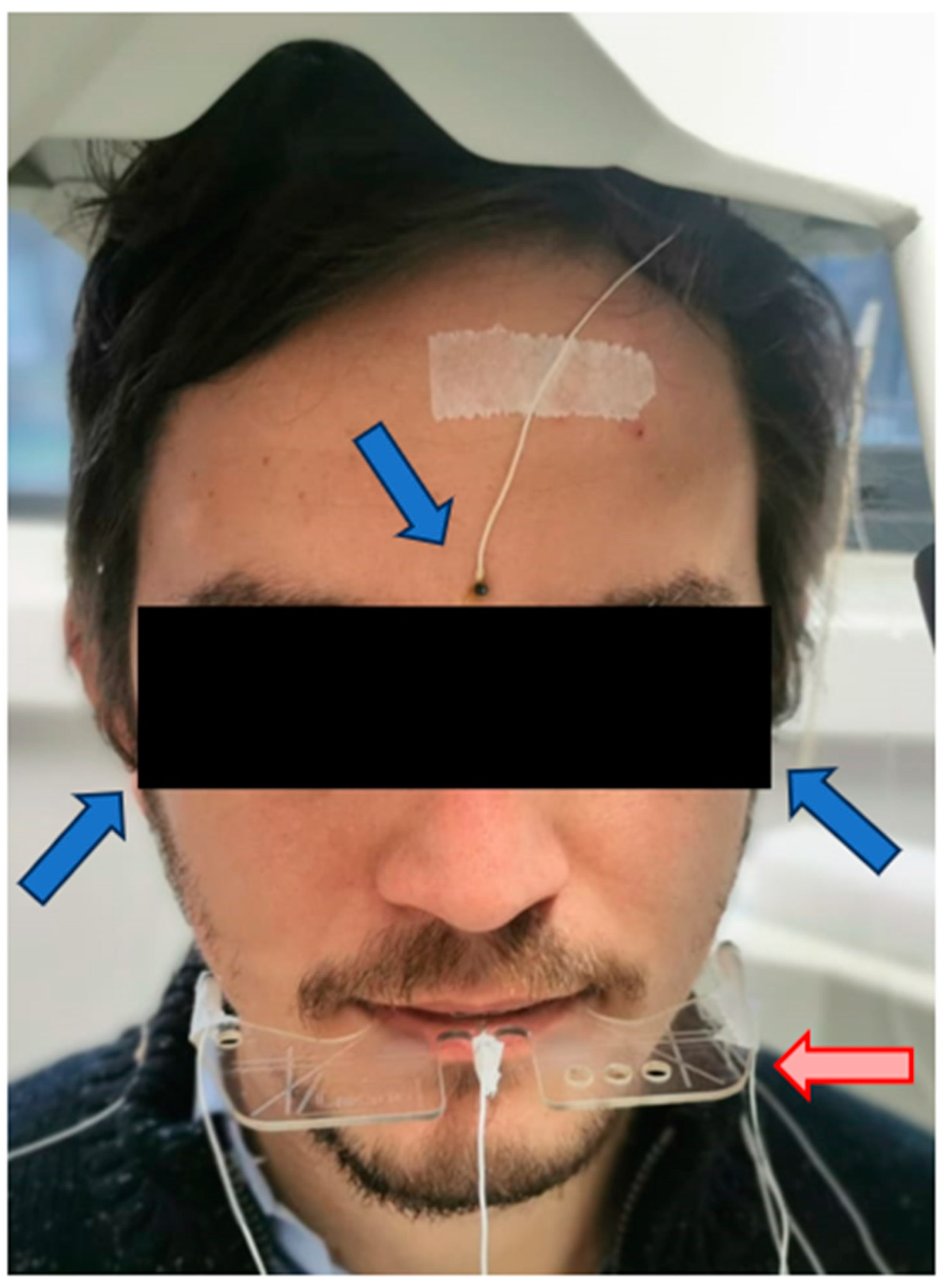
The AG 501 can correct the movements and the inclination of the head to obtain the absolute movements of the mandible, compared to a system generated by reference sensors. This procedure is called Head Correction. For this, three reference sensors are placed at points that are independent of mandibular movement; for example, the cutaneous points of the right and left mastoids, process and glabella [
39] (
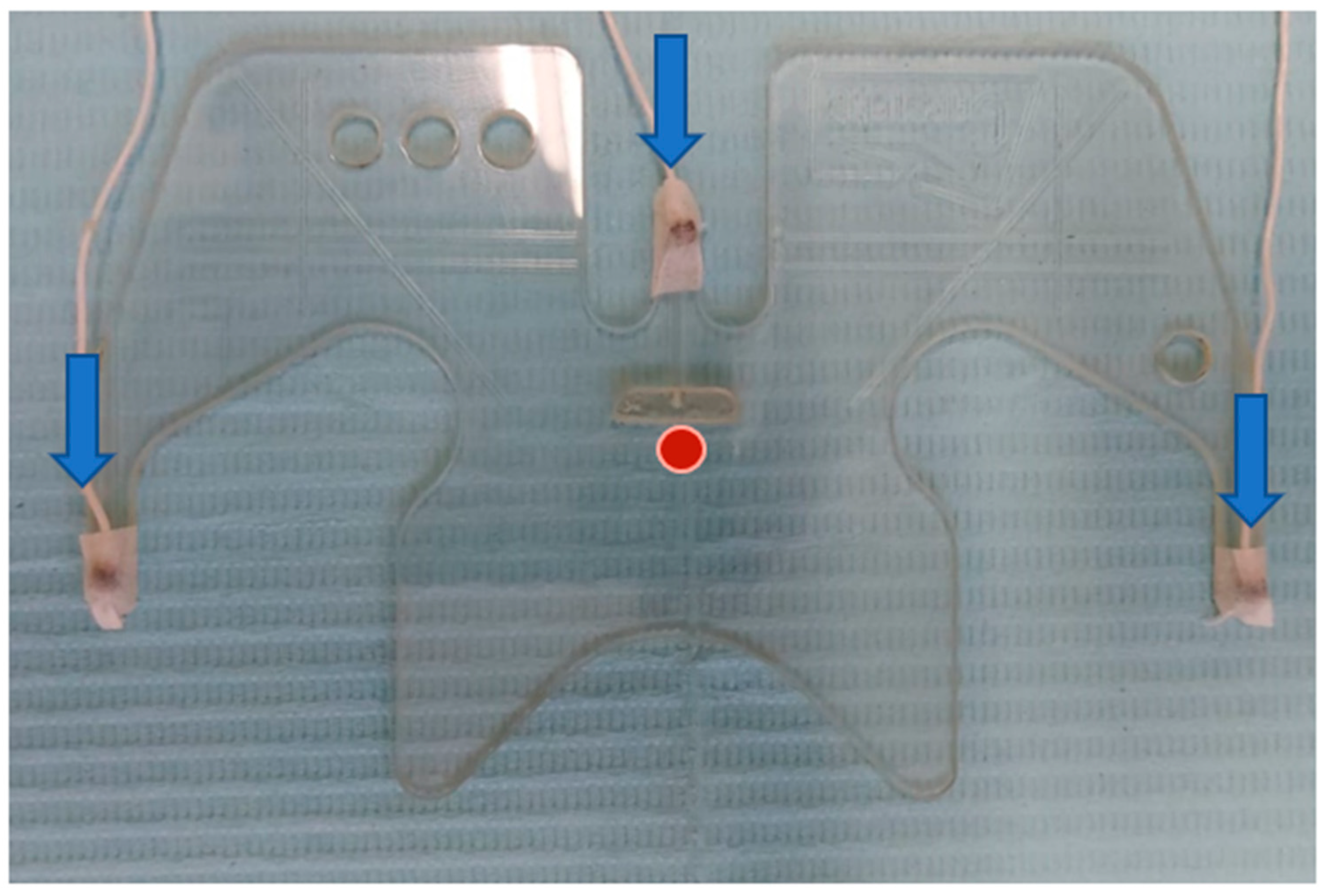
Figure 2). In addition, the device has an accessory, called the bite plane, which aligns the horizontal plane of the system with the occlusal plane of the patient. This eliminates possible parallax errors and locates the origin of the system in the occlusal plane at the midline of the upper incisors on the vestibular side [
40]. This is a support that accommodates three sensors that remain outside the mouth in a fixed configuration. The participant is asked to hold the bite plane, with the incisors immediately behind the stop in the center of the bite plane in front of the central sensor (
Figure 3). With the three reference sensors and the three bite plane sensors, a first recording is conducted to perform the head correction. From this recording, the others are corrected to put them in line with the reference system.
3.2. Data Acquisition
The AG501 generates binary files, with three-dimensional coordinates of the active sensors given in millimeters. These can be extracted and processed using routines specifically developed for this purpose. Given that the masticatory movement is to be analyzed, the recording sensor must be placed at some point on the mandible. Typically, it is placed on the gums, underneath the interincisal line on the vestibular side [
34].
4. Border Movements
There are limits to the movements that the mandible can perform. These are defined by the border movements on the sagittal, coronal and horizontal planes [
41]. To describe the border movements, the participant is asked to place the mandible in extreme positions on each plane, describing Posselt’s envelope of motion [
42]. Border movements tend to be used as a clinical tool to evaluate the state of the masticatory system [
43]. A limitation of mandibular border movement is a symptom of temporomandibular joint disorder [
44]. Yu et al. [
45] analyzed the border movements in patients undergoing orthodontic treatment, observing a greater range after the treatment.
Fuentes et al. [
38] developed a protocol to record Posselt’s envelope of motion. The sensor was placed on the gum on the vestibular side of the lower incisors, on the interincisal line. The patient was asked to perform border movements in the sagittal and coronal plane. In
Figure 4, the Sagittal view of Posselt’s envelope of motion can be seen.
Area of Posselt’s Envelope of Motion
Once Posselt’s envelope of motion has been defined, it can be analyzed to extract characteristics to compare the study groups.
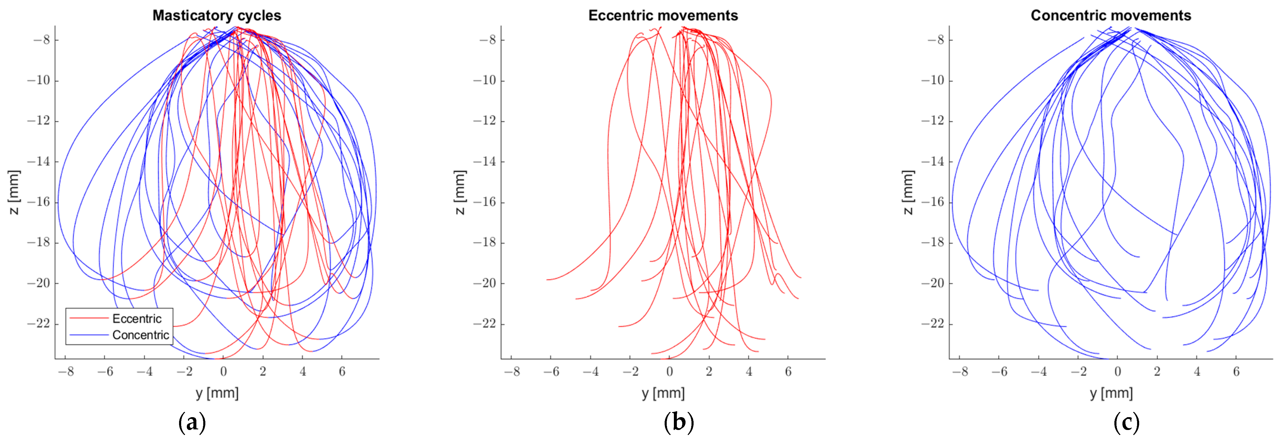
Chuhuaicura et al. [
46] in their study on eccentric and concentric border movements, found significant differences in the described area concentrically and eccentrically from the frontal and sagittal polygons, with the area being greater concentrically in both cases. In this study, subjects were asked to perform border movements in the traditional way [
41] and then in reverse, with a movement sensor attached to the interincisal line on the lower incisors. Based on these movements, the polygon area was calculated with a Matlab function, polyarea, with the coordinates of the movement sensor as an input. After performing the border movements, the subjects performed peanut chewing. With the same procedure, the area of each chewing cycle was obtained and compared to the area obtained for the border movements. Chuhuaicura et al. found that for the frontal polygon, the chewing area is around 10% of the frontal border movement area, for sagittal it is around 5% and for horizontal it is around 15%.
Figure 5 provides an example of eccentric and concentric movements during mastication.
5. Kinematic Analysis
It is possible to perform kinematic analyses with sensor coordinates. If we define the position of sensor S as:
We can define the speed and acceleration of this point as [
47]:
where
n indicates the number of the sample within the file generated by the AG501. Δ
t will be determined by the sampling frequency (fs) of the device, 1250 Hz, for which Equations (2) and (3) can be re-written as:
Patients with a temporomandibular joint disorder show a lower mastication speed than healthy subjects [
48,
49,
50]. In line with these observations, Bakke et al. [
51] found that patients with unilateral pain in the temporomandibular joint showed an increase in mastication speed after undergoing treatment.
6. Masticatory Cycles
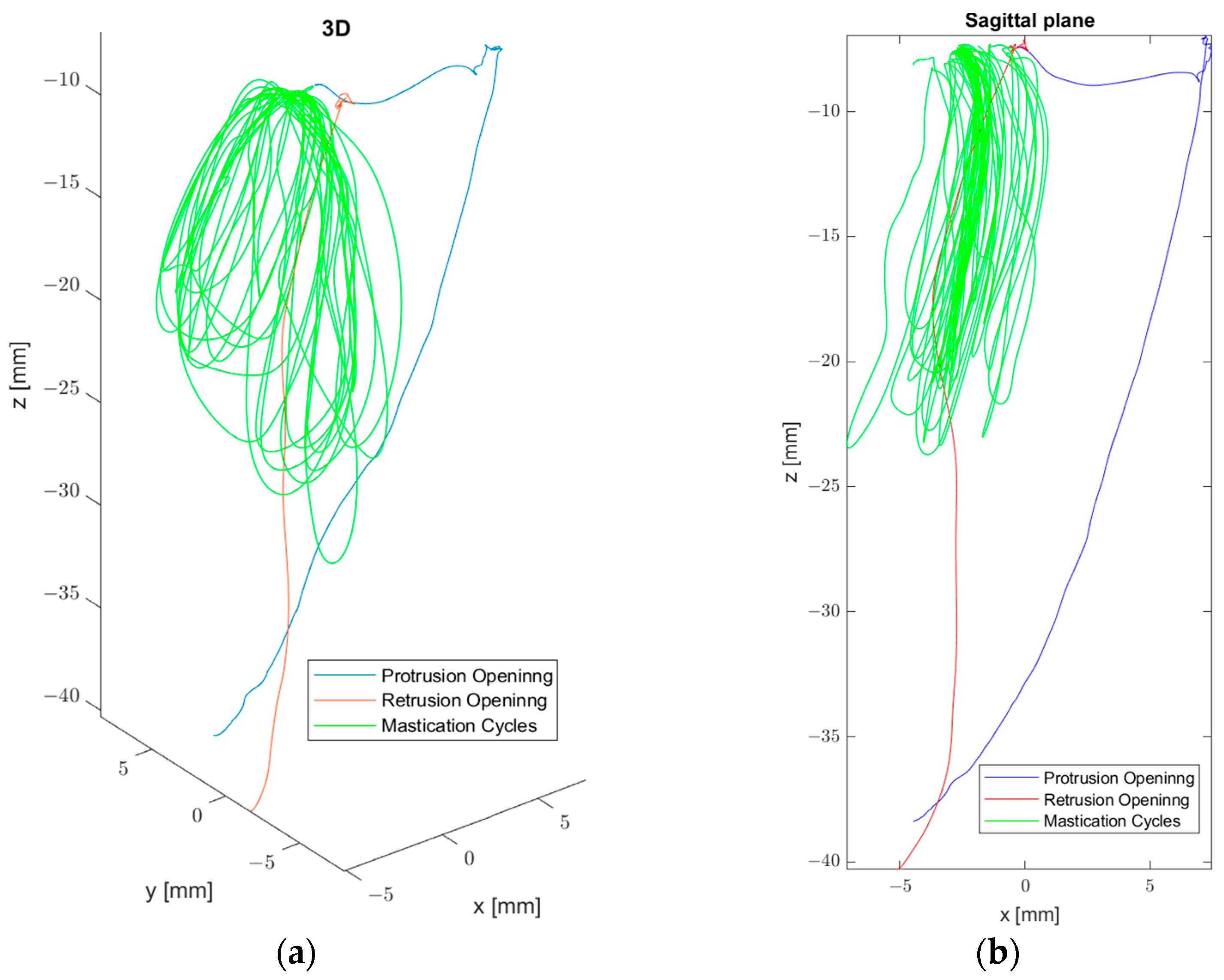
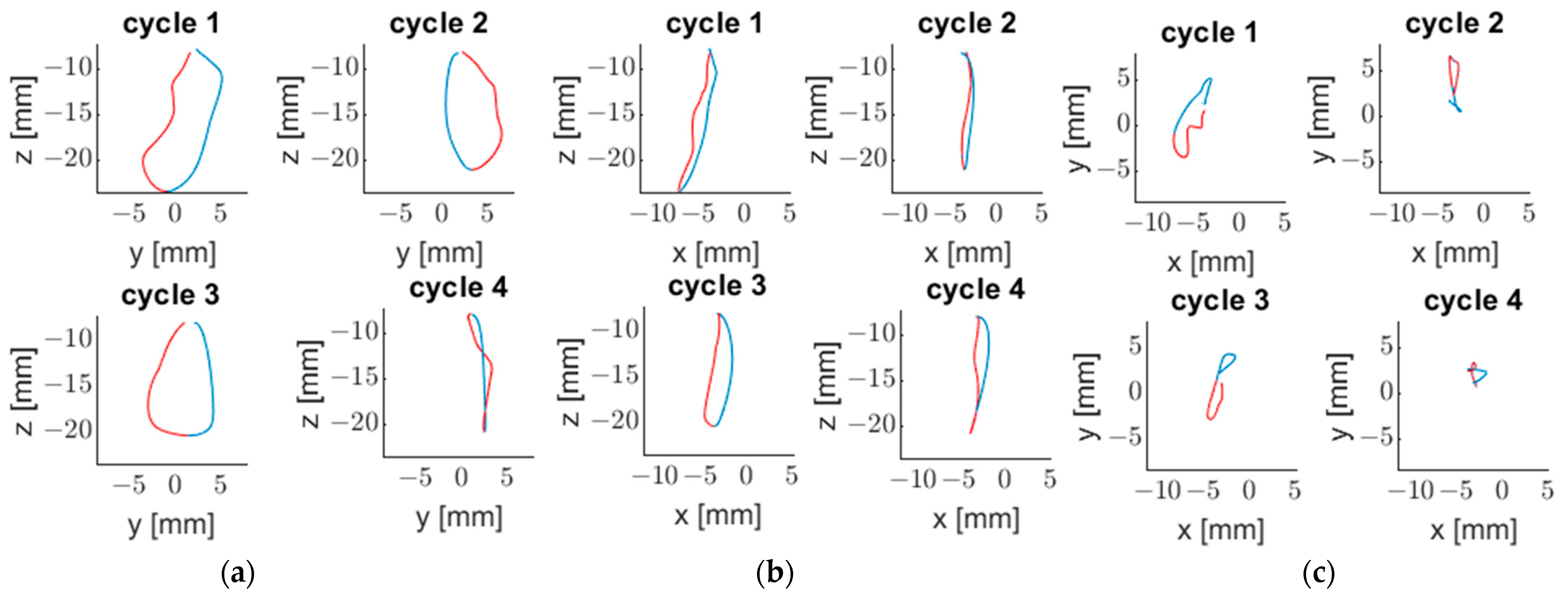
Vargas-Arguto et al. [
52] managed to differentiate each masticatory cycle using Matlab routines (
Figure 6).
Eleven healthy participants (five men and six women) with a full denture up to the first molar of each hemiarch were included. They were asked to chew 3.7 g of peanut and carrot disks (1 × 2 cm) until they had the need to swallow. Three records were made for each food. Chewing cycles were identified using the vertical position of the sensor. Using a threshold value, the beginning and the end of each cycle was established. With the masticatory cycles defined and the previously mentioned tools, each cycle can be analyzed in isolation. Using Matlab routines, Vargas-Arguto et al. [
52] identified the number of cycles, the masticatory cycle frequency, the speed of the ascent and descent of each cycle, using the
Section 5 equations, and the area of each, using the same method as Chuhuaicura et al. [
46]. They noted that men presented a greater masticatory frequency and mandibular speed than women. Only significant differences in the horizontal polygon area between peanut and carrot chewing were found.
The study of movement patterns is relevant to understanding the neural control of mastication and for denture design [
53]. In their study, Lepley et al. [
54] found that patients with poor masticatory performance presented irregular patterns and that they differed from those with greater masticatory efficiency. Flores-Orozco et al. [
55] found that a large mandibular opening and a small angle of lateral excursion are associated with greater masticatory efficiency.
Mastication patterns can be divided into four types: alternating bilateral, simultaneous bilateral, unilateral preference and chronic unilateral [
56]. Several studies have endeavored to identify factors that determine the preference for a mastication side. Christensen & Radue [
57] analyzed the relation between the side of mastication preference and the dominant hand, but did not find a significant relation. García et al. [
58] reported that the choice of one of these patterns is influenced by the consistency of the food. Paphangkorakit et al. [
59] indicated that when a concerted effort is required to cut and grind the food, subjects have a side of preference (for example with pork), whereas with more fragile foods (almonds) they tend toward bilateral mastication. Hannam et al. [
60] suggested that the preference for a side may be influenced by the ability to place the mandible on that side.
7. Analysis of Mouth Opening
Mouth opening is an important indication of the functionality of the temporomandibular joint (TMJ) [
61]. Marinelli et al. [
40] designed a protocol to analyze mouth opening using EMA by using four parameters: vertical distance, Euclidean distance, trajectory and angle. This protocol was tested over a mandibular phantom, with movement sensors placed on the interincisal line between the first molar and last premolar on the left and right. With the sensors placed, four openings were performed, simulating 1, 2, 3 and 4 cm of mouth opening from the maximum intercuspation position (MIP) in vertical distance.
Vertical distance was obtained via Equation (6):
where
zj is the vertical position of the interincisal sensor at each opening and
zPMI is the vertical position of the sensor with the phantom closed.
Euclidean distance is the distance between the reached position and MIP:
Trajectory is the length of the path traveled by the sensor during the opening:
For the opening angle, the data in the premolar and interincisal sensors were used. In the first place, two vectors were defined.
where
S1,
S2 and
S3 are the interincisal, right premolar and left premolar sensors, respectively. With these two vectors, the perpendicular vector was calculated based on the cross product.
With this vector defined, the inclination angle can be calculated for every opening with respect to MIP as follows:
In this research, the openings achieved according to Equation (6) were 9.8, 20.2, 30.4 and 38.9 mm. In concordance with this, the Euclidean distances were 10.2, 21.5, 33.0 and 43.1 mm and the trajectories were 16.7, 28.1, 39 and 48.7 mm, respectively. The opening angles were 5.6, 12, 18.3 and 23.8 degrees, respectively.
Mouth opening has been used to assess the effect of surgical procedures [
62] and the efficacy of anti-inflammatory drugs used in postoperative surgical removals [
63].
8. Electromyography
Gómez et al. [
64] reported a protocol to simultaneously record electromyography (EMG) and electromagnetic articulography, which together with the previously presented variables provides a comprehensive examination of masticatory function. In a recent work, Lezcano et al. [
65] reported a recording protocol in which they synchronized the recordings of EMA and EMG.
Ranges of motion are influenced by the muscles of mastication [
66]. As mentioned previously, mastication is a process coordinated by the centers of the brainstem, which receive feedback from a set of sensory receptors. The texture of the food influences the force, the duration of the masticatory cycle and the number of cycles before swallowing [
67]. Some studies have found a link between the characteristics of the food and the electromyographic activity of the elevator muscles during mastication [
68,
69,
70,
71]. The level of dentition, age [
72] or the presence of temporomandibular joint disorders [
73,
74] also affect electromyographic activity.
9. Discussion
As previously described, the analysis of masticatory movement is of great interest for both understanding masticatory function and for the diagnosis of disorders or assessing the effectiveness of a treatment.
In their works on border movements, Kataoka et al. [
44] and Yu et al. [
45] used a video system to record mandibular movement. Kataoka used a system based on LEDs and a facebow, while Yu used two video cameras placed in front of and beside the patient with a 90° angle between them. Two resin reference balls were used to label the cutting edges between the nose and the center point of the mandibular incisors. The method presented by Yu is simple; however, it lacks a head correction procedure, and the markers are placed on the patient’s skin, which may move during chewing. Kataoka makes use of a facebow which enables head correction, but it could result in unnatural chewing. Ferrario et al. [
11] avoid both problems using reference markers and six high-resolution cameras. The movement was recorded using three extraoral markers placed on an equilateral triangular antenna, anchored on the mandibular anterior gingival line just out of dental contact. However, due to this arrangement, the antenna may be displaced by the lips, making it difficult to capture natural movements during chewing. Lepley et al. [
54] use the Optotrak 3020
® (Northern Digital, Waterloo, ON, Canada) system with a single diode attached to the subjects’ chin and glasses with a diode arrangement to correct for head movement. This system covers the previously mentioned limitations but needs a 2 m space between the subject and the cameras. Also, the movement marker is attached to the patient’s skin.
The ARCUSdigma device is considered the gold standard for recording mandibular movement [
75]. It is based on the use of an ultrasound, with emitters mounted on a facebow fixed to the mandible [
76]. Sójka et al. [
76] used this device to evaluate mandibular movement. Given that the device must be placed inside the mandible, this interferes with natural movement during mastication. An advantage of this system is the possibilty to record condylar movement [
77].
A kinesiograph also uses a facebow with a magnet attached to the incisors. This device senses the maginetic field of the magnet to record movement. It has been used to describe mandibular movement [
78] and Posselt polygons [
79]. However, this device has been questioned due to inconsistent results [
80].
In previous paragraphs, we described parameters used to study chewing and devices designed to achieve this. Posselt polygons, kinematic analysis, mouth opening and electromiographic activity [
81,
82,
83] have been the objects of several studies. These variables have been analyzed with different devices to try to overcome certain problems that arise when recording mandibular movement, head movement, faithful tracking of mandibular movement and modifying the natural movement during chewing. Border movements have been analyzed using EMA in several studies [
19,
38], and a new variable has been introduced by analyzing border movements eccentrically and concentrically [
46], as well as through symmetry analysis [
84]. In addition, it has been made possible to analyze masticatory kinematics, differentiation of each cycle and its stage of ascent and descent in the area described [
52].
Regarding sensor placement, these are attached to the gums to avoid skin movement. Due to their dimensions, they do not interfere with movement, and head movement can be filtered using the head correction procedure. Three-dimensional electromagnetic articulography allows for analysis of reliable accuracy [
37].
Future studies should involve groups of greater diversity, with different skeletal classes or that suffer from a temporomandibular joint disorders. In addition, studies can be carried out on patients who are undergoing treatment and observe whether there are significant differences between the variables that are analyzed before and after the treatment. The protocol for the use of EMA in conjunction with EMG was made by analyzing the vertical dimension statically, which is why a future study should be conducted where electromyographic activity is recorded during masticatory movements.
An obstacle to the use of EMA is the interference of metallic elements with the magnetic fields, which is why the analysis of subjects with metallic prostheses is limited. This issue is also shared with the kinesiograph. In addition, the sensors must be 8 mm apart so as not to interfere with each other [
24]. A disadvantage with respect to the ARCUSdigma device is the posibility of measuring condylar movement. Due to its costs and complexity, EMA is limited to research use only.
10. Conclusions
Electromagnetic articulography is a tool that allows for complete analysis of the various aspects used to evaluate movement during mastication, along with EMG. The differentiation of each cycle makes it possible to visualize the change in mastication patterns as the food is crushed and forms an alimentary bolus. In addition, it allows the analysis of new variables, such as the frequency of cycles, speed of ascent and descent and the area of each cycle. Several studies have already been conducted with this technology, but more analyses are needed with a greater diversity of subjects and cases.
11. Future Directions
The use of EMA and data postprocessing offers new possibilities in the assessment of functional movements of mastication, and combined with the EMG it is possible to cover all aspects of past research, like mouth opening, preference side, number of cycles, kinematic analysis, etc., with a unique technique. However, a new paradigm can be introduced in the analysis of masticatory movement as the normalization of both mandibular opening, area description and EMG activity. Normalization is a technique that could allow for comparison of parameters between patients, avoiding differences due to body size, age or gender. This can be conducted cycle by cycle since individualization is possible. With these possibilities, a standardization of mandibular movement records and variables of interest can be performed to avoid differences between studies due to different recording methods or devices. A potential challenge in using EMA in the field of dentistry is that the metal components of prosthetics can interfere with the equipment. A deep study of the presence of metal elements and their interference with EMA must be conducted.
Author Contributions
Conceptualization, R.F. and F.M.; methodology, C.V.; software, F.M.; validation, F.M., P.N. and R.F.; formal analysis, F.M. and R.F.; investigation, F.M., R.F., P.N., C.V. and J.A.; resources, R.F.; data curation, F.M. and P.N.; writing—original draft preparation, F.M. and C.V.; writing—review and editing, F.M., C.V. and J.A.; visualization, C.V.; supervision, R.F.; project administration, R.F. All authors have read and agreed to the published version of the manuscript.
Funding
This research received no external funding.
Institutional Review Board Statement
The study was conducted in accordance with the Declaration of Helsinki and approved by the Ethics Committee of Universidad de La Frontera, protocol code no. 079_22; approved on 12 March 2022.
Informed Consent Statement
Informed consent was obtained from all subjects involved in the study.
Data Availability Statement
The data presented in this study are available on request from the corresponding author. The data are not publicly available due to no public database is available.
Conflicts of Interest
The authors declare no conflict of interest.
References
- Prinz, J.F.; Lucas, P.W. Swallow Thresholds in Human Mastication. Arch. Oral Biol. 1995, 40, 401–403. [Google Scholar] [CrossRef]
- Dellow, P.G.; Lund, J.P. Evidence for Central Timing of Rhythmical Mastication. J. Physiol. 1971, 215, 1–13. [Google Scholar] [CrossRef]
- Lund, J.P. Mastication and Its Control by the Brain Stem. Crit. Rev. Oral Biol. Med. 1991, 2, 33–64. [Google Scholar] [CrossRef] [PubMed]
- Gonçalves, T.M.S.V.; Schimmel, M.; van der Bilt, A.; Chen, J.; van der Glas, H.W.; Kohyama, K.; Hennequin, M.; Peyron, M.; Woda, A.; Leles, C.R.; et al. Consensus on the Terminologies and Methodologies for Masticatory Assessment. J. Oral Rehabil. 2021, 48, 745–761. [Google Scholar] [CrossRef]
- Yurkstas, A.A. The Masticatory Act. J. Prosthet. Dent. 1965, 15, 248–260. [Google Scholar] [CrossRef]
- Woo, J.; Tong, C.; Yu, R. Chewing Difficulty Should Be Included as a Geriatric Syndrome. Nutrients 2018, 10, 1997. [Google Scholar] [CrossRef] [PubMed]
- Nakayama, E.; Tohara, H.; Sakai, M.; Iida, M.; Abe, K.; Ueda, K. Kinematic Features of Mandibular Movement during Mastication in Geriatric Individuals Who Are Provided with a Dysphagia Diet at Long-Term Care Facilities. Nutrients 2023, 15, 2273. [Google Scholar] [CrossRef] [PubMed]
- Hennequin, M.; Allison, P.; Veyrune, J.; Faye, M.; Peyron, M. Clinical Evaluation of Mastication: Validation of Video versus Electromyography. Clin. Nutr. 2005, 24, 314–320. [Google Scholar] [CrossRef] [PubMed]
- Allison, P.J.; Peyron, M.A.; Faye, M.; Hennequin, M. Video Evaluation for Mastication Validation InPersons with Down Syndrome. Dysphagia 2004, 19, 95–99. [Google Scholar] [CrossRef]
- van der Bilt, A.; Abbink, J.H. The Influence of Food Consistency on Chewing Rate and Muscular Work. Arch. Oral Biol. 2017, 83, 105–110. [Google Scholar] [CrossRef]
- Ferrario, V.F.; Sforza, C.; Lovecchio, N.; Mian, F. Quantification of Translational and Gliding Components in Human Temporomandibular Joint during Mouth Opening. Arch. Oral Biol. 2005, 50, 507–515. [Google Scholar] [CrossRef]
- Buschang, P.H.; Hayasaki, H.; Throckmorton, G.S. Quantification of Human Chewing-Cycle Kinematics. Arch. Oral Biol. 2000, 45, 461–474. [Google Scholar] [CrossRef] [PubMed]
- Rahman, F.; Femiano, F.; Louis, P.J.; Kau, C.H. An Evaluation of Jaw Tracking Movements in Patients with Total Joint Replacements versus a Control Group. Medicina 2022, 58, 738. [Google Scholar] [CrossRef]
- Karlsson, S.; Carlsson, G.E. Characteristics of Mandibular Masticatory Movement in Young and Elderly Dentate Subjects. J. Dent. Res. 1990, 69, 473–476. [Google Scholar] [CrossRef] [PubMed]
- Piancino, M.G.; Isola, G.; Merlo, A.; Dalessandri, D.; Debernardi, C.; Bracco, P. Chewing Pattern and Muscular Activation in Open Bite Patients. J. Electromyogr. Kinesiol. 2012, 22, 273–279. [Google Scholar] [CrossRef] [PubMed]
- Leles, C.R.; Compagnoni, M.A.; Souza, R.F.; de Barbosa, D.B. Kinesiographic Study of Mandibular Movements during Functional Adaptation to Complete Dentures. J. Appl. Oral Sci. 2003, 11, 311–318. [Google Scholar] [CrossRef]
- Jankelson, B. Measurement Accuracy of the Mandibular Kinesiograph—A Computerized Study. J. Prosthet. Dent. 1980, 44, 656–666. [Google Scholar] [CrossRef]
- Peyron, M.-A.; Mioche, L.; Renon, P.; Abouelkaram, S. Masticatory Jaw Movement Recordings: A New Method to Investigate Food Texture. Food Qual. Prefer. 1996, 7, 229–237. [Google Scholar] [CrossRef]
- Vargas-Agurto, S.A.; Lezcano, M.F.; Álvarez, G.; Navarro, P.; Fuentes, R. Análisis Tridimensional de Movimientos Mandibulares Bordeantes En Participantes Dentados Totales. Int. J. Morphol. 2020, 38, 983–989. [Google Scholar] [CrossRef]
- Hixon, T.J. An Electromagnetic Method for Transducing Jaw Movements during Speech. J. Acoust. Soc. Am. 1971, 49, 603–606. [Google Scholar] [CrossRef]
- Schönle, P.W.; Gräbe, K.; Wenig, P.; Höhne, J.; Schrader, J.; Conrad, B. Electromagnetic Articulography: Use of Alternating Magnetic Fields for Tracking Movements of Multiple Points inside and Outside the Vocal Tract. Brain Lang. 1987, 31, 26–35. [Google Scholar] [CrossRef] [PubMed]
- Bourdiol, P.; Mishellany-Dutour, A.; Peyron, M.-A.; Woda, A. Tongue-Mandible Coupling Movements during Saliva Swallowing. J. Oral Rehabil. 2014, 41, 199–205. [Google Scholar] [CrossRef]
- Maassen, B.; van Lieshout, P. Speech Motor Control: New Developments in Basic and Applied Research; Oxford University Press: Oxford, UK, 2010; ISBN 978-0-19-923579-7. [Google Scholar]
- Rebernik, T.; Jacobi, J.; Jonkers, R.; Noiray, A.; Wieling, M. A Review of Data Collection Practices Using Electromagnetic Articulography. Lab. Phonol. J. Assoc. Lab. Phonol. 2021, 12, 6. [Google Scholar] [CrossRef]
- Dromey, C.; Hunter, E.; Nissen, S.L. Speech Adaptation to Kinematic Recording Sensors: Perceptual and Acoustic Findings. J. Speech Lang. Hear. Res. 2018, 61, 593–603. [Google Scholar] [CrossRef]
- Hasegawa-Johnson, M. Electromagnetic Exposure Safety of the Carstens Aerticulograph AG100. J. Acoust. Soc. Am. 1998, 104, 2529–2532. [Google Scholar] [CrossRef]
- Smith, S.; Aasen, R. The Effects of Electromagnetic Fields on Cardiac Pacemakers. IEEE Trans. Broadcast. 1992, 38, 136–139. [Google Scholar] [CrossRef]
- Tognola, G.; Parazzini, M.; Sibella, F.; Paglialonga, A.; Ravazzani, P. Electromagnetic Interference and Cochlear Implants. Ann. Ist. Super. Sanita 2007, 43, 241–247. [Google Scholar]
- Zhang, Y.; Jones, P.L.; Jetley, R. A Hazard Analysis for a Generic Insulin Infusion Pump. J. Diabetes Sci. Technol. 2010, 4, 263–283. [Google Scholar] [CrossRef] [PubMed]
- Joglar, J.A.; Nguyen, C.; Garst, D.M.; Katz, W.F. Safety of Electromagnetic Articulography in Patients With Pacemakers and Implantable Cardioverter-Defibrillators. J. Speech Lang. Hear. Res. 2009, 52, 1082–1087. [Google Scholar] [CrossRef]
- Katz, W.F.; Bharadwaj, S.V.; Gabbert, G.J.; Loizou, P.C.; Tobey, E.A.; Poroy, O. EMA Compatibility of the Clarion 1.2 Cochlear Implant System. Acoust. Res. Lett. Online 2003, 4, 100–105. [Google Scholar] [CrossRef]
- Mücke, D.; Hermes, A.; Roettger, T.B.; Becker, J.; Niemann, H.; Dembek, T.A.; Timmermann, L.; Visser-Vandewalle, V.; Fink, G.R.; Grice, M.; et al. The Effects of Thalamic Deep Brain Stimulation on Speech Dynamics in Patients with Essential Tremor: An Articulographic Study. PLoS ONE 2018, 13, e0191359. [Google Scholar] [CrossRef] [PubMed]
- Hoole, P.; Nguyen, N. Electromagnetic Articulography. In Coarticulation; Hardcastle, W.J., Hewlett, N., Eds.; Cambridge University Press: Cambridge, UK, 1999; pp. 260–269. ISBN 978-0-521-44027-1. [Google Scholar]
- Fuentes, R.; Arias, A.; Lezcano, M.F.; Saravia, D.; Kuramochi, G.; Dias, F.J. Systematic Standardized and Individualized Assessment of Masticatory Cycles Using Electromagnetic 3D Articulography and Computer Scripts. BioMed Res. Int. 2017, 2017, 7134389. [Google Scholar] [CrossRef]
- Savariaux, C.; Badin, P.; Samson, A.; Gerber, S. A Comparative Study of the Precision of Carstens and Northern Digital Instruments Electromagnetic Articulographs. J. Speech Lang. Hear. Res. 2017, 60, 322–340. [Google Scholar] [CrossRef]
- Stella, M.; Stella, A.; Sigona, F.; Bernardini, P.; Grimaldi, M.; Fivela, B.G. Electromagnetic Articulography with AG500 and AG501. In Proceedings of the Interspeech 2013 ISCA, Lyon, France, 25–29 August 2013; pp. 1316–1320. [Google Scholar]
- Lezcano, M.F.; Dias, F.; Arias, A.; Fuentes, R. Accuracy and Reliability of AG501 Articulograph for Mandibular Movement Analysis: A Quantitative Descriptive Study. Sensors 2020, 20, 6324. [Google Scholar] [CrossRef]
- Fuentes, R.; Arias, A.; Saravia, D.; Lezcano, M.F.; Dias, F.J. An Innovative Method to Analyse the Range of Border Mandibular Movements Using 3D Electromagnetic Articulography (AG501) and MATLAB. Biomed. Res. 2017, 28, 4239–4247. [Google Scholar]
- Álvarez, G.; Dias, F.J.; Lezcano, F.; Arias, A.; Navarro, P.; Fuentes, R. Description of Tongue Movements on Swallowing Patterns. Arch. Oral Biol. 2020, 113, 104706. [Google Scholar] [CrossRef]
- Marinelli, F.; Lezcano, M.F.; Alarcón, J.; Navarro, P.; Fuentes, R. A Novel Technique to Accurately Measure Mouth Opening Using 3D Electromagnetic Articulography. Bioengineering 2022, 9, 577. [Google Scholar] [CrossRef] [PubMed]
- Okeson, J.P. Management of Temporomandibular Disorders and Occlusion, 5th ed.; Mosby: St. Louis, MO, USA, 2003; ISBN 978-0-323-01477-9. [Google Scholar]
- Posselt, U. Terminal Hinge Movement of the Mandible. J. Prosthet. Dent. 2001, 86, 2–9. [Google Scholar] [CrossRef] [PubMed]
- Koolstra, J.H.; Naeije, M.; Van Eijden, T.M.G.J. The Three-Dimensional Active Envelope of Jaw Border Movement and Its Determinants. J. Dent. Res. 2001, 80, 1908–1912. [Google Scholar] [CrossRef]
- Kataoka, T.; Kawanabe, N.; Shiraga, N.; Hashimoto, T.; Deguchi, T.; Miyawaki, S.; Takano-Yamamoto, T.; Yamashiro, T. The Influence of Craniofacial Morphology on Mandibular Border Movements. CRANIO® 2013, 31, 14–22. [Google Scholar] [CrossRef]
- Yu, J.-H.; Lin, C.-C.; Chen, Y.-H. Changes in Mandibular Border Movements in Adult Patients after Correction of Functional Anterior Crossbite. J. Dent. Sci. 2017, 12, 139–144. [Google Scholar] [CrossRef]
- Chuhuaicura, P.; Lezcano, M.F.; Dias, F.J.; Fuentes, A.D.; Arias, A.; Fuentes, R. Mandibular Border Movements: The Two Envelopes of Motion. J. Oral Rehabil. 2021, 48, 384–391. [Google Scholar] [CrossRef] [PubMed]
- Stewart, J. Calculus: Early Transcendentals, 7th ed.; Brooks/Cole, Cengage Learning: Belmont, CA, USA, 2012; ISBN 978-0-538-49790-9. [Google Scholar]
- Radke, J.C.; Kamyszek, G.J.; Kull, R.S.; Velasco, G.R. TMJ Symptoms Reduce Chewing Amplitude and Velocity, and Increase Variability. CRANIO® 2019, 37, 12–19. [Google Scholar] [CrossRef]
- Kuwahara, T.; Bessette, R.W.; Maruyama, T. Chewing Pattern Analysis in TMD Patients with Unilateral and Bilateral Internal Derangement. CRANIO® 1995, 13, 167–172. [Google Scholar] [CrossRef] [PubMed]
- Hansdottir, R.; Bakke, M. Joint Tenderness, Jaw Opening, Chewing Velocity, and Bite Force in Patients with Temporomandibular Joint Pain and Matched Healthy Control Subjects. J. Orofac. Pain 2004, 18, 108–113. [Google Scholar]
- Bakke, M.; Hansdottir, R. Mandibular Function in Patients with Temporomandibular Joint Pain: A 3-Year Follow-Up. Oral Surg. Oral Med. Oral Pathol. Oral Radiol. Endodontology 2008, 106, 227–234. [Google Scholar] [CrossRef]
- Vargas-Agurto, S.; Lezcano, M.F.; Álvarez, G.; Navarro, P.; Arias, A.; Fuentes, R. Análisis Cinemático de La Masticación de Alimentos Duros y Blandos En Participantes Dentados Utilizando Articulografía Electromagnética 3D. Int. J. Morphol. 2021, 39, 935–940. [Google Scholar] [CrossRef]
- Wilding, R.J.C.; Lewin, A. A Model for Optimum Functional Human Jaw Movements Based on Values Associated with Preferred Chewing Patterns. Arch. Oral Biol. 1991, 36, 519–523. [Google Scholar] [CrossRef]
- Lepley, C.; Throckmorton, G.; Parker, S.; Buschang, P.H. Masticatory Performance and Chewing Cycle Kinematics—Are They Related? Angle Orthod. 2010, 80, 295–301. [Google Scholar] [CrossRef]
- Flores-Orozco, E.I.; Rovira-Lastra, B.; Willaert, E.; Peraire, M.; Martinez-Gomis, J. Relationship between Jaw Movement and Masticatory Performance in Adults with Natural Dentition. Acta Odontol. Scand. 2016, 74, 103–107. [Google Scholar] [CrossRef] [PubMed]
- Genaro, K.F.; Berretin-Felix, G.; Rehder, M.I.B.C.; Marchesan, I.Q. Avaliação Miofuncional Orofacial: Protocolo MBGR. Rev. CEFAC 2009, 11, 237–255. [Google Scholar] [CrossRef]
- Christensen, L.V.; Radue, J.T. Lateral Preference in Mastication: A Feasibility Study. J. Oral Rehabil. 1985, 12, 421–427. [Google Scholar] [CrossRef] [PubMed]
- Garcia, D.G.B.; Benevides, S.D.; Araujo, R.P.; Ribeiro, C.D.O.; Mello, S.M.F. Mastigação Habitual e Atividade Eletromiográfica Dos Músculos Masseter e Temporal Em Escolares de 7 a 12 Anos. Rev. CEFAC 2014, 16, 1928–1935. [Google Scholar] [CrossRef]
- Paphangkorakit, J.; Thothongkam, N.; Supanont, N. Chewing-Side Determination of Three Food Textures. J. Oral Rehabil. 2006, 33, 2–7. [Google Scholar] [CrossRef] [PubMed]
- Hannam, A.G.; De Cou, R.E.; Scott, J.D.; Wood, W.W. The Relationship between Dental Occlusion, Muscle Activity and Associated Jaw Movement in Man. Arch. Oral Biol. 1977, 22, 25–32. [Google Scholar] [CrossRef]
- Zawawi, K.H.; Al-Badawi, E.A.; Lobo, S.L.; Melis, M.; Mehta, N.R. An Index for the Measurement of Normal Maximum Mouth Opening. J. Can. Dent. Assoc. 2003, 69, 737–741. [Google Scholar]
- Balakrishnan, G.; Narendar, R.; Kavin, T.; Venkataraman, S.; Gokulanathan, S. Incidence of Trismus in Transalveolar Extraction of Lower Third Molar. J. Pharm. Bioallied Sci. 2017, 9, 222. [Google Scholar] [CrossRef]
- Paiva-Oliveira, J.G.; Bastos, P.R.H.O.; Cury Pontes, E.R.J.; Da Silva, J.C.L.; Delgado, J.A.B.; Oshiro-Filho, N.T. Comparison of the Anti-Inflammatory Effect of Dexamethasone and Ketorolac in the Extractions of Third Molars. Oral Maxillofac. Surg. 2016, 20, 123–133. [Google Scholar] [CrossRef] [PubMed]
- Gomez, J.; Lezcano, M.F.; Peldoza, V.; Fuentes, R. Electromiografía de Superficie y Articulografía Electromangética Para Análisis de Dimensión Vertical. Reporte de Protocolo. Av. EN Odontoestomatol. 2019, 36, 151–159. [Google Scholar] [CrossRef]
- Lezcano, M.F.; Dias, F.; Farfán-Beltrán, N.; Manzanares-Céspedes, M.C.; Cerda, C.; Fuentes, R. Synchronization of Surface Electromyography and 3D Electromagnetic Articulography Applied to Study the Biomechanics of the Mandible: Proof of Concept. Appl. Sci. 2023, 13, 5851. [Google Scholar] [CrossRef]
- Nascimento, G.K.B.O.; da Cunha, D.A.; de Lima, L.M.; de Moraes, K.J.R.; Pernambuco, L.d.A.; Régis, R.M.F.L.; da Silva, H.J. Eletromiografia de Superfície Do Músculo Masseter Durante a Mastigação: Uma Revisão Sistemática. Rev. CEFAC 2012, 14, 725–731. [Google Scholar] [CrossRef]
- Mioche, L.; Bourdiol, P.; Martin, J.-F.; Noël, Y. Variations in Human Masseter and Temporalis Muscle Activity Related to Food Texture during Free and Side-Imposed Mastication. Arch. Oral Biol. 1999, 44, 1005–1012. [Google Scholar] [CrossRef] [PubMed]
- Horio, T.; Kawamura, Y. Effects of Texture of Food on Chewing Patterns in the Human Subject. J. Oral Rehabil. 1989, 16, 177–183. [Google Scholar] [CrossRef] [PubMed]
- Agrawal, K.R.; Lucas, P.W.; Bruce, I.C.; Prinz, J.F. Food Properties That Influence Neuromuscular Activity During Human Mastication. J. Dent. Res. 1998, 77, 1931–1938. [Google Scholar] [CrossRef] [PubMed]
- Mathevon, E.; Mioche, L.; Brown, W.E.; Culioli, J. Texture analysis of beef cooked at various temperatures by mechanical measurements, sensory assessments and electromyography. J. Texture Stud. 1995, 26, 175–192. [Google Scholar] [CrossRef]
- Diaz-Tay, J.; Jayasinghe, N.; Lucas, P.W.; McCallum, J.C.; Jones, J.T. Association between Surface Electromyography of Human Jaw-Closing Muscle and Quantified Food Breakdown. Arch. Oral Biol. 1991, 36, 893–898. [Google Scholar] [CrossRef]
- Gaszynska, E.; Kopacz, K.; Fronczek-Wojciechowska, M.; Padula, G.; Szatko, F. Electromyographic Activity of Masticatory Muscles in Elderly Women – a Pilot Study. Clin. Interv. Aging 2017, 12, 111–116. [Google Scholar] [CrossRef]
- Valentino, R.; Cioffi, I.; Vollaro, S.; Cimino, R.; Baiano, R.; Michelotti, A. Jaw Muscle Activity Patterns in Women with Chronic TMD Myalgia during Standardized Clenching and Chewing Tasks. CRANIO® 2021, 39, 157–163. [Google Scholar] [CrossRef] [PubMed]
- Mapelli, A.; Zanandréa Machado, B.C.; Giglio, L.D.; Sforza, C.; De Felício, C.M. Reorganization of Muscle Activity in Patients with Chronic Temporomandibular Disorders. Arch. Oral Biol. 2016, 72, 164–171. [Google Scholar] [CrossRef]
- Wieckiewicz, M.; Zietek, M.; Nowakowska, D.; Wieckiewicz, W. Comparison of Selected Kinematic Facebows Applied to Mandibular Tracing. BioMed Res. Int. 2014, 2014, 818694. [Google Scholar] [CrossRef]
- Sójka, A.; Huber, J.; Kaczmarek, E.; Hędzelek, W. Evaluation of Mandibular Movement Functions Using Instrumental Ultrasound System: Mandibular Movement Analysis. J. Prosthodont. 2017, 26, 123–128. [Google Scholar] [CrossRef] [PubMed]
- Dragus, A.C.; Mihai, A.; Tanase, G.; Burlibasa, M.; Cristache, C.M. Intraindividual Left–Right Side Differences of Sagittal Condylar Inclination (SCI) in Different Skeletal Classes. Healthcare 2023, 11, 1341. [Google Scholar] [CrossRef] [PubMed]
- Manfredini, D.; Cocilovo, F.; Favero, L.; Ferronato, G.; Tonello, S.; Guarda-Nardini, L. Surface Electromyography of Jaw Muscles and Kinesiographic Recordings: Diagnostic Accuracy for Myofascial Pain: Surface electromyography of jaw muscles. J. Oral Rehabil. 2011, 38, 791–799. [Google Scholar] [CrossRef]
- Frisoli, M.; Edelhoff, J.M.; Gersdorff, N.; Nicolet, J.; Braidot, A.; Engelke, W. Comparative Study Using Video Analysis and an Ultrasonic Measurement System to Quantify Mandibular Movement. CRANIO® 2017, 35, 19–29. [Google Scholar] [CrossRef]
- Cooper, B.C.; Adib, F. An Assessment of the Usefulness of Kinesiograph as an Aid in the Diagnosis of TMD: A Review of Manfredini et al.’s Studies. CRANIO® 2015, 33, 46–66. [Google Scholar] [CrossRef] [PubMed]
- van der Bilt, A.; van Kampen, F.M.C.; Cune, M.S. Masticatory Function with Mandibular Implant-Supported Overdentures Fitted with Different Attachment Types. Eur. J. Oral Sci. 2006, 114, 191–196. [Google Scholar] [CrossRef]
- Flores-Orozco, E.I.; Rovira-Lastra, B.; Peraire, M.; Salsench, J.; Martinez-Gomis, J. Reliability of a Visual Analog Scale for Determining the Preferred Mastication Side. J. Prosthet. Dent. 2016, 115, 203–208. [Google Scholar] [CrossRef]
- Eberhard, L.; Braun, S.; Wirth, A.; Schindler, H.-J.; Hellmann, D.; Giannakopoulos, N.N. The Effect of Experimental Balancing Interferences on Masticatory Performance. J. Oral Rehabil. 2014, 41, 346–352. [Google Scholar] [CrossRef]
- Lezcano, M.F.; Dias, F.J.; Chuhuaicura, P.; Navarro, P.; Fuentes, R. Symmetry of Mandibular Movements: A 3D Electromagnetic Articulography Technique Applied on Asymptomatic Participants. J. Prosthet. Dent. 2021, 125, 746–752. [Google Scholar] [CrossRef]
| Disclaimer/Publisher’s Note: The statements, opinions and data contained in all publications are solely those of the individual author(s) and contributor(s) and not of MDPI and/or the editor(s). MDPI and/or the editor(s) disclaim responsibility for any injury to people or property resulting from any ideas, methods, instructions or products referred to in the content. |
© 2023 by the authors. Licensee MDPI, Basel, Switzerland. This article is an open access article distributed under the terms and conditions of the Creative Commons Attribution (CC BY) license (https://creativecommons.org/licenses/by/4.0/).


 and
and 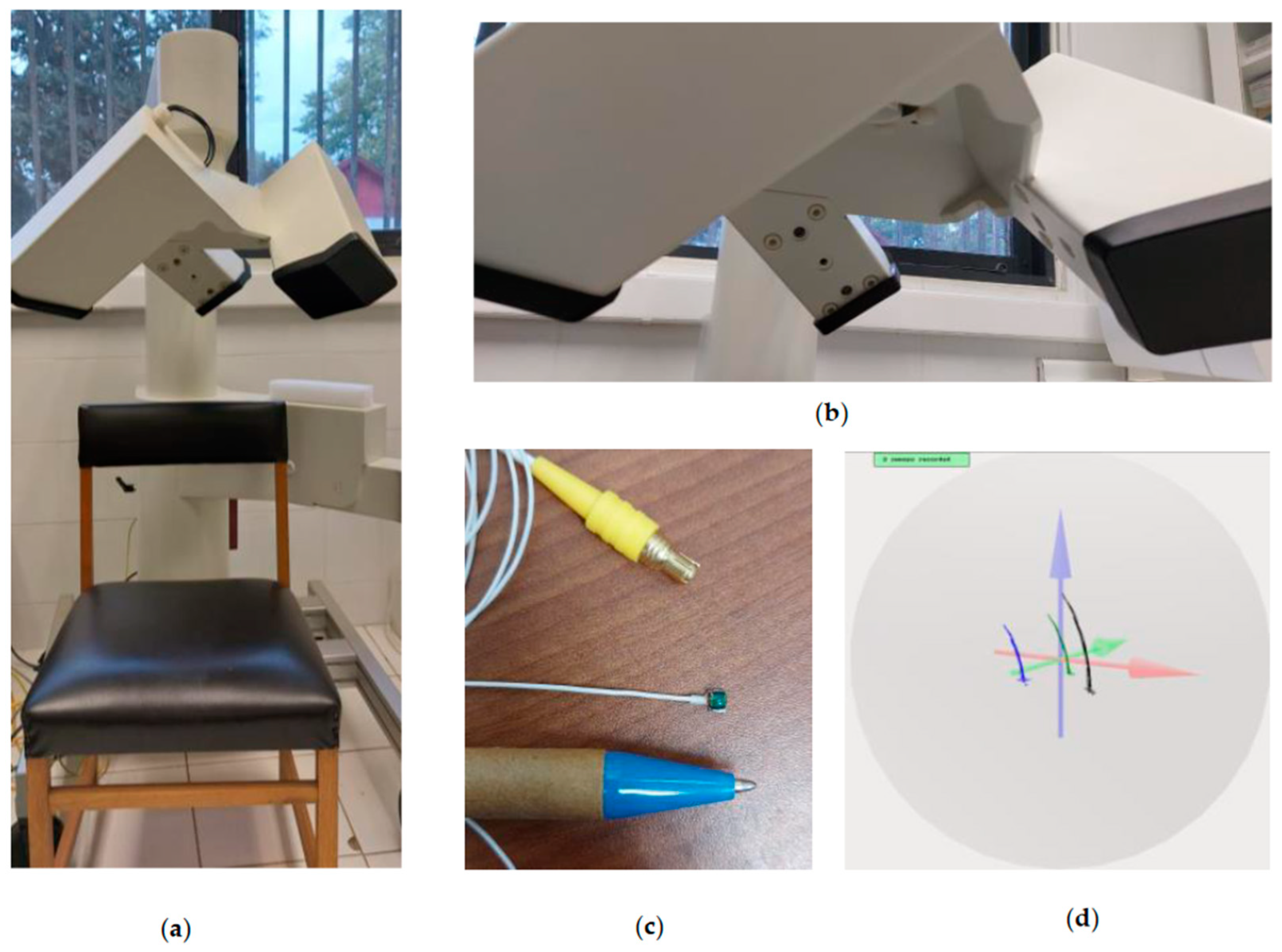


