
A Monoblock Light-Scattering Milk Fat Percentage and Somatic Cell Count Sensor for Use in Milking Systems

 , ,
, , 
Abstract
:1. Introduction
2. Sensor Design
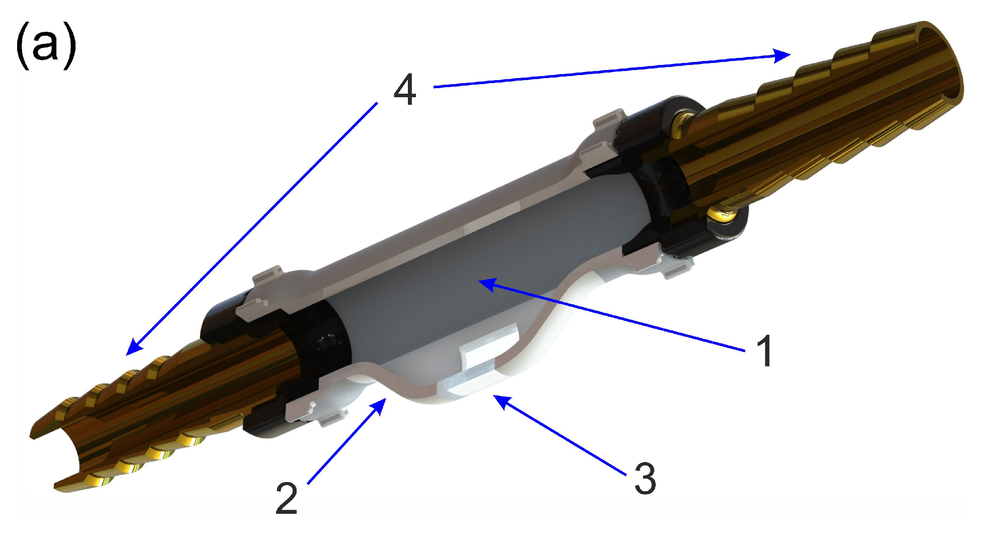
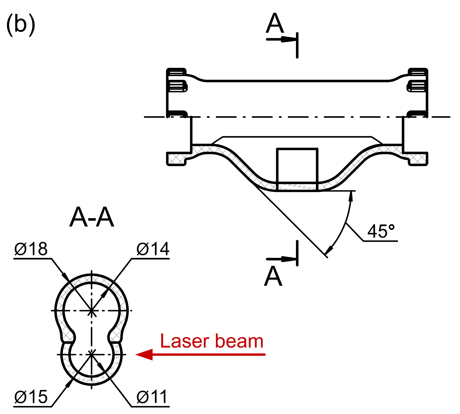
2.1. Device Layout
2.2. Principle of Operation
3. Materials and Milking Equipment
3.1. Milk Samples and Somatic Cells
3.2. Milking Equipment
4. Results
4.1. Fat Percentage Measurement
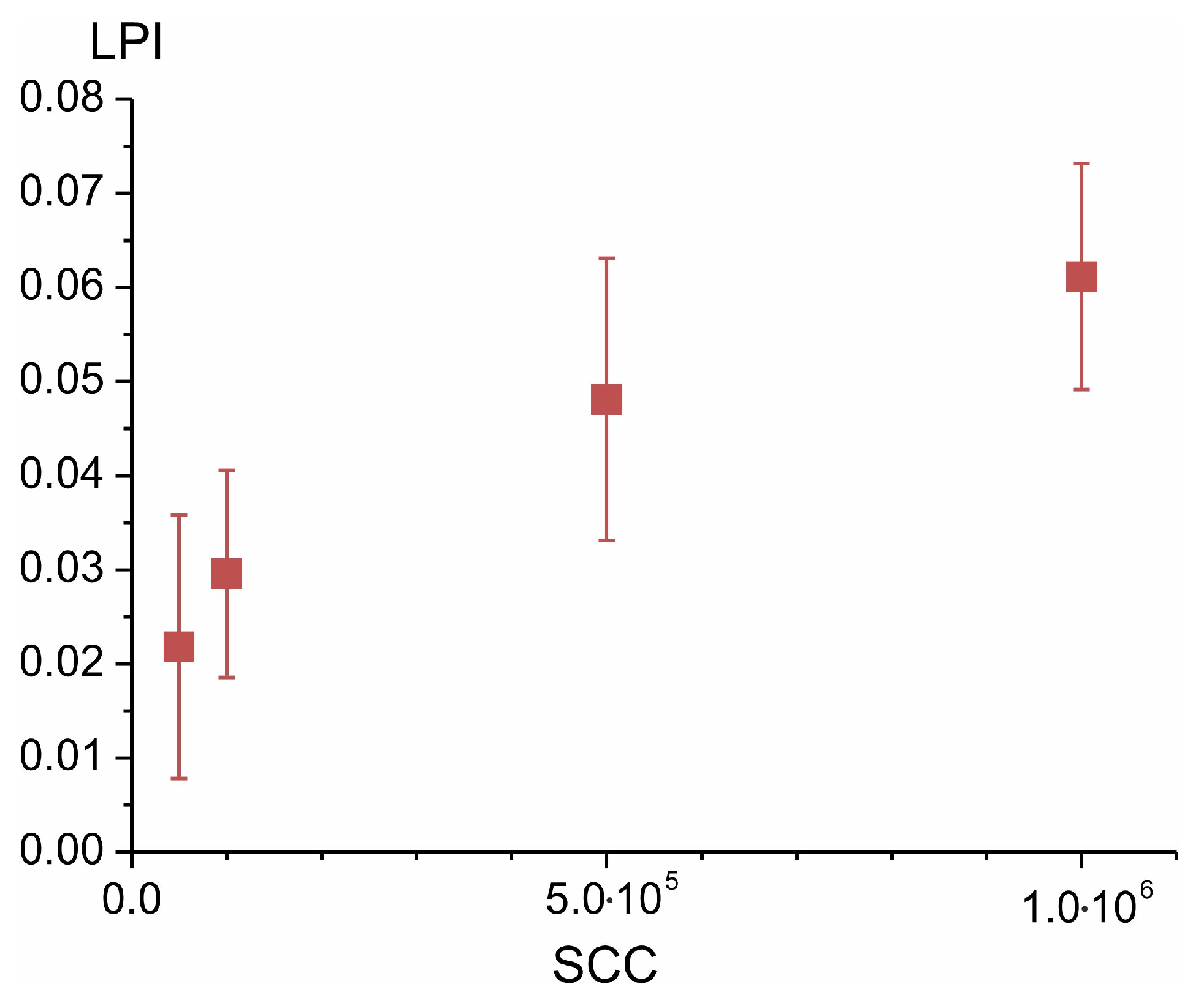
4.2. SCC Estimation
4.3. Test Measurements with the Sensor for Raw Milk: Uninfected versus Mastitis Case
5. Discussion
6. Conclusions
Author Contributions
Funding
Institutional Review Board Statement
Informed Consent Statement
Data Availability Statement
Conflicts of Interest
Appendix A. Theoretical Simulation of Light Scattering in Milk


References
- Truong, T.; Lopez, C.; Bhandari, B.; Prakash, S. (Eds.) Dairy Fat Products and Functionality; Springer Nature Switzerland AG: Cham, Switzerland, 2020. [Google Scholar] [CrossRef]
- Harding, F. (Ed.) Milk Quality; Springer: Boston, MA, USA, 1995. [Google Scholar] [CrossRef]
- Alsaftli, Z. The Obstacles to Using Milk Composition as Management Tool in Dairy Cattle Farms. J. Adv. Dairy. Res. 2020, 8, 233. [Google Scholar]
- El-Tahawy, A.S.; El-Far, A.H. Influences of somatic cell count on milk composition and dairy farm profitability. Int. J. Dairy Technol. 2020, 63, 463–469. [Google Scholar] [CrossRef]
- Cinar, M.; Serbester, U.; Ceyhan, A.; Gorgulu, M. Effect of Somatic Cell Count on Milk Yield and Composition of First and Second Lactation Dairy Cows. Ital. J. Anim. Sci. 2015, 14, 3646. [Google Scholar] [CrossRef]
- ICAR Certifications for Milk Meters for Cow Sheep Goats. Available online: https://www.icar.org/index.php/certifications/icar-certifications-for-milk-meters-for-cow-sheep-goats (accessed on 24 May 2016).
- ISO 5707:2007; Milking Machine Installations—Construction and Performance. ISO: Geneva, Switzerland, 2007. Available online: https://www.iso.org/standard/37190.html (accessed on 17 December 2015).
- Hu, Y.T.; Ting, Y.; Hu, J.Y.; Hsieh, S.C. Techniques and methods to study functional characteristics of emulsion systems. J. Food Drug Anal. 2017, 25, 16. [Google Scholar] [CrossRef]
- Palberg, T.; Ballauff, M. (Eds.) Optical Methods and Physics of Colloidal Dispersions. In Proceedings of the International Workshop on Optical Methods and the Physics of Colloidal Dispersions, Held in Memory of Prof. Dr. Klaus Schätzel, Mainz, Germany, 30 September 1996; Steinkopff: Heidelberg, Germany, 1997. [Google Scholar] [CrossRef]
- Chaikov, L.L.; Kovalenko, K.V.; Krivokhizha, S.V.; Kudryavtseva, A.D.; Tareeva, M.V.; Tcherniega, N.V.; Shevchenko, M.A. Structure of Water Microemulsion Particles: Study by Optical Methods. Phys. Wave Phen. 2019, 27, 87. [Google Scholar] [CrossRef]
- Michels, R.; Foschum, F.; Kienle, A. Optical properties of fat emulsions. Opt. Express 2008, 16, 5907. [Google Scholar] [CrossRef]
- Xu, R. Particle Characterization: Light Scattering Methods; Kluwer Academic Publishers: New York, NY, USA, 2002. [Google Scholar]
- Khosroshahi, M.E.; Patel, Y.; Woll-Morison, V. Non-destructive assessment of milk quality using pulsed UV photoacoustic, fluorescence and near FTIR spectroscopy. Laser Phys. Lett. 2022, 19, 075602. [Google Scholar] [CrossRef]
- Lu, R. (Ed.) Light Scattering Technology for Food Property, Quality and Safety Assessment; CRC Press: Boca Raton, FL, USA, 2016. [Google Scholar]
- van den Berg, F.; Lyndgaard, C.B.; Sørensen, K.M.; Engelsen, S.B. Process analytical technology in the food industry. Trends Food Sci. Technol. 2013, 31, 27–35. [Google Scholar] [CrossRef]
- Burmistrov, D.E.; Pavkin, D.Y.; Khakimov, A.R.; Ignatenko, D.N.; Nikitin, E.A.; Lednev, V.N.; Lobachevsky, Y.P.; Gudkov, S.V.; Zvyagin, A.V. Application of Optical Quality Control Technologies in the Dairy Industry: An Overview. Photonics 2021, 8, 551. [Google Scholar] [CrossRef]
- Uusitalo, S.; Diaz-Olivares, J.; Sumen, J.; Hietala, E.; Adriaens, I.; Saeys, W.; Utriainen, M.; Frondelius, L.; Pastell, M.; Aernouts, B. Evaluation of MEMS NIR Spectrometers for On-Farm Analysis of Raw Milk Composition. Foods 2021, 10, 2686. [Google Scholar] [CrossRef]
- Afilab Milk Analyzer. Available online: https://www.afimilk.com/parlor-automation/ (accessed on 6 June 2023).
- US Patent Application Publications. Spectroscopic Fluid Analyzer. US 2004/0179194A1, 29 March 2004. Current Assignee: Sae Afikim Milking Systems Agricultural Cooperative Ltd., Afimilk ACAL Agricultural Research Organization of Israel Ministry of Agriculture. Application Granted: 26 June 2007. Available online: https://patents.google.com/patent/US20040179194A1/en?assignee=afikim&oq=afikim (accessed on 26 June 2007).
- Kucheryavskiy, S.; Melenteva, A.; Bogomolov, A. Determination of fat and total protein content in milk using conventional digital imaging. Talanta 2014, 121, 144–152. [Google Scholar] [CrossRef]
- Jain, P.; Sarma, S.E. Light Scattering and Transmission Measurement Using Digital Imaging for Online Analysis of Constituents in Milk. In Proceedings of the SPIE 9525, Optical Measurement Systems for Industrial Inspection, Munich, Germany, 21 June 2015. [Google Scholar] [CrossRef]
- Katsumata, T.; Aizawa, H.; Komuro, S.; Ito, S.; Matsumoto, T. Quantitative analysis of fat and protein concentrations of milk based on fibre-optic evaluation of back scattering intensity. Int. Dairy J. 2020, 109, 104743. [Google Scholar] [CrossRef]
- Ohtani, S.; Wang, T.; Nishimura, K.; Irie, M. Milk Fat Analysis by Fiber-optic Spectroscopy. Asian Australas. J. Anim. Sci. 2005, 18, 580–583. [Google Scholar] [CrossRef]
- Angrasari, F.; Arifin, A.; Abdullah, B. Fabrication of Milk Fat Sensor based on Plastic Optical Fiber. J. Phys. Conf. Ser. 2019, 1341, 082038. [Google Scholar] [CrossRef]
- Shkirin, A.V.; Ignatenko, D.N.; Chirikov, S.N.; Vorobev, A.V.; Gudkov, S.V. Application of Laser Polarimetric Scatterometry in the Study of Water-Based Multicomponent Bioorganic Systems on the Example of Cow Milk. Phys. Wave Phen. 2022, 30, 186–195. [Google Scholar] [CrossRef]
- Widmer, J.; Descloux, L.; Brügger, C.; Jäger, M.L.; Berger, T.; Egger, L. Direct labeling of milk cells without centrifugation for counting total and differential somatic cells using flow cytometry. J. Dairy Sci. 2022, 105, 8705–8717. [Google Scholar] [CrossRef]
- Li, N.; Richoux, R.; Perruchot, M.H.; Boutinaud, M.; Mayol, J.F.; Gagnaire, V. Flow Cytometry Approach to Quantify the Viability of Milk Somatic Cell Counts after Various Physico-Chemical Treatments. PLoS ONE 2015, 10, e0146071. [Google Scholar] [CrossRef]
- Ramezani, M.; Ferrentino, G.; Morozova, K.; Scampicchio, M. Multiple Light Scattering Measurements for Online Monitoring of Milk Fermentation. Foods 2021, 10, 1582. [Google Scholar] [CrossRef] [PubMed]
- Hass, R.; Münzberg, M.; Bressel, L.; Reich, O. Industrial applications of Photon Density Wave spectroscopy for in-line particle sizing. Appl. Opt. 2013, 52, 1423–1431. [Google Scholar] [CrossRef]
- Photon Density Wave Spectroscopy. Available online: https://www.nanopat.eu/process-analytical-technologies/photon-density-wave-spectroscopy/ (accessed on 1 November 2020).
- Ruiz, S.V.; Hass, R.; Reich, O. Optical monitoring of milk fat phase transition within homogenized fresh milk by Photon Density Wave spectroscopy. Int. Dairy J. 2012, 26, 120–126. [Google Scholar] [CrossRef]
- Static Multiple Light Scattering (SMLS). Available online: https://formulaction.com/technologies/static-multiple-light-scattering-smls/ (accessed on 3 February 2023).
- Turbiscan LAB. Product Description. Available online: https://technex.nl/en/producten/turbiscan/ (accessed on 1 July 2022).
- Zhu, H.; Fu, H.; Yan, P.; Li, X.; Zhang, L.; Wang, X.; Chai, C. Study on the release of GMZ bentonite colloids by static multiple light scattering technique. Colloids Surf. A Physicochem. Eng. Asp. 2022, 640, 128374. [Google Scholar] [CrossRef]
- Shkirin, A.V.; Astashev, M.E.; Ignatenko, D.N.; Suyazov, N.V.; Vedunova, M.V.; Gudkov, S.V. Laser Scatterometric Device for Inline Measurement of Fat Percentage and the Concentration Level of Large-Scale Impurities in Milk. Appl. Sci. 2022, 12, 12517. [Google Scholar] [CrossRef]
- Nocedal, J.; Wright, S. Numerical Optimization; Springer Science+Business Media: New York, NY, USA, 2006. [Google Scholar] [CrossRef]
- Kirsanov, V.V.; Pavkin, D.Y.; Ruzin, S.S.; Tsymbal, A.A. Comparative technical and economic assessment of automated and robotized milking plants. Agric. Eng. 2020, 3, 39–43. [Google Scholar] [CrossRef]
- Kondratieva, O.; Fedorov, A.; Slinko, O.; Voytyuk, V. Improving the technological support of dairy cattle breeding. BIO Web Conf. 2021, 37, 00090. [Google Scholar] [CrossRef]
- Tuchin, V.V. Tissue Optics: Light Scattering Methods and Instruments for Medical Diagnostics, 3rd ed.; SPIE: Bellingham, WA, USA, 2015. [Google Scholar] [CrossRef]
- Muinonen, K.; Markkanen, J.; Väisänen, T.; Peltoniemi, J.; Penttilä, A. Multiple scattering of light in discrete random media using incoherent interactions. Opt. Lett. 2018, 43, 683–686. [Google Scholar] [CrossRef]
- Jönsson, J.; Berrocal, E. Multi-Scattering software: Part I: Online accelerated Monte Carlo simulation of light transport through scattering media. Opt. Express 2020, 28, 37612–37638. [Google Scholar] [CrossRef]
- Amador-Espejo, G.G.; Suàrez-Berencia, A.; Juan, B.; Bárcenas, M.E.; Trujillo, A.J. Effect of moderate inlet temperatures in ultra-high-pressure homogenization treatments on physicochemical and sensory characteristics of milk. J. Dairy Sci. 2014, 97, 659–671. [Google Scholar] [CrossRef]
- Mishchenko, M.I.; Travis, L.D.; Lacis, A.A. Scattering, Absorption, and Emission of Light by Small Particles; Cambridge University Press: Cambridge, UK, 2002. [Google Scholar]
- Maksimova, I.L.; Romanov, S.V.; Izotova, V.F. The effect of multiple scattering in disperse media on polarization characteristics of scattered light. Opt. Spectrosc. 2002, 92, 915–923. [Google Scholar] [CrossRef]


Disclaimer/Publisher’s Note: The statements, opinions and data contained in all publications are solely those of the individual author(s) and contributor(s) and not of MDPI and/or the editor(s). MDPI and/or the editor(s) disclaim responsibility for any injury to people or property resulting from any ideas, methods, instructions or products referred to in the content. |
© 2023 by the authors. Licensee MDPI, Basel, Switzerland. This article is an open access article distributed under the terms and conditions of the Creative Commons Attribution (CC BY) license (https://creativecommons.org/licenses/by/4.0/).
Share and Cite
Shkirin, A.V.; Astashev, M.E.; Ignatenko, D.N.; Suyazov, N.V.; Chirikov, S.N.; Kirsanov, V.V.; Pavkin, D.Y.; Lobachevsky, Y.P.; Gudkov, S.V. A Monoblock Light-Scattering Milk Fat Percentage and Somatic Cell Count Sensor for Use in Milking Systems. Sensors 2023, 23, 8618. https://doi.org/10.3390/s23208618
Shkirin AV, Astashev ME, Ignatenko DN, Suyazov NV, Chirikov SN, Kirsanov VV, Pavkin DY, Lobachevsky YP, Gudkov SV. A Monoblock Light-Scattering Milk Fat Percentage and Somatic Cell Count Sensor for Use in Milking Systems. Sensors. 2023; 23(20):8618. https://doi.org/10.3390/s23208618
Chicago/Turabian StyleShkirin, Alexey V., Maxim E. Astashev, Dmitry N. Ignatenko, Nikolai V. Suyazov, Sergey N. Chirikov, Vladimir V. Kirsanov, Dmitriy Y. Pavkin, Yakov P. Lobachevsky, and Sergey V. Gudkov. 2023. "A Monoblock Light-Scattering Milk Fat Percentage and Somatic Cell Count Sensor for Use in Milking Systems" Sensors 23, no. 20: 8618. https://doi.org/10.3390/s23208618