Review

Emerging Methods in Biosensing of Immunoglobin G—A Review

1
School of Chemical and Materials Engineering (SCME), National University of Sciences and Technology (NUST), Islamabad 44000, Pakistan
2
College of Computational Sciences and Natural Sciences, Minerva University, San Francisco, CA 94103, USA
*
Author to whom correspondence should be addressed.
Sensors 2023, 23(2), 676; https://doi.org/10.3390/s23020676
Submission received: 12 November 2022 / Revised: 30 December 2022 / Accepted: 1 January 2023 / Published: 6 January 2023
(This article belongs to the Special Issue Biosensors for Diagnostic Applications)

Abstract

:
Human antibodies are produced due to the activation of immune system components upon exposure to an external agent or antigen. Human antibody G, or immunoglobin G (IgG), accounts for 75% of total serum antibody content. IgG controls several infections by eradicating disease-causing pathogens from the body through complementary interactions with toxins. Additionally, IgG is an important diagnostic tool for certain pathological conditions, such as autoimmune hepatitis, hepatitis B virus (HBV), chickenpox and MMR (measles, mumps, and rubella), and coronavirus-induced disease 19 (COVID-19). As an important biomarker, IgG has sparked interest in conducting research to produce robust, sensitive, selective, and economical biosensors for its detection. To date, researchers have used different strategies and explored various materials from macro- to nanoscale to be used in IgG biosensing. In this review, emerging biosensors for IgG detection have been reviewed along with their detection limits, especially electrochemical biosensors that, when coupled with nanomaterials, can help to achieve the characteristics of a reliable IgG biosensor. Furthermore, this review can assist scientists in developing strategies for future research not only for IgG biosensors but also for the development of other biosensing systems for diverse targets.

Graphical Abstract

1. Introduction

1.1. Introduction to Immunoglobins and Immunoglobin G (IgG)

Antibody or immunoglobin (Ig) is a small protective protein produced by the human immune system upon the manifestation of foreign substances known as antigens and constitutes 20% of plasma protein. Immunoglobins eradicate new antigens in the body by recognizing and binding with them. There are multiple kinds of antigens: They could be viruses, bacteria, toxins, etc., and these antibodies instruct plasma cells to produce specific antibodies [1]. Antibodies were discovered due to the work of Emil von Behring’s discovery of antitoxins for diphtheria, tetanus, and anthrax [2]. Antibodies are further classified into five diverse types: Immunoglobin M (IgM), Immunoglobin D (IgD), Immunoglobin G (IgG), Immunoglobin A (IgA), and Immunoglobin E (IgE), as shown in Figure 1. These couple glycoproteins are composed of 82–96% protein and 4–18% carbohydrate and differ in heavy chain structure with varying effector functions [3].
IgG alone accounts for about 10–20% of plasma proteins and 70% to 75% of the total immunoglobulins in plasma, making it one of the primary antibodies and highest concentrations of proteins in human serum [4]. The subclasses of IgG can further be categorized on the basis of decreasing abundance of Immunoglobin G 1 (IgG1), Immunoglobin G 2 (IgG2), Immunoglobin G 3 (IgG3), and Immunoglobin G 4 (IgG4), which differ based on varying primary amino acid sequences and structural differences in the hinge and heavy chain constant regions [5,6,7]. The basic structure of an antibody contains two light chains (LCs) which are similar and two indistinguishable heavy chains (HCs) while making a Y-shaped compact structure composed of two heterodimers (linked together via a disulfide bridge). Human HCs for each subtype of an antibody differ from each other, and LCs have a constant region (CL) plus a variable region (VL) [8].
Figure 1. The diagram shows the isotypes, subclasses, and allotypes of immunoglobulins in humans with respective letters and numbers assigned to each type and subtypes of immunoglobulins. Adopted and used with permission from [9].
Sensors 23 00676 g001

1.1.1. The Basic Biology of IgG

The molecular mass of IgG is approximately 160 kDa, which happens to be due to the presence of light and heavy chains each being two in number [10,11]. The fragment antigen binding (Fab) region present inside the IgG structure contains a paratope and can help in pathogen inhibition through recognition events whereas the fragment crystallizable (Fc) region is in charge of the interaction with different accessory molecules to moderate indirect effector operations, including the complement–dependent cytotoxicity mechanism (CDC), antibody-dependent cellular cytotoxicity, and phagocytosis (ADCC and ADCP) [12,13].
Extensive research in the 20th century led to the discovery of IgG subclasses in the 1960s by using rabbit antisera specific to human IgG myeloma proteins [3,7]. Although there is a 90% similarity between IgG subclasses at an amino acid level, they differ with respect to their antigen binding event, half-life, formation of immune complex, placental transport, and effector cell triggering [14]. The detailed differences in the structure of IgG subtypes and allotypes have been shown in Figure 2a–d along with regions and various components of IgG (Figure 2e), and schematics for the structure of other immunoglobins (IgE, IgD, IgA, and IgM) have also been shown (Figure 2f).

1.1.2. Importance of IgG

The modern world has improved the resolution, throughput, and sensitivity of the instrumentation needed for analyzing glycoproteins. However, even with these advancements, precision in instrumentation still suffers [16]. IgG affects therapeutic antibodies, and the same effect is passed on to our immune systems, which play a part in protection against pathogens [16,17]. Serological tests are simpler to develop, more practical, and provide faster results [18]. Advancements in serological tests have marked immunoglobins, e.g., IgM, IgG, and IgA, etc., as important biomarker tools [19,20]. Moreover, our literature review identifies positive results with IgG and IgM tests either exclusively or combined [21]. Recent studies emphasized the importance of glycosylation in the therapeutic effects of IVIg [22,23]. Further evidence identifying anti-inflammatory properties has been shown in mouse models [24,25]. The importance of IgG is further characterized by improving immunity, treatment of pathological diseases and biomarkers, and its role in vaccines and biosensors for diagnostics.

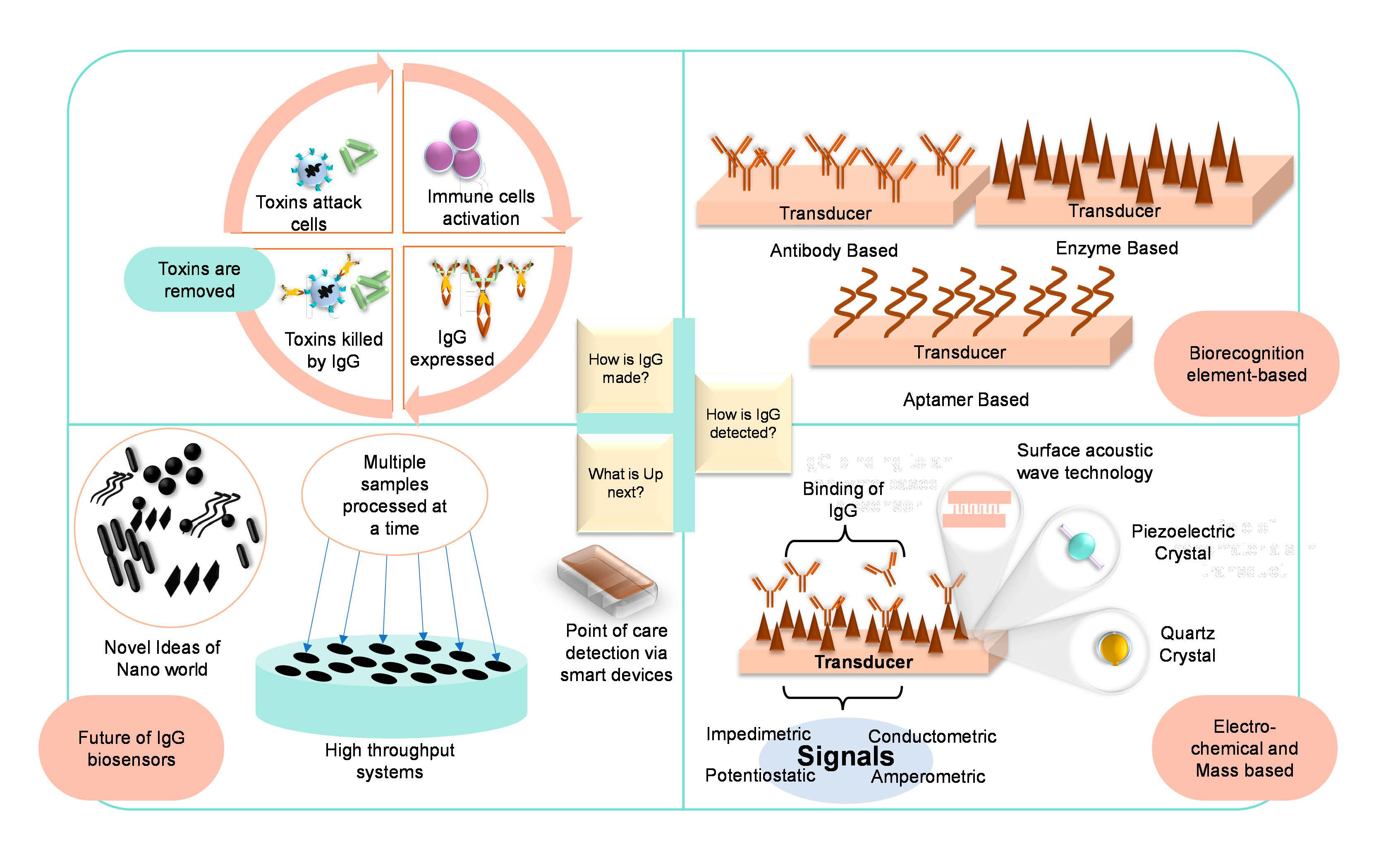
1.1.3. Role of IgG in Immunity

Based on the kind of molecule involved in providing the defense mechanism against foreign entities, human immunity has been categorized into two types, i.e., cell-mediated immunity and humoral immunity, where cell-mediated immunity involves the role of T cells while humoral immunity works because of antibodies [26,27]. IgG is an important class of antibodies that comprises 75% of all the immunoglobins present in the plasma and plays its function in humoral immunity [28]. The ability of IgG to bind with a diverse number of molecules comes from the primary amino acid sequences, hence diversifying the capability of IgG to tackle pathogens. The general defense mechanism of IgG against pathogens is a different mechanism, and three of them are depicted in Figure 3.

1.1.4. IgG as an Important Biomarker and Different Pathological Conditions

As evident from the literature, IgG is an important biomarker for multiple pathological conditions. The recent onset of SARS-CoV-2 once again shifted the focus onto IgG serum level studies, as it fluctuates upon the infection. Baoqing et al., have studied responses of IgG and IgM glycoproteins in COVID-19 patients and elevations in levels of N and S protein (of virus)-specific IgG and IgM after the onset of symptoms in non-ICU (nonintensive care unit) patients [29]. Because serological methods cannot be used in preliminary stages of the infection (up to the sixth day), they can make the diagnosis in the later stages (from the ninth day to the third month) of the disease. Therefore, IgG can be used to understand that people who do not show symptoms have had this disease rather than a COVID-19 diagnosis. Zhang et al., reported that the isotyping of immune response based on IgG response and NLR (neutrophil to lymphocyte ratio) can be a useful tool to differentiate COVID-19 patients on the foundation of disease severity [30]. Hongyan et al., stated that the quantification of IgG antibodies can be an important tool to evaluate the prognosis and seriousness of COVID-19 infection [31].
Apart from that, MOG-IgG has been confirmed to have a link with neuromyelitis optica (NMO), a central nervous system disorder [14]. Stefan et al., studied the correlation between serum IgG levels and efficiency in checkpoint inhibition and reported that IgG and its subclasses could serve as an important biomarker in metastatic melanoma patients to predict the success of checkpoint inhibition [2]. Dani et al., have reported the association of IgG serum levels with Shigella (a type of bacteria that is the leading cause of diarrhea and death related to this disease), and identification of the threshold level of these protective antibodies can help in the development of vaccines for children as well as infants. During infection, IgG is produced to enhance serotype-specific immunity, and levels of antibodies fluctuated in dose-response manners when different vaccines were tested under phase three studies and CHIM (controlled human infection model) clinical trials [32]. In addition to that, the presence of serum antibodies has also been reported as an important hallmark for neuromyelitis optica (NMO) [33]. So, it can be inferred that high or low levels of serum IgG are associated with a broad spectrum of diseases and can be used as an important biomarker that emphasizes the importance of IgG biosensing.

1.2. Introduction to Biosensors

Biosensors are generally referred to as small but sensitive devices that measure biomolecules (also known as an analyte) with the help of multiple output signals generated by various chemical or biological reactions [34]. The usage of biosensors has been in the healthcare sector, food industry, and environmental monitoring for a few decades back [35]. In the healthcare industry, biosensors are being used during both the diagnosis and treatment stages of any disease as well as in research labs [36]. The working principle of a biosensor is highlighted by the main components of a typical biosensor, and these components are as follows [37]:
  • Analyte or substrate: A biomolecule that needs to be recognized, i.e., IgG antibody, is an analyte that can be detected via various biosensors.
  • Receptor or biorecognition elements: These are the molecules that have an affinity to bind to the analyte. Enzymes, antibodies, antiantibodies, aptamers (DNA or RNA), and proteins are a few examples of receptors. The phenomenon of the affinity binding of an analyte to a receptor is known as biorecognition. As biorecognition elements are specific to their analyte and according to the need of the time, one more biorecognition element is used in a biosensor, and based on this, biosensors can be divided into different categories as listed in Figure 4 [38].
  • Transducer: Transducers help in the conversion of the biorecognition phenomenon into a measurable output signal, and this process is known as the transduction mechanism or signalization. Based on multiple transduction events happening in various biosensors, they have been classified as shown in Figure 5 [39].
  • Electronics: Electronics are the part of a biosensor that deals with the processing of the transduction mechanism and prepares it for display. Complex electronic circuits work here that help in the amplification of transduction signals from analog to digital form.
  • Display: Presenting the data in a readable form is referred to as the display, which is a combination of both hardware and software, and it displays output signals in accordance with the user’s need either in the form of graphs, numeric, images, or tables, and complete schematics of a biosensor component are shown in Figure 6.

2. Biosensors for the Detection of IgG

The effort to develop an IgG biosensor has been there for a few decades, and it was further highlighted due to the COVID-19 outbreak in 2019 because of the strong link between serum IgG titer and SARS-CoV-2 infection. Some of the biosensors are highlighted in Table 1.

2.1. Electrochemical Biosensors for IgG Detection

Electrochemical biosensing has an enormous impact on the biosensing of IgG, and a broad range of studies have been conducted to attain the unique, selective, sensitive, and economical characteristics of a biosensor. Liang et al., has reported a molecular imprinted electrochemical biosensor for selective and extremely sensitive detection of IgG [57]. The GCE (glassy carbon electrode) was modified using a MoS2@N-GQDs-IL (molybdenum disulfide @nitrogen-doped graphene quantum dots with ionic liquid) nanocomposite. Further testing was carried out by using cyclic voltammetry (CV), EIS (electrochemical impedance spectroscopy), and DPV (differential pulse voltammetry), and some of the results are compiled in Figure 7 [57].
The recent outbreak of COVID-19 had impacted scientists in a way to explore economic, rapid, sensitive, and selective methods for the detection of viral particles. During that point in time and up until now, nanomaterials have been explored to achieve qualities in a biosensor. Meanwhile, the direct detection of IgG antibodies to SARS-CoV-2 in blood using an incredibly accurate label-free nanosensor based on integrated graphene and Au nanostars was proposed by Seyyed et al. This platform was unique and gives a response for IgG produced against SARS-CoV-2 spike protein within a minute [58]. Likewise, PDA-AgNPs-PDA-Au (polydopamine silver nanoparticles polydopamine gold) film has been developed for the detection of horse IgG [59]. Other than that, a number of other electrochemical sensors have been proposed until today for the detection of IgG, as listed in the sections given below.

2.1.1. Potentiostatic Biosensors for IgG Detection

Potentiostatic biosensors are the type of electrochemical biosensor in which the potentiostat detects the voltage difference between the working electrode (WE) and reference electrode (RE) by adjusting the potential difference of the counter electrode with respect to the WE while the resultant potential difference is determined by the concentration of an analyte [60,61,62]. These sensors are an important type known as redox-potential biosensors because the redox reaction is the main principle governing these sensors. The response towards an analyte is determined by ion conduction and ion exchange reactions at the solution interface/membrane; hence, the measurement of the free ions via these biosensors requires calibration on regular basis [63]. A light addressable potentiometric sensor (LAPS) was proposed by Jintao et al., based on the covalently functionalized membrane for the detection of IgG. A linear relationship between the potential shift and the concentration of IgG was observed, hence reporting a detection range of 0–150 μg/mL [64].

2.1.2. Conductometric Biosensors for IgG Sensing

Conductometric sensors measure the conductivity difference occurring due to a biological event in an analyte solution [65]. It can detect both electroactive and electroactive species, and the conductivity electrodes can be in direct contact with the solution or insulated by a thin layer [66]. Conductometric sensors have been widely used, particularly as gas sensors and for evaluating engine oil quality. However, their use in microbial detection in food has been restricted [67]. Conductometric biosensors are useful because they work at a low working voltage, do not require the use of a reference electrode, perform quick analysis, have a reasonably high sensitivity and precision for detection, and can be manufactured using low-cost technology [68,69,70].
The conducting polymer, which works as an electrochemical transducer to transform biological impulses into electrical signals, is an essential component of a conductometric biosensor. A few examples of conducting polymers are polyaniline, polythiophene, polyacetylene, and polypyrrole (PPy) [71,72]. As explained in the following literature review, these conducting polymers have distinct and extensive uses in the medical field. Polyaniline may be used to detect cholesterol in blood via an enzymatic approach, and nonenzymatic detection of IgG in humans is viable by utilizing polyaniline gold nanospheres as nanolabels [73,74]. Noninvasive usage of a PPy layer on the gold surfaces of disposable screen-printed electrodes (SPE) utilizing 1H-pyrrole-1-propanoic acid revealed new applications for conductometric biosensors [75]. Despite the fact that this type of biosensing is noninvasive and research is being conducted on nonenzymatic forms of detection, such as cholesterol detection, the current market uses an enzymatic form of detection, which has some drawbacks in the form of fluctuations in conductivity due to enzymatic reactions and the conductivity of the electrolyte solution [76]. Okafor et al., proposed a conductometric biosensor based on polyaniline (PANI) for the detection of Johne’s disease-specified IgG antibodies [77]. This sensor was further compared with the detection of IgG with ELISA, and this study confirmed the moderate agreement between both techniques [78]. A single PANI wire was used to propose yet another conductometric biosensor for the detection of IgG and myoglobin with an LOD of 3ng/mL for IgG [79].
Nonetheless, the conductometric technique gives remarkable results in IgG detection, particularly when the technology is repurposed from contaminants in the environment to pollutants in the human body in the future.

2.1.3. Impedimetric Biosensors for IgG Detection

Impedimetric biosensors are electrochemical sensors that measure changes in capacitance and conductance on the surface of an electrode [80]. Based on the type of biorecognition element, impedimetric biosensors can be categorized into cell-based impedimetric biosensors, immunobinding-based, nucleic acid-based, and enzyme-based biosensors [81]. They have been used to measure multiple biological molecules because of the unique properties of each type of impedimetric biosensor, i.e., sensitivity, selectivity, and reproducibility [82]. As mentioned above the output signal for this type of biosensor is the impedance of conductance, and these signals are always proportional to the concentration and activity of the analyte [83] as studied by Zou et al. They have explored the role of a nanointerdigitated electrode array (nIDA) coupled with a microfluidic system integrated onto a polymer substrate which can be used for impedimetric biosensing of protein, cells, and genes [84].
During the early 2000s, it was reported that impedimetric biosensors are less frequent. Still, studies are reported to use these sensors for sensitive sensing, as the hybridization of DNA was monitored via this category of sensors. In addition, a very low concentration of about 10 pg/mL of antibodies was detected via an impedimetric biosensor that used polypyrrol film [85]. Furthermore, multiple studies have reported impedimetric sensors for IgG detection specifically. In this regard, a dengue virus IgG impedimetric biosensor based on a screen-printed glassy carbon electrode has shown satisfactory performance up to a minimum concentration of 2.81 ng/mL of viral IgG antibodies. The general schematics of this sensor have been shown in Figure 8 along with the graph of signals generated for the biosensor [86].
Similarly, Schrattenecker et al., have introduced the generation of a redox pair Hexaamineruthenium (II)(III) during impedimetric studies via a gold electrode, and the detection range of IgG for this specific sensor was 11.3 ng/mL to 113 μg/mL. The use of an alternative redox couple for an impedimetric biosensor with high stability was proposed for a stable IgG biosensor with redox-probe Hexaammineruthenium (II)/(III) [87]. Protein G has a binding affinity with IgG, so this interaction of both proteins was used to develop a highly sensitive biosensor with a limit of detection up to 1 × 10–14 mol/L [41]. Moreover, measles-specific primary IgG antibodies have been reported to be sensed via an impedimetric immunosensor with the help of a label-free approach [42]. Another label-free impedimetric biosensor was proposed by Honglan et al., with an LOD of 5 ng·mL−1 and a linear range of 10 ng·mL−1 to 1.0 μg·mL−1 [88]. Bhasin et at. proposed a simple dipping and reading model for the detection of proteins. “O2VBR” increases signal amplitudes by irreversibly oxidizing the VBR (virus bioresistor) channel for the detection of DJ-1 and two different IgG antibodies. IgG antiglycan antibody detection was made possible using impedimetric biosensors on a GPI phosphoglycan bioreceptor with an LOD of 0.31 IU mL−1 [89].

2.1.4. Amperometric Biosensors for IgG Detection

Similar to other types of electrochemical biosensors, amperometric biosensors generate the signals after an electrochemical event, but their output signals are in the form of either current or potential. The change in current or voltage fluctuates based on the analyte concentration, as studied by Malhotra et al.; the amperometric biosensor’s working electrode is typically made of either a noble metal (gold, titanium, nickel, etc.), ITO (indium tin oxide), or coating of carbon with bioreceptor elements. Upon applying the potential, the current is produced via enzymatic conversion or absorption of proteins at the surface of an electrode [90].
The approach of amperometric sensing for IgG is an old one, and most work has been performed since the 2000s. Dutra et al., described the importance of epoxy graphite biocomposite consisting of silver and TCNQ (tetracyanoquidimethane). This material has enhanced the selectivity of IgG sensing by reducing the potential down to 0.28 V [91].
The detection of rabbit IgG as a model analyte has been developed utilizing a separation-free homogenous immunosensor that is disposable and mediator-free and is based on a conducting polymer-covered, screen-printed carbon electrode. This sensor’s foundation was an analysis of the accessible antibody binding sites using free and tagged antigens in a competitive experiment. The catalytic current was measured amperometrically at/0.35 V vs. Ag/AgCl and revealed a linear range of RIgG concentrations ranging from 0.5 to 2 mg/mL with a standard deviation of 9/0.0145. The detection threshold was found to be 0.33 mg/mL [92]. Based on improved stiff biocomposites, highly sensitive electrochemical amperometric immunosensors for IgG detection have also been reported [44]. IgG regulation is not only important for the human body but also plays a significant role in monitoring FPT (failure of passive transfer) in calves. Hence, Robinson et al., discussed the creation of a label-free impedimetric immunosensor for bovine IgG in serum and showed how it might be used to estimate the FPT in newborn calves. The designed sensors successfully distinguished between newborn calf sera both pre- and postcolostrum feeding and showed quick and accurate IgG detection. Such technology might make it possible to determine FPT quickly, enhancing the vigor and survival rate of calves [93].

2.2. Mass-Based Biosensors

2.2.1. Magnetoelectric Biosensors

Magnetoelectric biosensors work on the principle of the magnetoelectric effect which refers to the fluctuation in a material’s electric polarization due to the applied magnetic field (direct effect) or change in the magnetic field under the influence of an electric field (converse effect). The magnetoelectric effect is prominent in materials that exhibit ferromagnetic and ferroelectric properties simultaneously, known as multiferroic materials. The following effect is also prominent if a material acquires electric polarization under the action of a magnetic field [94].
The ME effect was first observed experimentally in the single-phase multiferroic material Cr2O3 in 1961. Numerous studies were then conducted all over the world to improve the coupling capability of ferroelectric and magnetic orderings in a single-phase material system [95]. Combining magnetoelectric detection methods with other techniques helps in the extremely sensitive detection of biomolecules. As reported by Mulvaney et al., combined chip-based magnetoelectric biosensing with FFD (fluidic force discrimination) gave high sensitivity to the detection of biomolecules, including IgG, in plasma up to femtomolar concentrations with an incubation time of only 5 min for a single protein [96].

2.2.2. Piezoelectric Biosensors

Piezoelectricity, also known as the piezoelectric effect, is a physical phenomenon that describes a material’s ability to produce voltage when mechanically stressed. The effect is also effective in the opposite situation. When a voltage is applied to the surface of a piezoelectric material, it causes mechanical stress or oscillation. Anisotropic crystals, or crystals with no center of symmetry, are typical piezoelectric materials [97]. In comparison to methods such as surface plasmon resonance, piezoelectric biosensors have low manufacturing costs and simple assay mechanisms. These biosensors might attract users because of their ability to detect macromolecules in diagnosis, and further improvements can increase or enhance their application [98]. A piezoelectric platform was developed for the attachment of IgGNS1 to detect the nonstructural protein of dengue fever. This system easily detected NS1 protein levels only up to 10-fold dilutions [99,100].

2.2.3. Quartz Crystal Microbalance (QCM)

The quartz crystal microbalance (QCM) refers to a biosensing platform with a mechanical transduction part and operates under the rule of mass detection. Recent years have been benchmark years for this platform because of its unique ability to virtually detect any biomolecule in a label-free mode [101]. These biosensors are rapid and sensitive for diagnostic purposes [102].
Using an external electric field to apply to quartz results in mechanical stresses in the crystal according to the piezoelectric effect, which is the basis for QCM. The crystal oscillates in a direction perpendicular to the plate surface when an alternating voltage is applied to it [102,103,104]. Numerous inherent advantages exist for disease biosensing with the QCM mass-based detection principle. Since mass is a fundamental property of all matter, QCM systems can detect almost any kind of molecule, making them an ideal platform for identifying the various kinds of disease biomarkers. The frequency response instantly reflects molecular recognition events at the crystal surface without the need for labeling procedures, leading to a quick detection time that is typically between 30 min and 1 h [102].
According to the literature, QCM has been used to improve the binding of protein A and IgG molecules, SAM molecules and protein A, and other compositions for better disease biosensing, e.g., through the coating of the gold surface of the quartz crystal with a 35 nm polystyrene film and then treating it with an acid [105,106]. Similar to the combined use of aptamers and antibodies mentioned in the Biosensors Based on Biorecognition Element section, QCM can utilize enzyme-based methodology coupled with antibodies for improving the sensitivity of the amplification system by using anti-IgG–horseradish peroxidase enzyme conjugate [107]. In this case, although the detection of IgG is limited, it opens doors to show IgG detection could be the next step with simple modifications and appropriate coupling of the biorecognition element, hence proving that QCM has the potential for IgG detection. Kata et al., designed, fabricated, and characterized QCM biosensors based on a multichannel monolithic platform. Anti-IgG antibodies were immobilized to target IgG detection. This platform was reported to be label-free with real-time detection [108]. In another study, QCM was coupled with protein A, and the determined LOD was 5.07 pg/mL (standard deviation of 0.18pg/mL) with a linear range of 5.0 pg/mL–20.0 ng/mL [109]. Dai et al., reported that, with the addition of hexacyanoferrate and iron(III) ions at pH 4, the growth of “snow ball”-like objects greatly enhanced the response of a QCM biosensor, providing an LOD of 2.4 nM, which was facilitated by a surface-captured analyte (IgG from urine) [110].

2.2.4. Surface Acoustic Waves (SAW)

The manipulation of surface acoustic waves results in yet another type of mass-based biosensing (SAWs). SAWs are elastic waves that spread across the surface of a solid using piezoelectric crystals. They are similar to ocean waves in that they are guided by an electromechanical coupling, and SAW devices are widely used in communication and sensing [111,112]. They can record remarkably small frequency shifts brought on by incredibly small mass loadings because their frequency ranges from several hundred MHz to GHz [113].
SAWs have been utilized in biosensors due to their ability to miniaturize designs with high thermal stability, high sensitivity, quick response times, low cost, ease of fabrication, and wire-free integration [113,114,115]. Due to their special qualities, they are widely regarded as smart transducers that can be combined with a wide range of recognition layers, including functional polymers, carbon nanotubes, graphene sheets, metal oxide coatings, and biological receptors [115,116,117]. SAW sensors can therefore detect a wide variety of sensing targets, including small gas molecules, large bioanalytes, and even entire cell structures [113].
With developments in biosensing technology, SAW has been developed with precise control for promising utility in biosensors. For instance, the shear-horizontal surface acoustic wave (SH-SAW) biosensor is an inventive pathogen detection platform for detecting the anti-SARS-CoV-2 nucleocapsid antibody (IgG-based) [118]. Although there are still cases of poor sensitivity and an inability to detect small molecules, Yao et al., developed a method for quantifying Escherichia coli L-asparaginase by immobilizing amide-coupled polyclonal antibodies, which significantly improved sensitivity [119,120]. Although compared to other biosensing techniques, SAWs have certain drawbacks, as they stand to be a relatively modern method of biosensing; nonetheless, they stand their ground with new advancements [111].

2.3. Biosensors Based on Biorecognition Element

Biosensors are also categorized based on the biorecognition element used to modify the transducer of biosensors. Some of these sensors are compiled in Table 2 while the rest of the explanation has been provided in upcoming sections of the paper.

2.3.1. Antibodies-Based IgG Detection

An innovative type of antiviral intervention is through monoclonal antibodies (mAbs) that can attach to and “neutralize” a virus in infected people [131]. Neutralizing mAbs are recombinant proteins generated from convalescent patients or humanized-mice B cells. High-throughput screening of these B cells enables the discovery of antibodies with the specificity and affinity to attach to a virus and inhibit viral entrance, hence eliminating disease associated with productive infection. These mAbs are referred to as “neutralizing”. They can eventually be utilized as passive immunotherapy (described later) to reduce virulence [131,132].
These antibodies are versatile and are employed as a biorecognition material in biosensors to create antibody-based biosensors or immunosensors, which employ a transducer to convert the antibody–antigen binding event to a quantifiable physical signal [133]. Immunosensors have transformed diagnostics by detecting a variety of analytes, including disease indicators, dietary and environmental toxins, biological warfare agents, and illegal substances [134]. Immunosensors are classified into two types: nonlabelled immunosensors and labeled immunosensors. Non-labeled immunosensors are constructed so that the antigen–antibody complex may be identified immediately by detecting the physical changes caused by the complex’s creation. A tagged immunosensor, on the other hand, incorporates a sensitively detectable label. The immunocomplex is thus determined sensitively using label measurement [135]. Immunosensors are frequently employed in clinical diagnostics to detect or quantify disease-related chemicals, such as carcinogenic molecules, proteins, LDLs, food pathogens, bacteria, and viruses, because of their higher affinity of the antibody–antigen complex with high selectivity [90,136]. Recent advances in immunosensors have resulted in remarkable ideas, such as immunosensors for Chagas’ disease antibodies and detecting lymphocytes and the total content of immunoglobulin [135,137].
An IgG–IgM immunochromatographic assay was developed for the detection of IgG and IgM against SARS-CoV-2 while using antihuman antibodies as biorecognition components. This simultaneous and rapid detection method was able to give a response within 15 min with sensitivity and specificity of 85.29% and 100.00% [138]. Piotr et al., demonstrated the detection of goat IgG by using biotin–avidin–biotin interactions and antibody anti-goat IgG interactions [139]. Wang et al., studied the importance of the Mach–Zehnder interferometer (MZI) for the detection of IgG. A low detection limit interferometric optical fiber biosensor for IgG/anti-IgG immunosensing was proposed [47].

2.3.2. Aptamer-Based IgG Detection

Genosensors, also known as DNA/RNA sensors, are small analytical instruments that employ genetic material analysis to identify pathogens, such as bacteria and viruses, illnesses, and/or propensity to diseases [140]. Immobilized DNA/RNA probes are used as a recognition element in nanosensors, allowing particular hybridization reactions to occur, often via DNA–DNA or DNA–RNA molecular recognition [140]. Biosensors are not limited to antibodies as biorecognition elements but may also utilize other elements such as DNA or RNA (Aptamers) with the same transduction capabilities [136]. Nucleic acids, deoxyribonucleic acid (DNA), and ribonucleic acid (RNA) convey genetic information that is read in cells to produce RNA and proteins for normal human body function [141].
Aptamers are single-stranded oligonucleotides that fold into certain structures and bind to specific targets, such as proteins. They frequently block protein–protein interactions in binding proteins, which may result in therapeutic effects, such as antagonism [141]. As extremely specific strands have been observed against a wide range of biomarkers, nucleic acid aptamers offer significant potential as diagnostic reagents. They provide enticing advantages, such as repeatable creation by chemical synthesis, regulated modification with labels and functions that provide varied ways for detection and directed immobilization, and strong biochemical and thermal resistance. Aptamers against immunoglobulin targets (IgA, IgM, IgG, and IgE) have a distinct diagnostic domain; hence, multiple aptamers have been identified and employed in conjunction with diverse detection modalities [142]. According to another study, immunosensors use DNA and RNA as biorecognition elements by simultaneously detecting two tumor markers, carcinoembryonic antigen (CEA) and alpha-fetoprotein (AFP) [137]. It is also useful in the identification of autoimmune disorders with IgG by detecting early molecular processes that were not discovered by previous techniques, such as physician assessment [143].

2.3.3. Enzyme-Based IgG Detection

Enzymes are biological catalysts (also known as biocatalysts) that help living organisms accelerate metabolic processes [144]. Enzymes are catalysts composed of a lengthy chain of amino acids linked together by a peptide bond, commonly known as protein. These are utilized to start any reaction by adhering to a specified spot on the substrate. Enzymes are specific in their function, and their activity is affected by substrate concentration as well as other physical factors, such as temperature and pH [145]
Enzymes are an important biorecognition material for biosensors [146]. Enzyme-based biosensors measure alterations in proton concentration (H+), gas release or uptake (e.g., CO2, NH3, O2, etc.), light emission, absorption or reflectance, heat emission, and other factors that happen during substrate consumption or product formation of enzymatic reactions to detect the presence of analytes. The transducer then converts these variations into quantifiable signals (electrical, optical, or thermal signals) that can be used to identify potentially interesting analytes [147]. However, there are many techniques for using enzymes in biosensors: the enzyme converting the analyte into a sensor-detectable product; detection of an enzyme blocked or activated via the analyte; and monitoring enzyme property alteration [148]. Recent research demonstrates how enzyme-based biosensors have been successfully applied in IgG detection. For example, given the quick catalytic reaction and redox cycling, the detection limit for mouse IgG and prostate-specific antigen was reduced to 1 fg/mL [149]. According to the literature, using hydrazine, EC redox cycling was applied in an ALP-based biosensor to reduce p-quinone imine [129]. Finally, the pandemic increased the inclusion of enzymatic biosensors in the industry as SARS-CoV-2 uses enzyme-linked immunosorbent assay (ELISA) to detect IgG levels in the blood to determine the type of SARS-CoV-2 [150].

2.3.4. Biomimetic IgG Detection

Biomimicry is an applied science that draws inspiration for human issue solutions from the study of natural designs, processes, and systems [151,152]. Fluid-drag reduction swimsuits inspired by the structure of shark skin, airplane shapes inspired by the appearance of birds, low-drag wind turbines inspired by humpback whales, and sturdy building constructions inspired by the backbone of turban shells are some examples of biomimetic investigations [153]. This method of seeking inspiration from nature for human applications is also applied in biosensors.
Biomimetic biosensors use artificial receptors, also known as biocompatible materials, that are not sourced from biological creatures but have compositions and qualities that are comparable to those made by living species [154]. They are utilized as sensors in tissue reactions, bone cement in bone-bonding systems, joint replacements, skin restoration devices, and drug delivery, to highlight a few applications [154]. Successful biomaterials either: (1) restore a natural function where the original material is missing or unable to perform as intended or (2) maintain an environment that is most suited for procedures such as cell culture, tissue growth, biomolecular assays, and biotechnology-based manufacturing [155]. An example of biomimetic biosensors is the use of heterogeneous catalysis of imine hydrogenation [156]. A similar device was used to quantify a carcinoembryonic antigen (CEA) and -fetoprotein using antibodies that had been marked with gold nanoparticles (AFP) [157]. A highly sensitive glycoprotein sensor based on molecularly imprinted polymers (MIP) that were electropolymerized in the presence of template molecules (bovine serum albumin) is another application (BSA) [158].
Dianping et al., proposed a biomimetic interface based on glucose oxidase for the detection of IgG. The biomimetic interface was fabricated onto a gold electrode by its modification with polydendrime, Au/SiO2 nanocomposite, Fc-anti-IgG antibodies, and GOx (glucose oxidase) as a backfiller. Electrochemical testing as well as human sample testing were performed to check the reproducibility, sensitivity, and selectivity of an IgG sensor with a reported detection range from 3.5 × 10⁻⁵ to 1.2 × 10⁻³ mol/L [130]. These applications of biorecognition elements show immense potential in improving the biomedical industry and improving the cost and accessibility of diagnostics in the future via interdisciplinary biorecognition element utility.

3. Conclusions and Future Perspective

The presented review emphasizes the significance of various biosensors for the detection of IgG antibodies and their working principles, as IgG plays a crucial role in different pathological conditions and its use as a biomarker tool for diagnostic applications. Here, a comparison has been made between the use of biosensors with respect to the transduction properties in mass-based biosensors (magnetoelectric, piezoelectric, quartz-crystal microbalance, and surface acoustic waves) and biorecognition elements (antibodies, aptamers, enzymes, and biomimetic materials). The initial discussion was based on the classification of biosensors while highlighting the advantages and drawbacks of electrochemical biosensors (potentiostatic, conductometric, impedimetric, and amperometric) with an in-depth literature review of how biosensors have evolved over the decade. Biosensors, especially electrochemical biosensors, have seen major developments over the years with their initial use as glucose sensors for IgG detection in SARS-CoV-2. Although the sensors utilize similar principles, they differ in sensitivity, response rate, cost, and biochemical and thermal resistance using diverse biomaterials or a pair of biorecognition materials with a complimentary transducer. Biosensors offer applications to multiple industries for drug delivery, food safety monitoring, toxicology, medicine, and technology; however, with IgG biosensors, we have seen a popular demand in disease prevention for diagnosis of prostate cancer, tumor markers, autoimmune diseases, and COVID-19. Although IgG biosensors have many advantages, they do have certain disadvantages, such as detection limit, detection time, selectivity, and difficulty in maintaining a high throughput system. Many of these issues are controllable thanks to recent advances in nanomaterial production, such as carbon nanotubes, nanodiamonds, and metallic organic frameworks. Furthermore, the balance between specificity and versatility is problematic because single IgG molecules are used by an expensive biosensor to maintain specificity, and to make affordable biosensors, IgG detection with protein molecules must be expanded using adaptable machine learning models that are programmed iteratively, leading to a more efficient method of IgG detection. Finally, with more suitable nanomaterials and well-designed software, IgG detection could be possible in handheld devices that can be utilized for point-of-care detection in every home.

Author Contributions

Writing—original draft, methodology, software, validation, and formal analysis, T.A. and S.H.B.; writing—review and editing, U.L.; writing—review and editing, W.M.; project administration, W.M. All authors have read and agreed to the published version of the manuscript.

Funding

This research received no external funding.

Institutional Review Board Statement

Not applicable.

Informed Consent Statement

Not applicable.

Data Availability Statement

Not applicable.

Acknowledgments

This work was conducted at the School of Chemical and Materials Engineering (SCME), National University of Sciences and Technology, Sector H-12, Islamabad. Waheed Miran acknowledges the support from the SCME, NUST.

Conflicts of Interest

The authors declare no conflict of interest.

References

  1. Vaillant, A.A.J.; Jamal, Z.; Ramphul, K. Immunoglobulin. In StatPearls [Internet]; StatPearls Publishing: Tampa, FL, USA, 2021. [Google Scholar]
  2. Diem, S.; Fässler, M.; Bomze, D.; Ali, O.H.; Berner, F.; Niederer, R.; Hillmann, D.; Mangana, J.; Levesque, M.P.; Dummer, R.; et al. Immunoglobulin G and Subclasses as Potential Biomarkers in Metastatic Melanoma Patients Starting Checkpoint Inhibitor Treatment. J. Immunother. 2019, 42, 89–93. [Google Scholar] [CrossRef] [PubMed] [Green Version]
  3. Rispens, T.; Vidarsson, G. Human IgG Subclasses. In Antibody Fc; Elsevier: Amsterdam, The Netherlands, 2014; pp. 159–177. [Google Scholar]
  4. Dietzen, D.J. Amino acids, peptides, and proteins. In Principles and Applications of Molecular Diagnostics; Elsevier: Amsterdam, The Netherlands, 2018; pp. 345–380. [Google Scholar]
  5. Schur, P.H. IgG subclasses. A historical perspective. Monogr. Allergy 1988, 23, 1–11. [Google Scholar] [PubMed]
  6. Bournazos, S.; Gupta, A.; Ravetch, J.V. The role of IgG Fc receptors in antibody-dependent enhancement. Nat. Rev. Immunol. 2020, 20, 633–643. [Google Scholar] [CrossRef]
  7. Napodano, C.; Marino, M.; Stefanile, A.; Pocino, K.; Scatena, R.; Gulli, F.; Rapaccini, G.L.; Delli Noci, S.; Capozio, G.; Rigante, D.; et al. Immunological Role of IgG Subclasses. Immunol. Investig. 2021, 50, 427–444. [Google Scholar] [CrossRef] [PubMed]
  8. Chiu, M.L.; Goulet, D.R.; Teplyakov, A.; Gilliland, G.L. Antibody Structure and Function: The Basis for Engineering Therapeutics. Antibodies 2019, 8, 55. [Google Scholar] [CrossRef] [Green Version]
  9. Irani, V.; Guy, A.; Andrew, D.; Beeson, J.; Ramsland, P.; Richards, J. Molecular properties of human IgG subclasses and their implications for designing therapeutic monoclonal antibodies against infectious diseases. Mol. Immunol. 2015, 207, 171–182. [Google Scholar] [CrossRef]
  10. Palmeira, P.; Quinello, C.; Silveira-Lessa, A.L.; Zago, C.A.; Carneiro-Sampaio, M. IgG Placental Transfer in Healthy and Pathological Pregnancies. Clin. Dev. Immunol. 2012, 2012, 985646. [Google Scholar] [CrossRef]
  11. Liu, H.; May, K. Disulfide bond structures of IgG molecules. mAbs 2012, 4, 17–23. [Google Scholar] [CrossRef] [Green Version]
  12. Dübel, S. Therapeutic Antibodies—From Past to Future. In Handbook of Therapeutic Antibodies; Wiley: Hoboken, NJ, USA, 2007; pp. 2–16. [Google Scholar]
  13. Moldenhauer, G. Selection Strategies I: Monoclonal Antibodies. In Handbook of Therapeutic Antibodies; Wiley: Hoboken, NJ, USA, 2007; pp. 18–44. [Google Scholar]
  14. Vidarsson, G.; Dekkers, G.; Rispens, T. IgG subclasses and allotypes: From structure to effector functions. Front. Immunol. 2014, 5, 520. [Google Scholar] [CrossRef] [Green Version]
  15. Schroeder, H.W.; Cavacini, L. Structure and function of immunoglobulins. J. Allergy Clin. Immunol. 2010, 125, S41–S52. [Google Scholar] [CrossRef]
  16. Sjögren, J.; Lood, R.; Nägeli, A. On enzymatic remodeling of IgG glycosylation; unique tools with broad applications. Glycobiology 2019, 30, 254–267. [Google Scholar] [CrossRef] [PubMed]
  17. Sjögren, J.; Collin, M. Bacterial glycosidases in pathogenesis and glycoengineering. Future Microbiol. 2014, 9, 1039–1051. [Google Scholar] [CrossRef] [PubMed]
  18. Deeks, J.J.; Dinnes, J.; Takwoingi, Y.; Davenport, C.; Spijker, R.; Taylor-Phillips, S.; Adriano, A.; Beese, S.; Dretzke, J.; di Ruffano, L.F.; et al. Antibody tests for identification of current and past infection with SARS-CoV-2. Cochrane Database Syst. Rev. 2020, 2020, CD013652. [Google Scholar]
  19. Peeling, R.W.; Wedderburn, C.J.; Garcia, P.J.; Boeras, D.; Fongwen, N.; Nkengasong, J.; Sall, A.; Tanuri, A.; Heymann, D.L. Serology testing in the COVID-19 pandemic response. Lancet Infect. Dis. 2020, 20, e245–e249. [Google Scholar] [CrossRef] [PubMed]
  20. Chen, M.; Qin, R.; Jiang, M.; Yang, Z.; Wen, W.; Li, J. Clinical applications of detecting IgG, IgM or IgA antibody for the diagnosis of COVID-19: A meta-analysis and systematic review. Int. J. Infect. Dis. 2021, 104, 415–422. [Google Scholar] [CrossRef] [PubMed]
  21. Cheng, M.P.; Papenburg, J.; Desjardins, M.; Kanjilal, S.; Quach, C.; Libman, M.; Dittrich, S.; Yansouni, C.P. Diagnostic testing for severe acute respiratory syndrome–related coronavirus 2: A narrative review. Ann. Intern. Med. 2020, 172, 726–734. [Google Scholar] [CrossRef] [Green Version]
  22. van de Bovenkamp, F.S.; Hafkenscheid, L.; Rispens, T.; Rombouts, Y. The emerging importance of IgG Fab glycosylation in immunity. J. Immunol. 2016, 196, 1435–1441. [Google Scholar] [CrossRef] [PubMed] [Green Version]
  23. Schwab, I.; Nimmerjahn, F.J. Role of sialylation in the anti-inflammatory activity of intravenous immunoglobulin–F (ab′) 2 versus Fc sialylation. Clin. Exp. Immunol. 2014, 178 (Suppl. 1), 97. [Google Scholar] [CrossRef] [PubMed] [Green Version]
  24. Kaneko, Y.; Nimmerjahn, F.; Ravetch, J.V. Anti-inflammatory activity of immunoglobulin G resulting from Fc sialylation. Science 2006, 313, 670–673. [Google Scholar] [CrossRef] [PubMed] [Green Version]
  25. Schwab, I.; Mihai, S.; Seeling, M.; Kasperkiewicz, M.; Ludwig, R.J.; Nimmerjahn, F.J. Broad requirement for terminal sialic acid residues and FcγRIIB for the preventive and therapeutic activity of intravenous immunoglobulins in vivo. Eur. J. Immunol. 2014, 44, 1444–1453. [Google Scholar] [CrossRef] [PubMed]
  26. Janeway, C.A. Immunobiology 5: The Immune System in Health and Disease, 5th ed.; Janeway, C.A., Travers, J.P., Walport, M., Shlomchik, M.J., Eds.; Garland: Edinburgh, UK; Churchill Livingstone: New York, NY, USA, 2001. [Google Scholar]
  27. Annunziato, F.; Romagnani, C.; Romagnani, S. The 3 major types of innate and adaptive cell-mediated effector immunity. J. Allergy Clin. Immunol. 2015, 135, 626–635. [Google Scholar] [CrossRef] [PubMed]
  28. Leusen, J.H.W.; Nimmerjahn, F. The Role of IgG in Immune Responses. In Molecular and Cellular Mechanisms of Antibody Activity; Nimmerjahn, F., Ed.; Springer: New York, NY, USA, 2013; pp. 85–112. [Google Scholar]
  29. Sun, B.; Feng, Y.; Mo, X.; Zheng, P.; Wang, Q.; Li, P.; Peng, P.; Liu, X.; Chen, Z.; Huang, H.; et al. Kinetics of SARS-CoV-2 specific IgM and IgG responses in COVID-19 patients. Emerg. Microbes Infect. 2020, 9, 940–948. [Google Scholar] [CrossRef] [PubMed]
  30. Zhang, B.; Zhou, X.; Zhu, C.; Song, Y.; Feng, F.; Qiu, Y.; Feng, J.; Jia, Q.; Song, Q.; Zhu, B.; et al. Immune Phenotyping Based on the Neutrophil-to-Lymphocyte Ratio and IgG Level Predicts Disease Severity and Outcome for Patients With COVID-19. Front. Mol. Biosci. 2020, 7, 157. [Google Scholar] [CrossRef] [PubMed]
  31. Hou, H.; Wang, T.; Zhang, B.; Luo, Y.; Mao, L.; Wang, F.; Wu, S.; Sun, Z. Detection of IgM and IgG antibodies in patients with coronavirus disease 2019. Clin. Transl. Immunol. 2020, 9, e01136. [Google Scholar] [CrossRef]
  32. Cohen, D.; Meron-Sudai, S.; Bialik, A.; Asato, V.; Goren, S.; Ariel-Cohen, O.; Reizis, A.; Hochberg, A.; Ashkenazi, S. Serum IgG antibodies to Shigella lipopolysaccharide antigens—A correlate of protection against shigellosis. Hum. Vaccines Immunother. 2019, 15, 1401–1408. [Google Scholar] [CrossRef] [Green Version]
  33. Bradl, M.; Misu, T.; Takahashi, T.; Watanabe, M.; Mader, S.; Reindl, M.; Adzemovic, M.; Bauer, J.; Berger, T.; Fujihara, K.; et al. Neuromyelitis optica: Pathogenicity of patient immunoglobulin in vivo. Ann. Neurol. 2009, 66, 630–643. [Google Scholar] [CrossRef]
  34. González-Navarro, F.F.; Stoytcheva, M.; Renteria, L.; Belanche, L.; Flores Rios, B.; Ibarra Esquer, J. Glucose Oxidase Biosensor Modeling and Predictors Optimization by Machine Learning Methods. Sensors 2016, 16, 1483. [Google Scholar] [CrossRef] [Green Version]
  35. Mehrotra, P. Biosensors and their applications—A review. J. Oral Biol. Craniofacial Res. 2016, 6, 153–159. [Google Scholar] [CrossRef] [Green Version]
  36. Mohankumar, P.; Ajayan, J.; Mohanraj, T.; Yasodharan, R. Recent developments in biosensors for healthcare and biomedical applications: A review. Measurement 2021, 167, 108293. [Google Scholar] [CrossRef]
  37. Bhalla, N.; Jolly, P.; Formisano, N.; Estrela, P. Introduction to biosensors. Essays Biochem. 2016, 60, 1–8. [Google Scholar]
  38. Kirchhain, A.; Bonini, A.; Vivaldi, F.; Poma, N.; Di Francesco, F. Latest developments in non-faradic impedimetric biosensors: Towards clinical applications. TrAC Trends Anal. Chem. 2020, 133, 116073. [Google Scholar] [CrossRef]
  39. Vaidya, A.M.; Annapure, U.S. Chapter 38—Enzymes in Biosensors for Food Quality Assessment. In Enzymes in Food Biotechnology; Kuddus, M., Ed.; Academic Press: Cambridge, MA, USA, 2019; pp. 659–674. [Google Scholar]
  40. Chen, M.; Song, Z.; Han, R.; Li, Y.; Luo, X. Low fouling electrochemical biosensors based on designed Y-shaped peptides with antifouling and recognizing branches for the detection of IgG in human serum. Biosens. Bioelectron. 2021, 178, 113016. [Google Scholar] [CrossRef] [PubMed]
  41. Honda, H.; Kusaka, Y.; Wu, H.; Endo, H.; Tsuya, D.; Ohnuki, H. Toward a Practical Impedimetric Biosensor: A Micro-Gap Parallel Plate Electrode Structure That Suppresses Unexpected Device-to-Device Variations. ACS Omega 2022, 7, 11017–11022. [Google Scholar] [CrossRef] [PubMed]
  42. Mashazi, P.; Tetyana, P.; Vilakazi, S.; Nyokong, T. Electrochemical impedimetric immunosensor for the detection of measles-specific IgG antibodies after measles infections. Biosens. Bioelectron. 2013, 49, 32–38. [Google Scholar] [CrossRef] [PubMed]
  43. Dong, S.; Tong, M.; Zhang, D.; Huang, T. The strategy of nitrite and immunoassay human IgG biosensors based on ZnO@ZIF-8 and ionic liquid composite film. Sens. Actuators B Chem. 2017, 251, 650–657. [Google Scholar] [CrossRef]
  44. Montes, R.; Céspedes, F.; Baeza, M. Highly sensitive electrochemical immunosensor for IgG detection based on optimized rigid biocomposites. Biosens. Bioelectron. 2016, 78, 505–512. [Google Scholar] [CrossRef]
  45. Guo, Z.; Qin, Y.; Chen, P.; Hu, J.; Zhou, Y.; Zhao, X.; Liu, Z.; Fei, Y.; Jiang, X.; Wu, X.J. Hyperboloid-Drum Microdisk Laser Biosensors for Ultrasensitive Detection of Human IgG. Small 2020, 16, 2000239. [Google Scholar] [CrossRef]
  46. Melnik, E.; Bruck, R.; Müellner, P.; Schlederer, T.; Hainberger, R.; Lämmerhofer, M. Human IgG detection in serum on polymer based Mach-Zehnder interferometric biosensors. J. Biophotonics 2016, 9, 218–223. [Google Scholar] [CrossRef]
  47. Wang, B.-T.; Wang, Q. An interferometric optical fiber biosensor with high sensitivity for IgG/anti-IgG immunosensing. Opt. Commun. 2018, 426, 388–394. [Google Scholar] [CrossRef]
  48. Mendes, J.P.; Coelho, L.C.C.; Jorge, P.A.S.; Pereira, C.M. Differential Refractometric Biosensor for Reliable Human IgG Detection: Proof of Concept. Biosensors 2022, 12, 515. [Google Scholar] [CrossRef]
  49. Wang, Q.; Jing, J.-Y.; Wang, B.-T. Highly sensitive SPR biosensor based on graphene oxide and staphylococcal protein a co-modified TFBG for human IgG detection. IEEE Trans. Instrum. Meas. 2018, 68, 3350–3357. [Google Scholar] [CrossRef]
  50. Wang, Q.; Wang, B.-T. Surface plasmon resonance biosensor based on graphene oxide/silver coated polymer cladding silica fiber. Sens. Actuators B Chem. 2018, 275, 332–338. [Google Scholar] [CrossRef]
  51. Wang, Q.; Wang, X.-Z.; Song, H.; Zhao, W.-M.; Jing, J.-Y. A dual channel self-compensation optical fiber biosensor based on coupling of surface plasmon polariton. Opt. Laser Technol. 2020, 124, 106002. [Google Scholar] [CrossRef]
  52. Wang, B.T.; Wang, Q. Sensitivity-Enhanced Optical Fiber Biosensor Based on Coupling Effect between SPR and LSPR. IEEE Sens. J. 2018, 18, 8303–8310. [Google Scholar] [CrossRef]
  53. Hageneder, S.; Jungbluth, V.; Soldo, R.; Petri, C.; Pertiller, M.; Kreivi, M.; Weinhäusel, A.; Jonas, U.; Dostalek, J. Responsive Hydrogel Binding Matrix for Dual Signal Amplification in Fluorescence Affinity Biosensors and Peptide Microarrays. ACS Appl. Mater. Interfaces 2021, 13, 27645–27655. [Google Scholar] [CrossRef] [PubMed]
  54. Iwanaga, M. All-Dielectric Metasurface Fluorescence Biosensors for High-Sensitivity Antibody/Antigen Detection. ACS Nano 2020, 14, 17458–17467. [Google Scholar] [CrossRef] [PubMed]
  55. Zhang, H.; Kikuchi, N.; Ohshima, N.; Kajisa, T.; Sakata, T.; Izumi, T.; Sone, H. Design and Fabrication of Silicon Nanowire-Based Biosensors with Integration of Critical Factors: Toward Ultrasensitive Specific Detection of Biomolecules. ACS Appl. Mater. Interfaces 2020, 12, 51808–51819. [Google Scholar] [CrossRef]
  56. Zhou, L.; Kato, F.; Ogi, H. Sensitive label-free immunoglobulin G detection using a MEMS quartz crystal microbalance biosensor with a 125 MHz wireless quartz resonator. Jpn. J. Appl. Phys. 2021, 60, SDDB03. [Google Scholar] [CrossRef]
  57. Axin Liang, A.; Huipeng Hou, B.; Shanshan Tang, C.; Liquan Sun, D.; Aiqin Luo, E. An advanced molecularly imprinted electrochemical sensor for the highly sensitive and selective detection and determination of Human IgG. Bioelectrochemistry 2021, 137, 107671. [Google Scholar] [CrossRef]
  58. Alireza Hashemi, S.; Bahrani, S.; Mojtaba Mousavi, S.; Omidifar, N.; Ghaleh Golab Behbahan, N.; Arjmand, M.; Ramakrishna, S.; Bagheri Lankarani, K.; Moghadami, M.; Shokripour, M.; et al. Ultra-precise label-free nanosensor based on integrated graphene with Au nanostars toward direct detection of IgG antibodies of SARS-CoV-2 in blood. J. Electroanal. Chem. 2021, 894, 115341. [Google Scholar] [CrossRef]
  59. Wang, N.; Zhang, D.; Deng, X.; Sun, Y.; Wang, X.; Ma, P.; Song, D. A novel surface plasmon resonance biosensor based on the PDA-AgNPs-PDA-Au film sensing platform for horse IgG detection. Spectrochim. Acta Part A Mol. Biomol. Spectrosc. 2018, 191, 290–295. [Google Scholar] [CrossRef] [PubMed]
  60. Bembnowicz, P.; Yang, G.; Anastasova, S.; Spehar-Délèze, A.; Vadgama, P. Wearable electronic sensor for potentiometric and amperometric measurements. In Proceedings of the 2013 IEEE International Conference on Body Sensor Networks, Cambridge, MA, USA, 6–9 May 2013; pp. 1–5. [Google Scholar]
  61. Abdullah, S.; Tonello, S.; Borghetti, M.; Sardini, E.; Serpelloni, M. Potentiostats for Protein Biosensing: Design Considerations and Analysis on Measurement Characteristics. J. Sens. 2019, 2019, 6729329. [Google Scholar] [CrossRef]
  62. Yunus, S.; Jonas, A.M.; Lakard, B. Potentiometric Biosensors. In Encyclopedia of Biophysics; Roberts, G.C.K., Ed.; Springer: Berlin/Heidelberg, Germany, 2013; pp. 1941–1946. [Google Scholar]
  63. Colomer i Farrarons, J.; Miribel-Català, P.L.; Rodríguez-Villarreal, I.; Samitier i Martí, J.J. Portable bio-devices: Design of electrochemical instruments from miniaturized to implantable devices. In Chapter 18 in: Serra, Pier Andrea. 2011. New Perspectives in Biosensors Technology and Applications; IntechOpen: London, UK, 2011; pp. 373–400. [Google Scholar]
  64. Liang, J.; Guan, M.; Huang, G.; Qiu, H.; Chen, Z.; Li, G.; Huang, Y. Highly sensitive covalently functionalized light-addressable potentiometric sensor for determination of biomarker. Mater. Sci. Eng. C 2016, 63, 185–191. [Google Scholar] [CrossRef]
  65. Jaffrezic-Renault, N.; Dzyadevych, S.V. Conductometric Microbiosensors for Environmental Monitoring. Sensors 2008, 8, 2569–2588. [Google Scholar] [CrossRef] [Green Version]
  66. Narayan, R. Medical Biosensors for Point of Care (POC) Applications; Woodhead Publishing: Sawston, UK, 2016. [Google Scholar]
  67. Adley, C.C.; Ryan, M.P. 14—Conductometric biosensors for high throughput screening of pathogens in food. In High Throughput Screening for Food Safety Assessment; Bhunia, A.K., Kim, M.S., Taitt, C.R., Eds.; Woodhead Publishing: Sawston, UK, 2015; pp. 315–326. [Google Scholar]
  68. Odobašić, A.; Šestan, I.; Begić, S. Biosensors for Determination of Heavy Metals in Waters; IntechOpen: London, UK, 2019. [Google Scholar]
  69. Dzyadevych, S.; Jaffrezic-Renault, N. 6—Conductometric biosensors. In Biological Identification; Schaudies, R.P., Ed.; Woodhead Publishing: Sawston, UK, 2014; pp. 153–193. [Google Scholar]
  70. Kłos, A.; Wierzba, S. Application of conductometric and pH metric measurements in determining the kinetics and equilibrium parameters of the heterophasic ion exchange: Metal cation-proton. Electrochem. Commun. 2019, 102, 5–12. [Google Scholar] [CrossRef]
  71. Namsheer, K.; Rout, C.S. Conducting polymers: A comprehensive review on recent advances in synthesis, properties and applications. RSC Adv. 2021, 11, 5659–5697. [Google Scholar]
  72. Khan, R.; Mohammad, A.; Asiri, A.M. Advanced Biosensors for Health Care Applications; Elsevier: Amsterdam, The Netherlands, 2019. [Google Scholar]
  73. Dhand, C.; Dwivedi, N.; Mishra, S.; Solanki, P.R.; Mayandi, V.; Beuerman, R.W.; Ramakrishna, S.; Lakshminarayanan, R.; Malhotra, B.D. Polyaniline-based biosensors. Nanobiosen. Dis. Diagn. 2015, 4, 25–46. [Google Scholar]
  74. Tang, J.; Tang, D. Non-enzymatic electrochemical immunoassay using noble metal nanoparticles: A review. Microchim. Acta 2015, 182, 2077–2089. [Google Scholar] [CrossRef]
  75. Pasinszki, T.; Krebsz, M. Biosensors for non-invasive detection of celiac disease biomarkers in body fluids. Biosensors 2018, 8, 55. [Google Scholar] [CrossRef] [Green Version]
  76. Chadha, U.; Bhardwaj, P.; Agarwal, R.; Rawat, P.; Agarwal, R.; Gupta, I.; Panjwani, M.; Singh, S.; Ahuja, C.; Selvaraj, S.K.; et al. Recent progress and growth in Biosensors Technology: A Critical Review. J. Ind. Eng. Chem. 2022, 109, 21–51. [Google Scholar] [CrossRef]
  77. Bhasin, A.; Choi, E.J.; Drago, N.P.; Garrido, J.E.; Sanders, E.C.; Shin, J.; Andoni, I.; Kim, D.-H.; Fang, L.; Weiss, G.A.; et al. Enhancing the Sensitivity of the Virus BioResistor by Overoxidation: Detecting IgG Antibodies. Anal. Chem. 2021, 93, 11259–11267. [Google Scholar] [CrossRef] [PubMed]
  78. Okafor, C.; Grooms, D.; Alocilja, E.; Bolin, S. Comparison between a conductometric biosensor and ELISA in the evaluation of Johne’s disease. Sensors 2014, 14, 19128–19137. [Google Scholar] [CrossRef] [PubMed] [Green Version]
  79. Lee, I.; Luo, X.; Cui, X.T.; Yun, M. Highly sensitive single polyaniline nanowire biosensor for the detection of immunoglobulin G and myoglobin. Biosens. Bioelectron. 2011, 26, 3297–3302. [Google Scholar] [CrossRef] [PubMed] [Green Version]
  80. Kim, M.; Iezzi Jr, R.; Shim, B.S.; Martin, D.C. Impedimetric biosensors for detecting vascular endothelial growth factor (VEGF) based on poly (3, 4-ethylene dioxythiophene)(PEDOT)/gold nanoparticle (Au NP) composites. Front. Chem. 2019, 7, 234. [Google Scholar] [CrossRef]
  81. Guan, J.-G.; Miao, Y.-Q.; Zhang, Q.-J. Impedimetric biosensors. J. Biosci. Bioeng. 2004, 97, 219–226. [Google Scholar] [CrossRef]
  82. Bahadır, E.B.; Sezgintürk, M.K. A review on impedimetric biosensors. Artif. Cells Nanomed. Biotechnol. 2016, 44, 248–262. [Google Scholar] [CrossRef]
  83. Yang, L.; Guiseppi-Elie, A. Impedimetric Biosensors for Nano- and Microfluidics. In Encyclopedia of Microfluidics and Nanofluidics; Li, D., Ed.; Springer: Boston, MA, USA, 2008; pp. 811–823. [Google Scholar]
  84. Zou, Z.; Kai, J.; Rust, M.J.; Han, J.; Ahn, C.H. Functionalized nano interdigitated electrodes arrays on polymer with integrated microfluidics for direct bio-affinity sensing using impedimetric measurement. Sens. Actuators A Phys. 2007, 136, 518–526. [Google Scholar] [CrossRef]
  85. Pohanka, M.; Skládal, P. Electrochemical biosensors—Principles and applications. J. Appl. Biomed. 2008, 6, 57–64. [Google Scholar] [CrossRef] [Green Version]
  86. Siew, Q.Y.; Pang, E.L.; Loh, H.-S.; Tan, M.T.T. Highly sensitive and specific graphene/TiO2 impedimetric immunosensor based on plant-derived tetravalent envelope glycoprotein domain III (EDIII) probe antigen for dengue diagnosis. Biosens. Bioelectron. 2021, 176, 112895. [Google Scholar] [CrossRef]
  87. Schrattenecker, J.D.; Heer, R.; Melnik, E.; Maier, T.; Fafilek, G.; Hainberger, R. Hexaammineruthenium (II)/(III) as alternative redox-probe to Hexacyanoferrat (II)/(III) for stable impedimetric biosensing with gold electrodes. Biosens. Bioelectron. 2019, 127, 25–30. [Google Scholar] [CrossRef]
  88. Qi, H.; Wang, C.; Cheng, N. Label-free electrochemical impedance spectroscopy biosensor for the determination of human immunoglobulin G. Microchim. Acta 2010, 170, 33–38. [Google Scholar] [CrossRef]
  89. Echeverri, D.; Garg, M.; Varón Silva, D.; Orozco, J. Phosphoglycan-sensitized platform for specific detection of anti-glycan IgG and IgM antibodies in serum. Talanta 2020, 217, 121117. [Google Scholar] [CrossRef]
  90. Malhotra, B.D.; Ali, M.A. (Eds.) Chapter 1—Nanomaterials in Biosensors: Fundamentals and Applications. In Nanomaterials for Biosensors; William Andrew Publishing: Norwich, NY, USA, 2018; pp. 1–74. [Google Scholar]
  91. Dutra, R.; Coelho, G.; Silva, V.; Ledingham, W.J. A reusable amperometric biosensor based on a novel silver-epoxy electrode for immunoglobulin detection. Biotechnol. Lett. 2000, 22, 579–583. [Google Scholar] [CrossRef]
  92. Darain, F.; Park, S.-U.; Shim, Y.-B. Disposable amperometric immunosensor system for rabbit IgG using a conducting polymer modified screen-printed electrode. Biosens. Bioelectron. 2003, 18, 773–780. [Google Scholar] [CrossRef]
  93. Robinson, C.; Creedon, N.; Sayers, R.; Kennedy, E.; O’Riordan, A. Electrochemical detection of bovine immunoglobulins G to determine passive transfer of antibodies to calves. Anal. Methods 2020, 12, 2655–2660. [Google Scholar] [CrossRef]
  94. Kopyl, S.; Surmenev, R.; Surmeneva, M.; Fetisov, Y.; Kholkin, A. Magnetoelectric effect: Principles and applications in biology and medicine—A review. Mater. Today Bio 2021, 12, 100149. [Google Scholar] [CrossRef]
  95. Gao, J.; Jiang, Z.; Zhang, S.; Mao, Z.; Shen, Y.; Chu, Z. Review of Magnetoelectric Sensors. Actuators 2021, 10, 109. [Google Scholar] [CrossRef]
  96. Mulvaney, S.P.; Cole, C.L.; Kniller, M.D.; Malito, M.; Tamanaha, C.R.; Rife, J.C.; Stanton, M.W.; Whitman, L.J. Rapid, femtomolar bioassays in complex matrices combining microfluidics and magnetoelectronics. Biosens. Bioelectron. 2007, 23, 191–200. [Google Scholar] [CrossRef]
  97. Pohanka, M. Overview of Piezoelectric Biosensors, Immunosensors and DNA Sensors and Their Applications. Materials 2018, 11, 448. [Google Scholar] [CrossRef] [PubMed] [Green Version]
  98. Pohanka, M. The Piezoelectric Biosensors: Principles and Applications, a Review. Int. J. Electrochem. Sci. 2017, 12, 496–506. [Google Scholar] [CrossRef]
  99. Narita, F.; Wang, Z.; Kurita, H.; Li, Z.; Shi, Y.; Jia, Y.; Soutis, C. A Review of Piezoelectric and Magnetostrictive Biosensor Materials for Detection of COVID-19 and Other Viruses. Adv. Mater. 2021, 33, 2005448. [Google Scholar] [CrossRef]
  100. Pirich, C.L.; de Freitas, R.A.; Torresi, R.M.; Picheth, G.F.; Sierakowski, M.R. Piezoelectric immunochip coated with thin films of bacterial cellulose nanocrystals for dengue detection. Biosens. Bioelectron. 2017, 92, 47–53. [Google Scholar] [CrossRef]
  101. Afzal, A.; Mujahid, A.; Schirhagl, R.; Bajwa, S.Z.; Latif, U.; Feroz, S. Gravimetric viral diagnostics: QCM based biosensors for early detection of viruses. Chemosensors 2017, 5, 7. [Google Scholar] [CrossRef]
  102. Lim, H.J.; Saha, T.; Tey, B.T.; Tan, W.S.; Ooi, C.W. Quartz crystal microbalance-based biosensors as rapid diagnostic devices for infectious diseases. Biosens. Bioelectron. 2020, 168, 112513. [Google Scholar] [CrossRef]
  103. Chen, J.Y.; Penn, L.S.; Xi, J. Quartz crystal microbalance: Sensing cell-substrate adhesion and beyond. Biosens. Bioelectron. 2018, 99, 593–602. [Google Scholar] [CrossRef]
  104. Tothill, I.E. 14—Emerging bio-sensing methods for mycotoxin analysis. In Determining Mycotoxins and Mycotoxigenic Fungi in Food and Feed; De Saeger, S., Ed.; Woodhead Publishing: Sawston, UK, 2011; pp. 359–384. [Google Scholar]
  105. Atashbar, M.; Bejcek, B.; Vijh, A.; Singamaneni, S. QCM biosensor with ultra thin polymer film. Sens. Actuators B Chem. 2005, 107, 945–951. [Google Scholar] [CrossRef]
  106. Noi, K.; Iijima, M.; Kuroda, S.I.; Ogi, H. Ultrahigh-sensitive wireless QCM with bio-nanocapsules. Sens. Actuators B Chem. 2019, 293, 59–62. [Google Scholar] [CrossRef]
  107. De Saeger, S. Determining Mycotoxins and Mycotoxigenic Fungi in Food and Feed; Elsevier: Amsterdam, The Netherlands, 2011. [Google Scholar]
  108. Waiwijit, U.; Wisitsoraat, A.; Sangworasil, M.; Pintavirooj, C.; Tuantranont, A. Real-time multianalyte biosensors based on interference-free multichannel monolithic quartz crystal microbalance. Biosens. Bioelectron. 2015, 67, 576–581. [Google Scholar]
  109. Akter, R.; Rhee, C.K.; Rahman, M.A. A highly sensitive quartz crystal microbalance immunosensor based on magnetic bead-supported bienzymes catalyzed mass enhancement strategy. Biosens. Bioelectron. 2015, 66, 539–546. [Google Scholar] [CrossRef] [PubMed]
  110. Wei-Wei, D.; Zhi-Hong, M.; Jing, Z.; Xian-Li, L.; Na, Z. Prussian Blue-Enhanced Piezoelectric Homogeneous Immunoassay for Determination of Immunoglobulin G. Chin. J. Anal. Chem. 2012, 40, 847–851. [Google Scholar]
  111. Bandhu, L.; Nash, G.R. Controlling the properties of surface acoustic waves using graphene. Nano Res. 2016, 9, 685–691. [Google Scholar] [CrossRef] [Green Version]
  112. Mandal, D.; Banerjee, S. Surface acoustic wave (SAW) sensors: Physics, materials, and applications. Sensors 2022, 22, 820. [Google Scholar] [CrossRef]
  113. Mujahid, A.; Dickert, F.L. Surface acoustic wave (SAW) for chemical sensing applications of recognition layers. Sensors 2017, 17, 2716. [Google Scholar] [CrossRef] [PubMed] [Green Version]
  114. Devkota, J.; Ohodnicki, P.R.; Greve, D.W. SAW sensors for chemical vapors and gases. Sensors 2017, 17, 801. [Google Scholar] [CrossRef] [Green Version]
  115. Viespe, C.; Miu, D. Characteristics of surface acoustic wave sensors with nanoparticles embedded in polymer sensitive layers for VOC detection. Sensors 2018, 18, 2401. [Google Scholar] [CrossRef] [Green Version]
  116. Marcu, A.; Nicolae, I.; Viespe, C. Active surface geometrical control of noise in nanowire-SAW sensors. Sens. Actuators B Chem. 2016, 231, 469–473. [Google Scholar] [CrossRef]
  117. Viespe, C.; Miu, D. Surface acoustic wave sensor with Pd/ZnO bilayer structure for room temperature hydrogen detection. Sensors 2017, 17, 1529. [Google Scholar] [CrossRef]
  118. Peng, Y.-C.; Cheng, C.-H.; Yatsuda, H.; Liu, S.-H.; Liu, S.-J.; Kogai, T.; Kuo, C.-Y.; Wang, R.Y. A novel rapid test to detect Anti-SARS-CoV-2 N protein IgG based on shear horizontal surface acoustic wave (SH-SAW). Diagnostics 2021, 11, 1838. [Google Scholar] [CrossRef] [PubMed]
  119. Huang, Y.; Das, P.K.; Bhethanabotla, V.R. Surface acoustic waves in biosensing applications. Sens. Actuators Rep. 2021, 3, 100041. [Google Scholar] [CrossRef]
  120. Yao, H.; Fernández, C.S.; Xu, X.; Wynendaele, E.; De Spiegeleer, B. A Surface Acoustic Wave (SAW) biosensor method for functional quantification of E. coli l-asparaginase. Talanta 2019, 203, 9–15. [Google Scholar] [CrossRef]
  121. Kim, J.P.; Lee, B.Y.; Hong, S.; Sim, S.J. Ultrasensitive carbon nanotube-based biosensors using antibody-binding fragments. Anal. Biochem. 2008, 381, 193–198. [Google Scholar] [CrossRef] [PubMed]
  122. Chen, Z.; Zhang, Z.; Zhai, X.; Li, Y.; Lin, L.; Zhao, H.; Bian, L.; Li, P.; Yu, L.; Wu, Y.; et al. Rapid and Sensitive Detection of anti-SARS-CoV-2 IgG, Using Lanthanide-Doped Nanoparticles-Based Lateral Flow Immunoassay. Anal. Chem. 2020, 92, 7226–7231. [Google Scholar] [CrossRef] [PubMed]
  123. Cady, N.C.; Tokranova, N.; Minor, A.; Nikvand, N.; Strle, K.; Lee, W.T.; Page, W.; Guignon, E.; Pilar, A.; Gibson, G.N. Multiplexed detection and quantification of human antibody response to COVID-19 infection using a plasmon enhanced biosensor platform. Biosens. Bioelectron. 2021, 171, 112679. [Google Scholar] [CrossRef] [PubMed]
  124. Vu, C.-A.; Hu, W.-P.; Yang, Y.-S.; Chan, H.W.-H.; Chen, W.-Y. Signal Enhancement of Silicon Nanowire Field-Effect Transistor Immunosensors by RNA Aptamer. ACS Omega 2019, 4, 14765–14771. [Google Scholar] [CrossRef] [Green Version]
  125. Zhang, Z.; Wang, X.; Wei, X.; Zheng, S.W.; Lenhart, B.J.; Xu, P.; Li, J.; Pan, J.; Albrecht, H.; Liu, C. Multiplex quantitative detection of SARS-CoV-2 specific IgG and IgM antibodies based on DNA-assisted nanopore sensing. Biosens. Bioelectron. 2021, 181, 113134. [Google Scholar] [CrossRef]
  126. Yoshida, Y.; Sakai, N.; Masuda, H.; Furuichi, M.; Nishikawa, F.; Nishikawa, S.; Mizuno, H.; Waga, I. Rabbit antibody detection with RNA aptamers. Anal. Biochem. 2008, 375, 217–222. [Google Scholar] [CrossRef]
  127. Yang, H.; Hou, Q.; Ding, C. Denatured bovine serum albumin hydrogel–based electrochemical biosensors for detection of IgG. Microchim. Acta 2022, 189, 400. [Google Scholar] [CrossRef]
  128. Roda, A.; Arduini, F.; Mirasoli, M.; Zangheri, M.; Fabiani, L.; Colozza, N.; Marchegiani, E.; Simoni, P.; Moscone, D. A challenge in biosensors: Is it better to measure a photon or an electron for ultrasensitive detection? Biosens. Bioelectron. 2020, 155, 112093. [Google Scholar] [CrossRef]
  129. Das, J.; Jo, K.; Lee, J.W.; Yang, H. Electrochemical Immunosensor Using p-Aminophenol Redox Cycling by Hydrazine Combined with a Low Background Current. Anal. Chem. 2007, 79, 2790–2796. [Google Scholar] [CrossRef]
  130. Tang, D.; Niessner, R.; Knopp, D. Flow-injection electrochemical immunosensor for the detection of human IgG based on glucose oxidase-derivated biomimetic interface. Biosens. Bioelectron. 2009, 24, 2125–2130. [Google Scholar] [CrossRef]
  131. Shanmugaraj, B.; Siriwattananon, K.; Wangkanont, K.; Phoolcharoen, W. Perspectives on monoclonal antibody therapy as potential therapeutic intervention for Coronavirus disease-19 (COVID-19). Asian Pac. J. Allergy Immunol. 2020, 38, 10–18. [Google Scholar] [PubMed]
  132. Taylor, P.C.; Adams, A.C.; Hufford, M.M.; de la Torre, I.; Winthrop, K.; Gottlieb, R.L. Neutralizing monoclonal antibodies for treatment of COVID-19. Nat. Rev. Immunol. 2021, 21, 382–393. [Google Scholar] [CrossRef] [PubMed]
  133. Hillman, Y.; Gershberg, J.; Lustiger, D.; Even, D.; Braverman, D.; Dror, Y.; Ashur, I.; Vernick, S.; Sal-Man, N.; Wine, Y. Monoclonal Antibody-Based Biosensor for Point-of-Care Detection of Type III Secretion System Expressing Pathogens. Anal. Chem. 2021, 93, 928–935. [Google Scholar] [CrossRef]
  134. Sharma, S.; Byrne, H.; O’Kennedy, R.J. Antibodies and antibody-derived analytical biosensors. Essays Biochem. 2016, 60, 9–18. [Google Scholar] [PubMed]
  135. Mollarasouli, F.; Kurbanoglu, S.; Ozkan, S.A. The Role of Electrochemical Immunosensors in Clinical Analysis. Biosensors 2019, 9, 86. [Google Scholar] [CrossRef] [PubMed] [Green Version]
  136. Sanz, C.G.; Buoro, R.M.; Bacil, R.P.; da Silva, I.S.; Rendelucci, A.D.; Costa, F.P.; Serrano, S.H.P. Sensing Materials: Electrochemical Applications of DNA Sensors and Biosensors. In Reference Module in Biomedical Sciences; Elsevier: Amsterdam, The Netherlands, 2021. [Google Scholar]
  137. Wang, H.; Shen, G.; Yu, R. CHAPTER 9—Aspects of recent development of immunosensors. In Electrochemical Sensors, Biosensors and their Biomedical Applications; Zhang, X., Ju, H., Wang, J., Eds.; Academic Press: San Diego, CA, USA, 2008; pp. 237–260. [Google Scholar]
  138. Zeng, L.; Li, Y.; Liu, J.; Guo, L.; Wang, Z.; Xu, X.; Song, S.; Hao, C.; Liu, L.; Xin, M.; et al. Rapid, ultrasensitive and highly specific biosensor for the diagnosis of SARS-CoV-2 in clinical blood samples. Mater. Chem. Front. 2020, 4, 2000–2005. [Google Scholar] [CrossRef]
  139. Popov, P.; Honaker, L.W.; Kooijman, E.E.; Mann, E.K.; Jákli, A.I. A liquid crystal biosensor for specific detection of antigens. Sens. Bio-Sens. Res. 2016, 8, 31–35. [Google Scholar] [CrossRef] [Green Version]
  140. Brazaca, L.C.; Dos Santos, P.L.; de Oliveira, P.R.; Rocha, D.P.; Stefano, J.S.; Kalinke, C.; Abarza Muñoz, R.A.; Bonacin, J.A.; Janegitz, B.C.; Carrilho, E. Biosensing strategies for the electrochemical detection of viruses and viral diseases—A review. Anal. Chim. Acta 2021, 1159, 338384. [Google Scholar] [CrossRef]
  141. Minchin, S.; Lodge, J. Understanding biochemistry: Structure and function of nucleic acids. Essays Biochem. 2019, 63, 433–456. [Google Scholar] [CrossRef] [Green Version]
  142. Bognár, Z.; Gyurcsányi, R.E. Aptamers against Immunoglobulins: Design, Selection and Bioanalytical Applications. Int. J. Mol. Sci. 2020, 21, 5748. [Google Scholar] [CrossRef]
  143. Zhang, X.; Zambrano, A.; Lin, Z.-T.; Xing, Y.; Rippy, J.; Wu, T. Immunosensors for Biomarker Detection in Autoimmune Diseases. Arch. Immunol. Ther. Exp. 2016, 65, 111–121. [Google Scholar] [CrossRef]
  144. Robinson, P.K. Enzymes: Principles and biotechnological applications. Essays Biochem. 2015, 59, 1–41. [Google Scholar] [CrossRef]
  145. Uçak, İ.; Afreen, M. Chapter 13—Enzymes. In Nutraceutical and Functional Food Components, 2nd ed.; Galanakis, C.M., Ed.; Academic Press: Cambridge, MA, USA, 2022; pp. 537–571. [Google Scholar]
  146. Cajigas, S.; Soto, D.; Orozco, J. Biosensors: Biosensors with Signal Amplification. In Reference Module in Biomedical Sciences; Elsevier: Amsterdam, The Netherlands, 2021. [Google Scholar]
  147. Nguyen, H.H.; Lee, S.H.; Lee, U.J.; Fermin, C.D.; Kim, M. Immobilized Enzymes in Biosensor Applications. Materials 2019, 12, 121. [Google Scholar] [CrossRef] [Green Version]
  148. Liu, H.; Ge, J.; Ma, E.; Yang, L. 10—Advanced biomaterials for biosensor and theranostics. In Biomaterials in Translational Medicine, Yang, L., Bhaduri, S.B., Webster, T.J., Eds.; Academic Press: Cambridge, MA, USA, 2019; pp. 213–255. [Google Scholar]
  149. Yang, H. Enzyme-based ultrasensitive electrochemical biosensors. Curr. Opin. Chem. Biol. 2012, 16, 422–428. [Google Scholar] [CrossRef]
  150. Santiago, I. Trends and innovations in biosensors for COVID-19 mass testing. ChemBioChem 2020, 21, 2880–2889. [Google Scholar] [CrossRef]
  151. El-Zeiny, R.M.A. Biomimicry as a problem solving methodology in interior architecture. Procedia-Soc. Behav. Sci. 2012, 50, 502–512. [Google Scholar] [CrossRef]
  152. Ilieva, L.; Ursano, I.; Traista, L.; Hoffmann, B.; Dahy, H. Biomimicry as a Sustainable Design Methodology—Introducing the ‘Biomimicry for Sustainability’Framework. Biomimetics 2022, 7, 37. [Google Scholar] [CrossRef]
  153. Hwang, J.; Jeong, Y.; Park, J.M.; Lee, K.H.; Hong, J.W.; Choi, J. Biomimetics: Forecasting the future of science, engineering, and medicine. Int. J. Nanomed. 2015, 10, 5701. [Google Scholar]
  154. Gessinger, G. From Idea to Product to Market-The Flow. In Materials and Innovative Product Development: Using Common Sense; Butterworth-Heinemann: Oxford, UK, 2009. [Google Scholar]
  155. Glaser, D.E.; Viney, C. Biomimetic materials. In Biomaterials Science; Elsevier: Amsterdam, The Netherlands, 2013; pp. 349–360. [Google Scholar]
  156. Chen, J.; Zhang, Z.; Bao, Z.; Su, Y.; Xing, H.; Yang, Q.; Ren, Q. Functionalized metal–organic framework as a biomimetic heterogeneous catalyst for transfer hydrogenation of imines. ACS Appl. Mater. Interfaces 2017, 9, 9772–9777. [Google Scholar] [CrossRef]
  157. Leng, C.; Lai, G.; Yan, F.; Ju, H. Gold nanoparticle as an electrochemical label for inherently crosstalk-free multiplexed immunoassay on a disposable chip. Anal. Chim. Acta 2010, 666, 97–101. [Google Scholar] [CrossRef]
  158. Wang, X.; Dong, J.; Ming, H.; Ai, S. Sensing of glycoprotein via a biomimetic sensor based on molecularly imprinted polymers and graphene–Au nanoparticles. Analyst 2013, 138, 1219–1225. [Google Scholar] [CrossRef]
Figure 2. Differences in the structures of IgG subtypes and allotypes (ad), adapted with permission from [14]. Some of the differences are due to hinge region while others are unique due to their heavy and light chains. Detailed insight of the regions of IgG. (e) The light chain (shown in green) vs. the heavy chain (shown in purple). Antigen specificity is determined by complementarity determining regions (CDR, striped lines). The glycosylation patterns are shown in yellow, as they also have impact on the function of IgG, adapted with permission from [9]. The comparison of IgG structure can be made with other immunoglobins shown in (f), adapted with permission from [15].
Sensors 23 00676 g002
Figure 3. The first mechanism is the neutralization of the pathogen’s ability to enter host cells, and toxins are stopped to enter the host cells. IgG binds itself to viral epitopes via Fab domain. This event is responsible for the blockage of vial entry, fusion, and maturation inside the host cells. The second mechanism is the binding of antigen-bound IgG with phagocytic cells, which leads to the ingestion of both viral particles and phagocytes; this mechanism is known as opsonization. The last mechanism involves the recognition of antigen-bound IgG molecules by complement proteins (Cq1) and activation of complement system, which leads to virus eradication.
Sensors 23 00676 g003
Figure 4. Classification of biosensors based on biorecognition elements.
Sensors 23 00676 g004
Figure 5. Classification of biosensors based on transduction mechanisms.
Sensors 23 00676 g005
Figure 6. The components and working principle of a typical biosensor represent levels of components, the types of components, and the purpose of each level. For instance, a bioreceptor is a component with types, such as aptamers, enzymes, etc., with the purpose of biorecognition. Adapted from [37].
Sensors 23 00676 g006
Figure 7. In figure (AC), the samples were bare GCE (a), MoS2/GCE (b), MoS2@N− GQDs/GCE (c), and MoS2@N−GQDs−IL/GCE (d), giving different Nyquist plots in (A) and voltammograms in (B) and (C). In (D) (i) shows the differential pulse voltammograms with varying concentrations of MIPs NPs/MoS2@N−GQDs−IL/GCE electrode in PBS while in (D) (ii,iii) shows the calibration curves for human IgG at MIPs NPs/MoS2@N−GQDs−IL/GCE. Results were used with permission from [57].
Sensors 23 00676 g007
Figure 8. An impedimetric sensor for detecting DENV IgG proteins using G/TiO₂/PSE-modified nanocomposite. The graph represents a Nyquist plot with the red indicating G/TiO₂-modified SPCE and the blue indicating G/TiO2/PSE/cEDIII/milk/IgG. Used with permission from [86].
Sensors 23 00676 g008
Table 1. Comparison of various kinds of biosensors based on transduction mechanisms for IgG detection.
Biosensor Type Biorecognition Element Transduction Platform and Material Used Application AreaAdvantagesDetection Range (LOD) Ref.
Electrochemical biosensorY-shaped peptidesPEDOT-citrate/AuNPs modified electrodeAntifouling biosensor for the detection of IgG Remarkable performance with high specificity, sensitivity, stability, and selectivity 100 pg mL−1 to 10 μg mL−1
(32 pg/mL)
[40]
Protein G Microgap parallel plate electrode Highly reproducible biosensing Higher reproducibility than conventional interdigitated electrode sensing 1 × 10–13 to 1 × 10⁻⁷ mol/L (f 1 × 10–14 mol/L)[41]
Measles-specific antigen Gold surface Serodiagnosis of measles Good stability of coated antigens1 μg mL−1[42]
Label freeZnO@ZIF-8/IL composite filmComposite film for the detection of nitrite and IgGHighly selective and good reproducibility 0.1–10 and 10–400 ng/mL (0.03 ng/mL)[43]
Rigid biocomposites Biosensor for rabbit IgG detectionFirst antigen–antibody ratio-based assay---[44]
Optical biosensorGoat antihuman IgGHD microdisk (Active WGM)Optical sensor for practical and research applicationsIncredibly low detection limit and a benchmark for high throughput systems0.007 [aM]–0.667 [mM] (0.06 [aM])[45]
Label-free binding assayHigh index contrast polymer material systemSuitable alternative to inorganic optical biosensorsCost effectivity 5–200 nM (3.1 nM)[46]
Secondary antihIgG antibody (H + L) Down to 100 pM (30 pM)
Staphylococcal protein A and goat antihuman IgGSingle-mode fiber with large core-offset fusion splice based on Mach–Zehnder interferometerSensor for biomedical applications Low detection limit with simple fabrication method and higher sensitivity.
Simpler fabrication, high sensitivity, low detection limit, reusability.
(47 ng/mL)[47]
Label freeA correction system based on a nonimprinted polymer (NIP)-coated LPFGHighly selective and specific transduction platform for the detection of biomolecules Reliability and good sensitivity 0.25 nmol/L
1 to 100 nmol/L,
[48]
Label freeGO-SPA-modified TFBG-SPR biosensorBiosensor for biochemistry field Small-size, label-free, and sensitive biosensor0.096 dB/(μg/mL) and 0.5 μg/mL
(0.5 μg/mL)
[49]
Protein A and goat antihuman IgG antibodyGraphene oxide/silver film SPRSensor for future biomedical and biochemistry applicationsHigh sensitivity with real-time monitoring low detection limit with multiple usabilities0.4985 nm/(μg/mL) (0.04 μg/mL)[50]
Goat antihuman immunoglobulin G (IgG)Two different sensing channels with different modifications---No temperature sensitivity, high accuracy, and sensitivity15 ng/mL[51]
Goat antihuman IgGAu film-coated photonic crystal fiber (Au-PCF) with gold NPs and protein A---Good recognition performance and sensitivity 37 ng/mL[52]
Alkyne-terminated peptidesThermoresponsive hydrogel attached to the metallic surface through SAM of linker moleculeSignal enhancement in fluorescence biosensor Higher fluorescence up to five folds ---[53]
Protein AG-Cys Meta surface sensor systemDual channel fiber biosensor High throughput with good sensitivity 5 pg/mL[54]
Solution-gate FET biosensorOvalbumin moleculesSilicon nanowire-based Early detection of diseasesRapid and ultrasensitive detection6 aM to 600 nM (6 aM)[55]
Mass-based biosensor Protein A 125 MHz AT-cut quartz resonators Biosensor for potential application in clinical disease diagnosis Cost effective, low power requirement, good reliability, and sensitivity. 1 ng mL–1 or less[56]
Table 2. Comparison of various kinds of biosensors based on biorecognition elements for IgG detection.
Transduction Platform and Material Used Detection Mechanism AdvantagesDetection Range (LOD) Ref.
Antibody and proteins-based IgG biosensors
Fab-modified CNT–FETField emission transistor (FET) based detection Low LOD as compared to biosensors using whole antibodies(∼7 fM level)[121]
Lanthanide-doped polystyrene nanoparticles (LNPs)Lateral flow immunoassayIdentification of suspicious cases, good for monitoring and evaluating progress of diseased condition upon treatment---[122]
Multiplexed grating-coupled fluorescent plasmonic platform Fluorescent immunoassay Sensitivity and selectivity up to 100% ---[123]
Aptamer-based IgG biosensors
Silicon nanowireFET based assayImmunoassay for both direct and sandwich detection of rabbit IgG---[124]
DNA assisted nanoporesLFAReliable quantification, high accuracy, automated assay with dynamic range---[125]
Aptamers were designed to study their specificitySPR analysis Good selectivity of rabbit IgG ---[126]
MXene with bovine serum albumin (BSA)Electrochemical detection Quantitative as well as sensitive detection 0.1 ng/mL to 10 µg/mL (23 pg/mL)[127]
Enzyme-based IgG biosensors
Enzyme paper-based biosensorAmperometry and chemiluminescence (CL)-based detection Suitable for point-of-care detection(12 fM)[128]
One-electrode, one-enzyme formatElectrochemical Immunosensing Significantly low limit of detection 100 fg/mL to 100 μg/mL[129]
Biomimetic IgG biosensors
Glucose oxidase-based biomimetic interfaceFlow injection electrochemical detection Acceptable sensitivity, selectivity, and reproducibility. 3.5 × 10⁻⁵ to 1.2 × 10⁻³ mol/L (--)[130]
Disclaimer/Publisher’s Note: The statements, opinions and data contained in all publications are solely those of the individual author(s) and contributor(s) and not of MDPI and/or the editor(s). MDPI and/or the editor(s) disclaim responsibility for any injury to people or property resulting from any ideas, methods, instructions or products referred to in the content.

