
In Vivo Penetrating Microelectrodes for Brain Electrophysiology
Abstract
:1. Introduction
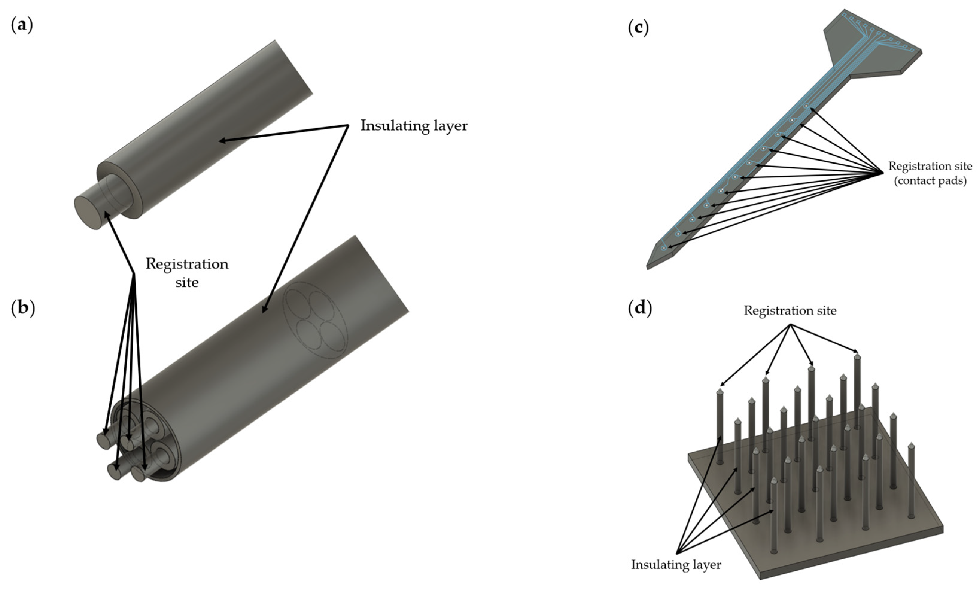
2. Types of In Vivo Microelectrodes
3. Materials for Microelectrodes

| No. | Material | Atomic Number | Electrical Resistivity (20 °C, nΩ⋅m) | Electrical Conductivity (20 °C, 106 S/m) | Thermal Conductivity, W/(m⋅K) | Thermal Expansion (25 °C, µm/(m⋅K)) | Melting Point, °C | Biocompatibility | Description |
|---|---|---|---|---|---|---|---|---|---|
| 1 | Gold (Au) | 79 | 22.14 | 44.2 | 318 | 14.2 | 1064 | Nontoxic | Absence of gliosis [46]; widely used as a conductive material in the manufacture of microelectrodes. |
| 2 | Platinum (Pt) | 78 | 105 | 9.3 | 71.6 | 8.8 | 1768 | Nontoxic | Absence of gliosis [46]; platinum and platinum–iridium (Pt-Ir) widely used as conductive materials in the manufacture of microelectrodes. |
| 3 | Iridium (Ir) | 77 | 47.1 | 21 | 147 | 6.4 | 2446 | Nontoxic | Used as a conductive material in the manufacture of microelectrodes; has a high melting point and is rarely used for film microelectrodes [139]; iridium oxide (IrO2) has improved properties for electrical stimulation and registration of neuronal activity compared to iridium [140,141,142,143]. |
| 4 | Tungsten (W) | 74 | 52.8 | 8.9 | 173 | 4.5 | 3422 | Nontoxic | Could be used as a conductive material in the manufacture of microelectrodes [46,144]. |
| 5 | Tantalum (Ta) | 73 | 131 | 7.7 | 57.5 | 6.3 | 3017 | Nontoxic | Could be used as a conductive material in the manufacture of microelectrodes [145,146]. |
| 6 | Silver (Ag) | 47 | 15.87 | 62.1 | 429 | 18.9 | 961 | Toxic | Silver (Ag) and silver chloride (AgCl) not recommended for microelectrode manufacturing [49]; may cause an allergic reaction [49]; formation of necrotic tissue [47,48]. |
| 7 | Copper (Cu) | 29 | 16.78 | 58.7 | 401 | 16.5 | 1084 | Toxic | Formation of necrotic tissue [147,148]; not recommended for microelectrode manufacturing [49,147]; may cause an allergic reaction [49]. |
| 8 | Nickel (Ni) | 28 | 69.3 | 14.3 | 90.9 | 13.4 | 1455 | Toxic | Not recommended for microelectrode manufacturing [147]; causes localized necrosis [147]; may cause an allergic reaction [49]. |
| 9 | Iron (Fe) | 26 | 96.1 | 10 | 80.4 | 11.8 | 1538 | Toxic | Not recommended for microelectrode manufacturing [45,49]. |
| 10 | Titanium (Ti) | 22 | 420 | 2.4 | 21.9 | 8.6 | 1668 | Nontoxic | Titanium and titanium nitride (TiN) could be used as conductive materials in the manufacture of microelectrodes [149,150,151]; titanium can oxidize, and for this reason, it is most often used as an adhesive layer [117,152,153]. |
| 11 | Aluminum (Al) | 13 | 26.5 | 36.9 | 237 | 23.1 | 660 | Relatively harmless | Not recommended for microelectrode manufacturing [49,147]. |
| 12 | Graphene | - | 10 | 0.1 | 3500–5300 | (−4)–(−3) | 4236 | Nontoxic | Could be used as a conductive material in the manufacture of transparent microelectrodes [71,154,155]. |
| 13 | Stainless steel | - | 690 | 1.45 | 15 | 14.4–17.3 | 1400–1530 | Relatively harmless | Suitable for use as a conductive material in the manufacture of microelectrodes [48,156]. |
| 14 | Nichrome | - | 1100 | 0.909 | 13 | 14 | 1400 | Nontoxic | Could be used as a conductive material in the manufacture of microelectrodes [48]. |
| No. | Name | Young’s Modulus (GPa) | Description |
|---|---|---|---|
| 1 | Flexible polyimide (Pi) | 2.5 [116] 2.3–8.5 [139] | High thermal and chemical stability; polyimide thin films can be applied to microelectrode manufacturing; biocompatible [117]; it should be noted that certain types of polyimides readily absorb water, when polyimide is used as an electrical insulator, excessive water penetration can lead to corrosion and short circuits [157,158]; no FDA approval data. |
| 2 | Parylene-C | 2.76 [120,139,159] | Organic hydrophobic polymer; high biocompatibility; well-shaped; sputtering ability [160,161,162]; FDA-approved. |
| 3 | SU-8 | 2.0 [127,128] 2.87–4.40 [163] | Well-shaped; used in microfluidics [164,165]; no FDA approval data. |
| 4 | PDMS | 0.00132–0.00297 [132] 0.00036–0.00087 [139] | Used in microfluidic and nonpenetrating microelectrodes [166]; has high viscoelasticity, high gas permeability, a low dielectric constant and a low Young’s modulus, which can be changed by changing the curing temperature [167]; FDA-approved. |
Young’s Modulus
4. Microelectrode Impedance
5. The Effects of Microelectrode Implantation on Brain Tissue
- Disruption of the blood–brain barrier (BBB);
- Tissue deformation;
- Chronic inflammation after microelectrode implantation;
- Neuronal cells loss.
5.1. Disruption of the Blood–Brain Barrier and Inflammation
- The microelectrode type, size (thickness, diameter) and tethered or untethered form are important factors affecting both the lifetime of the microelectrode and the quality of the obtained data (change in impedance and signal-to-noise ratio).
- Applying untethered microwire electrodes with a small size (<15 µm of thickness, diameter) could reduce BBB damage, glial scarring and neuronal loss, thereby potentially extending the lifetime of a long-term implanted microelectrode and improving the quality of the obtained data.
- Coating microelectrodes with anti-inflammatory drugs or their additional injection can reduce chronic neurotoxicity mediated by pro-inflammatory cytokines.
5.2. Chronic Inflammation
6. Modern Advances in the Field of In Vivo Penetrating Microelectrodes for Brain Electrophysiology
7. Conclusions
Supplementary Materials
Author Contributions
Funding
Institutional Review Board Statement
Informed Consent Statement
Data Availability Statement
Conflicts of Interest
References
- Schwartz, A.B.; Cui, X.T.; Weber, D.J.; Moran, D.W. Brain-controlled interfaces: Movement restoration with neural prosthetics. Neuron 2006, 52, 205–220. [Google Scholar] [CrossRef] [PubMed] [Green Version]
- Hubel, D.H.; Wiesel, T.N. Receptive fields, binocular interaction and functional architecture in the cat’s visual cortex. J. Physiol. 1962, 160, 106–154. [Google Scholar] [CrossRef] [PubMed]
- Hubel, D.H. Tungsten Microelectrode for Recording from Single Units. Science 1957, 125, 549–550. [Google Scholar] [CrossRef] [PubMed]
- Lovinger, D.M. Communication networks in the brain: Neurons, receptors, neurotransmitters, and alcohol. Alcohol Res. Health J. Natl. Inst. Alcohol Abus. Alcohol. 2008, 31, 196–214. [Google Scholar]
- Buzsáki, G. Large-scale recording of neuronal ensembles. Nat. Neurosci. 2004, 7, 446–451. [Google Scholar] [CrossRef]
- Park, H.; Takmakov, P.; Lee, H. Electrochemical Evaluations of Fractal Microelectrodes for Energy Efficient Neurostimulation. Sci. Rep. 2018, 8, 4375. [Google Scholar] [CrossRef] [Green Version]
- Dragas, J.; Viswam, V.; Shadmani, A.; Chen, Y.; Bounik, R.; Stettler, A.; Radivojevic, M.; Geissler, S.; Obien, M.; Müller, J.; et al. A Multi-Functional Microelectrode Array Featuring 59760 Electrodes, 2048 Electrophysiology Channels, Stimulation, Impedance Measurement and Neurotransmitter Detection Channels. IEEE J。 Solid-State Circuits 2017, 52, 1576–1590. [Google Scholar] [CrossRef] [Green Version]
- Flavin, M.T.; Paul, M.A.; Lim, A.S.; Lissandrello, C.A.; Ajemian, R.; Lin, S.J.; Han, J. Electrochemical modulation enhances the selectivity of peripheral neurostimulation in vivo. Proc. Natl. Acad. Sci. USA 2022, 119, e2117764119. [Google Scholar] [CrossRef]
- Iwasa, S.N.; Shi, H.H.; Hong, S.H.; Chen, T.; Marquez-Chin, M.; Iorio-Morin, C.; Kalia, S.K.; Popovic, M.R.; Naguib, H.E.; Morshead, C.M. Novel Electrode Designs for Neurostimulation in Regenerative Medicine: Activation of Stem Cells. Bioelectricity 2020, 2, 348–361. [Google Scholar] [CrossRef]
- Mills, J.O.; Jalil, A.; Stanga, P.E. Electronic retinal implants and artificial vision: Journey and present. Eye 2017, 31, 1383–1398. [Google Scholar] [CrossRef] [Green Version]
- Downey, J.E.; Schwed, N.; Chase, S.M.; Schwartz, A.B.; Collinger, J.L. Intracortical recording stability in human brain–computer interface users. J. Neural Eng. 2018, 15, 046016. [Google Scholar] [CrossRef] [PubMed]
- Nakatani, S.; Araki, N.; Hoshino, T.; Fukayama, O.; Mabuchi, K. Brain-controlled cycling system for rehabilitation following paraplegia with delay-time prediction. J. Neural Eng. 2021, 18, 016022. [Google Scholar] [CrossRef] [PubMed]
- Mosharov, E.V.; Sulzer, D. Analysis of exocytotic events recorded by amperometry. Nat. Methods 2005, 2, 651–658. [Google Scholar] [CrossRef] [PubMed]
- Peters, J.L.; Miner, L.H.; Michael, A.C.; Sesack, S.R. Ultrastructure at carbon fiber microelectrode implantation sites after acute voltammetric measurements in the striatum of anesthetized rats. J. Neurosci. Methods 2004, 137, 9–23. [Google Scholar] [CrossRef] [PubMed]
- Kita, J.M.; Wightman, R.M. Microelectrodes for studying neurobiology. Curr. Opin. Chem. Biol. 2008, 12, 491–496. [Google Scholar] [CrossRef] [PubMed] [Green Version]
- Kim, G.H.; Kim, K.; Lee, E.; An, T.; Choi, W.; Lim, G.; Shin, J.H. Recent Progress on Microelectrodes in Neural Interfaces. Materials 2018, 11, 1995. [Google Scholar] [CrossRef] [PubMed] [Green Version]
- Nam, Y.; Wheeler, B.C. In vitro microelectrode array technology and neural recordings. Crit. Rev. Biomed. Eng. 2011, 39, 45–61. [Google Scholar] [PubMed] [Green Version]
- Taylor, A.M.; Blurton-Jones, M.; Rhee, S.W.; Cribbs, D.H.; Cotman, C.W.; Jeon, N.L. A microfluidic culture platform for CNS axonal injury, regeneration and transport. Nat. Methods 2005, 2, 599–605. [Google Scholar] [CrossRef]
- Valor, L.M.; Charlesworth, P.; Humphreys, L.; Anderson, C.N.; Grant, S.G. Network activity-independent coordinated gene expression program for synapse assembly. Proc. Natl. Acad. Sci. USA 2007, 104, 4658–4663. [Google Scholar] [CrossRef] [Green Version]
- Bebarova, M. Advances in patch clamp technique: Towards higher quality and quantity. Gen. Physiol. Biophys. 2012, 31, 131–140. [Google Scholar] [CrossRef] [Green Version]
- Guan, B.; Chen, X.; Zhang, H. Two-electrode voltage clamp. Methods Mol. Biol. 2013, 998, 79–89. [Google Scholar] [PubMed]
- Slutzky, M.W.; Jordan, L.R.; Lindberg, E.W.; Lindsay, K.E.; Miller, L.E. Decoding the rat forelimb movement direction from epidural and intracortical field potentials. J. Neural Eng. 2011, 8, 036013. [Google Scholar] [CrossRef] [PubMed] [Green Version]
- Obaid, A.; Hanna, M.-E.; Wu, Y.-W.; Kollo, M.; Racz, R.; Angle, M.R.; Müller, J.; Brackbill, N.; Wray, W.; Franke, F.; et al. Massively parallel microwire arrays integrated with CMOS chips for neural recording. Sci. Adv. 2020, 6, eaay2789. [Google Scholar] [CrossRef] [PubMed] [Green Version]
- Kipke, D.R.; Vetter, R.J.; Williams, J.C.; Hetke, J.F. Silicon-substrate intracortical microelectrode arrays for long-term recording of neuronal spike activity in cerebral cortex. IEEE Trans. Neural Syst. Rehabil. Eng. 2003, 11, 151–155. [Google Scholar] [CrossRef] [PubMed]
- Rousche, P.J.; Normann, R.A. Chronic recording capability of the Utah Intracortical Electrode Array in cat sensory cortex. J. Neurosci. Methods 1998, 82, 1–15. [Google Scholar] [CrossRef]
- Du, Z.J.; Kolarcik, C.L.; Kozai, T.D.Y.; Luebben, S.D.; Sapp, S.A.; Zheng, X.S.; Nabity, J.A.; Cui, X.T. Ultrasoft microwire neural electrodes improve chronic tissue integration. Acta Biomater. 2017, 53, 46–58. [Google Scholar] [CrossRef] [Green Version]
- Ferguson, J.E.; Boldt, C.; Redish, A.D. Creating low-impedance tetrodes by electroplating with additives. Sens. Actuators A Phys. 2009, 156, 388–393. [Google Scholar] [CrossRef] [Green Version]
- Tseng, W.-T.; Yen, C.-T.; Tsai, M.-L. A bundled microwire array for long-term chronic single-unit recording in deep brain regions of behaving rats. J. Neurosci. Methods 2011, 201, 368–376. [Google Scholar] [CrossRef]
- Szostak, K.M.; Grand, L.; Constandinou, T.G. Neural Interfaces for Intracortical Recording: Requirements, Fabrication Methods, and Characteristics. Front. Neurosci. 2017, 11, 665. [Google Scholar] [CrossRef] [Green Version]
- Saxena, T.; Karumbaiah, L.; Gaupp, E.A.; Patkar, R.; Patil, K.; Betancur, M.; Stanley, G.B.; Bellamkonda, R.V. The impact of chronic blood-brain barrier breach on intracortical electrode function. Biomaterials 2013, 34, 4703–4713. [Google Scholar] [CrossRef]
- Karumbaiah, L.; Saxena, T.; Carlson, D.; Patil, K.; Patkar, R.; Gaupp, E.A.; Betancur, M.; Stanley, G.B.; Carin, L.; Bellamkonda, R.V. Relationship between intracortical electrode design and chronic recording function. Biomaterials 2013, 34, 8061–8074. [Google Scholar] [CrossRef]
- Iannacci, J. Introduction to MEMS and RF-MEMS: From the early days of microsystems to modern RF-MEMS passives. In Rf-Mems Technology for High-Performance Passives; IOP Publishing Ltd.: Brtistol, UK, 2017; pp. 2053–2563. [Google Scholar]
- Panescu, D. MEMS in medicine and biology. IEEE Eng. Med. Biol. Mag. 2006, 25, 19–28. [Google Scholar] [CrossRef]
- Najafi, K.; Ji, J.; Wise, K.D. Scaling limitations of silicon multichannel recording probes. IEEE Trans. Biomed. Eng. 1990, 37, 1–11. [Google Scholar] [CrossRef]
- Pérez-Prieto, N.; Delgado-Restituto, M. Recording Strategies for High Channel Count, Densely Spaced Microelectrode Arrays. Front. Neurosci. 2021, 15, 681085. [Google Scholar] [CrossRef]
- McNamara, M.; Ersöz, A.; Han, M. A Diagnostic Circuit for Crosstalk Detection in Microelectrode Arrays. In Proceedings of the 2021 10th International IEEE/EMBS Conference on Neural Engineering (NER), Virtual, 4–6 May 2021; pp. 544–547. [Google Scholar]
- House, P.A.; MacDonald, J.D.; Tresco, P.A.; Normann, R.A. Acute microelectrode array implantation into human neocortex: Preliminary technique and histological considerations. Neurosurg. Focus 2006, 20, E4. [Google Scholar] [CrossRef]
- Hatsopoulos, N.G.; Donoghue, J.P. The science of neural interface systems. Annu. Rev. Neurosci. 2009, 32, 249–266. [Google Scholar] [CrossRef] [Green Version]
- Cody, P.A.; Eles, J.R.; Lagenaur, C.F.; Kozai, T.D.Y.; Cui, X.T. Unique electrophysiological and impedance signatures between encapsulation types: An analysis of biological Utah array failure and benefit of a biomimetic coating in a rat model. Biomaterials 2018, 161, 117–128. [Google Scholar] [CrossRef]
- Leber, M.; Korner, J.; Reiche, C.F.; Yin, M.; Bhandari, R.; Franklin, R.; Negi, S.; Solzbacher, F. Advances in Penetrating Multichannel Microelectrodes Based on the Utah Array Platform. Adv. Exp. Med. Biol. 2019, 1101, 1–40. [Google Scholar]
- Woeppel, K.; Hughes, C.; Herrera, A.J.; Eles, J.R.; Tyler-Kabara, E.C.; Gaunt, R.A.; Collinger, J.L.; Cui, X.T. Explant Analysis of Utah Electrode Arrays Implanted in Human Cortex for Brain-Computer-Interfaces. Front. Bioeng. Biotechnol. 2021, 9, 759711. [Google Scholar] [CrossRef]
- Maynard, E.M.; Nordhausen, C.T.; Normann, R.A. The Utah intracortical Electrode Array: A recording structure for potential brain-computer interfaces. Electroencephalogr. Clin. Neurophysiol. 1997, 102, 228–239. [Google Scholar] [CrossRef]
- Majeed, S.; Naqvi, S.T.R.; ul Haq, M.N.; Ashiq, M.N. Electroanalytical techniques in biosciences: Conductometry, coulometry, voltammetry, and electrochemical sensors. In Analytical Techniques in Biosciences; Egbuna, C., Patrick-Iwuanyanwu, K.C., Shah, M.A., Ifemeje, J.C., Rasul, A., Eds.; Academic Press: Cambridge, MA, USA, 2022; pp. 157–178. [Google Scholar]
- Shin, H.; Son, Y.; Chae, U.; Kim, J.; Choi, N.; Lee, H.J.; Woo, J.; Cho, Y.; Yang, S.H.; Lee, C.J.; et al. Multifunctional multi-shank neural probe for investigating and modulating long-range neural circuits in vivo. Nat. Commun. 2019, 10, 3777. [Google Scholar] [CrossRef] [Green Version]
- Dymond, A.M.; Kaechele, L.E.; Jurist, J.M.; Crandall, P.H. Brain tissue reaction to some chronically implanted metals. J. Neurosurg. 1970, 33, 574–580. [Google Scholar] [CrossRef]
- Stensaas, S.S.; Stensaas, L.J. Histopathological evaluation of materials implanted in the cerebral cortex. Acta Neuropathol. 1978, 41, 145–155. [Google Scholar] [CrossRef]
- Robinson, F.R.; Johnson, M.T. Histopathological Studies of Tissue Reactions to Various Metals Implanted in Cat Brains; ASD Technical Report; United States. Air Force. Systems Command. Aeronautical Systems Division: Greene, OH, USA, 1961; Volume 61, p. 13. [Google Scholar]
- Babb, T.L.; Kupfer, W. Phagocytic and metabolic reactions to chronically implanted metal brain electrodes. Exp. Neurol. 1984, 86, 171–182. [Google Scholar] [CrossRef]
- Geddes, L.A.; Roeder, R. Criteria for the selection of materials for implanted electrodes. Ann. Biomed. Eng. 2003, 31, 879–890. [Google Scholar] [CrossRef]
- Saini, M.; Singh, Y.; Arora, P.; Arora, V.; Jain, K. Implant biomaterials: A comprehensive review. World J. Clin. Cases 2015, 3, 52–57. [Google Scholar] [CrossRef]
- Kim, R.; Hong, N.; Nam, Y. Gold nanograin microelectrodes for neuroelectronic interfaces. Biotechnol. J. 2013, 8, 206–214. [Google Scholar] [CrossRef]
- Woldring, S.; Dirken, M.N. Spontaneous unit-activity in the superficial cortical layers. Acta Physiol. Et Pharmacol. Neerl. 1950, 1, 369–379. [Google Scholar]
- Czeschik, A.; Offenhäusser, A.; Wolfrum, B. Fabrication of MEA-based nanocavity sensor arrays for extracellular recording of action potentials. Phys. Status Solidi (A) 2014, 211, 1462–1466. [Google Scholar] [CrossRef]
- Xie, C.; Lin, Z.; Hanson, L.; Cui, Y.; Cui, B. Intracellular recording of action potentials by nanopillar electroporation. Nat. Nanotechnol. 2012, 7, 185–190. [Google Scholar] [CrossRef] [PubMed] [Green Version]
- Mathieson, K.; Kachiguine, S.; Adams, C.; Cunningham, W.; Gunning, D.; Shea, V.O.; Smith, K.M.; Chichilnisky, E.J.; Litke, A.M.; Sher, A.; et al. Large-area microelectrode arrays for recording of neural signals. IEEE Trans. Nucl. Sci. 2004, 51, 2027–2031. [Google Scholar] [CrossRef]
- Jun, S.B.; Hynd, M.R.; Dowell-Mesfin, N.; Smith, K.L.; Turner, J.N.; Shain, W.; Kim, S.J. Low-density neuronal networks cultured using patterned poly-l-lysine on microelectrode arrays. J. Neurosci. Methods 2007, 160, 317–326. [Google Scholar] [CrossRef] [PubMed] [Green Version]
- Park, S.; Song, Y.J.; Boo, H.; Chung, T.D. Nanoporous Pt Microelectrode for Neural Stimulation and Recording: In Vitro Characterization. J. Phys. Chem. C 2010, 114, 8721–8726. [Google Scholar] [CrossRef]
- Takayama, Y.; Moriguchi, H.; Kotani, K.; Suzuki, T.; Mabuchi, K.; Jimbo, Y. Network-wide integration of stem cell-derived neurons and mouse cortical neurons using microfabricated co-culture devices. Bio. Syst. 2012, 107, 1–8. [Google Scholar] [CrossRef] [PubMed]
- Dowben, R.M.; Rose, J.E. A metal-filled microelectrode. Science 1953, 118, 22–24. [Google Scholar] [CrossRef]
- Beder, O.E.; Eade, G. An investigation of tissue tolerance to titanium metal implants in dogs. Surgery 1956, 39, 470–473. [Google Scholar]
- Duane, W.; Fricke, H.; Stenström, W. The Absorption of X-Rays by Chemical Elements of High Atomic Numbers. Proc. Natl. Acad. Sci. USA 1920, 6, 607–612. [Google Scholar] [CrossRef] [Green Version]
- Devi, M.; Vomero, M.; Fuhrer, E.; Castagnola, E.; Gueli, C.; Nimbalkar, S.; Hirabayashi, M.; Kassegne, S.; Stieglitz, T.; Sharma, S. Carbon-based neural electrodes: Promises and challenges. J. Neural Eng. 2021, 18, 041007. [Google Scholar] [CrossRef]
- Buschbeck, E.K.; Duc Le, A.; Kelley, C.; Hoque, M.A.; Alvarez, N.T. Functionalized carbon nanotube microfibers for chronic neural implants. J. Neurosci. Methods 2021, 364, 109370. [Google Scholar] [CrossRef]
- Zestos, A.G.; Venton, B.J. Carbon Nanotube-Based Microelectrodes for Enhanced Neurochemical Detection. ECS Trans. 2017, 80, 1497–1509. [Google Scholar] [CrossRef]
- Budai, D. Carbon Fiber-based Microelectrodes and Microbiosensors. In Intelligent and Biosensors; IntechOpen: London, UK, 2010; pp. 269–288. [Google Scholar]
- Vafaiee, M.; Mohammadpour, R.; Vossoughi, M.; Asadian, E.; Janahmadi, M.; Sasanpour, P. Carbon Nanotube Modified Microelectrode Array for Neural Interface. Front. Bioeng. Biotechnol. 2020, 8, 582713. [Google Scholar] [CrossRef] [PubMed]
- Bareket-Keren, L.; Hanein, Y. Carbon nanotube-based multi electrode arrays for neuronal interfacing: Progress and prospects. Front. Neural Circuits 2012, 6, 122. [Google Scholar] [CrossRef] [PubMed] [Green Version]
- Park, D.-W.; Brodnick, S.K.; Ness, J.P.; Atry, F.; Krugner-Higby, L.; Sandberg, A.; Mikael, S.; Richner, T.J.; Novello, J.; Kim, H.; et al. Fabrication and utility of a transparent graphene neural electrode array for electrophysiology, in vivo imaging, and optogenetics. Nat. Protoc. 2016, 11, 2201–2222. [Google Scholar] [CrossRef]
- Du, X.; Wu, L.; Cheng, J.; Huang, S.; Cai, Q.; Jin, Q.; Zhao, J. Graphene microelectrode arrays for neural activity detection. J. Biol. Phys. 2015, 41, 339–347. [Google Scholar] [CrossRef] [PubMed]
- Rastogi, S.K.; Bliley, J.; Shiwarski, D.J.; Raghavan, G.; Feinberg, A.W.; Cohen-Karni, T. Graphene Microelectrode Arrays for Electrical and Optical Measurements of Human Stem Cell-Derived Cardiomyocytes. Cell. Mol. Bioeng. 2018, 11, 407–418. [Google Scholar] [CrossRef]
- Körbitzer, B.; Krauß, P.; Belle, S.; Schneider, J.J.; Thielemann, C. Electrochemical Characterization of Graphene Microelectrodes for Biological Applications. ChemNanoMat 2019, 5, 427–435. [Google Scholar] [CrossRef]
- Biran, R.; Martin, D.C.; Tresco, P.A. Neuronal cell loss accompanies the brain tissue response to chronically implanted silicon microelectrode arrays. Exp. Neurol. 2005, 195, 115–126. [Google Scholar] [CrossRef] [PubMed]
- Kozai, T.D.; Langhals, N.B.; Patel, P.R.; Deng, X.; Zhang, H.; Smith, K.L.; Lahann, J.; Kotov, N.A.; Kipke, D.R. Ultrasmall implantable composite microelectrodes with bioactive surfaces for chronic neural interfaces. Nat. Mater. 2012, 11, 1065–1073. [Google Scholar] [CrossRef] [Green Version]
- Polikov, V.S.; Tresco, P.A.; Reichert, W.M. Response of brain tissue to chronically implanted neural electrodes. J. Neurosci. Methods 2005, 148, 1–18. [Google Scholar] [CrossRef]
- Fattahi, P.; Yang, G.; Kim, G.; Abidian, M.R. A review of organic and inorganic biomaterials for neural interfaces. Adv. Mater. (Deerfield Beach Fla.) 2014, 26, 1846–1885. [Google Scholar] [CrossRef]
- Guimard, N.K.; Gomez, N.; Schmidt, C.E. Conducting polymers in biomedical engineering. Prog. Polym. Sci. 2007, 32, 876–921. [Google Scholar] [CrossRef]
- Meng, Q.; Cai, K.; Chen, Y.; Chen, L. Research progress on conducting polymer based supercapacitor electrode materials. Nano Energy 2017, 36, 268–285. [Google Scholar] [CrossRef]
- Kim, D.-H.; Wiler, J.A.; Anderson, D.J.; Kipke, D.R.; Martin, D.C. Conducting polymers on hydrogel-coated neural electrode provide sensitive neural recordings in auditory cortex. Acta Biomater. 2010, 6, 57–62. [Google Scholar] [CrossRef] [PubMed]
- Ludwig, K.A.; Uram, J.D.; Yang, J.; Martin, D.C.; Kipke, D.R. Chronic neural recordings using silicon microelectrode arrays electrochemically deposited with a poly(3,4-ethylenedioxythiophene) (PEDOT) film. J. Neural Eng. 2006, 3, 59–70. [Google Scholar] [CrossRef] [PubMed]
- Yang, J.; Kim, D.H.; Hendricks, J.L.; Leach, M.; Northey, R.; Martin, D.C. Ordered surfactant-templated poly(3,4-ethylenedioxythiophene) (PEDOT) conducting polymer on microfabricated neural probes. Acta Biomater. 2005, 1, 125–136. [Google Scholar] [CrossRef]
- Castagnola, V.; Descamps, E.; Lecestre, A.; Dahan, L.; Remaud, J.; Nowak, L.G.; Bergaud, C. Parylene-based flexible neural probes with PEDOT coated surface for brain stimulation and recording. Biosens. Bioelectron. 2015, 67, 450–457. [Google Scholar] [CrossRef]
- Kayser, L.V.; Lipomi, D.J. Stretchable Conductive Polymers and Composites Based on PEDOT and PEDOT:PSS. Adv. Mater. 2019, 31, 1806133. [Google Scholar] [CrossRef] [Green Version]
- Carli, S.; Bianchi, M.; Zucchini, E.; Di Lauro, M.; Prato, M.; Murgia, M.; Fadiga, L.; Biscarini, F. Electrodeposited PEDOT:Nafion Composite for Neural Recording and Stimulation. Adv. Healthc. Mater. 2019, 8, 1900765. [Google Scholar] [CrossRef]
- Kshirsagar, P.; Dickreuter, S.; Mierzejewski, M.; Burkhardt, C.J.; Chassé, T.; Fleischer, M.; Jones, P.D. Transparent Graphene/PEDOT:PSS Microelectrodes for Electro- and Optophysiology. Adv. Mater. Technol. 2019, 4, 1800318. [Google Scholar] [CrossRef] [Green Version]
- Cho, Y.U.; Lee, J.Y.; Jeong, U.-J.; Park, S.H.; Lim, S.L.; Kim, K.Y.; Jang, J.W.; Park, J.H.; Kim, H.W.; Shin, H.; et al. Ultra-Low Cost, Facile Fabrication of Transparent Neural Electrode Array for Electrocorticography with Photoelectric Artifact-Free Optogenetics. Adv. Funct. Mater. 2022, 32, 2105568. [Google Scholar] [CrossRef]
- Ludwig, K.A.; Langhals, N.B.; Joseph, M.D.; Richardson-Burns, S.M.; Hendricks, J.L.; Kipke, D.R. Poly(3,4-ethylenedioxythiophene) (PEDOT) polymer coatings facilitate smaller neural recording electrodes. J. Neural Eng. 2011, 8, 014001. [Google Scholar] [CrossRef] [PubMed]
- Cui, X.; Martin, D.C. Electrochemical deposition and characterization of poly(3,4-ethylenedioxythiophene) on neural microelectrode arrays. Sens. Actuators B Chem. 2003, 89, 92–102. [Google Scholar] [CrossRef]
- Wilks, S.J.; Richardson-Burns, S.M.; Hendricks, J.L.; Martin, D.C.; Otto, K.J. Poly(3,4-ethylenedioxythiophene) as a Micro-Neural Interface Material for Electrostimulation. Front. Neuroeng. 2009, 2, 7. [Google Scholar] [CrossRef] [PubMed] [Green Version]
- George, P.M.; Lyckman, A.W.; LaVan, D.A.; Hegde, A.; Leung, Y.; Avasare, R.; Testa, C.; Alexander, P.M.; Langer, R.; Sur, M. Fabrication and biocompatibility of polypyrrole implants suitable for neural prosthetics. Biomaterials 2005, 26, 3511–3519. [Google Scholar] [CrossRef] [PubMed]
- Cui, X.; Hetke, J.F.; Wiler, J.A.; Anderson, D.J.; Martin, D.C. Electrochemical deposition and characterization of conducting polymer polypyrrole/PSS on multichannel neural probes. Sens. Actuators A Phys. 2001, 93, 8–18. [Google Scholar] [CrossRef]
- Stauffer, W.R.; Cui, X.T. Polypyrrole doped with 2 peptide sequences from laminin. Biomaterials 2006, 27, 2405–2413. [Google Scholar] [CrossRef] [PubMed]
- Han, L.; Yan, L.; Wang, M.; Wang, K.; Fang, L.; Zhou, J.; Fang, J.; Ren, F.; Lu, X. Transparent, Adhesive, and Conductive Hydrogel for Soft Bioelectronics Based on Light-Transmitting Polydopamine-Doped Polypyrrole Nanofibrils. Chem. Mater. 2018, 30, 5561–5572. [Google Scholar] [CrossRef]
- Dimaki, M.; Vazquez, P.; Olsen, M.H.; Sasso, L.; Rodriguez-Trujillo, R.; Vedarethinam, I.; Svendsen, W.E. Fabrication and characterization of 3D micro- and nanoelectrodes for neuron recordings. Sensors 2010, 10, 10339–10355. [Google Scholar] [CrossRef] [PubMed] [Green Version]
- Das, D.; Dutta, D.; Kundu, P. Nickel nanocatalyst supported on sulfonated polyaniline: Potentials toward methanol oxidation and as anode materials of DMFCs. J. Mater. Chem. A Mater. Energy Sustain. 2015, 3, 11349–11357. [Google Scholar] [CrossRef]
- Wang, H.; Lin, J.; Shen, Z.X. Polyaniline (PANi) based electrode materials for energy storage and conversion. J. Sci. Adv. Mater. Devices 2016, 1, 225–255. [Google Scholar] [CrossRef] [Green Version]
- Campbell, P.K.; Jones, K.E.; Huber, R.J.; Horch, K.W.; Normann, R.A. A silicon-based, three-dimensional neural interface: Manufacturing processes for an intracortical electrode array. IEEE Trans. Biomed. Eng. 1991, 38, 758–768. [Google Scholar] [CrossRef] [PubMed]
- Sawahata, H.; Yamagiwa, S.; Moriya, A.; Dong, T.; Oi, H.; Ando, Y.; Numano, R.; Ishida, M.; Koida, K.; Kawano, T. Single 5 μm diameter needle electrode block modules for unit recordings in vivo. Sci. Rep. 2016, 6, 35806. [Google Scholar] [CrossRef] [PubMed]
- Kita, Y.; Tsuruhara, S.; Kubo, H.; Yamashita, K.; Seikoba, Y.; Idogawa, S.; Sawahata, H.; Yamagiwa, S.; Leong, X.L.A.; Numano, R.; et al. Three-micrometer-diameter needle electrode with an amplifier for extracellular in vivo recordings. Proc. Natl. Acad. Sci. USA 2021, 118, e2008233118. [Google Scholar] [CrossRef] [PubMed]
- Fujishiro, A.; Kaneko, H.; Kawashima, T.; Ishida, M.; Kawano, T. In vivo neuronal action potential recordings via three-dimensional microscale needle-electrode arrays. Sci. Rep. 2014, 4, 4868. [Google Scholar] [CrossRef]
- Yamashita, K.; Sawahata, H.; Yamagiwa, S.; Yokoyama, S.; Numano, R.; Koida, K.; Kawano, T. A floating 5 μm-diameter needle electrode on the tissue for damage-reduced chronic neuronal recording in mice. Lab. A Chip. 2022, 22, 747–756. [Google Scholar] [CrossRef]
- Bettinger, C.J.; Ecker, M.; Kozai, T.D.Y.; Malliaras, G.G.; Meng, E.; Voit, W. Recent advances in neural interfaces—Materials chemistry to clinical translation. MRS Bull. 2020, 45, 655–668. [Google Scholar] [CrossRef]
- Ciancone, M.T.; Rebec, G.V. A simple device for the reliable production of varnish-insulated, high-impedance tungsten microelectrodes. J. Neurosci. Methods 1989, 27, 77–79. [Google Scholar] [CrossRef]
- Fang, J.; Fu, R.; Gu, X.; Zhang, X.; Li, G. Characterization of Glass Insulating Thick Films with Ag Conductors for Multilayer Packages. Materials 2021, 14, 494. [Google Scholar] [CrossRef]
- Liu, B.; Rolland, J.P.; DeSimone, J.M.; Bard, A.J. Fabrication of Ultramicroelectrodes Using A “Teflon-like” Coating Material. Anal. Chem. 2005, 77, 3013–3017. [Google Scholar] [CrossRef]
- Song, E.; Fang, H.; Jin, X.; Zhao, J.; Jiang, C.; Yu, K.J.; Zhong, Y.; Xu, D.; Li, J.; Fang, G.; et al. Thin, Transferred Layers of Silicon Dioxide and Silicon Nitride as Water and Ion Barriers for Implantable Flexible Electronic Systems. Adv. Electron. Mater. 2017, 3, 1700077. [Google Scholar] [CrossRef]
- Khodakarami, S.; Zhao, H.; Rabbi, K.F.; Miljkovic, N. Scalable Corrosion-Resistant Coatings for Thermal Applications. ACS Appl. Mater. Interfaces 2021, 13, 4519–4534. [Google Scholar] [CrossRef] [PubMed]
- Schmidt, S.; Haensch, T.; Frank, R.; Jahnke, H.-G.; Robitzki, A.A. Reactive Sputtered Silicon Nitride as an Alternative Passivation Layer for Microelectrode Arrays in Sensitive Bioimpedimetric Cell Monitoring. ACS Appl. Mater. Interfaces 2021, 13, 59185–59195. [Google Scholar] [CrossRef] [PubMed]
- Cogan, S.F.; Edell, D.J.; Guzelian, A.A.; Ping Liu, Y.; Edell, R. Plasma-enhanced chemical vapor deposited silicon carbide as an implantable dielectric coating. J. Biomed. Mater. Res. Part A 2003, 67A, 856–867. [Google Scholar] [CrossRef]
- Deku, F.; Frewin, C.L.; Stiller, A.; Cohen, Y.; Aqeel, S.; Joshi-Imre, A.; Black, B.; Gardner, T.J.; Pancrazio, J.J.; Cogan, S.F. Amorphous Silicon Carbide Platform for Next Generation Penetrating Neural Interface Designs. Micromachines 2018, 9, 480. [Google Scholar] [CrossRef] [PubMed]
- Song, E.; Lee, Y.K.; Li, R.; Li, J.; Jin, X.; Yu, K.J.; Xie, Z.; Fang, H.; Zhong, Y.; Du, H.; et al. Transferred, Ultrathin Oxide Bilayers as Biofluid Barriers for Flexible Electronic Implants. Adv. Funct. Mater. 2018, 28, 1702284. [Google Scholar] [CrossRef]
- Sagar, S.; Mohammadian, N.; Park, S.; Majewski, L.A.; Das, B.C. Ultra-thin anodized aluminium dielectric films: The effect of citric acid concentration and low-voltage electronic applications. Nanotechnology 2020, 31, 255705. [Google Scholar] [CrossRef]
- Roy, R.K.; Lee, K.-R. Biomedical applications of diamond-like carbon coatings: A review. J. Biomed. Mater. Res. Part B Appl. Biomater. 2007, 83B, 72–84. [Google Scholar] [CrossRef]
- Szymanski, L.J.; Kellis, S.; Liu, C.Y.; Jones, K.T.; Andersen, R.A.; Commins, D.; Lee, B.; McCreery, D.B.; Miller, C.A. Neuropathological effects of chronically implanted, intracortical microelectrodes in a tetraplegic patient. J. Neural Eng. 2021, 18, 0460b9. [Google Scholar] [CrossRef]
- Nguyen, J.K.; Park, D.J.; Skousen, J.L.; Hess-Dunning, A.E.; Tyler, D.J.; Rowan, S.J.; Weder, C.; Capadona, J.R. Mechanically-compliant intracortical implants reduce the neuroinflammatory response. J. Neural Eng. 2014, 11, 056014. [Google Scholar] [CrossRef] [Green Version]
- Xiang, Z.; Yen, S.-C.; Xue, N.; Sun, T.; Tsang, W.M.; Zhang, S.; Liao, L.-D.; Thakor, N.V.; Lee, C. Ultra-thin flexible polyimide neural probe embedded in a dissolvable maltose-coated microneedle. J. Micromechanics Microengineering 2014, 24, 065015. [Google Scholar] [CrossRef] [Green Version]
- Lee, K.-K.; He, J.; Singh, A.; Massia, S.; Ehteshami, G.; Kim, B.; Raupp, G. Polyimide-based intracortical neural implant with improved structural stiffness. J. Micromechanics Microengineering 2003, 14, 32–37. [Google Scholar] [CrossRef]
- Kim, B.J.; Meng, E. Review of polymer MEMS micromachining. J. Micromechanics Microengineering 2015, 26, 013001. [Google Scholar] [CrossRef]
- Hiçyılmaz, A.; Çelik Bedeloğlu, A. Applications of polyimide coatings: A review. SN Appl. Sci. 2021, 3, 363. [Google Scholar] [CrossRef]
- Constantin, C.P.; Aflori, M.; Damian, R.F.; Rusu, R.D. Biocompatibility of Polyimides: A Mini-Review. Materials 2019, 12, 3166. [Google Scholar] [CrossRef]
- Hsu, J.M.; Rieth, L.; Normann, R.A.; Tathireddy, P.; Solzbacher, F. Encapsulation of an integrated neural interface device with Parylene C. IEEE Trans. Biomed. Eng. 2009, 56, 23–29. [Google Scholar] [CrossRef]
- Ortigoza-Diaz, J.; Scholten, K.; Larson, C.; Cobo, A.; Hudson, T.; Yoo, J.; Baldwin, A.; Weltman Hirschberg, A.; Meng, E. Techniques and Considerations in the Microfabrication of Parylene C Microelectromechanical Systems. Micromachines 2018, 9, 422. [Google Scholar] [CrossRef] [Green Version]
- Marszalek, T.; Gazicki-Lipman, M.; Ulanski, J. Parylene C as a versatile dielectric material for organic field-effect transistors. Beilstein J. Nanotechnol. 2017, 8, 1532–1545. [Google Scholar] [CrossRef] [Green Version]
- Satti, A.T.; Park, J.; Park, J.; Kim, H.; Cho, S. Fabrication of Parylene-Coated Microneedle Array Electrode for Wearable ECG Device. Sensors 2020, 20, 5183. [Google Scholar] [CrossRef]
- de la Oliva, N.; Mueller, M.; Stieglitz, T.; Navarro, X.; Del Valle, J. On the use of Parylene C polymer as substrate for peripheral nerve electrodes. Sci. Rep. 2018, 8, 5965. [Google Scholar] [CrossRef] [Green Version]
- Golda-Cepa, M.; Engvall, K.; Hakkarainen, M.; Kotarba, A. Recent progress on parylene C polymer for biomedical applications: A review. Prog. Org. Coat. 2020, 140, 105493. [Google Scholar] [CrossRef]
- Altuna, A.; Bellistri, E.; Cid, E.; Aivar, P.; Gal, B.; Berganzo, J.; Gabriel, G.; Guimerà, A.; Villa, R.; Fernández, L.J.; et al. SU-8 based microprobes for simultaneous neural depth recording and drug delivery in the brain. Lab. Chip. 2013, 13, 1422–1430. [Google Scholar] [CrossRef] [Green Version]
- Cho, S.; Lu, H.M.; Cauller, L.; Romero-Ortega, M.I.; Lee, J.; Hughes, G.A. Biocompatible SU-8-Based Microprobes for Recording Neural Spike Signals From Regenerated Peripheral Nerve Fibers. IEEE Sens. J. 2008, 8, 1830–1836. [Google Scholar] [CrossRef]
- Altuna, A.; Menendez de la Prida, L.; Bellistri, E.; Gabriel, G.; Guimerá, A.; Berganzo, J.; Villa, R.; Fernández, L.J. SU-8 based microprobes with integrated planar electrodes for enhanced neural depth recording. Biosens. Bioelectron. 2012, 37, 1–5. [Google Scholar] [CrossRef] [PubMed] [Green Version]
- Nemani, K.V.; Moodie, K.L.; Brennick, J.B.; Su, A.; Gimi, B. In vitro and in vivo evaluation of SU-8 biocompatibility. Mater. Sci. Eng. C Mater. Biol. Appl. 2013, 33, 4453–4459. [Google Scholar] [CrossRef] [PubMed]
- Chen, Z.; Lee, J.B. Biocompatibility of SU-8 and Its Biomedical Device Applications. Micromachines 2021, 12, 794. [Google Scholar] [CrossRef] [PubMed]
- Vitale, F.; Vercosa, D.G.; Rodriguez, A.V.; Pamulapati, S.S.; Seibt, F.; Lewis, E.; Yan, J.S.; Badhiwala, K.; Adnan, M.; Royer-Carfagni, G.; et al. Fluidic Microactuation of Flexible Electrodes for Neural Recording. Nano Lett. 2018, 18, 326–335. [Google Scholar] [CrossRef]
- Kim, J.M.; Im, C.; Lee, W.R. Plateau-Shaped Flexible Polymer Microelectrode Array for Neural Recording. Polymers 2017, 9, 690. [Google Scholar] [CrossRef] [Green Version]
- Miranda, I.; Souza, A.; Sousa, P.; Ribeiro, J.; Castanheira, E.M.S.; Lima, R.; Minas, G. Properties and Applications of PDMS for Biomedical Engineering: A Review. J. Funct. Biomater. 2021, 13, 2. [Google Scholar] [CrossRef]
- Ariati, R.; Sales, F.; Souza, A.; Lima, R.A.; Ribeiro, J. Polydimethylsiloxane Composites Characterization and Its Applications: A Review. Polymers 2021, 13, 4258. [Google Scholar] [CrossRef]
- Park, S.; Mondal, K.; Treadway, R.M.; Kumar, V.; Ma, S.; Holbery, J.D.; Dickey, M.D. Silicones for Stretchable and Durable Soft Devices: Beyond Sylgard-184. ACS Appl. Mater. Interfaces 2018, 10, 11261–11268. [Google Scholar] [CrossRef]
- Li, J.; Huang, D.; Chen, Y.; Li, Z. Low-Cost, Metal-Based Micro-Needle Electrode Array (M-MNEA): A Three-Dimensional Intracortical Neural Interface. In Proceedings of the 2019 20th International Conference on Solid-State Sensors, Actuators and Microsystems & Eurosensors XXXIII (TRANSDUCERS & EUROSENSORS XXXIII), Berlin, Germany, 23–27 June 2019; pp. 1635–1638. [Google Scholar]
- Lee, J.H.; Kim, H.; Kim, J.H.; Lee, S.-H. Soft implantable microelectrodes for future medicine: Prosthetics, neural signal recording and neuromodulation. Lab. A Chip. 2016, 16, 959–976. [Google Scholar] [CrossRef] [PubMed]
- Kim, B.J.; Meng, E. Micromachining of Parylene C for bioMEMS. Polym. Adv. Technol. 2016, 27, 564–576. [Google Scholar] [CrossRef]
- Weltman, A.; Yoo, J.; Meng, E. Flexible, Penetrating Brain Probes Enabled by Advances in Polymer Microfabrication. Micromachines 2016, 7, 180. [Google Scholar] [CrossRef] [PubMed] [Green Version]
- Klein, J.D.; Clauson, S.L.; Cogan, S.F. Morphology and charge capacity of sputtered iridium oxide films. J. Vac. Sci. Technol. A 1989, 7, 3043–3047. [Google Scholar] [CrossRef]
- Robblee, L.S.; Lefko, J.L.; Brummer, S. Activated Ir: An Electrode Suitable for Reversible Charge Injection in Saline Solution. J. Electrochem. Soc. 1983, 130, 731–733. [Google Scholar] [CrossRef]
- Weiland, J.D.; Anderson, D.J.; Humayun, M.S. In vitro electrical properties for iridium oxide versus titanium nitride stimulating electrodes. IEEE Trans. Biomed. Eng. 2002, 49, 1574–1579. [Google Scholar] [CrossRef]
- Cogan, S.F.; Ehrlich, J.; Plante, T.D.; Smirnov, A.; Shire, D.B.; Gingerich, M.; Rizzo, J.F. Sputtered iridium oxide films for neural stimulation electrodes. J. Biomed. Mater. Res. Part B Appl. Biomater. 2009, 89, 353–361. [Google Scholar] [CrossRef]
- Prochazka, A.; Mushahwar, V.K.; McCreery, D.B. Neural prostheses. J. Physiol. 2001, 533, 99–109. [Google Scholar] [CrossRef]
- May, G.A.; Shamma, S.A.; White, R.L. A tantalum-on-sapphire microelectrode array. IEEE Trans. Electron. Devices 1979, 26, 1932–1939. [Google Scholar] [CrossRef]
- Lerner, H.; Zahradnik, R.T.; Buchbinder, M. Miniature implantable tantalum/tantalum oxide stimulating electrodes. IEEE Trans. Biomed. Eng. 1982, 29, 290–292. [Google Scholar] [CrossRef]
- Agnew, W.F.; McCreery, D.B. Neural Prostheses: Fundamental Studies; Prentice Hall: Englewood Cliffs, NJ, USA, 1990. [Google Scholar]
- Bickford, R.G.; Fischer, G.; Sayre, G.P. Histologic changes in the cat’s brain after introduction of metallic and plastic coated wire used in electro-encephalography. Proc. Staff Meet. Mayo Clin. 1957, 32, 14–21. [Google Scholar] [PubMed]
- Ryynänen, T.; Kujala, V.; Ylä-Outinen, L.; Korhonen, I.; Tanskanen, J.M.A.; Kauppinen, P.; Aalto-Setälä, K.; Hyttinen, J.; Kerkelä, E.; Narkilahti, S.; et al. All Titanium Microelectrode Array for Field Potential Measurements from Neurons and Cardiomyocytes—A Feasibility Study. Micromachines 2011, 2, 394–409. [Google Scholar] [CrossRef] [Green Version]
- Olczak, K.P.; Otto, K.J. Performance of Titanium Nitride Electrodes Used for Chronic Peripheral Nerve Stimulation. In ECS Meeting Abstracts; IOP Publishing: Bristol, UK, 2020; Volume MA2020-02, p. 1576. [Google Scholar] [CrossRef]
- Cogan, S.F. Neural stimulation and recording electrodes. Annu. Rev. Biomed. Eng. 2008, 10, 275–309. [Google Scholar] [CrossRef] [PubMed]
- Norlin, P.; Kindlundh, M.; Mouroux, A.; Yoshida, K.; Hofmann, U.G. A 32-site neural recording probe fabricated by DRIE of SOI substrates. J. Micromechanics Microengineering 2002, 12, 414–419. [Google Scholar] [CrossRef] [Green Version]
- Ejserholm, F.; Köhler, P.; Granmo, M.; Schouenborg, J.; Bengtsson, M.; Wallman, L. μ-Foil Polymer Electrode Array for Intracortical Neural Recordings. IEEE J. Transl. Eng. Health Med. 2014, 2, 1500207. [Google Scholar] [CrossRef] [PubMed]
- Cho, Y.U.; Lim, S.L.; Hong, J.-H.; Yu, K.J. Transparent neural implantable devices: A comprehensive review of challenges and progress. NPJ Flex. Electron. 2022, 6, 53. [Google Scholar] [CrossRef]
- Lu, Y.; Liu, X.; Hattori, R.; Ren, C.; Zhang, X.; Komiyama, T.; Kuzum, D. Ultra-low Impedance Graphene Microelectrodes with High Optical Transparency for Simultaneous Deep 2-photon Imaging in Transgenic Mice. Adv. Funct. Mater. 2018, 28, 1800002. [Google Scholar] [CrossRef] [PubMed]
- Green, J.D. A Simple Microelectrode for recording from the Central Nervous System. Nature 1958, 182, 962. [Google Scholar] [CrossRef]
- Hara, M.; Togashi, Y. Tritiated water permeation and sorption in polyimide film. J. Nucl. Mater. 2012, 429, 325–328. [Google Scholar] [CrossRef]
- Okamoto, K.-I.; Tanihara, N.; Watanabe, H.; Tanaka, K.; Kita, H.; Nakamura, A.; Kusuki, Y.; Nakagawa, K. Sorption and diffusion of water vapor in polyimide films. J. Polym. Sci. Part B Polym. Phys. 1992, 30, 1223–1231. [Google Scholar] [CrossRef]
- Shin, J.H.; Kim, G.B.; Lee, E.J.; An, T.; Shin, K.; Lee, S.E.; Choi, W.; Lee, S.; Latchoumane, C.; Shin, H.S.; et al. Carbon-nanotube-modified electrodes for highly efficient acute neural recording. Adv. Healthc. Mater. 2014, 3, 245–252. [Google Scholar] [CrossRef] [PubMed]
- Kim, B.J.; Kuo, J.T.; Hara, S.A.; Lee, C.D.; Yu, L.; Gutierrez, C.A.; Hoang, T.Q.; Pikov, V.; Meng, E. 3D Parylene sheath neural probe for chronic recordings. J. Neural Eng. 2013, 10, 045002. [Google Scholar] [CrossRef] [Green Version]
- Agorelius, J.; Tsanakalis, F.; Friberg, A.; Thorbergsson, P.T.; Pettersson, L.M.; Schouenborg, J. An array of highly flexible electrodes with a tailored configuration locked by gelatin during implantation-initial evaluation in cortex cerebri of awake rats. Front. Neurosci. 2015, 9, 331. [Google Scholar] [CrossRef] [PubMed]
- Wu, X.; Yang, X.; Song, L.; Wang, Y.; Li, Y.; Liu, Y.; Yang, X.; Wang, Y.; Pei, W.; Li, W. A Modified Miniscope System for Simultaneous Electrophysiology and Calcium Imaging in vivo. Front. Integr. Neurosci. 2021, 15, 682019. [Google Scholar] [CrossRef] [PubMed]
- Robin, C.J.; Vishnoi, A.; Jonnalagadda, K.N. Mechanical Behavior and Anisotropy of Spin-Coated SU-8 Thin Films for MEMS. J. Microelectromechanical Syst. 2014, 23, 168–180. [Google Scholar] [CrossRef]
- Kotzar, G.; Freas, M.; Abel, P.; Fleischman, A.; Roy, S.; Zorman, C.; Moran, J.M.; Melzak, J. Evaluation of MEMS materials of construction for implantable medical devices. Biomaterials 2002, 23, 2737–2750. [Google Scholar] [CrossRef]
- Seymour, J.P.; Kipke, D.R. Neural probe design for reduced tissue encapsulation in CNS. Biomaterials 2007, 28, 3594–3607. [Google Scholar] [CrossRef]
- Kozai, T.D.; Kipke, D.R. Insertion shuttle with carboxyl terminated self-assembled monolayer coatings for implanting flexible polymer neural probes in the brain. J. Neurosci. Methods 2009, 184, 199–205. [Google Scholar] [CrossRef] [Green Version]
- Johnston, I.D.; McCluskey, D.K.; Tan, C.K.L.; Tracey, M.C. Mechanical characterization of bulk Sylgard 184 for microfluidics and microengineering. J. Micromechanics Microengineering 2014, 24, 035017. [Google Scholar] [CrossRef]
- Wellman, S.M.; Eles, J.R.; Ludwig, K.A.; Seymour, J.P.; Michelson, N.J.; McFadden, W.E.; Vazquez, A.L.; Kozai, T.D.Y. A Materials Roadmap to Functional Neural Interface Design. Adv. Funct. Mater. 2018, 28, 1701269. [Google Scholar] [CrossRef] [PubMed]
- Yamashita, K.; Sawahata, H.; Yamagiwa, S.; Numano, R.; Koida, K.; Kawano, T. Floating 5-μm-Diameter Needle for Low Invasive Chronic Recording. In Proceedings of the 2019 20th International Conference on Solid-State Sensors, Actuators and Microsystems & Eurosensors XXXIII (TRANSDUCERS & EUROSENSORS XXXIII), Berlin, Germany, 23–27 June 2019; pp. 302–305. [Google Scholar]
- Sridharan, A.; Rajan, S.D.; Muthuswamy, J. Long-term changes in the material properties of brain tissue at the implant-tissue interface. J. Neural Eng. 2013, 10, 066001. [Google Scholar] [CrossRef] [PubMed] [Green Version]
- Muthuswamy, J.; Gilletti, A.; Jain, T.; Okandan, M. Microactuated neural probes to compensate for brain micromotion. In Proceedings of the 25th Annual International Conference of the IEEE Engineering in Medicine and Biology Society (IEEE Cat. No.03CH37439), Cancun, Mexico, 17–21 September 2003; pp. 1941–1943. [Google Scholar]
- Seymour, J.P.; Langhals, N.B.; Anderson, D.J.; Kipke, D.R. Novel multi-sided, microelectrode arrays for implantable neural applications. Biomed. Microdevices 2011, 13, 441–451. [Google Scholar] [CrossRef] [PubMed] [Green Version]
- Aregueta-Robles, U.A.; Woolley, A.J.; Poole-Warren, L.A.; Lovell, N.H.; Green, R.A. Organic electrode coatings for next-generation neural interfaces. Front. Neuroeng. 2014, 7, 15. [Google Scholar] [CrossRef] [PubMed]
- Yin, P.; Liu, Y.; Xiao, L.; Zhang, C. Advanced Metallic and Polymeric Coatings for Neural Interfacing: Structures, Properties and Tissue Responses. Polymers 2021, 13, 2843. [Google Scholar] [CrossRef]
- Viswam, V.; Obien, M.E.J.; Franke, F.; Frey, U.; Hierlemann, A. Optimal Electrode Size for Multi-Scale Extracellular-Potential Recording from Neuronal Assemblies. Front. Neurosci. 2019, 13, 385. [Google Scholar] [CrossRef] [PubMed] [Green Version]
- Fan, B.; Wolfrum, B.; Robinson, J.T. Impedance scaling for gold and platinum microelectrodes. J. Neural Eng. 2021, 18, 056025. [Google Scholar] [CrossRef]
- Wang, A.; Jung, D.; Lee, D.; Wang, H. Impedance Characterization and Modeling of Subcellular to Micro-sized Electrodes with Varying Materials and PEDOT:PSS Coating for Bioelectrical Interfaces. ACS Appl. Electron. Mater. 2021, 3, 5226–5239. [Google Scholar] [CrossRef]
- Neto, J.P.; Baião, P.; Lopes, G.; Frazão, J.; Nogueira, J.; Fortunato, E.; Barquinha, P.; Kampff, A.R. Does Impedance Matter When Recording Spikes with Polytrodes? Front. Neurosci. 2018, 12, 715. [Google Scholar] [CrossRef]
- Macdonald, D.D. Reflections on the history of electrochemical impedance spectroscopy. Electrochim. Acta 2006, 51, 1376–1388. [Google Scholar] [CrossRef]
- Bandarenka, A.S. Exploring the interfaces between metal electrodes and aqueous electrolytes with electrochemical impedance spectroscopy. Analyst 2013, 138, 5540–5554. [Google Scholar] [CrossRef] [PubMed]
- Myllymaa, S.; Pirinen, S.; Myllymaa, K.; Suvanto, M.; Pakkanen, T.A.; Pakkanen, T.T.; Lappalainen, R. Improving electrochemical performance of flexible thin film electrodes with micropillar array structures. Meas. Sci. Technol. 2012, 23, 125701. [Google Scholar] [CrossRef]
- Merrill, D.R.; Bikson, M.; Jefferys, J.G.R. Electrical stimulation of excitable tissue: Design of efficacious and safe protocols. J. Neurosci. Methods 2005, 141, 171–198. [Google Scholar] [CrossRef]
- Ates, M. Review study of electrochemical impedance spectroscopy and equivalent electrical circuits of conducting polymers on carbon surfaces. Prog. Org. Coat. 2011, 71, 1–10. [Google Scholar] [CrossRef]
- Magar, H.S.; Hassan, R.Y.A.; Mulchandani, A. Electrochemical Impedance Spectroscopy (EIS): Principles, Construction, and Biosensing Applications. Sensors 2021, 21, 6578. [Google Scholar] [CrossRef]
- Wang, A.; Jung, D.; Park, J.; Junek, G.; Wang, H. Electrode–Electrolyte Interface Impedance Characterization of Ultra-Miniaturized Microelectrode Arrays Over Materials and Geometries for Sub-Cellular and Cellular Sensing and Stimulation. IEEE Trans. NanoBioscience 2019, 18, 248–252. [Google Scholar] [CrossRef]
- Yang, S.; Yang, Y.; Geng, L.; Adedokum, G.; Xie, D.; Liu, R.; Xu, L. Surface Modification to Improve the Electrochemical Performance of Neural Microelectrode Arrays. In Proceedings of the 2021 5th IEEE Electron Devices Technology & Manufacturing Conference (EDTM), Chengdu, China, 8–11 April 2021; pp. 1–3. [Google Scholar]
- Oka, H.; Shimono, K.; Ogawa, R.; Sugihara, H.; Taketani, M. A new planar multielectrode array for extracellular recording: Application to hippocampal acute slice. J. Neurosci. Methods 1999, 93, 61–67. [Google Scholar] [CrossRef]
- Harris, A.; Paolini, A. Correlation of Impedance and Effective Electrode Area of Iridium Oxide Neural Electrodes. Aust. J. Chem. 2017, 70, 1016–1024. [Google Scholar] [CrossRef]
- Yamagiwa, S.; Fujishiro, A.; Sawahata, H.; Numano, R.; Ishida, M.; Kawano, T. Layer-by-layer assembled nanorough iridium-oxide/platinum-black for low-voltage microscale electrode neurostimulation. Sens. Actuators B Chem. 2015, 206, 205–211. [Google Scholar] [CrossRef] [Green Version]
- Shad, E.; Molinas, M.; Ytterdal, T. A Low-power and Low-noise Multi-purpose Chopper Amplifier with High CMRR and PSRR. In Proceedings of the 2020 42nd Annual International Conference of the IEEE Engineering in Medicine & Biology Society (EMBC), Montreal, QC, Canada, 20–24 July 2020; pp. 3998–4001. [Google Scholar]
- Naderi, K.; Shad, E.; Molinas, M.; Heidari, A. A Power Efficient Low-noise and High Swing CMOS Amplifier for Neural Recording Application. In Proceedings of the 2020 42nd Annual International Conference of the IEEE Engineering in Medicine & Biology Society (EMBC), Montreal, QC, Canada, 20–24 July 2020; pp. 4298–4301. [Google Scholar]
- Ruiz-Amaya, J.; Rodriguez-Perez, A.; Delgado-Restituto, M. A Low Noise Amplifier for Neural Spike Recording Interfaces. Sensors 2015, 15, 25313–25335. [Google Scholar] [CrossRef] [Green Version]
- Wang, T.; Lai, M.; Twigg, C.M.; Peng, S. A Fully Reconfigurable Low-Noise Biopotential Sensing Amplifier With 1.96 Noise Efficiency Factor. IEEE Trans. Biomed. Circuits Syst. 2014, 8, 411–422. [Google Scholar] [CrossRef] [PubMed]
- Ng, K.A.; Greenwald, E.; Xu, Y.P.; Thakor, N.V. Implantable neurotechnologies: A review of integrated circuit neural amplifiers. Med. Biol. Eng. Comput. 2016, 54, 45–62. [Google Scholar] [CrossRef] [PubMed] [Green Version]
- Fang, H.; Yu, K.J.; Gloschat, C.; Yang, Z.; Song, E.; Chiang, C.-H.; Zhao, J.; Won, S.M.; Xu, S.; Trumpis, M.; et al. Capacitively coupled arrays of multiplexed flexible silicon transistors for long-term cardiac electrophysiology. Nat. Biomed. Eng. 2017, 1, 0038. [Google Scholar] [CrossRef] [Green Version]
- Pinion, C.W.; Christesen, J.D.; Cahoon, J.F. Understanding the vapor–liquid–solid mechanism of Si nanowire growth and doping to synthetically encode precise nanoscale morphology. J. Mater. Chem. C 2016, 4, 3890–3897. [Google Scholar] [CrossRef]
- Szarowski, D.H.; Andersen, M.D.; Retterer, S.; Spence, A.J.; Isaacson, M.; Craighead, H.G.; Turner, J.N.; Shain, W. Brain responses to micro-machined silicon devices. Brain Res. 2003, 983, 23–35. [Google Scholar] [CrossRef]
- Edell, D.J.; Toi, V.V.; McNeil, V.M.; Clark, L.D. Factors influencing the biocompatibility of insertable silicon microshafts in cerebral cortex. IEEE Trans. Biomed. Eng. 1992, 39, 635–643. [Google Scholar] [CrossRef] [PubMed]
- Sanders, J.E.; Stiles, C.E.; Hayes, C.L. Tissue response to single-polymer fibers of varying diameters: Evaluation of fibrous encapsulation and macrophage density. J. Biomed. Mater. Res. 2000, 52, 231–237. [Google Scholar] [CrossRef]
- Bernatchez, S.F.; Parks, P.J.; Gibbons, D.F. Interaction of macrophages with fibrous materials in vitro. Biomaterials 1996, 17, 2077–2086. [Google Scholar] [CrossRef]
- Stice, P.; Gilletti, A.; Panitch, A.; Muthuswamy, J. Thin microelectrodes reduce GFAP expression in the implant site in rodent somatosensory cortex. J. Neural Eng. 2007, 4, 42–53. [Google Scholar] [CrossRef]
- Thelin, J.; Jörntell, H.; Psouni, E.; Garwicz, M.; Schouenborg, J.; Danielsen, N.; Linsmeier, C.E. Implant size and fixation mode strongly influence tissue reactions in the CNS. PLoS ONE 2011, 6, e16267. [Google Scholar] [CrossRef]
- Woolley, A.J.; Desai, H.A.; Otto, K.J. Chronic intracortical microelectrode arrays induce non-uniform, depth-related tissue responses. J. Neural Eng. 2013, 10, 026007. [Google Scholar] [CrossRef] [PubMed] [Green Version]
- Welkenhuysen, M.; Andrei, A.; Ameye, L.; Eberle, W.; Nuttin, B. Effect of insertion speed on tissue response and insertion mechanics of a chronically implanted silicon-based neural probe. IEEE Trans. Biomed. Eng. 2011, 58, 3250–3259. [Google Scholar] [CrossRef] [PubMed]
- Kozai, T.D.; Jaquins-Gerstl, A.S.; Vazquez, A.L.; Michael, A.C.; Cui, X.T. Brain tissue responses to neural implants impact signal sensitivity and intervention strategies. ACS Chem. Neurosci. 2015, 6, 48–67. [Google Scholar] [CrossRef] [PubMed] [Green Version]
- Lecomte, A.; Descamps, E.; Bergaud, C. A review on mechanical considerations for chronically-implanted neural probes. J. Neural Eng. 2018, 15, 031001. [Google Scholar] [CrossRef]
- Ersen, A.; Elkabes, S.; Freedman, D.S.; Sahin, M. Chronic tissue response to untethered microelectrode implants in the rat brain and spinal cord. J. Neural Eng. 2015, 12, 016019. [Google Scholar] [CrossRef]
- Takeuchi, H.; Jin, S.; Wang, J.; Zhang, G.; Kawanokuchi, J.; Kuno, R.; Sonobe, Y.; Mizuno, T.; Suzumura, A. Tumor necrosis factor-alpha induces neurotoxicity via glutamate release from hemichannels of activated microglia in an autocrine manner. J. Biol. Chem. 2006, 281, 21362–21368. [Google Scholar] [CrossRef] [PubMed] [Green Version]
- Marin, C.; Fernández, E. Biocompatibility of intracortical microelectrodes: Current status and future prospects. Front. Neuroeng. 2010, 3, 8. [Google Scholar] [CrossRef] [Green Version]
- Yamagiwa, S.; Sawahata, H.; Oi, H.; Numano, R.; Ishida, M.; Koida, K.; Kawano, T. Ultra high-aspect-ratio neuroprobe: 5-μm-diameter and 400-μm-length needle detects action potentials in VIVO. In Proceedings of the 2017 IEEE 30th International Conference on Micro Electro Mechanical Systems (MEMS), Las Vegas, NV, USA, 22–26 January 2017; pp. 553–556. [Google Scholar]
- Yagi, S.; Yamagiwa, S.; Kubota, Y.; Sawahata, H.; Numano, R.; Imashioya, T.; Oi, H.; Ishida, M.; Kawano, T. Dissolvable Base Scaffolds Allow Tissue Penetration of High-Aspect-Ratio Flexible Microneedles. Adv. Healthc. Mater. 2015, 4, 1949–1955. [Google Scholar] [CrossRef]
- Biran, R.; Martin, D.C.; Tresco, P.A. The brain tissue response to implanted silicon microelectrode arrays is increased when the device is tethered to the skull. J. Biomed. Mater. Res. Part A 2007, 82A, 169–178. [Google Scholar] [CrossRef]
- Jun, J.J.; Steinmetz, N.A.; Siegle, J.H.; Denman, D.J.; Bauza, M.; Barbarits, B.; Lee, A.K.; Anastassiou, C.A.; Andrei, A.; Aydın, Ç.; et al. Fully integrated silicon probes for high-density recording of neural activity. Nature 2017, 551, 232–236. [Google Scholar] [CrossRef] [Green Version]
- Yuste, R. From the neuron doctrine to neural networks. Nat. Rev. Neurosci. 2015, 16, 487–497. [Google Scholar] [CrossRef] [PubMed]
- Harris, K.D.; Quiroga, R.Q.; Freeman, J.; Smith, S.L. Improving data quality in neuronal population recordings. Nat. Neurosci. 2016, 19, 1165–1174. [Google Scholar] [CrossRef] [PubMed] [Green Version]
- Rapeaux, A.B.; Constandinou, T.G. Implantable brain machine interfaces: First-in-human studies, technology challenges and trends. Curr. Opin. Biotechnol. 2021, 72, 102–111. [Google Scholar] [CrossRef] [PubMed]
- Saha, S.; Mamun, K.A.; Ahmed, K.; Mostafa, R.; Naik, G.R.; Darvishi, S.; Khandoker, A.H.; Baumert, M. Progress in Brain Computer Interface: Challenges and Opportunities. Front. Syst. Neurosci. 2021, 15, 578875. [Google Scholar] [CrossRef]
- Rios, G.; Lubenov, E.V.; Chi, D.; Roukes, M.L.; Siapas, A.G. Nanofabricated Neural Probes for Dense 3-D Recordings of Brain Activity. Nano Lett. 2016, 16, 6857–6862. [Google Scholar] [CrossRef] [PubMed]
- Steinmetz, N.A.; Aydin, C.; Lebedeva, A.; Okun, M.; Pachitariu, M.; Bauza, M.; Beau, M.; Bhagat, J.; Böhm, C.; Broux, M.; et al. Neuropixels 2.0: A miniaturized high-density probe for stable, long-term brain recordings. Science, 2021; 372, eabf4588. [Google Scholar]
- Raducanu, B.C.; Yazicioglu, R.F.; Lopez, C.M.; Ballini, M.; Putzeys, J.; Wang, S.; Andrei, A.; Rochus, V.; Welkenhuysen, M.; Helleputte, N.V.; et al. Time Multiplexed Active Neural Probe with 1356 Parallel Recording Sites. Sensors 2017, 17, 2388. [Google Scholar] [CrossRef] [Green Version]
- Fiáth, R.; Meszéna, D.; Somogyvári, Z.; Boda, M.; Barthó, P.; Ruther, P.; Ulbert, I. Recording site placement on planar silicon-based probes affects signal quality in acute neuronal recordings. Sci. Rep. 2021, 11, 2028. [Google Scholar] [CrossRef]
- Fiáth, R.; Raducanu, B.C.; Musa, S.; Andrei, A.; Lopez, C.M.; van Hoof, C.; Ruther, P.; Aarts, A.; Horváth, D.; Ulbert, I. A silicon-based neural probe with densely-packed low-impedance titanium nitride microelectrodes for ultrahigh-resolution in vivo recordings. Biosens. Bioelectron. 2018, 106, 86–92. [Google Scholar] [CrossRef] [Green Version]
- Fiáth, R.; Raducanu, B.C.; Musa, S.; Andrei, A.; Lopez, C.M.; Welkenhuysen, M.; Ruther, P.; Aarts, A.; Ulbert, I. Fine-scale mapping of cortical laminar activity during sleep slow oscillations using high-density linear silicon probes. J. Neurosci. Methods 2019, 316, 58–70. [Google Scholar] [CrossRef] [Green Version]
- Guan, S.; Wang, J.; Gu, X.; Zhao, Y.; Hou, R.; Fan, H.; Zou, L.; Gao, L.; Du, M.; Li, C.; et al. Elastocapillary self-assembled neurotassels for stable neural activity recordings. Sci. Adv. 2019, 5, eaav2842. [Google Scholar] [CrossRef] [Green Version]
- Musk, E. An Integrated Brain-Machine Interface Platform with Thousands of Channels. J. Med. Internet Res. 2019, 21, e16194. [Google Scholar] [CrossRef] [PubMed]
- Angotzi, G.N.; Boi, F.; Lecomte, A.; Miele, E.; Malerba, M.; Zucca, S.; Casile, A.; Berdondini, L. SiNAPS: An implantable active pixel sensor CMOS-probe for simultaneous large-scale neural recordings. Biosens. Bioelectron. 2019, 126, 355–364. [Google Scholar] [CrossRef] [PubMed]
- Zhang, B.; Deng, C.; Cai, C.; Li, X. In Vivo Neural Interfaces—From Small- to Large-Scale Recording. Front. Nanotechnol. 2022, 4, 885411. [Google Scholar] [CrossRef]
- Kollo, M.; Racz, R.; Hanna, M.-E.; Obaid, A.; Angle, M.R.; Wray, W.; Kong, Y.; Müller, J.; Hierlemann, A.; Melosh, N.A.; et al. CHIME: CMOS-Hosted in vivo Microelectrodes for Massively Scalable Neuronal Recordings. Front. Neurosci. 2020, 14, 834. [Google Scholar] [CrossRef] [PubMed]
- Chen, G.; Gatti, M.; Cheng, M.M.C. Titanium Nitride Nanotubes Electrodes for Chronic Neural Stimulation. In Proceedings of the 2019 20th International Conference on Solid-State Sensors, Actuators and Microsystems & Eurosensors XXXIII (TRANSDUCERS & EUROSENSORS XXXIII), Berlin, Germany, 23–27 June 2019; pp. 1662–1665. [Google Scholar]
- Mora Lopez, C.; Putzeys, J.; Raducanu, B.C.; Ballini, M.; Wang, S.; Andrei, A.; Rochus, V.; Vandebriel, R.; Severi, S.; Van Hoof, C.; et al. A Neural Probe with Up to 966 Electrodes and Up to 384 Configurable Channels in 0.13 μm SOI CMOS. IEEE Trans. Biomed. Circuits Syst. 2017, 11, 510–522. [Google Scholar] [CrossRef] [PubMed] [Green Version]
- Stringer, C.; Pachitariu, M.; Steinmetz, N.; Reddy, C.B.; Carandini, M.; Harris, K.D. Spontaneous behaviors drive multidimensional, brainwide activity. Science 2019, 364, eaav7893. [Google Scholar] [CrossRef] [PubMed]
- Fiáth, R.; Meszéna, D.; Boda, M.; Bartho, P.; Ruther, P.; Ulbert, I. Recording site placement on planar silicon-based probes affects neural signal quality: Edge sites enhance acute recording performance. bioRxiv 2020. [Google Scholar] [CrossRef]
- Chung, J.E.; Joo, H.R.; Fan, J.L.; Liu, D.F.; Barnett, A.H.; Chen, S.; Geaghan-Breiner, C.; Karlsson, M.P.; Karlsson, M.; Lee, K.Y.; et al. High-Density, Long-Lasting, and Multi-region Electrophysiological Recordings Using Polymer Electrode Arrays. Neuron 2019, 101, 21–31.e5. [Google Scholar] [CrossRef]
- Sahasrabuddhe, K.; Khan, A.A.; Singh, A.P.; Stern, T.M.; Ng, Y.; Tadić, A.; Orel, P.; LaReau, C.; Pouzzner, D.; Nishimura, K.; et al. The Argo: A high channel count recording system for neural recording in vivo. J. Neural Eng. 2020, 18, 015002. [Google Scholar] [CrossRef]
- Steinmetz, N.A.; Koch, C.; Harris, K.D.; Carandini, M. Challenges and opportunities for large-scale electrophysiology with Neuropixels probes. Curr. Opin. Neurobiol. 2018, 50, 92–100. [Google Scholar] [CrossRef]
- Steinmetz, N.A.; Zatka-Haas, P.; Carandini, M.; Harris, K.D. Distributed coding of choice, action and engagement across the mouse brain. Nature 2019, 576, 266–273. [Google Scholar] [CrossRef] [PubMed]
- Putzeys, J.; Raducanu, B.C.; Carton, A.; De Ceulaer, J.; Karsh, B.; Siegle, J.H.; Van Helleputte, N.; Harris, T.D.; Dutta, B.; Musa, S.; et al. Neuropixels Data-Acquisition System: A Scalable Platform for Parallel Recording of 10 000+ Electrophysiological Signals. IEEE Trans. Biomed. Circuits Syst. 2019, 13, 1635–1644. [Google Scholar] [CrossRef]
- Wang, S.; Garakoui, S.K.; Chun, H.; Salinas, D.G.; Sijbers, W.; Putzeys, J.; Martens, E.; Craninckx, J.; Helleputte, N.V.; Lopez, C.M. A Compact Quad-Shank CMOS Neural Probe with 5120 Addressable Recording Sites and 384 Fully Differential Parallel Channels. IEEE Trans. Biomed. Circuits Syst. 2019, 13, 1625–1634. [Google Scholar] [CrossRef] [PubMed]
- Herbawi, A.S.; Christ, O.; Kiessner, L.; Mottaghi, S.; Hofmann, U.G.; Paul, O.; Ruther, P. CMOS Neural Probe With 1600 Close-Packed Recording Sites and 32 Analog Output Channels. J. Microelectromechanical Syst. 2018, 27, 1023–1034. [Google Scholar] [CrossRef]
- Dorigo, D.D.; Moranz, C.; Graf, H.; Marx, M.; Wendler, D.; Shui, B.; Herbawi, A.S.; Kuhl, M.; Ruther, P.; Paul, O.; et al. Fully Immersible Subcortical Neural Probes with Modular Architecture and a Delta-Sigma ADC Integrated Under Each Electrode for Parallel Readout of 144 Recording Sites. IEEE J. Solid-State Circuits 2018, 53, 3111–3125. [Google Scholar] [CrossRef]
- Perge, J.A.; Homer, M.L.; Malik, W.Q.; Cash, S.; Eskandar, E.; Friehs, G.; Donoghue, J.P.; Hochberg, L.R. Intra-day signal instabilities affect decoding performance in an intracortical neural interface system. J. Neural Eng. 2013, 10, 036004. [Google Scholar] [CrossRef] [Green Version]
- Lacour, S.P.; Courtine, G.; Guck, J. Materials and technologies for soft implantable neuroprostheses. Nat. Rev. Mater. 2016, 1, 16063. [Google Scholar] [CrossRef] [Green Version]
- Chen, R.; Canales, A.; Anikeeva, P. Neural recording and modulation technologies. Nat. Rev. Mater. 2017, 2, 16093. [Google Scholar] [CrossRef]
- Barz, F.; Trouillet, V.; Paul, O.; Ruther, P. CMOS-Compatible, Flexible, Intracortical Neural Probes. IEEE Trans. Biomed. Eng. 2020, 67, 1366–1376. [Google Scholar] [CrossRef]
- Liu, J.; Fu, T.-M.; Cheng, Z.; Hong, G.; Zhou, T.; Jin, L.; Duvvuri, M.; Jiang, Z.; Kruskal, P.; Xie, C.; et al. Syringe-injectable electronics. Nat. Nanotechnol. 2015, 10, 629–636. [Google Scholar] [CrossRef] [Green Version]
- Zhou, T.; Hong, G.; Fu, T.-M.; Yang, X.; Schuhmann, T.G.; Viveros, R.D.; Lieber, C.M. Syringe-injectable mesh electronics integrate seamlessly with minimal chronic immune response in the brain. Proc. Natl. Acad. Sci. USA 2017, 114, 5894–5899. [Google Scholar] [CrossRef] [PubMed] [Green Version]
- Fu, T.M.; Hong, G.; Viveros, R.D.; Zhou, T.; Lieber, C.M. Highly scalable multichannel mesh electronics for stable chronic brain electrophysiology. Proc. Natl. Acad. Sci. USA 2017, 114, E10046–E10055. [Google Scholar] [CrossRef] [PubMed] [Green Version]
- Han, J.J. Synchron receives FDA approval to begin early feasibility study of their endovascular, brain-computer interface device. Artif. Organs 2021, 45, 1134–1135. [Google Scholar] [CrossRef] [PubMed]
- Oxley, T.J.; Opie, N.L.; John, S.E.; Rind, G.S.; Ronayne, S.M.; Wheeler, T.L.; Judy, J.W.; McDonald, A.J.; Dornom, A.; Lovell, T.J.H.; et al. Minimally invasive endovascular stent-electrode array for high-fidelity, chronic recordings of cortical neural activity. Nat. Biotechnol. 2016, 34, 320–327. [Google Scholar] [CrossRef] [PubMed]
- Wei, X.; Luan, L.; Zhao, Z.; Li, X.; Zhu, H.; Potnis, O.; Xie, C. Nanofabricated Ultraflexible Electrode Arrays for High-Density Intracortical Recording. Adv. Sci. 2018, 5, 1700625. [Google Scholar] [CrossRef] [PubMed]
- Hanson, T.; Diaz-Botia, C.; Kharazia, V.; Maharbiz, M.M.; Sabes, P. The “Sewing Machine” for Minimally Invasive Neural Recording. bioRxiv 2019. [Google Scholar] [CrossRef]
- Atkinson, D.; D’Souza, T.; Rajput, J.S.; Tasnim, N.; Muthuswamy, J.; Marvi, H.; Pancrazio, J.J. Advances in Implantable Microelectrode Array Insertion and Positioning. Neuromodulation Technol. Neural Interface 2022, 25, 789–795. [Google Scholar] [CrossRef] [PubMed]
- Abaya, T.V.F.; Diwekar, M.; Blair, S.; Tathireddy, P.; Rieth, L.; Clark, G.A.; Solzbacher, F. Characterization of a 3D optrode array for infrared neural stimulation. Biomed. Opt. Express 2012, 3, 2200–2219. [Google Scholar] [CrossRef] [PubMed]
- Abaya, T.V.; Diwekar, M.; Blair, S.; Tathireddy, P.; Rieth, L.; Solzbacher, F. Deep-tissue light delivery via optrode arrays. J. Biomed. Opt. 2014, 19, 015006. [Google Scholar] [CrossRef]
- Schwaerzle, M.; Pothof, F.; Paul, O.; Ruther, P. High-resolution neural depth probe with integrated 460 NM light emitting diode for optogenetic applications. In Proceedings of the 2015 Transducers—2015 18th International Conference on Solid-State Sensors, Actuators and Microsystems (TRANSDUCERS), Anchorage, AK, USA, 21–25 June 2015; pp. 1774–1777. [Google Scholar]
- Shin, G.; Gomez, A.M.; Al-Hasani, R.; Jeong, Y.R.; Kim, J.; Xie, Z.; Banks, A.; Lee, S.M.; Han, S.Y.; Yoo, C.J.; et al. Flexible Near-Field Wireless Optoelectronics as Subdermal Implants for Broad Applications in Optogenetics. Neuron 2017, 93, 509–521.e3. [Google Scholar] [CrossRef] [Green Version]
- Zhao, H.; Soltan, A.; Maaskant, P.; Dong, N.; Sun, X.; Degenaar, P. A Scalable Optoelectronic Neural Probe Architecture with Self-Diagnostic Capability. IEEE Trans. Circuits Syst. I Regul. Pap. 2018, 65, 2431–2442. [Google Scholar] [CrossRef] [PubMed]
- Reddy, J.W.; Kimukin, I.; Towe, E.; Chamanzar, M. Flexible, Monolithic, High-Density µLED Neural Probes for Simultaneous Optogenetics Stimulation and Recording. In Proceedings of the 2019 9th International IEEE/EMBS Conference on Neural Engineering (NER), San Francisco, CA, USA, 20–23 March 2019; pp. 831–834. [Google Scholar]
- Mendrela, A.E.; Kim, K.; English, D.; McKenzie, S.; Seymour, J.P.; Buzsáki, G.; Yoon, E. A High-Resolution Opto-Electrophysiology System With a Miniature Integrated Headstage. IEEE Trans. Biomed. Circuits Syst. 2018, 12, 1065–1075. [Google Scholar] [CrossRef] [PubMed]
- Wu, F.; Stark, E.; Ku, P.-C.; Wise, K.D.; Buzsáki, G.; Yoon, E. Monolithically Integrated μLEDs on Silicon Neural Probes for High-Resolution Optogenetic Studies in Behaving Animals. Neuron 2015, 88, 1136–1148. [Google Scholar] [CrossRef] [PubMed] [Green Version]
- Kim, K.; English, D.; McKenzie, S.; Wu, F.; Stark, E.; Seymour, J.; Ku, P.C.; Wise, K.; Buzsaki, G.; Yoon, E. GaN-on-Si μLED optoelectrodes for high-spatiotemporal-accuracy optogenetics in freely behaving animals. In Proceedings of the 2016 IEEE International Electron Devices Meeting (IEDM), San Francisco, CA, USA, 3–7 December 2016; pp. 26.5.1–26.5.4. [Google Scholar]
- Ko, E.; Vöröslakos, M.; Buzsáki, G.; Yoon, E. flexLiTE: Flexible micro-LED integrated optoelectrodes for minimally-invasive chronic deep-brain study. bioRxiv 2022, 503006. [Google Scholar] [CrossRef]
- Ji, B.; Guo, Z.; Wang, M.; Yang, B.; Wang, X.; Li, W.; Liu, J. Flexible polyimide-based hybrid opto-electric neural interface with 16 channels of micro-LEDs and electrodes. Microsyst. Nanoeng. 2018, 4, 27. [Google Scholar] [CrossRef] [PubMed] [Green Version]
- Ji, B.W.; Kang, X.Y.; Wang, M.H.; Bao, B.F.; Tian, H.C.; Yang, B.; Chen, X.; Wang, X.L.; Liu, J.Q. Photoelectric neural interface combining wire-bonding μleds with iridium oxide microelectrodes for optogenetics. In Proceedings of the 2017 IEEE 30th International Conference on Micro Electro Mechanical Systems (MEMS), Las Vegas, NV, USA, 22–26 January 2017; pp. 538–541. [Google Scholar]
- Schwaerzle, M.; Paul, O.; Ruther, P. Compact silicon-based optrode with integrated laser diode chips, SU-8 waveguides and platinum electrodes for optogenetic applications. J. Micromechanics Microengineering 2017, 27, 065004. [Google Scholar] [CrossRef]
- Han, M.; Karatum, O.; Nizamoglu, S. Optoelectronic Neural Interfaces Based on Quantum Dots. ACS Appl. Mater. Interfaces 2022, 14, 20468–20490. [Google Scholar] [CrossRef] [PubMed]
- Zhao, Y.; Liu, C.; Liu, Z.; Luo, W.; Li, L.; Cai, X.; Liang, D.; Su, Y.; Ding, H.; Wang, Q.; et al. Wirelessly Operated, Implantable Optoelectronic Probes for Optogenetics in Freely Moving Animals. IEEE Trans. Electron. Devices 2019, 66, 785–792. [Google Scholar] [CrossRef]
- Kampasi, K.; Ladner, I.; Zhou, J.; Calónico Soto, A.; Hernandez, J.; Patra, S.; Haque, R.-u. POEMS (Polymeric Opto-Electro-Mechanical Systems) for advanced neural interfaces. Mater. Lett. 2021, 285, 129015. [Google Scholar] [CrossRef]
- Sharma, K.; Jäckel, Z.; Schneider, A.; Paul, O.; Diester, I.; Ruther, P. Multifunctional optrode for opsin delivery, optical stimulation, and electrophysiological recordings in freely moving rats. J. Neural Eng. 2021, 18, 066013. [Google Scholar] [CrossRef]
- Kim, K.; Vöröslakos, M.; Seymour, J.P.; Wise, K.D.; Buzsáki, G.; Yoon, E. Artifact-free and high-temporal-resolution in vivo opto-electrophysiology with microLED optoelectrodes. Nat. Commun. 2020, 11, 2063. [Google Scholar] [CrossRef] [PubMed]
- Hunt, D.L.; Lai, C.; Smith, R.D.; Lee, A.K.; Harris, T.D.; Barbic, M. Multimodal in vivo brain electrophysiology with integrated glass microelectrodes. Nat. Biomed. Eng. 2019, 3, 741–753. [Google Scholar] [CrossRef] [PubMed]
- Kim, K.; Vöröslakos, M.; Fernández-Ruiz, A.; Parizi, S.S.; Ko, E.; Hendrix, B.; Seymour, J.P.; Wise, K.D.; Buzsáki, G.; Yoon, E. HectoSTAR microLED optoelectrodes for large-scale, high-precision in invo opto-electrophysiology. bioRxiv 2020, 334227. [Google Scholar] [CrossRef]
- Reddy, J.W.; Kimukin, I.; Stewart, L.T.; Ahmed, Z.; Barth, A.L.; Towe, E.; Chamanzar, M. High Density, Double-Sided, Flexible Optoelectronic Neural Probes with Embedded μLEDs. Front. Neurosci. 2019, 13, 745. [Google Scholar] [CrossRef] [PubMed]
- Horváth, Á.C.; Borbély, S.; Boros, Ö.C.; Komáromi, L.; Koppa, P.; Barthó, P.; Fekete, Z. Infrared neural stimulation and inhibition using an implantable silicon photonic microdevice. Microsyst. Nanoeng. 2020, 6, 44. [Google Scholar] [CrossRef] [PubMed]
- Shin, H.; Lee, H.J.; Chae, U.; Kim, H.; Kim, J.; Choi, N.; Woo, J.; Cho, Y.; Lee, C.J.; Yoon, E.-S.; et al. Neural probes with multi-drug delivery capability. Lab. A Chip. 2015, 15, 3730–3737. [Google Scholar] [CrossRef]
- Lee, H.J.; Son, Y.; Kim, J.; Lee, C.J.; Yoon, E.-S.; Cho, I.-J. A multichannel neural probe with embedded microfluidic channels for simultaneous in vivo neural recording and drug delivery. Lab. A Chip. 2015, 15, 1590–1597. [Google Scholar] [CrossRef]
- Pongrácz, A.; Fekete, Z.; Márton, G.; Bérces, Z.; Ulbert, I.; Fürjes, P. Deep-brain silicon multielectrodes for simultaneous in vivo neural recording and drug delivery. Sens. Actuators B Chem. 2013, 189, 97–105. [Google Scholar] [CrossRef]
- Xiang, Z.; Wang, H.; Pastorin, G.; Lee, C. Development of a Flexible and Disposable Microneedle-Fluidic-System with Finger-Driven Drug Loading and Delivery Functions for Inflammation Treatment. J. Microelectromechanical Syst. 2015, 24, 565–574. [Google Scholar] [CrossRef]
- Wang, H.; Pastorin, G.; Lee, C. Toward Self-Powered Wearable Adhesive Skin Patch with Bendable Microneedle Array for Transdermal Drug Delivery. Adv. Sci. 2016, 3, 1500441. [Google Scholar] [CrossRef] [Green Version]
- Kang, Y.N.; Chou, N.; Jang, J.-W.; Choe, H.K.; Kim, S. A 3D flexible neural interface based on a microfluidic interconnection cable capable of chemical delivery. Microsyst. Nanoeng. 2021, 7, 66. [Google Scholar] [CrossRef] [PubMed]
- Shin, H.; Jeong, S.; Lee, J.-H.; Sun, W.; Choi, N.; Cho, I.-J. 3D high-density microelectrode array with optical stimulation and drug delivery for investigating neural circuit dynamics. Nat. Commun. 2021, 12, 492. [Google Scholar] [CrossRef] [PubMed]
- Chou, N.; Shin, H.; Kim, K.; Chae, U.; Jang, M.; Jeong, U.-J.; Hwang, K.-S.; Yi, B.; Lee, S.E.; Woo, J.; et al. A Multimodal Multi-Shank Fluorescence Neural Probe for Cell-Type-Specific Electrophysiology in Multiple Regions across a Neural Circuit. Adv. Sci. 2022, 9, 2103564. [Google Scholar] [CrossRef] [PubMed]
- Rubehn, B.; Wolff, S.B.E.; Tovote, P.; Lüthi, A.; Stieglitz, T. A polymer-based neural microimplant for optogenetic applications: Design and first in vivo study. Lab. A Chip. 2013, 13, 579–588. [Google Scholar] [CrossRef]
- Hengsteler, J.; Mandal, B.; van Nisselroy, C.; Lau, G.P.S.; Schlotter, T.; Zambelli, T.; Momotenko, D. Bringing Electrochemical Three-Dimensional Printing to the Nanoscale. Nano Lett. 2021, 21, 9093–9101. [Google Scholar] [CrossRef]
- Somers, P.; Liang, Z.; Johnson, J.E.; Boudouris, B.W.; Pan, L.; Xu, X. Rapid, continuous projection multi-photon 3D printing enabled by spatiotemporal focusing of femtosecond pulses. Light Sci. Appl. 2021, 10, 199. [Google Scholar] [CrossRef]
- Saleh, M.S.; Ritchie, S.M.; Nicholas, M.A.; Gordon, H.L.; Hu, C.; Jahan, S.; Yuan, B.; Bezbaruah, R.; Reddy, J.W.; Ahmed, Z.; et al. CMU Array: A 3D nanoprinted, fully customizable high-density microelectrode array platform. Sci. Adv. 2022, 8, eabj4853. [Google Scholar] [CrossRef]
- Roy, T.; Salazar de Troya, M.A.; Worsley, M.A.; Beck, V.A. Topology optimization for the design of porous electrodes. Struct. Multidiscip. Optim. 2022, 65, 171. [Google Scholar] [CrossRef]
- Sigmund, O.; Maute, K. Topology optimization approaches. Struct. Multidiscip. Optim. 2013, 48, 1031–1055. [Google Scholar] [CrossRef]
- Beckers, M. Topology optimization using a dual method with discrete variables. Struct. Optim. 1999, 17, 14–24. [Google Scholar] [CrossRef]
- Lin, L.; Kollipara, P.S.; Zheng, Y. Digital manufacturing of advanced materials: Challenges and perspective. Mater. Today 2019, 28, 49–62. [Google Scholar] [CrossRef]
- Muldoon, K.; Song, Y.; Ahmad, Z.; Chen, X.; Chang, M.-W. High Precision 3D Printing for Micro to Nano Scale Biomedical and Electronic Devices. Micromachines 2022, 13, 642. [Google Scholar] [CrossRef] [PubMed]
- Liu, Y.; Li, X.; Chen, J.; Yuan, C. Micro/Nano Electrode Array Sensors: Advances in Fabrication and Emerging Applications in Bioanalysis. Front. Chem. 2020, 8, 573865. [Google Scholar] [CrossRef] [PubMed]


Publisher’s Note: MDPI stays neutral with regard to jurisdictional claims in published maps and institutional affiliations. |
© 2022 by the authors. Licensee MDPI, Basel, Switzerland. This article is an open access article distributed under the terms and conditions of the Creative Commons Attribution (CC BY) license (https://creativecommons.org/licenses/by/4.0/).
Share and Cite
Erofeev, A.; Antifeev, I.; Bolshakova, A.; Bezprozvanny, I.; Vlasova, O. In Vivo Penetrating Microelectrodes for Brain Electrophysiology. Sensors 2022, 22, 9085. https://doi.org/10.3390/s22239085
Erofeev A, Antifeev I, Bolshakova A, Bezprozvanny I, Vlasova O. In Vivo Penetrating Microelectrodes for Brain Electrophysiology. Sensors. 2022; 22(23):9085. https://doi.org/10.3390/s22239085
Chicago/Turabian StyleErofeev, Alexander, Ivan Antifeev, Anastasia Bolshakova, Ilya Bezprozvanny, and Olga Vlasova. 2022. "In Vivo Penetrating Microelectrodes for Brain Electrophysiology" Sensors 22, no. 23: 9085. https://doi.org/10.3390/s22239085