
Design and Implementation of Embedded-Based Vein Image Processing System with Enhanced Denoising Capabilities

Abstract
:1. Introduction
- Improved results by adding a function to remove body hair that affects the image processing results.
- Reduction of construction cost by implementing an embedded system-based vein image processing system.
- Presenting a new research direction by combining digital hair removal, which has been used to check skin condition, with the field of intravenous projection.
2. Related Work
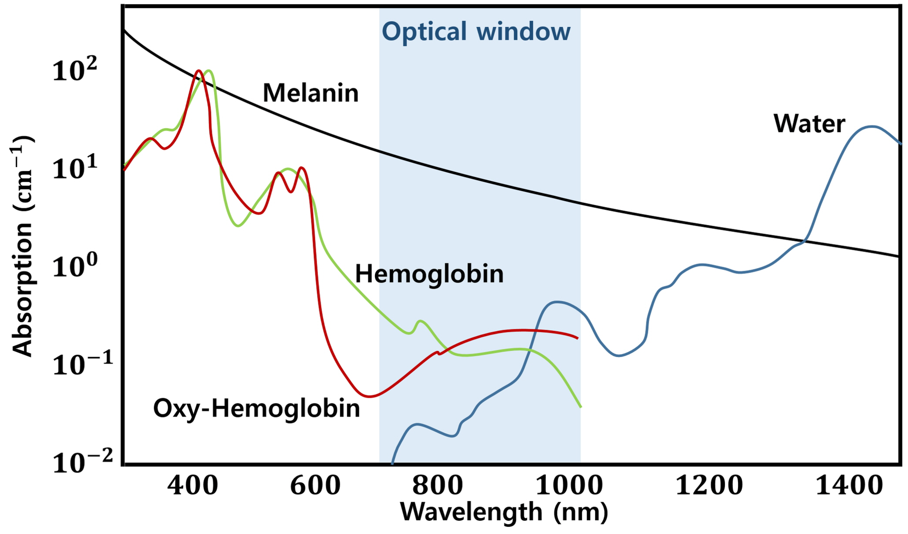
2.1. Vein Imaging
2.2. Related Research Trends
2.3. Used Algorithm
2.3.1. Morphology
2.3.2. Telea Inpainting
2.3.3. Histogram Normalization and Equalization
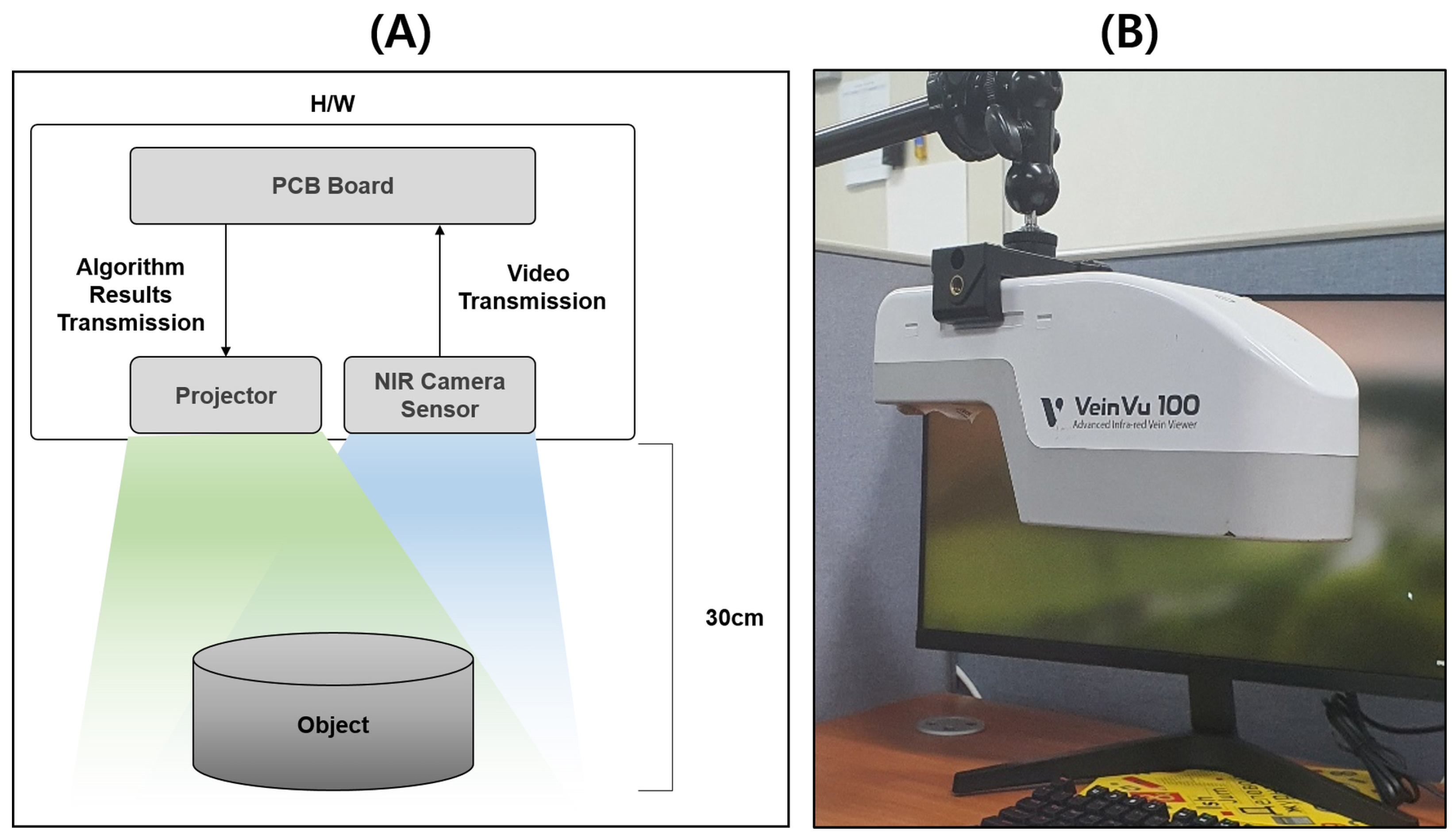
3. Proposed Intravenous Projector System
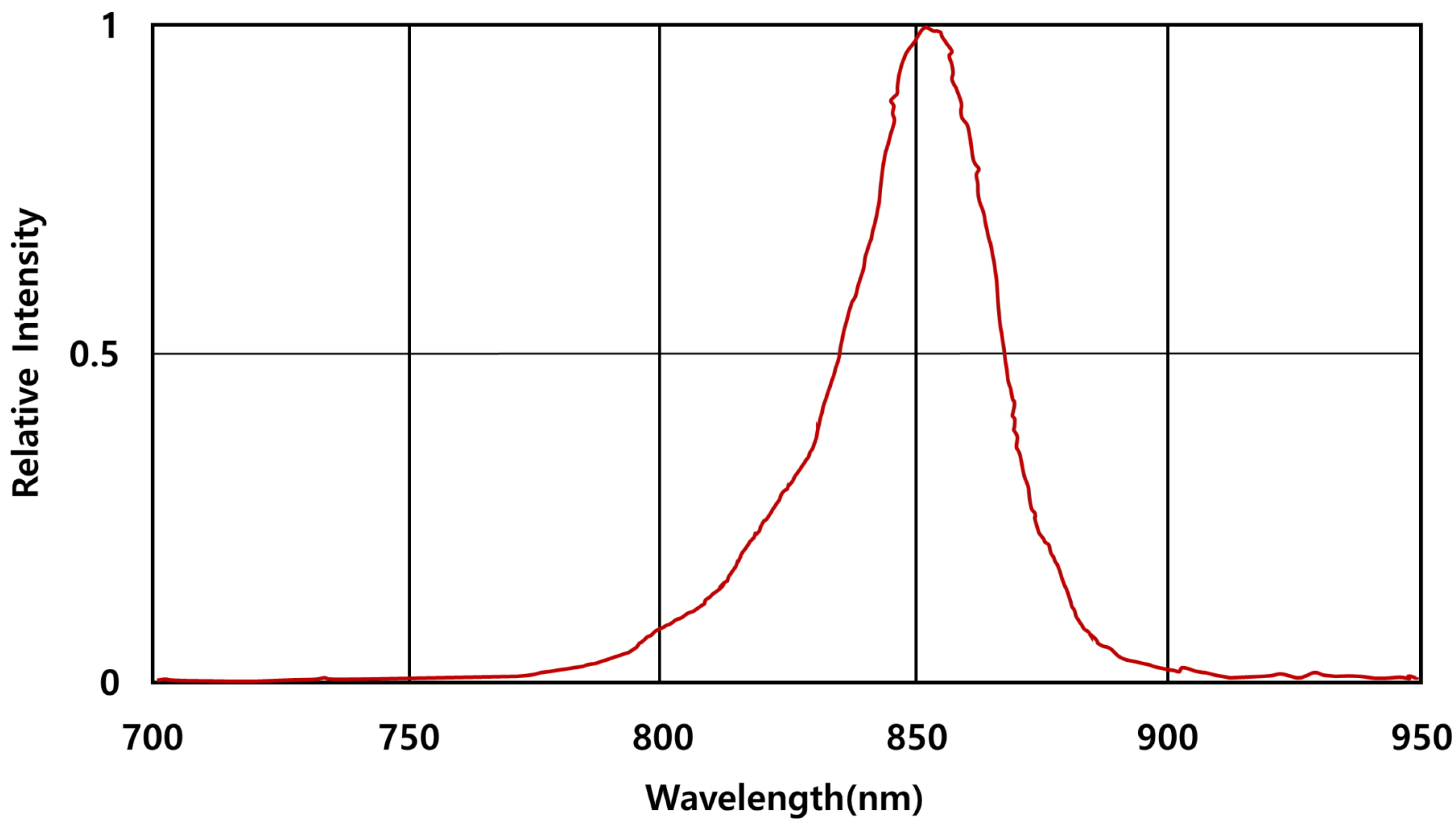
3.1. Video Shooting and Image Transfer
3.2. Video Shooting and Image Transfer
3.3. Vein Image Processing and Projector Image Delivery and Transmission
3.4. Building Embedded System Environment
3.4.1. Cross-Compiling for System Optimization and Saving Computing Resources
3.4.2. Utilization of Image Processing Library for System Optimization and Saving of Computing Resources
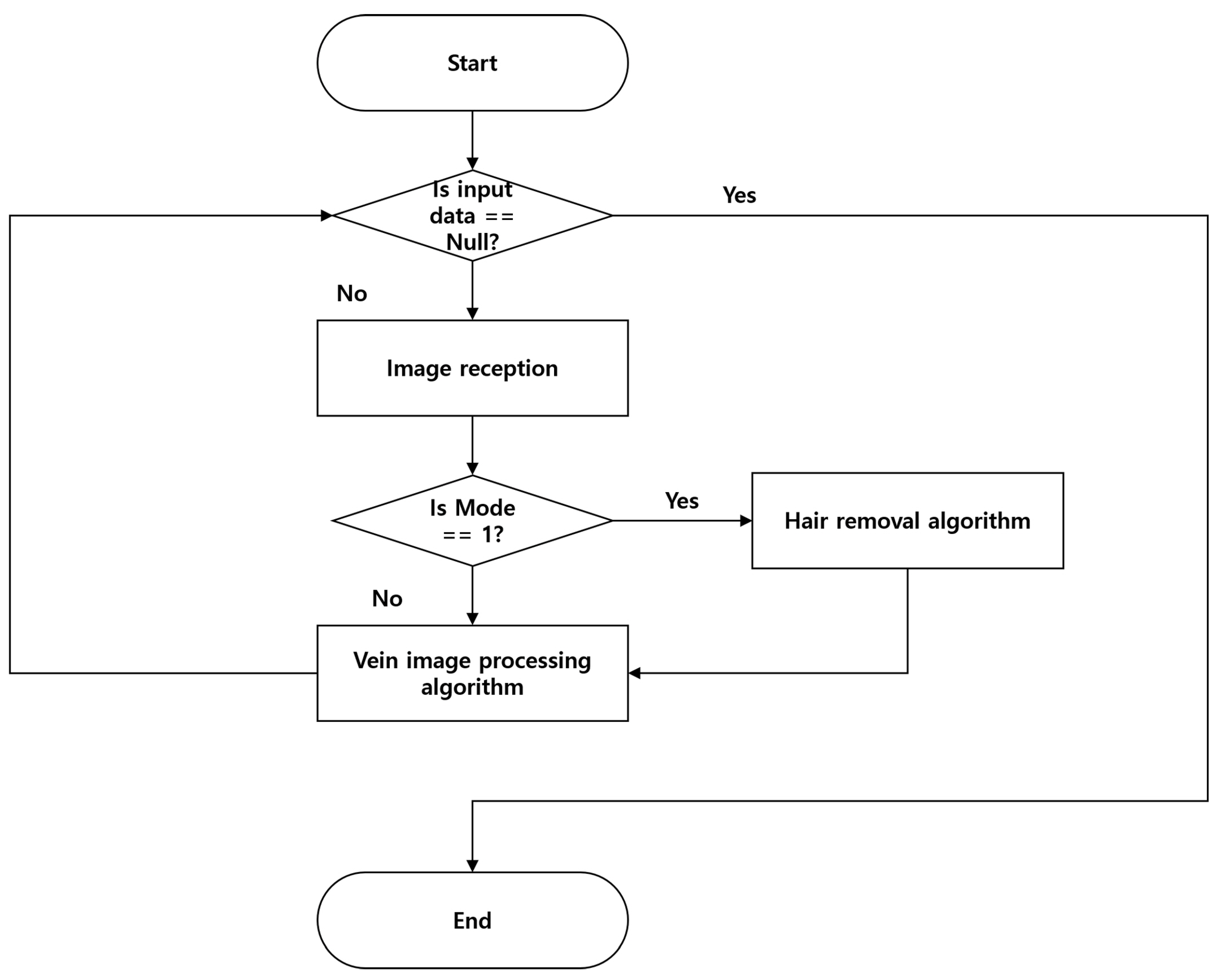
4. Proposed Vein Image Processing Algorithm
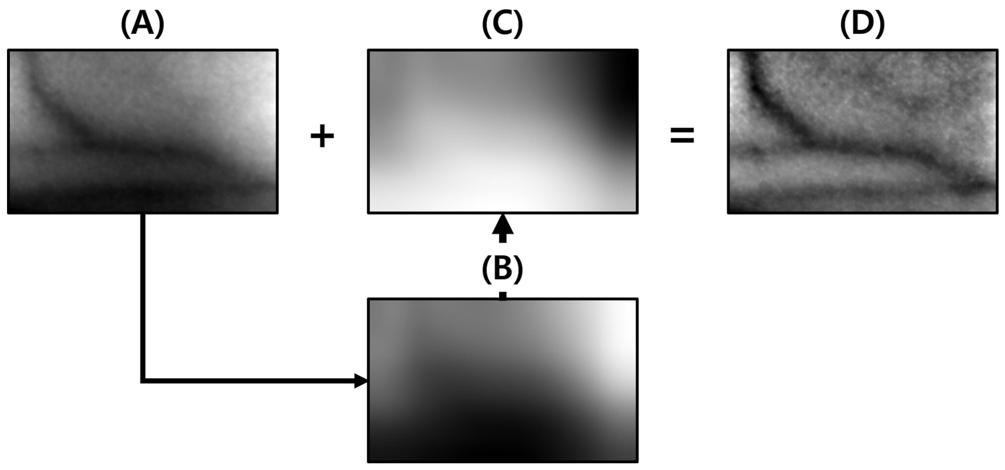
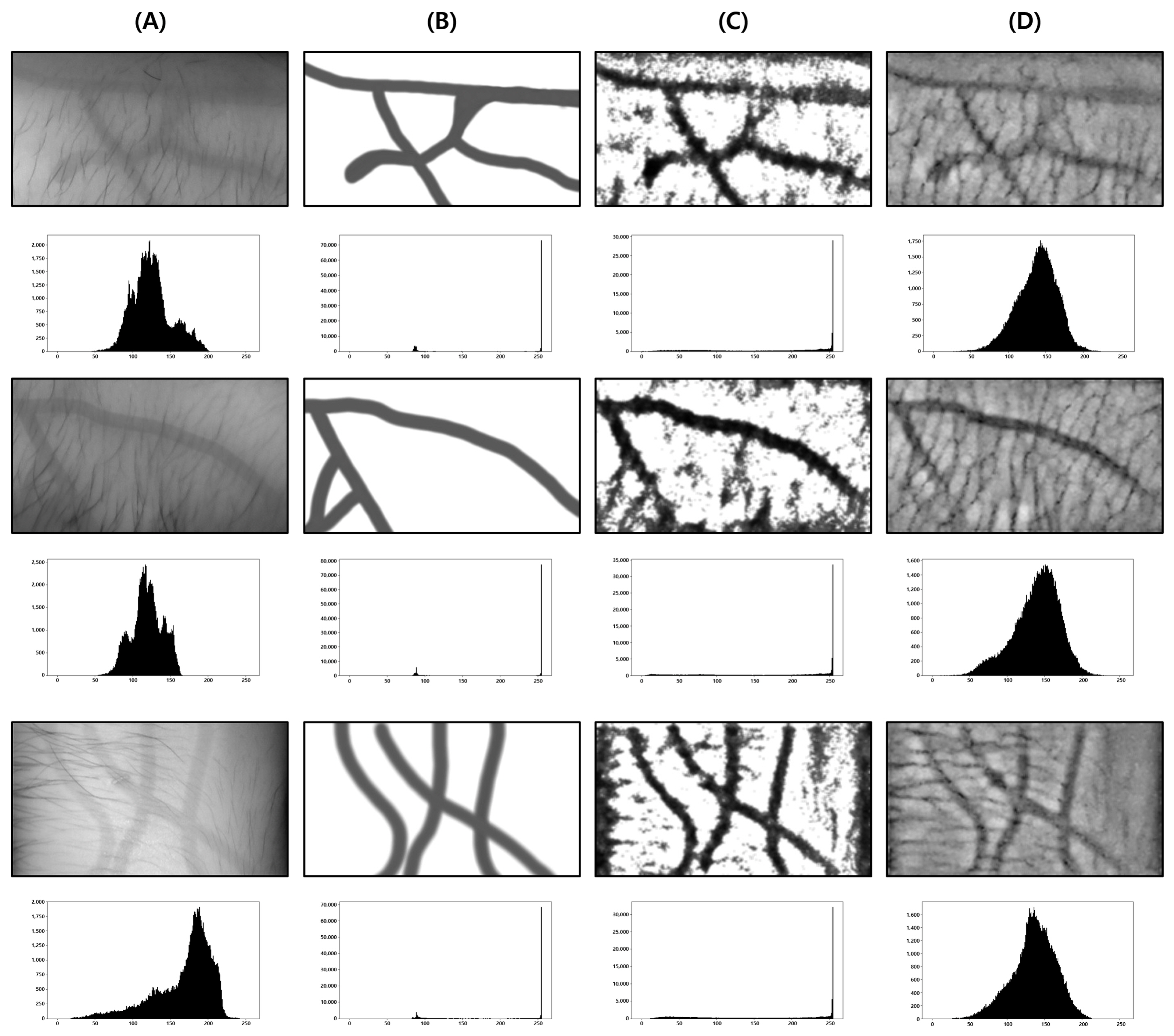
4.1. Light Component Removal
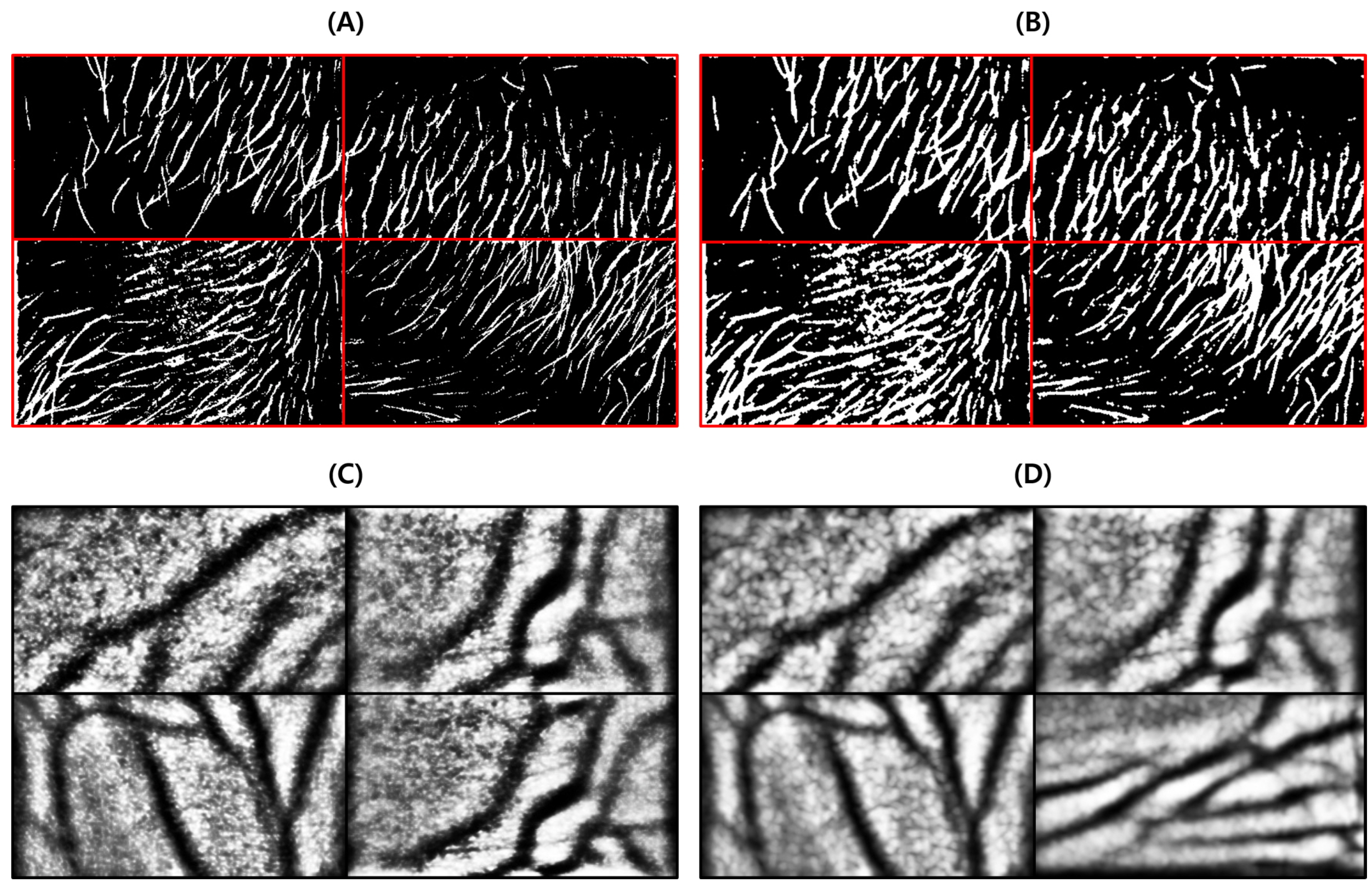
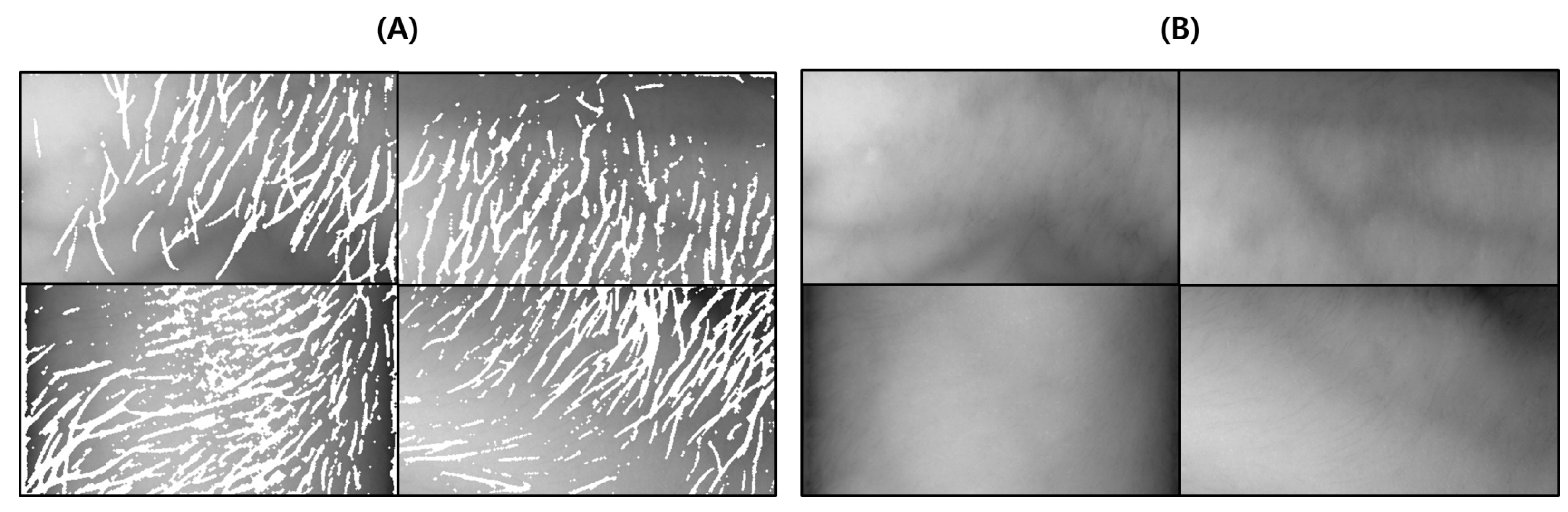
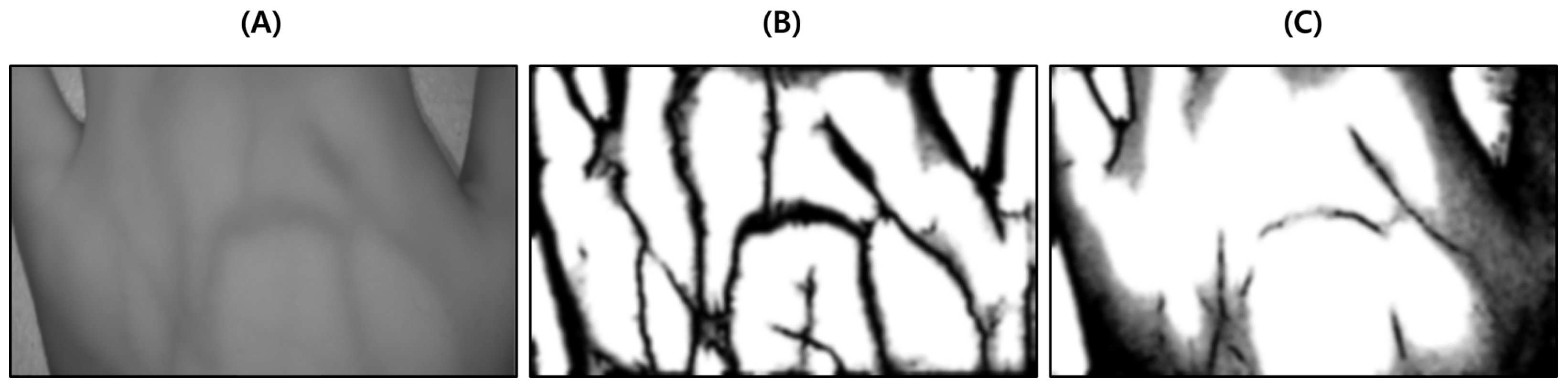
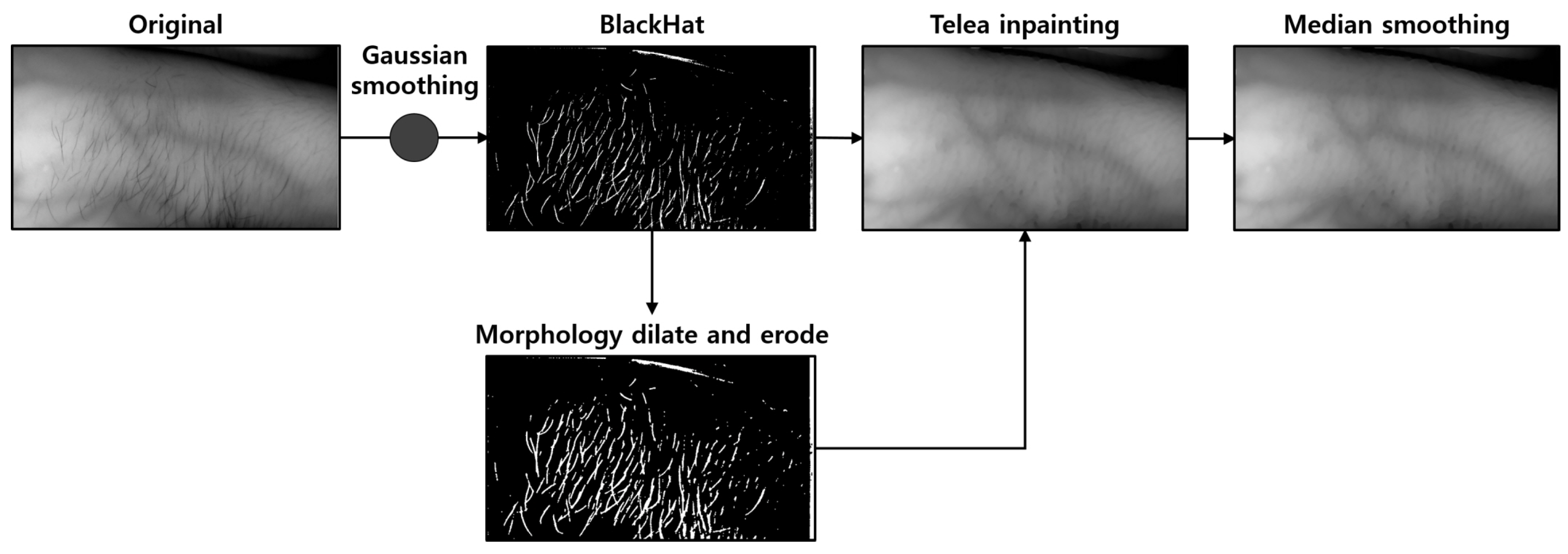
4.2. Hair Noise Removal
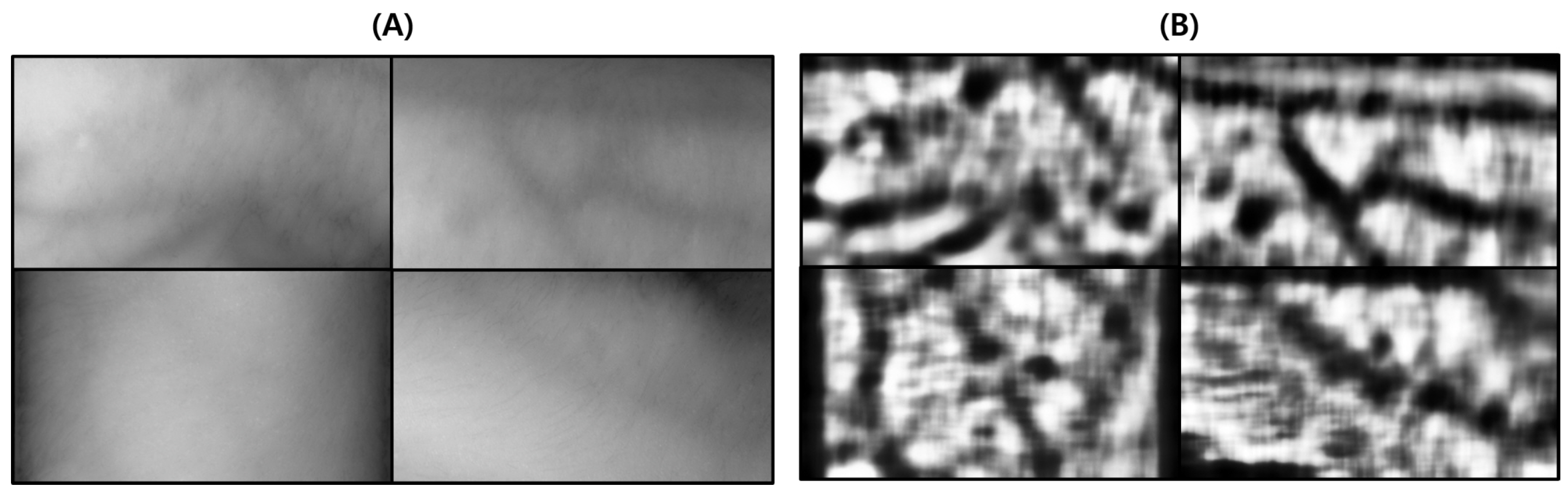
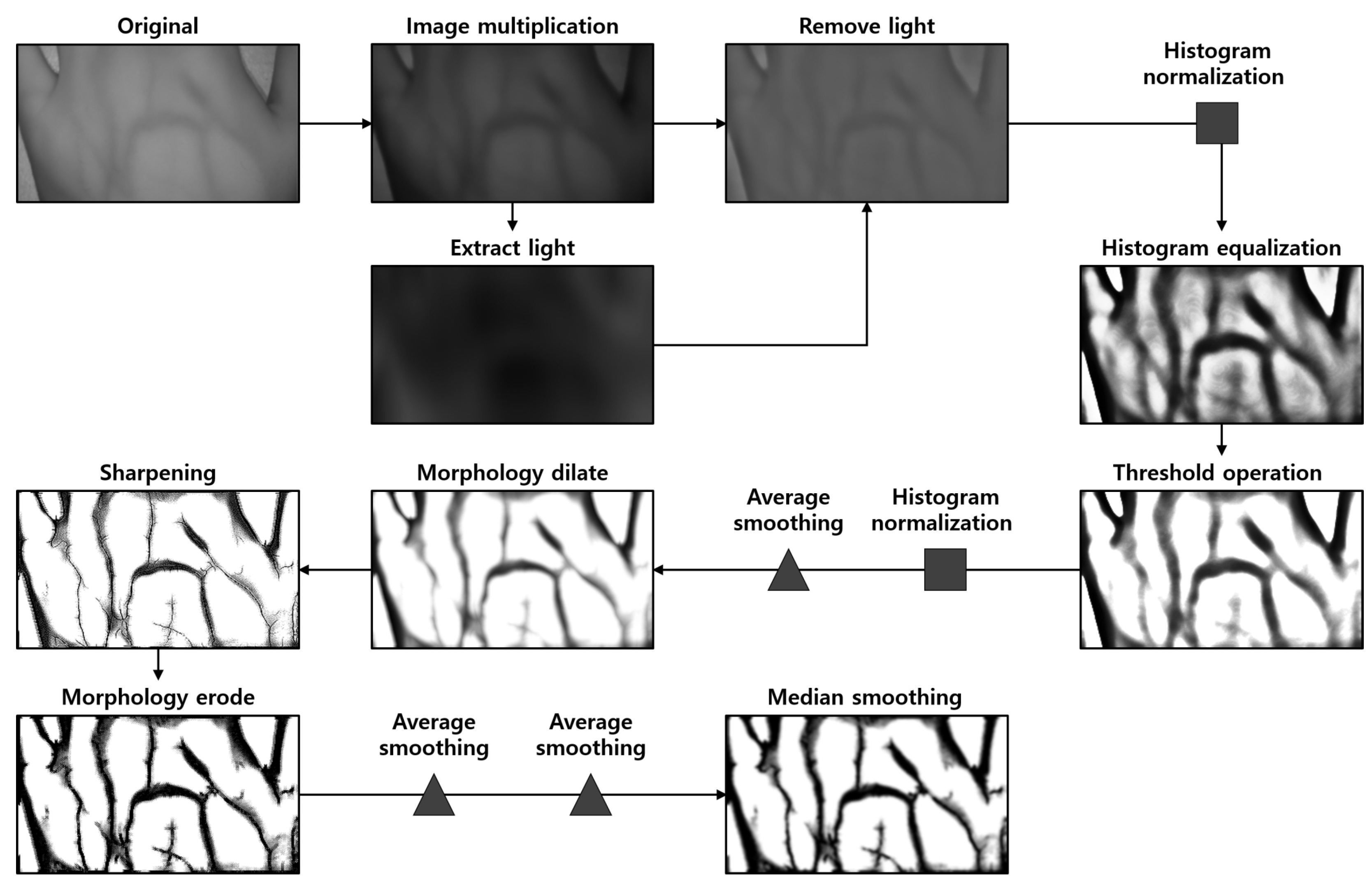
4.3. Vein Image Processing
- Extracting and removing light components to apply image processing algorithms uniformly to the entire image;
- Histogram normalization and equalization for the contrast enhancement of venous components;
- Threshold processing to remove noise such as skin wrinkles and fat layers in addition to vein components in the image;
- Morphological dilation to connect disconnected vein components and remove noise;
- Sharpening to restore thickened vein components attributed to morphological dilation;
- Median smoothing for image quality improvement and small noise removal.
5. Intravenous Projector System Performance Evaluation
5.1. Experimental Evaluation Environment
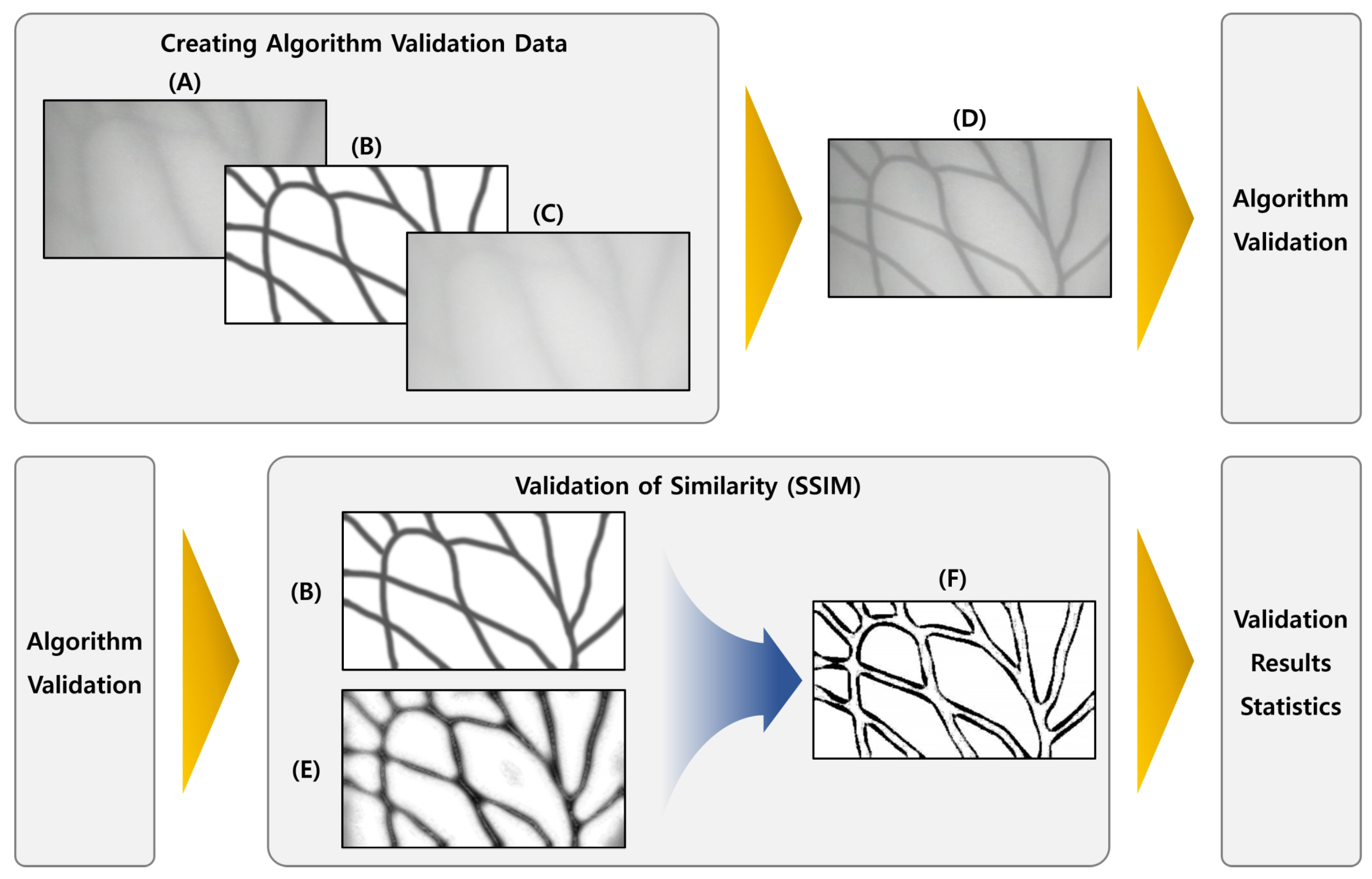
5.2. Dataset
Dataset for Verification of Vein Image Processing Algorithm
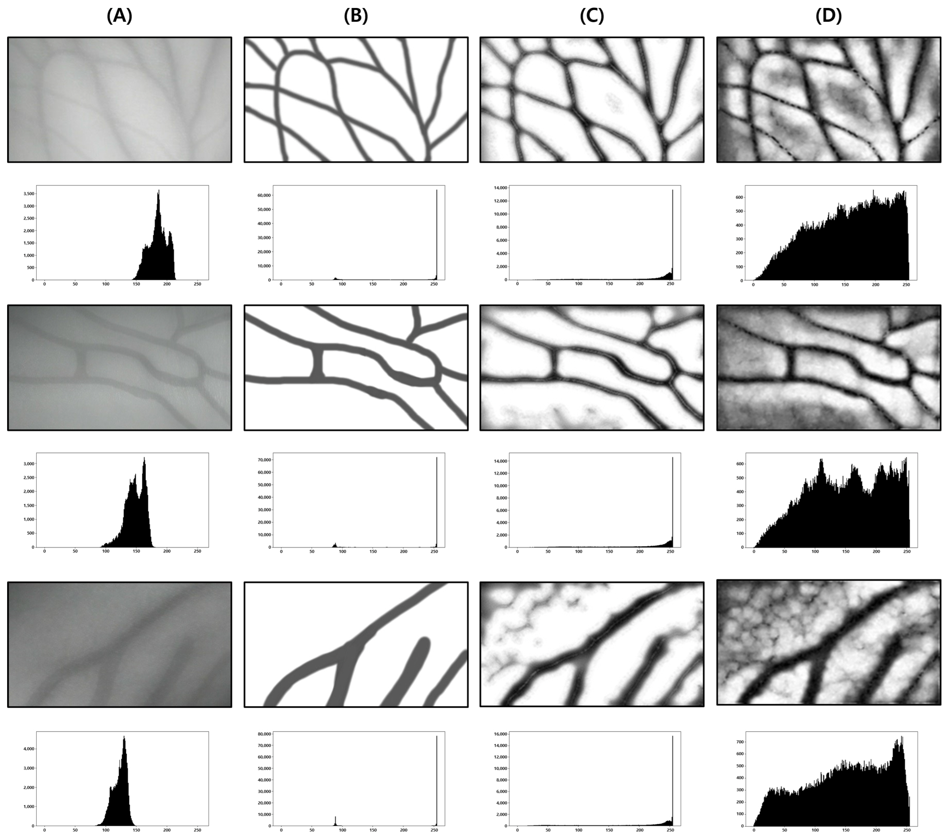
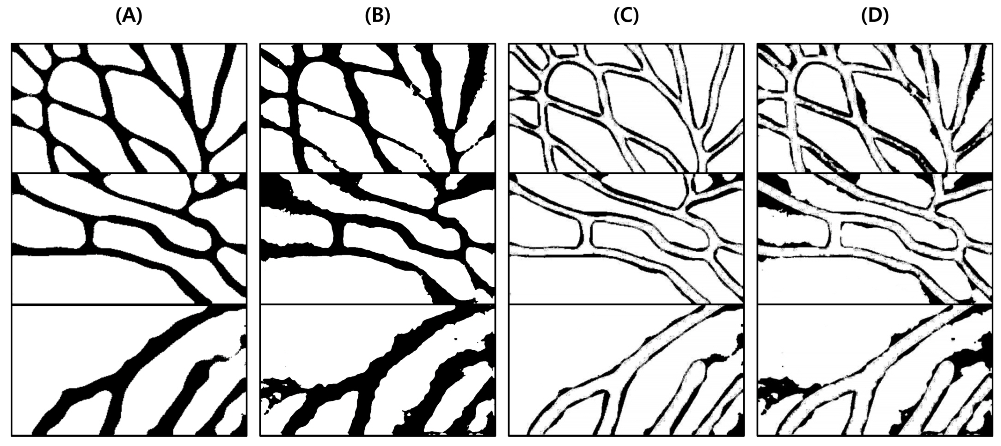
5.3. Functional Evaluation of Suggested Algorithm
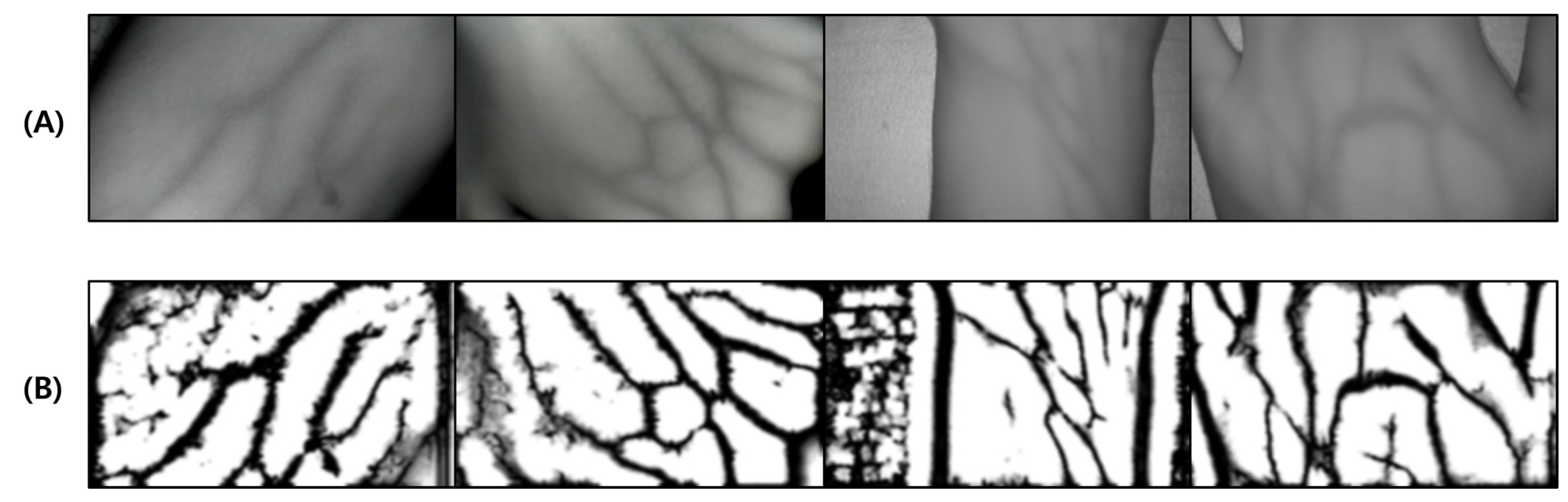
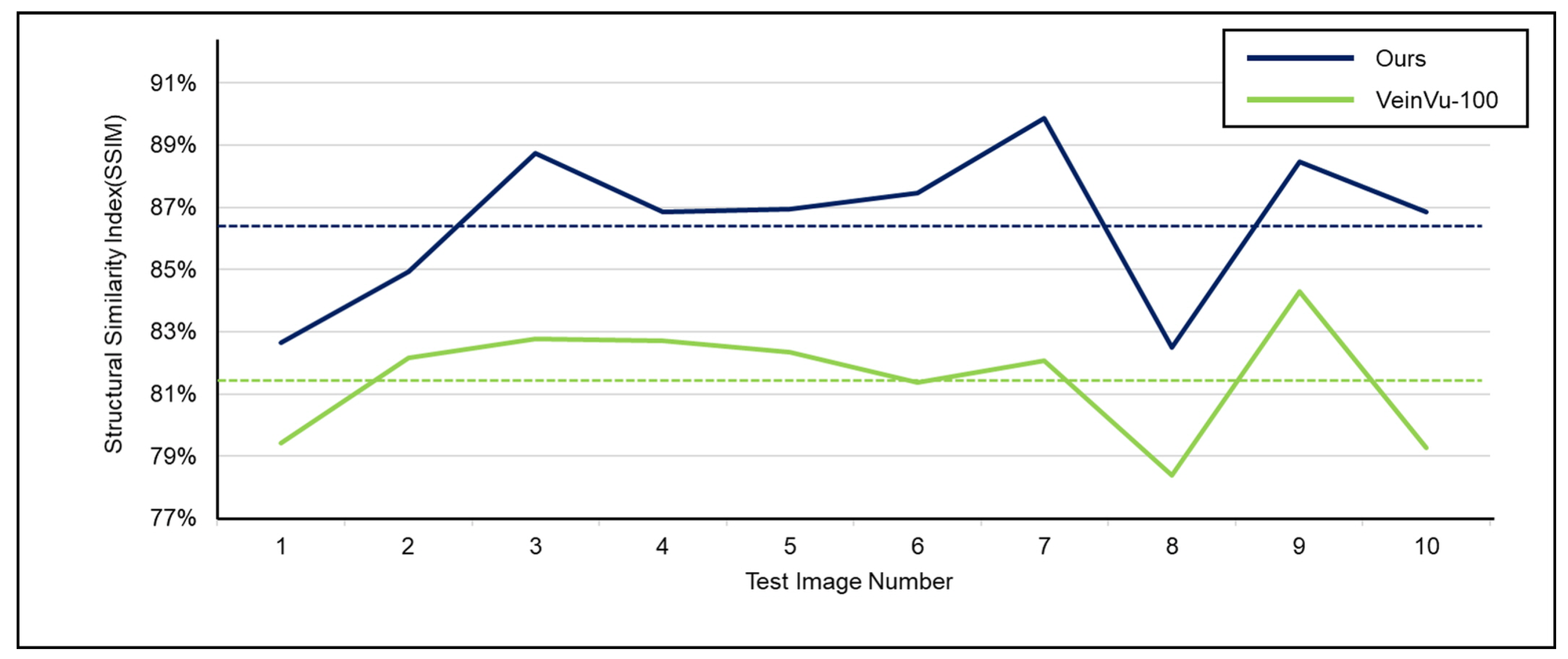
5.3.1. Proposed Algorithm Accuracy Verification
- To clearly distinguish the location of the vein output from each algorithm, it is converted into a binary image by applying a threshold to the resulting image.
- The similarity between binary images and expert-marked vein location images is compared using SSIM.
- The average of the similarities obtained through comparison of images of the entire data set is calculated.
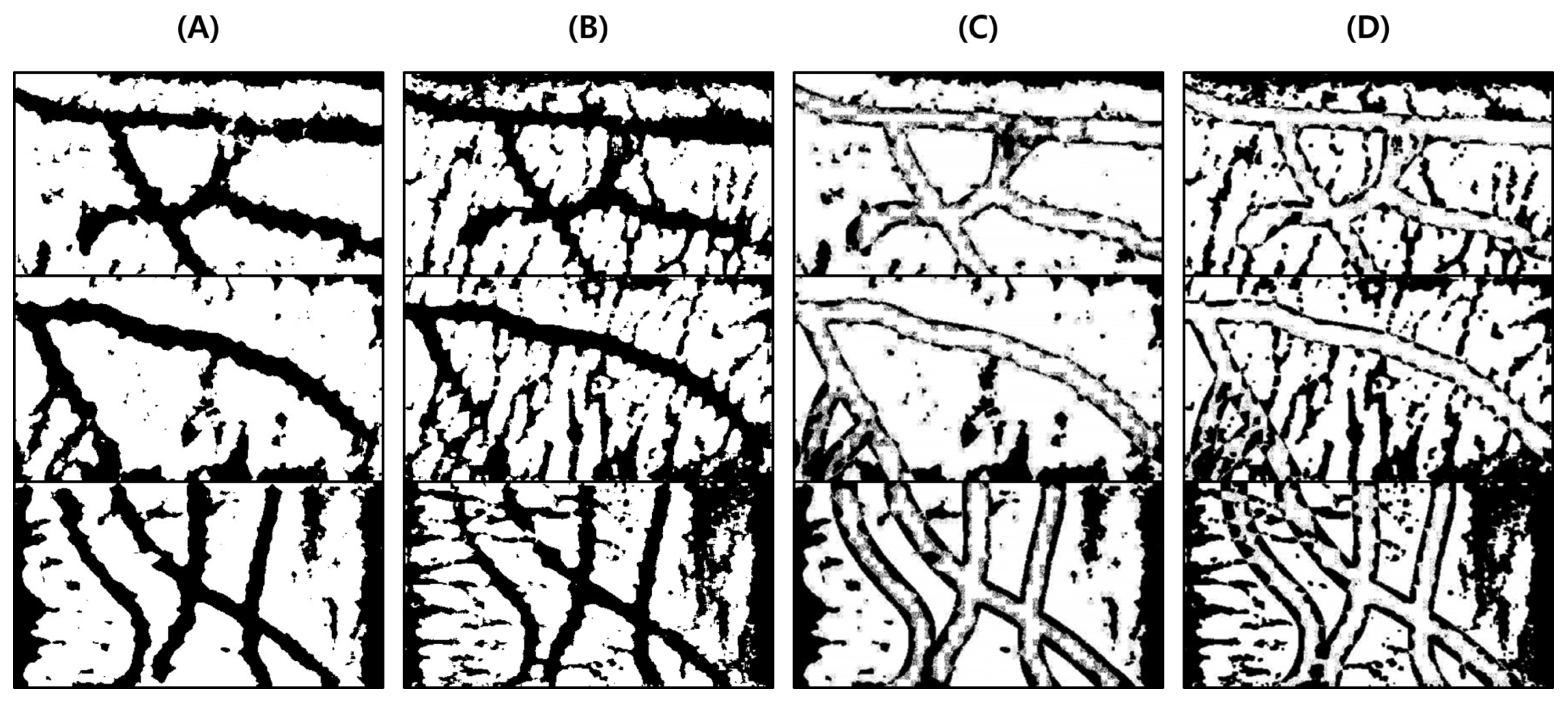
5.3.2. Performance Verification of Vein Image Processing Algorithm Based on Hair Noise Removal
5.3.3. Simple Vein Image Processing Algorithm Performance Verification
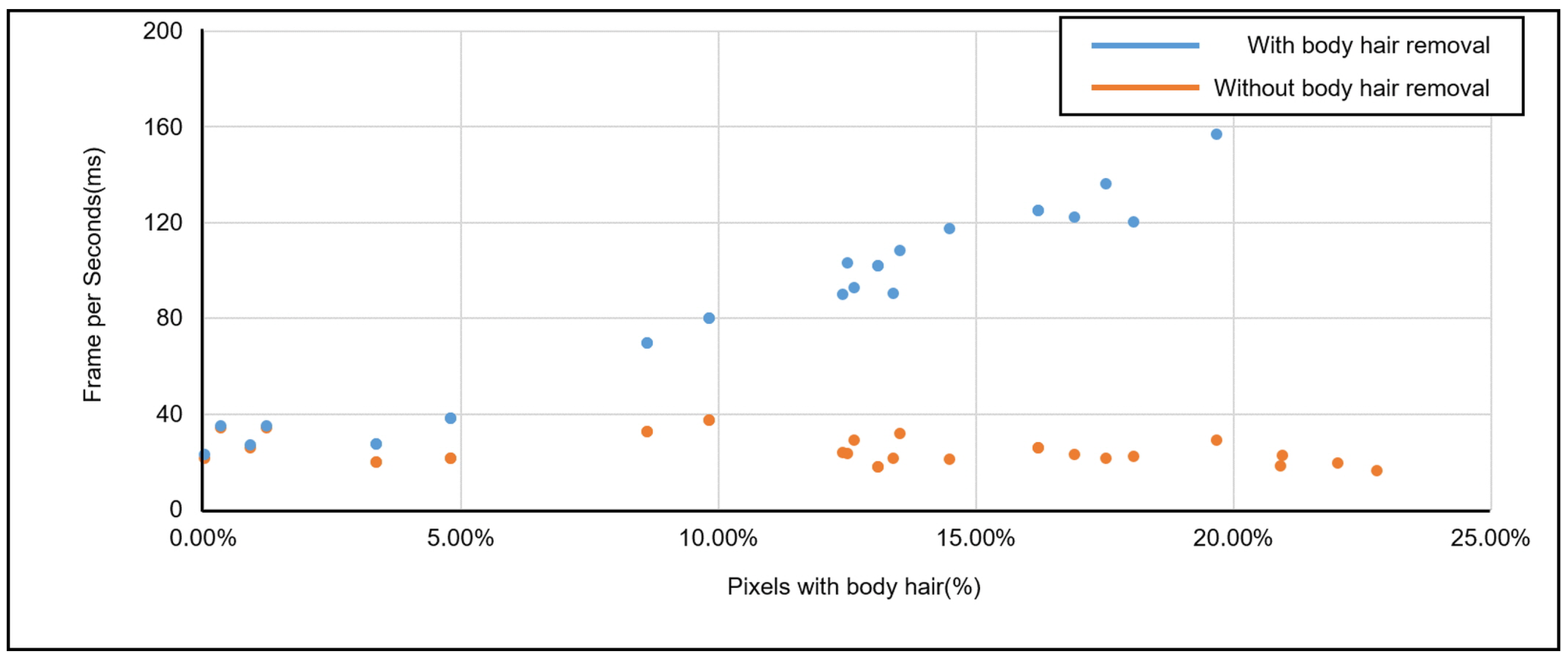
5.3.4. Vein Image Processing Algorithm Speed Performance Verification
6. Conclusions and Discussion
6.1. Conclusions
6.2. Discussion
Author Contributions
Funding
Institutional Review Board Statement
Informed Consent Statement
Data Availability Statement
Conflicts of Interest
References
- Ai, D.; Yang, J.; Fan, J.; Zhao, Y.; Song, X.; Shen, J.; Shao, L.; Wang, Y. Augmented reality based real-time subcutaneous vein imaging system. Biomed. Opt. Express 2016, 7, 2565–2585. [Google Scholar] [CrossRef] [Green Version]
- Kim, K.; Jeong, H.W.; Lee, Y. Performance Evaluation of Dorsal Vein Network of Hand Imaging Using Relative Total Variation-Based Regularization for Smoothing Technique in a Miniaturized Vein Imaging System: A Pilot Study. Int. J. Environ. Res. Public Health 2021, 18, 1548. [Google Scholar] [CrossRef] [PubMed]
- Li, W.; Raj, A.N.J.; Tjahjadi, T.; Zhuang, Z. Digital hair removal by deep learning for skin lesion segmentation. Pattern Recognit. 2021, 117, 107994. [Google Scholar] [CrossRef]
- Attia, M.; Hossny, M.; Zhou, H.; Nahavandi, S.; Asadi, H.; Yazdabadi, A. Digital hair segmentation using hybrid convolutional and recurrent neural networks architecture. Comput. Methods Programs Biomed. 2019, 177, 17–30. [Google Scholar] [CrossRef] [PubMed]
- Kim, M.J.; Park, J.M.; Rhee, N.; Je, S.M.; Hong, S.H.; Lee, Y.M.; Chung, S.P.; Kim, S.H. Efficacy of VeinViewer in pediatric peripheral intravenous access: A randomized controlled trial. Eur. J. Pediatr. 2012, 171, 1121–1125. [Google Scholar] [CrossRef] [PubMed]
- Karaaltin, M.V. Utilizing the vein viewer technology to map out a venous flap preoperatively. J. Reconstr. Microsurg. 2013, 29, 423–424. [Google Scholar] [CrossRef] [PubMed]
- Bardou, D.; Bouaziz, H.; Lv, L.; Zhang, T. Hair removal in dermoscopy images using variational autoencoders. Skin Res. Technol. 2022, 28, 445–454. [Google Scholar] [CrossRef] [PubMed]
- Yildiz, M.Z.; Boyraz, Ö.F. Development of a low-cost microcomputer based vein imaging system. Infrared Phys. Technol. 2019, 98, 27–35. [Google Scholar] [CrossRef]
- Telea, A. An image inpainting technique based on the fast marching method. J. Graph. Tools 2004, 9, 23–34. [Google Scholar] [CrossRef]
- Bradski, G. The openCV library. Dr. Dobb’s J. Softw. Tools Prof. Program. 2000, 25, 120–123. [Google Scholar]
- Sangiovanni-Vincentelli, A.; Martin, G. Platform-based design and software design methodology for embedded systems. IEEE Des. Test Comput. 2001, 18, 23–33. [Google Scholar] [CrossRef]
- Pulli, K.; Baksheev, A.; Kornyakov, K.; Eruhimov, V. Real-time computer vision with OpenCV. Commun. ACM 2012, 55, 61–69. [Google Scholar] [CrossRef]
- Parubochyi, V.; Shuwar, R. Fast self-quotient image method for lighting normalization based on modified Gaussian filter kernel. Imaging Sci. J. 2018, 66, 471–478. [Google Scholar] [CrossRef]
- Otsu, N. A threshold selection method from gray-level histograms. IEEE Trans. Syst. Man Cybern. 1979, 9, 62–66. [Google Scholar] [CrossRef] [Green Version]
- Pi, R. Raspberry pi 3 Model b. 2015. Available online: https://www.raspberrypi.org (accessed on 22 April 2022).
- Wang, Z.; Bovik, A.C.; Sheikh, H.R.; Simoncelli, E.P. Image quality assessment: From error visibility to structural similarity. IEEE Trans. Image Process. 2004, 13, 600–612. [Google Scholar] [CrossRef] [PubMed] [Green Version]
- De Bruijne, M. Machine learning approaches in medical image analysis: From detection to diagnosis. Med. Image Anal. 2016, 33, 94–97. [Google Scholar] [CrossRef] [PubMed]
- Hasan, A.H. DNA Repair Genes (APE1 and XRCC1) Polymorphisms–Cadmium Interaction in Fuel Station Workers. J. Pharm. Negat. Results 2022, 13, 32–37. [Google Scholar]


| Item | Specifications |
|---|---|
| Compiler | arm-Linux-gnueabihf-g++ |
| OpenCV version | OpenCV 3.4.1 |
| Included library | opencv2/opencv |
| opencv2/imgproc | |
| opencv2/video | |
| opencv2/core | |
| opencv2/imgproc | |
| opencv2/highgui |
| Item | Specifications |
|---|---|
| CPU | Quad core ARM Cortex-A53 CPU Broadcom (1.4 GHz) |
| RAM | 1GB LPDDR2 SDRAM |
| GPU | Video core IV |
| Storage | 128 GB SD Card |
| OS | Raspbian STRETCH LITE (1.8 G) |
| LAN | 2.4 GHz and 5 GHz IEEE 802.11.b/g/n/ac Wireless LAN and Bluetooth 4.2/BLE |
| Size | 120 mm × 75 mm × 34 mm |
| Weight | 75 g |
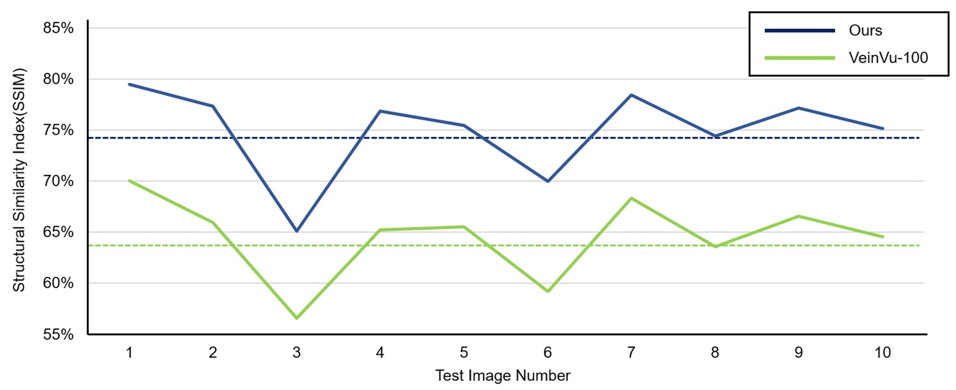
| Item | Ours | Veinvu-100 | |
|---|---|---|---|
| Images with hair | Max | 79.46% | 70.03% |
| Min | 65.07% | 56.59% | |
| Average | 74.93% | 64.55% | |
| Item | Ours | Veinvu-100 | |
|---|---|---|---|
| Images without hair | Max | 89.86% | 84.30% |
| Min | 82.49% | 78.38% | |
| Average | 86.52% | 81.48% | |
Publisher’s Note: MDPI stays neutral with regard to jurisdictional claims in published maps and institutional affiliations. |
© 2022 by the authors. Licensee MDPI, Basel, Switzerland. This article is an open access article distributed under the terms and conditions of the Creative Commons Attribution (CC BY) license (https://creativecommons.org/licenses/by/4.0/).
Share and Cite
Lee, J.; Jeong, I.; Kim, K.; Cho, J. Design and Implementation of Embedded-Based Vein Image Processing System with Enhanced Denoising Capabilities. Sensors 2022, 22, 8559. https://doi.org/10.3390/s22218559
Lee J, Jeong I, Kim K, Cho J. Design and Implementation of Embedded-Based Vein Image Processing System with Enhanced Denoising Capabilities. Sensors. 2022; 22(21):8559. https://doi.org/10.3390/s22218559
Chicago/Turabian StyleLee, Jongwon, Incheol Jeong, Kapyol Kim, and Jinsoo Cho. 2022. "Design and Implementation of Embedded-Based Vein Image Processing System with Enhanced Denoising Capabilities" Sensors 22, no. 21: 8559. https://doi.org/10.3390/s22218559