
The Characteristics Analysis of a Microfluid-Based EGFET Biosensor with On-Chip Sensing Film for Lactic Acid Detection


Abstract
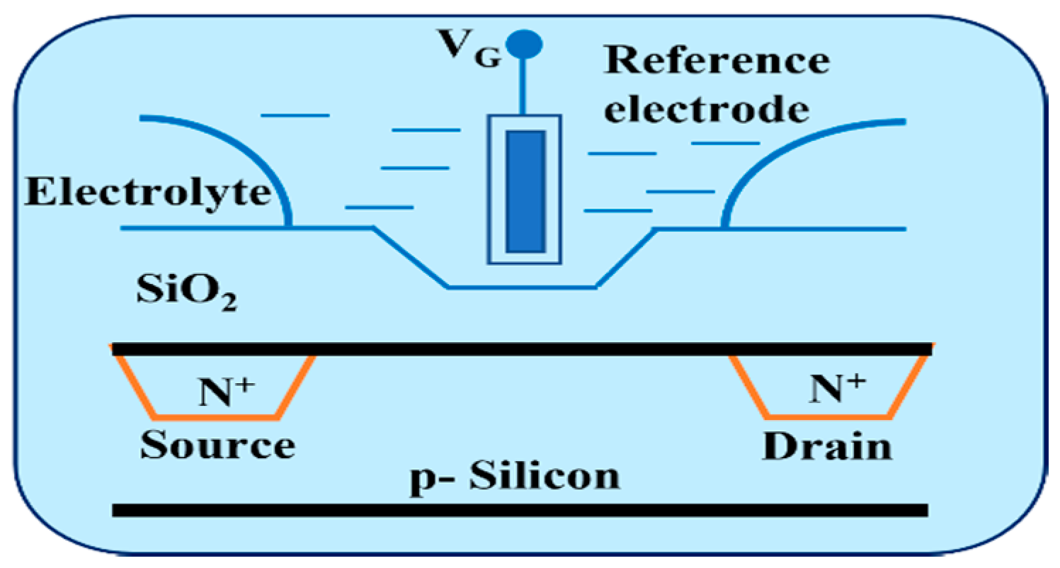
:1. Introduction
2. Materials
2.1. Materials
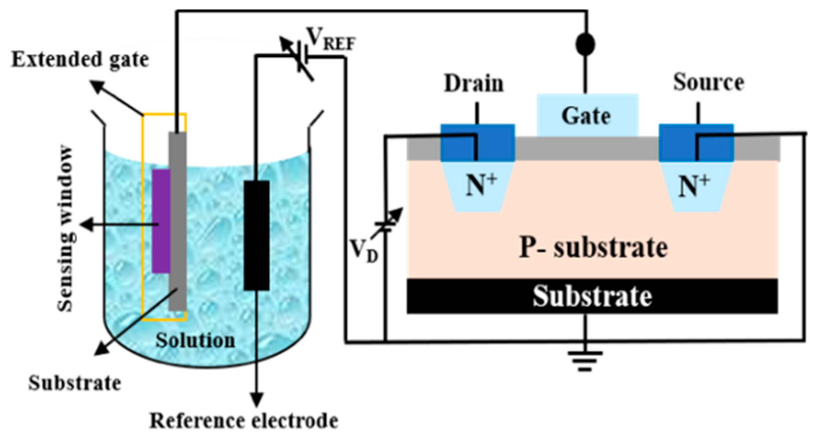
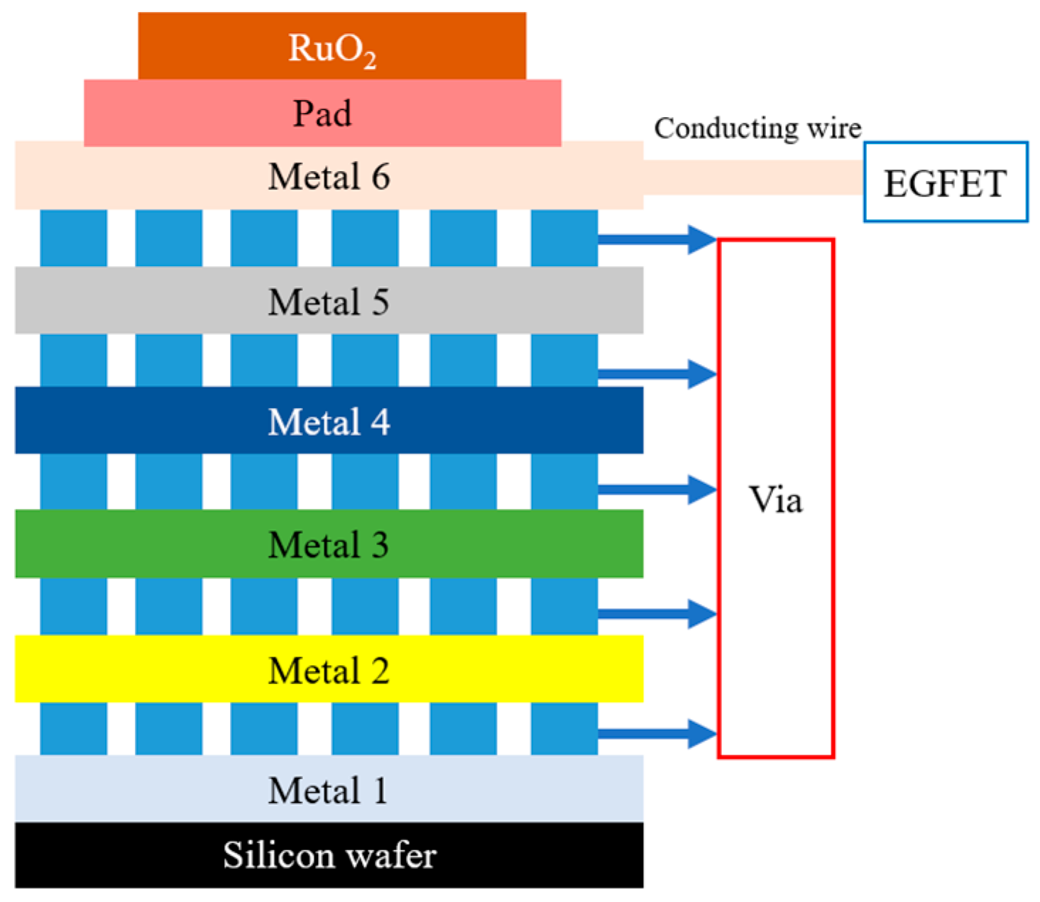
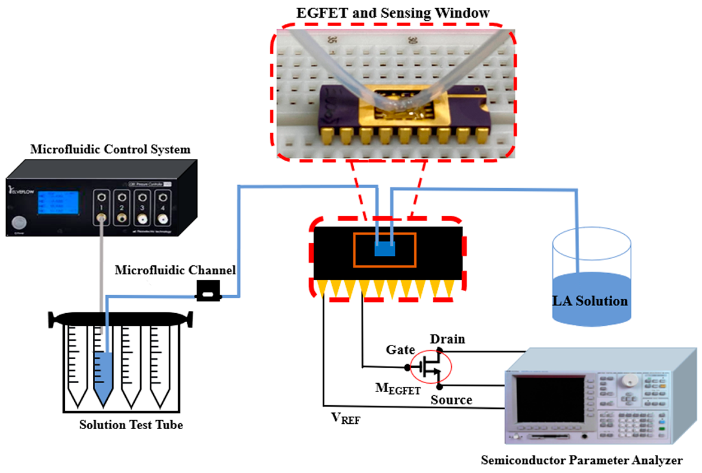
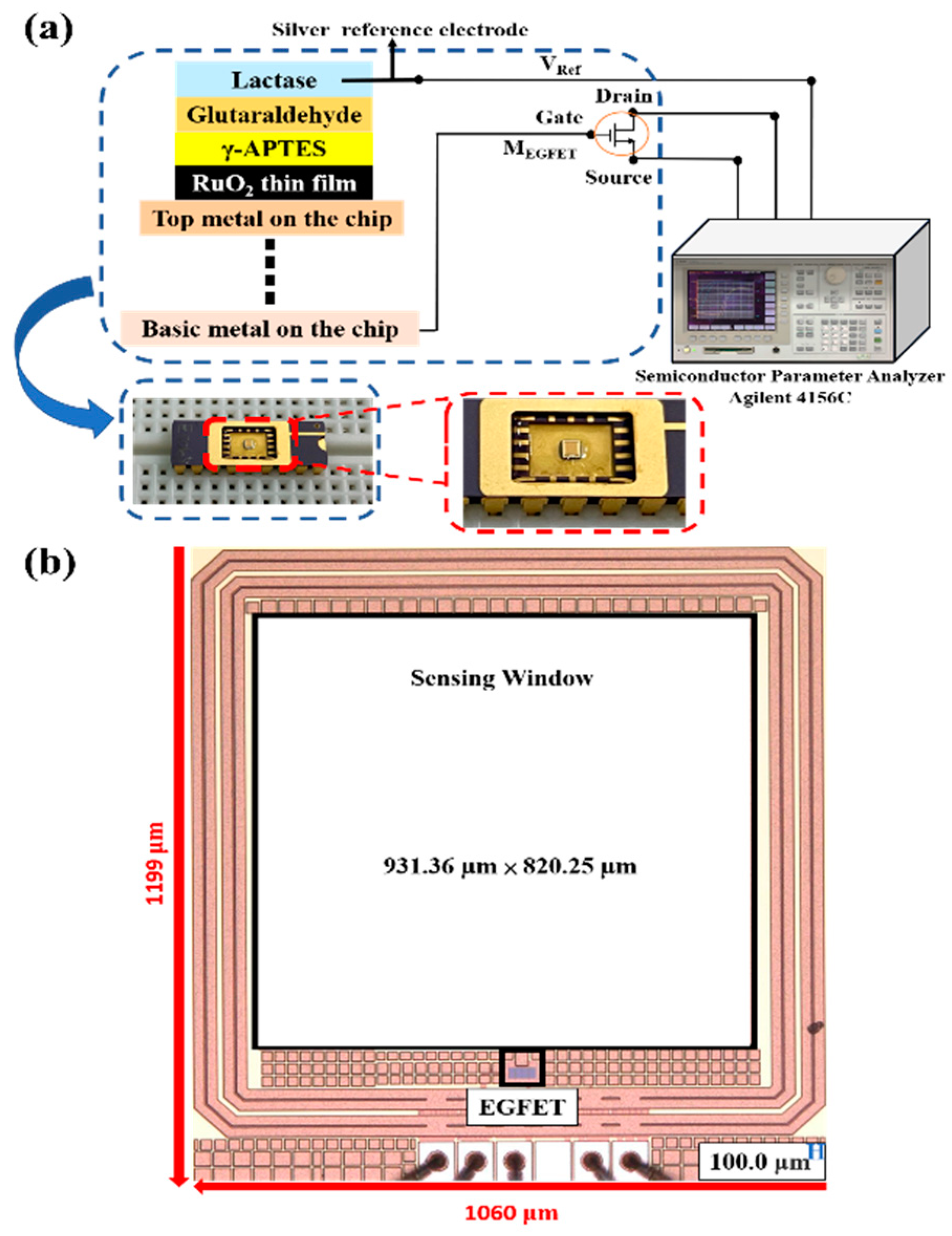
2.2. The OCSW Integrates the Design of EGFET
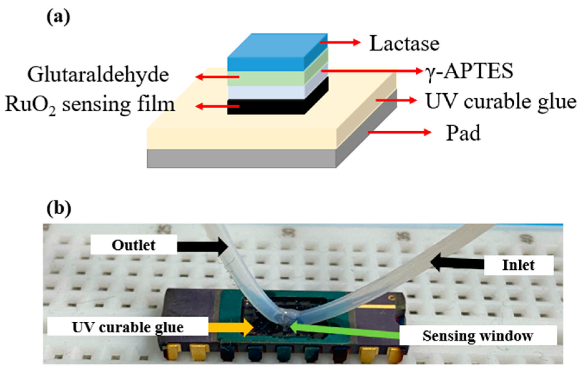
2.3. Fabrication of the LA Sensing Film
- E0 is the reference electrode potential;
- R is the universal gas constant, equal to 8.31 J/(Kmol);
- T is the absolute temperature;
- F is the Faradays constant, equal to 96485.33 C/mol;
- [Ru (OH)3] is the activity of RuIII at absolute temperature;
- [RuO2] is the activity of RuIV at absolute temperature;
- H+ represents the activity at absolute temperature
2.4. Sensitivity Measurement of LA Sensing Window
3. Results and Discussion
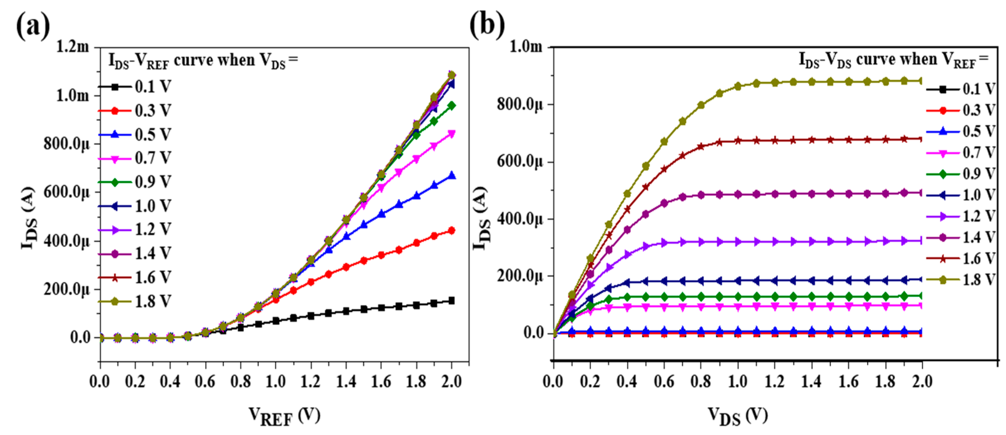
3.1. Sensing Analysis of OCSW
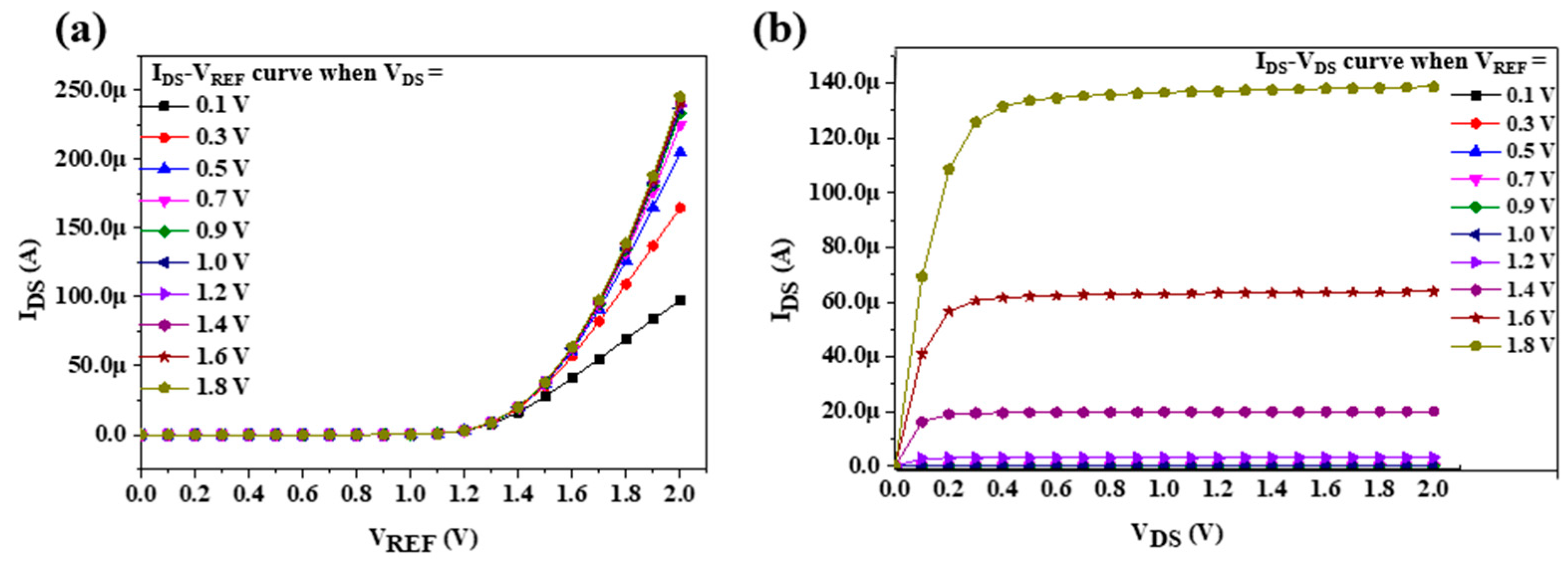
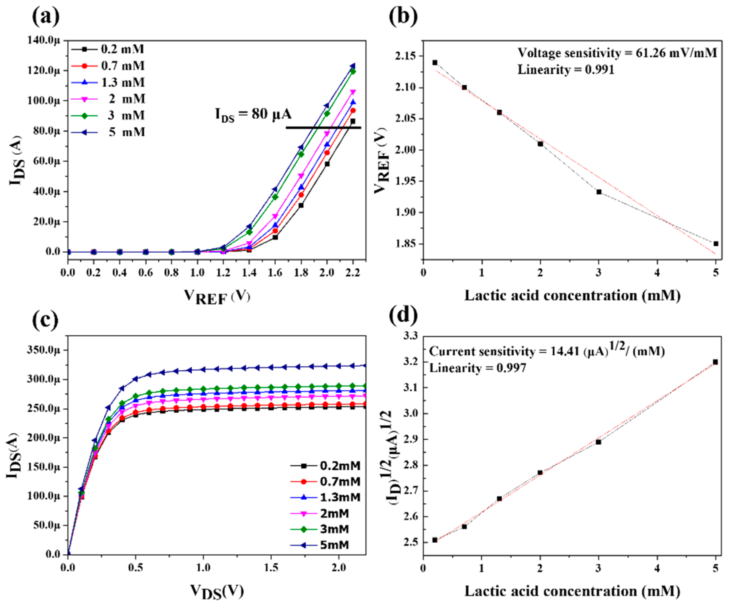
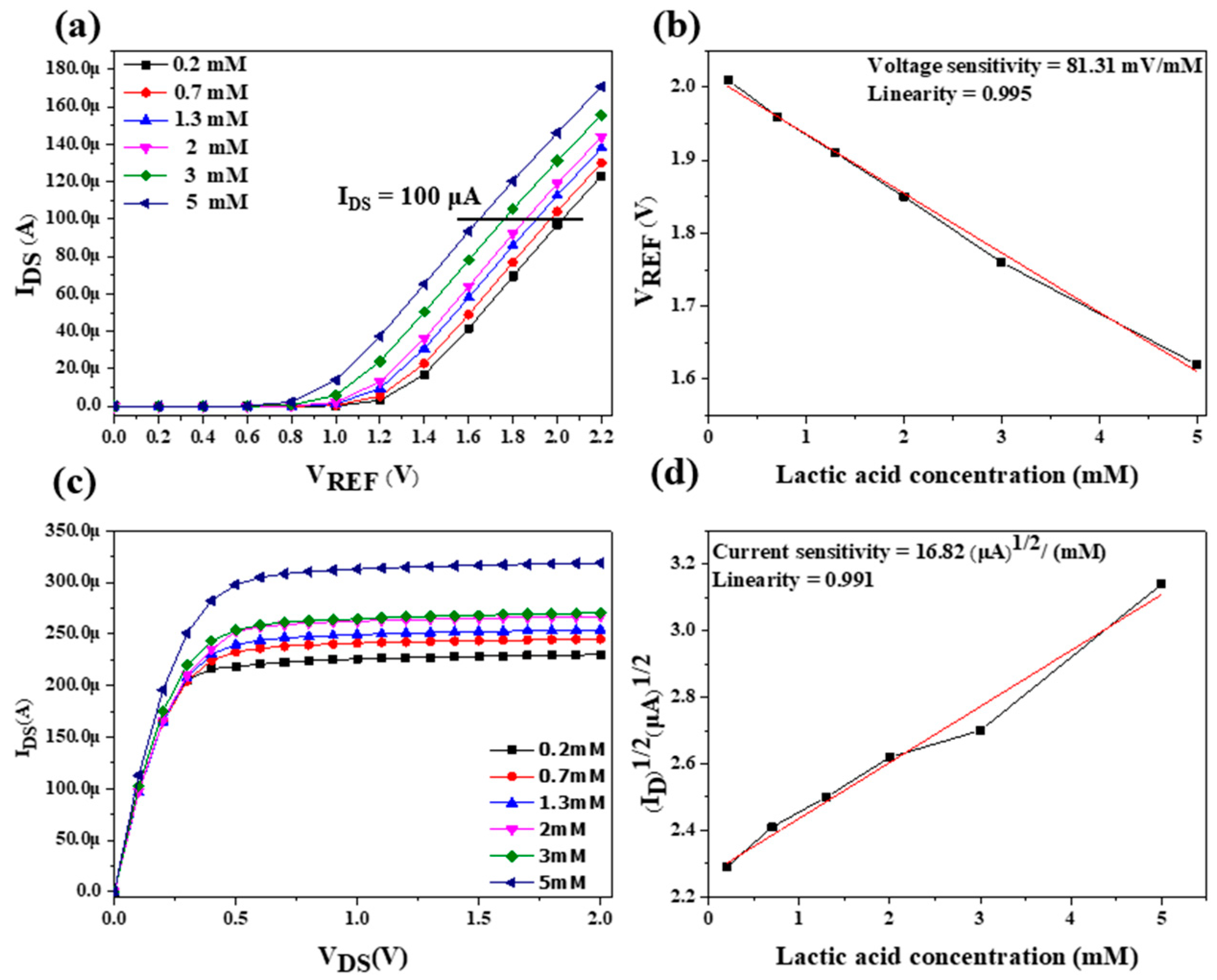
3.2. Sensitivity and Linearity of LA Biosensor
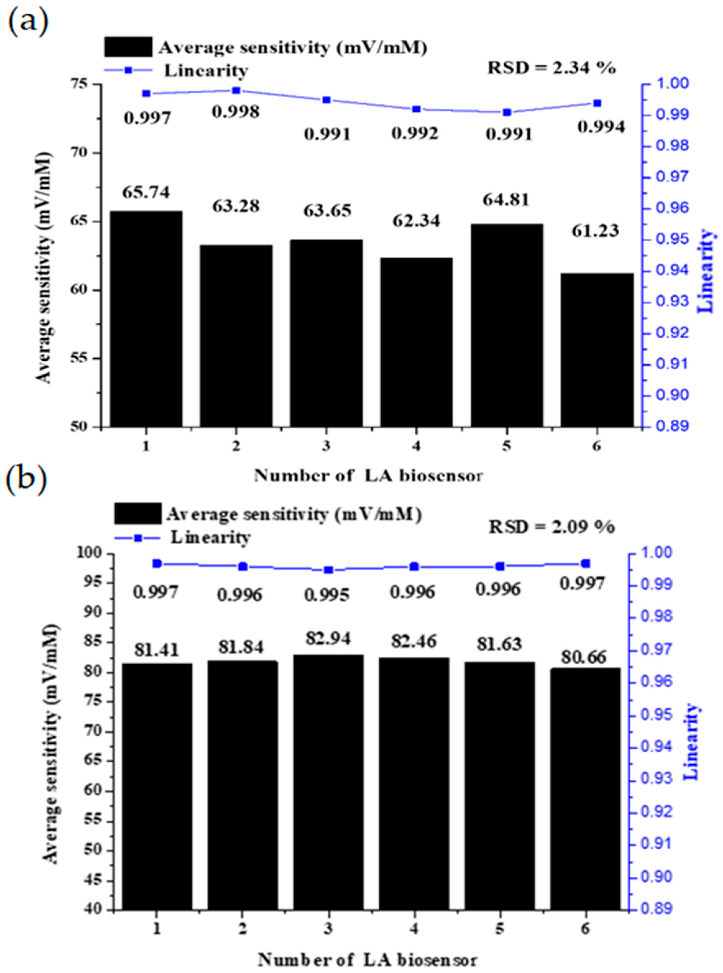
3.3. Reproducibility
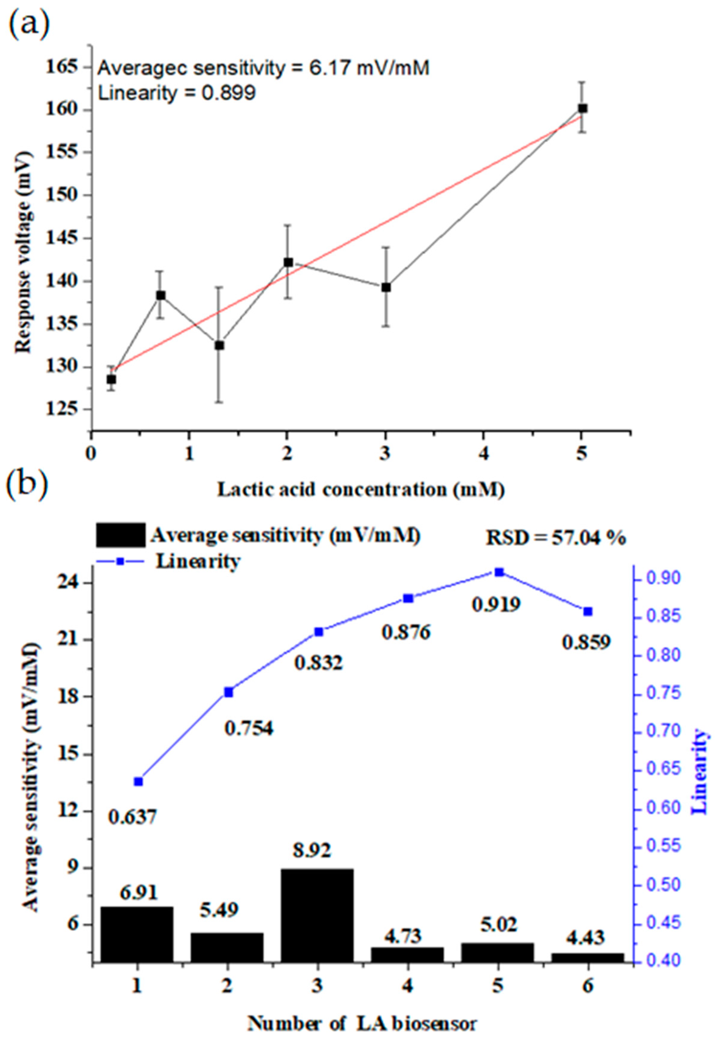
3.4. Sensing Characteristics Analysis of the Standard Sensor
4. Conclusions
Author Contributions
Funding
Institutional Review Board Statement
Informed Consent Statement
Data Availability Statement
Acknowledgments
Conflicts of Interest
References
- Jing, K.H.; Arshad, M.K.M.d.; Huda, A.R.N.; Ruslinda, R.; Gopinath, S.C.B.; Nuzaihan, M.N.M.; Ayub, R.M.; Fathil, M.F.M.; Othman, N.; Hashim, U. Gate Dielectric Scaling in MOSFETs Device. In Proceedings of the 2016 AIP Conference, Penang, Malaysia, 6 July 2016. [Google Scholar]
- Liu, J.; Ohsato, H.; Wang, X.; Liao, M.; Koide, Y. Design and Fabrication of High-Performance Diamond Triple-Gate Field-Effect Transistors. Sci. Rep. 2016, 6, 34757. [Google Scholar]
- Kahng, D. A Historical Perspective on the Development of MOS Transistors and Related Devices. IEEE Trans. Electron Devices 1976, 23, 655–657. [Google Scholar] [CrossRef]
- Bergveld, P. Development of an Ion-sensitive Solid-State Device for Neurophysiological Measurements. IEEE Trans. Biomed. Eng. 1970, BME-17, 70–71. [Google Scholar]
- Goda, T. Chemically Induced pH Perturbations for Analyzing Biological Barriers Using Ion-Sensitive Field-Effect Transistors. Sensors 2021, 21, 7277–7292. [Google Scholar] [PubMed]
- Xu, G.; Abbott, J.; Ham, D. Optimization of CMOS-ISFET-Based Biomolecular Sensing: Analysis and Demonstration in DNA Detection. IEEE Trans. Electron Devices 2016, 63, 3249–3256. [Google Scholar]
- Zhao, S.; Shi, C.; Hu, H.; Li, Z.; Xiao, G.; Yang, Q.; Sun, P.; Cheng, L.; Niu, W.; Bi, J.; et al. ISFET and Dex-Agnps Based Portable Sensor for Reusable and Real-Time Determinations of Concanavalin A and Glucose on Smartphone. Biosens. Bioelectron. 2020, 151, 111962. [Google Scholar]
- Chaudhary, R.; Sharma, A.; Sinha, S.; Yadav, J.; Sharma, R.; Mukhiya, R.; Khanna, V.K. Fabrication and Characterization of Al Gate N-MOSFET, on-chip Fabricated with Si3N4 ISFET. In Proceedings of the 19th International Symposium on VLSI Design and Test, Ahmedabad, India, 26–29 June 2015; pp. 1–4. [Google Scholar]
- Spiegel, J.v.d.; Lauks, I.; Chan, P.; Babic, D. The Extended Gate Chemically Sensitive Field Effect Transistor as Multi-Species Microprobe. Sens. Actuators 1983, 4, 291–298. [Google Scholar] [CrossRef]
- Chou, J.C.; Chen, C.W. Fabrication and Application of Ruthenium-Doped Titanium Dioxide Films as Electrode Material for Ion-Sensitive Extended-Gate FETs. IEEE Sens. J. 2009, 9, 277–284. [Google Scholar] [CrossRef]
- Wrege, R.; Peter, C.; Wesling, B.N.; Rambo, C.R.; Schneider, M.C.; Galup-Montoro, C. A CMOS Test Chip with Simple Post-Processing Steps for Dry Characterization of ISFET Arrays. IEEE Sens. J. 2021, 21, 4755–4763. [Google Scholar]
- Manaresi, N.; Romani, A.; Medoro, G.; Altomare, L.; Leonardi, A.; Tartagni, M.; Guerrieri, R. A CMOS Chip for Individual Cell Manipulation and Detection. IEEE J. Solid-State Circuits 2003, 38, 2297–2305. [Google Scholar]
- Miscourides, N.; Georgiou, P. ISFET Arrays in CMOS: A Head-to-Head Comparison Between Voltage and Current Mode. IEEE Sens. J. 2019, 19, 1224–1238. [Google Scholar] [CrossRef]
- Kim, T.; Bao, C.; Hausmann, M.; Siqueira, G.; Zimmerman, T.; Kim, W.S. 3D Printed Disposable Wireless Ion Sensors with Biocompatible Cellulose Composites. Adv. Electron. Mater. 2019, 5, 7. [Google Scholar] [CrossRef]
- Massey, R.S.; Prakash, R. A Low-Temperature-Processed, Soft-Fluidic OEGFET Saliva Aptasensor for Cortisol. IEEE J. Flex. Electron. 2022, 1, 64–72. [Google Scholar] [CrossRef]
- Tseng, S.C.; Wu, T.Y.; Chou, J.C.; Liao, Y.H.; Lai, C.H.; Chen, J.S.; Huang, M.S. Research of Non-Ideal Effect and Dynamic Measurement of The Flexible-Arrayed Chlorine Ion Sensor. IEEE Sens. J. 2016, 16, 4683–4690. [Google Scholar] [CrossRef]
- Sadig, H.R.; Li, C. Applying a Novel Polymeric Precursor Derived by Capillary-Gravitational Coating in Fabrication of Nanostructured Tri- Metal Oxide-Based pH Sensing Electrode. IEEE Sens. J. 2020, 20, 12512–12521. [Google Scholar] [CrossRef]
- Zhang, Q.; Gu, D.; Li, H.; Xu, Z.; Sun, H.; Li, J.; Shen, L. Energy Release From RuO2//RuO2 Supercapacitors Under Dynamic Discharge Conditions. Electrochim. Acta 2021, 367, 8. [Google Scholar] [CrossRef]
- Asbani, B.; Robert, K.; Roussel, P.; Brousse, T.; Lethien, C. Asymmetric Mmicro-Supercapacitors Based on Electrodeposited RuO2 and Sputtered VN Films. Energy Storage Mater. 2021, 37, 207–214. [Google Scholar] [CrossRef]
- Huang, M.J.; Chen, W.H.; Cheng, C.; Chen, S.R.; Lin, J.Y.; Yang, C.R. Integration of RuO2 /Conductive Fiber Composites within Carbonized Micro-Electrode Array for Supercapacitors. J. Alloys Compd. 2021, 869, 9. [Google Scholar] [CrossRef]
- Wang, L.; Li, L.; Zhang, T.; Liu, X.; Ao, J.P. Enhanced pH Sensitivity of AlGaN/GaN Ion-Sensitive Field Effect Transistor with Al2O3 Synthesized by Atomic Layer Deposition. Appl. Surf. Sci. 2018, 427, 1199–1202. [Google Scholar] [CrossRef]
- Singh, K.; Pang, S.T.; Pan, T.M. Amorphous ZnSnxOy Fabricated at Room-Temperature for Flexible pH-EGFET Sensor. IEEE Trans. Electron Devices 2021, 68, 793–797. [Google Scholar] [CrossRef]
- Elyasi, A.; Fouladian, M.; Jamasb, S. Counteracting Threshold-Voltage Drift in Ion-Selective Field Effect Transistors (ISFETs) Using Threshold-Setting Ion Implantation. IEEE J. Electron Devices Soc. 2018, 6, 747–754. [Google Scholar] [CrossRef]
- Hickey, D.P.; Reid, R.C.; Milton, R.D.; Minteer, S.D. A Self-Powered Amperometric Lactate Biosensor Based on Lactate Oxidase Immobilized in Dimethylferrocene-Modified LPEI. Biosens. Bioelectron. 2016, 77, 26–31. [Google Scholar] [CrossRef] [PubMed] [Green Version]
- Rathee, K.; Dhull, V.; Dhull, R.; Singh, S. Biosensors Based on Electrochemical Lactate Detection: A Comprehensive Review. Biochem. Biophys. Rep. 2016, 5, 35–54. [Google Scholar] [CrossRef] [Green Version]
- Todd, J.J. Lactate: Valuable for Physical Performance and Maintenance of Brain Function During Exercise. Biosci. Horiz. Int. J. Stud. Res. 2014, 7, 7. [Google Scholar] [CrossRef]
- Chiu, T.K.; Lei, K.F.; Hsieh, C.H.; Hsiao, H.B.; Wang, H.M.; Wu, M.H. Development of a Microfluidic-Based Optical Sensing Device for Label-Free Detection of Circulating Tumor Cells (CTCs) Through Their Lactic Acid Metabolism. Sensors 2015, 15, 6789–6806. [Google Scholar] [CrossRef] [PubMed]
- Tsai, Y.C.; Chen, S.Y.; Liaw, H.W. Immobilization of Lactate Dehydrogenase Within Multiwalled Carbon Nanotube-Chitosan Nanocomposite for Application to Lactate Biosensors. Sens. Actuators B Chem. 2007, 125, 474–481. [Google Scholar] [CrossRef]
- Parra, A.; Casero, E.; Vázquez, L.; Pariente, F.; Lorenzo, E. Design and Characterization of a Lactate Biosensor Based on Immobilized Lactate Oxidase onto Gold Surfaces. Anal. Chim. Acta 2006, 555, 308–315. [Google Scholar] [CrossRef]
- Mazzei, F.; Azzoni, A.; Cavalieri, B.; Botre, F.; Botre, C. A Multi-Enzyme Bioelectrode for The Rapid Determination of Total Lactate Concentration in Tomatoes, Tomato Juice and Tomato Paste. Food Chem. 1996, 55, 413–418. [Google Scholar] [CrossRef]
- Salim, A.; Lim, S. Review of Recent Metamaterial Microfluidic Sensors. Sensors 2018, 18, 232. [Google Scholar] [CrossRef] [Green Version]
- Booth, J.C.; Orloff, N.D.; Mateu, J.; Janezic, M.; Rinehart, M.; Beall, J.A. Quantitative Permittivity Measurements of Nanoliter Liquid Volumes in Microfluidic Channels to 40 GHz. IEEE Trans. Instrum. Meas. 2010, 59, 3279–3288. [Google Scholar] [CrossRef]
- Nien, Y.H.; Su, Z.Y.; Ho, C.S.; Chou, J.C.; Lai, C.H.; Kuo, P.Y.; Kang, Z.X.; Dong, Z.X.; Lai, T.Y.; Wnag, C.H. The Analysis of Potentiometric Flexible Arrayed Urea Biosensor Modified by Graphene Oxide and γ-Fe2O3 Nanoparticles. IEEE Trans. Electron Devices 2020, 67, 5104–5110. [Google Scholar] [CrossRef]
- Singh, K.; Lou, B.S.; Her, J.L.; Pang, S.T.; Pan, T.M. Super Nernstian pH Response and Enzyme-Free Detection of Glucose Using Sol-Gel Derived RuOx on PET Flexible-Based Extended-Gate Field-Effect Transistor. Sens. Actuators B Chem. 2019, 298, 9. [Google Scholar] [CrossRef]
- Lonsdale, W.; Shylendra, S.P.; Brouwer, S.; Wajrak, M.; Alameh, K. Application of Ruthenium Oxide pH Sensitive Electrode to Samples with High Redox Interference. Sens. Actuators B Chem. 2018, 273, 1222–1225. [Google Scholar] [CrossRef]
- Kurzweil, P. Precious Metal Oxides for Electrochemical Energy Converters: Pseudocapacitance and pH Dependence of Redox Processes. J. Power Source 2009, 190, 189–200. [Google Scholar] [CrossRef]
- Chou, J.C.; Tsai, Y.H.; Chen, C.C. Development of a Disposable All-Solid-State Ascorbic Acid Biosensor and Miniaturized Reference Electrode Fabricated on Single Substrate. IEEE Sens. J. 2008, 8, 1571–1577. [Google Scholar] [CrossRef]
- Kuo, P.Y.; Chen, Y.Y. A Novel Low Unity-Gain Frequency and Llow Power Consumption Instrumentation Amplifier Design for RuO₂ Uric Acid Biosensor Measurement. IEEE Trans. Instrum. Meas. 2021, 70, 9. [Google Scholar] [CrossRef]
- Chou, J.C.; Ye, G.C.; Wu, D.G.; Chen, C.C. Fabrication of the Array Chlorine Ion Sensor Based on Microfluidic Device Framework. Solid-State Electron. 2012, 77, 87–92. [Google Scholar] [CrossRef]
- Samphao, A.; Butmee, P.; Jitcharoen, J.; Svorc, L.; Raber, G.; Kalcher, K. Flow-Injection Amperometric Determination of Glucose Using a Biosensor Based on Immobilization of Glucose Oxidase onto Au Seeds Decorated on Core Fe3O4 Nanoparticles. Talanta 2015, 142, 35–42. [Google Scholar] [CrossRef]
- Morales, M.A.; Halpern, J.M. Guide to Selecting a Biorecognition Element for Biosensors. Bioconjug. Chem. 2018, 29, 3231–3239. [Google Scholar] [CrossRef]
- Chou, J.C.; Lin, S.H.; Lai, T.Y.; Kuo, P.Y.; Lai, C.H.; Nien, Y.H.; Su, T.Y. A Facile Fabrication of a Potentiometric Arrayed Glucose Biosensor Based on Nafion-GOx/GO/AZO. Sensors 2020, 20, 964. [Google Scholar] [CrossRef] [Green Version]
- Chou, J.C.; Lee, K.T.; Lai, C.H.; Kuo, P.Y.; Nien, Y.H.; Huang, Y.H.; Kang, Z.X. Novel Potentiometric Non-Enzymatic Ascorbic Acid Sensor Based on Molybdenum Oxide Film and Copper Nanoparticles. IEEE Sens. J. 2022, 22, 50–60. [Google Scholar] [CrossRef]
- Madden, J.; Vaughan, E.; Thompson, M.; Riordan, A.O.; Galvin, P.; Lacopino, D.; Teixeira, S.R. Electrochemical Sensor for Enzymatic Lactate Detection Based on Laser-scribed Graphitic Carbon Modified with Platinum, Chitosan and Lactate Oxidase. Talanta 2022, 246, 8. [Google Scholar] [CrossRef]
- Chou, J.C.; Yan, S.J.; Liao, Y.H.; Lai, C.H.; Wu, Y.X.; Wu, C.Y. Remote Detection for Glucose and Lactate Based on Flexible Sensor Array. IEEE Sens. J. 2018, 18, 3467–3474. [Google Scholar] [CrossRef]
- Chou, J.C.; Chen, H.Y.; Liao, Y.H.; Lai, C.H.; Yan, S.J.; Wu, C.Y.; Wu, Y.X. Sensing Characteristic of Arrayed Flexible Indium Gallium Zinc Oxide Lactate Biosensor Modified by GO and Magnetic Beads. IEEE Trans. Nanotechnol. 2018, 17, 147–153. [Google Scholar] [CrossRef]
- Diallo, A.K.; Djeghlaf, L.; Mazenq, L.; Launay, J.; Sant, W.; Temple-Boyer, P. Development of pH-Based ElecFET Biosensors for Lactate Ion Detection. Biosens. Bioelectron. 2013, 40, 291–296. [Google Scholar] [CrossRef] [Green Version]
- Wang, G.Y.; Lian, K.; Chu, T.Y. Electrolyte-Gated Field Effect Transistors in Biological Sensing: A Survey of Electrolytes. IEEE J. Electron Devices Soc. 2021, 9, 939–950. [Google Scholar] [CrossRef]
- Avci, I.; Oğuz, M.; Şen, M. An Extended Gate Field Effect Transistor (EGFET) pH Microsensor. In Proceedings of the 2021 Medical Technologies Congress, Antalya, Turkey, 4–6 November 2021. [Google Scholar]
- Wägli, P.; Homsy, A.; De Rooij, N.F. Norland Optical Adhesive (NOA81) Microchannels with Adjustable Wetting Behavior and High Chemical Resistance Against a Range of Mid-Infrared-Transparent Organic Solvents. Sens. Actuators B Chem. 2011, 156, 994–1001. [Google Scholar] [CrossRef]


| Flow Rate (μL/min) | Voltage Sensitivity (mV/mM) | Linearity |
|---|---|---|
| 0 | 61.26 | 0.991 |
| 10 | 62.32 | 0.993 |
| 20 | 72.84 | 0.992 |
| 30 | 81.31 | 0.995 |
| 40 | 67.43 | 0.985 |
| 50 | 52.29 | 0.978 |
| Measurements | Sensitivity (mV/mM) | Linearity | RSD% | Cost | Solution Amounts |
|---|---|---|---|---|---|
| Static | 61.26 | 0.991 | 2.34 | lower | more |
| Dynamic | 81.31 | 0.995 | 2.09 | higher | less |
| Sensing Equipment | Sensing Films | Linear Range (mM) | Sensitivity (mV/mM) | Linearity | RSD% | Reference |
|---|---|---|---|---|---|---|
| EGFET (static) | Lactate/APTES/RuO2 | 0.2–5 | 61.26 | 0.991 | 2.34 | In current study |
| EGFET (dynamic) | Lactate/APTES/RuO2 | 0.2–5 | 81.31 | 0.995 | N/A | In current study |
| LSG | LOx/Pt/CS | 0.2–3 | 35.8 μA/mM/cm2 | 0.999 | 2.9 | [44] 2022 |
| LT1167 | Lactate/MBs/ GPTS/GO/NiO | 0.2–3 | 46.70 | 0.998 | N/A | [45] 2018 |
| LT1167 | Lactate/MBs/IGZO | 0.3–3 | 70.37 | 0.967 | N/A | [46] 2018 |
| ElecFET | LOD/Si3N4 | 1–6 | 20.00 | N/A | N/A | [47] 2013 |
Publisher’s Note: MDPI stays neutral with regard to jurisdictional claims in published maps and institutional affiliations. |
© 2022 by the authors. Licensee MDPI, Basel, Switzerland. This article is an open access article distributed under the terms and conditions of the Creative Commons Attribution (CC BY) license (https://creativecommons.org/licenses/by/4.0/).
Share and Cite
Kuo, P.-Y.; Chang, C.-H.; Lai, W.-H.; Wang, T.-H. The Characteristics Analysis of a Microfluid-Based EGFET Biosensor with On-Chip Sensing Film for Lactic Acid Detection. Sensors 2022, 22, 5905. https://doi.org/10.3390/s22155905
Kuo P-Y, Chang C-H, Lai W-H, Wang T-H. The Characteristics Analysis of a Microfluid-Based EGFET Biosensor with On-Chip Sensing Film for Lactic Acid Detection. Sensors. 2022; 22(15):5905. https://doi.org/10.3390/s22155905
Chicago/Turabian StyleKuo, Po-Yu, Chun-Hung Chang, Wei-Hao Lai, and Tai-Hui Wang. 2022. "The Characteristics Analysis of a Microfluid-Based EGFET Biosensor with On-Chip Sensing Film for Lactic Acid Detection" Sensors 22, no. 15: 5905. https://doi.org/10.3390/s22155905