Article

Biosensors for Klebsiella pneumoniae with Molecularly Imprinted Polymer (MIP) Technique

by
Chuchart Pintavirooj
1,
Naphatsawan Vongmanee
1,
Wannisa Sukjee
2,
Chak Sangma
2 and
Sarinporn Visitsattapongse
1,*
1
Department of Biomedical Engineering, School of Engineering, King Mongkut’s Institute of Technology Ladkrabang, Bangkok 10520, Thailand
2
Department of Chemistry, Faculty of Science, Kasetsart University, Bangkok 10900, Thailand
*
Author to whom correspondence should be addressed.
Sensors 2022, 22(12), 4638; https://doi.org/10.3390/s22124638
Submission received: 20 May 2022 / Revised: 12 June 2022 / Accepted: 14 June 2022 / Published: 20 June 2022
(This article belongs to the Section Biosensors)

Abstract

:
Nosocomial infection is one of the most important problems that occurs in hospitals, as it directly affects susceptible patients or patients with immune deficiency. Klebsiella pneumoniae (K. pneumoniae) is the most common cause of nosocomial infections in hospitals. K. pneumoniae can cause various diseases such as pneumonia, urinary tract infections, septicemias, and soft tissue infections, and it has also become highly resistant to antibiotics. The principal routes for the transmission of K. pneumoniae are via the gastrointestinal tract and the hands of hospital personnel via healthcare workers, patients, hospital equipment, and interventional procedures. These bacteria can spread rapidly in the hospital environment and tend to cause nosocomial outbreaks. In this research, we developed a MIP-based electrochemical biosensor to detect K. pneumoniae. Quantitative detection was performed using an electrochemical technique to measure the changes in electrical signals in different concentrations of K. pneumoniae ranging from 10 to 105 CFU/mL. Our MIP-based K. pneumoniae sensor was found to achieve a high linear response, with an R2 value of 0.9919. A sensitivity test was also performed on bacteria with a similar structure to that of K. pneumoniae. The sensitivity results show that the MIP-based K. pneumoniae biosensor with a gold electrode was the most sensitive, with a 7.51 (% relative current/log concentration) when compared with the MIP sensor applied with Pseudomonas aeruginosa and Enterococcus faecalis, where the sensitivity was 2.634 and 2.226, respectively. Our sensor was also able to achieve a limit of detection (LOD) of 0.012 CFU/mL and limit of quantitation (LOQ) of 1.61 CFU/mL.

1. Introduction

At present, nosocomial infections or healthcare-associated infections are a significant issue and occur in patients under medical care in hospitals [1]. These infections frequently transpire in patients living in developed and developing countries [2] during hospital stays lasting 48–72 h or around 3 days, thus causing a prolonged stay in the hospital [3]. Prevalent infections include central line-associated bloodstream infections, catheter-associated urinary tract infections, surgical site infections, and ventilator-associated pneumonia [4]. Nosocomial pathogens include bacteria, viruses, and fungal parasites [5]. Nosocomial infections can affect the body in various ways and lead to many serious illnesses, and the premature mortality rate is frequently increasing and directly affects immunocompromised patients at high risk [1,3,4]. It has been reported that 1 out of 10 patients gets infected by nosocomial infection [6]. During hospitalization, these infections can be transmitted to patients by healthcare workers, other patients, hospital equipment, or interventional procedures [7].
K. pneumoniae is the most common and well-known nosocomial infection in hospital [8]. It is a Gram-negative bacterium with a rod shape of 2.0 by 0.5 µm in size on average [9]. K. pneumoniae infections include sepsis [10], pneumonia [11], urinary tract infections [12], bloodstream infections [13], wound or surgical site infections [14], meningitis [15], and hepatic abscess [16]. Moreover, the K. pneumoniae infections acquired in hospitals are difficult to treat [17] as many K. pneumoniae strains have acquired additional genetic traits and become highly resistant to treatment [18].
Recently, traditional laboratory techniques and biological methodologies have been used to identify K. pneumoniae, including microscopic determination [19], biochemical examination [20], and polymerase chain reaction (PCR), which has gained wide popularity for K. pneumoniae diagnostics [16]. However, these techniques are labor-intensive and time-consuming and also require expensive equipment and trained personnel. These technologies are best for diagnostics in the laboratory but are not suitable for on-site detection. Hence, rapid, accurate, and sensitive identification of K. pneumoniae is required for determining the most appropriate therapy and controlling the spread of the pathogen [21]. Nowadays, most research about K. pneumoniae detection is based on foodborne pathogens and uses complex methods for analysis.
In this work, we used electrochemical biosensors to analyze the detection result. Electrochemical sensors are one of the most common sensor types [22,23]. This type of sensor works with a combination of three electrodes, namely a working electrode, a counter electrode, and a reference electrode. A signal occurs when the chemical interaction changes on the surface of the electrode. This sensor has crucial advantages such as its size, portability, economic features, and ability to be used as a point-of-care device by patients themselves at home or in a doctor’s clinic, which also makes the electrochemical sensors suitable for sensing applications [24,25].
The molecular imprinting polymer technique is a molecular recognition using polymer synthesis to be an artificial receptor compatible with a specific molecule. MIPs are designed to mimic biological recognition such as enzyme and substrate, antigen, and antibody. This method has been widely used in various applications such as pharmaceutics [26], the food industry [27], drug delivery systems [26], and biosensors. MIP-based electrochemical sensors are currently being used for detection, and it is cost-effective to design sensors that can recognize biomolecules [28] such as bacteria [29,30], viruses [29,31], proteins [32], enzymes [33], amino acids [34], antibodies [35], and drugs [36]. However, the use of larger and more complex structures for the imprinting of bacterial cells challenges some characteristics and natural structures of microorganisms [37]. The MIP method is principally based on the specific molecular recognition of a template molecule, and a MIP can be synthesized using different types and combinations of functional monomers, cross-linkers, initiators, and solvents in the optimum ratio [28]. Due to its multiple advantages, including its easy synthesis and cost-effectiveness for preparation, and high stability, high affinity, and high selectivity to the template molecule [28,38,39,40], MIP-based electrochemical sensors have drawn researcher attention recently. Sharma et al. [41] described the synthesis of MIP and electrochemical sensing of K. pneumoniae. Pyrrole was used as monomer to imprint K. pneumoniae, which was later removed ultrasonication. The developed MIP-based sensor of Sharma et al. achieved a sensitivity of 3 μA mL CFU/cm2, LOD 1.352 CFU/mL, and linear detection range of 1 to 105 CFU per mL. Sukjee et al. [42] presented a MIP sensor for measuring SARS-CoV-2 whole-virus particles in the environment. Four monomers with the optimized ratio of acrylamide (AAM), methacrylic acid (MAA), methyl methacrylate (MMA), and N-vinylpyrrolidone (NVP) were used in the study. This sensor can detect SARS-CoV-2 at concentrations as low as 0.1 fM in a buffer and samples prepared from reservoir water with a three log-scale linearity. Türkmen et al. [43] applied molecular imprinting polymer technique with surface plasmon resonance to detect Pseudomonas sp. bacteria in real-time. The detection of limit value was calculated as 0.5 × 10² CFU/mL. This sensor system provided inexpensive, easy, fast, high sensitivity, and selectivity with excellent potential for bacterial analysis in food samples. A more in-depth review of MIP-based electrochemical sensors can be referred to in previous works [44,45,46,47].
In this work, we studied the use of electrochemical biosensors for K. pneumoniae detection using the MIP technique. The analysis in this work assesses electrochemical biosensor-based cyclic voltammetry (CV) detection methods depending on the observation of current changes happening at the sensor surface due to the biochemical interactions on the electrode [48]. This technique has many features that are suitable for K. pneumoniae detection and offers rapid response, selectivity, high sensitivity, and ease of use. The composites of functionalized material for K. pneumoniae detection such as the imprinting process and conductive nanoparticles and the specificity for binding K. pneumoniae are also determined in this work. Our work is in fact similar to the research work of Sharma et al. [41]. What differentiate our works from Sharma et al. study, however, is that our study explores on different types of monomers and the optimization of the monomer ratio used in our MIP-based electrochemical biosensors which make our sensor more suitable and reliable for K. pneumoniae detection.

2. Materials and Methods

2.1. K. pneumoniae Culturing and Growth Conditions

K. pneumoniae (TISTR 1383) was prepared in a biosafety cabinet and streaked on a Luria–Bertani (LB) agar plate before being incubated at 37 °C for 16 h. A single colony was isolated and inoculated into the LB broth and incubated at 240 rpm at 37 °C for 16 h. The K. pneumoniae culture was serially diluted to quantify the bacterial colony-forming units (CFU/mL). The value of the original stocks was determined using the viable count spread plate method. This method can produce stocks with approximately 4.2 × 10⁹ CFU/mL. For template imprinting, 1 µL K. pneumoniae stock with approximately 4.2 × 10⁶ CFU was used for coating on the working electrode. Dilution was performed in a liquid suspension of phosphate-buffered saline (PBS) at pH 7.4 to a concentration range of 101–105 CFU/mL as the analytical range for this work as shown in Figure 1.

2.2. K. pneumoniae Fixation for Imprinting on the Electrode and Scanning Electron Microscope (SEM) Sample Preparation

First, the bacteria grown in LB broth were washed with PBS to remove mucus or any other contaminant before imprinting on the working electrode [49]. To remove mucus, the electrode was washed with PBS 5 times. The electrode was fixed with 1% glutaraldehyde and then processed for 60 min, followed by 3% glutaraldehyde overnight. The electrode was then dehydrated with sequential concentrations of ethanol of 30%, 50%, 70%, 80%, 90%, 95%, and 100%. Each step lasted for 15 min, except 100% which lasted for 30 min.
A Quanta 450 FEI instrument was used for the SEM analysis to examine the morphology of the K. pneumoniae. Prior to SEM analysis, the bacterial samples were fixed on glass slides, dehydrated with the ethanol series mentioned above, and then dried in a critical point dryer machine to replace water with carbon dioxide (CO2) in the cells. After that, the samples were coated with 40–60 nm of gold material to obtain electrolytic conductance, resulting in high-quality SEM visualization.

2.3. Estimating Number of Imprinted K. pneumoniae Cells

The number of imprinted K. pneumoniae cells has been show to affect the performance of the MIP sensor. Ideally, the number of imprinted K. pneumoniae cells can be estimated from the working electrode area/K. pneumoniae cell area ratio. In practice, however, due to the randomly arranged distribution of K. pneumoniae in the solution, the number of imprinted K. pneumoniae cells in our study was adjusted by 10-fold dilution. The area of our designed working electrode on SPE was 12.56 mm2, and the area of K. pneumoniae was approximately 8.5 × 10⁻⁷ mm2/cell. In calculation, 1.48 × 10⁷ K. pneumoniae cells were imprinted on the working electrode. In practice, however, 106 cells are normally needed for the imprinting process to prevent overlap of the imprinted bacteria on working electrode.

2.4. Polymers Synthesis for Bacterial Sample Detection

Dynamic light scattering (DLS) was used to investigate the type of charge around the surface of K. pneumoniae by adding a K. pneumoniae culture into a DLS chamber for potential measurement. The result of the DLS procedure showed that the voltage around the surface of K. pneumoniae was approximately −10 mV. The polymer synthesized for K. pneumoniae detection was made from positive monomers, as the surface of K. pneumoniae was negatively charged. The optimal ratio of the functional monomer was needed to ensure a high binding capacity for the bacterial cell and cross-linking agent for material hardening. Functional monomers were bound with K. pneumoniae to form a pre-polymerization complex. The formation of this complex is essential for detecting K. pneumoniae in a sample. MAM and AAM were chosen as positive monomers. NVP was chosen as a hydrophobic monomer. A hydrophobic group is better than a hydrophilic group because when bacteria imprint on a prepolymer complex, the hydrophilic group will destroy the structure and shape of the template. Azobis(isobutyronitrile) (AIBN) was selected as an initiator for complete polymerization. Additionally, N,N′-(1,2-dihydroxyethylene) bisacrylamide (DHEBA) was chosen as a cross-linker for template hardening on the prepolymer complex and Dimethyl sulfoxide (DMSO) was used as a solvent.

2.5. Fabrication of Screen-Printed Electrode (SPE)

We investigated two different types of MIP sensor electrodes, including a screen-printed electrode coated with carbon and one with gold and reduced graphene oxide (GO). GO has lower conductivity than graphene, so reduced GO is more often employed as an electrode modifier in the electrochemical biosensing area [50]. The steps followed for K. pneumoniae template imprinting are shown in Figure 2. The screen-printed electrode (SPE) in this work had gold as the working and counter electrode material and silver as the reference electrode material (DRP-220BT, Dropsens, Asturias, Spain). The sensitive layer was coated on the working SPE (4-mm diameter). This biosensor was made from polymer and graphene oxide. Table 1 shows the ratio in composition of the monomer mixture with two positive monomers (MAM and AAM) and one hydrophobic polymer (NVP). Each polymer condition was mixed with 1.5 mg of the free radical initiator AIBN and 47 mg of the cross-linker DHEBA. Further, 300 μL of DMSO was mixed in. The mixture was pre-polymerized at 70 °C to obtain a gel solution. Then, the prepolymer gel was mixed with 0.15 mg/mL GO at a ratio of 2:3 before coating on the WE. Then, 1 µL of prepolymer was mixed with GO coated on the working electrode (WE). Additionally, 1 µL of K. pneumoniae template was dropped on the polymer mixed with GO for imprinting on the SPE, followed by exposure to UV light for 3 h and incubation at 55 °C for 15 h to complete the template self-assembly and polymerization process. After that, the bacterial template was removed by washing with 10% acetic acid for 30 min followed by distilled water at 50 °C for 30 min [31]. All procedures were conducted in a biosafety cabinet.

2.6. Electrochemical Characterization

The cyclic voltammetry (CV) experiment was conducted using AutoLab PGSTAT302N (Methrom Dropsens, Asturias, Spain). K. pneumoniae samples were prepared by diluting the stock to the desired concentrations of 101–105 CFU/mL in 0.01 M phosphate-buffered saline (PBS) containing 5 mM K4Fe(CN)6/K3Fe(CN)6 in a ratio of 1:1 as a redox couple. The CV potential was scanned from −0.3 to +0.6 V at a scan rate of 50 mV/s. The process started with adding 10 μL of K. pneumoniae analyte on the prepolymer gel with GO on a gold electrode followed by measuring the current change with a potentiostat and drying on the electrode before the next CV measurement at any concentration.

3. Results

3.1. SEM Images of K. pneumoniae MIPs on the SPE Electrode

To confirm the successful imprinting of K. pneumoniae on the MIP sensor, an electron microscope was used. Figure 3a shows the rod-shaped morphology of K. pneumoniae in an SEM image. Figure 3b shows the cavity of the imprinted K. pneumoniae cell template with a size of approximately 0.5 × 1.7 µm on the working electrode after the imprinting process. The zeta potential of K. pneumoniae showed an average of approximately −10.6 mV. This result showed that a negative charge existed around the surface of K. pneumoniae; thus, we chose positive monomers to prepare the polymer for K. pneumoniae detection.

3.2. Polymers Synthesis and Selectivity for K. pneumoniae Detection

As mentioned above, the polymer synthesized for K. pneumoniae detection was made from positive monomers because there is a negative charge around the surface of K. pneumoniae. The resulting bacterial surface charges are shown in Table 2 Functional positively charged monomers must interact with the target molecule in order to form a pre-polymerization complex, which is essential for the formation of specific binding sites to the template. In this work, we chose MAM and AAM as positive monomers and NVP as a hydrophobic monomer. A hydrophobic group is better than a hydrophilic group because when bacteria imprint on a prepolymer, this type cannot destroy the structure and shape of the template. AIBN was selected as a radio initiator for complete polymerization. DHEBA was selected as a cross-linker for template hardening on the prepolymer. DMSO was used as a solvent, as it is a colorless liquid and a powerful solvent that dissolves both polar and non-polar compounds and is used in a wide range of organic solvents, as well as water. Furthermore, it can easily dissolve many substances that have extremely poor solubility in water.
For selectivity testing, Pseudomonas aeruginosa (P. aeruginosa) bacteria, which are of similar shape, size, and surface charge to K. pneumoniae, were chosen. To compare the signals that occur, Gram-positive Enterococcus faecalis (E. faecalis) bacteria, which cause nosocomial infections in hospitals, were also selected for selectivity testing. The surface charges of these bacteria are shown in Table 2.

3.3. Cyclic Voltammogram in Each Condition of Monomers on Carbon Electrode

As the optimal ratio of monomers is crucial for the effective performance of a K. pneumoniae MIP sensor, 10 rough ratios of MAM, AAM, and NVP, named conditions 1 to 10, were investigated. The ratios of the mixed monomers MAM:AAM:NVP in conditions 1 to 10 were 2:3:1, 4:2:1, 2:5:1, 2:7:1, 2:2:1, 1:1:1, 1:2:1, 1:4:1, 2:1:1, and 4:1:1, respectively. From the cyclic voltammograms, the blank electrode current and peak current of each concentration for each condition were measured and are shown in Table 3. To compare the performance of the electrodes, percent relative current was used, which is defined as follows:
%   R e l a t i v e   C u r r e n t = ( I I 0 ) I 0 × 100
where I is the current for each MIP-based sensor of various concentrations of K. pneumoniae and I0 is the current of the sensor when without of the K. pneumoniae concentration or the sensor is blank. A characteristic graph of % relative current versus log concentration was plotted. A comparison of the linearity condition of the monomers in all 10 polymer conditions on the carbon electrode is shown in Figure 4.
This characteristic graph of % relative current versus log of concentration shows that all conditions did not give a linear response, especially at low concentrations of K. pneumoniae. Specifically, the graphs of conditions 1, 2, 3, 4, 6, 7, 8, and 10 (ratios of 2:3:1, 4:2:1, 2:5:1, 2:7:1, 1:1:1, 1:2:1, 1:4:1, and 4:1:1 respectively) show a negative response at low concentration, and conditions 5 and 9 demonstrated a positive response. In conditions 5 and 9 (MAM:AAM:NVP ratio of 2:2:1 and 2:1:1), the best results were based on its positive response in the characteristic graph at a higher concentration level. However, conditions 5 and 9 performed poorly for K. pneumoniae detection at a low concentration. Hence, conditions 5 and 9 were selected for further investigation.

3.4. Study on Type of Electrode: Gold and Carbon Electrode

The type of electrode used was further investigated to find the optimal MIP sensor for K. pneumoniae detection. Conditions 5 and 9 were selected to test the gold electrode, as shown in Figure 5a and Figure 5b, respectively. In Table 4 and Figure 6, the data shows a characteristic graph of % relative current versus log of concentration on the gold electrode. Based on the graph, it is clear that condition 9 provides an optimal condition for the MIP sensor, as it has the highest linearity and specificity. Condition 5 also proved that it could provide a comparable and acceptable performance. When considering the magnitude of current that will result in a better dynamic range, condition 9 proved to be superior to condition 5 and, hence, was selected to be the optimal MIP sensor for K. pneumoniae detection.

3.5. Specificity Test

The cyclic voltammograms from the carbon electrode and gold electrode experiments showed the ability to differentiate the signals from different K. pneumoniae concentrations and were used to confirm the successful imprinting. The decrease in current gain changed with the increasing K. pneumoniae concentration in the analyte sample on the electrode, indicating that the absorption of K. pneumoniae on the surface of the working electrode biosensor increased the impedance. The specificity of K. pneumoniae was validated by comparing with the signals of current for P. aeruginosa and E. faecalis as a cross-selectivity measurement. Both types of bacteria are found in almost the same environment, and they have a similar morphology to K. pneumoniae. At high concentrations of P. aeruginosa and E. faecalis, the cyclic voltammograms showed that the current did not change by more than 5%, whereas the result of K. pneumoniae at each concentration showed the current changing by more than 5%. Figure 7 shows that the linearity observed at up to four log-scale concentrations ranged from 101 to 105 CFU/mL. In practice, the LOD for K. pneumoniae was found to be approximately 10 CFU/mL. In the reusable electrode test, the K. pneumoniae SPE could be used two times with the same electrode, but the second time, the performance was reduced by 15%. In calculation, the LOD was calculated from 3.3 × (σ/slope) and the LOQ was defined as 10 × (σ/slope). Our sensor was able to achieve an LOD of 0.012 CFU/mL and LOQ of 1.61 CFU/mL.

4. Discussion

This research was focused on the design and fabrication of an MIP-based sensor for the detection of K. pneumoniae, a common cause of nosocomial infection. The results demonstrate that the sensor can be used to detect K. pneumoniae with high sensitivity, linearity, and specificity. There are, however, a number of issues that need to be discussed for completeness:
(i).
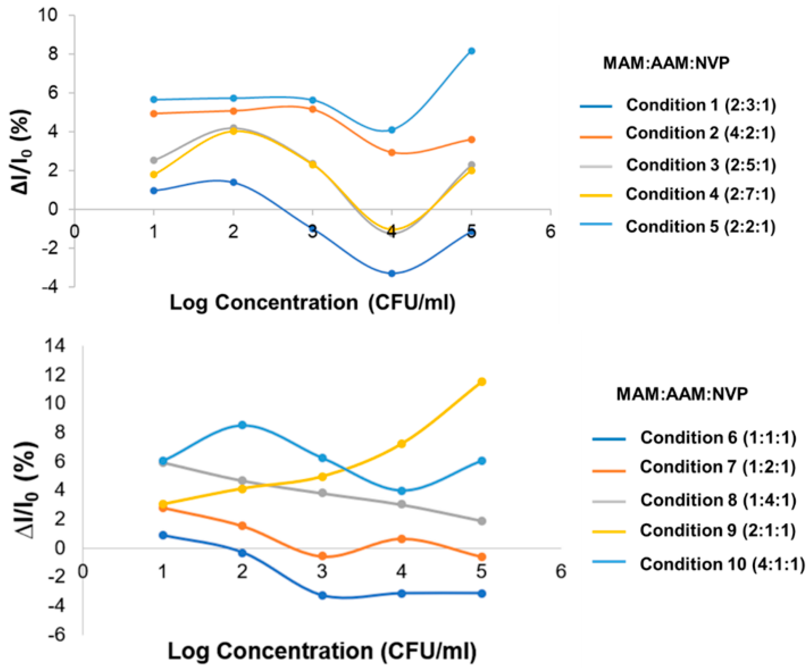
In the research, various ratios of three functional monomers, constituting the polymer—namely MAM, AAM, and NVP—were evaluated for optimization, and the results demonstrated that the combination of the three monomers at a ratio of 2:1:1 (condition 9) gave the best linearity performance in K. pneumoniae detection.
(ii).
Testing on the gold electrode provides an effective performance for MIP sensor for K. pneumoniae with high sensitivity calculated by slope of linearity equation of 7.51 (% relative current/log concentration). For the reproducibility of this sensor, it can give approximately 5% of an error bar in the linearity range of log concentration levels.
(iii).
To demonstrate the specificity of our MIP-based K. pneumoniae sensor, we tested it with E. faecalis and P. aeruginosa, which are found in most environments. The sensitivity of the MIP-based K. pneumoniae biosensor with the gold electrode was the highest at a value 7.51 (% relative current/log concentration) compared with the MIP sensor applied with P. aeruginosa and E. faecalis. The sensitivity values of the MIP sensor applied with P. aeruginosa and E. faecalis were 2.634 and 2.226 (% relative current/log concentration), respectively.
(iv).
The experimental results show that the MIP-based K. pneumoniae sensor provides not only a linear response but also high specificity. Our optimal choice for the best linearity was obtained with an MAM:AAM:NVP ratio of 2:1:1 in the gold electrode MIP sensor with an R2 value of 0.9919 and best sensitivity of 7.51 (% relative current/log concentration). The LOD for K. pneumoniae was found to be approximately 10 CFU/mL in a practice experiment and was found to be 0.012 CFU/mL by calculation, which was superior to [41] where the LOD was reported to be 1.352 CFU/mL. For the LOQ of our MIP sensor, it achieved 1.61 CFU/mL.

5. Conclusions

This work demonstrated a method to develop biosensors based on MIP specifically for the rapid detection of K. pneumoniae and using electrochemical biosensors for analysis. Various conditions have been optimized for our designed MIP-based sensor, including the ratio of monomers and the type of electrode. The optimal ratio of MAM:AAM:NVP to detect K. pneumoniae was found to be 2:1:1. Both the carbon electrode and the gold electrode, used with a 2:1:1 ratio for the polymer, can achieve an acceptable performance. The sensitivity of the gold electrode was 7.51 (% relative current/log concentration). The best linearity response was also found in the gold electrode with an R2 value of 0.9919. Our MIP-based sensor also had a detection limit of 10 CFU/mL, which was found to be sufficient for detecting K. pneumoniae in real-world applications. Specificity testing of the MIP-based sensor on the rod-type bacteria was also conducted, which confirmed that the MIP demonstrates a lower sensitivity response in other rod-type bacteria. The MIP-based K pneumoniae sensor is easy to analyze, provides fast detection for diagnostics, and has a high accuracy and specificity for K. pneumoniae detection.

Author Contributions

Conceptualization, S.V. and N.V.; methodology, S.V. and W.S.; software, C.P.; validation, C.P., N.V. and S.V.; formal analysis, C.S.; investigation, N.V.; resources, S.V.; data curation, C.P.; writing—original draft preparation, N.V., C.P. and S.V.; writing—review and editing, N.V., C.P. and S.V.; visualization, C.P.; supervision, S.V.; project administration, C.P. and S.V.; funding acquisition, N.V. and S.V. All authors have read and agreed to the published version of the manuscript.

Funding

This research and innovation activity was funded by the KMITL research fund, grant number 2563-02-01-034 and National Research Council of Thailand (NRCT).

Institutional Review Board Statement

Not applicable.

Informed Consent Statement

Not applicable.

Conflicts of Interest

The authors declare no conflict of interest.

References

  1. Haque, M.; Sartelli, M.; McKimm, J.; Abu Bakar, M. Health care-associated infections—An overview. Infect. Drug Resist. 2018, 11, 2321–2333. [Google Scholar] [CrossRef] [PubMed] [Green Version]
  2. Vilar-Compte, D.; Camacho-Ortiz, A.; Ponce-de-León, S. Infection Control in Limited Resources Countries: Challenges and Priorities. Curr. Infect. Dis. Rep. 2017, 19, 20. [Google Scholar] [CrossRef] [PubMed]
  3. Khan, H.A.; Baig, F.K.; Mehboob, R. Nosocomial infections: Epidemiology, prevention, control and surveillance. Asian Pac. J. Trop. Biomed. 2017, 7, 478–482. [Google Scholar] [CrossRef]
  4. Boev, C.; Kiss, E. Hospital-Acquired Infections: Current Trends and Prevention. Crit. Care Nurs. Clin. N. Am. 2017, 29, 51–65. [Google Scholar] [CrossRef] [PubMed]
  5. Donkor, E.S. Nosocomial Pathogens: An In-Depth Analysis of the Vectorial Potential of Cockroaches. Trop Med. Infect. Dis. 2019, 4, 14. [Google Scholar] [CrossRef] [PubMed] [Green Version]
  6. Huynh, D.T.N.; Kim, A.-Y.; Kim, Y.-R. Identification of Pathogenic Factors in Klebsiella pneumoniae Using Impedimetric Sensor Equipped with Biomimetic Surfaces. Sens. Actuators B Chem. 2017, 17, 1406. [Google Scholar] [CrossRef] [PubMed]
  7. Ssekitoleko, R.T.; Oshabaheebwa, S.; Munabi, I.G.; Tusabe, M.S.; Namayega, C.; Ngabirano, B.A.; Matovu, B.; Mugaga, J.; Reichert, W.M.; Joloba, M.L. The role of medical equipment in the spread of nosocomial infections: A cross-sectional study in four tertiary public health facilities in Uganda. BMC Public Health 2020, 20, 1561. [Google Scholar] [CrossRef] [PubMed]
  8. Podschun, R.; Ullmann, U. Klebsiella spp. as nosocomial pathogens: Epidemiology, taxonomy, typing methods, and pathogenicity factors. Clin. Microbiol. Rev. 1998, 11, 589–603. [Google Scholar] [CrossRef] [Green Version]
  9. Lenchenko, E.; Blumenkrants, D.; Sachivkina, N.; Shadrova, N.; Ibragimova, A. Morphological and adhesive properties of Klebsiella pneumoniae biofilms. Vet. World 2020, 13, 197–200. [Google Scholar] [CrossRef] [Green Version]
  10. Sawano, T.; Tsubokura, M.; Leppold, C.; Ozaki, A.; Fujioka, S.; Nemoto, T.; Kato, S.; Oikawa, T.; Kanazawa, Y. Klebsiella pneumoniae sepsis deteriorated by uncontrolled underlying disease in a decontamination worker in Fukushima, Japan. J. Occup Health 2016, 58, 320–322. [Google Scholar] [CrossRef] [PubMed] [Green Version]
  11. Farver, C.F.; Zander, D.S. Bacterial Diseases. In Pulmonary Pathology, 2nd ed.; Elsevier: Amsterdam, The Netherlands, 2016; pp. 163–200. [Google Scholar]
  12. Cristea, O.M.; Avramescu, C.S.; Balasoiu, M.; Popescu, F.D.; Popescu, F.; Amzoiu, M.O. Urinary tract infection with Klebsiella pneumoniae in Patients with Chronic Kidney Disease. Curr. Health Sci. J. 2017, 43, 137–148. [Google Scholar] [PubMed]
  13. Xu, M.; Fu, Y.; Kong, H.; Chen, X.; Chen, Y.; Li, L.; Yang, Q. Bloodstream infections caused by Klebsiella pneumoniae: Prevalence of blaKPC, virulence factors and their impacts on clinical outcome. BMC Infect. Dis. 2018, 18, 358. [Google Scholar] [CrossRef] [PubMed] [Green Version]
  14. Zhao, Q.; Guo, L.; Wang, L.F.; Zhao, Q.; Shen, D.X. Prevalence and characteristics of surgical site hypervirulent Klebsiella pneumoniae isolates. J. Clin. Lab. Anal. 2020, 34, e23364. [Google Scholar] [CrossRef] [PubMed]
  15. Fang, C.T.; Chen, Y.C.; Chang, S.C.; Sau, W.Y.; Luh, K.T. Klebsiella pneumoniae meningitis: Timing of antimicrobial therapy and prognosis. QJM 2000, 93, 45–53. [Google Scholar] [CrossRef] [Green Version]
  16. Wang, Y.; Yan, W.; Wang, Y.; Xu, J.; Ye, C. Rapid, sensitive and reliable detection of Klebsiella pneumoniae by label-free multiple cross displacement amplification coupled with nanoparticles-based biosensor. J. Microbiol. Methods 2018, 149, 80–88. [Google Scholar] [CrossRef]
  17. Paczosa, M.K.; Mecsas, J. Klebsiella pneumoniae: Going on the Offense with a Strong Defense. Microbiol. Mol. Biol. Rev. 2016, 80, 629–661. [Google Scholar] [CrossRef] [Green Version]
  18. Nirwati, H.; Sinanjung, K.; Fahrunissa, F.; Wijaya, F.; Napitupulu, S.; Hati, V.P.; Hakim, M.S.; Meliala, A.; Aman, A.T.; Nuryastuti, T. Biofilm formation and antibiotic resistance of Klebsiella pneumoniae isolated from clinical samples in a tertiary care hospital, Klaten, Indonesia. BMC Proc. 2019, 13, 20. [Google Scholar] [CrossRef]
  19. Osman, E.A.; El-Amin, N.; Adrees, E.A.E.; Al-Hassan, L.; Mukhtar, M. Comparing conventional, biochemical and genotypic methods for accurate identification of Klebsiella pneumoniae in Sudan. Access Microbiol. 2020, 2, acmi000096. [Google Scholar] [CrossRef]
  20. Murdoch, D.R.; O’Brien, K.L.; Driscoll, A.J.; Karron, R.A.; Bhat, N.; The Pneumonia Methods Working Group; The PERCH Core Team. Laboratory methods for determining pneumonia etiology in children. Clin. Infect. Dis. 2012, 54, 146–152. [Google Scholar] [CrossRef] [Green Version]
  21. Ahmed, A.; Rushworth, J.V.; Hirst, N.A.; Millner, P.A. Biosensors for whole-cell bacterial detection. Clin. Microbiol. Rev. 2014, 27, 631–646. [Google Scholar] [CrossRef] [Green Version]
  22. Choudhury, S. Chapter 12—Molecular tools for the detection of waterborne pathogens. In Waterborne Pathogens; Butterworth-Heinemann: Oxford, UK, 2020; pp. 219–235. [Google Scholar]
  23. Stetter, J.; Penrose, W.; Yao, S. Sensors, Chemical Sensors, Electrochemical Sensors, and ECS. J. Electrochem. Soc. 2003, 150, S11. [Google Scholar] [CrossRef]
  24. Grieshaber, D.; MacKenzie, R.; Voros, J.; Reimhult, E. Electrochemical Biosensors—Sensor Principles and Architectures. Sensors 2008, 8, 1400–1458. [Google Scholar] [CrossRef] [PubMed]
  25. da Silva, E.T.S.; Souto, D.E.P.; Barragan, J.T.C.; Giarola, J.D.F.; de Moraes, A.C.; Kubota, L.T. Electrochemical Biosensors in Point-of-Care Devices: Recent Advances and Future Trends. Spec. Issue Electrochem. Biosensing 2017, 4, 778–794. [Google Scholar] [CrossRef]
  26. Abbas Zaidi, S. Molecular imprinting: A useful approach for drug delivery. Mater. Sci. Energy Technol. 2020, 3, 72–77. [Google Scholar]
  27. Regal, P.; Díaz-Bao, M.; Barreiro, R.; Cepeda, A.; Fente, C. Application of molecularly imprinted polymers in food analysis: Clean-up and chromatographic improvements. Open Chem. 2012, 10, 766–784. [Google Scholar] [CrossRef]
  28. Saylan, Y.; Akgonullu, S.; Yavuz, H.; Unal, S.; Denizli, A. Molecularly Imprinted Polymer Based Sensors for Medical Applications. Sensors 2019, 19, 1279. [Google Scholar] [CrossRef] [Green Version]
  29. Cui, F.; Zhou, Z.; Zhou, H.S. Molecularly Imprinted Polymers and Surface Imprinted Polymers Based Electrochemical Biosensor for Infectious Diseases. Sensors 2020, 20, 996. [Google Scholar] [CrossRef] [Green Version]
  30. Xu, M.; Wang, R.; Li, Y. An electrochemical biosensor for rapid detection of E. coli O157:H7 with highly efficient bi-functional glucose oxidase-polydopamine nanocomposites and Prussian blue modified screen-printed interdigitated electrodes. Analyst 2016, 141, 5441–5449. [Google Scholar] [CrossRef] [Green Version]
  31. Tancharoen, C.; Sukjee, W.; Thepparit, C.; Jaimipuk, T.; Auewarakul, P.; Thitithanyanont, A.; Sangma, C. Electrochemical Biosensor Based on Surface Imprinting for Zika Virus Detection in Serum. ACS Sens. 2019, 4, 69–75. [Google Scholar] [CrossRef]
  32. Sellergren, B. Imprinted Polymers with Memory for Small Molecules, Proteins, or Crystals The author is grateful to Dr. Andrew Hall and Dr. Gunter Buchel for linguistic advice. Angew Chem. Int. Ed. Engl. 2000, 39, 1031–1037. [Google Scholar] [CrossRef]
  33. Saylan, Y.; Yilmaz, F.; Ozgur, E.; Derazshamshir, A.; Yavuz, H.; Denizli, A. Molecular Imprinting of Macromolecules for Sensor Applications. Sensors 2017, 17, 898. [Google Scholar] [CrossRef] [PubMed]
  34. Janiak, D.S.; Kofinas, P. Molecular imprinting of peptides and proteins in aqueous media. Anal. Bioanal. Chem. 2007, 389, 399–404. [Google Scholar] [CrossRef] [PubMed]
  35. Ansell, R.J.; Ramstrom, O.; Mosbach, K. Towards artificial antibodies prepared by molecular imprinting. Clin. Chem 1996, 42, 1506–1512. [Google Scholar] [CrossRef] [PubMed] [Green Version]
  36. Turner, N.W.; Jeans, C.W.; Brain, K.R.; Allender, C.J.; Hlady, V.; Britt, D.W. From 3D to 2D: A review of the molecular imprinting of proteins. Biotechnol. Prog. 2006, 22, 1474–1489. [Google Scholar] [CrossRef] [PubMed]
  37. Bole, A.L.; Manesiotis, P. Advanced Materials for the Recognition and Capture of Whole Cells and Microorganisms. Adv. Mater. 2016, 28, 5349–5366. [Google Scholar] [CrossRef] [PubMed]
  38. Li, R.; Feng, Y.; Pan, G.; Liu, L. Advances in Molecularly Imprinting Technology for Bioanalytical Applications. Sensors 2019, 19, 177. [Google Scholar] [CrossRef] [Green Version]
  39. El-Schich, Z.; Zhang, Y.; Feith, M.; Beyer, S.; Sternbaek, L.; Ohlsson, L.; Stollenwerk, M.; Wingren, A.G. Molecularly imprinted polymers in biological applications. Biotechniques 2020, 69, 406–419. [Google Scholar] [CrossRef]
  40. Vasapollo, G.; Sole, R.D.; Mergola, L.; Lazzoi, M.R.; Scardino, A.; Scorrano, S.; Mele, G. Molecularly imprinted polymers: Present and future prospective. Int. J. Mol. Sci. 2011, 12, 5908–5945. [Google Scholar] [CrossRef] [Green Version]
  41. Sharma, R.; Lakshmi, G.B.V.S.; Kumar, A.; Solanki, P. Polypyrrole Based Molecularly Imprinted Polymer Platform for Klebsiella pneumonia Detection. ECS Sens. Plus 2022, 1, 010603. [Google Scholar] [CrossRef]
  42. Sukjee, W.; Thitithanyanont, A.; Manopwisedjaroen, S.; Seetaha, S.; Thepparit, C.; Sangma, C. Virus MIP-composites for SARS-CoV-2 detection in the aquatic environment. Mater. Lett. 2022, 315, 131973. [Google Scholar] [CrossRef]
  43. Türkmen, D.; Yılmaz, T.; Bakhshpour, M.; Denizli, A. An Alternative Approach for Bacterial Growth Control: Pseudomonas spp. Imprinted Polymer-Based Surface Plasmon Resonance Sensor. IEEE Sens. J. 2022, 22, 3001–3008. [Google Scholar] [CrossRef]
  44. Ramanavicius, S.; Morkvenaite-Vilkonciene, I.; Samukaite-Bubniene, U.; Ratautaite, V.; Plikusiene, I.; Viter, R.; Ramanavicius, A. Electrochemically Deposited Molecularly Imprinted Polymer-Based Sensors. Sensors 2022, 22, 1282. [Google Scholar] [CrossRef] [PubMed]
  45. Ramanavicius, S.; Jagminas, A.; Ramanavicius, A. Advances in Molecularly Imprinted Polymers Based Affinity Sensors (Review). Polymers 2021, 13, 974. [Google Scholar] [CrossRef] [PubMed]
  46. Idil, N.; Mattiasson, B. Imprinting of Microorganisms for Biosensor Applications. Sensors 2017, 17, 708. [Google Scholar] [CrossRef] [Green Version]
  47. Jamalipour Soufi, G.; Iravani, S.; Varma, R.S. Molecularly imprinted polymers for the detection of viruses: Challenges and opportunities. Analyst 2021, 146, 3087–3100. [Google Scholar] [CrossRef]
  48. Fahmy Taha, M.H.; Ashraf, H.; Caesarendra, W. A Brief Description of Cyclic Voltammetry Transducer-Based Non-Enzymatic Glucose Biosensor Using Synthesized Graphene Electrodes. Appl. Syst. Innov. 2020, 3, 32. [Google Scholar] [CrossRef]
  49. Liu, B.Y.; Zhang, G.M.; Li, X.L.; Chen, H. Effect of glutaraldehyde fixation on bacterial cells observed by atomic force microscopy. Scanning 2012, 34, 6–11. [Google Scholar] [CrossRef]
  50. Smart, A.A.; Crew, A.; Pemberton, R.; Hughes, G.; Doran, O.; Hart, J.P. Screen-printed carbon-based biosensors and their applications in agri-food safety. TrAC Trends Anal. Chem. 2020, 127, 115898. [Google Scholar] [CrossRef]
Figure 1. Schematic representation of K. pneumoniae on LB agar and the serial dilution method.
Sensors 22 04638 g001
Figure 2. Schematic representation of the preparation of polymer-GO on gold electrode for K. pneumoniae detection.
Sensors 22 04638 g002
Figure 3. (a) Magnified SEM image at 10,000×; (b) the surface of whole K. pneumoniae on the SPE, with a size of approximately 0.5 × 1.7 µm (SEM image at 50,000×).
Sensors 22 04638 g003
Figure 4. Comparison of linearity range for conditions 1 to 10 on the carbon electrode.
Sensors 22 04638 g004
Figure 5. (a) Cyclic voltammogram of K. pneumoniae at concentration levels in condition 5 on gold electrode; (b) cyclic voltammogram of K. pneumoniae at concentration levels in condition 9 on gold electrode.
Sensors 22 04638 g005
Figure 6. Comparison of linearity range for condition 5 and condition 9 on the gold electrode (* is referred to multiplication).
Sensors 22 04638 g006
Figure 7. The plot of current changes as a linear function of log K. pneumoniae concentration gains from MIP composite on the gold electrode compared with the results from the control experiments using P. aeruginosa and E. faecalis. The number of replicated measurements is 3. (* is referred to multiplication).
Sensors 22 04638 g007
Table 1. The amount of each functional monomer in various conditions *.
ConditionRatio (n:n:n)Methacrylamide (MAM) (mg)Acrylamide (AAM) (mg)N-Vinylpyrrolidone (NVP) (µL)
12:3:11721.310.7
24:2:13414.210.7
32:5:11735.510.7
42:7:11749.710.7
52:2:11714.210.7
61:1:18.57.110.7
71:2:18.514.210.7
81:4:18.528.410.7
92:1:1177.110.7
104:1:1347.110.7
* 47 mg of N,N′-(1,2-dihydroxyethylene)bisacrylamide (DHEBA) has been used as crosslinker.
Table 2. The zeta potential around the surface of bacterial samples from dynamic light scattering.
RecordBacteria SampleAverage Zeta Potential (mV)
1K. pneumoniae−10.6
2P. aeruginosa−10.1
3E. faecalis−16.2
Table 3. The current data of conditions 1–10 on the carbon electrode.
ConditionConcentrationCurrent (µA)∆I (µA)(∆I/I0) × 100%
Condition 1 (MAM:AAM:NVP) (2:3:1)Blank52.24--
10 CFU/mL51.730.510.97
102 CFU/mL51.510.731.40
103 CFU/mL52.75−0.52−0.99
104 CFU/mL53.95−1.71−3.27
105 CFU/mL52.83−0.59−1.13
Condition 2 (MAM:AAM:NVP) (4:2:1)Blank45.56--
10 CFU/mL43.312.254.94
102 CFU/mL43.252.315.08
103 CFU/mL43.222.355.15
104 CFU/mL44.221.342.95
105 CFU/mL43.921.653.61
Condition 3 (MAM:AAM:NVP) (2:5:1)Blank54.81--
10 CFU/mL53.421.392.54
102 CFU/mL52.512.304.20
103 CFU/mL53.521.292.36
104 CFU/mL55.49−0.67−1.23
105 CFU/mL53.561.262.29
Condition 4 (MAM:AAM:NVP) (2:7:1)Blank53.87--
10 CFU/mL52.900.971.80
102 CFU/mL51.702.174.03
103 CFU/mL52.621.252.32
104 CFU/mL54.43−0.56−1.03
105 CFU/mL52.791.092.01
Condition 5 (MAM:AAM:NVP) (2:2:1)Blank19.04--
10 CFU/mL17.961.085.66
102 CFU/mL17.941.095.74
103 CFU/mL17.961.085.65
104 CFU/mL18.250.784.12
105 CFU/mL17.481.568.17
Condition 6 (MAM:AAM:NVP) (1:1:1)Blank49.06--
10 CFU/mL48.630.420.87
102 CFU/mL49.22−0.16−0.33
103 CFU/mL50.67−1.62−3.30
104 CFU/mL50.60−1.54−3.15
105 CFU/mL50.60−1.54−3.14
Condition 7 (MAM:AAM:NVP) (1:2:1)Blank46.26--
10 CFU/mL44.971.292.79
102 CFU/mL45.560.701.52
103 CFU/mL46.52−0.26−0.57
104 CFU/mL45.960.300.65
105 CFU/mL46.54−0.27−0.59
Condition 8 (MAM:AAM:NVP) (1:4:1)Blank49.94--
10 CFU/mL46.982.965.92
102 CFU/mL47.622.334.66
103 CFU/mL48.051.903.80
104 CFU/mL48.441.503.01
105 CFU/mL48.990.951.90
Condition 9 (MAM:AAM:NVP) (2:1:1)Blank52.25--
10 CFU/mL50.671.583.02
102 CFU/mL50.102.154.11
103 CFU/mL49.672.584.94
104 CFU/mL48.483.777.22
105 CFU/mL46.246.0111.50
Condition 10 (MAM:AAM:NVP) (4:1:1)Blank49.88--
10 CFU/mL46.873.016.03
102 CFU/mL45.644.248.50
103 CFU/mL46.773.116.23
104 CFU/mL47.901.983.97
105 CFU/mL46.863.026.05
Table 4. The current data of conditions 5 and 9 on gold electrode.
ConditionConcentrationCurrent (µA)∆I (µA)(∆I/I0) × 100%
Condition 5 (MAM:AAM:NVP) (2:2:1)Blank65.14--
10 CFU/mL62.232.914.47
102 CFU/mL59.955.207.98
103 CFU/mL55.889.2614.22
104 CFU/mL53.6911.4617.59
105 CFU/mL51.8513.2920.40
Condition 9 (MAM:AAM:NVP) (2:1:1)Blank52.48--
10 CFU/mL40.1412.3423.51
102 CFU/mL34.8117.6733.66
103 CFU/mL31.6020.8839.78
104 CFU/mL27.6424.8347.32
105 CFU/mL24.5827.8953.15
Publisher’s Note: MDPI stays neutral with regard to jurisdictional claims in published maps and institutional affiliations.

