
Field-Effect Transistor-Based Biosensors for Environmental and Agricultural Monitoring

 ,
,  , , , , and
Abstract
:1. Introduction
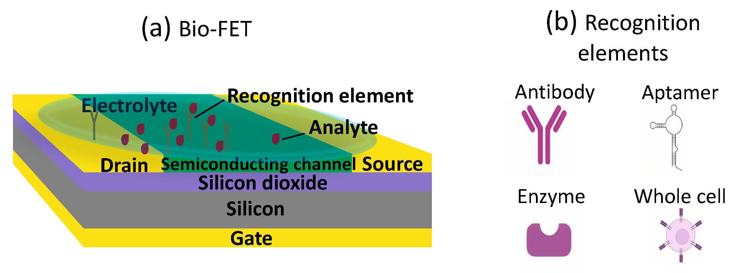
2. Bio-FETs
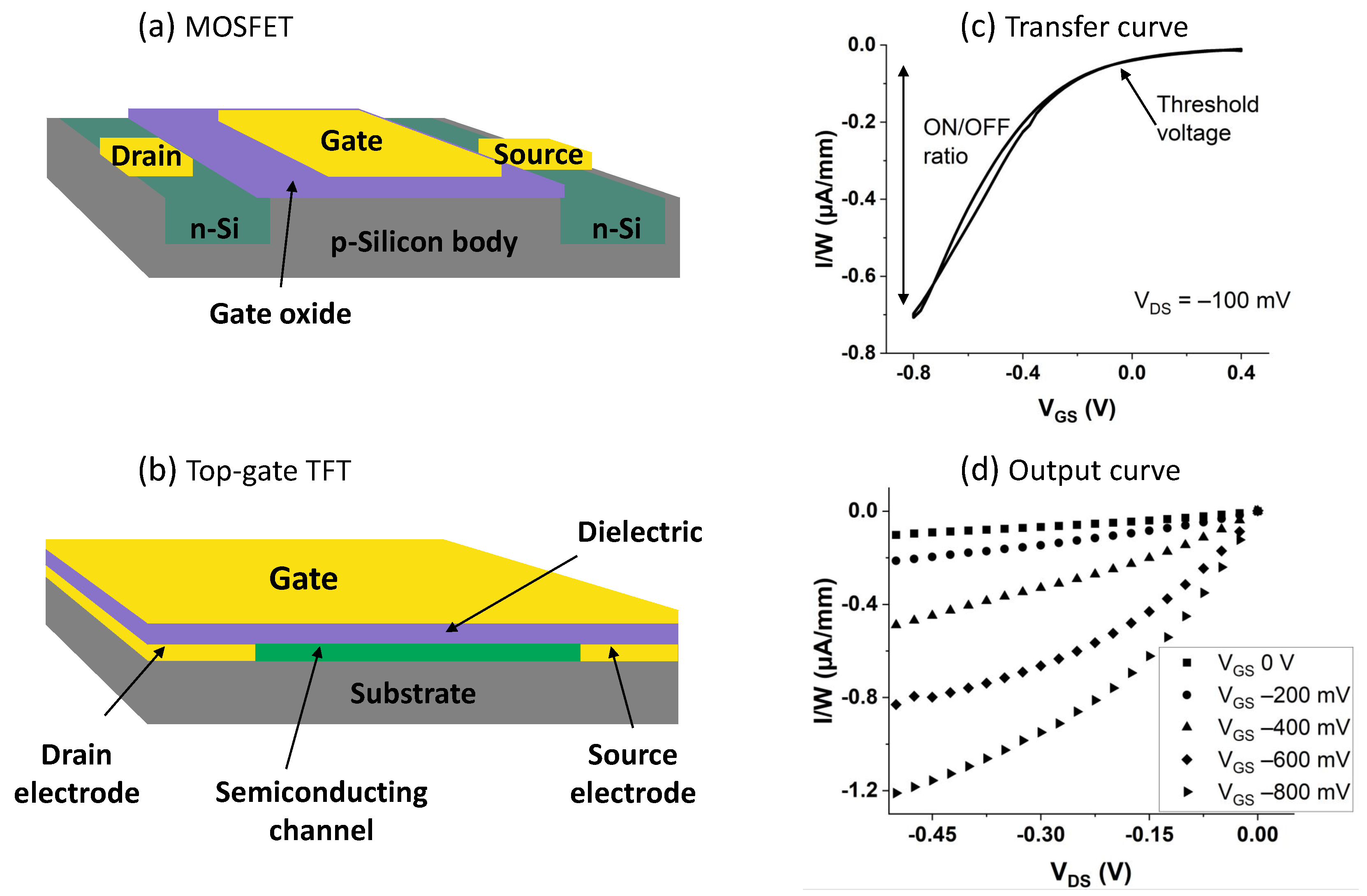
2.1. Bio-FET Operation and Configurations
2.2. Materials
2.2.1. Substrates
2.2.2. Electrodes
2.2.3. Active Materials
2.2.4. Dielectric Materials
2.2.5. Electrolytes
2.3. Fabrication Methods

| Configuration | Substrate | Source/Drain | Gate | Active Material | Recognition Element | Analyte | Ref. |
|---|---|---|---|---|---|---|---|
| Bottom-gate FET | Si/SiO2 wafer | Ag | Al | CNTs | Antibodies | Salmonella | [116] |
| Bottom-gate FET | Si/SiO2 wafer | Cr/Au | Si | CNTs | Antibodies | Salmonella | [92] |
| Bottom-gate FET | Si/SiO2 wafer | Ti/Au | Si | CNTs | Aptamers | Escherichia coli | [117] |
| Bottom-gate FET | Si/SiO2 wafer | Ti/Au | Cr/Au | CNTs | Antibodies | Domoic acid | [118] |
| Bottom-gate FET | Si/SiO2 wafer | Ti/Au | - | CNTs | Hydrogel | Aspergillus niger activity | [103] |
| Bottom-gate FET | Si/SiO2 wafer | Cr/Au | Si | CNTs | DNA | P-Ethylphenol | [101] |
| Bottom-gate FET | Si/SiO2 wafer | Ti/Pt | Si | CNTs | Ag-ZnOs | Methyl parathion | [119] |
| Bottom-gate FET | Si/SiO2 wafer | Ti/Au | Cr/Au | CNTs | Antibodies | Atrazine | [88] |
| EG-FET | PI | Cr/Au | Cr/Au planar | CNTs | Enzymes | Acetylcholine | [55] |
| EG-FET | Quartz | Cr/Au | Au wire | Pentacene | Antibodies | Plum Pox Virus | [53] |
| EG-FET | Si/SiO2 wafer | Ti/Au | Pt microelectrodes | Poly(DPP-DTT) | n.a. | Glyphosate and diuron | [82] |
| ECT | Si/SiO2 wafer | Ni/Au | Ag/AgCl needle | Graphene | TCA | Cu2+ ions | [52] |
| ECT | Si/SiO2 wafer | Au | Ag/AgCl needle | Au-NP | Cells | Cell membrane depolarization | [120] |
| ECT | Glass | Cr/Au | Cr/Au | Graphene | Enzymes | Trichlorfon | [51] |
| ECT | Glass | Cr/Au | GCE | Graphene | ZrO2/rGO | Methyl parathion | [8] |
| ECT | Si/SiO2 wafer | Ti/Au | Ti/Au planar | PEDOT:PSS | CNPs-SF patch | Limonin | [110] |
| ECT | Cotton thread | - | Ag wire | PEDOT:PSS | n.a. | Ions | [121] |
| ECT | PET | n.a. | Ag/AgCl needle | PBTTT + P3HT | Ion exchange gel | Extracellular signals | [90] |
| ECT | PEN | Ti/Au | Ti/Au planar | PEDOT:PSS | Enzymes + PtNPs | Glucose and Sucrose | [122] |
| ECT | PEN | Ag | Ag/AgCl planar | PEDOT:PSS | Ion-selective membrane | Potassium | [123] |
| ISFET | Si/SiO2 wafer | n.a. | n.a. | Si | Enzymes | Indole alkaloids | [87] |
| ISFET | Si/SiO2 wafer | Poly-Si/Al | Si | Si | Enzymes | Glycoalkaloids | [98] |
2.4. Functionalization Methods
3. Bio-FETs in Environmental Applications
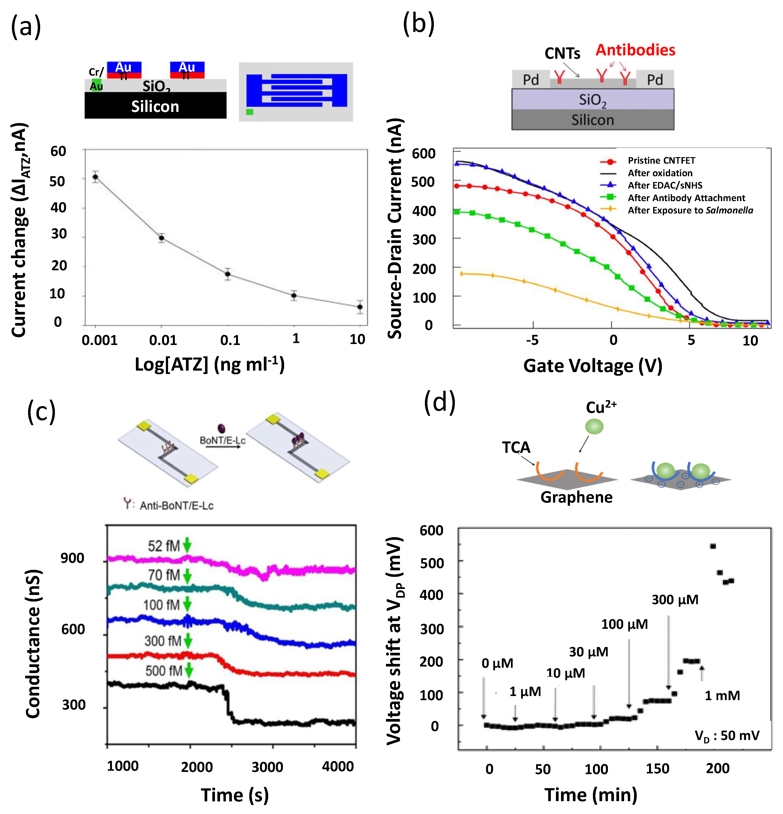
3.1. Pesticides
3.2. Bacteria and Toxins
3.3. Metals
3.4. Other Chemicals
4. Bio-FETs in Agricultural Plants Applications
4.1. Abiotic Stresses
4.2. Biotic Stresses
4.3. Plant Metabolites and pH Measurements
5. Discussion
6. Conclusions
Author Contributions
Funding
Institutional Review Board Statement
Informed Consent Statement
Data Availability Statement
Conflicts of Interest
References
- United Nations, Department of Economic and Social Affairs, Population Division. World Population Prospects 2019; United Nations Publications: New York, NY, USA, 2019. [Google Scholar]
- SDG, UN. Available online: http://unstats.un.org/sdgs/report/2020/ (accessed on 1 March 2022).
- Manisalidis, I.; Stavropoulou, E.; Stavropoulos, A.; Bezirtzoglou, E. Environmental and health impacts of air pollution: A review. Front. Public Health 2020, 8, 14. [Google Scholar] [CrossRef] [PubMed] [Green Version]
- Ghorbanpour, M.; Bhargava, P.; Varma, A.; Choudhary, D.K. Biogenic Nano-Particles and Their Use in Agro-Ecosystems; Springer: Berlin/Heidelberg, Germany, 2020. [Google Scholar]
- Crinnion, W.J. Chlorinated pesticides: Threats to health and importance of detection. Altern. Med. Rev. 2009, 14, 347–359. [Google Scholar] [PubMed]
- Matich, E.K.; Soria, N.G.C.; Aga, D.S.; Atilla-Gokcumen, G.E. Applications of metabolomics in assessing ecological effects of emerging contaminants and pollutants on plants. J. Hazard. Mater. 2019, 373, 527–535. [Google Scholar] [CrossRef]
- Lee, K.; Park, J.; Lee, M.S.; Kim, J.; Hyun, B.G.; Kang, D.J.; Na, K.; Lee, C.Y.; Bien, F.; Park, J.U. In-situ synthesis of carbon nanotube–graphite electronic devices and their integrations onto surfaces of live plants and insects. Nano Lett. 2014, 14, 2647–2654. [Google Scholar] [CrossRef] [PubMed]
- Tao, T.; Zhou, Y.; Ma, M.; He, H.; Gao, N.; Cai, Z.; Chang, G.; He, Y. Novel graphene electrochemical transistor with ZrO2/rGO nanocomposites functionalized gate electrode for ultrasensitive recognition of methyl parathion. Sens. Actuators B Chem. 2021, 328, 128936. [Google Scholar] [CrossRef]
- UNEP. About UN Environment Programme; UNEP: Nairobi, Kenya, 2022. [Google Scholar]
- Zörb, C.; Geilfus, C.M.; Dietz, K.J. Salinity and crop yield. Plant Biol. 2019, 21, 31–38. [Google Scholar] [CrossRef]
- Boyer, J.S. Plant productivity and environment. Science 1982, 218, 443–448. [Google Scholar] [CrossRef]
- De Souza Machado, A.A.; Kloas, W.; Zarfl, C.; Hempel, S.; Rillig, M.C. Microplastics as an emerging threat to terrestrial ecosystems. Glob. Chang. Biol. 2018, 24, 1405–1416. [Google Scholar] [CrossRef] [Green Version]
- Pesticides Action Network Europe. Banned and Hazardous Pesticides in European Food: Report Highlights; Pesticides Action Network Europe: Brussels, Belgium, 2020; Available online: https://www.pan-europe.info/press-releases/2020/09/banned-and-hazardous-pesticides-european-food-report-highlights (accessed on 15 July 2021).
- Lepp, N.W. Effect of Heavy Metal Pollution on Plants: Metals in the Environment; Springer Science & Business Media: Berlin/Heidelberg, Germany, 2012; Volume 2. [Google Scholar]
- Ivanković, T.; Hrenović, J. Surfactants in the environment. Arh. Hig. Rada Toksikol. 2010, 61, 95–109. [Google Scholar] [CrossRef] [Green Version]
- Johnson, A.C.; Jin, X.; Nakada, N.; Sumpter, J.P. Learning from the past and considering the future of chemicals in the environment. Science 2020, 367, 384–387. [Google Scholar] [CrossRef] [Green Version]
- Rose, R.L.; Hodgson, E.; Roe, R.M. Chapter 28—Pesticides. In Toxicology; Marquardt, H., Schäfer, S.G., McClellan, R., Welsch, F., Eds.; Academic Press: San Diego, CA, USA, 1999; pp. 663–697. [Google Scholar] [CrossRef]
- National Institute of Environmental Health Sciences, NIH. Pesticides. Available online: https://www.niehs.nih.gov/health/topics/agents/pesticides/index.cfm (accessed on 13 July 2021).
- Pujari, M.; Kapoor, D. Heavy metals in the ecosystem: Sources and their effects. In Heavy Metals in the Environment; Elsevier: Amsterdam, The Netherlands, 2021; pp. 1–7. [Google Scholar]
- Asati, A.; Pichhode, M.; Nikhil, K. Effect of heavy metals on plants: An overview. Int. J. Appl. Innov. Eng. Manag. 2016, 5, 56–66. [Google Scholar]
- Motsara, M. Guide to Laboratory Establishment for Plant Nutrient Analysis; Scientific Publishers: Stevenson Ranch, CA, USA, 2015. [Google Scholar]
- Ayers, R.S.; Westcot, D.W. Water Quality for Agriculture; Food and Agriculture Organization of the United Nations Rome: Rome, Italy, 1985; Volume 29. [Google Scholar]
- Xu, M.L.; Gao, Y.; Han, X.X.; Zhao, B. Detection of pesticide residues in food using surface-enhanced Raman spectroscopy: A review. J. Agric. Food Chem. 2017, 65, 6719–6726. [Google Scholar] [CrossRef] [PubMed]
- Ahmad, L.; Nabi, F. Agriculture 5.0: Artificial Intelligence, IoT and Machine Learning; CRC Press: Boca Raton, FL, USA, 2021. [Google Scholar]
- Shu, L.; Hancke, G.P.; Abu-Mahfouz, A.M. Guest editorial: Sustainable and intelligent precision agriculture. IEEE Trans. Ind. Inform. 2021, 17, 4318–4321. [Google Scholar] [CrossRef]
- Almalki, F.A.; Soufiene, B.O.; Alsamhi, S.H.; Sakli, H. A low-cost platform for environmental smart farming monitoring system based on IoT and UAVs. Sustainability 2021, 13, 5908. [Google Scholar] [CrossRef]
- Yin, H.; Cao, Y.; Marelli, B.; Zeng, X.; Mason, A.J.; Cao, C. Soil Sensors and Plant Wearables for Smart and Precision Agriculture. Adv. Mater. 2021, 33, 2007764. [Google Scholar] [CrossRef]
- Araújo, S.O.; Peres, R.S.; Barata, J.; Lidon, F.; Ramalho, J.C. Characterising the Agriculture 4.0 Landscape—Emerging Trends, Challenges and Opportunities. Agronomy 2021, 11, 667. [Google Scholar] [CrossRef]
- Irimia-Vladu, M. “Green” electronics: Biodegradable and biocompatible materials and devices for sustainable future. Chem. Soc. Rev. 2014, 43, 588–610. [Google Scholar] [CrossRef] [Green Version]
- Roper, J.M.; Garcia, J.F.; Tsutsui, H. Emerging Technologies for Monitoring Plant Health in Vivo. ACS Omega 2021, 6, 5101–5107. [Google Scholar] [CrossRef]
- Zulkifli, S.N.; Rahim, H.A.; Lau, W.J. Detection of contaminants in water supply: A review on state-of-the-art monitoring technologies and their applications. Sens. Actuators B Chem. 2018, 255, 2657–2689. [Google Scholar] [CrossRef]
- Mahlein, A.K. Plant disease detection by imaging sensors–parallels and specific demands for precision agriculture and plant phenotyping. Plant Dis. 2016, 100, 241–251. [Google Scholar] [CrossRef] [Green Version]
- Farber, C.; Mahnke, M.; Sanchez, L.; Kurouski, D. Advanced spectroscopic techniques for plant disease diagnostics. A review. TrAC Trends Anal. Chem. 2019, 118, 43–49. [Google Scholar] [CrossRef]
- Walia, A.; Waadt, R.; Jones, A.M. Genetically encoded biosensors in plants: Pathways to discovery. Annu. Rev. Plant Biol. 2018, 69, 497–524. [Google Scholar] [CrossRef] [PubMed]
- Paolucci, M.; Landini, M.P.; Sambri, V. Conventional and molecular techniques for the early diagnosis of bacteraemia. Int. J. Antimicrob. Agents 2010, 36, S6–S16. [Google Scholar] [CrossRef] [Green Version]
- Hamed, S.; Ibba, P.; Petrelli, M.; Ciocca, M.; Lugli, P.; Petti, L. Transistor-based plant sensors for agriculture 4.0 measurements. In Proceedings of the 2021 IEEE International Workshop on Metrology for Agriculture and Forestry (MetroAgriFor), Trento-Bolzano, Italy, 3–5 November 2021; pp. 69–74. [Google Scholar]
- Elli, G.; Ciocca, M.; Lugli, P.; Petti, L. Field-effect-transistor based biosensors: A review of their use in environmental monitoring applications. In Proceedings of the 2021 IEEE International Workshop on Metrology for Agriculture and Forestry (MetroAgriFor), Trento-Bolzano, Italy, 3–5 November 2021; pp. 102–107. [Google Scholar]
- Jócsák, I.; Végvári, G.; Vozáry, E. Electrical impedance measurement on plants: A review with some insights to other fields. Theor. Exp. Plant Physiol. 2019, 31, 359–375. [Google Scholar] [CrossRef] [Green Version]
- Hussain, M.I.; El-Keblawy, A.; Akhtar, N.; Elwakil, A.S. Electrical Impedance Spectroscopy in Plant Biology. In Sustainable Agriculture Reviews 52; Springer: Berlin/Heidelberg, Germany, 2021; pp. 395–416. [Google Scholar]
- Houssin, T.; Follet, J.; Follet, A.; Dei-Cas, E.; Senez, V. Label-free analysis of water-polluting parasite by electrochemical impedance spectroscopy. Biosens. Bioelectron. 2010, 25, 1122–1129. [Google Scholar] [CrossRef]
- Ehosioke, S.; Nguyen, F.; Rao, S.; Kremer, T.; Placencia-Gomez, E.; Huisman, J.A.; Kemna, A.; Javaux, M.; Garré, S. Sensing the electrical properties of roots: A review. Vadose Zone J. 2020, 19, e20082. [Google Scholar] [CrossRef]
- Tran, D.; Dutoit, F.; Najdenovska, E.; Wallbridge, N.; Plummer, C.; Mazza, M.; Raileanu, L.E.; Camps, C. Electrophysiological assessment of plant status outside a Faraday cage using supervised machine learning. Sci. Rep. 2019, 9, 17073. [Google Scholar] [CrossRef]
- Najdenovska, E.; Dutoit, F.; Tran, D.; Plummer, C.; Wallbridge, N.; Camps, C.; Raileanu, L.E. Classification of plant electrophysiology signals for detection of spider mites infestation in tomatoes. Appl. Sci. 2021, 11, 1414. [Google Scholar] [CrossRef]
- Tran, D.; Camps, C. Early Diagnosis of Iron Deficiency in Commercial Tomato Crop Using Electrical Signals. Front. Sustain. Food Syst. 2021, 5, 46. [Google Scholar] [CrossRef]
- De Moraes, A.C.M.; Kubota, L.T. Recent trends in field-effect transistors-based immunosensors. Chemosensors 2016, 4, 20. [Google Scholar] [CrossRef] [Green Version]
- Nigam, V.K.; Shukla, P. Enzyme based biosensors for detection of environmental pollutants—A review. J. Microbiol. Biotechnol. 2015, 25, 1773–1781. [Google Scholar] [CrossRef] [PubMed]
- Sanati, A.; Jalali, M.; Raeissi, K.; Karimzadeh, F.; Kharaziha, M.; Mahshid, S.S.; Mahshid, S. A review on recent advancements in electrochemical biosensing using carbonaceous nanomaterials. Microchim. Acta 2019, 186, 773. [Google Scholar] [CrossRef] [PubMed]
- Shkodra, B.; Petrelli, M.; Costa Angeli, M.A.; Garoli, D.; Nakatsuka, N.; Lugli, P.; Petti, L. Electrolyte-gated carbon nanotube field-effect transistor-based biosensors: Principles and applications. Appl. Phys. Rev. 2021, 8, 041325. [Google Scholar] [CrossRef]
- Pohanka, M. The piezoelectric biosensors: Principles and applications. Int. J. Electrochem. Sci. 2017, 12, 496–506. [Google Scholar] [CrossRef]
- Huertas, C.S.; Calvo-Lozano, O.; Mitchell, A.; Lechuga, L.M. Advanced evanescent-wave optical biosensors for the detection of nucleic acids: An analytic perspective. Front. Chem. 2019, 7, 724. [Google Scholar] [CrossRef] [Green Version]
- Wang, R.; Wang, Y.; Qu, H.; Zheng, L. An Acetylcholinesterase-Functionalized Biosensor for Sensitive Detection of Organophosphorus Pesticides Based on Solution-Gated Graphene Transistors. ACS Agric. Sci. Technol. 2021, 1, 372–378. [Google Scholar] [CrossRef]
- Takagiri, Y.; Ikuta, T.; Maehashi, K. Selective detection of Cu2+ ions by immobilizing thiacalix[4]arene on graphene field-effect transistors. ACS Omega 2019, 5, 877–881. [Google Scholar] [CrossRef] [Green Version]
- Berto, M.; Vecchi, E.; Baiamonte, L.; Condò, C.; Sensi, M.; Di Lauro, M.; Sola, M.; De Stradis, A.; Biscarini, F.; Minafra, A.; et al. Label free detection of plant viruses with organic transistor biosensors. Sens. Actuators B Chem. 2019, 281, 150–156. [Google Scholar] [CrossRef]
- Li, H.; Shi, W.; Song, J.; Jang, H.J.; Dailey, J.; Yu, J.; Katz, H.E. Chemical and biomolecule sensing with organic field-effect transistors. Chem. Rev. 2018, 119, 3–35. [Google Scholar] [CrossRef]
- Bhatt, V.D.; Joshi, S.; Becherer, M.; Lugli, P. Flexible, low-cost sensor based on electrolyte gated carbon nanotube field effect transistor for organo-phosphate detection. Sensors 2017, 17, 1147. [Google Scholar] [CrossRef] [Green Version]
- Scuratti, F.; Bonacchini, G.E.; Bossio, C.; Salazar-Rios, J.M.; Talsma, W.; Loi, M.A.; Antognazza, M.R.; Caironi, M. Real-Time Monitoring of Cellular Cultures with Electrolyte-Gated Carbon Nanotube Transistors. ACS Appl. Mater. Interfaces 2019, 11, 37966–37972. [Google Scholar] [CrossRef] [PubMed]
- Zhou, L.; Wang, K.; Sun, H.; Zhao, S.; Chen, X.; Qian, D.; Mao, H.; Zhao, J. Novel Graphene Biosensor Based on the Functionalization of Multifunctional Nano-bovine Serum Albumin for the Highly Sensitive Detection of Cancer Biomarkers. Nano-Micro Lett. 2019, 11, 20. [Google Scholar] [CrossRef] [PubMed] [Green Version]
- Poghossian, A.; Schöning, M.J. Label-Free Sensing of Biomolecules with Field-Effect Devices for Clinical Applications. Electroanalysis 2014, 26, 1197–1213. [Google Scholar] [CrossRef]
- Hernandez-Vargas, G.; Sosa-Hernández, J.E.; Saldarriaga-Hernandez, S.; Villalba-Rodríguez, A.M.; Parra-Saldivar, R.; Iqbal, H. Electrochemical biosensors: A solution to pollution detection with reference to environmental contaminants. Biosensors 2018, 8, 29. [Google Scholar] [CrossRef] [PubMed] [Green Version]
- McConnell, E.M.; Nguyen, J.; Li, Y. Aptamer-based biosensors for environmental monitoring. Front. Chem. 2020, 8, 434. [Google Scholar] [CrossRef] [PubMed]
- Justino, C.I.; Duarte, A.C.; Rocha-Santos, T.A. Recent progress in biosensors for environmental monitoring: A review. Sensors 2017, 17, 2918. [Google Scholar] [CrossRef] [PubMed] [Green Version]
- Griesche, C.; Baeumner, A.J. Biosensors to support sustainable agriculture and food safety. TrAC Trends Anal. Chem. 2020, 128, 115906. [Google Scholar] [CrossRef]
- Kundu, M.; Krishnan, P.; Kotnala, R.; Sumana, G. Recent developments in biosensors to combat agricultural challenges and their future prospects. Trends Food Sci. Technol. 2019, 88, 157–178. [Google Scholar] [CrossRef]
- Arora, K. Advances in nano based biosensors for food and agriculture. In Nanotechnology, Food Security and Water Treatment; Springer: Cham, Switzerland, 2018; pp. 1–52. [Google Scholar]
- Petti, L.; Münzenrieder, N.; Vogt, C.; Faber, H.; Büthe, L.; Cantarella, G.; Bottacchi, F.; Anthopoulos, T.D.; Tröster, G. Metal oxide semiconductor thin-film transistors for flexible electronics. Appl. Phys. Rev. 2016, 3, 021303. [Google Scholar] [CrossRef] [Green Version]
- Wang, D.; Noël, V.; Piro, B. Electrolytic gated organic field-effect transistors for application in biosensors—A Review. Electronics 2016, 5, 9. [Google Scholar] [CrossRef]
- Golio, M.; Golio, J. RF and Microwave Passive and Active Technologies; CRC Press: Boca Raton, FL, USA, 2018. [Google Scholar]
- Lee, M.L.; Fitzgerald, E.A.; Bulsara, M.T.; Currie, M.T.; Lochtefeld, A. Strained Si, SiGe, and Ge channels for high-mobility metal-oxide-semiconductor field-effect transistors. J. Appl. Phys. 2005, 97, 011101. [Google Scholar] [CrossRef]
- Lüssem, B.; Riede, M.; Leo, K. Doping of organic semiconductors. Phys. Status Solidi 2013, 210, 9–43. [Google Scholar] [CrossRef]
- Zhao, Y.; Guo, Y.; Liu, Y. 25th anniversary article: Recent advances in n-type and ambipolar organic field-effect transistors. Adv. Mater. 2013, 25, 5372–5391. [Google Scholar] [CrossRef] [PubMed]
- Zhang, S. Review of modern field effect transistor technologies for scaling. J. Phys. Conf. Ser. 2020, 1617, 012054. [Google Scholar] [CrossRef]
- Kuo, Y. Thin film transistor technology—Past, present, and future. Electrochem. Soc. Interface 2013, 22, 55. [Google Scholar] [CrossRef]
- Myronov, M. Molecular Beam Epitaxy of High Mobility Silicon, Silicon Germanium and Germanium Quantum Well Heterostructures. In Molecular Beam Epitaxy; Elsevier: Amsterdam, The Netherlands, 2018; pp. 37–54. [Google Scholar]
- Benda, V.; Grant, D.A.; Gowar, J. Discrete and Integrated Power Semiconductor Devices: Theory and Applications; John Wiley & Sons: Hoboken, NJ, USA, 1999. [Google Scholar]
- Rivnay, J.; Inal, S.; Salleo, A.; Owens, R.M.; Berggren, M.; Malliaras, G.G. Organic electrochemical transistors. Nat. Rev. Mater. 2018, 3, 17086. [Google Scholar] [CrossRef]
- Bergveld, P. Thirty years of ISFETOLOGY: What happened in the past 30 years and what may happen in the next 30 years. Sens. Actuators B Chem. 2003, 88, 1–20. [Google Scholar] [CrossRef] [Green Version]
- Polk, B.J. ChemFET arrays for chemical sensing microsystems. In Proceedings of the SENSORS, 2002 IEEE, Orlando, FL, USA, 12–14 June 2002; Volume 1, pp. 732–735. [Google Scholar]
- Winie, T.; Arof, A.K.; Thomas, S. Polymer Electrolytes: Characterization Techniques and Energy Applications; John Wiley & Sons: Hoboken, NJ, USA, 2019. [Google Scholar]
- Enderby, J.; Neilson, G. The structure of electrolyte solutions. Rep. Prog. Phys. 1981, 44, 593. [Google Scholar] [CrossRef]
- Joshi, S.; Bhatt, V.D.; Rani, H.; Becherer, M.; Lugli, P. Understanding the influence of in-plane gate electrode design on electrolyte gated transistor. Microelectron. Eng. 2018, 199, 87–91. [Google Scholar] [CrossRef]
- Oldham, K.B. A Gouy–Chapman–Stern model of the double layer at a (metal)/(ionic liquid) interface. J. Electroanal. Chem. 2008, 613, 131–138. [Google Scholar] [CrossRef]
- Le Gall, J.; Vasilijević, S.; Battaglini, N.; Mattana, G.; Noël, V.; Brayner, R.; Piro, B. Algae-functionalized hydrogel-gated organic field-effect transistor. Application to the detection of herbicides. Electrochim. Acta 2021, 372, 137881. [Google Scholar] [CrossRef]
- Schöning, M.J.; Poghossian, A. Recent advances in biologically sensitive field-effect transistors (BioFETs). Analyst 2002, 127, 1137–1151. [Google Scholar] [CrossRef] [PubMed] [Green Version]
- Pachauri, V.; Ingebrandt, S. Biologically sensitive field-effect transistors: From ISFETs to NanoFETs. Essays Biochem. 2016, 60, 81–90. [Google Scholar]
- Antonisse, M.M.; Reinhoudt, D.N. Potentiometric anion selective sensors. Electroanal. Int. J. Devoted Fundam. Pract. Asp. Electroanal. 1999, 11, 1035–1048. [Google Scholar] [CrossRef]
- Jiao, Y.; Wang, X.; Chen, Y.; Castellano, M.J.; Schnable, J.C.; Schnable, P.S.; Dong, L. In-Planta Nitrate Detection Using Insertable Plant Microsensor. In Proceedings of the 2019 20th IEEE International Conference on Solid-State Sensors, Actuators and Microsystems & Eurosensors XXXIII (TRANSDUCERS & EUROSENSORS XXXIII), Berlin, Germany, 23–27 June 2019; pp. 37–40. [Google Scholar]
- Arkhypova, V.; Soldatkin, O.; Mozhylevska, L.; Konvalyuk, I.; Kunakh, V.; Dzyadevych, S. Enzyme biosensor based on pH-sensitive field-effect transistors for assessment of total indole alkaloids content in tissue culture of Rauwolfia serpentina. Electrochem. Sci. Adv. 2021, e2100152. [Google Scholar] [CrossRef]
- Belkhamssa, N.; Justino, C.I.; Santos, P.S.; Cardoso, S.; Lopes, I.; Duarte, A.C.; Rocha-Santos, T.; Ksibi, M. Label-free disposable immunosensor for detection of atrazine. Talanta 2016, 146, 430–434. [Google Scholar] [CrossRef] [Green Version]
- Takemoto, A.; Araki, T.; Uemura, T.; Noda, Y.; Yoshimoto, S.; Izumi, S.; Tsuruta, S.; Sekitani, T. Printable Transparent Microelectrodes toward Mechanically and Visually Imperceptible Electronics. Adv. Intell. Syst. 2020, 2, 2000093. [Google Scholar] [CrossRef]
- Bischak, C.G.; Flagg, L.Q.; Ginger, D.S. Ion exchange gels allow organic electrochemical transistor operation with hydrophobic polymers in aqueous solution. Adv. Mater. 2020, 32, 2002610. [Google Scholar] [CrossRef]
- Coppedè, N.; Janni, M.; Bettelli, M.; Maida, C.L.; Gentile, F.; Villani, M.; Ruotolo, R.; Iannotta, S.; Marmiroli, N.; Marmiroli, M.; et al. An in vivo biosensing, biomimetic electrochemical transistor with applications in plant science and precision farming. Sci. Rep. 2017, 7, 16195. [Google Scholar] [CrossRef] [Green Version]
- Lerner, M.B.; Goldsmith, B.R.; McMillon, R.; Dailey, J.; Pillai, S.; Singh, S.R.; Johnson, A.C. A carbon nanotube immunosensor for Salmonella. Aip Adv. 2011, 1, 042127. [Google Scholar] [CrossRef] [Green Version]
- Diacci, C.; Lee, J.W.; Janson, P.; Dufil, G.; Méhes, G.; Berggren, M.; Simon, D.T.; Stavrinidou, E. Real-Time Monitoring of Glucose Export from Isolated Chloroplasts Using an Organic Electrochemical Transistor. Adv. Mater. Technol. 2020, 5, 1900262. [Google Scholar] [CrossRef] [Green Version]
- Huang, I.Y.; Huang, R.S. Fabrication and characterization of a new planar solid-state reference electrode for ISFET sensors. Thin Solid Films 2002, 406, 255–261. [Google Scholar] [CrossRef]
- Bellando, F.; Mele, L.J.; Palestri, P.; Zhang, J.; Ionescu, A.M.; Selmi, L. Sensitivity, noise and resolution in a BEOL-modified foundry-made ISFET with miniaturized reference electrode for wearable point-of-care applications. Sensors 2021, 21, 1779. [Google Scholar] [CrossRef] [PubMed]
- Khanna, V.K. Remedial and adaptive solutions of ISFET non-ideal behaviour. Sens. Rev. 2013, 33, 228–237. [Google Scholar] [CrossRef]
- Teramoto, A.; Yamada, A. A simple estimation method of photosynthetic condition based on pH measurement using ISFET sensor. In Proceedings of the IEEE MHS2013, Nagoya, Japan, 10–13 November 2013; pp. 1–3. [Google Scholar]
- Korpan, Y.I.; Raushel, F.M.; Nazarenko, E.A.; Soldatkin, A.P.; Jaffrezic-Renault, N.; Martelet, C. Sensitivity and specificity improvement of an ion sensitive field effect transistors-based biosensor for potato glycoalkaloids detection. J. Agric. Food Chem. 2006, 54, 707–712. [Google Scholar] [CrossRef]
- Pal, K. Nanofabrication for Smart Nanosensor Applications; Elsevier: Amsterdam, The Netherlands, 2020. [Google Scholar]
- Ajayan, P.M. Nanotubes from Carbon. Chem. Rev. 1999, 99, 1787–1799. [Google Scholar] [CrossRef]
- Wang, H.; Wang, Y.; Hou, X.; Xiong, B. Bioelectronic Nose Based on Single-Stranded DNA and Single-Walled Carbon Nanotube to Identify a Major Plant Volatile Organic Compound (p-Ethylphenol) Released by Phytophthora Cactorum Infected Strawberries. Nanomaterials 2020, 10, 479. [Google Scholar] [CrossRef] [Green Version]
- Lee, N.H.; Nahm, S.H.; Choi, I.S. Real-Time Monitoring of a Botulinum Neurotoxin Using All-Carbon Nanotube-Based Field-Effect Transistor Devices. Sensors 2018, 18, 4235. [Google Scholar] [CrossRef] [Green Version]
- Park, M.; Kim, H.S.; Kim, T.; Kim, J.; Seo, S.; Lee, B.Y. Real-time monitoring of microbial activity using hydrogel-hybridized carbon nanotube transistors. Sens. Actuators B Chem. 2018, 263, 486–492. [Google Scholar] [CrossRef]
- Li, Y.; Zhu, Y.; Wang, C.; He, M.; Lin, Q. Selective detection of water pollutants using a differential aptamer-based graphene biosensor. Biosens. Bioelectron. 2019, 126, 59–67. [Google Scholar] [CrossRef]
- Gosling, J.H.; Makarovsky, O.; Wang, F.; Cottam, N.D.; Greenaway, M.T.; Patanè, A.; Wildman, R.D.; Tuck, C.J.; Turyanska, L.; Fromhold, T.M. Universal mobility characteristics of graphene originating from charge scattering by ionised impurities. Commun. Phys. 2021, 4, 30. [Google Scholar] [CrossRef]
- Dufil, G.; Bernacka-Wojcik, I.; Armada-Moreira, A.; Stavrinidou, E. Plant Bioelectronics and Biohybrids: The Growing Contribution of Organic Electronic and Carbon-Based Materials. Chem. Rev. 2022, 122, 4847–4883. [Google Scholar] [CrossRef] [PubMed]
- Amato, D.; Montanaro, G.; Vurro, F.; Coppedé, N.; Briglia, N.; Petrozza, A.; Janni, M.; Zappettini, A.; Cellini, F.; Nuzzo, V. Towards In Vivo Monitoring of Ions Accumulation in Trees: Response of an in Planta Organic Electrochemical Transistor Based Sensor to Water Flux Density, Light and Vapor Pressure Deficit Variation. Appl. Sci. 2021, 11, 4729. [Google Scholar] [CrossRef]
- Nguy, T.P.; Hayakawa, R.; Kilinc, V.; Petit, M.; Yemineni, S.N.; Higuchi, M.; Raimundo, J.M.; Charrier, A.M.; Wakayama, Y. Electrolyte-gated-organic field effect transistors functionalized by lipid monolayers with tunable pH sensitivity for sensor applications. Appl. Phys. Express 2019, 13, 011005. [Google Scholar] [CrossRef]
- Zeglio, E.; Inganäs, O. Active materials for organic electrochemical transistors. Adv. Mater. 2018, 30, 1800941. [Google Scholar] [CrossRef]
- Saraf, N.; Barkam, S.; Peppler, M.; Metke, A.; Vázquez-Guardado, A.; Singh, S.; Emile, C.; Bico, A.; Rodas, C.; Seal, S. Microsensor for limonin detection: An indicator of citrus greening disease. Sens. Actuators B Chem. 2019, 283, 724–730. [Google Scholar] [CrossRef]
- Paska, Y.; Stelzner, T.; Christiansen, S.; Haick, H. Enhanced sensing of nonpolar volatile organic compounds by silicon nanowire field effect transistors. ACS Nano 2011, 5, 5620–5626. [Google Scholar] [CrossRef]
- Wang, B.; Cancilla, J.C.; Torrecilla, J.S.; Haick, H. Artificial sensing intelligence with silicon nanowires for ultraselective detection in the gas phase. Nano Lett. 2014, 14, 933–938. [Google Scholar] [CrossRef]
- Mikolajick, T.; Heinzig, A.; Trommer, J.; Pregl, S.; Grube, M.; Cuniberti, G.; Weber, W.M. Silicon nanowires—A versatile technology platform. Phys. Status Solidi (RRL)—Rapid Res. Lett. 2013, 7, 793–799. [Google Scholar] [CrossRef]
- Zabet-Khosousi, A.; Dhirani, A.A. Charge transport in nanoparticle assemblies. Chem. Rev. 2008, 108, 4072–4124. [Google Scholar] [CrossRef]
- Lee, E.H.; Lee, S.W.; Saraf, R.F. Noninvasive measurement of membrane potential modulation in microorganisms: Photosynthesis in green algae. ACS Nano 2014, 8, 780–786. [Google Scholar] [CrossRef] [PubMed]
- Villamizar, R.A.; Maroto, A.; Rius, F.X.; Inza, I.; Figueras, M.J. Fast detection of Salmonella Infantis with carbon nanotube field effect transistors. Biosens. Bioelectron. 2008, 24, 279–283. [Google Scholar] [CrossRef] [PubMed]
- So, H.M.; Park, D.W.; Jeon, E.K.; Kim, Y.H.; Kim, B.S.; Lee, C.K.; Choi, S.Y.; Kim, S.C.; Chang, H.; Lee, J.O. Detection and titer estimation of Escherichia coli using aptamer-functionalized single-walled carbon-nanotube field-effect transistors. Small 2008, 4, 197–201. [Google Scholar] [CrossRef] [PubMed]
- Marques, I.; Pinto da Costa, J.; Justino, C.; Santos, P.; Duarte, K.; Freitas, A.; Cardoso, S.; Duarte, A.; Rocha-Santos, T. Carbon nanotube field effect transistor biosensor for the detection of toxins in seawater. Int. J. Environ. Anal. Chem. 2017, 97, 597–605. [Google Scholar] [CrossRef]
- Kumar, T.V.; Pillai, S.K.R.; Chan-Park, M.B.; Sundramoorthy, A.K. Highly selective detection of an organophosphorus pesticide, methyl parathion, using Ag–ZnO–SWCNT based field-effect transistors. J. Mater. Chem. C 2020, 8, 8864–8875. [Google Scholar] [CrossRef]
- Lee, S.W.; Lee, E.H.; Thiel, G.; Van Etten, J.L.; Saraf, R.F. Noninvasive measurement of electrical events associated with a single chlorovirus infection of a microalgal cell. ACS Nano 2016, 10, 5123–5130. [Google Scholar] [CrossRef]
- Janni, M.; Coppede, N.; Bettelli, M.; Briglia, N.; Petrozza, A.; Summerer, S.; Vurro, F.; Danzi, D.; Cellini, F.; Marmiroli, N.; et al. In Vivo Phenotyping for the Early Detection of Drought Stress in Tomato. Plant Phenom. 2019, 2019, 6168209. [Google Scholar] [CrossRef] [Green Version]
- Diacci, C.; Abedi, T.; Lee, J.W.; Gabrielsson, E.O.; Berggren, M.; Simon, D.T.; Niittylä, T.; Stavrinidou, E. Diurnal in vivo xylem sap glucose and sucrose monitoring using implantable organic electrochemical transistor sensors. Iscience 2021, 24, 101966. [Google Scholar] [CrossRef]
- Strand, E.J.; Bihar, E.; Gleason, S.M.; Han, S.; Schreiber, S.W.; Renny, M.N.; Malliaras, G.G.; McLeod, R.R.; Whiting, G.L. Printed Organic Electrochemical Transistors for Detecting Nutrients in Whole Plant Sap. Adv. Electron. Mater. 2021, 8, 2100853. [Google Scholar] [CrossRef]
- Wu, C.Y.; Hsieh, H.; Lee, Y.C. Contact photolithography at sub-micrometer scale using a soft photomask. Micromachines 2019, 10, 547. [Google Scholar] [CrossRef] [Green Version]
- Smith, B. Optical projection lithography. In Nanolithography; Elsevier: Amsterdam, The Netherlands, 2014; pp. 1–41. [Google Scholar]
- Chen, Y. Nanofabrication by electron beam lithography and its applications: A review. Microelectron. Eng. 2015, 135, 57–72. [Google Scholar] [CrossRef]
- Zabihipour, M.; Lassnig, R.; Strandberg, J.; Berggren, M.; Fabiano, S.; Engquist, I.; Andersson Ersman, P. High yield manufacturing of fully screen-printed organic electrochemical transistors. NPJ Flex. Electron. 2020, 4, 15. [Google Scholar] [CrossRef]
- Angeli, M.A.C.; Ciocca, M.; Petti, L.; Lugli, P. Advances in printing technologies for soft robotics devices applications. In Advances in Chemical Engineering; Elsevier: Amsterdam, The Netherlands, 2021; Volume 57, pp. 45–89. [Google Scholar]
- Duan, S.; Gao, X.; Wang, Y.; Yang, F.; Chen, M.; Zhang, X.; Ren, X.; Hu, W. Scalable Fabrication of Highly Crystalline Organic Semiconductor Thin Film by Channel-Restricted Screen Printing toward the Low-Cost Fabrication of High-Performance Transistor Arrays. Adv. Mater. 2019, 31, 1807975. [Google Scholar] [CrossRef] [PubMed]
- Wang, R.; Cao, Y.; Qu, H.; Wang, Y.; Zheng, L. Label-free detection of Cu (II) in fish using a graphene field-effect transistor gated by structure-switching aptamer probes. Talanta 2022, 237, 122965. [Google Scholar] [CrossRef]
- Belkhamssa, N.; da Costa, J.P.; Justino, C.I.; Santos, P.S.; Cardoso, S.; Duarte, A.C.; Rocha-Santos, T.; Ksibi, M. Development of an electrochemical biosensor for alkylphenol detection. Talanta 2016, 158, 30–34. [Google Scholar] [CrossRef]
- Cohen, E.; Lightfoot, E. Coating Processes. In Kirk-Othmer Encyclopedia of Chemical Technology; John Wiley & Sons, Inc.: Hoboken, NJ, USA, 2000; pp. 1–68. [Google Scholar]
- Brinker, C.J. Dip coating. In Chemical Solution Deposition of Functional Oxide Thin Films; Springer: Berlin/Heidelberg, Germany, 2013; pp. 233–261. [Google Scholar]
- Sande, M.G.; Rodrigues, J.L.; Ferreira, D.; Silva, C.J.; Rodrigues, L.R. Novel biorecognition elements against pathogens in the design of state-of-the-art diagnostics. Biosensors 2021, 11, 418. [Google Scholar] [CrossRef]
- Rhoades, R.; Pflanzer, R.G. Human Physiology; Saunders College Publishing: New York, NY, USA, 1992. [Google Scholar]
- Douaki, A.; Demelash Abera, B.; Cantarella, G.; Shkodra, B.; Mushtaq, A.; Ibba, P.; Inam, A.; Petti, L.; Lugli, P. Flexible screen printed aptasensor for rapid detection of furaneol: A comparison of CNTs and AgNPs effect on aptasensor performance. Nanomaterials 2020, 10, 1167. [Google Scholar] [CrossRef]
- Lee, G.; Lim, J.; Park, J.; Choi, S.; Hong, S.; Park, H. Neurotransmitter detection by enzyme-immobilized CNT-FET. Curr. Appl. Phys. 2009, 9, S25–S28. [Google Scholar] [CrossRef]
- Morales, M.A.; Halpern, J.M. Guide to selecting a biorecognition element for biosensors. Bioconjug. Chem. 2018, 29, 3231–3239. [Google Scholar] [CrossRef]
- Arkhypova, V.N.; Dzyadevych, S.V.; Soldatkin, A.P.; Anna, V.; Martelet, C.; Jaffrezic-Renault, N. Development and optimisation of biosensors based on pH-sensitive field effect transistors and cholinesterases for sensitive detection of solanaceous glycoalkaloids. Biosens. Bioelectron. 2003, 18, 1047–1053. [Google Scholar] [CrossRef]
- Wang, W.; Narain, R.; Zeng, H. Chapter 10—Hydrogels; Elsevier Inc.: Amsterdam, The Netherlands, 2020. [Google Scholar]
- Jin, J.H.; Kim, J.; Jeon, T.; Shin, S.K.; Sohn, J.R.; Yi, H.; Lee, B.Y. Real-time selective monitoring of allergenic Aspergillus molds using pentameric antibody-immobilized single-walled carbon nanotube-field effect transistors. RSC Adv. 2015, 5, 15728–15735. [Google Scholar] [CrossRef]
- Joshi, S.; Bhatt, V.D.; Wu, H.; Becherer, M.; Lugli, P. Flexible lactate and glucose sensors using electrolyte-gated carbon nanotube field effect transistor for non-invasive real-time monitoring. IEEE Sens. J. 2017, 17, 4315–4321. [Google Scholar] [CrossRef]
- McNaught, A.D.; Wilkinson, A. Compendium of Chemical Terminology; Blackwell Science Oxford: Oxford, UK, 1997; Volume 1669. [Google Scholar]
- Ratner, B.D. The biocompatibility manifesto: Biocompatibility for the twenty-first century. J. Cardiovasc. Transl. Res. 2011, 4, 523–527. [Google Scholar] [CrossRef] [PubMed] [Green Version]
- Lee, G.; Wei, Q.; Zhu, Y. Emerging Wearable Sensors for Plant Health Monitoring. Adv. Funct. Mater. 2021, 31, 2106475. [Google Scholar] [CrossRef]
- Michela, J.; Claudia, C.; Federico, B.; Sara, P.; Filippo, V.; Nicola, C.; Manuele, B.; Davide, C.; Loreto, F.; Zappettini, A. Real-time monitoring of Arundo donax response to saline stress through the application of in vivo sensing technology. Sci. Rep. 2021, 11, 18598. [Google Scholar] [CrossRef]
- Vurro, F.; Janni, M.; Coppedè, N.; Gentile, F.; Manfredi, R.; Bettelli, M.; Zappettini, A. Development of an In Vivo Sensor to Monitor the Effects of Vapour Pressure Deficit (VPD) Changes to Improve Water Productivity in Agriculture. Sensors 2019, 19, 4667. [Google Scholar] [CrossRef] [Green Version]
- Wang, R.; Yuan, Y.; Yen, H.; Grieneisen, M.; Arnold, J.; Wang, D.; Wang, C.; Zhang, M. A review of pesticide fate and transport simulation at watershed level using SWAT: Current status and research concerns. Sci. Total Environ. 2019, 669, 512–526. [Google Scholar] [CrossRef]
- ALS Europe. Overview of Pesticides Classes. Available online: https://www.alsglobal.eu/specialities/pesticides-testing/overview-of-pesticide-classes (accessed on 30 March 2022).
- Mahajan, R.; Verma, S.; Chandel, S.; Chatterjee, S. Organophosphate pesticide: Usage, environmental exposure, health effects, and microbial bioremediation. In Microbial Biodegradation and Bioremediation; Elsevier: Amsterdam, The Netherlands, 2022; pp. 473–490. [Google Scholar]
- Fan, A.M. Biomarkers in toxicology, risk assessment, and environmental chemical regulations. In Biomarkers in Toxicology; Elsevier: Amsterdam, The Netherlands, 2014; pp. 1057–1080. [Google Scholar]
- Shannon, M.W.; Borron, S.W.; Burns, M.J.; Haddad, L.M.; Winchester, J.F. Haddad and Winchester’s Clinical Management of Poisoning and Drug Overdose; Saunders: Philadephia, PA, USA; Elsevier: Amsterdam, The Netherlands, 2007. [Google Scholar]
- Bethsass, J.; Colangelo, A. European Union bans atrazine, while the United States negotiates continued use. Int. J. Occup. Environ. Health 2006, 12, 260–267. [Google Scholar] [CrossRef]
- Lasserre, J.P.; Fack, F.; Revets, D.; Planchon, S.; Renaut, J.; Hoffmann, L.; Gutleb, A.C.; Muller, C.P.; Bohn, T. Effects of the endocrine disruptors atrazine and PCB 153 on the protein expression of MCF-7 human cells. J. Proteome Res. 2009, 8, 5485–5496. [Google Scholar] [CrossRef]
- Chowdhury, I.F.; Rohan, M.; Stodart, B.J.; Chen, C.; Wu, H.; Doran, G.S. Persistence of atrazine and trifluralin in a clay loam soil undergoing different temperature and moisture conditions. Environ. Pollut. 2021, 276, 116687. [Google Scholar] [CrossRef]
- Percival, S.L.; Williams, D.W. Cyanobacteria. In Microbiology of Waterborne Diseases; Elsevier: Amsterdam, The Netherlands, 2014; pp. 79–88. [Google Scholar]
- Guimarães, A.; De Assis, H.S.; Boeger, W. The effect of trichlorfon on acetylcholinesterase activity and histopathology of cultivated fish Oreochromis niloticus. Ecotoxicol. Environ. Saf. 2007, 68, 57–62. [Google Scholar] [CrossRef] [PubMed]
- Sumampouw, O.J.; Risjani, Y. Bacteria as indicators of environmental pollution. Environment 2014, 51, 52. [Google Scholar]
- Ahmed, W.; Hargreaves, M.; Goonetilleke, A.; Katouli, M. Population similarity analysis of indicator bacteria for source prediction of faecal pollution in a coastal lake. Mar. Pollut. Bull. 2008, 56, 1469–1475. [Google Scholar] [CrossRef] [PubMed] [Green Version]
- Zhang, Z.; Yu, L.; Xu, L.; Hu, X.; Li, P.; Zhang, Q.; Ding, X.; Feng, X. Biotoxin sensing in food and environment via microchip. Electrophoresis 2014, 35, 1547–1559. [Google Scholar] [CrossRef] [PubMed]
- Long, S.C.; Tauscher, T. Watershed issues associated with Clostridium botulinum: A literature review. J. Water Health 2006, 4, 277–288. [Google Scholar] [CrossRef] [PubMed]
- Espelund, M.; Klaveness, D. Botulism outbreaks in natural environments—An update. Front. Microbiol. 2014, 5, 287. [Google Scholar] [CrossRef] [Green Version]
- Mos, L. Domoic acid: A fascinating marine toxin. Environ. Toxicol. Pharmacol. 2001, 9, 79–85. [Google Scholar] [CrossRef]
- National Oceanic and Atmospheric Administration, NOAA. What Is a Harmful Algal Bloom? Available online: https://www.noaa.gov/what-is-harmful-algal-bloom (accessed on 20 March 2022).
- Thakur, B.; Zhou, G.; Chang, J.; Pu, H.; Jin, B.; Sui, X.; Yuan, X.; Yang, C.H.; Magruder, M.; Chen, J. Rapid detection of single E. coli bacteria using a graphene-based field-effect transistor device. Biosens. Bioelectron. 2018, 110, 16–22. [Google Scholar] [CrossRef] [Green Version]
- Bradl, H. Heavy Metals in the Environment: Origin, Interaction and Remediation; Elsevier: Amsterdam, The Netherlands, 2005. [Google Scholar]
- Falina, S.; Syamsul, M.; Rhaffor, N.A.; Sal Hamid, S.; Mohamed Zain, K.A.; Abd Manaf, A.; Kawarada, H. Ten Years Progress of Electrical Detection of Heavy Metal Ions (HMIs) Using Various Field-Effect Transistor (FET) Nanosensors: A Review. Biosensors 2021, 11, 478. [Google Scholar] [CrossRef]
- Kinuthia, G.K.; Ngure, V.; Beti, D.; Lugalia, R.; Wangila, A.; Kamau, L. Levels of heavy metals in wastewater and soil samples from open drainage channels in Nairobi, Kenya: Community health implication. Sci. Rep. 2020, 10, 8434. [Google Scholar] [CrossRef]
- Gumpu, M.B.; Sethuraman, S.; Krishnan, U.M.; Rayappan, J.B.B. A review on detection of heavy metal ions in water–an electrochemical approach. Sens. Actuators B Chem. 2015, 213, 515–533. [Google Scholar] [CrossRef]
- Bilal, M.; Adeel, M.; Rasheed, T.; Zhao, Y.; Iqbal, H.M. Emerging contaminants of high concern and their enzyme-assisted biodegradation—A review. Environ. Int. 2019, 124, 336–353. [Google Scholar] [CrossRef] [PubMed]
- An, J.H.; Park, S.J.; Kwon, O.S.; Bae, J.; Jang, J. High-performance flexible graphene aptasensor for mercury detection in mussels. ACS Nano 2013, 7, 10563–10571. [Google Scholar] [CrossRef] [PubMed]
- Kim, T.H.; Lee, J.; Hong, S. Highly selective environmental nanosensors based on anomalous response of carbon nanotube conductance to mercury ions. J. Phys. Chem. C 2009, 113, 19393–19396. [Google Scholar] [CrossRef]
- Bansod, B.; Kumar, T.; Thakur, R.; Rana, S.; Singh, I. A review on various electrochemical techniques for heavy metal ions detection with different sensing platforms. Biosens. Bioelectron. 2017, 94, 443–455. [Google Scholar] [CrossRef] [PubMed]
- Hu, G.; Wu, J.; Ma, C.; Liang, Z.; Liu, W.; Liu, M.; Wu, J.Z.; Jia, C.L. Controlling the Dirac point voltage of graphene by mechanically bending the ferroelectric gate of a graphene field effect transistor. Mater. Horizons 2019, 6, 302–310. [Google Scholar] [CrossRef]
- Wu, X.; Cobbina, S.J.; Mao, G.; Xu, H.; Zhang, Z.; Yang, L. A review of toxicity and mechanisms of individual and mixtures of heavy metals in the environment. Environ. Sci. Pollut. Res. 2016, 23, 8244–8259. [Google Scholar] [CrossRef]
- Ruiz, D.; Endocrine Society. Endocrine Disrupting Chemicals (EDCs). 2022. Available online: https://www.endocrine.org/patient-engagement/endocrine-library/edcs (accessed on 10 April 2022).
- Adeel, M.; Song, X.; Wang, Y.; Francis, D.; Yang, Y. Environmental impact of estrogens on human, animal and plant life: A critical review. Environ. Int. 2017, 99, 107–119. [Google Scholar] [CrossRef]
- Nazari, E.; Suja, F. Effects of 17β-estradiol (E2) on aqueous organisms and its treatment problem: A review. Rev. Environ. Health 2016, 31, 465–491. [Google Scholar] [CrossRef]
- Ying, G.G.; Williams, B.; Kookana, R. Environmental fate of alkylphenols and alkylphenol ethoxylates—A review. Environ. Int. 2002, 28, 215–226. [Google Scholar] [CrossRef]
- Kalsoom, T.; Ramzan, N.; Ahmed, S.; Ur-Rehman, M. Advances in sensor technologies in the era of smart factory and industry 4.0. Sensors 2020, 20, 6783. [Google Scholar] [CrossRef] [PubMed]
- Munirathinam, S. Industry 4.0: Industrial internet of things (IIOT). In Advances in Computers; Elsevier: Amsterdam, The Netherlands, 2020; Volume 117, pp. 129–164. [Google Scholar]
- Dillon, T.; Wu, C.; Chang, E. Cloud computing: Issues and challenges. In Proceedings of the 2010 24th IEEE International Conference on Advanced Information Networking and Applications, Perth, Australia, 20–23 April 2010; pp. 27–33. [Google Scholar]
- Gröger, C. Building an industry 4.0 analytics platform. Datenbank-Spektrum 2018, 18, 5–14. [Google Scholar] [CrossRef]
- Lichtenthaler, H.K. Vegetation stress: An introduction to the stress concept in plants. J. Plant Physiol. 1996, 148, 4–14. [Google Scholar] [CrossRef]
- Brilli, F.; Loreto, F.; Baccelli, I. Exploiting plant volatile organic compounds (VOCs) in agriculture to improve sustainable defense strategies and productivity of crops. Front. Plant Sci. 2019, 10, 264. [Google Scholar] [CrossRef] [PubMed]
- Friederich, P.; León, S.; Perea, J.D.; Roch, L.M.; Aspuru-Guzik, A. The influence of sorbitol doping on aggregation and electronic properties of PEDOT: PSS: A theoretical study. Mach. Learn. Sci. Technol. 2020, 2, 01LT01. [Google Scholar] [CrossRef]
- García, J.A.; Glasa, M.; Cambra, M.; Candresse, T. P lum pox virus and sharka: A model potyvirus and a major disease. Mol. Plant Pathol. 2014, 15, 226–241. [Google Scholar] [CrossRef] [PubMed]
- Wang, H.; Ramnani, P.; Pham, T.; Villarreal, C.C.; Yu, X.; Liu, G.; Mulchandani, A. Gas biosensor arrays based on single-stranded DNA-functionalized single-walled carbon nanotubes for the detection of volatile organic compound biomarkers released by huanglongbing disease-infected citrus trees. Sensors 2019, 19, 4795. [Google Scholar] [CrossRef] [Green Version]
- Teklić, T.; Parađiković, N.; Špoljarević, M.; Zeljković, S.; Lončarić, Z.; Lisjak, M. Linking abiotic stress, plant metabolites, biostimulants and functional food. Ann. Appl. Biol. 2021, 178, 169–191. [Google Scholar] [CrossRef]
- Stitt, M.; Zeeman, S.C. Starch turnover: Pathways, regulation and role in growth. Curr. Opin. Plant Biol. 2012, 15, 282–292. [Google Scholar] [CrossRef]
- Arkhypova, V.N.; Dzyadevych, S.V.; Soldatkin, A.P.; Korpan, Y.I.; Anna, V.; Gravoueille, J.M.; Martelet, C.; Jaffrezic-Renault, N. Application of enzyme field effect transistors for fast detection of total glycoalkaloids content in potatoes. Sens. Actuators B Chem. 2004, 103, 416–422. [Google Scholar] [CrossRef]
- Herrmann, V.; Tesche, M. In vivo pH measurement in the xylem of broad-leaved trees using ion-sensitive field-effect transistors. Trees 1992, 6, 13–18. [Google Scholar] [CrossRef]
- Izumi, R.; Ono, A.; Ishizuka, H.; Terao, K.; Takao, H.; Kobayashi, T.; Kataoka, I.; Shimokawa, F. Biological information (pH/EC) sensor device for quantitatively monitoring plant health conditions. In Proceedings of the 2017 IEEE SENSORS, Scotland, UK, 29 October–1 November 2017; pp. 1–3. [Google Scholar]
- Trung, T.Q.; Lee, N.E. Flexible and stretchable physical sensor integrated platforms for wearable human-activity monitoringand personal healthcare. Adv. Mater. 2016, 28, 4338–4372. [Google Scholar] [CrossRef] [PubMed]
- Kattepur, A.K.; Voon, L.K.; Yong, J.W. ISFET Biosensors for In Situ Measurement of pH in Plants. HKIE Trans. 2007, 14, 18–22. [Google Scholar] [CrossRef]
- Torsi, L.; Magliulo, M.; Manoli, K.; Palazzo, G. Organic field-effect transistor sensors: A tutorial review. Chem. Soc. Rev. 2013, 42, 8612–8628. [Google Scholar] [CrossRef]
- Ebata, H.; Izawa, T.; Miyazaki, E.; Takimiya, K.; Ikeda, M.; Kuwabara, H.; Yui, T. Highly soluble[1]benzothieno[3,2-b]benzothiophene (BTBT) derivatives for high-performance, solution-processed organic field-effect transistors. J. Am. Chem. Soc. 2007, 129, 15732–15733. [Google Scholar] [CrossRef]
- Stavrinidou, E.; Gabrielsson, R.; Gomez, E.; Crispin, X.; Nilsson, O.; Simon, D.T.; Berggren, M. Electronic plants. Sci. Adv. 2015, 1, e1501136. [Google Scholar] [CrossRef] [Green Version]
- Adhikari, R.Y.; Terrell, J.; Targos, J.; Huffman, K.A.; Wang, H.; Cradlebaugh, J. Electrical characterization of leaf-based wires & supercapacitors. RSC Adv. 2019, 9, 27289–27293. [Google Scholar]
- Lew, T.T.S.; Park, M.; Cui, J.; Strano, M.S. Plant nanobionic sensors for arsenic detection. Adv. Mater. 2021, 33, 2005683. [Google Scholar] [CrossRef]
- Parker, D.; Daguerre, Y.; Dufil, G.; Mantione, D.; Solano, E.; Cloutet, E.; Hadziioannou, G.; Näsholm, T.; Berggren, M.; Pavlopoulou, E.; et al. Biohybrid plants with electronic roots via in vivo polymerization of conjugated oligomers. Mater. Horizons 2021, 8, 3295–3305. [Google Scholar] [CrossRef]
- Nahle, S.; Safar, R.; Grandemange, S.; Foliguet, B.; Lovera-Leroux, M.; Doumandji, Z.; Le Faou, A.; Joubert, O.; Rihn, B.; Ferrari, L. Single wall and multiwall carbon nanotubes induce different toxicological responses in rat alveolar macrophages. J. Appl. Toxicol. 2019, 39, 764–772. [Google Scholar] [CrossRef] [Green Version]
- Barbarino, M.; Giordano, A. Assessment of the carcinogenicity of carbon nanotubes in the respiratory system. Cancers 2021, 13, 1318. [Google Scholar] [CrossRef] [PubMed]
- Geim, A.K.; Novoselov, K.S. The rise of graphene. In Nanoscience and Technology: A Collection of Reviews from Nature Journals; World Scientific: Singapore, 2010; pp. 11–19. [Google Scholar]
- Jin, X.; Liu, C.; Xu, T.; Su, L.; Zhang, X. Artificial intelligence biosensors: Challenges and prospects. Biosens. Bioelectron. 2020, 165, 112412. [Google Scholar] [CrossRef] [PubMed]
- El Naqa, I.; Murphy, M.J. What is machine learning? In Machine Learning in Radiation Oncology; Springer: Berlin/Heidelberg, Germany, 2015; pp. 3–11. [Google Scholar]
- Tarca, A.L.; Carey, V.J.; Chen, X.w.; Romero, R.; Drăghici, S. Machine learning and its applications to biology. PLoS Comput. Biol. 2007, 3, e116. [Google Scholar] [CrossRef]
- Bian, L.; Sorescu, D.C.; Chen, L.; White, D.L.; Burkert, S.C.; Khalifa, Y.; Zhang, Z.; Sejdic, E.; Star, A. Machine-learning identification of the sensing descriptors relevant in molecular interactions with metal nanoparticle-decorated nanotube field-effect transistors. ACS Appl. Mater. Interfaces 2018, 11, 1219–1227. [Google Scholar] [CrossRef]
- Yoshizumi, T.; Goda, T.; Yatabe, R.; Oki, A.; Matsumoto, A.; Oka, H.; Washio, T.; Toko, K.; Miyahara, Y. Field-effect transistor array modified by a stationary phase to generate informative signal patterns for machine learning-assisted recognition of gas-phase chemicals. Mol. Syst. Des. Eng. 2019, 4, 386–389. [Google Scholar] [CrossRef]
- Volkov, A.G.; Ranatunga, D.R.A. Plants as environmental biosensors. Plant Signal. Behav. 2006, 1, 105–115. [Google Scholar] [CrossRef] [Green Version]
- Calvo, P.; Sahi, V.P.; Trewavas, A. Are plants sentient? Plant Cell Environ. 2017, 40, 2858–2869. [Google Scholar] [CrossRef]


| Analyte | Recognition Element | Range of Detection | Application | Device Lifetime | Ref. |
|---|---|---|---|---|---|
| Atrazine | Anti-atrazine antibodies | –10 g/mL | Detection in aqueous samples | Disposable | [88] |
| Acetylcholine | Acetylcholinesterase | –1 mM | Malathion inhibition sensing | n.a. | [55] |
| Glyphosate—diuron | Cyanobacteria | mM | Pesticides influence on cyanobacteria activity | Few hours | [82] |
| Methyl parathion | Ag-ZnOs | –0.1 mM | Detection in rice and soil | 35 days | [119] |
| Salmonella | Anti-Salmonella antibodies | cfu/mL | Detection in complex nutrient broth | Disposable | [92] |
| Aspergillus niger | Malt extract agar hydrogel | n.a. | Real-time monitoring of microbial growth/activity | 3 days | [103] |
| Escherichia coli | RNA-based E. coli aptamers | n.a. | Detection and titer estimation | n.a. | [117] |
| Salmonella infantis | Anti-Salmonella antibodies | 100–500 cfu/mL | Fast detection in solution | 24 h | [116] |
| ions | TCA | –1 mM | Selective detection | Few hours | [52] |
| Domoic acid | Anti-DA antibodies | 10– g/mL | Detection in spiked artificial seawater | Disposable | [118] |
| BoNT | Anti-BoNT/E-Lc antibodies-peptides | – mM | Real-time monitoring of toxin | n.a. | [102] |
| Ions | n.a. | n.a. | Detection of WFD, VPD and light | 10 days | [107] |
| Indole alkaloids | Acetylcholinesterase | 2–15 (g/mL) | Indole alkaloids detection | 10 to 20 measurements | [87] |
| Glucose and Sucrose | Invertase, mutarotase and glucose oxidase | –1 mM | Metabolite monitoring | 2 days | [122] |
| Ions | n.a. | n.a. | Measuring saline stress | 37 days | [146] |
| Potassium | Potassium-specific ion selective membrane | – mM | Nutrients detection | 4 months | [123] |
| Methyl parathion | ZrO2/rGO | –10 (g/mL) | Pesticide detection | 28 days | [8] |
| Action potential | Ion exchange gel | n.a. | Recording extracellular signals | n.a. | [90] |
| Glucose | Glucose oxidase | –5 mM | Signaling molecule monitoring | n.a. | [93] |
| Leaf electric potential | n.a. | n.a. | Plant response to dark and light | n.a. | [89] |
| p-Ethylphenol | ssDNA | n.a. | Plant pathogen identification | n.a. | [101] |
| Ions | n.a. | n.a. | Drought stress | 23 days | [121] |
| Nitrate | Nitrate-specific ion selective membrane | 0.1–1000 ppm | Nutrient concentration detection | 160 h | [86] |
| Ions | n.a. | n.a. | Vapor Pressure Deficit | 15 days | [147] |
Publisher’s Note: MDPI stays neutral with regard to jurisdictional claims in published maps and institutional affiliations. |
© 2022 by the authors. Licensee MDPI, Basel, Switzerland. This article is an open access article distributed under the terms and conditions of the Creative Commons Attribution (CC BY) license (https://creativecommons.org/licenses/by/4.0/).
Share and Cite
Elli, G.; Hamed, S.; Petrelli, M.; Ibba, P.; Ciocca, M.; Lugli, P.; Petti, L. Field-Effect Transistor-Based Biosensors for Environmental and Agricultural Monitoring. Sensors 2022, 22, 4178. https://doi.org/10.3390/s22114178
Elli G, Hamed S, Petrelli M, Ibba P, Ciocca M, Lugli P, Petti L. Field-Effect Transistor-Based Biosensors for Environmental and Agricultural Monitoring. Sensors. 2022; 22(11):4178. https://doi.org/10.3390/s22114178
Chicago/Turabian StyleElli, Giulia, Saleh Hamed, Mattia Petrelli, Pietro Ibba, Manuela Ciocca, Paolo Lugli, and Luisa Petti. 2022. "Field-Effect Transistor-Based Biosensors for Environmental and Agricultural Monitoring" Sensors 22, no. 11: 4178. https://doi.org/10.3390/s22114178