
Synthesis of (Z)-3-Allyl-5-(4-nitrobenzylidene)-2-sulfanylidene-1,3-thiazolidin-4-one and Determination of Its Crystal Structure


 ,
,  , , and
Abstract
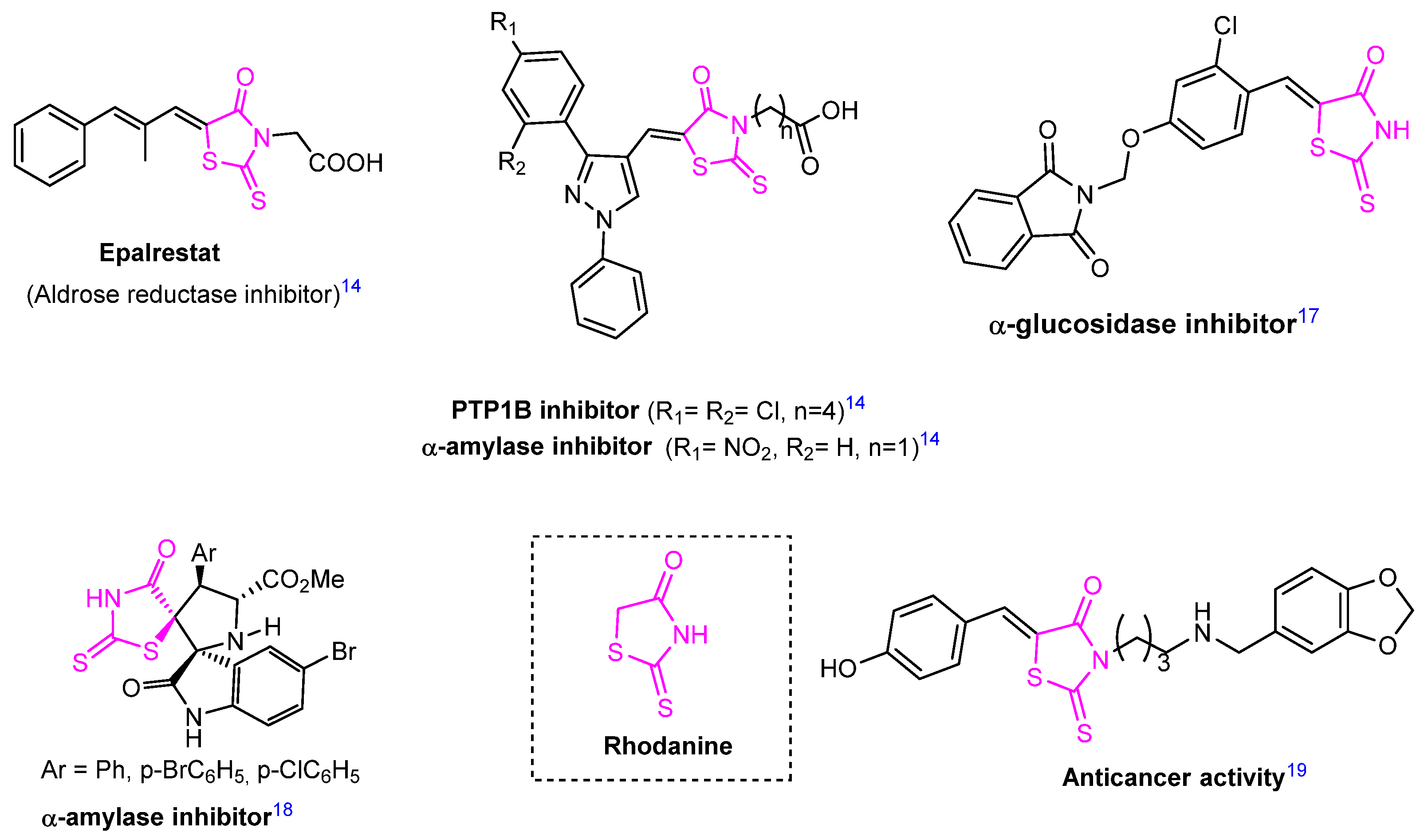
:1. Introduction
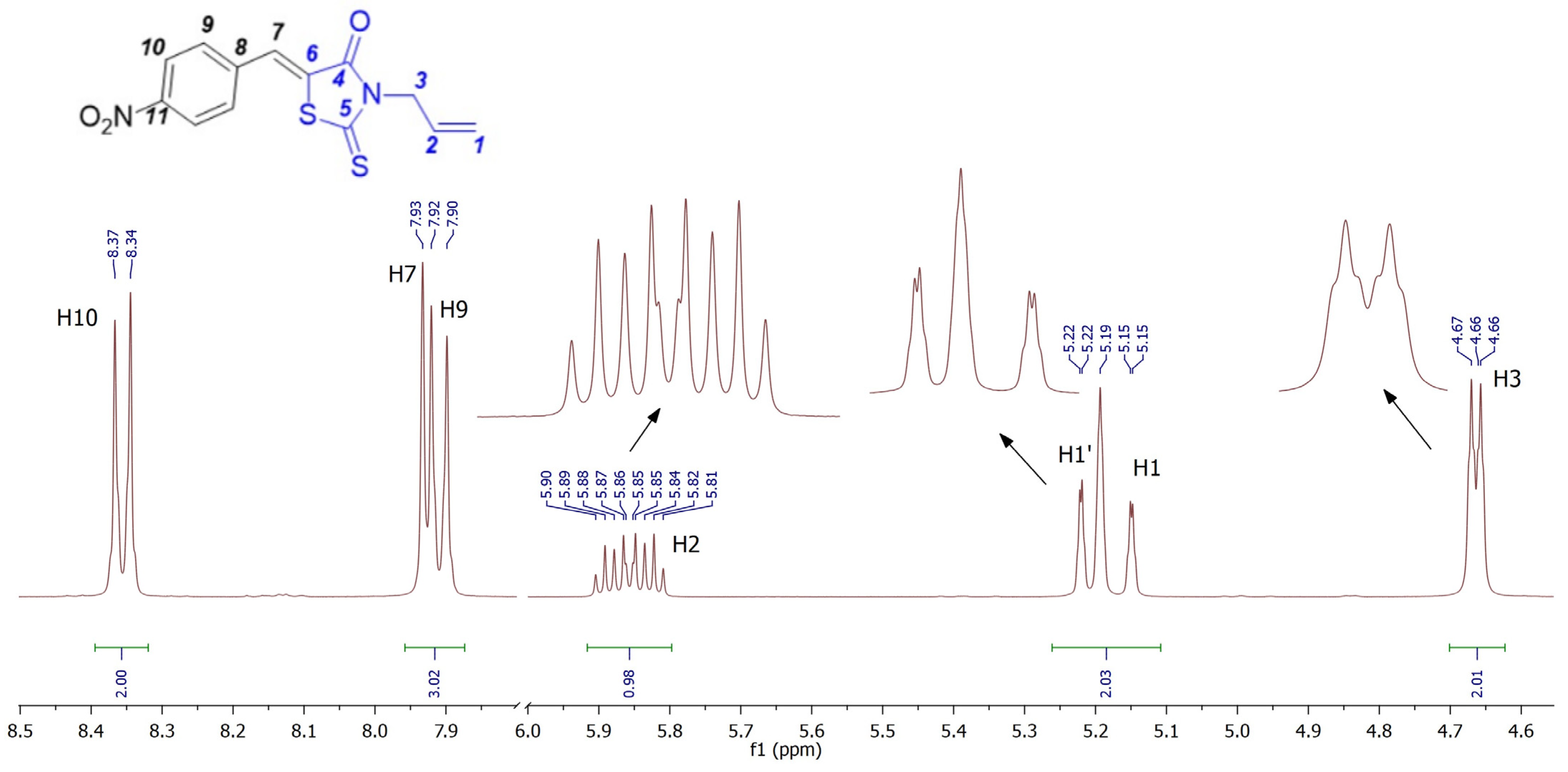
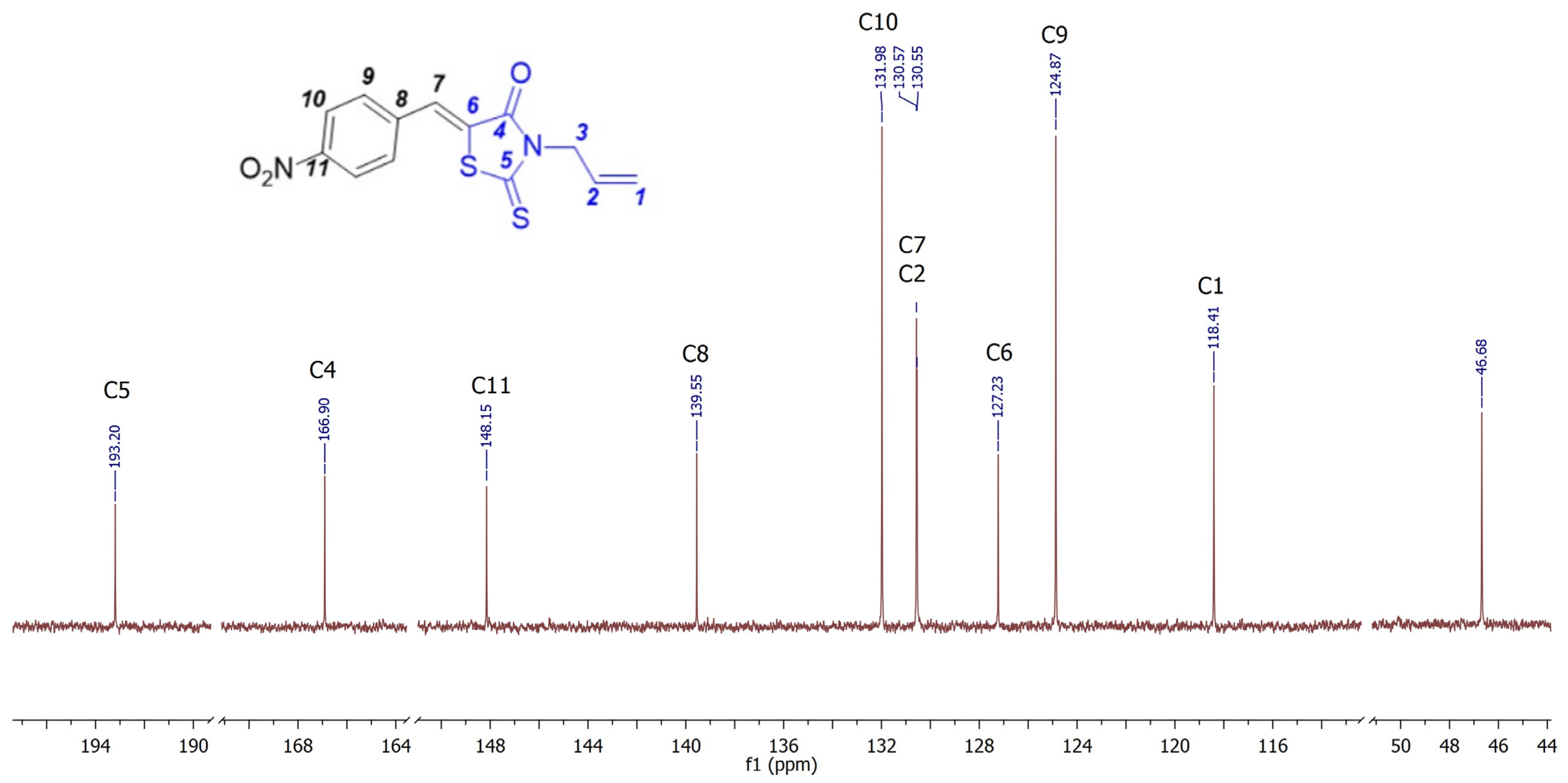
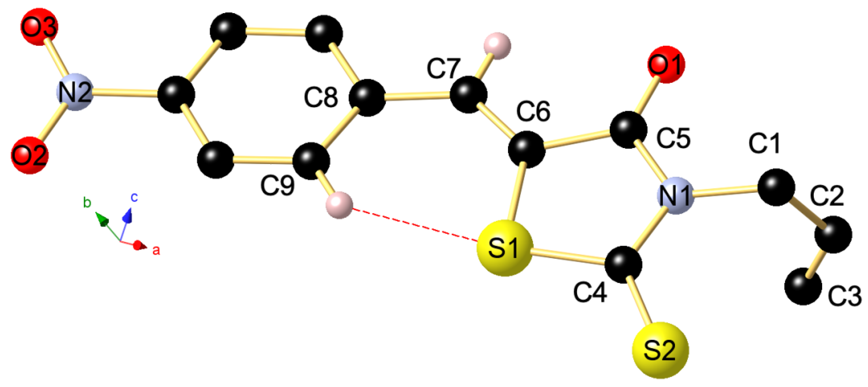
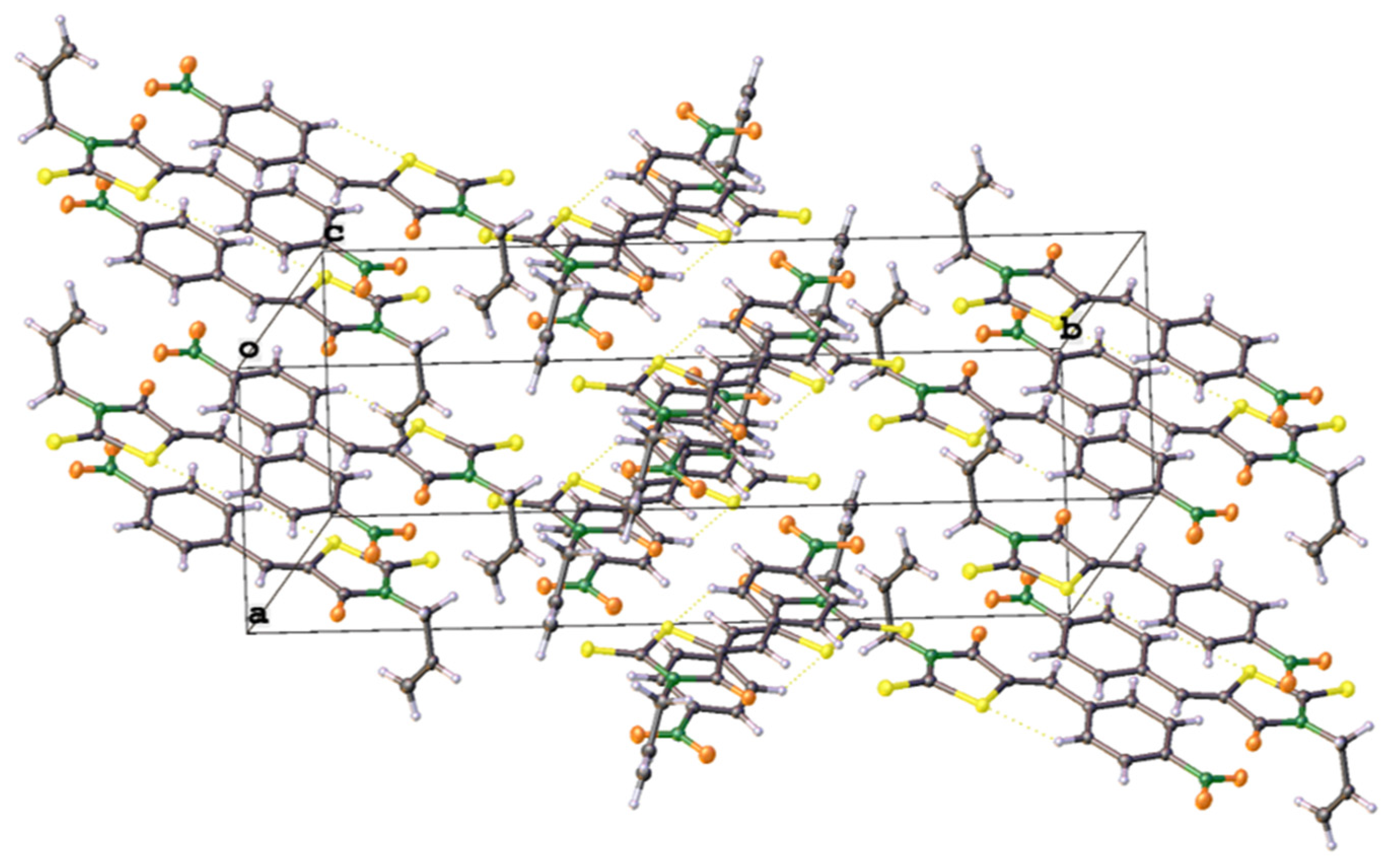
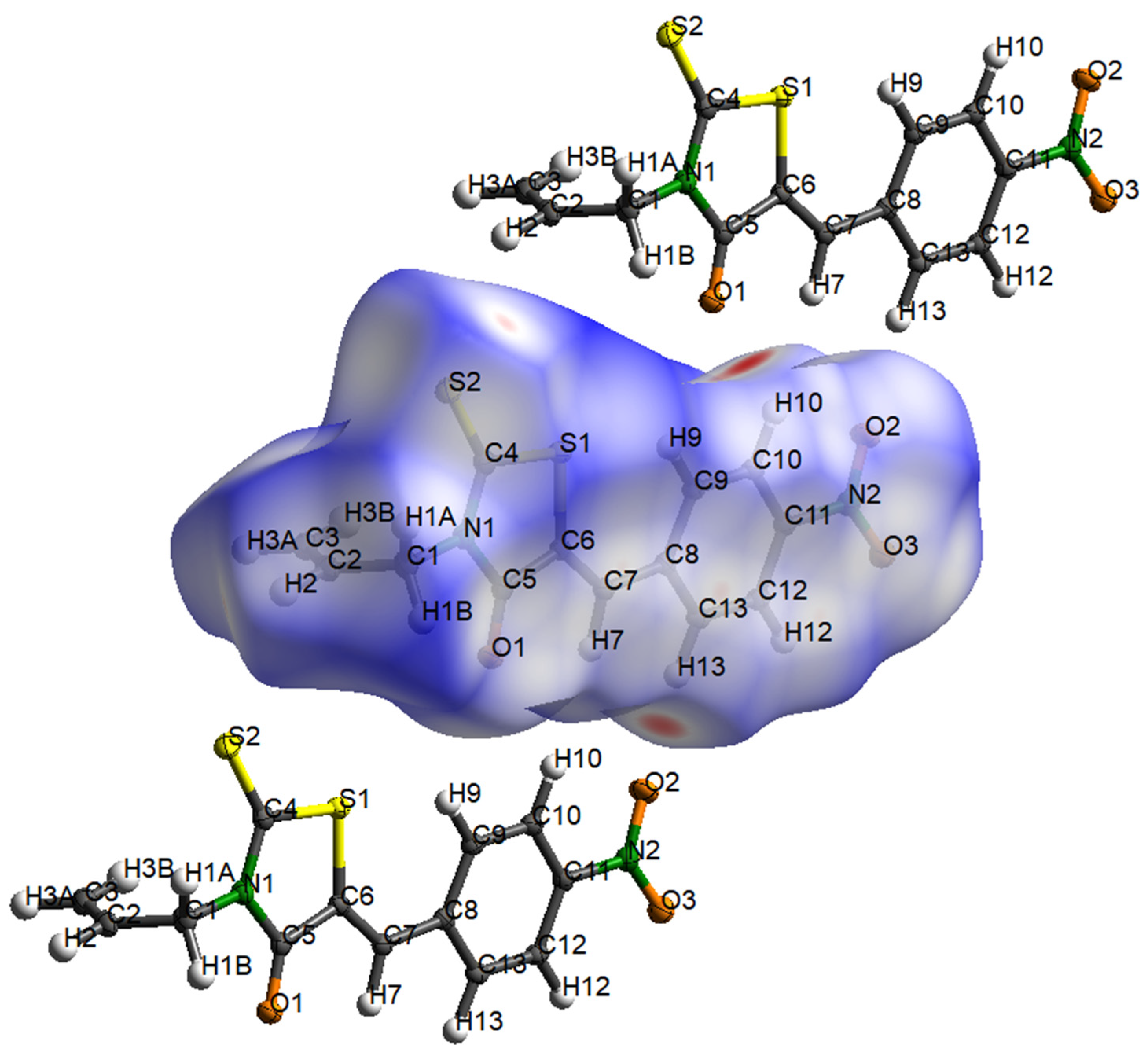
2. Results and Discussion
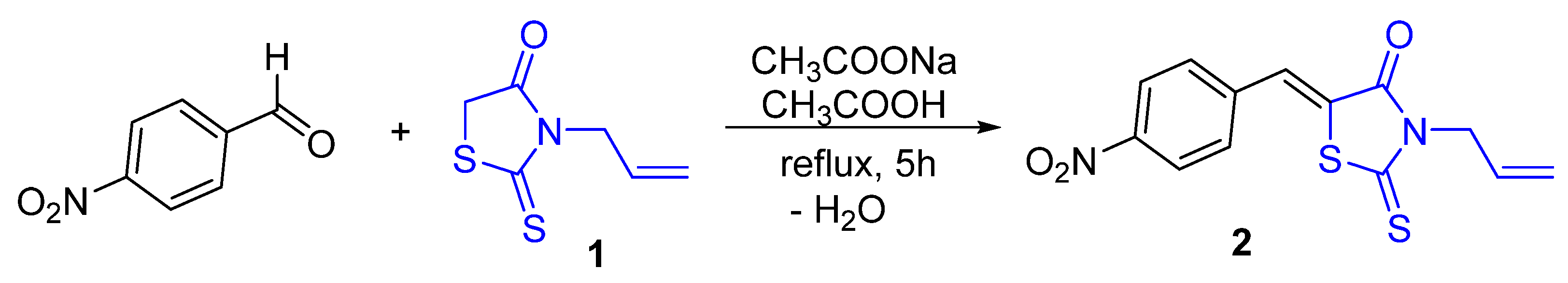
3. Materials and Methods
4. Conclusions
Supplementary Materials
Author Contributions
Funding
Data Availability Statement
Acknowledgments
Conflicts of Interest
References
- Brown, F.C.; Bradsher, C.K.; Bond, S.M.; Potter, M. Rhodanine Derivatives. J. Am. Chem. Soc. 1951, 73, 2357–2359. [Google Scholar] [CrossRef]
- Kaminskyy, D.; Kryshchyshyn, A.; Lesyk, R. Recent Developments with Rhodanine as a Scaffold for Drug Discovery. Expert Opin. Drug Discov. 2017, 12, 1233–1252. [Google Scholar] [CrossRef]
- Mousavi, S.M.; Zarei, M.; Hashemi, S.A.; Babapoor, A.; Amani, A.M. A Conceptual Review of Rhodanine: Current Applications of Antiviral Drugs, Anticancer and Antimicrobial Activities. Artif. Cells Nanomed. Biotechnol. 2019, 47, 1132–1148. [Google Scholar] [CrossRef] [PubMed]
- Bin Ahmad Kamar, A.K.D.; Ju Yin, L.; Tze Liang, C.; Tjin Fung, G.; Avupati, V.R. Rhodanine Scaffold: A Review of Antidiabetic Potential and Structure–Activity Relationships (SAR). Med. Drug Discov. 2022, 15, 100131. [Google Scholar] [CrossRef]
- Naufal, M.; Hermawati, E.; Syah, Y.M.; Hidayat, A.T.; Hidayat, I.W.; Al-Anshori, J. Structure–Activity Relationship Study and Design Strategies of Hydantoin, Thiazolidinedione, and Rhodanine-Based Kinase Inhibitors: A Two-Decade Review. ACS Omega 2024, 9, 4186–4209. [Google Scholar] [CrossRef] [PubMed]
- Bourahla, K.; Guihéneuf, S.; Limanton, E.; Paquin, L.; Le Guével, R.; Charlier, T.; Rahmouni, M.; Durieu, E.; Lozach, O.; Carreaux, F.; et al. Design and Microwave Synthesis of New (5Z) 5-Arylidene-2-Thioxo-1,3-Thiazolinidin-4-One and (5Z) 2-Amino-5-Arylidene-1,3-Thiazol-4(5H)-One as New Inhibitors of Protein Kinase DYRK1A. Pharmaceuticals 2021, 14, 1086. [Google Scholar] [CrossRef] [PubMed]
- Khodair, A.I.; Alzahrani, F.M.; Awad, M.K.; Al-Issa, S.A.; Al-Hazmi, G.H.; Nafie, M.S. Design, Synthesis, Computational Investigations, and Antitumor Evaluation of N-Rhodanine Glycosides Derivatives as Potent DNA Intercalation and Topo II Inhibition against Cancer Cells. ACS Omega 2023, 8, 13300–13314. [Google Scholar] [CrossRef]
- Jiang, H.; Zhang, W.-J.; Li, P.-H.; Wang, J.; Dong, C.-Z.; Zhang, K.; Chen, H.-X.; Du, Z.-Y. Synthesis and Biological Evaluation of Novel Carbazole-Rhodanine Conjugates as Topoisomerase II Inhibitors. Bioorg. Med. Chem. Lett. 2018, 28, 1320–1323. [Google Scholar] [CrossRef] [PubMed]
- Szczepański, J.; Tuszewska, H.; Trotsko, N. Anticancer Profile of Rhodanines: Structure–Activity Relationship (SAR) and Molecular Targets—A Review. Molecules 2022, 27, 3750. [Google Scholar] [CrossRef]
- Yin, L.J.; Bin Ahmad Kamar, A.K.D.; Fung, G.T.; Liang, C.T.; Avupati, V.R. Review of Anticancer Potentials and Structure-Activity Relationships (SAR) of Rhodanine Derivatives. Biomed. Pharmacother. 2022, 145, 112406. [Google Scholar] [CrossRef]
- Krátký, M.; Nováčková, K.; Svrčková, K.; Švarcová, M.; Štěpánková, Š. New 3-Amino-2-Thioxothiazolidin-4-One-Based Inhibitors of Acetyl- and Butyryl-Cholinesterase: Synthesis and Activity. Future Med. Chem. 2024, 16, 59–74. [Google Scholar] [CrossRef]
- Krátký, M.; Štěpánková, Š.; Vorčáková, K.; Vinšová, J. Synthesis and in Vitro Evaluation of Novel Rhodanine Derivatives as Potential Cholinesterase Inhibitors. Bioorg. Chem. 2016, 68, 23–29. [Google Scholar] [CrossRef] [PubMed]
- Available online: https://en.wikipedia.org/wiki/epalrestat (accessed on 9 February 2024).
- Hotta, N.; Sakamoto, N.; Shigeta, Y.; Kikkawa, R.; Goto, Y. Clinical Investigation of Epalrestat, an Aldose Reductase Inhibitor, on Diabetic Neuropathy in Japan: Multicenter Study. J. Diabetes Complicat. 1996, 10, 168–172. [Google Scholar] [CrossRef] [PubMed]
- Sun, L.; Wang, P.; Xu, L.; Gao, L.; Li, J.; Piao, H. Discovery of 1,3-Diphenyl-1H-Pyrazole Derivatives Containing Rhodanine-3-Alkanoic Acid Groups as Potential PTP1B Inhibitors. Bioorg. Med. Chem. Lett. 2019, 29, 1187–1193. [Google Scholar] [CrossRef] [PubMed]
- Bansal, G.; Singh, S.; Monga, V.; Thanikachalam, P.V.; Chawla, P. Synthesis and Biological Evaluation of Thiazolidine-2,4-Dione-Pyrazole Conjugates as Antidiabetic, Anti-Inflammatory and Antioxidant Agents. Bioorg. Chem. 2019, 92, 103271. [Google Scholar] [CrossRef] [PubMed]
- Wang, G.; Peng, Y.; Xie, Z.; Wang, J.; Chen, M. Synthesis, α-Glucosidase Inhibition and Molecular Docking Studies of Novel Thiazolidine-2,4-Dione or Rhodanine Derivatives. Med. Chem. Commun. 2017, 8, 1477–1484. [Google Scholar] [CrossRef] [PubMed]
- Toumi, A.; Boudriga, S.; Hamden, K.; Sobeh, M.; Cheurfa, M.; Askri, M.; Knorr, M.; Strohmann, C.; Brieger, L. Synthesis, Antidiabetic Activity and Molecular Docking Study of Rhodanine-Substitued Spirooxindole Pyrrolidine Derivatives as Novel α-Amylase Inhibitors. Bioorg. Chem. 2021, 106, 104507. [Google Scholar] [CrossRef]
- Dago, C.; Ambeu, C.; Coulibaly, W.-K.; Békro, Y.-A.; Mamyrbékova, J.; Defontaine, A.; Baratte, B.; Bach, S.; Ruchaud, S.; Guével, R.; et al. Synthetic Development of New 3-(4-Arylmethylamino)Butyl-5-Arylidene-Rhodanines under Microwave Irradiation and Their Effects on Tumor Cell Lines and against Protein Kinases. Molecules 2015, 20, 12412–12435. [Google Scholar] [CrossRef]
- Ali Muhammad, S.; Ravi, S.; Thangamani, A. Synthesis and Evaluation of Some Novel N-Substituted Rhodanines for Their Anticancer Activity. Med. Chem. Res. 2016, 25, 994–1004. [Google Scholar] [CrossRef]
- Akhavan, M.; Foroughifar, N.; Pasdar, H.; Bekhradnia, A. Green Synthesis, Biological Activity Evaluation, and Molecular Docking Studies of Aryl Alkylidene 2, 4-Thiazolidinedione and Rhodanine Derivatives as Antimicrobial Agents. Comb. Chem. High Throughput Screen. 2020, 22, 716–727. [Google Scholar] [CrossRef] [PubMed]
- Tomašić, T.; Zidar, N.; Mueller-Premru, M.; Kikelj, D.; Mašič, L.P. Synthesis and Antibacterial Activity of 5-Ylidenethiazolidin-4-Ones and 5-Benzylidene-4,6-Pyrimidinediones. Eur. J. Med. Chem. 2010, 45, 1667–1672. [Google Scholar] [CrossRef]
- Hesse, S. Synthesis of 5-Arylidenerhodanines in L-Proline-Based Deep Eutectic Solvent. Beilstein J. Org. Chem. 2023, 19, 1537–1544. [Google Scholar] [CrossRef] [PubMed]
- Pearson, R.G. Recent Advances in the Concept of Hard and Soft Acids and Bases. J. Chem. Educ. 1987, 64, 561. [Google Scholar] [CrossRef]
- Moers, F.G.; Bosman, W.P.J.H.; Beurskens, P.T. Crystal Structure of 3-Methylrhodaninecopper(I) Iodide. J. Cryst. Mol. Struct. 1972, 2, 23–29. [Google Scholar] [CrossRef]
- Moers, F.G.; Goossens, J.W.M.; Langhout, J.P.M. Rhodanine Complexes of Copper(I), Palladium(II) and Platinum(II). J. Inorg. Nucl. Chem. 1973, 35, 855–859. [Google Scholar] [CrossRef]
- Moers, F.G.; Smits, J.M.M.; Beurskens, P.T. Crystal Structure of Bis-(Rhodanine)Copper(I) Iodide, C6H6CuIN2O2S4. J. Crystallogr. Spectrosc. Res. 1986, 16, 101–106. [Google Scholar] [CrossRef]
- Fabretti, A.C.; Peyronel, G.; Franchini, G.C. Copper(I) Complexes of Rhodanine. Transit. Met. Chem. 1978, 3, 125–127. [Google Scholar] [CrossRef]
- Arar, W.; Khatyr, A.; Knorr, M.; Brieger, L.; Krupp, A.; Strohmann, C.; Efrit, M.L.; Ben Akacha, A. Synthesis, Crystal Structures and Hirshfeld Analyses of Phosphonothioamidates (EtO)2P(=O)C(=S)N(H)R (R = Cy, Bz) and Their Coordination on CuI and HgX2 (X = Br, I). Phosphorus Sulfur Silicon Relat. Elem. 2021, 196, 845–858. [Google Scholar] [CrossRef]
- Hameau, A.; Guyon, F.; Knorr, M.; Enescu, M.; Strohmann, C. Self-Assembly of Dithiolene-Based Coordination Polymers of Mercury(II): Dithioether versus Thiocarbonyl Bonding. Monatsh. Chem. 2006, 137, 545–555. [Google Scholar] [CrossRef]
- Guyon, F.; Hameau, A.; Khatyr, A.; Knorr, M.; Amrouche, H.; Fortin, D.; Harvey, P.D.; Strohmann, C.; Ndiaye, A.L.; Huch, V.; et al. Syntheses, Structures, and Photophysical Properties of Mono- and Dinuclear Sulfur-Rich Gold(I) Complexes. Inorg. Chem. 2008, 47, 7483–7492. [Google Scholar] [CrossRef]
- Hameau, A.; Guyon, F.; Khatyr, A.; Knorr, M.; Strohmann, C. 4,5-Bis(Methylthio)-1,3-Dithiole-2-Thione, a Versatile Sulphur-Rich Building Block for the Self-Assembly of Cu(I) and Ag(I) Coordination Polymers: Dithioether versus Thiocarbonyl Bonding. Inorg. Chim. Acta 2012, 388, 60–70. [Google Scholar] [CrossRef]
- Arar, W.; Viau, L.; Jourdain, I.; Knorr, M.; Strohmann, C.; Scheel, R.; Ben Akacha, A. Synthesis of Catena-Bis(μ-Bromo)-(O-Methyl-N-Phenylthiocarbamate)-Dicopper(I) and Its Reactivity towards PAr3 (Ar = Ph, p-Tol). Molbank 2023, 2023, M1655. [Google Scholar] [CrossRef]
- Gouveia, F.L.; De Oliveira, R.M.B.; De Oliveira, T.B.; Da Silva, I.M.; Do Nascimento, S.C.; De Sena, K.X.F.R.; De Albuquerque, J.F.C. Synthesis, Antimicrobial and Cytotoxic Activities of Some 5-Arylidene-4-Thioxo-Thiazolidine-2-Ones. Eur. J. Med. Chem. 2009, 44, 2038–2043. [Google Scholar] [CrossRef] [PubMed]
- El Ajlaoui, R.; Ouafa, A.; Mojahidi, S.; El Ammari, L.; Saadi, M.; El Mostapha, R. Unexpected Synthesis of Novel 3-Allyl-5-(Arylidene)-2-Thioxo-Thiazolidin-4-Ones in Reactions of 3-Allylrhodanine with 2-Arylidene-4-Methyl-5-Oxopyrazolidinium Ylides. Synth. Commun. 2015, 45, 2035–2042. [Google Scholar] [CrossRef]
- Momose, Y.; Meguro, K.; Ikeda, H.; Hatanaka, C.; Oi, S.; Sohda, T. Studies on Antidiabetic Agents. X. Synthesis and Biological Activities of Pioglitazone and Related Compounds. Chem. Pharm. Bull. 1991, 39, 1440–1445. [Google Scholar] [CrossRef]
- El Ajlaoui, R.; Rakib, E.M.; Mojahidi, S.; Saadi, M.; El Ammari, L. (Z)-3-Allyl-5-(3-Methoxybenzylidene)-2-Sulfanylidene-1,3-Thiazolidin-4-One. IUCrData 2016, 1, x160052. [Google Scholar] [CrossRef]
- El Ajlaoui, R.; Belkhouya, N.; Rakib, E.M.; Mojahidi, S.; Saadi, M.; El Ammari, L. (Z)-3-Allyl-5-(4-Fluorobenzylidene)-2-Sulfanylidenethiazolidin-4-one. IUCrData 2016, 1, x161236. [Google Scholar] [CrossRef]
- El Ajlaoui, R.; Rakib, E.M.; Mojahidi, S.; Saadi, M.; El Ammari, L. Crystal Structure of (Z)-3-Allyl-5-(4-Chlorobenzylidene)-2-Sulfanylidene-1,3-Thiazolidin-4-one. Acta Crystallogr. Sect. E Crystallogr. Commun. 2015, 71, o1012. [Google Scholar] [CrossRef]
- Germain, G.; Piret, P.; Van Meersche, M.; De Kerf, J. Structure d’une Mérocyanine: C10H12S3N2O. Acta Crystallogr. 1962, 15, 373–382. [Google Scholar] [CrossRef]
- El Ajlaoui, R.; Rakib, E.M.; Mojahidi, S.; Saadi, M.; El Ammari, L. Crystal Structure of (E)-3-Allyl-2-Sulfanylidene-5-[(Thiophen-2-Yl)Methylidene]Thiazolidin-4-one. Acta Crystallogr. Sect. E Crystallogr. Commun. 2015, 71, o433–o434. [Google Scholar] [CrossRef]
- Dolomanov, O.V.; Bourhis, L.J.; Gildea, R.J.; Howard, J.A.K.; Puschmann, H. OLEX2: A complete structure solution, refinement and analysis program. J. Appl. Crystallogr. 2009, 42, 339–341. [Google Scholar] [CrossRef]
- Spackman, M.A.; Jayatilaka, D. Hirshfeld Surface Analysis. CrystEngComm 2009, 11, 19–32. [Google Scholar] [CrossRef]
- Spackman, P.R.; Turner, M.J.; McKinnon, J.J.; Wolff, S.K.; Grimwood, D.J.; Jayatilaka, D.; Spackman, M.A. CrystalExplorer: A Program for Hirshfeld Surface Analysis, Visualization and Quantitative Analysis of Molecular Crystals. J. Appl. Crystallogr. 2021, 54, 1006–1011. [Google Scholar] [CrossRef] [PubMed]
- Sheldrick, G.M. Crystal Structure Refinement with SHELXL. Acta Crystallogr. C Struct. Chem. 2015, 71, 3–8. [Google Scholar] [CrossRef]
- Sheldrick, G.M. SHELXT—Integrated Space-Group and Crystal-Structure Determination. Acta Crystallogr. A Found Adv. 2015, 71, 3–8. [Google Scholar] [CrossRef]


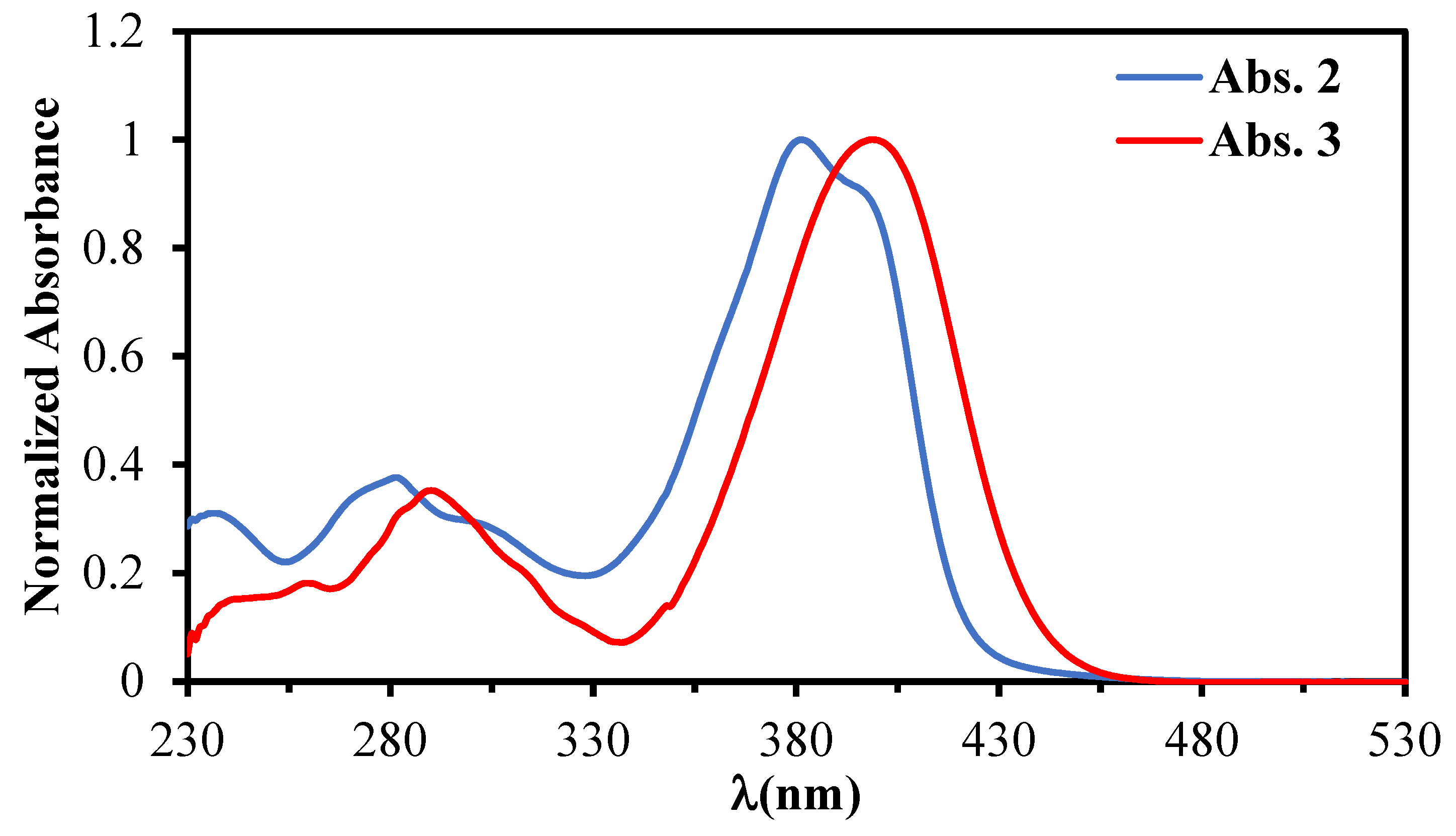
| Comp. | Absorption: λabs nm (ε × 10−3M−1cm−1) |
|---|---|
| 2 | 239 (5.5), 281 (6.7), 303 sh (4.8), 381 (17.9), 399 sh (16.0) |
| 3 | 242 (2.8), 262 (3.2), 294 (6.1), 313 sh (3.6), 399 (18.1) |
Disclaimer/Publisher’s Note: The statements, opinions and data contained in all publications are solely those of the individual author(s) and contributor(s) and not of MDPI and/or the editor(s). MDPI and/or the editor(s) disclaim responsibility for any injury to people or property resulting from any ideas, methods, instructions or products referred to in the content. |
© 2024 by the authors. Licensee MDPI, Basel, Switzerland. This article is an open access article distributed under the terms and conditions of the Creative Commons Attribution (CC BY) license (https://creativecommons.org/licenses/by/4.0/).
Share and Cite
Moreno, B.; Jourdain, I.; Knorr, M.; Boudriga, S.; Strohmann, C.; Schrimpf, T. Synthesis of (Z)-3-Allyl-5-(4-nitrobenzylidene)-2-sulfanylidene-1,3-thiazolidin-4-one and Determination of Its Crystal Structure. Molbank 2024, 2024, M1783. https://doi.org/10.3390/M1783
Moreno B, Jourdain I, Knorr M, Boudriga S, Strohmann C, Schrimpf T. Synthesis of (Z)-3-Allyl-5-(4-nitrobenzylidene)-2-sulfanylidene-1,3-thiazolidin-4-one and Determination of Its Crystal Structure. Molbank. 2024; 2024(1):M1783. https://doi.org/10.3390/M1783
Chicago/Turabian StyleMoreno, Bastien, Isabelle Jourdain, Michael Knorr, Sarra Boudriga, Carsten Strohmann, and Tobias Schrimpf. 2024. "Synthesis of (Z)-3-Allyl-5-(4-nitrobenzylidene)-2-sulfanylidene-1,3-thiazolidin-4-one and Determination of Its Crystal Structure" Molbank 2024, no. 1: M1783. https://doi.org/10.3390/M1783